Article

Efficient Methods for Evaluation on Ploidy Level of Cucurbita pepo L. Regenerant Plants Obtained in Unpollinated Ovule Culture In Vitro

1
Federal State Budgetary Scientific Institution Federal Scientific Vegetable Center (FSBSI FSVC), VNIISSOK, 143072 Moscow, Russia
2
South Siberian Botanical Garden, Altai State University, 656049 Barnaul, Russia
*
Author to whom correspondence should be addressed.
Horticulturae 2022, 8(11), 1083; https://doi.org/10.3390/horticulturae8111083
Submission received: 27 September 2022 / Revised: 4 November 2022 / Accepted: 13 November 2022 / Published: 16 November 2022
(This article belongs to the Section Propagation and Seeds)

Abstract

:
An important stage in doubled haploid (DH) production is to evaluate and to differentiate the ploidy level of regenerant plants at least two–three times during the technology. Therefore, rapid and reliable methods are necessary for particular species taken into the technology. In this study, Cucurbita pepo regenerants obtained through unpollinated ovule culture in vitro were evaluated including three different methods: direct chromosome counting in apical meristems, flow cytometry of the cell nucleus, and estimation of morphological parameters of the abaxial epidermis. Methods were optimized for each of three evaluations, and main criteria were determined for ploidy level differentiation. As a result, four ploidy levels, namely, 2n, 3n, 4n, and 8n, were defined among regenerant plants adapted to ex vitro conditions, while true haploids were only found among plants that remained in the in vitro culture. In total, 32.35%, 26.47%, 33.82%, 4.41%, and 2.94% of regenerant plants of courgette and patisson were diploid, triploid, tetraploid, octaploid, and aneuploid, respectively. According to results of flow cytometry of the cell nucleus, two cytotypes in diploid samples with DNA content of 2C = 1.07 ± 0.03 pg for courgette belonging to subsp. pepo and 2C = 0.95 ± 0.03 pg for patisson samples belonging to subsp. ovifera were revealed. The images of metaphase chromosomes of haploid, triploid, and tetraploid C. pepo specimens obtained using the propion–lacmoid chromosome staining method were presented for the first time. Parameters of abaxial epidermis in diploid samples of courgette and patisson grown in open-field and greenhouse conditions were described and compared. It was shown that the most robust parameter not depending on external factors was the number of chloroplasts in stomatal guard cells, which contained 9.41 to 11.31, 14.84 to 16.3, and up to 17.58 chloroplasts in diploid, triploid, and tetraploid samples, respectively. The application of several methods for estimation enables avoiding the misidentification of ploidy levels in adapted regenerant plants produced with the use of DH technology.

1. Introduction

Cucurbita pepo L. is an annual herbaceous plant belonging to the family Cucurbitaceae Jussand is widely distributed throughout the world due to its economic importance. Most of these vegetable crops are grown for their fruits (Latin name: pepo, peponium) and for the seeds and oil extracted from them. The area harvested under pumpkins, squash, and gourds occupies two million hectares globally, with most of the production grown in India and China [1]. The range of pumpkin crops in the world’s major seed companies is represented exclusively by F1 hybrids. In addition to the heterosis effect, the production of hybrid seeds allows the protection of the plant breeder’s rights. In this regard, aligned lineage material becomes particularly valuable in the breeding process [2,3]. The use of doubled haploid production techniques (DH-technology) makes it possible to create a 100% homozygous line within 1–2 years. In addition, each DH is a distinct genotypic and phenotypic class with a unique combination of homozygous alleles.
Three different methods are commonly used to produce DHs in C. pepo cultures: parthenogenesis in situ stimulated by irradiated pollen, gynogenesis in vitro (in vitro culture of unpollinated ovules/ovaries), and androgenesis in vitro (anther culture and isolated microspore culture) [4,5,6]. Since, in all three methods, the process takes place in haploid cells, the forming embryoid or callus will initially have a haploid set of chromosomes. At the second stage, under the influence of certain factors/agents, the chromosomes are doubled, and the resulting organism ideally acquires a diploid set of chromosomes with a homozygous allele state.
Chromosome doubling in initially haploid embryoids/plants is a rather complicated and still poorly studied process in most cultures. Quite often, haploids pass to the diploid level spontaneously while still in culture [7]. This is thought to be an inherited trait in many species that reduce costs in the production of DH plants. Possible mechanisms for spontaneous doubling are most commonly cited as cell fusion, somatic cell fusion, endomitosis, and endoreduplication [8,9,10,11,12,13]. According to the literature, cucurbits are characterized by low levels of spontaneous chromosome doubling when using DH technology [4,14]. The resulting plantlets are usually haploid (up to 70%), but mixoploid or even spontaneous DHs can also be obtained. It has also been noted that the yield of doubled haploids due to spontaneous chromosome doubling is higher with unpollinated ovule or anther cultivation technologies than with parthenogenesis in situ stimulated by irradiated pollen [2,15,16]. Prolonged in vitro culture will also facilitate the transition to the diploid level. The method of chromosome doubling developed for cucumber, using direct plant regeneration from haploid leaf explants, is based on this [17,18]. This method was even more effective than colchicine treatment [19].
When large numbers of haploid plants are present among the regenerants, DH protocols should include an additional step in which various anti-microtubule doubling agents that promote ploidy increase, such as colchicine, amiprophos methyl (APM), oryzalin, and trifluralin [20,21]. The use of caffeine for the same purpose has also been reported [22]. The chromosome doubling procedure is rather laborious and has been reported in reviews [9,12,23]. Quite often, the use of antimitotic agents fails to achieve the desired result and transfer the plants to a double haploid level, and the treatment procedure has to be repeated. The resulting plants have to be constantly tested for ploidy level.
Regardless of the DH technology used, and whether the antimitotic treatment or regeneration step is included from haploid leaves, haploids, DH, and plants of different ploidy can be obtained as a result [17,19,24,25,26]. The formation of triploid, tetraploid, and mixoploid plants in progeny using haploid techniques is also a fairly well-known development, including in members of the Cucurbitaceae family [2,6,15,27].
Evaluating and differentiating the obtained regenerant plants for ploidy level is an extremely important stage of DH technology, and testing should be carried out at least two or three times at different stages of the technology. A quick and qualitative method for ploidy assessment of the regenerant plants is of great interest. At present, ploidy of the obtained regenerant plants can be determined using several methods: direct counting of chromosomes (microscopy of cytological preparations), flow cytometry of cell nuclei (determination of chromatin content in cell nuclei), or a set of indirect morphological signs of leaf abaxial epidermis.
Direct chromosome counting is the most accurate, but also the most time-consuming method. It is especially difficult for cultures with a large number of small-sized chromosomes, such as representatives of the Cucurbita genus. The process of preparing a cytological preparation is rather long and multistage, and it requires laborious work of a cytologist. Regardless of the species, genotype, or source of tissue for analysis, there are three basic operations for handling mitotic chromosomes that remain the same: collection and preprocessing of material, fixation of material, and chromosome staining [28]. The necessary data on chromosome sets for most plants are now readily available and collected in public resources such as the Chromosome Counts Database (CCDB; http://ccdb.tau.ac.il/ (accessed on 27 May 2022)) [29] and the Index to Plant Chromosome Numbers (IPCN, http://legacy.tropicos.org/Project/IPCN) (accessed on 27 May 2022) [30].
The small size of mitotic chromosomes and their relatively large number in the genus Cucurbita make them difficult to count accurately, even though they are usually well separated. For a long time there was even disagreement among researchers about the number of chromosomes in different members of the genus [31,32]. Later, it was reported that, although little is known about the cytology of the genus Cucurbita because its cytological studies are extremely difficult, all species have a diploid chromosome set (2n = 2x = 40), which distinguishes it from other species of the family Cucurbitaceae with significantly fewer chromosomes [33,34,35,36]. Despite tremendous progress in cytogenetic studies, as well as the advent of new dyes and high-resolution microscopes, members of the genus Cucurbita are still poorly understood cytogenetically. At present, we are only able to find images of chromosomes for Cucurbita moschata [37,38]. We could not find similar studies for C. pepo, which confirms the difficulties of karyological analysis in this species.
Flow cytometry of cell nuclei (determination of chromatin content in cell nuclei) is a relatively simple and extremely fast method that allows several hundred samples to be analyzed in a working day and requires a small amount of material to be analyzed. Advantages also include the ability to perform the assay at any stage of plant development in vitro and in vivo. In addition, flow cytometry is the only method that provides detailed information on the presence of mixoploidia in tissues, as well as their proportion in the genotype under study [39]. A significant disadvantage is the high cost of the device and consumables, as well as the need to standardize the technique for a particular species by a specialist, for correct interpretation of the results obtained.
There are a limited number of publications on application of the flow cytometry method for representatives of genus Cucurbita, including those collected in the Database of C-values of plant DNA (https://cvalues.science.kew.org/ (accessed on 27 May 2022)) [40], where the minimal genome size (1C) is presented in C. foetidissima (0.34 pg), and the maximal is presented in C. pepo (0.54 pg) [41]. There are limited publications on the use of flow cytometry in the analysis of progeny obtained using DH technologies in C. pepo species [27,42].
Indirect morphological signs of abaxial leaf epidermis (includes the number of stomata guard cells per 1 mm2, the size of these stomata, and the number of chloroplasts in the stomata guard cells) has been used for the identification of plant ploidy in practical plant breeding for more than 70 years. Initially, stomatal guard cell length (SL) was used as a differentiating indicator because it was observed that this indicator correlated with ploidy level in a number of species, including Trifolium hybridum [43], Lolium perenne [44], Brassica oleracea L. ssp. gemmifera [45], and Hordeum vulgare L. [46]. Frandsen (1968) estimated the average number of chloroplasts in a pair of stomatal guard cells (CN) and correlated it with the number of chromosomes in potato, and it was found that the average number of chloroplasts increased 1.8–1.9 times after each doubling of chromosome number [47]. Thus, it has been shown that ploidy identification can be achieved on the basis of the number of chloroplasts in the stomatal guard cells [48,49]. At present, the method of ploidy identification using a set of indirect morphological characteristics of abaxial leaf epidermis is actively used for most agricultural plants, including the evaluation of DH-derived plants [28]. The most commonly used indicators are the length and width of stomata guard cells, their number in the microscopic field of view or per 1 mm2, and the chloroplast number (each side of the guard cells).
Choe et al. (2012) [50] showed that identification of haploids and diploids using a set of indirect morphological characteristics of the leaf abaxial epidermis is three times faster and 20 times cheaper than flow cytometry, with this method requiring little investment or training.
Morphological characteristics of the abaxial leaf epidermis are used to identify ploidy in the genus Cucurbita in plants obtained using DH technologies [51,52,53]. Epidermal staining with 10% Lugol’s iodine solution [51] or 1% AgNO3 [52,54,55,56] is recommended to facilitate chloroplast counting in stomatal guard cells.
Having searched the published data for C. pepo using a variety of haploid techniques, predominantly haploid and diploid plants were formed among the generated regenerant plants and only occasionally mixoploid plants were diagnosed [6]. We could find only two reports of induced tetraploidy in haploid induction attempts in C. pepo specimens [6,42]. Notably, it is in these studies that ploidy identification was performed using the flow cytometry of cell nuclei (recommended as one of the most accurate and fastest for this species), whereas other studies preferred cytological analysis (extremely difficult in this species) and ploidy analysis using abaxial epidermal indicators (extremely dependent in this species on cultivation conditions and requiring prior standardization). The mechanism of triploid and tetraploid formation is still unclear and requires further study, but seems to be related to the long-term in vitro culture, precisely to the effect of growth regulators. The low detection rate can be attributed to the problematic nature of ploidy identification in this species.
Thus, each of the three methods of assessment for ploidy differs in terms of the cost of consumables, the cost of the necessary equipment, and the time required to obtain a result for a single sample. Although all three methods have been used in few published protocols for C. pepo, there are no photographs of metaphase chromosomes in haploid, diploid, triploid, and tetraploid summer squash plants in the literature, along with very limited information on genome size (2C) reference values in plants with different ploidy levels to use protocols on flow cytometry of cell nuclei and abaxial epidermis indices. In this study, the aim was to evaluate C. pepo regenerant plants obtained in an unpollinated ovule culture in vitro by ploidy level using three different methods: direct chromosome counts in apical meristems, flow cytometry of cell nuclei, and abaxial epidermis morphological parameters. It was necessary to identify evaluation criteria and optimize the methodology for each of the three methods.

2. Materials and Methods

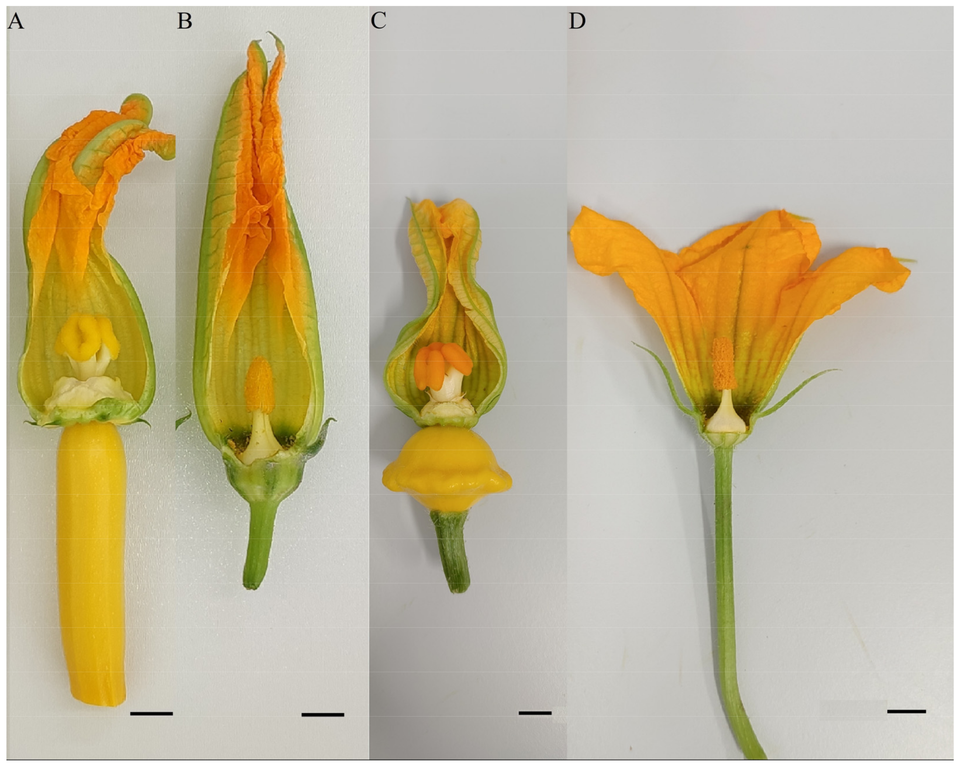
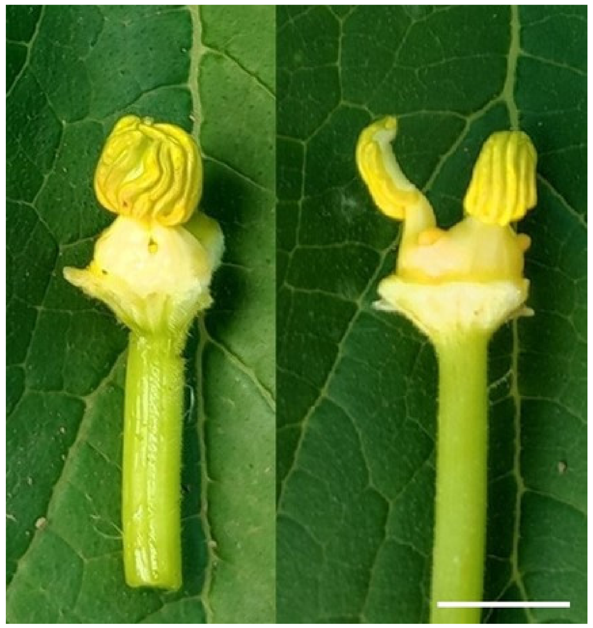
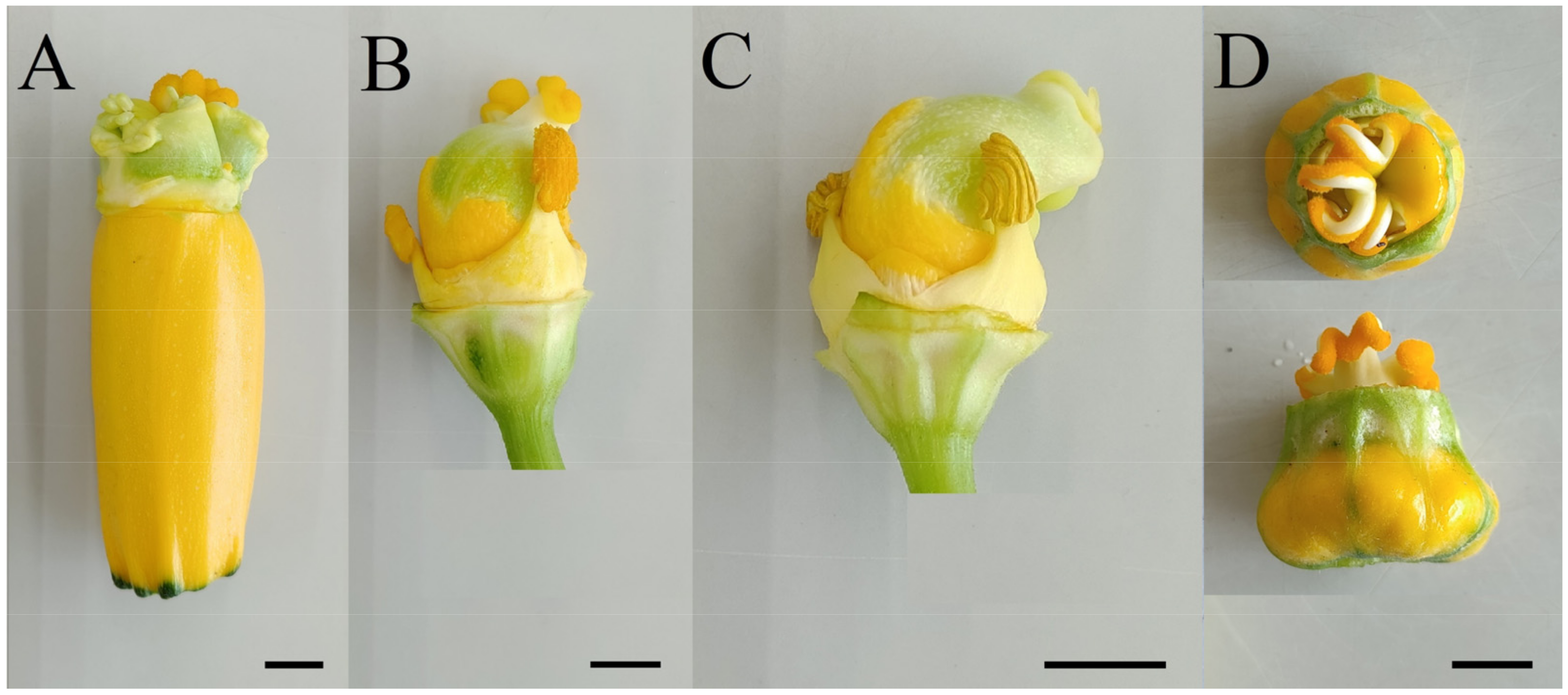
2.1. Plant Material and Growing Conditions of Donor Plants

We used gynogenic plants, regenerants of summer squash obtained in isolated ovule culture in vitro according to the method optimized in the laboratory of reproductive biotechnology in agricultural plant breeding of FSBSI FSVC [27,57]. Genotypes of initial donor plants of courgette (Faraon, Yakor’, Gold Rush F1) and patisson (Disk, Cheburashka, Sunny De Light F1) were used as control plants for ploidy determination. Donor and ex vitro adapted regenerant plants were grown from May to August in 2021–2022 in the Moscow region in film greenhouses and in open field conditions.

2.2. Ploidy Study of Plants Using Flow Cytometry of Cell Nuclei

DNA content was determined using flow cytometry techniques with propidium iodide. Fresh young leaves of donor plants and regenerant plants obtained in an in vitro culture of unpollinated ovules were used for the study. Leaves were ground with a blade in 1 mL of a modified Tris-MgCl2 buffer [58], containing 0.2 M Tris base, 4 mM MgCl2, 0.5% Triton X-100, 50 µg/mL RNase, 0.5% polyvinylpyrrolidone K15, and 50 µg/mL propidium iodide (pH 7.5). Fluorescence data of isolated nuclei were detected using a Partec CyFlow PA flow cytometer (Partec, GmbH, Münster, Germany) with a 532 nm laser source. Histograms were visualized and processed using Flowing Software 2.5.1 software (University of Turku, Turku, Finland). Statistical data were calculated in XLStat v. 24.4 software (Addinsoft). Ficus benjamina (2C = 0.90 pg) was used as an internal standard to determine DNA content [59,60].
To determine ploidy, diploid samples of donor plants were used as an external standard.
Ploidy was determined by the index of difference between the peaks of diploid standard of the same species and the sample:
Index = Sample   peak   mean   Standard   peak   mean .
The DNA content (2C, pg) was calculated as follows [61]:
2 C ,   pg = Sample   peak   mean   Standard   peak   mean × 2 C   Standard .

2.3. Propion–Lacmoid Method of Chromosome Staining

Cytological studies of courgette and patisson samples were performed using the modernized propion–lacmoid method [62] through preparation of pressed preparations from the stem apical meristem (axillary buds and young leaves) and root tips.
Plant material was not pretreated due to the large number of small-sized zucchini chromosomes. Fixation (in propionic acid) and staining (with lacmoid) of the material were performed simultaneously in standard propion–lacmoid solution for 24 h. Only intensively growing young tissues of apical meristems were used for fixation to provide a large number of dividing cells.
To prepare propion–lacmoid, 5 g of lacmoid was added to 50 mL of 50% propionic acid, and then left in the dark for 3–5 days, shaking the flask periodically at RT. Subsequently, the solution was filtered into a dark glass vessel and stored in a dark place.
Maceration of stained tissues was carried out by boiling in 40% propionic acid solution: stem meristem—20–30 s; large root meristem—45–60 s; small root meristem—20–30 s from the boiling point.
After boiling, the material was allowed to cool for 1–3 min, and part of the material was transferred to a drop of 40% propionic acid on a slide and covered with a coverslip. Strips of filter paper pressed on the edges of coverslip were used to crush the contents of the preparation. The flat end of the wooden handle of the preparaval needle was used to “flatten” the whole surface of the coverslip horizontally and vertically by pressing. Such a method of cell flattening is applicable for objects with small chromosomes of most vegetable crops, including pumpkin plants. For preservation, the preparation was melted using paraffin and left to differentiate for 30 or more minutes.
The preparations were viewed using an Axio Imager A2 microscope (Carl Zeiss Microscopy GmbH, Jena, Germany) with a 20× and 40× objective; chromosome counting and photography were performed with a 100× objective (with oil immersion). The number of chromosomes of each form was counted at 25 metaphases at a magnification of 100 × 10 × 1 in cells with a solid, uncompressed envelope.
The most successful microscopic objects were photographed using an Axio Imager A2 equipped with an AxioCam MRc5 camera (Carl Zeiss Microscopy GmbH, Jena, Germany).
The obtained images were processed and documented using AxioVision software, version 4.8 (Carl Zeiss MicroImaging, Jena, Germany).

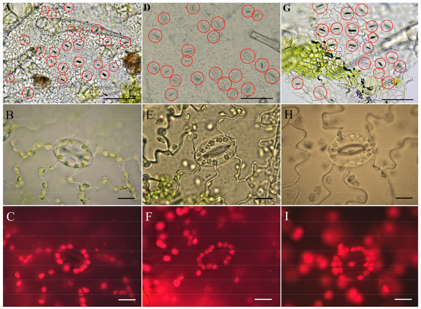
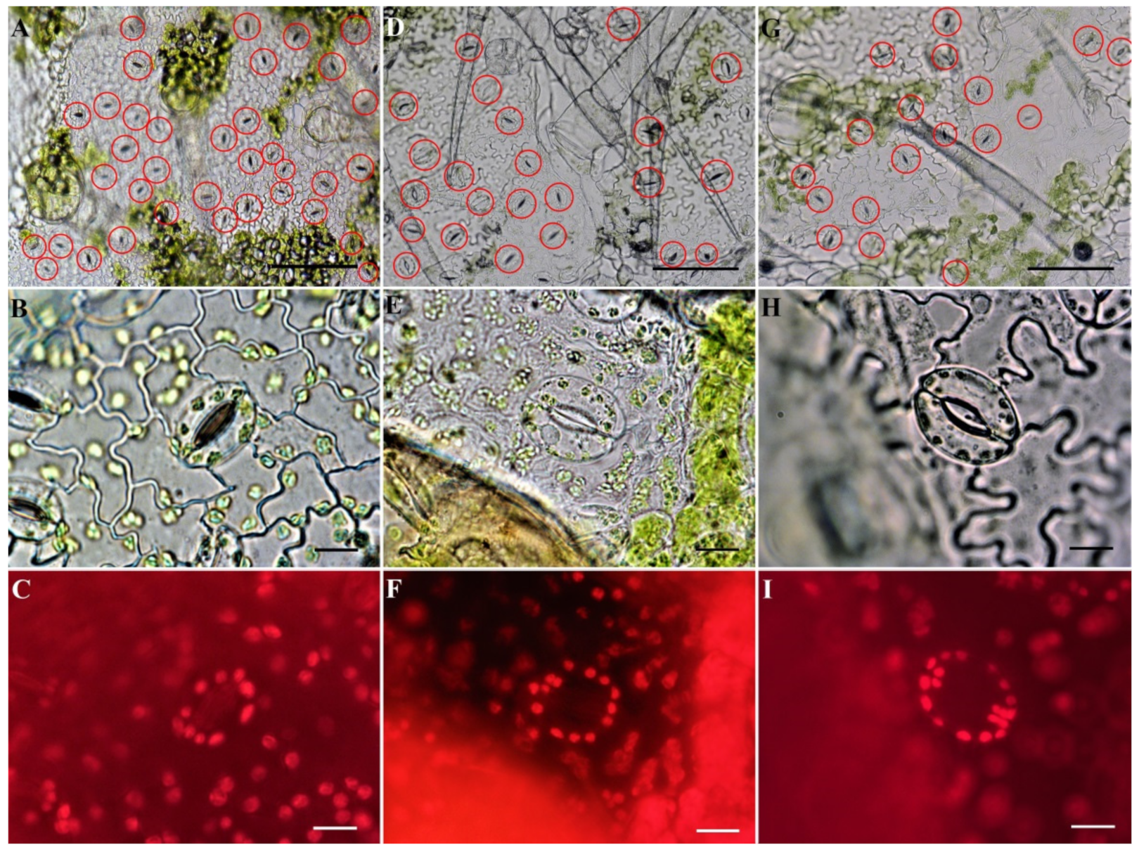
2.4. Identification of Ploidy of Regenerant Plants Based on Morphometric Parameters of the Epidermis on the Abaxial Side of the Leaf

The epidermal layer of cells was removed from the abaxial side of leaves with sharp tweezers, washed in distilled water, placed on a slide in a drop of water, covered with a coverslip, and viewed under a fluorescent microscope Axio Imager A2 with Filter Set 14: EX BP 510/560, BS FT 580, EM LP 590 (Carl Zeiss Microscopy GmbH, Jena, Germany). Chloroplasts exhibiting very strong autofluorescence in red, with a peak of approximately 680 nm, were counted.
Photodocumentation of the obtained preparations was performed using an AxioCam MRc5 camera (Carl Zeiss Microscopy GmbH, Jena, Germany) using 20×, 40×, and 100× lenses (with oil immersion). Using a 20× lens, we counted the number of stomata in the microscope field of view (at least fivefold replications). The length and width of stomata guard cells were measured using a 40× lens (10-fold repetition), and the stomatal index (stomata length/stomata width) was calculated. With the 100× oil immersion lens, the number of chloroplasts located in the two stomatal guard cells was counted (10-fold repetition for each plant).

2.5. Statistical Analysis

The experimental data were processed using conventional mathematical and statistical methods with a package of application programs (Microsoft Excel and Statistika 7.0).

3. Results and Discussion

3.1. Analysis of Plant Regenerants Using Flow Cytometry of Cell Nuclei

We optimized a protocol of the flow cytometry method for the analysis of C. pepo plants using modified Tris-MgCl2 buffer for plant nucleus extraction and propidium iodide. As a result, four ploidy levels, namely, 2n, 3n, 4n, and 8n, were defined among regenerant plants adapted to ex vitro conditions, while true haploids were only found among plants that remained in vitro culture. In total, 32.35%, 26.47%, 33.82%, 4.41%, and 2.94% of regenerant plants of courgette and patisson were diploid, triploid, tetraploid, octaploid, and aneuploid, respectively.
According to the results of flow cytometry of cell nuclei, two cytotypes in diploid samples with DNA content of 2C = 1.07 ± 0.03 pg for courgette and 2C = 0.95 ± 0.03 pg for patisson were revealed (Figure 1a,b). Furthermore, two cytotypes were identified for triploids and tetraploids, in proportion to the diploid cytotypes. Thus, in tetraploids, cytotypes with DNA content of 2C = 2.08 ± 0.05 pg and 2C = 1.84 ± 0.04 pg were common (Figure 1d,e). Triploids have a major cytotype with DNA content of 2C = 1.62 ± 0.02 pg, rarely 2C = 1.50 ± 0.02 pg (Figure 1g). An octaploid cytotype of 2C = 3.61 ± 0.04 pg was detected for the three courgette samples (Figure 1h).
Samples with a difficult-to-diagnose ploidy were also noted, characterized by two peaks, where the second peak contained a greater number of nuclei. In this case, as shown in practice, during subsequent acclimatization in greenhouse conditions, the first peak “disappeared”, indirectly indicating the presence of cells with two ploidy levels; one of these cell types, apparently, was subsequently reduced (Figure 1i).
It is known that C. pepo species can be characterized as one of the most polymorphic, with genotypes differing greatly in coloring, size, and fruit shape. Genotypes of initial donor plants Faraon, Yakor’, and Gold Rush F1 taken as control samples for flow cytometry analysis can be referred to subsp. pepo according to fruit shape, whereas Disk and Sunny De Light can be referred to subsp. ovifera. As the DNA content of Faraon, (Zucchini)—1.03 pg, Yakor’ (Vegetable Marrow)—1.05 pg, and Gold Rush F1 (yellow Zucchini)—1.08 pg was determined in control samples, in contrast to Disk (Scallop)—0.92 pg, Cheburashka (Scallop)—0.93 pg, and Sunny De Light (yellow Scallop)—0.95 pg, we can conclude that the identified cytotypes were characteristic of a particular variety.
There are very limited publications in the literature assessing the ploidy level of courgette and patisson using flow cytometry. According to the literature data for C. pepo species, the number of chromosomes is known to be 2C = 2x = 40, and the DNA content for different genotypes varies from 1.06 to 1.10 pg [41,63]. At the same time, according to the results of other researchers, the genome size (2C) was 1.18 pg for C. pepo and 0.97 pg for C. moschata [64]. Within C. pepo species, there was a fairly strong variability in this index, with a genome size of 0.74 pg for pumpkin and 1.05 pg for gourd [65]. Significant intraspecific variation in genome size has been identified in this species, even among cultivars with similar morphology [63,66,67]. Analysis of the literature confirms the high level of polymorphism of C. pepo species and its insufficient study.
Previously, it was suggested that the size of plants and fruits correlates with changes in genome size in plant species [66,68]. It is known that the flowers (Figure 2), fruits, and seeds are larger in subsp. pepo than in subsp. ovifera [69]. In our study, samples belonging to subsp. pepo were also found to have higher DNA content. These results are also in agreement with Gasmanova et al. (2007) [67] who found that C. pepo subsp. texana (=ovifera) cultivars had less DNA than C. pepo subsp. pepo.
It should be noted that, although flow cytometric determination of ploidy is undoubtedly an excellent method for ploidy analysis, it is somewhat problematic in Cucurbitaceae. As previously shown in watermelon [70], cucumber [71], and hull-less pumpkin [42], endoreduplication is often observed in young tissues. These additional peaks can partially obscure the presence of the first, often smaller G1 peak representing ploidy level. In one study [72], a high level of C. pepo endoploidy was observed depending on the explant chosen for the study. In our study, endopolyploidy with the presence of three or more peaks on the histograms was inherent in all studied samples.
The regenerant plants obtained by gynogenesis were distributed according to the donor plant cytotype from which they were obtained in proportion to the ploidy level. The method of flow cytometry made it possible to identify ploidy in C. pepo regenerants obtained using DH technologies rather accurately and quickly.

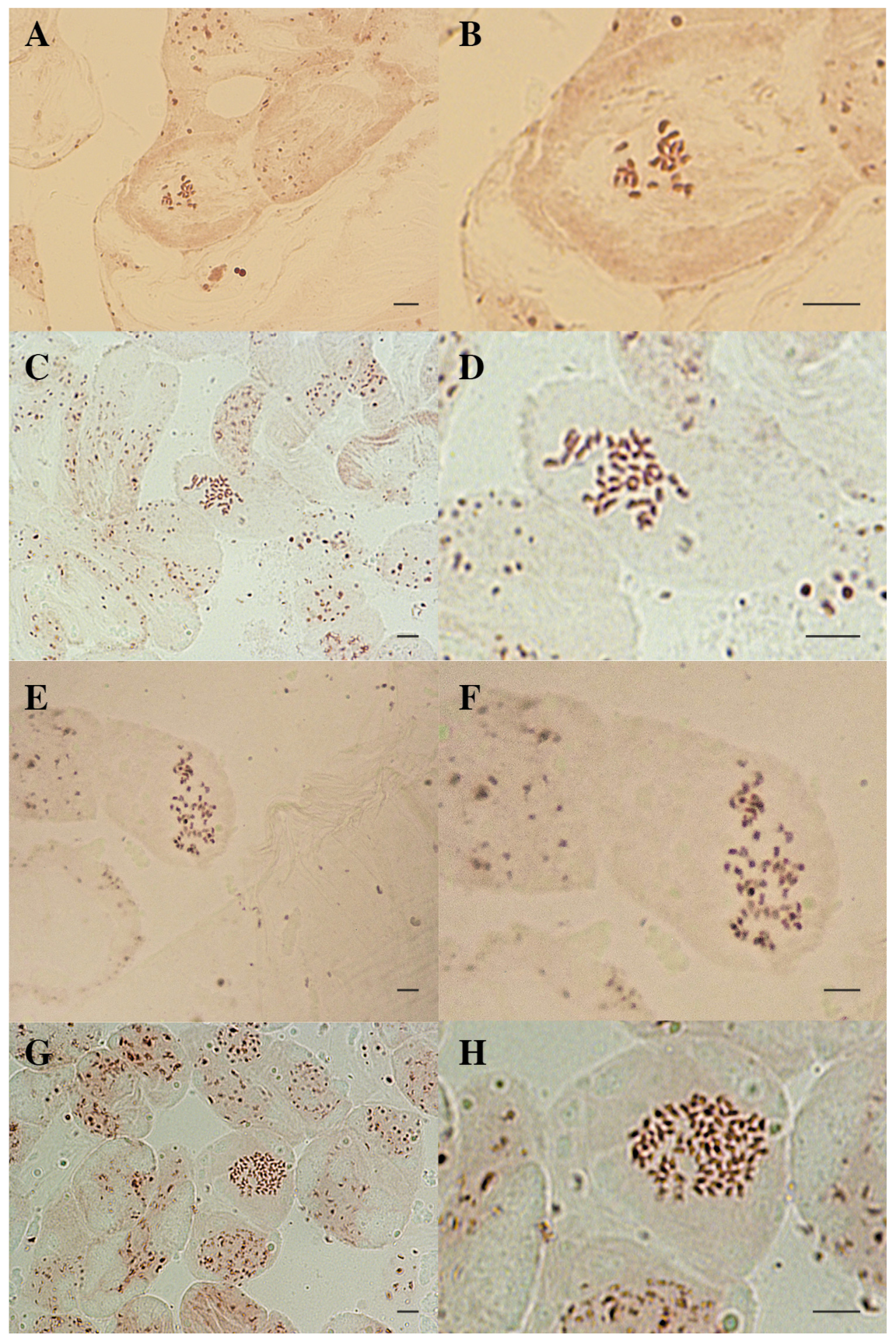
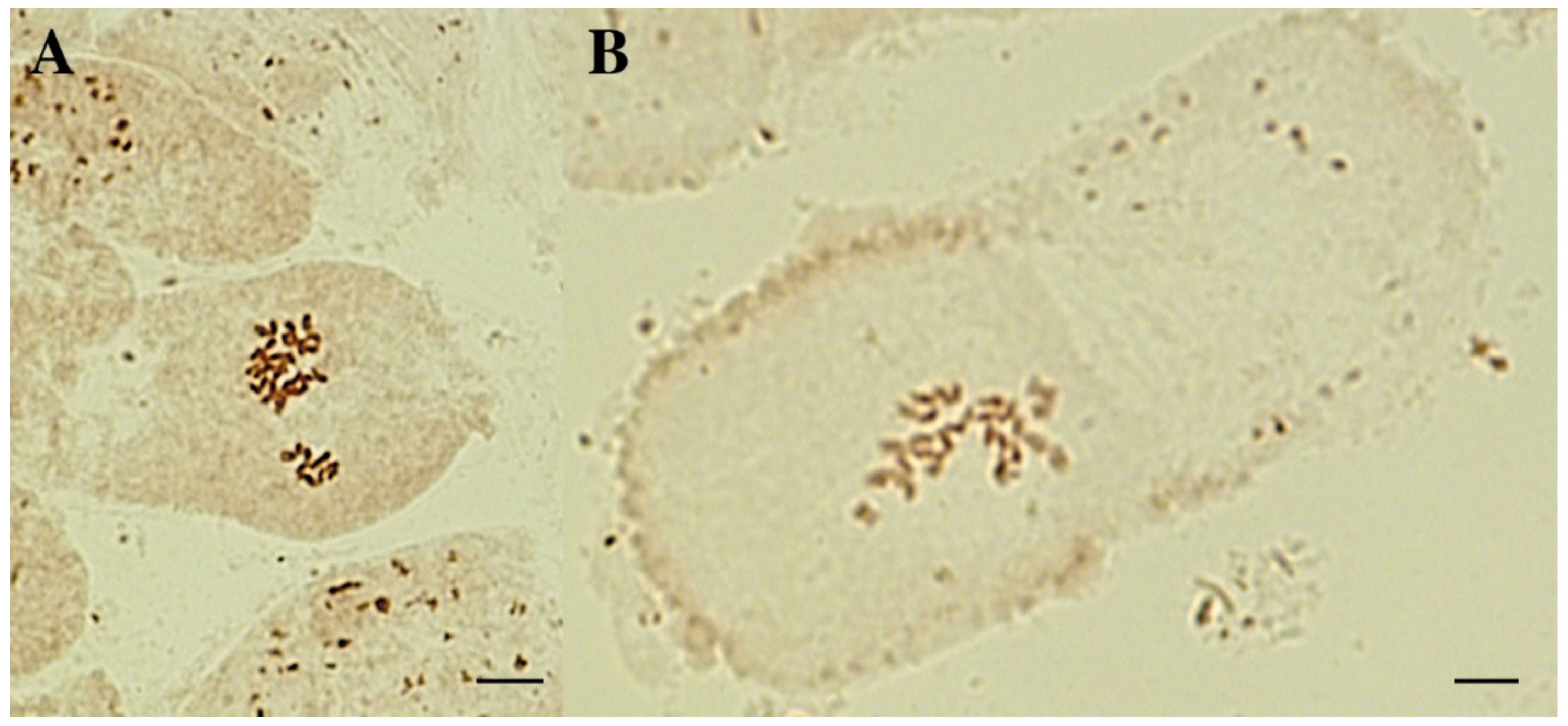
3.2. Karyological Analysis of Courgette Regenerant Plants Obtained in the Culture of Unpollinated Ovules In Vitro

Microscopic counting of chromosomes in apical meristems is the reference, and it is the only unambiguous tool for determining the chromosome number of a plant. Accordingly, we used this analysis as a confirmatory method after ploidy determination using flow cytometry of cell nuclei. C. pepo proved to be a difficult subject cytologically due to the large number of chromosomes, their small size, the low frequency of mitosis, and the small number of metaphases with good chromosome dispersion. In our study, axillary buds/top meristems or very young leaflets were the most convenient explants for preparation of crushed preparations in courgette and patisson. It was from them that preparations with the maximum number of dividing cells could be obtained, whereas, from young roots such preparations were much worse. At the same time, young shoot apices could be plucked with tweezers under sterile conditions, even in in vitro culture specimens. However, we would like to note that haploid specimens in our study were most often very small/stunted and died frequently after taking a part of the apical meristems for chromosomal analysis. Preparations of cells containing a haploid set of chromosomes were the hardest to obtain. The frequency of mitosis in haploid plants was extremely low.
When selecting the optimal technique for cytological analysis, we were guided by the convenience and rapidity of its implementation. Our upgraded technique of chromosome staining in meristem cells using propion–lacmoid was successfully applied in cytological analysis of courgette and patisson regenerant plants obtained by gynogenesis. The undoubted convenience of this technique is the absence of the need for a fluorescence microscope, easy availability and small amount of inexpensive reagents, and the absence of a separate fixation step (in this method, it occurs simultaneously with staining and takes no more than 24 h), after which a maceration step in 40% propionic acid and preparation of a crushed preparation can be performed. C. pepo preparations can be viewed under an ordinary light microscope using a 100× lens with oil immersion.
We managed to obtain microphotographs of courgette and patisson chromosomes of different ploidy levels, which will be of interest to other researchers working with this species, as they could not be found in the published literature. Figure 3 shows C. pepo cells containing 20, 40, 60, and 80 chromosomes corresponding to haploid (n), diploid (2n), triploid (3n), and tetraploid (4n) ploidy levels, respectively (Figure 3).
The ploidy of all these plants coincided with the ploidy level determined using the flow cytometry method. We were unable to obtain high-quality preparations from octaploid plants (8n) confirmed by flow cytometry; these plants developed rather poorly and died soon afterward.
Among the analyzed samples from plants still in the in vitro culture, it was possible to detect mixoploidy and which cells of different ploidy levels (n and 2n; 2n and 3n; 3n and 4n) could meet in one preparation. Aneuploid specimens were also found in a sample of plants (Figure 4); most often, these plants had problems with the formation of generative organs, and a slightly decreased DNA content (0.86–0.89 pg) could be noted on flow cytometry analysis. Formation of gynandromorphic flowers was noted, male flowers were usually sterile, and various abnormalities were observed in female flowers (Figure 5 and Figure 6). It was also impossible to obtain offspring from self-pollination in such plants.
It is known from the literature [28,39] that regenerants of some species tend to present a nonhomogeneous ploidy level (mixoploid); after spontaneous or induced chromosome doubling, they may be subfertile or even sterile. At the same time, prolonged exposure to in vitro culture conditions, particularly plant growth regulators, can promote more than one round of chromosome doubling in cells and the emergence of polyploid plants [28].
At present, we have been able to find images of chromosomes only for Cucurbita moschata [37,38]. Studies by Waminal et al. (2011) [38] showed that the complete set of chromosomes of C. moschata is 2n = 2x = 40, with chromosome length ranging from 1.05 ± 0.30 μm to 1.78 ± 0.03 μm and a total length of 25.19 ± 0.30 μm. Thirty-six metacentric and four sub-metacentric chromosomes were identified. We could not find similar studies for C. pepo, confirming the difficulties of karyological analysis in this species. Despite this, this is the assay most often chosen by researchers when testing plants obtained using DH technologies, but cytological illustrations in these publications are quite often lacking or are of poor quality [16,54,55,56,73,74]. Lack of a convenient chromosome staining technique can lead to unreliable results.
In the literature on the evaluation of DH-derived regenerant plants in the genus Cucurbita, the most common method used was chromosome counting in root tips using the Feulgen technique [52,74,75,76], or staining with 1–2% acetocarmine solution [16,73,74]. At the same time, it was mandatory to pretreat the material for 2–3 h with a 0.05% aqueous solution of colchicine [16,73,74] or α-mono bromo naphthalene [54,55,56] followed by fixation in Carnoy’s fixative solution for 48 h. After all these steps, the material had to be washed in 70% ethanol, and only then (after maceration in 1 N HCl) could the pressed preparation be prepared and stained. Such a long and multistep procedure of preparation with a large number of necessary reagents did not ensure obtaining high-quality preparations, on which chromosomes could be easily counted. Despite the rather small size (about 2 μm), when using the propion–lacmoid method of staining, the chromosomes are quite clearly visible and differ from the cells published by other authors, where dark-colored dots are taken as chromosomes [16,52,75,76]. Such dots are quite frequent and abundant in stained meristem cells of courgette and, as can be seen, are not related to ploidy level; their number is quite different even in nearby cells of the same preparation (Figure 3).
According to our assumption, it is difficult to obtain a high-quality cytological preparation in species of the genus Cucurbita; in most studies conducted using DH technologies, triploids and tetraploids were not diagnosed among regenerant plants, whereas, in our studies, they were formed in rather large numbers. Thus, propion–lacmoid method of chromosome staining has clear advantages over other methods of karyological analysis and can be used for routine analysis of ploidy level in C. pepo samples, and it is recommended for inclusion into protocols for DH plant production.

3.3. Abaximal Epidermis Indices in C. pepo Regenerant Plants

Courgette and patisson regenerant plants obtained in the culture of unpollinated ovules in vitro were evaluated according to the main indicators of abaxial epidermis: stomata guard cell size (width and length), stomata density (number of stoma per mm2), and chloroplast number (each side of the guard cells). Five to six leaves from the shoot tip were taken for the analysis, since it is known from literature data that leaves of different age can differ in the studied parameters. Leaves of C. pepo are large and the epidermis on the lower side of the leaf can be separated easily using tweezers. We analyzed the preparations without using additional staining (10% Lugol’s iodine solution or 1% AgNO3), as suggested in published protocols [51,52,54,55,56]. To count the number of chloroplasts in stomata guard cells, we used a fluorescence microscope with a set of filters to detect natural chloroplast autofluorescence (Figure 7 and Figure 8).
Initially, the main indices of the abaxial epidermis were measured in control specimens of courgette and patisson grown under open-field and greenhouse conditions. Among the selected control specimens within the patisson group (subsp. ovifera) and courgette group (subsp. pepo), the genotype factor did not significantly influence the studied parameters, but there was a significant difference between plants of the same genotype but grown in different conditions (Table 1). SL in plants grown in the greenhouse was 1.2 times higher, while the SD was 1.99 times lower compared to plants grown in the field under sunny and hot summer conditions. The variation of these traits was comparable with the data obtained by other researchers [51,52,53] for C. pepo species on haploid and diploid samples (Table 1). Thus, the use of abaxial epidermis indices (SL and SD) for identification of plant ploidy can be used only for plants grown under the same conditions. At the same time, no significant difference in the number of chloroplasts in stomatal guard cells (CN) in plants grown under different conditions was found. In addition, a significant difference in abaxial epidermis indices between summer squash samples belonging to different varieties was found. The length (SL) and width (SW) of stomatal guard cells were smaller in patisson, while stomata density (SD) was greater. For patisson grown under open-field conditions, the SL was on average 20.00 µm, and the number of stomata averaged 550.55 per 1 mm2; for courgette, the SL was 24.96 µm, while the SD was less as 446.89 prs/mm2. The number of chloroplasts in stomatal guard cells of diploid control samples did not depend on genotype and growth conditions and averaged 10.7 in both varieties. The CN indicator turned out to be the most stable, and it can be recommended for ploidy identification. The results obtained confirmed the data published for other crops, indicating that growing conditions can influence abaxial epidermis parameters [77,78]. This should be taken into account in ploidy analysis when SL, SW, SI, and SD are used.
Courgette and patisson regenerant plants obtained in the unpollinated ovule culture in vitro were grown in a greenhouse; in diploid samples, the average indices of the abaxial epidermis practically did not differ from those of control donor plants grown in the same conditions (Table 2). In the analyzed sample of regenerant plants, samples differing in the number of chloroplasts in stomata closing cells of abaxial leaf epidermis from control diploid samples (containing from 8 to 12) were found. In courgette, the number of chloroplasts in regenerant plants obtained in the unpollinated ovule culture varied from 7 to 24. Comparing the data obtained (Table 2, Figure 7) with the data obtained by analysis of flow cytometry of cell nuclei and direct chromosome counting, it was concluded that plants containing on average 11.31 chloroplasts in stomata closing cells would have a diploid set of chromosomes, whereas 17.66 would have a tetraploid set. In triploid specimens, there was a very large variation in all abaximal epidermal indices. Stomata varying greatly in size and containing seven to 24 chloroplasts could be observed per microscope field of view, but the average CN value for triploid specimens was 14.84 pc. The groups of plants with diploid, triploid, and tetraploid ploidy differed significantly in SL, SD, and CN parameters. A similar pattern was observed for plants with different ploidy level in patisson (Table 2, Figure 8). Diploid and tetraploid samples differed 1.87 times by CN. According to SW indices, it was reliably possible to separate all three studied groups according to ploidy level in patisson, whereas, in courgette, it was only reliably possible to separate diploid samples from samples with high ploidy level according to this parameter.
Thus, abaxial epidermal indices can be used to identify ploidy in C. pepo. The use of an indirect indicator such as the number of chloroplasts in stomatal guard cells (CN) can serve as a sufficiently accurate, relatively fast and inexpensive way to identify ploidy in regenerant plants of courgette and patisson.

4. Conclusions

Our study represents the first analysis of ploidy in C. pepo regenerant plants obtained in an unpollinated ovule culture in vitro using three methods of ploidy identification (flow cytometry of cell nuclei, direct chromosome counting, and use of abaxial epidermis parameters). In this study, we identified four levels of ploidy among ex vitro adapted regenerant plants (2n, 3n, 4n, and 8n), and haploids (n) were detected only among plants in the in vitro culture. Of the courgette and patisson samples examined, 32.35% were diploid, while significant proportions were triploid at 26.47% and tetraploid at 33.82%. A protocol was optimized for the analysis of C. pepo plants using flow cytometry of cell nuclei. Two cytotypes were identified for diploid samples with a DNA content of 2C = 1.07 ± 0.03 pg for courgette samples belonging to subsp. pepo and a second cytotype of 2C = 0.95 ± 0.03 pg for patisson samples belonging to subsp. ovifera. Photographs of metaphase chromosomes of haploid, triploid, and tetraploid C. pepo specimens were presented for the first time. The propion–lacmoid method of chromosome staining showed its advantages for routine analysis for ploidy levels in C. pepo specimens and can be recommended for inclusion in protocols for DH plant derivation. Abaxial epidermal indices in diploid courgette and patisson specimens grown under different conditions (field and greenhouse) were described and compared. It was shown that the most stable parameter, independent of growing conditions, was the number of chloroplasts in stomata guard cells. For patisson and courgette, SN averages were 9.41 to 11.31 pcs for diploid samples, 14.84 to 16.3 pcs for triploid samples, and up to 17.58 pcs for tetraploid samples. The use of several methods of estimation can allow avoiding errors in the identification of ploidy of regenerant plants obtained using DH technologies.

Author Contributions

Conceptualization, E.D. and A.D.; flow cytometry analysis, M.S.; chromosome analysis, L.K.; morphological analysis, S.B. and A.E.; data curation, E.D and A.D.; writing—original draft preparation, E.D.; visualization, A.E., L.K., and S.B. All authors have read and agreed to the published version of the manuscript.

Funding

The reported study was funded by RFBR, project number 20-316-9005.

Conflicts of Interest

The authors declare no conflict of interest.

References

  1. FAOSTAT. Available online: https://www.fao.org/ (accessed on 22 May 2022).
  2. Hooghvorst, I.; Nogués, S. Opportunities and Challenges in Doubled Haploids and Haploid Inducer-Mediated Genome-Editing Systems in Cucurbits. Agronomy 2020, 10, 1441. [Google Scholar] [CrossRef]
  3. Dhillon, N.P.; Laenoi, S.; Srimat, S.; Pruangwitayakun, S.; Mallappa, A.; Kapur, A.; Yadav, K.K.; Hegde, G.; Schafleitner, R.; Schreinemachers, P. Sustainable Cucurbit Breeding and Production in Asia Using Public–Private Partnerships by the World Vegetable Center. Agronomy 2020, 10, 1171. [Google Scholar] [CrossRef]
  4. Dong, Y.-Q.; Zhao, W.-X.; Li, X.-H.; Liu, X.-C.; Gao, N.-N.; Huang, J.-H.; Wang, W.-Y.; Xu, X.-L.; Tang, Z.-H. Androgenesis, Gynogenesis, and Parthenogenesis Haploids in Cucurbit Species. Plant Cell Rep. 2016, 35, 1991–2019. [Google Scholar] [CrossRef] [PubMed]
  5. Kurtar, E.S.; Balkaya, A.; Kandemir, D. A Productive Direct Regeneration Protocol for a Wide Range of Winter Squash (Cucurbita maxima Duch.) and Pumpkin (Cucurbita moschata Duch.) Lines. Rom. Biotechnol. Lett. 2020, 25, 1348–1355. [Google Scholar] [CrossRef]
  6. Domblides, E.A.; Ermolaev, A.S.; Belov, S.N. Obtaining Doubled Haploids of Cucurbita Pepo L. Ovoŝi Ross. 2021. [Google Scholar] [CrossRef]
  7. Palmer, C.E.; Keller, W.A.; Arnison, P.G. Utilization of Brassica Haploids. In In Vitro Haploid Production in Higher Plants: Volume 3—Important Selected Plants; Current Plant Science and Biotechnology in Agriculture; Jain, S.M., Sopory, S.K., Veilleux, R.E., Eds.; Springer Netherlands: Dordrecht, The Netherlands, 1996; pp. 173–192. ISBN 978-94-017-1858-5. [Google Scholar]
  8. Testillano, P.; Georgiev, S.; Mogensen, H.L.; Coronado, M.J.; Dumas, C.; Risueno, M.C.; Matthys-Rochon, E. Spontaneous Chromosome Doubling Results from Nuclear Fusion during in Vitro Maize Induced Microspore Embryogenesis. Chromosoma 2004, 112, 342–349. [Google Scholar] [CrossRef] [PubMed] [Green Version]
  9. Kasha, K.J. Chromosome Doubling and Recovery of Doubled Haploid Plants. In Haploids in Crop Improvement II.; Biotechnology in Agriculture and Forestry; Don Palmer, C.E., Keller, W.A., Kasha, K.J., Eds.; Springer: Berlin/Heidelberg, Germany, 2005; pp. 123–152. ISBN 978-3-540-26889-5. [Google Scholar]
  10. Seguí-Simarro, J.M.; Nuez, F. Pathways to Doubled Haploidy: Chromosome Doubling during Androgenesis. Cytogenet. Genome Res. 2008, 120, 358–369. [Google Scholar] [CrossRef]
  11. Daghma, D.E.S.; Hensel, G.; Rutten, T.; Melzer, M.; Kumlehn, J. Cellular Dynamics during Early Barley Pollen Embryogenesis Revealed by Time-Lapse Imaging. Front. Plant Sci. 2014, 5, 675. [Google Scholar] [CrossRef] [Green Version]
  12. Ahmadi, B.; Ebrahimzadeh, H. In Vitro Androgenesis: Spontaneous vs. Artificial Genome Doubling and Characterization of Regenerants. Plant Cell Rep. 2020, 39, 299–316. [Google Scholar] [CrossRef]
  13. Khan, P.S.S.V.; Vijayalakshmi, G.; Raja, M.M.; Naik, M.L.; Germanà, M.A.; Terry, R.G. Doubled Haploid Production in Onion (Allium cepa L.): From Gynogenesis to Chromosome Doubling. Plant Cell Tissue Organ Cult. PCTOC 2020, 142, 1–22. [Google Scholar] [CrossRef]
  14. Dryanovska, O.A. Induced Callus in Vitro from Ovaries and Anthers of Species from the Cucurbitaceae Family. Dokl. Na Bolg. Akad. Na Nauk. 1985, 38, 1243. [Google Scholar]
  15. Gałązka, J.; Niemirowicz-Szczytt, K. Review of Research on Haploid Production in Cucumber and Other Cucurbits. Folia Hortic. 2013, 25, 67–78. [Google Scholar] [CrossRef] [Green Version]
  16. Shalaby, T.A. Factors Affecting Haploid Induction through in Vitro Gynogenesis in Summer Squash (Cucurbita pepo L.). Sci. Hortic. 2007, 115, 1–6. [Google Scholar] [CrossRef]
  17. Faris, N.M.; Rakoczy-Trojanowska, M.; Malepszy, S.; Niemirowicz-Szczytt, K. Diploidization of Cucumber (Cucumis Sativus L.) Haploids by in Vitro Culture of Leaf Explant. In Progress in Biotechnology; Food Biotechnology; Bielecki, S., Tramper, J., Polak, J., Eds.; Elsevier: Amsterdam, The Netherlands, 2000; Volume 17, pp. 49–54. [Google Scholar]
  18. Niemirowicz-Szczytt, K.; Faris, N.M.; Nikolova, V.; Rakoczy-Trojanowska, M.; Malepszy, S. Optimization of Cucumber (Cucumis sativus L.) Haploid Production and Doubling. In Proceedings of the Cucurbitaceae, Warsaw, Poland, 24–28 July 1995; Volume 94, pp. 169–171. [Google Scholar]
  19. Sztangret-Wiśniewska, J.; Gałecka, T.; Korzeniewska, A.; Marzec, L.; Kołakowska, G.; Piskurewicz, U.; Śmiech, M.; Niemirowicz-Szczytt, K. Characteristics of Double-Haploid Cucumber (Cucumis sativus L.) Lines Resistant to Downy Mildew (Pseudoperonospora Cubensis [Berk. et Curt.] Rostovzev). In Proceedings of the Cucurbitaceae 2006, Asheville, NC, USA, 17–21 September 2006; pp. 515–526. [Google Scholar]
  20. Gürel, S.; Gürel, E.; Kaya, Z. Doubled Haploid Plant Production from Unpollinated Ovules of Sugar Beet (Beta Vulgaris L.). Plant Cell Rep. 2000, 19, 1155–1159. [Google Scholar] [CrossRef] [PubMed]
  21. Grzebelus, E.; Adamus, A. Effect of Anti-Mitotic Agents on Development and Genome Doubling of Gynogenic Onion (Allium Cepa L.) Embryos. Plant Sci. 2004, 167, 569–574. [Google Scholar] [CrossRef]
  22. Broughton, S.; Castello, M.; Liu, L.; Killen, J.; Hepworth, A.; O’Leary, R. The Effect of Caffeine and Trifluralin on Chromosome Doubling in Wheat Anther Culture. Plants 2020, 9, 105. [Google Scholar] [CrossRef] [Green Version]
  23. Castillo, A.M.; Cistué, L.; Vallés, M.P.; Soriano, M. Chromosome Doubling in Monocots. In Advances in Haploid Production in Higher Plants; Touraev, A., Forster, B.P., Jain, S.M., Eds.; Springer Netherlands: Dordrecht, The Netherlands, 2009; pp. 329–338. ISBN 978-1-4020-8854-4. [Google Scholar]
  24. Claveria, E.; Garcia-Mas, J.; Dolcet-Sanjuan, R. Optimization of Cucumber Doubled Haploid Line Production Using in Vitro Rescue of in Vivo Induced Parthenogenic Embryos. J. Am. Soc. Hortic. Sci. 2005, 130, 555–560. [Google Scholar] [CrossRef] [Green Version]
  25. Lim, W.; Earle, E.D. Effect of in Vitro and in Vivo Colchicine Treatments on Pollen Production and Fruit Set of Melon Plants Obtained by Pollination with Irradiated Pollen. Plant Cell Tissue Organ Cult. 2008, 95, 115–124. [Google Scholar] [CrossRef]
  26. Lim, W.; Earle, E.D. Enhanced Recovery of Doubled Haploid Lines from Parthenogenetic Plants of Melon (Cucumis melo L.). Plant Cell Tissue Organ Cult. PCTOC 2009, 98, 351–356. [Google Scholar] [CrossRef]
  27. Domblides, E.; Shmykova, N.; Khimich, G.; Korotseva, I.; Kan, L.; Domblides, A.; Pivovarov, V.; Soldatenko, A. Production of Doubled Haploid Plants of Cucurbitaceae Family Crops through Unpollinated Ovule Culture in Vitro. Acta Hortic. 2020, 1294, 19–28. [Google Scholar] [CrossRef]
  28. Ochatt, S.J.; Seguí-Simarro, J.M. Analysis of Ploidy in Haploids and Doubled Haploids. In Doubled Haploid Technology: Volume 1: General Topics, Alliaceae, Cereals; Methods in Molecular Biology; Segui-Simarro, J.M., Ed.; Springer US: New York, NY, USA, 2021; pp. 105–125. ISBN 978-1-07-161315-3. [Google Scholar]
  29. Rice, A.; Glick, L.; Abadi, S.; Einhorn, M.; Kopelman, N.M.; Salman-Minkov, A.; Mayzel, J.; Chay, O.; Mayrose, I. The Chromosome Counts Database (CCDB)–a Community Resource of Plant Chromosome Numbers. New Phytol. 2015, 206, 19–26. [Google Scholar] [CrossRef] [PubMed]
  30. Goldblatt, P.; Lowry, P.P. The Index to Plant Chromosome Numbers (Ipcn): Three Decades of Publication by the Missouri Botanical Garden Come to an End. Ann. Mo. Bot. Gard. 2011, 98, 226–227. [Google Scholar] [CrossRef]
  31. Vavilov, N.I. Intergeneric Hybrids of Melons, Watermelons and Squashes. Proc. Appl. Bot. Genet. Breed. 1925, 14, 1–35. [Google Scholar]
  32. Whitaker, T.W. Cytological and Phylogenetic Studies in the Cucurbitaceae. Bot. Gaz. 1933, 94, 780–790. [Google Scholar] [CrossRef]
  33. Whitaker, T.W.; Davis, G.N. Cucurbits.; Leonard Hill (Books) Ltd.: London, UK; Interscience Publishers Inc.: New York, NY, USA, 1962. [Google Scholar]
  34. Varghese, B.M. Studies on Cytology and Evolution of South Indian Cucurbitaceae. Ph.D. Thesis, University of Kerala, Thiruvananthapuram, India, 1973. [Google Scholar]
  35. Jeffrey, C. A Review of the Cucurbitaceae. Bot. J. Linn. Soc. 1980, 81, 233–247. [Google Scholar] [CrossRef]
  36. Metwally, E.I.; Haroun, S.A.; El-Fadly, G.A. Interspecific Cross between Cucurbita pepo L. and Cucurbita Martinezii through in Vitro Embryo Culture. Euphytica 1996, 90, 1–7. [Google Scholar] [CrossRef]
  37. Kurtar, E.S.; Balkaya, A.; Kandemir, D. Evaluation of Haploidization Efficiency in Winter Squash (Cucurbita maxima Duch.) and Pumpkin (Cucurbita moschata Duch.) through Anther Culture. Plant Cell Tissue Organ Cult. PCTOC 2016, 127, 497–511. [Google Scholar] [CrossRef]
  38. Waminal, N.E.; Kim, N.-S.; Kim, H.H. Dual-Color FISH Karyotype Analyses Using RDNAs in Three Cucurbitaceae Species. Genes Genom. 2011, 33, 521–528. [Google Scholar] [CrossRef]
  39. Bohanec, B. Ploidy Determination Using Flow Cytometry. In Doubled Haploid Production in Crop Plants; Springer: Berlin/Heidelberg, Germany, 2003; pp. 397–403. [Google Scholar]
  40. Pellicer, J.; Leitch, I.J. The Plant DNA C-values Database (Release 7.1): An Updated Online Repository of Plant Genome Size Data for Comparative Studies. New Phytol. 2019. [Google Scholar] [CrossRef] [Green Version]
  41. Arumuganathan, K.; Earle, E.D. Nuclear DNA Content of Some Important Plant Species. Plant Mol. Biol. Rep. 1991, 9, 208–218. [Google Scholar] [CrossRef]
  42. Košmrlj, K.; Murovec, J.; Bohanec, B. Haploid Induction in Hull-Less Seed Pumpkin through Parthenogenesis Induced by X-Ray-Irradiated Pollen. J. Am. Soc. Hortic. Sci. 2013, 138, 310–316. [Google Scholar] [CrossRef] [Green Version]
  43. Evans, A.M. The Production and Identification of Polyploids in Red Clover, White Clover and Lucerne. New Phytol. 1955, 54, 149–162. [Google Scholar] [CrossRef]
  44. Speckmann, G.J.; Post, J.; Dijkstra, H. The Length of Stomata as an Indicator for Polyploidy in Rye-Grasses. Euphytica 1965, 14, 225–230. [Google Scholar] [CrossRef]
  45. Dore, C.; Lescure, J.-C.; Charlot, F. Evaluation Du Niveau de Ploïdie Des Plantes d’une Population de Choux de Bruxelles (Brassica oleracea L. Ssp. gemmifera) d’origine Pollinique. Agronomie 1986, 6, 797–801. [Google Scholar] [CrossRef] [Green Version]
  46. Borrino, E.M.; Powell, W. Stomatal Guard Cell Length as an Indictor of Ploidy in Microspore-Derived Plants of Barley. Genome 1988, 30, 158–160. [Google Scholar] [CrossRef]
  47. Frandsen, N.O. Die Plastidenzahl Als Merkmal Bei Der Kartoffel. Theor. Appl. Genet. 1968, 38, 153–167. [Google Scholar] [CrossRef]
  48. Jacobs, J.P.; Yoder, J.I. Ploidy Levels in Transgenic Tomato Plants Determined by Chloroplast Number. Plant Cell Rep. 1989, 7, 662–664. [Google Scholar] [CrossRef]
  49. Singsit, C.; Veilleux, R.E. Chloroplast Density in Guard Cells of Leaves of Anther-Derived Potato Plants Grown in Vitro and in Vivo. HortScience 1991, 26, 592–594. [Google Scholar] [CrossRef] [Green Version]
  50. Choe, E.; Carbonero, C.H.; Mulvaney, K.; Rayburn, A.L.; Mumm, R.H. Improving in Vivo Maize Doubled Haploid Production Efficiency through Early Detection of False Positives. Plant Breed. 2012, 131, 399–401. [Google Scholar] [CrossRef]
  51. Ebrahimzadeh, H.; Lotfi, M.; Sadat-Hosseini, M. Parthenogenetic Haploid Plant Production in Styrian Pumpkin by Gamma Irradiated Pollen. Int. J. Hortic. Sci. Technol. 2021, 8, 305–314. [Google Scholar] [CrossRef]
  52. Kurtar, E.S.; SarI, N.; Abak, K. Obtention of Haploid Embryos and Plants through Irradiated Pollen Technique in Squash (Cucurbita pepo L.). Euphytica 2002, 127, 335–344. [Google Scholar] [CrossRef]
  53. Kurtar, E.S.; Seymen, M.; Çetın, A.N.; Türkmen, Ö. Dihaploidization in Promising Summer Squash Genotypes (Cucurbita pepo L.) via Irradiated Pollen Technique. Yuzuncu Yil Univ. J. Agric. Sci. 2021, 31, 42–51. [Google Scholar] [CrossRef]
  54. Kurtar, E.S.; Seymen, M. Induction of Parthenogenesis by Irradiated Pollen in Cucurbita Species. In Doubled Haploid Technology: Volume 3: Emerging Tools, Cucurbits, Trees, Other Species; Segui-Simarro, J.M., Ed.; Methods in Molecular Biology; Springer US: New York, NY, 2021; pp. 111–121. ISBN 978-1-07-161331-3. [Google Scholar]
  55. Kurtar, E.S.; Seymen, M. Gynogenesis in Cucurbita Species. In Doubled Haploid Technology: Volume 3: Emerging Tools, Cucurbits, Trees, Other Species; Methods in Molecular Biology; Segui-Simarro, J.M., Ed.; Springer US: New York, NY, USA, 2021; pp. 123–133. ISBN 978-1-07-161331-3. [Google Scholar]
  56. Kurtar, E.S.; Seymen, M. Anther Culture in Cucurbita Species. In Doubled Haploid Technology: Volume 3: Emerging Tools, Cucurbits, Trees, Other Species; Methods in Molecular Biology; Segui-Simarro, J.M., Ed.; Springer US: New York, NY, USA, 2021; pp. 111–121. ISBN 978-1-07-161331-3. [Google Scholar]
  57. Domblides, E.A.; Shmykova, N.A.; Zayachkovskaya, T.V.; Khimich, G.A.; Korotseva, I.B.; Kan, L.Y.; Domblides, A.S. Doubled Haploid Plant Production from in Vitro Culture of Unpollinated Ovules of Summer Squash (Cucurbita pepo L.). In Proceedings of the VII International Scientific and Practical Conference Dedicated to the 30th Anniversary of the Department of Plant Biotechnology of the Nikitskii Botanical Garden, Simferopol, Russia, 25 September–1 October 2016; pp. 28–29. [Google Scholar]
  58. Pfosser, M.; Heberle-Bors, E.; Amon, A.; Lelley, T. Evaluation of Sensitivity of Flow Cytometry in Detecting Aneuploidy in Wheat Using Disomic and Ditelosomic Wheat–Rye Addition Lines. Cytom. J. Int. Soc. Anal. Cytol. 1995, 21, 387–393. [Google Scholar] [CrossRef] [PubMed]
  59. Skaptsov, M.V.; Smirnov, S.V.; Kutsev, M.G.; Pshennikova, L.M.; Shmakov, A.I. The First Data Report about DNA Content of Some Representatives of the Genus Trapa L. from Primorye Territory. Прoблемы Бoтаники Южнoй Сибири И Мoнгoлии 2020, 19, 87–89. [Google Scholar] [CrossRef]
  60. Sokoloff, D.D.; Skaptsov, M.V.; Vislobokov, N.A.; Smirnov, S.V.; Shmakov, A.I.; Remizowa, M.V. Morphological Characterization of Diploid and Triploid Acorus Calamus (Acoraceae) from Southern Western Siberia, Parthenocarpy in Sterile Plants and Occurrence of Aneuploidy. Bot. J. Linn. Soc. 2021, 195, 189–215. [Google Scholar] [CrossRef]
  61. Doležel, J.; Greilhuber, J.; Suda, J. Estimation of Nuclear DNA Content in Plants Using Flow Cytometry. Nat. Protoc. 2007, 2, 2233–2244. [Google Scholar] [CrossRef]
  62. Solovyova, L.V. Laboratory Manual on the Fruit Plant Cytology; Moscow State University: Moscow, Russia, 1982. [Google Scholar]
  63. Rayburn, A.L.; Kushad, M.M.; Wannarat, W. Intraspecific Genome Size Variation in Pumpkin (Cucurbita pepo Subsp. pepo). Hortscience 2008, 43, 949–951. [Google Scholar] [CrossRef]
  64. Barow, M.; Meister, A. Lack of Correlation between AT Frequency and Genome Size in Higher Plants and the Effect of Nonrandomness of Base Sequences on Dye Binding. Cytom. J. Int. Soc. Anal. Cytol. 2002, 47, 1–7. [Google Scholar] [CrossRef]
  65. Marie, D.; Brown, S.C. A Cytometric Exercise in Plant DNA Histograms, with 2C Values for 70 Species. Biol. Cell 1993, 78, 41–51. [Google Scholar] [CrossRef]
  66. Tatum, T.C.; Nunez, L.; Kushad, M.M.; Rayburn, A.L. Genome Size Variation in Pumpkin (Cucurbita Sp.). Ann. Appl. Biol. 2006, 149, 145–151. [Google Scholar] [CrossRef]
  67. Gasmanova, N.; Lebeda, A.; Dolezalova, I.; Paris, H.S. Variability of Nuclear DNA Content among and within Three Cucurbit Genera. Acta Hortic. 2007, 731, 83. [Google Scholar] [CrossRef]
  68. Rayburn, A.L.; Price, H.J.; Smith, J.; Gold, J.R. C-band Heterochromatin and DNA Content in Zea Mays. Am. J. Bot. 1985, 72, 1610–1617. [Google Scholar] [CrossRef]
  69. Paris, H.S.; Lebeda, A.; Křistkova, E.; Andres, T.C.; Nee, M.H. Parallel Evolution Under Domestication and Phenotypic Differentiation of the Cultivated Subspecies of Cucurbita pepo (Cucurbitaceae). Econ. Bot. 2012, 66, 71–90. [Google Scholar] [CrossRef]
  70. Sarı, N.; Abak, K.; Pitrat, M.; Rode, J.C.; Vaulx, R.D. Induction of Parthenogenetic Haploid Embryos after Pollination by Irradiated Pollen in Watermelon. HortScience 1994, 29, 1189–1190. [Google Scholar] [CrossRef] [Green Version]
  71. Gilissen, L.J.W.; Van Staveren, M.J.; Creemers-Molenaar, J.; Verhoeven, H.A. Development of Polysomaty in Seedlings and Plants of Cucumis sativus L. Plant Sci. 1993, 91, 171–179. [Google Scholar] [CrossRef]
  72. Barow, M.; Meister, A. Endopolyploidy in Seed Plants Is Differently Correlated to Systematics, Organ, Life Strategy and Genome Size. Plant Cell Environ. 2003, 26, 571–584. [Google Scholar] [CrossRef]
  73. Shalaby, T.A. Embryogenesis and Plantlets Regeneration from Anther Culture of Squash Plants (Cucurbita pepo L.) as Affected by Different Genotypes. J. Agric. Res. Tanta Univ. 2006, 32, 173–183. [Google Scholar]
  74. Metwally, E.I.; Moustafa, S.A.; El-Sawy, B.I.; Haroun, S.A.; Shalaby, T.A. Production of Haploid Plants from in Vitro Culture of Unpollinated Ovules of Cucurbita pepo. Plant Cell Tissue Organ Cult. 1998, 52, 117–121. [Google Scholar] [CrossRef]
  75. Kurtar, E.S.; Balkaya, A.; Ozbakir, M.; Ofluoglu, T. Induction of Haploid Embryo and Plant Regeneration via Irradiated Pollen Technique in Pumpkin (Cucurbita moschata Duchesne Ex. Poir). Afr. J. Biotechnol. 2009, 8, 5944–5951. [Google Scholar] [CrossRef]
  76. Kurtar, E.S.; Balkaya, A. Production of in Vitro Haploid Plants from in Situ Induced Haploid Embryos in Winter Squash (Cucurbita maxima Duchesne Ex Lam.) via Irradiated Pollen. Plant Cell Tissue Organ Cult. PCTOC 2010, 102, 267–277. [Google Scholar] [CrossRef]
  77. Zachleder, V.; Cepák, V. Variations in Chloroplast Nucleoid Number during the Cell Cycle in the AlgaScenedesmus Quadricauda Grown under Different Light Conditions. Protoplasma 1987, 141, 74–82. [Google Scholar] [CrossRef]
  78. Zhang, Z.; Guo, Y.; Ai, X.; Zhang, F.; He, Q.; Sun, X.; Jiao, Z. Effects of sunlight and temperature on ultrastructure and functions of chloroplast of cucumber in solar greenhouse. Ying Yong Sheng Tai Xue Bao J. Appl. Ecol. 2003, 14, 1287–1290. [Google Scholar]
Figure 1. Examples of histograms in cytofluorimetric study of C. pepo (internal standardization): (a) standard (Ficus benjamina 2C = 0.90 pg) and diploid sample with cytotype 2C = 1.07 pg; (b) combined histogram of diploid study with cytotypes 2C = 1.07 pg and 2C = 0.95 pg; (c) standard and triploid sample with cytotype 2C = 1.62 pg; (d) standard and tetraploid sample with cytotype 2C = 1.84 pg; (e) standard and tetraploid sample with cytotype 2C = 2.08 pg; (f) pooled histogram of tetraploid study with cytotypes 2C = 1.84 pg and 2C = 2.08 pg; (g) pooled histogram of diploid and triploid studies with cytotypes 2C = 1.08 pg and 2C = 1.50 pg; (h) pooled histogram of tetraploid and octaploid studies with cytotypes 2C = 2.08 pg and 2C = 3.61 pg; (i) example of endopolyploidy.
Horticulturae 08 01083 g001
Figure 2. Normal flowers of C. pepo: (A) female flower of courgette; (B) male flower of courgette; (C) female flower of patisson; (D) male flower of patisson. Scale bars = 1 cm.
Horticulturae 08 01083 g002
Figure 3. Metaphases in meristems of C. pepo regenerant plants of different ploidy levels: (A,B) haploid (2n = x =20); (C,D) diploid (2n = 2x = 40); (E,F) triploid (2n = 3x = 60); (G,H) tetraploid (2n = 4x= 80). Scale bars = 10 µm.
Horticulturae 08 01083 g003
Figure 4. Metaphases in meristems of C. pepo regenerant plants with an aneuploid chromosome set: (A) aneuploid with 36 chromosomes; (B) aneuploid with 38 chromosomes. Scale bars = 10 µm.
Horticulturae 08 01083 g004
Figure 5. Abnormal male flowers of C. pepo (corolla removed). Scale bars = 1 cm.
Horticulturae 08 01083 g005
Figure 6. Abnormal female and gynandromorphic flowers of C. pepo (corolla removed): (A) abnormal female flower of courgette; (B,C) gynandromorphic flowers of courgette; (D) abnormal female flower of patisson. Scale bars = 1 cm.
Horticulturae 08 01083 g006
Figure 7. Epidermis of abaxial surface of leaves of courgette regenerant plants of different ploidy level obtained in unpollinated ovule culture in vitro: (AC) plant with diploid set of chromosomes (2n = 2x = 40); (DF) plant with triploid set of chromosomes (2n = 3x = 60); (GI) plant with tetraploid set of chromosomes (2n = 4x = 80); (A,D,G) stomata cells in regenerant plants of different ploidy using 20× lens; (B,E,H) stomata of regenerant plants of different ploidy levels using a 100× lens; (C,F,I) autofluorescence of chloroplasts using a set of fluorescence filters using a 100× lens. In the figures A,D,G stomata indicated by the circles.
Horticulturae 08 01083 g007
Figure 8. Epidermis of abaxial leaf surface of patisson regenerant plants of different ploidy level obtained in unpollinated ovule culture in vitro: (AC) plant with diploid chromosomes; (DF) plant with triploid chromosomes; (GI) plant with tetraploid chromosomes; (A,D,G) stomata in regenerant plants of different ploidy level using 20× lens; (B,E,H) stomata of regenerant plants of different ploidy levels using a 100× lens; (C,F,I) autofluorescence of chloroplasts using a set of fluorescence filters using a 100× lens. In the figures A,D,G stomata indicated by the circles.
Horticulturae 08 01083 g008
Table 1. Parameters of abaxial epidermis in donor C. pepo plants grown under different conditions.
ParametersGrowing Condition
(Factor B)
Factor ATwo-Way ANOVA
subsp. oviferasubsp. pepo
SL
(µm)
Field20.00 b/B24.96 b/AFactor A 2 ***, Factor B ***,
Factor A × Factor B ns
Greenhouse25.25 a/B30.41a/A
SW
(µm)
Field15.91 b/B17.96 b/AFactor A ***, Factor B ***,
Factor A × Factor B ns
Greenhouse18.59 a/B20.06 a/A
SI
(SL/SW)
Field1.26 b/B1.39 b/AFactor A ***, Factor B ***,
Factor A × Factor B ns
Greenhouse1.36 a/B1.51 a/A
SD
(number/per mm2)
Field550.55 a/A446.89 a/BFactor A ***, Factor B ***,
Factor A × Factor B ***
Greenhouse275.50 b/A276.10 b/A
CNField10.81 a/A11.11 a/AFactor A ns, Factor B ns,
Factor A × Factor B ns
Greenhouse10.71 a/A10.28 a/A
Note: Values in the table are the mean of three genotyges; 2 *** significant at 0.5% probability levels, respectively; ns: non-significant. Values with the same lowercase letters in columns (comparison between all growing conditions within the same indicator and genotype) and uppercase letters in rows (comparison between all genotypes of the two varieties within the same indicator and growing conditions) are not significantly different with 95% probability according to Duncan’s multiple range test (MRT). SL—stomatal length; SW—stomatal width; SI—stomatal index (stomata length/stomata width); SD—stomatal density; CN—chloroplast number (number/guard cell pair).
Table 2. Indices of abaxial epidermis in courgette (C. pepo subsp. pepo) and patisson (C. pepo subsp. ovifera) regenerant plants obtained in unpollinated ovule culture in vitro depending on ploidy level.
PloidySL
(µm)
SW
(µm)
SI
(SL/SW)
SD
(Number/per mm2)
CN
C. pepo subsp. pepo
2n29.01 c19.66 a1.48 b251.8 a11.31 c
3n31.83 b20.36 b1.57 a195.57 b14.84 b
4n33.30 a20.98 b1.59 a163.76 c17.66 a
C. pepo subsp. ovifera
2n25.95 c18.81 c1.39 a264.32 a9.41 c
3n32.00 b26.65 b1.21 b191.83 b16.30 b
4n35.83 a29.09 a1.24 b173.71 c17.58 a
Note: Values in the table are the mean. Values with the same letter in rows are not significantly different with 95% probability according to Duncan’s multiple range test (MRT). SL—stomatal length; SW—stomatal width; SI—Stomatal index (stomata length/stomata width); SD—stomatal density; CN—chloroplast number (number/guard cell pair).
Publisher’s Note: MDPI stays neutral with regard to jurisdictional claims in published maps and institutional affiliations.

