Article

State of the Art and Elucidation of Postharvest LED Lighting on the Metabolism of Brassica Sprouts

by
Lorena Martínez-Zamora
1,2,
Noelia Castillejo
1,
Marina Cano-Lamadrid
1 and
Francisco Artés-Hernández
1,*
1
Postharvest and Refrigeration Group, Department of Agronomical Engineering & Institute of Plant Biotechnology, Universidad Politécnica de Cartagena, Cartagena, 30203 Murcia, Spain
2
Department of Food Technology, Food Science, and Nutrition, Faculty of Veterinary Sciences, University of Murcia, Espinardo, 30071 Murcia, Spain
*
Author to whom correspondence should be addressed.
Horticulturae 2022, 8(11), 1065; https://doi.org/10.3390/horticulturae8111065
Submission received: 26 October 2022 / Revised: 7 November 2022 / Accepted: 10 November 2022 / Published: 13 November 2022
(This article belongs to the Special Issue Food Quality and Safety of Fresh and Fresh-Cut Produce)

Abstract

:
Brassicaceae sprouts are important sources of vitamins, phenolic compounds, minerals, glucosinolates, and isothiocyanates. LEDs illumination have been demonstrated to increase yield and the phytochemical content of young plants. In the present work, rocket, radish, and tatsoi seeds were germinated in darkness for 7 days at 20 °C and 90% RH. After harvesting, sprouts were stored for 5 days at 5 °C under different LEDs treatments: White, Blue, Green, Orange, and Red. Darkness was used as control. The respiration rate and the sulforaphane content were monitored as a reference of the primary and secondary metabolism changes to evaluate the influence of LEDs. The application of Blue and Green LEDs increased the CO2 emission by ~25–45% compared to Darkness while no C2H4 emission was detected. The biosynthesis of sulforaphane was also increased by ~15–25%, under different wavelengths, although a clear tendency was not found among species. The state of the art of this research field was reviewed to elucidate the knowledge on it. Conclusively, the primary and secondary metabolism of plants, specifically in sprouts, can be stimulated using postharvest LED lighting.

Graphical Abstract

1. Introduction

Food trends have been shifting in recent years toward a healthier lifestyle, for which food companies need to establish strategies aligned with such consumer demands concerning food choice and consumption. Knowing how food has been produced or the impact it has on human body, wellbeing, or the environment, will have an increasing influence on consumption decisions [1,2]. For these reasons, fruit and vegetable consumption, as well as their derivatives and their new presentations (sprouts, microgreens, baby leaves, dips, smoothies, juices, etc.), are daily increasing due to the growing social demand for plant-based foods whose intake has demonstrated to provide important health benefits [3,4].
In this context, Brassicaceae vegetables such as broccoli, radish, turnip, cauliflower, rocket, tatsoi, cabbage, and mustard, among others, have widely known as important nutritious sources of vitamins, phenolic compounds, minerals, proteins, and sulfur compounds (glucosinolates, and their derivatives, isothiocyanates) [5]. In fact, phytochemicals with great bio-activity (plant secondary metabolites as phenolics, carotenoids, glucosinolates or isothiocyanates) are highly concentrated in the young plants of such veggies, specifically, in the sprouts and microgreens, which are in the initial growing steps [6]. In this sense, their health-promoting properties (antioxidant, anti-inflammatory, and anti-carcinogenic) which can reduce the risk of suffering the most important chronic diseases are even ten-fold more concentrated than that in the adult plant [7], which makes the inclusion of these products really appreciated for a healthy balanced diet.
Specifically, rocket—Eruca vesicaria (L.) Cav.—is a plant also known as arugula, which is a part of the cuisine of Mediterranean countries from ancient years. This vegetable is rich in vitamin A, antioxidants, anticarcinogenic compounds, for which the National Cancer Institute notes that it may neutralize free radicals, protecting the body against some diseases, including some types of cancer [8]. Radish—Raphanus sativus L.—from a subfamily of the crucifers, shares with these foods its antimicrobial action and its antioxidant, expectorant, depurative, immunostimulant, and anticarcinogenic properties. Its vitamins, minerals, and phytochemicals undoubtedly make it a great food for maintaining health [9]. The Asian variety tatsoi –Brassica rapa subsp. narinosa– is rich in vitamins A, C, and K, β-carotene, Ca, P, Fe, K, and fiber, giving it all the benefits it stands for [10,11].
Plant metabolism can be divided into two main branches: the central or carbon metabolism (primary), which involves the vital pathways and reactions for plant survival, and the specialized metabolism (secondary), which is in charge to manage the interactions between the plant and its environment for development and for phytochemicals production [12]. In this sense, plant photosynthetic activity results in the production of primary metabolites involved in normal plant growth, development, and reproduction. Since the last 50 years, photosynthetically active radiation has been delimited from 400 to 700 nm, because it is considered that the region of light is able to assimilate a higher quantum yield of CO2 [13,14]. In this sense, the interactions between plants and its environment trigger specific and specialized metabolic pathways that evolved from primary metabolism and play a key role in the biosynthesis of metabolites through the secondary carbon metabolism [12,15].
Parallelly, several plant growth and phytochemical-promoting techniques have been recently studied due to their positive impact as elicitors of secondary metabolites [16,17], which can be increased up to ten-fold in the case of young plants, such as sprouts or microgreens. Different illuminations, as an essential part of plant development, but applied in a monitored way, combining different wavelengths or intensities, have demonstrated to be useful tools to increase bioactive compounds and yield, not only during growing, where the scientific publications found are extensive [18,19,20,21], but also after harvesting, during shelf-life including refrigerated transportation and retailing [22,23,24].
As a matter of fact, during the last twenty years, the application of LED lighting and the study of its use during plant growth has been widely investigated by numerous scientists. For instance, recently, the application of full-spectrum and red-blue LEDs significantly increased the growth and photosynthesis of soybean sprouts compared to fluorescent lights [19]. Moreover, the biomass of broccoli, red radish, red cabbage, and white mustard sprouts was increased after growing under white LEDs (400–700 nm) [25]. Using similar wavelengths range, the leaf area, the pubescence of ground tissues, the number of stomata, and the thickness of the leaves were also increased in wheat sprouts growth under LEDs [26]. Also the yield of purslane microgreens was enhanced by 21% under the growth of blue + red and blue + red + far-red LEDs compared to fluorescent lights [27]. Similar results were also shown in carrot sprouts under the same LED conditions during growing [28], and in canola sprouts under red LEDs compared to blue, and white LEDs [29].
In this sense, the novelty of this study is to elucidate how postharvest LED treatments can impact the primary and secondary metabolism of cruciferous young plants. Therefore, our main objective is to evaluate the effect of different wavelengths of the visible light spectra on respiration rates and changes in sulforaphane content in refrigerated shelf-life in three varieties of Brassicaceae sprouts: rocket, radish, and tatsoi. In addition, we compared the behavior of such species regarding their primary and secondary metabolism. A review of the state-of-the-art was carried out to elucidate the knowledge presents in this research field.

2. Materials and Methods

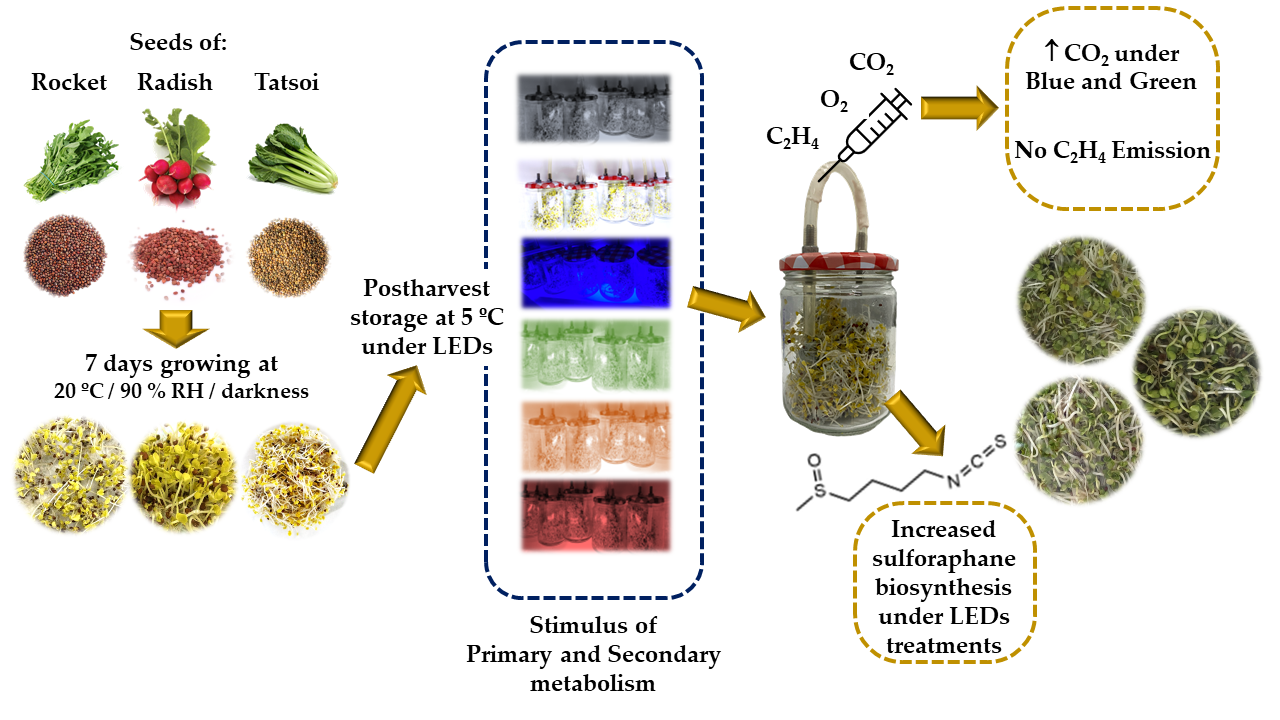
2.1. Plant Material, Seed Germination, and Minimal Processing

Rocket—Eruca vesicaria (L.) Cav.—radish (Raphanus sativus L.), and tatsoi (Brassica rapa subsp. narinosa) seeds were supplied by Intersemillas S.A. (Valencia, Spain). These three varieties were chosen between several Brassicaceae varieties because of their germination rate. In this sense, we previously germinated broccoli [24,30,31], kale [32], red cabbage [33], and mustard [34] sprouts, which were quite sensitive to fungal growth and required the application of antifungals. Therefore, based on our own experience, we used the most resistant varieties known to develop the present experience. As previously described by Martínez-Zamora et al. [28], three grams of seeds from each specie were weighed and washed with 40 mL of autoclaved distilled water. A laminar flow cabinet (Telstar Bio-II-A/M, Japan) was used for sowing, where seeds were arranged in 500 mL polypropylene trays (TR-500; 118 × 96 × 44 mm). Subsequently, a filter paper was used as a support at the bottom of the tray, which were moistened with autoclaved distilled water, and a 40 μm film was used for partially closing the tray to ensure high relative humidity (RH) in the trays. These trays were previously sterilized on a UV-C light equipment for 30 min. Conditioned trays with seeds were kept in the germination chamber (Sanyo MLR-350 H, Tokio, Japan) for 7 days at 20 °C, 90% RH, and under darkness conditions. During this period, sprouts were irrigated twice per day.
After growing for 7 days, sprouts were minimally processed in a cold room at 12 °C where they were disinfected for 1 min with a 150 ppm sodium hypochlorite solution and rinsed in cold water for 1 min. They were then placed on an absorbent filter paper for drying before packaging in 431.6 mL glass jars, where the respiration was monitored during postharvest storage, as it is shown in Figure 1.
On harvesting day, 50 sprouts were characterized, one replicate consisted of ten sprouts. The sprouts were horizontally arranged near a ruler and photographed. The photographs were processed using Image J software (Wayne Resband, Maryland, USA) to obtain the length of the hypocotyl (H) and root (R) of the sprouts, as previously described by Martínez-Zamora et al. [33]. The results were presented in mm. The growth rate was calculated by dividing H (mm) by the days of growth (d), expressing the results in mm/d.

2.2. Postharvest Light Treatments

Glass jars with 49.7 ± 0.4 g rocket sprouts, 49.9 ± 0.4 g radish sprouts, or 45.6 ± 0.5 g tatsoi sprouts, were kept for 5 days at 5.2 ± 0.2 °C and 85% RH under darkness conditions while a temperature of 6.1 ± 0.3 °C and 85% RH was reached when continuous light treatments were applied as summarized in Figure 1:
Darkness: used as control
White (400–650 nm; 5000–6500 K CCT and ~96% CRI) LED: 20.6 W m−2 = 1779.8 kJm−2 d−1 = 74.2 kJm−2 h−1
Blue (435 nm) LED: 20.5 W m−2 = 1771.2 kJm−2 d−1 = 73.8 kJm−2 h−1
Green (500 nm) LED: 20.4 W m−2 = 1762.6 kJm−2 d−1 = 73.4 kJm−2 h−1
Orange (610 nm) LED: 20.5 W m−2 = 1771.2 kJm−2 d−1 = 73.8 kJm−2 h−1
Red (660 nm) LED: 21 W m−2 = 1814.4 kJm−2 d−1 = 75.6 kJm−2 h−1
A Quantum-Photo Radiometer Data Logger DO 9721 (Delta Ohm, S.R.L., Venice, Italy) was used to measure the intensity of the lights. Therefore, the postharvest lighting treatments were: Darkness, White, Blue, Green, Orange, and Red, and their total dose for the 5 days shelf-life was ~8899.2 kJm−2. The photosynthetic photon flux density (PPFD) ranged between 93.8 and 96.6 µmol m−2 s−1. The jars within containers were kept under illumination treatments in a 5 m3 cold room at 5 °C and a RH of 90%. LED lights were purchased from LEDMurcia S.L. (Murcia, Spain). A photosynthetically active radiation (PAR) photon flux sensor QSO (Apogee instruments, Logan, UT, USA) connected to a ProCheck handheld reader (Decagon Devices Inc., Pullman, WA, USA) was used. The spectral parameters were determined with an illuminance spectrophotometer (CL-500A, Konica Minolta, Chiyoda, Tokyo, Japan). The different relative radiant power (%) used for such treatments are detailed in Figure 2.
Continuous lighting treatments were assayed in 30 × 60 × 60 cm (width × length × height) self-made metal containers inside a cold room at 5 °C, where the walls of containers were white in order to avoid light absorption, using the same structure previously described by Castillejo et al. [24]. The samples inside of containers were placed 20 cm from the light source, although such distance was slightly modified to reach the required intensities to be similar in all treatments, which were described above.

2.3. Respiration Rate and Ethylene Production

Samples of ~50 g of each sprout variety were stored in 431.6 mL glass jars and continuously flushed with humidified air at a flow rate of 20–30 mL min−1 to avoid CO2 accumulation. Three different experiments were carried out, one per Brassicaceae specie. The density of each specie was previously measured being 0.9040 g mL−1 for rocket and tatsoi sprouts and 0.9333 g mL−1 for radish sprouts.
The respiration rate was monitored every 12–24 h during the 96 h of refrigerated storage. The glass jars were hermetically closed, and after 45 min and 3 previous purges, a sample of 1 mL of the headspace (O2, CO2, and N2) was analyzed. Air sample was injected into a gas chromatograph, previously described by Álvarez-Hernández et al. [35], and equipped with a Hayesep column and CP Molsieve 5 Å column (1/8″, 80/100 mesh size; Teknokroma, Barcelona, Spain), with He as the carrier gas (5.5–6 bar). The gas concentration changes were used to calculate the O2 consumption and CO2 emission rates, and the results were expressed as mL kg−1 h−1. Five replicates (jars) were used for each lighting treatment and sampling time.
Similarly, the C2H4 production rate was daily determined every 12–24 h during the refrigerated shelf-life following the method described by Álvarez-Hernández et al. [35]. For C2H4 measurements, the oven, injector, and flame ionization detector (FID) temperatures were set at 80, 120, and 250 °C, respectively. Air and H2 were used as gas carriers at 350 and 35 mL min−1, respectively. A stainless-steel column packed with Porapak Q (1/8″, 80/100 mesh size; Teknokroma, Barcelona, Spain) was used. Gas calibration was done by comparison with an external gas mixture (CO2/O2/C2H4: 10%/10%/10 ppm) standard (Praxair, Molina de Segura, Spain). Results were expressed as µL kg−1 h−1. Five replicates (jars) were used for each lighting treatment and sampling time.

2.4. Sulforaphane Extraction and Analysis

Sulforaphane was extracted and analyzed following the method described by Martínez-Zamora et al. [30]. For that, 5 μL sulforaphane extracts were injected in a U-HPLC (Shimadzu, Kyoto, Japan) equipped with as described by Martínez-Zamora et al. [30] with a Gemini C18 column (250 mm x 4.6 mm, 5 µm particle size; Phenomenex, Torrance CA, USA) (column temperature at 36 °C) and SPDM-20A photodiode array detector. The mobile phases, 0.02 mol L−1 ammonium formate (A) and acetonitrile (B) with a 0.6 mL min−1 flow rate (55:45, v/v) were prepared to detect sulforaphane at 196 nm, as shown in Figures S1 and S2. DL-sulforaphane provided by Sigma-Aldrich (St. Louis, MO, USA) was used as standard (Figures S1 and S2), and results expressed as g kg−1 in fresh weight (FD) and dried weight (DW) were obtained as the mean of three replicates.

2.5. Scientific Literature Review

A comprehensive review according to related published manuscripts was performed using Web of Science (WoS), Scopus, PUBMED, and ScienceDirect databases in July 2022 to look for previous studies focused on the monitorization of respiration rate (related to primary metabolism) and sulforaphane content (related to secondary metabolism). Specific vocabulary included in the titles, abstract, and keywords was used for the bibliographic search, such as: light, respiration, sulforaphane, metabolism, phytochemical(s), bioactive compound(s), plant(s), sprout(s), and seed(s). Only research papers included in the Journal Citation Reports (JCR) of Institute for Scientific Information have been included.

2.6. Statistical Analysis

The experimental design was a one-factor analysis (T-light treatment) where analysis of variance (ANOVA) was performed using Statgraphics Plus software (vs. 5.1, Statpoint Technologies Inc., Warrenton, VA, USA). Statistical significance was set at p ≤ 0.05 and Tukey’s multiple range test was used to establish and separate means.

3. Results

3.1. Postharvest Sprouts Characterisation

After 7 germination days under darkness, the sowing seeds resulted in sprouts of similar characteristics and sizes (Table 1). Moisture content of studied species was ~90%, which was calculated after freeze drying the harvested sprouts. Radish sprouts were the longest, followed by rocket and tatsoi sprouts, which recorded similar growth rates during germination. Moreover, as appreciated on the pictures included in Table 1, the harvested sprouts from the three varieties showed a yellow color characterized by the absence of light during growing, which avoided the photosynthesis, that started during the postharvest period under the studied lighting treatments (Figure 3).

3.2. Influence of LED Lighting on the Primary Metabolism of Brassicaceae Sprouts

Figure 3 shows the different light spectrum studied had great influence in the color development of our sprouts, caused by concentration of photosynthetic pigments, as chlorophylls, responsible of green color. After 5 days at 5 °C, rocket, radish, and tatsoi sprouts under White, Blue, Green, Orange, and Red LED lighting, turned into a greener color from the initial yellow at harvest (Table 1), as well as, in those sprouts stored in Darkness during shelf-life (Figure 3).
In relation to the primary metabolism, and regarding the CO2 emissions showed in Figure 4, which were clearly affected by the light incidence compared to Darkness, the respiration rate was higher in radish than in rocket and tatsoi, although the respiration of different plants is not comparable. O2 consumption was directly proportional to CO2 emission and no differences were found between sprouts at different light treatments. Moreover, no C2H4 emissions were detected throughout the refrigerated shelf-life in any of the three Brassicaceae varieties studied, neither under the LED treatments assessed nor Darkness conditions.
As observed in Table 1 and Figure 3, rocket and tatsoi sprouts were 30 and 40% shorter than radish ones, and the leaves of radish sprouts were considerably bigger than the ones of rocket and tatsoi sprouts (Figure 3), which could be the reason why the respiration rate was more instable in the case of the little ones (Figure 4).
In this sense, rocket sprouts showed an increase in the respiration rate by ~49% and ~72%, respectively, compared to Darkness during the first day of storage under Blue or White LEDs (Figure 4A), which was not observed in the remaining LED treatments, neither throughout the refrigerated period. In the case of radish sprouts (Figure 4B), we can observe as full spectrum White LED lighting increased the CO2 release by a mean of ~43% from the beginning and for the first 75 h of storage regarding storage in Darkness.
If we focus in monochromatic lights, Blue and Green LEDs reported to stimulate the respiration rate in radish sprouts during the monitored period, and hence its primary metabolism, compared to Darkness by ~92% and ~68%, respectively. Similarly, Orange and Red LEDs increased the respiration rate by ~25 and ~45% compared to Darkness, respectively (Figure 4B).
Tatsoi sprouts also showed slight variations in their CO2 release under Blue and Green LEDs regarding Darkness (Figure 4C). These light treatments increased the respiration rate by ~77 and ~53%, respectively, during the first 48 h of the refrigerated storage. Furthermore, it is observed as after 72 h at 5 °C, the CO2 emissions of all the lighting treatments were equalized until the end of the study with no significant differences (p > 0.05). No great differences were found between the tatsoi sprouts stored under White LEDs and under Darkness conditions. By contrast, Orange and Red LEDs showed reductions in the respiration rate by ~50% after 48 h of refrigerated storage compared to Darkness.
Although the three Brassicaceae varieties showed different CO2 release behaviors, light from different wavelengths, especially Blue and Green LEDs, can increase the metabolism of Brassicaceae sprouts, which is related to the increase in the respiration rates under such illumination conditions.
The influence of different wavelengths of the visible spectrum into the main changes of the primary metabolism in different species at the first steps of growing are reviewed in Table 2. It is shown how illumination conditions, during growing and postharvest, can trigger primary metabolism pathways of plants and change its physiology and morphology.

3.3. Influence of LED Lighting on the Secondary Metabolism of Brassicaceae Sprouts

As observed in Figure 5, LED illumination can affect the sulforaphane accumulation in Brassicaceae sprouts. The sulforaphane content in rocket sprouts was 366 ± 20 mg kg−1 DW (Figure 5A) or 22 ± 3 mg kg−1 FW (Figure 5B), and only Blue LEDs positively influenced the biosynthesis of this phytochemical by ~16% (DW) and ~34% (FW) compared to Darkness storage, while the rest of the illumination treatments did not report relevant changes in its content after 5 days at 5 °C.
Radish sprouts were the richest Brassicaceae specie in sulforaphane in the present study, where they are compared to rocket and tatsoi, with 2.6 ± 0.1 g kg−1 DW (Figure 5C) and 0.3 ± 0.1 g kg−1 FW (Figure 5D), which represented almost ~7-fold the sulforaphane content of rocket sprouts or ~33-fold the sulforaphane content of tatsoi sprouts. In this case, only the incidence of postharvest Red LEDs improved the accumulation of sulforaphane by ~10% compared to Darkness, while the rest of LED treatment did not.
Tatsoi was the studied Brassicaceae with a fewer amount of this isothiocyanate, reporting 77.6 ± 5.5 mg kg−1 DW (Figure 5E), and 5.8 ± 0.5 mg kg−1 FW (Figure 5F). Sprouts stored under White LEDs increased their sulforaphane content by ~12.6%, while under Orange LEDs did it by ~11.8% compared to Darkness.
Since the primary metabolism contributes to the synthesis of intermediate compounds or co-factors, as precursors of plant secondary metabolites, it can explain our changes produced in the sulforaphane accumulation (Figure 5), in where some LED illumination affected the amount of this bioactive compound. To justify and discuss our results with previous bibliography found regarding the use of LED lighting to improve secondary metabolism in plants, last findings in this field are summarized in Table 3.

4. Discussion

According to sprout characterization, similar results regarding FW/DW, growth rate, and sprout length were previously obtained in our lab in radish and broccoli sprouts growth under darkness conditions for 10 days at 20 °C and 90% RH [30]. Other brassica species, as kale and red cabbage, reported similar values after growing for 7 days at 20 °C and 90% RH with 37.3 ± 7.7 [32] and 38.2 ± 4.3 mm [33], respectively.
Furthermore, since red and blue photons are more strongly absorbed than green or yellow ones, these can penetrate deeper into plant tissues [13,14]. The different ways to absorb photons can explain why the same sprouts under different wavelengths can report different tones of the same green color. In this sense, different green tones may strongly depend on chlorophyll production promoted by specific light wavelengths, like blue, as happens for soybean sprouts [19]. For instance, we can appreciate a blacker green in sprouts stored under Blue and White LEDs (Figure 3), while clearer and yellower green can be observed in the same sprouts under Orange and Red LEDs (Figure 3).
Although the effects of light on plants and their respiration have been studied for many years, studies on sprouts and young plants are still scarce. Furthermore, the application and influence of LED lighting in sprout growth and metabolism during their preharvest period has been widely investigated [36,37], although the influence of this technology after harvesting has not been deeply studied.
With regards to the second area (Table 2), Eaves and Forsyth [38] showed Brussels sprouts held in high CO2, light, and 200 ppm benzimidazole retained their color and high rates of respiration and photosynthesis for 6 days at 21 °C. In this way, it was already apparent 50 years ago that storage in darkness facilitated the loss of color (and chlorophylls) at a much faster rate than those horticultural commodities stored under light influence.
Other Brassicaceae vegetables, as pakchoi, were positively influenced under LEDs against senescence. Zhou et al. [39] showed a decrease in the respiration rate under 10 μmol m−2 s−1 White LED for 7 days at 20 °C. These results have been recently corroborated by Zhang et al. [40], who observed that high O2 and low CO2 levels within pakchoi under modified atmosphere packaging (MAP) and LED illumination reported a higher maintenance of the soluble sugar and the ascorbic acid contents, while preserving the color during the refrigerated storage. The respiration rate of mung bean sprouts, described and modelized by Chen et al. [41], was importantly affected by the temperature increase above 8–13 °C, but they did not study the influence of LED lighting.
Following this behavior, Ribas-Carbo et al. [42] corroborated the effect of light electron flow through the cyanide-resistant respiratory pathway in soybean cotyledons. In fact, the effect of continuous and variable light treatments was evaluated also by O2 isotope fractionation. Obtained results by the authors showed that light stimulated the primary metabolism by the increase in soluble sugars, starch concentrations, and chlorophylls, which was also associated to an increase in the O2 release. Furthermore, as light is involved in the activation of several genetic processes, it could operate to maintain the regulatory disulfide bond associated with the pyruvate pathway.
Apart from the light, temperature changes during postharvest storage demonstrated to be the most important factor to control the respiration and primary metabolism in early stages of plants during their retail sale period. In this sense, Young-Sang and Young-Ho [43] showed CO2 emissions, and hence respiration rate, were highly increased at temperatures higher than 8–12 °C, which was closely related to the decay level of soybean sprouts. The respiration rate of fresh chickpea sprouts was also monitored under MAP and different temperatures during postharvest storage [44]. Results obtained by Singh et al. [44] showed that CO2 emissions were positively affected by increasing temperature, which corroborates a great number of studies that low temperatures during postharvest can prolong the shelf-life of sprouts.
As other application way, pulsed light from UV spectrum (from 200 to 400 nm) demonstrated to decontaminate endive salad and mung bean sprouts [45], which did not alter the respiration rate, but it showed important improvement on the overall color and appearance of such products.
Mastropasqua et al. [46] reported the effect of White, Blue, and Red LED light application on sugars and starch production in radish, pumpkin, mung bean, and soybean sprouts. Regarding these parameters, increased values were obtained for radish and pumpkin sprouts, while no differences were obtained in mung bean and soybean sprouts compared to darkness conditions. In this sense, and according to the described bibliography, our results presented in Figure 4 can be justified by the fact that the application of lighting during postharvest has demonstrated to affect the primary metabolism, which accelerates plant respiration, as well as color changes due to increased photosynthesis of the plant by the incidence of light (Figure 3).
Table 2. State-of-art of main effects regarding sprout primary metabolism under different illumination wavelengths from the visible spectrum.
SpecieLight TreatmentPrimary Metabolism
Parameter Tested
Main FindingsRef.
Brussels sproutsSunlightRespiration and photosynthesis
Color retention
Increase of the respiration and the photosynthesis rates.
High color retention under light
[38]
Soybean sprouts5 min pulse of white light 350 µmol m−2 s−1 every 12 hRespiration
Total soluble sugars
Starch and chlorophylls
Slight increase of O2 release at the beginning. Soluble sugars, starch concentrations, and chlorophyll increased after illumination[42]
PakchoiWhite LED 10 μmol m−2 s−1Respiration rate
Chlorophyll and vitamin C
Respiration rate was lower under LED lighting. LED delayed senescence at 20 °C[39]
Mung bean sproutsPulsed light (17–25% UV) from 200 to 1100 nm at 0.1 and 1.0 J cm−2Color, respiration ratesNo increase in respiration rates, but a positive impact on quality[45]
Radish, soybean, mung bean, and pumpkin sproutsWhite, Blue, and Red LED 110 μmol m−2 s−1Dry matter
Soluble carbohydrates and starch
The soluble sugars and starch increased under LED lighting in radish and pumpkin, but not in mung bean and soybean.[46]
Kale sproutsWhite, Blue, Red, and Far-Red LED 1–100 μmol m−2 s−1GrowthThe growth was higher under Darkness conditions, followed by White, Red, Blue, and Far-Red LED lighting.[47]
Onion bulbsWhite LED 100 μmol m−2 s−1 and UV lamp at 254 nmSugar concentrationUV light reduced fructose and glucose concentrations[48]
Turnip, Cauliflower, and Mustard sproutsSeeds treated with laser (632 nm, Red) at 5 mW for 5 min and 500 mJ energyPhotosynthesis and respiration
Fresh weight
Photosynthesis, respiration, and total weight was increased [49]
Lemongrass sproutsSeeds treated with lasers (632 nm, Red) at 5 mW for 5 min and 500 mJ energyPhotosynthesis
Sugar, amino acid, organic acid, and essential oil analysis
Increase in photosynthesis, respiration, and fresh weight. The synthesis of primary metabolites as amino acids, organic acids, and essential oils was also increased [50]
PakchoiWhite LED 30 μmol m−2 s−1Respiration rate
Soluble sugars
The combination of LED and MAP reduced the respiration rate during 15 days of storage at 20 °C. It also increased the soluble sugars content.[40]
Broccoli sproutsWhite, Red, Yellow, Green, Blue, and Purple LEDs 60 μmol m−2 s−1Growth
Sugar content
LEDs inhibited the sprout growth. Red and Green LEDs decreased sugar content, while White, Blue, and Purple increased fructose content[51]
Okla et al. [50] and Almuhayawi et al. Authors confirm if you refer to the color of the first row where rocket, radish and tatsoi is. The color of the pictures no treated seeds with laser (632 nm, Red) at a power of 5 mW for 5 min and 500 mJ energy. First, treated lemongrass seeds [50] produced lemongrass sprouts with high levels of photosynthesis and respiration, while the synthesis of primary metabolites as amino acids, organic acids, and essential oils was also increased. Parallelly, Almuhayawi et al. [49] followed the same procedure in turnip, cauliflower, and mustard seeds, which resulted in sprouts with high photosynthesis and respiration rates. Therefore, a treatment with Red laser on the seeds have a remanent effect on the primary metabolism of young plants.
Accordingly, the use of LED lighting can stimulate the primary metabolism in plants, especially White LEDs and shorter wavelengths from the visible spectrum, as Blue and Green LEDs, which has been also justified by our own findings (Figure 4). Purple LED has also been reported as a plant metabolism stimulant, but has not been studied in this work. Nevertheless, it is difficult to find strong tendencies between cultivars from the same plant family as it showed different behaviors in cultivars of the Brassicaceae family [46,49]. Each variety and cultivar behave differently under the same light stimulus, making it necessary to individualize each study, as well as to establish the parameters necessary to stimulate its primary metabolism.
Specific and specialized metabolic pathways that evolved from primary carbon metabolism, derived from photosynthesis to the pyruvate and phosphoenolpyruvate, play a key role in the plant’s interaction with its environment [52]. In this sense, changes into primary metabolism can explain variations produced in the concentration of plant secondary carbon metabolites, derived for instance from the shikimic acid pathway, from which amino acids are derived to synthetize glucosinolates, and from which isothiocyanates as sulforaphane are derived [52].
Yoo et al. [48] observed sugar content of onion bulbs (part from plant primary metabolism) is partially reduced by the incidence of UV and visible LEDs, which was directly correlated with the increase in the biosynthesis of quercetin glycosides (part from secondary metabolism), especially when exposed to UV and visible UV LED lighting. Besides the changes in primary metabolism of pakchoi under White LED lighting, Zhang et al. [40] and Zhou et al. [39] showed the combination of LED and MAP reduced the degradation of ascorbic acid and chlorophylls [40], as well as its antioxidant and enzymatic activity, which were also increased under LEDs [39].
Wang et al. [53] demonstrated the positive effect of Red LED lighting in broccoli leaves, single and combined to Blue LEDs, on the biosynthesis of glucosinolates and sulforaphane accumulation, which was related to the overexpression of the HY5 gene and its cofactors involved in the synthesis of chlorophylls, carotenoids, flavonoids, phenolics, and glucosinolates. Jin et al. [54] showed the incidence of 12–13 μmol m−2 s−1 Green LEDs during the postharvest storage at 25 °C of broccoli florets, increased the accumulation of glucosinolates and sulforaphane compared to Darkness. Similarly, the total antioxidant capacity measured by DPPH, the total phenolic content, and the total chlorophyll content also experimented important increases under Green LEDs compared to fluorescent light and Darkness storage. Furthermore, also previous authors as Vitale et al. [19] have shown Blue and Red wavelengths enhance the expression of genes related to polyphenol synthesis, thus increasing phenolic compounds in light more than in dark-grown sprouts, which shows how biosynthetic pathways from pyruvate and phosphoenolpyruvate are stimulated under the influence of these wavelengths from the visible spectrum.
Apart from measuring changes in primary metabolism, in the study of Okla et al. [50], treated lemongrass seeds resulted in sprouts with improved synthesis of phenolic compounds, and hence in their total antioxidant capacity. Similarly in turnip, cauliflower, and mustard seeds, Almuhayawi et al. [49] showed sprouts with higher content in chlorophylls, carotenes, glucoraphanin, and sulforaphane, which corroborates the remanent effect of Red laser also on the secondary metabolism of such sprouts.
Table 3. State-of-art of main effects regarding sprout secondary metabolism under different illumination wavelengths from the visible spectrum.
SpecieLight TreatmentSecondary Metabolism
Parameter Tested
Main FindingsRef.
PakchoiWhite LED 10 μmol m−2 s−1Chlorophyll, vitamin C, and
antioxidant enzyme activity
Chlorophyll, vitamin C, and enzymatic activity were increased [39]
Kale sproutsWhite, Blue, Red, and Far-Red LED 1–100 μmol m−2 s−1Chlorophylls, anthocyanins,
glucosinolates, and total
antioxidant capacity
LED lighting increased the antioxidant capacity and secondary metabolites assessed [47]
Broccoli sproutsRed and Blue LEDs 350 and 41 μmol m−2 s−1Chlorophylls, carotenoids, and glucosinolatesChlorophylls, carotenoids, and glucoraphanin were highly biosynthesized after Blue LED treatments[55]
Broccoli sproutsRed, Green, and Blue LEDs 250 μmol m−2 s−1Chlorophylls, carotenoids, and glucosinolatesSecondary metabolites were highly biosynthesized under Blue LEDs [56]
Onion bulbsUV laser treatment and White LED 100 μmol m−2 s−1Quercetin glycosidesQuercetin glycosides concentrations increased the most when exposed to UV light and, to a lesser extent, when exposed to visible light[48]
Radish, soybean, mung bean, and pumpkin sproutsWhite, Blue, and Red LED 110 μmol m−2 s−1Polyphenols, chlorophyll, carotenoids, vitamin C, and anthocyaninsVitamin C, anthocyanins, carotenoids, and chlorophylls increased under White, Blue, and Red LEDs, but total phenolic content was maintained[46]
Turnip, Cauliflower, and Mustard sproutsSeeds were treated with He−Ne laser (632 nm; 5 mW; 5 min; 500 mJ) Chlorophylls, carotenoids, phenolic compounds, glucosinolates, and sulforaphaneLaser treatment on seeds before sowing increased the chlorophylls, carotenoids, total glucosinolates, glucoraphanin, and sulforaphane contents, and myrosinase activity[49]
Lemongrass sproutsSeeds were treated with lasers (632 nm; Red; 5 mW; 5 min; 500 mJ)Phenolic compounds and antioxidant capacityLaser treatment on seeds improved the synthesis of phenolic compounds and antioxidant capacity[50]
PakchoiWhite LED 30 μmol m−2 s−1Ascorbic acid, chlorophylls and
antioxidant capacity
LED + MAP reduced the degradation of ascorbic acid and chlorophylls, increasing the antioxidant capacity[40]
Broccoli sproutsBlue, Red, and Far-Red LEDs 35 μmol m−2 s−1Phenolic compoundsLED lighting increased the biosynthesis of phenolics[31]
Broccoli sproutsWhite, Yellow, and Green LEDs 35 μmol m−2 s−1 Phenolic compounds and glucosinolatesYellow LED lighting increased the biosynthesis of phenolics and glucosinolates[24]
Rocket sproutsPhotoperiod of 14 h 32 μmol m−2 s−1 Fluorescent light + 10 h 47.3 μmol m−2 s−1 White, Red, or Blue LEDsPhenolic acids, flavonoids, glucosinolates, and sulforaphaneThe application of White, Blue, Red LEDs for 10 h enhanced the biosynthesis of sulforaphane, glucosinolates, and phenolic compounds.[56]
Broccoli leavesRed and Blue LEDs 200 μmol m−2 s−1Glucosinolates and sulforaphaneRed LEDs promoted glucosinolates biosynthesis and sulforaphane accumulation, whereas Blue LEDs inhibited this effect[53]
Broccoli sproutsWhite, Red, Yellow, Green, Blue, and Purple LEDs 60 μmol m−2 s−1Anthocyanins and ascorbic acid content
Glucosinolates and sulforaphane
Yellow, Blue, and Purple LEDs increased glucoraphanin and anthocyanins contents. All the LED treatments increased ascorbic acid and sulforaphane contents.[51]
Mastropasqua et al. [46] related the application of 110 μmol m−2 s−1 White, Blue, and Red LED lighting with the high production of Vitamin C, anthocyanins, carotenoids, and chlorophylls, although total phenolic content was maintained compared to Darkness. In fact, these light treatments were effective in all the studied cultivars: radish, soybean, mung bean, and pumpkin sprouts.
Carvalho and Folta [47] showed as LED lighting in kale sprouts increased the total antioxidant capacity and secondary metabolites assessed (anthocyanins and chlorophylls) compared to Darkness. In fact, the total antioxidant capacity, linked to the biosynthesis of such compounds, exponentially increased under 50 μmol m−2 s−1 Red, Blue, White, and Far-Red LED lighting. Nevertheless, in this case, no differences were found regarding the glucosinolates biosynthesis, although a positive tendency was shown under Far-Red lighting. Far-Red light (usually ranged from 700 to 750 nm) is at the extreme red end of the visible spectrum, just before Infra-Red, being an important signal that plays a central role in modulating the response to Red light.
The bibliography is more extensive for broccoli sprouts, probably to be the most widely known and consumed Brassicaceae. In 2013, Kopsell et al. [55] showed chlorophylls, carotenoids, and glucoraphanin were biosynthesized after Blue LED treatments, single or combined to Red LEDs. These authors tried to adjust the correct combination between Blue, Red and Green LEDs to reach the best biosynthesis rate of phytochemicals in broccoli sprouts [56]. In fact, the best combinations studied were 5% Blue + 85% Red + 10% Green and 20% Blue + 80% Red, which obtained the highest rates in the biosynthesis of glucosinolates, chlorophylls, and carotenoids, as secondary metabolites, and minerals, as primary metabolites. In this sense, perception of light enriched in red and blue photons can trigger a metabolic cascade that produces healthy promoting compounds, as a response of the plant. Castillejo et al. [31] showed increases in the biosynthesis of phenolic compounds, and hence in the total antioxidant capacity of broccoli sprouts under 35 μmol m−2 s−1 Blue, Red, and Far-Red lighting for 14 days at 5 °C. The supplementation of a Blue + Red photoperiod with Far-Red LEDs reported also promising results in carrot sprouts, especially in the biosynthesis of phenolics and carotenoids [28].
Our previous findings have shown also 35 μmol m−2 s−1 Green and Yellow lighting for 14 days at 5 °C were able to stimulate the biosynthesis of phenolic and glucosinolate compounds of broccoli sprouts [24]. In fact, both LED treatments reported better results than a continuous illumination with Fluorescent or White LEDs, which demonstrated that also the medium region of the visible light spectra can enhance the biosynthesis of nutraceuticals applied at the correct dose. Moreover, this behavior has been also corroborated by Zhuang et al. [51] in broccoli sprouts. Latter authors applied 60 μmol m−2 s−1 White, Red, Yellow, Green, Blue, and Purple LEDs and obtained results as all the LED treatments were able to improve the biosynthesis of ascorbic acid, anthocyanins, and sulforaphane. Nevertheless, regarding the glucosinolates biosynthesis, Green and White LEDs did not stimulate the biosynthesis of glucoraphanin and gluconapin. Yellow, Blue, and Purple, followed by Red and White LEDs reported the best results in the biosynthesis of secondary metabolites, which was corroborated by the overexpression of the genes in charge of it.
We have recently demonstrated in rocket sprouts as the supplementation with a postharvest photoperiod for 10 h with White, Blue, and Red LEDs can enhance the biosynthesis of bioactive compounds as phenolics, glucosinolates, and sulforaphane [57]. The application of 47.3 μmol m−2 s−1 White, Red, or Blue LEDs positively affected the synthesis of caffeic, ferulic, and coumaric acids, but it did not affect to gallic and synaptic acid. In fact, the total phenolic acid content was increased after 14 days at 5 °C in rocket sprouts by ~22%, ~27%, and ~43% under White, Blue, and Red LEDs, respectively, compared to darkness conditions [57]. Furthermore, the photoperiod with these LEDs increased by ~23%, ~18%, and ~35%, respectively the synthesis of quercetin and kaempferol derivatives compared to darkness. Additionally, rutin content was enhanced by ~27% under White, ~33% under Blue, and ~55% under Red in comparison with darkness [57]. Regarding glucosinolates content, White lights increased their biosynthesis by >25%, while Blue and Red lighting reported increases by >45% compared to darkness, being glucoraphanin, glucoerucin, 4-methoxy-glucobrassicin, and glucobrassicin, the most affected compounds [57]. As main derivative from glucoraphanin, the sulforaphane content of rocket sprouts stored under a photoperiod supplemented with 10 h of White, Blue, and Red LEDs increased by ~2.7-fold, ~3.6-fold, and even ~8-fold, respectively, compared to darkness [57], which can explain the enhancement of sulforaphane in rocket sprouts under White and Blue LEDs in the present research.
As observed, the light-activation of genes affects the biosynthesis of sulforaphane, which is also appreciated in the results shown in the present study (Figure 5). Nevertheless, it must be considered that every specie, cultivar, and variety present a different behavior, and more studies must be performed to reach strong conclusions regarding the continuous illumination of Brassicaceae sprouts during postharvest storage to reach the highest nutritional values.

5. Conclusions

The positive influence of postharvest LED lighting on the plant primary and secondary metabolism in Brassicaceae sprouts has been corroborated. The respiration rate was greatly increased under Blue and Green LEDs regarding darkness, which evidences the acceleration of plant metabolism. Furthermore, although a clear tendency was not found regarding the sulforaphane biosynthesis, we can conclude that the secondary metabolism of Brassicaceae young plants is also stimulated under some types of LED lighting, which can be explained by the overexpression of genes to trigger the synthesis of secondary metabolites needing more research to clearly elucidate this fact. Specifically, in this work, Blue LEDs stimulates the biosynthesis of sulforaphane in rocket sprouts, while Red LEDs did it in radish sprouts after 5 days under refrigerated storage. In contrast, White, Green, and Orange LEDs reported a positive behavior in tatsoi sprouts during postharvest storage regarding the biosynthesis of this isothiocyanate. This fact opens future research to individualize the application of LED lights to each species, variety, and cultivar to obtain the best nutritional quality. At this stage, we recommend the postharvest illumination with visible spectrum LEDs, combined, or individually applied, continuously or in a photoperiod, according to their application possibilities, to stimulate the primary and secondary metabolism of cruciferous sprouts.

Supplementary Materials

The following supporting information can be downloaded at: https://www.mdpi.com/article/10.3390/horticulturae8111065/s1. Figure S1: Chromatogram of sulforaphane standard (detected at 196 nm) used for analysis. Figure S2. Spectrum of sulforaphane standard (detected at 196 nm) used for analysis.

Author Contributions

Conceptualization, F.A.-H., L.M.-Z. and N.C.; methodology, N.C. and L.M.-Z.; formal analysis, N.C. and L.M.-Z.; investigation, N.C., L.M.-Z. and M.C.-L.; data curation, N.C., L.M.-Z. and M.C.-L.; writing—original draft preparation, L.M.-Z.; writing—review and editing, F.A.-H., M.C.-L., N.C. and L.M.-Z.; visualization, F.A.-H.; supervision, F.A.-H.; funding acquisition, F.A.-H. All authors have read and agreed to the published version of the manuscript.

Funding

This research was funded by the Autonomous Community of the Region of Murcia, grant number 20849/PI/18, through the grant call for projects for the development of scientific and technical research by competitive groups, included in the Regional Programme for the Promotion of Scientific and Technical Research (Action Plan 2018) of the Seneca Foundation—Science and Technology Agency of the Region of Murcia (Spain).

Institutional Review Board Statement

Not applicable.

Informed Consent Statement

Not applicable.

Data Availability Statement

Not applicable.

Acknowledgments

Lorena Martínez-Zamora contract has been financed by the Programme for the Re-qualification of the Spanish University System during the three-year period 2021–2023, Margarita Salas modality for the University of Murcia. Marina Cano-Lamadrid contract (FJC2020-043764-I) has been co-financed by a Juan de la Cierva post-doctoral contracts-training 2020-2022 (FJC2020-043764-I). The technical assistance of Soner Ulutaş during the development of this study is highly appreciated.

Conflicts of Interest

The authors declare no conflict of interest.

References

  1. King, T.; Cole, M.; Farber, J.M.; Eisenbrand, G.; Zabaras, D.; Fox, E.M.; Hill, J.P. Food Safety for Food Security: Relationship between Global Megatrends and Developments in Food Safety. Trends Food Sci. Technol. 2017, 68, 160–175. [Google Scholar] [CrossRef]
  2. Barba, F.J.; Mariutti, L.R.B.; Bragagnolo, N.; Mercadante, A.Z.; Barbosa-Cánovas, G.V.; Orlien, V. Bioaccessibility of Bioactive Compounds from Fruits and Vegetables after Thermal and Nonthermal Processing. Trends Food Sci. Technol. 2017, 67, 195–206. [Google Scholar] [CrossRef]
  3. Plazzotta, S.; Manzocco, L.; Nicoli, M.C. Fruit and Vegetable Waste Management and the Challenge of Fresh-Cut Salad. Trends Food Sci. Technol. 2017, 63, 51–59. [Google Scholar] [CrossRef]
  4. Galieni, A.; Falcinelli, B.; Stagnari, F.; Datti, A.; Benincasa, P. Sprouts and Microgreens: Trends, Opportunities, and Horizons for Novel Research. Agronomy 2020, 10, 1424. [Google Scholar] [CrossRef]
  5. Johnson, T.L.; Dinkova-Kostova, A.T.; Fahey, J.W. Glucosinolates from the Brassica Vegetables and Their Health Effects; Elsevier Ltd.: Amsterdam, The Netherlands, 2015; ISBN 9780123849533. [Google Scholar]
  6. Pérez-Balibrea, S.; Moreno, D.A.; García-Viguera, C. Genotypic Effects on the Phytochemical Quality of Seeds and Sprouts from Commercial Broccoli Cultivars. Food Chem. 2011, 125, 348–354. [Google Scholar] [CrossRef]
  7. Abellán, A.; Domínguez-Perles, R.; Moreno, D.A.; García-Viguera, C. Sorting out the Value of Cruciferous Sprouts as Sources of Bioactive Compounds for Nutrition and Health. Nutrients 2019, 11, 429. [Google Scholar] [CrossRef] [Green Version]
  8. Bell, L.; Wagstaff, C. Rocket Science: A Review of Phytochemical & Health-Related Research in Eruca & Diplotaxis Species. Food Chem. X 2019, 1, 100002. [Google Scholar] [CrossRef] [PubMed]
  9. Gamba, M.; Asllanaj, E.; Raguindin, P.F.; Glisic, M.; Franco, O.H.; Minder, B.; Bussler, W.; Metzger, B.; Kern, H.; Muka, T. Nutritional and Phytochemical Characterization of Radish (Raphanus sativus): A Systematic Review. Trends Food Sci. Technol. 2021, 113, 205–218. [Google Scholar] [CrossRef]
  10. Zhang, Y.; Xiao, Z.; Ager, E.; Kong, L.; Tan, L. Nutritional Quality and Health Benefits of Microgreens, a Crop of Modern Agriculture. J. Futur. Foods 2021, 1, 58–66. [Google Scholar] [CrossRef]
  11. Kyriacou, M.C.; El-Nakhel, C.; Graziani, G.; Pannico, A.; Soteriou, G.A.; Giordano, M.; Ritieni, A.; De Pascale, S.; Rouphael, Y. Functional Quality in Novel Food Sources: Genotypic Variation in the Nutritive and Phytochemical Composition of Thirteen Microgreens Species. Food Chem. 2019, 277, 107–118. [Google Scholar] [CrossRef]
  12. Pott, D.M.; Osorio, S.; Vallarino, J.G. From Central to Specialized Metabolism: An Overview of Some Secondary Compounds Derived from the Primary Metabolism for Their Role in Conferring Nutritional and Organoleptic Characteristics to Fruit. Front. Plant Sci. 2019, 10, 835. [Google Scholar] [CrossRef] [Green Version]
  13. McCree, K.J. The Action Spectrum, Absorptance and Quantum Yield of Photosynthesis in Crop Plants. Agric. Meteorol. 1971, 9, 191–216. [Google Scholar] [CrossRef]
  14. McCree, K.J. Test of Current Definitions of Photosynthetically Active Radiation against Leaf Photosynthesis Data. Agric. Meteorol. 1972, 10, 443–453. [Google Scholar] [CrossRef]
  15. Bhatla, S.C.; Lal, M.A. Plant Physiology, Development and Metabolism; Springer: Berlin/Heidelberg, Germany, 2018. [Google Scholar]
  16. Angelova, Z.; Georgiev, S.; Roos, W. Elicitation of Plants. Biotechnol. Biotechnol. Equip. 2006, 20, 72–83. [Google Scholar] [CrossRef] [Green Version]
  17. Poulev, A.; O’Neal, J.M.; Logendra, S.; Pouleva, R.B.; Timeva, V.; Garvey, A.S.; Gleba, D.; Jenkins, I.S.; Halpern, B.T.; Kneer, R.; et al. Elicitation, a New Window into Plant Chemodiversity and Phytochemical Drug Discovery. J. Med. Chem. 2003, 46, 2542–2547. [Google Scholar] [CrossRef]
  18. Zhang, X.; Bian, Z.; Yuan, X.; Chen, X.; Lu, C. A Review on the Effects of Light-Emitting Diode (LED) Light on the Nutrients of Sprouts and Microgreens. Trends Food Sci. Technol. 2020, 99, 203–216. [Google Scholar] [CrossRef]
  19. Vitale, E.; Velikova, V.; Tsonev, T.; Ferrandino, I.; Capriello, T.; Arena, C. The Interplay between Light Quality and Biostimulant Application Affects the Antioxidant Capacity and Photosynthetic Traits of Soybean (Glycine max L. Merrill). Plants 2021, 10, 861. [Google Scholar] [CrossRef]
  20. Pennisi, G.; Sanyé-Mengual, E.; Orsini, F.; Crepaldi, A.; Nicola, S.; Ochoa, J.; Fernandez, J.A.; Gianquinto, G. Modelling Environmental Burdens of Indoor-Grown Vegetables and Herbs as Affected by Red and Blue LED Lighting. Sustainability 2019, 11, 4063. [Google Scholar] [CrossRef] [Green Version]
  21. Pennisi, G.; Orsini, F.; Castillejo, N.; Gómez, P.A.; Crepaldi, A.; Fernández, J.A.; Egea-Gilabert, C.; Artés-Hernández, F.; Gianquinto, G. Spectral Composition from Led Lighting during Storage Affects Nutraceuticals and Safety Attributes of Fresh-Cut Red Chard (Beta vulgaris) and Rocket (Diplotaxis tenuifolia) Leaves. Postharvest Biol. Technol. 2021, 175, 111500. [Google Scholar] [CrossRef]
  22. Janssen, R.J.P.; Krijn, M.P.C.M.; van den Bergh, T.; van Elmpt, R.F.M.; Nicole, C.C.S.; van Slooten, U. Optimizing Plant Factory Performance for Local Requirements. In Plant Factory Using Artificial Light: Adapting to Environmental Disruption and Clues to Agricultural Innovation; Anpo, M., Fukuda, H., Wada, T., Eds.; Elsevier: Amsterdam, The Netherlands, 2018; pp. 281–293. ISBN 9780128139745. [Google Scholar]
  23. Samuolienė, G.; Brazaitytė, A.; Viršilė, A.; Miliauskienė, J.; Vaštakaitė-Kairienė, V.; Duchovskis, P. Nutrient Levels in Brassicaceae Microgreens Increase Under Tailored Light-Emitting Diode Spectra. Front. Plant Sci. 2019, 10, 1–9. [Google Scholar] [CrossRef] [PubMed]
  24. Castillejo, N.; Martínez-Zamora, L.; Gómez, P.A.; Pennisi, G.; Orsini, F.; Art, F.; Crepaldi, A.; Fernández, J.A.; Artés-Hernández, F. Postharvest Yellow LED Lighting Affects Phenolics and Glucosinolates Biosynthesis in Broccoli Sprouts. J. Food Compos. Anal. 2021, 103, 104101. [Google Scholar] [CrossRef]
  25. Hernández-Cánovas, L.; Abellán-Victorio, Á.; Moreno, D.A. The Quality and Glucosinolate Composition of Cruciferous Sprouts under Elicitor Treatments Using MeJA and LED Lights. Proceedings 2020, 70, 67. [Google Scholar] [CrossRef]
  26. Han, T.; Astafurova, T.; Turanov, S.; Burenina, A.; Butenkova, A.; Surnina, E.; Valiev, D. Photomorphogenesis of Wheat Sprouts with LED Irradiation of Different Intensities. Light. Res. Technol. 2020, 52, 583–594. [Google Scholar] [CrossRef]
  27. Giménez, A.; Martínez, C.; Egea-Gilabert, C.; Gómez, P.A.; Artés-Hernández, F.; Pennisi, G.; Orsini, F.; Crepaldi, A.; Fernández, J.A. Combined Effect of Salinity and LED Lights on the Yield and Quality of Purslane (Portulaca oleracea L.) Microgreens. Horticulturae 2021, 7, 180. [Google Scholar] [CrossRef]
  28. Martínez-Zamora, L.; Castillejo, N.; Gómez, P.A.; Artés-Hernández, F. Amelioration Effect of Led Lighting in the Bioactive Compounds Synthesis during Carrot Sprouting. Agronomy 2021, 11, 304. [Google Scholar] [CrossRef]
  29. Park, C.H.; Kim, N.S.; Park, J.S.; Lee, S.Y.; Lee, J.W.; Park, S.U. Effects of Light-Emitting Diodes on the Accumulation of Glucosinolates and Phenolic Compounds in Sprouting Canola (Brassica napus L.). Foods 2019, 8, 76. [Google Scholar] [CrossRef] [Green Version]
  30. Martínez-Zamora, L.; Castillejo, N.; Artés-Hernández, F. Postharvest UV-B and UV-C Radiation Enhanced the Biosynthesis of Glucosinolates and Isothiocyanates in Brassicaceae Sprouts. Postharvest Biol. Technol. 2021, 181, 111650. [Google Scholar] [CrossRef]
  31. Castillejo, N.; Martínez-Zamora, L.; Gómez, P.A.; Pennisi, G.; Crepaldi, A.; Fernández, J.A.; Orsini, F.; Artés-Hernández, F. Postharvest LED Lighting: Effect of Red, Blue and Far Red on Quality of Minimally Processed Broccoli Sprouts. J. Sci. Food Agric. 2021, 101, 44–53. [Google Scholar] [CrossRef]
  32. Castillejo, N.; Martínez-Zamora, L.; Artés–Hernández, F. Periodical Uv-B Radiation Hormesis in Biosynthesis of Kale Sprouts Nutraceuticals. Plant Physiol. Biochem. 2021, 165, 274–285. [Google Scholar] [CrossRef]
  33. Martínez-Zamora, L.; Castillejo, N.; Artés–Hernández, F. UV-B Radiation as Abiotic Elicitor to Enhance Phytochemicals and Development of Red Cabbage Sprouts. Horticulturae 2021, 7, 567. [Google Scholar] [CrossRef]
  34. Artés–Hernández, F.; Miranda-Molina, F.D.; Klug, T.V.; Martínez–Hernández, G.B. Enrichment of Glucosinolate and Carotenoid Contents of Mustard Sprouts by Using Green Elicitors during Germination. J. Food Compos. Anal. 2022, 110, 104546. [Google Scholar] [CrossRef]
  35. Álvarez-Hernández, M.H.; Martínez-Hernández, G.B.; Avalos-Belmontes, F.; Rodríguez-Hernández, A.M.; Castillo-Campohermoso, M.A.; Artés-Hernández, F. An Innovative Ethylene Scrubber Made of Potassium Permanganate Loaded on a Protonated Montmorillonite: A Case Study on Blueberries. Food Bioprocess Technol. 2019, 12, 524–538. [Google Scholar] [CrossRef]
  36. Livadariu, O.; Maximilian, C.; Raiciu, A.D.; Șerbănică, C.P. An Assessment of the Type of Substrate and LEDs’ Irradiation Influence on Garden Cress Sprouts (Lepidium sativum L.). Appl. Sci. 2022, 12, 4732. [Google Scholar] [CrossRef]
  37. Korzeniewska, E.; Czarnecki, A.; Kiełbasa, P. Modelling of Carotenoids Content in Red Clover Sprouts Using Light of Di Ff Erent Wavelength and Pulsed Electric Field. Appl. Sci. 2020, 10, 4143. [Google Scholar] [CrossRef]
  38. Eaves, C.A.; Forsyth, F.R. The Influence of Light, Modified Atmospheres and Benzimidazole on Brussels Sprouts. J. Hortic. Sci. 1968, 43, 317–322. [Google Scholar] [CrossRef]
  39. Zhou, F.; Zuo, J.; Xu, D.; Gao, L.; Wang, Q.; Jiang, A. Low Intensity White Light-Emitting Diodes (LED) Application to Delay Senescence and Maintain Quality of Postharvest Pakchoi (Brassica campestris L. Ssp. Chinensis (L.) Makino Var. Communis Tsen et Lee). Sci. Hortic. 2020, 262, 109060. [Google Scholar] [CrossRef]
  40. Zhang, X.J.; Zhang, M.; Chitrakar, B.; Devahastin, S.; Guo, Z. Novel Combined Use of Red-White LED Illumination and Modified Atmosphere Packaging for Maintaining Storage Quality of Postharvest Pakchoi. Food Bioprocess Technol. 2022, 15, 590–605. [Google Scholar] [CrossRef]
  41. Chen, J.; Hu, Y.; Wang, J.; Yao, Y.; Hu, H. Respiration Rate Measurement and Chemical Kinetic Modelling for Mung Bean Sprouts. J. Food Process Eng. 2017, 40, 1–7. [Google Scholar] [CrossRef] [Green Version]
  42. Ribas-Carbo, M.; Robinson, S.A.; Gonzàlez-Meler, M.A.; Lennon, A.M.; Giles, L.; Siedow, J.N.; Berry, J.A. Effects of Light on Respiration and Oxygen Isotope Fractionation in Soybean Cotyledons. Plant Cell Environ. 2000, 23, 983–989. [Google Scholar] [CrossRef] [Green Version]
  43. Young-Sang, L.; Young-Ho, K. Changes in Post-Harvest Respiration, Growth and Vitamin C Content of Soybean Sprouts under Different Storage Temperature Conditions. Korean J. Crop Sci. 2004, 49, 410–414. [Google Scholar]
  44. Singh, R.; Kumar, A.; Mann, S.; Kulkarni, S.D. Respiration Rate Models of Fresh Chickpea Sprouts (Cicer arietinum L.) under Modified Atmosphere Packaging. Agric. Eng. 2012, 37, 49–57. [Google Scholar]
  45. Kramer, B.; Wunderlich, J.; Muranyi, P. Pulsed Light Decontamination of Endive Salad and Mung Bean Sprouts and Impact on Color and Respiration Activity. J. Food Prot. 2015, 78, 340–348. [Google Scholar] [CrossRef]
  46. Mastropasqua, L.; Dipierro, N.; Paciolla, C. Effects of Darkness and Light Spectra on Nutrients and Pigments in Radish, Soybean, Mung Bean and Pumpkin Sprouts. Antioxidants 2020, 9, 558. [Google Scholar] [CrossRef] [PubMed]
  47. Carvalho, S.D.; Folta, K.M. Sequential Light Programs Shape Kale (Brassica napus) Sprout Appearance and Alter Metabolic and Nutrient Content. Hortic. Res. 2014, 1, 8. [Google Scholar] [CrossRef] [PubMed] [Green Version]
  48. Yoo, K.S.; Lee, E.J.; Patil, B.S. Changes in Quercetin Glucoside Concentrations of Onion Bulbs by Scales, during Storage, and in Sprouting Leaves Exposed to UV. Postharvest Biol. Technol. 2013, 83, 65–71. [Google Scholar] [CrossRef]
  49. Almuhayawi, S.M.; Almuhayawi, M.S.; Al Jaouni, S.K.; Selim, S.; Hassan, A.H.A. Effect of Laser Light on Growth, Physiology, Accumulation of Phytochemicals, and Biological Activities of Sprouts of Three Brassica Cultivars. J. Agric. Food Chem. 2021, 69, 6240–6250. [Google Scholar] [CrossRef] [PubMed]
  50. Okla, M.K.; El-Tayeb, M.A.; Qahtan, A.A.; Abdel-Maksoud, M.A.; Elbadawi, Y.B.; Alaskary, M.K.; Balkhyour, M.A.; Hassan, A.H.A.; Abdelgawad, H. Laser Light Treatment of Seeds for Improving the Biomass Photosynthesis, Chemical Composition and Biological Activities of Lemongrass Sprouts. Agronomy 2021, 11, 478. [Google Scholar] [CrossRef]
  51. Zhuang, L.; Huang, G.; Li, X.; Xiao, J.; Guo, L. Effect of Different LED Lights on Aliphatic Glucosinolates Metabolism and Biochemical Characteristics in Broccoli Sprouts. Food Res. Int. 2022, 154, 111015. [Google Scholar] [CrossRef]
  52. Obata, T. Metabolons in Plant Primary and Secondary Metabolism. Phytochem. Rev. 2019, 18, 1483–1507. [Google Scholar] [CrossRef]
  53. Wang, J.; Mao, S.; Wu, Q.; Yuan, Y.; Liang, M.; Wang, S.; Huang, K.; Wu, Q. Effects of LED Illumination Spectra on Glucosinolate and Sulforaphane Accumulation in Broccoli Seedlings. Food Chem. 2021, 356, 129550. [Google Scholar] [CrossRef]
  54. Jin, P.; Yao, D.; Xu, F.; Wang, H.; Zheng, Y. Effect of Light on Quality and Bioactive Compounds in Postharvest Broccoli Florets. Food Chem. 2015, 172, 705–709. [Google Scholar] [CrossRef] [PubMed]
  55. Kopsell, D.A.; Sams, C.E. Increases in Shoot Tissue Pigments, Glucosinolates, and Mineral Elements in Sprouting Broccoli after Exposure to Short-Duration Blue Light from Light Emitting Diodes. J. Am. Soc. Hortic. Sci. 2013, 138, 31–37. [Google Scholar] [CrossRef]
  56. Kopsell, D.A.; Sams, C.E.; Barickman, T.C.; Morrow, R.C. Sprouting Broccoli Accumulate Higher Concentrations of Nutritionally Important Metabolites under Narrow-Band Light-Emitting Diode Lighting. J. Am. Soc. Hortic. Sci. 2014, 139, 469–477. [Google Scholar] [CrossRef] [Green Version]
  57. Martínez-Zamora, L.; Castillejo, N.; Artés–Hernández, F. Ultrasounds and a Postharvest Photoperiod to Enhance the Synthesis of Sulforaphane and Antioxidants in Rocket Sprouts. Antioxidants 2022, 11, 1490. [Google Scholar] [CrossRef] [PubMed]
Figure 1. Experimental design.
Horticulturae 08 01065 g001
Figure 2. Relative radiant power (%) applied during refrigerated storage: White (A), Blue (B), Green (C), Orange (D), and Red (E) LEDs.
Horticulturae 08 01065 g002
Figure 3. Color appearance of Brassicaceae sprouts after 5 days at 5 °C illuminated under different wavelengths of the electromagnetic spectrum.
Horticulturae 08 01065 g003
Figure 4. Respiration rate of Brassicaceae ((A): rocket; (B): radish; (C): tatsoi) sprouts under different wavelengths for 96 h at 5 °C. * denotes positive significant differences compared to Darkness.
Horticulturae 08 01065 g004
Figure 5. Sulforaphane content of Brassicaceae ((A,B): rocket; (C,D): radish; (E,F): tatsoi) sprouts under different wavelengths for 5 days at 5 °C. Left: Dry weight (DW). Right: Fresh weight (FW). Different capital letters denote significant differences (p < 0.05) among light treatments.
Horticulturae 08 01065 g005
Table 1. Initial sprout characterization after 7 days growing.
RocketRadishTatsoi
Fresh weight/Dry weight (FW/DW)11.75 ± 0.83 a7.20 ± 0.79 b9.82 ± 1.24 a
% Moisture91.46 ± 0.59 a85.99 ± 1.63 b89.72 ± 1.25 a
Hypocotyl length (mm)32.7 ± 3.0 b50.5 ± 5.5 a25.6 ± 3.8 b
Root length (mm)28.7 ± 0.1 b36.5 ± 4.3 a25.9 ± 4.8 b
Sprout length (mm)61.4 ± 3.0 b86.9 ± 9.2 a51.5 ± 1.5 c
Growth rate (mm/day)4.68 ± 0.43 b7.21 ± 0.79 a3.65 ± 0.54 c
Visual appearanceHorticulturae 08 01065 i001Horticulturae 08 01065 i002Horticulturae 08 01065 i003
a, b, c: different letters indicate significant differences (p < 0.05) among Brassicaceae sprouts.
Publisher’s Note: MDPI stays neutral with regard to jurisdictional claims in published maps and institutional affiliations.

