Article

Evolution of LysM-RLK Gene Family in Wild and Cultivated Peanut Species

1
Instituto de Agrobiotecnología del Litoral, Colectora Ruta Nacional No 168 km. 0, Paraje El Pozo, Santa Fe 3000, Argentina
2
Instituto de Investigaciones Agrobiotecnológicas (CONICET-UNRC), Ruta 36 Km 601, Río Cuarto 5800, Córdoba, Argentina
3
Agronomy Department, Plant Molecular & Cellular Biology Program, Genetics Institute, University of Florida, Gainesville, FL 32611, USA
*
Author to whom correspondence should be addressed.
Horticulturae 2022, 8(11), 1000; https://doi.org/10.3390/horticulturae8111000
Submission received: 30 September 2022 / Revised: 18 October 2022 / Accepted: 21 October 2022 / Published: 27 October 2022
(This article belongs to the Special Issue Horticultural Crops Genetics and Genomics)

Abstract

:
In legumes, a LysM-RLK perception of rhizobial lipo-chitooligosaccharides (LCOs) known as Nod factors (NFs), triggers a signaling pathway related to the onset of symbiosis development. On the other hand, activation of LysM-RLKs upon recognition of chitin-derived short-chitooligosaccharides initiates defense responses. In this work, we identified the members of the LysM-RLK family in cultivated (Arachis hypogaea L.) and wild (A. duranensis and A. ipaensis) peanut genomes, and reconstructed the evolutionary history of the family. Phylogenetic analyses allowed the building of a framework to reinterpret the functional data reported on peanut LysM-RLKs. In addition, the potential involvement of two identified proteins in NF perception and immunity was assessed by gene expression analyses. Results indicated that peanut LysM-RLK is a highly diverse family. Digital expression analyses indicated that some A. hypogaea LysM-RLK receptors were upregulated during the early and late stages of symbiosis. In addition, expression profiles of selected LysM-RLKs proteins suggest participation in the receptor network mediating NF and/or chitosan perception. The analyses of LysM-RLK in the non-model legume peanut can contribute to gaining insight into the molecular basis of legume–microbe interactions and to the understanding of the evolutionary history of this gene family within the Fabaceae.

1. Introduction

Plants are continuously subjected to multiple transient or persistent interactions with microbes. These interactions can produce a broad spectrum of outcomes for the host, ranging from beneficial to detrimental [1,2]. Legumes (plants of the Fabaceae family) are well known for their almost exclusive ability to form a mutualistic symbiosis with nitrogen-fixing bacteria known as rhizobia. In this symbiotic association, the rhizobia fix atmospheric nitrogen that is transferred to the host plant in exchange for carbohydrates and a stable environment. Besides this beneficial association, legumes, like all plants, must also actively defend against pathogenic microorganisms. Moreover, plants can simultaneously interact with pathogenic and mutualistic microorganisms [3]. Therefore, adequate microbial recognition and elicitation of a proper response are crucial for plant fitness.
Plant recognition of microbes is based on the detection of molecular signals produced by the microorganisms, or specific patterns associated with their cell structure. Interestingly, in the case of rhizobia and pathogenic fungi, detection involves the sensing of structurally related n-acetylglucosamine-containing molecules [4]. Rhizobia produce signal molecules known as Nod factors (NFs), which are lipo-chitooligosaccharides made up of four or five β-1-4-linked n-acetylglucosamine subunits, with an acyl chain of varying length at the C2 position of the non-reducing terminal residue and different species-specific substitutions at the reducing and non-reducing terminal residues [5]. On the other hand, phytopathogenic fungi are identified by plants through the perception of chitin-derived short-chain chitooligosaccharides (COs) [6]. However, even when NFs and COs are structurally related, there are apparent differences in the responses triggered after perception [6]. Legume recognition of rhizobial NFs triggers a signaling pathway that initiates genetic programs leading to rhizobial infection and nodule primordia formation. In contrast, COs with more than six subunits have been traditionally recognized as general elicitors of plant defense responses [7,8]. Additionally, chitosan (a deacetylated chitin derivative) has been described as an elicitor of plant defenses [9].
NFs and COs are perceived by plant receptors belonging to the Lysin motifs receptor-like kinases (LysM-RLKs) family [4,10]. LysM-RLKs are proteins consisting of three regions: one extracellular region with three LysM domains, one transmembrane region, and one highly conserved intracellular kinase domain. Ligand perception and signal transduction by LysM-RLKs in legumes constitutes a highly active and dynamic research area. Recent works suggest that ligand perception and downstream responses triggered by LysM-RLKs depend on combinatorial and/or sequential quantitative perception of diverse signaling molecules by spatio-temporally regulated networks of receptor/co-receptor complexes [2,3]. Considering that the LysM-RLK family is highly diverse and includes many family members in legumes (e.g., 18 in Lotus, 22 in Medicago), the number of combinations that could potentially be formed is vast. In addition, receptor activity is modulated by ligand binding and by a diverse variety of interactors [5], adding another layer of complexity to molecule perception. In this highly dynamic and complex scenario, a better characterization of the LysM-RLK family is required in order to understand the diverse roles of its members. However, identification of LysM-RLKs and their biological function in legumes has been mainly restricted to model species displaying similar infection mechanisms and nodule morphogenetic programs. Extending the studies to non-model species showing different infection mechanisms and nodule morphogenetic programs is required to unravel the biological function of its members and to clarify the evolution of the LysM-RLK family in legumes.
Peanut is an important oil and food legume crop worldwide, grown mainly in tropical and subtropical areas. The cultivated peanut A. hypogaea L., is an allotetraploid (2n = 4x = 40, AABB genome type; approximately 2.8 Gb, with a high repetitive DNA content) probably derived from a single hybridization event of two wild diploid species (2n = 2x = 20), A. duranensis (A genome) and A. ipaensis (B genome), followed by chromosome doubling [11,12]. Several lines of evidence indicate that the polyploidization event has occurred relatively recently (about 3.5 million years ago), and that genome changes in the polyploid peanut have been limited since then. Peanut-rhizobia symbiosis represents an interesting system for studying the evolution of symbiosis within the Fabaceae, due to the basal phylogenetic position of this species within the subfamily Papilionoideae, its particular infection mechanism, and its primitive aeschynomenoid nodule morphogenetic program [13]. Recently, the genomic sequence of cultivated peanut [14,15] and its diploid ancestors [16] were obtained, opening up the possibility of reaching a deep understanding of the molecular mechanisms governing the symbiotic program in this legume species. Since then, transcriptome analyses of nodulation have been performed [17,18], and the role of different molecules in the symbiotic signaling pathway has been proposed [19]. However, despite limited exceptions [13], there are no studies dealing with the identification of LysM-RLKs in this legume species. The aim of this work was to identify the members of the LysM-RLK family in cultivated (A. hypogaea L.) and wild (A. duranensis and A. ipaensis) peanut genomes, and to reconstruct the evolutionary history of the family. This phylogenetic framework allowed reinterpretation of the functional data reported for peanut LysM-RLKs. Finally, the potential participation of two peanut LysM-RLK in the receptor networks mediating NF and chitosan inoculation was assessed by gene expression analyses.

2. Materials and Methods

2.1. Identifying LysM-RLKs in Cultivated and Wild Peanut Genomes

Firstly, a search using the keyword “LysM” was conducted in PeanutBase “https://www.peanutbase.org/home” (accessed on 9 June 2022), which included A. duranensis, A. ipaensis, and A. hypogaea whole genomes [14,16]. Afterward, in order to retrieve unannotated or mis-annotated sequences, BlastP and tBlastN “https://peanutbase.org/pb_sequenceserver” (accessed on 9 June 2022) searches were performed by using the amino acid sequences of L. japonicus and M. truncatula’s LysM-RLKs as queries for the second step. The significant hits were further inspected for typical structural features of LysM-RLKs. Domain structure was analyzed using Pfam “http://pfam.xfam.org” (accessed on 9 May 2022) and Inter-ProScan “https://www.ebi.ac.uk/interpro/search/sequence/” (accessed on 9 June 2022) [20,21], and by an additional visual inspection of the alignments. To trace the three LysM domains within the sequences, typical CXC motifs located between LysM1-LysM2 and LysM2-LysM3 domains were identified. A conserved proline residue at the end of the third LysM domain indicated the end of the LysM domains [22]. Signal peptide and transmembrane domains were predicted, using SignalP “https://services.healthtech.dtu.dk/service.php?SignalP” (accessed on 9 June 2022) and TMHMM server v. 2.0 “https://dtu.biolib.com/DeepTMHMM” (accessed on 9 June 2022), respectively. Also, according to the domain organization, protein kinase domains were determined.
Afterward, a manual correction of gene structure predictions and annotations was performed according to [23].

2.2. Chromosomal Location of LysM-RLK Genes and the Synteny Analysis

The positional information of all identified LysM-RLK genes in wild and cultivated peanut was obtained from the corresponding reference genomes, and mapped to their chromosomal locations using Circos for comparison “http://circos.ca/” (accessed on 9 May 2022).
Gene duplication analysis was performed by multiple alignment of the peanut LysM-RLKs. Genes were considered duplicated according to the following criteria: similarity of the coding nucleotide sequences > 80% and identity between the sequences > 80%. Tandem-duplicated pairs were only considered when genes within a 200 kb genomic region belonged to the same phylogenetic group.

2.3. Digital Expression Analysis of LysM-RLK Genes

To assess the expression of LysM-RLKs genes in A. hypogaea within the progress of symbiosis, data were retrieved from peanut RNA-seq datasets (accession number GSE98997) [18]. Reported FPKM values of LysM-RLK genes in peanut plants were screened, and normalized count data were used to calculate the expression levels (log2FC) of each LysM-RLK gene at 1, 4, 8, 12 and 21 dpi. A heatmap was generated with the pheatmap R package.

2.4. Sequence Alignments and Phylogenetic Analyses

For all data sets, alignments of the predicted protein sequences were performed with the GUIDANCE algorithm [24], available at “http://guidance.tau.ac.il/” (accessed on 9 May 2022) with the MAFFT option and the following parameters: maxiterate = 1000, retree = 1, genafpair = true.
Phylogenies were inferred with a maximum likelihood (ML) approach using PhyML 3.0 [25], available at “http://www.atgc-montpellier.fr/”(accessed on 9 May 2022), and the substitution model that best fit the data was selected using smart model selection [26]. Branch confidence was evaluated using bootstrap analysis with 100 replicates. Finally, the tree was drawn with Itol v3 [27], available at “https://itol.embl.de/” (accessed on 9 May 2022).
Amino acid sequences of LysM-RLKs from Arabidopsis thaliana, L. japonicus, M. truncatula, Prunus persica, Brassica rapa and Solanum lycopersicum were retrieved from [23].

2.5. Ka/Ks Analysis

For the non-synonymous substitutions (Ka) and the synonymous substitutions (Ks) calculation, the LysM-RLKs gene pairs between cultivated peanut genome and the corresponding ancestor genome were identified and were aligned using the ParaAT [28], available at “http://cbb.big.ac.cn/software” (accessed on 9 May 2022) with multiple sequence aligner Muscle [29], following the instruction of the program. To create the input files for ParaAT, CDS and the amino acid sequences of the LysM-RLKs genes were retrieved, and the ortholog information of these genes was organized. Ka/Ks_Calculator [30], available at “http://evolution.genomics.org.cn/software.htm” (accessed on 9 May 2022) was then used to calculate the Ka and Ks numbers of each gene pair, using the model-averaging method (MA).

2.6. Plant Inoculation Assays

A. hypogaea L. (var. Runner cultivar Granoleico) seeds were surface sterilized and germinated. Peanut plants were inoculated with signaling molecules known for triggering rhizobial symbiosis (NFs from compatible rhizobia) or defense (chitosan, a deacetylated molecule derived from chitin).
For inoculation treatment, NFs from Bradyrhizobium SEMIA 6144 (peanut microsymbiont) were obtained, following the methodology proposed by [31]. Seven-day-old seedlings were inoculated by root immersion in 100 mL of an aqueous solution containing NFs (10−6 mol L−1) for 10 min. Afterward, the seedlings were placed in pots containing sterilized vermiculite located in growth chambers, under controlled conditions (light intensity of 200 mmol m−2 s−1, 16 h day/8 h night cycle). For chitosan inoculation, the same inoculation procedure was employed, using low molecular weight chitosan (Sigma, deacetylation ≥ 75%) 50 mg L−1 dissolved in acetic acid 0.1 M, pH: 6.8–7. The concentration of 50 mg L−1 chitosan was selected, since it had been previously demonstrated to elicit defense responses in peanut (results not shown). Control roots were similarly treated with sterile water or acetic acid 0.1 M.

2.7. RNA Extraction and Expression Analysis

Plants were harvested at 1, 8, 16, 24, and 72 h post inoculation (hpi) with the elicitor molecules. RNA from roots was extracted using TRIzol reagent (Invitrogen, Carlsbad, CA, USA), according to the manufacturer’s protocol. Each RNA sample was prepared from 2 replicates of 3 plants each, which were pooled to reduce noise arising from biological variations. RNA concentration was then measured using a NanoDrop spectrophotometer, and RNA integrity was determined by visualization in agarose gel. cDNA was synthesized from 1 μg of total RNA using the AccuScript Hi-Fi RT (Agilent, Santa Clara, CA, USA), as described by the manufacturer. The quantitative reverse-transcriptase polymerase chain reaction (qPCR) assays were performed using Power SYBR Green PCR Master Mix (Applied Biosystems, Foster City, CA, USA), according to the manufacturer’s instructions. Sequences of primers used for qPCR amplification were designed according to Ahy.YTK8KP and Ahy.IM7I4N sequences, and were synthesized by the Genbiotech company (Table S1). The primer efficiency in the PCR reactions was determined by linear regression analysis of 10-fold dilutions of a pool of cDNA samples, and denoted with a correlation coefficient (R2). Reactions were performed in a real-time thermocycler (Stratagene MX3000P; Agilent, Santa Clara, CA, USA) with settings of 95 °C for 3 min, and 40 cycles of 95 °C, 20 s and then 60 °C for 20 s. Results obtained from different treatments were standardized to the Actin mRNA level. Relative expression levels were calculated using the 2−ΔΔCt method [32].

2.8. Statistical Analysis

Data were subjected to analysis of variance (ANOVA). Statistical significance was determined by the LSD-Fisher test at p ≤ 0.05, using Infostat software (1.0, FCA, UNC, Argentina). Graph elaboration was performed by Prism-GraphPad version 9.2.0.

3. Results

3.1. Identification of LysM-RLKs in Reference Genomes of A. hypogaea, A. duranensis, and A. ipaensis

A two-step strategy search was performed against the PeanutBase database. A total number of 35, 19 and 19 gene models were annotated as LysM-RLKs according to the keyword “LysM” search in the genomes of A. hypogaea, A. duranensis, and A. ipaensis, respectively. Through BlastP and tBlastN searches using L. japonicus and M. truncatula LysM-RLKs as queries, six additional LysM-RLKs were identified. All the gene models found were then further confirmed by inspecting typical structural features of LysM-RLKs (the presence of 3 LysM domains in the extracellular region separated by CXC motifs, a transmembrane domain, and an intracellular region displaying homology with Serine/Threonine kinases). By using this strategy, a final number of 38, 20, and 21 LysM-RLKs were identified in the genomes of A. hypogaea, A. duranensis, and A. ipaensis, respectively (Table S2). Six of the total peanut sequences found were not annotated or were misannotated (see footnotes of Table S2 for details).

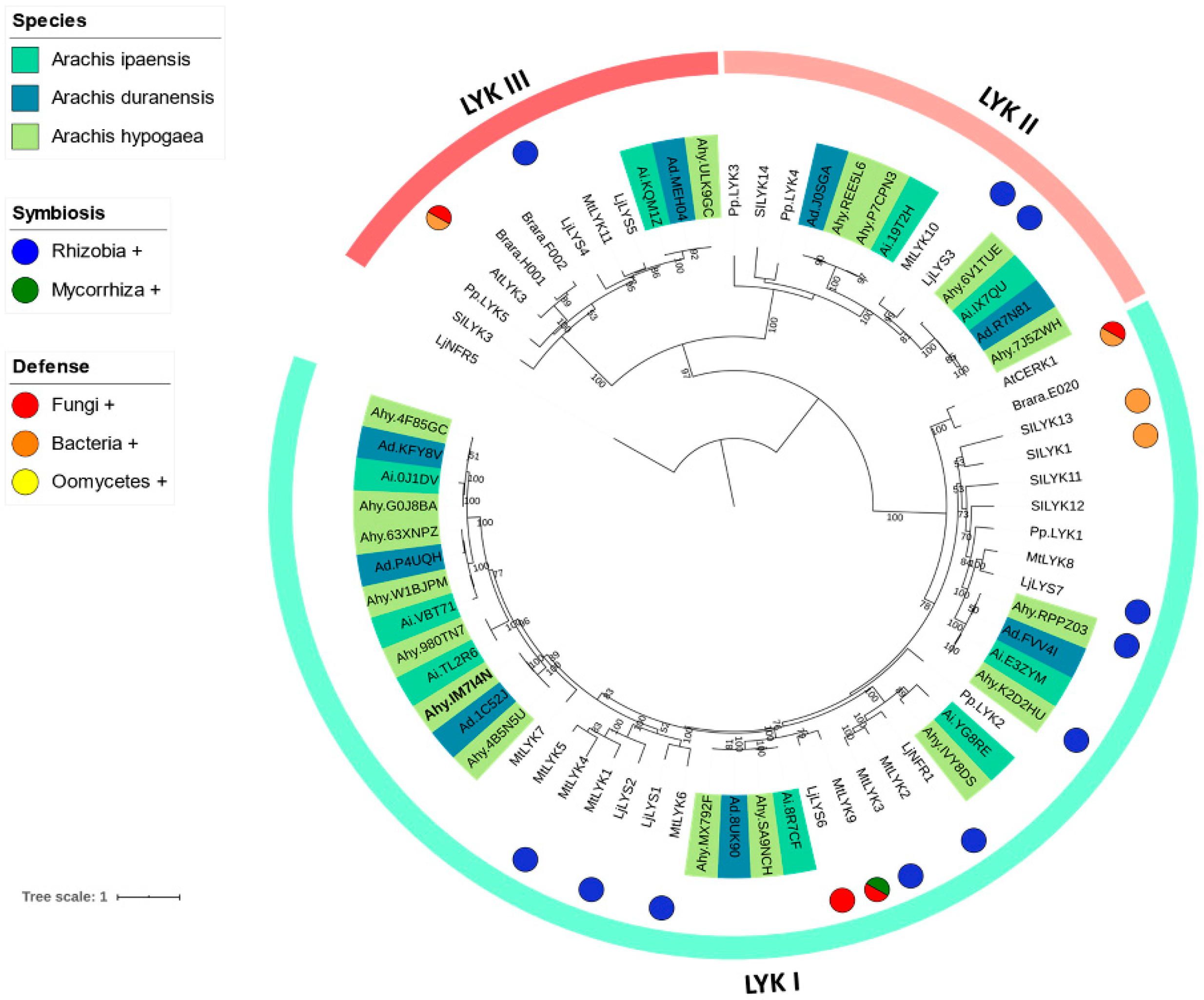
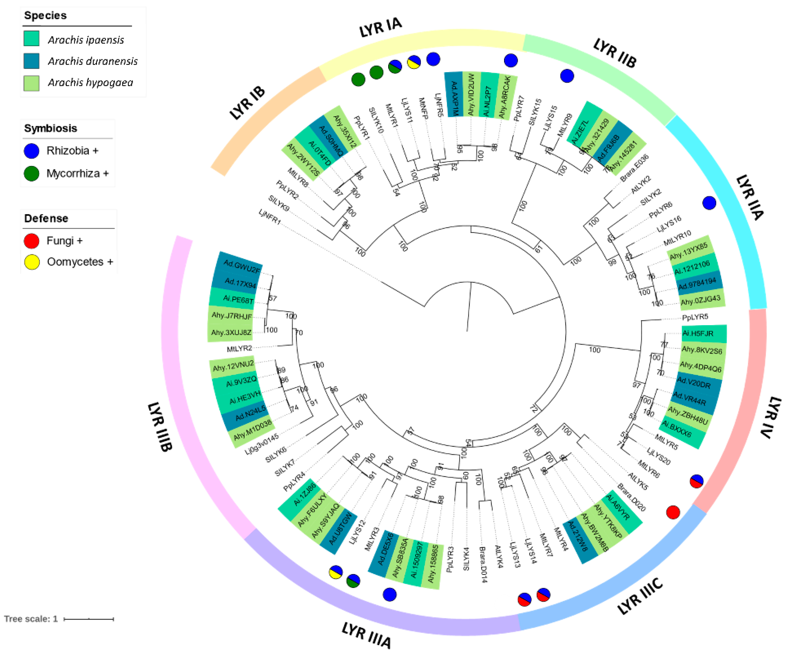
3.2. Phylogenetic Reconstruction of the LysM-RLK Family

In the evolutionary history of the LysM-RLK family, two well-supported groups are evident. The first group, called LYK [33], includes receptors with an active kinase domain [34,35,36,37]. The second group, called LYR [38], contains members lacking kinase activity (the loss of the glycine-rich loop and the “DFG” motif at the start of the activation loop) in the intracellular region, due to aberrations in the domain sequence [36,38].
In this work, we inferred phylogenies for LYKs and LYRs, including wild and cultivated peanut sequences and well-characterized LysM-RLKs from A. thaliana, L. japonicus, M. truncatula, P. persica, B. rapa and S. lycopersicum (species for which functional data is available). In LYK phylogeny, three sub-groups, LYKI, LYKII, and LYKIII (following the nomenclature proposed by [23] were observed (Figure 1). Of the total peanut LysM identified, 17 out of 38 from the genome of A. hypogaea, 8 out of 20 from A. duranensis, and 9 out of 21 from A. ipaensis belonged to the LYK group. All major LYK clades included copies from A. hypogaea, A. duranensis, and A. ipaensis. Similarly, in LYR phylogeny (Figure 2) well-supported clades were classified as LYRI (including LYRIA and LYRIB), LYRII (containing LYRIIA and LYRIIB), LYRIII (including LYRIII A, LYRIIIB and LYRIIIC) and LYRIV. Of the total peanut LysM identified, 21 out of the 38 from A. hypogaea, 12 out of 20 from A. duranensis, and 12 out of 21 from A. ipaensis belonged to the LYR group. All major LYR clades included copies from A. hypogaea, A. duranensis, and A. ipaensis.

3.3. Ka/Ks Analysis of LysM-RLK Genes

Out of the 38 LysM-RLK genes located on the cultivated peanut genome, 29 of them had their orthologs identified on their corresponding ancestor genome. Thus, a total of 29 pairs of the LysM-RLK orthologs between the tetraploid cultivated peanut genome and the wild ancestor peanut genomes were subjected to Ka/Ks calculation. Of these 29 pairs of orthologs, 8 pairs had no ratio calculated (Figure 3), due to their identical amino acid sequences between the copies in the pair. The remaining 21 pairs showed a Ka/Ks ratio that ranged from 0.001 to 1.7. Among these, only 2 pairs (located on chromosomes 4 and 5) had a Ka/Ks ratio above 1, indicating a positive selection, since the cultivated peanut was evolved. The remaining 19 pairs of LysM-RLK orthologs showed a Ka/Ks ratio below 1, suggesting that these genes were under negative selection.

3.4. Chromosomal Location and Synteny Analysis of Peanut LysM-RLK Genes

LysM-RLKs genes identified in A. duranensis were distributed on eight out of ten chromosomes, two genes each on ChrA01 and ChrA03, three genes each on ChrA05 and ChrA06, four genes each on ChrA04 and ChrA07. Two chromosomes (ChrA08 and ChrA09) contain only one gene. Some LysM-RLK genes located on ChrA06 and ChrA07 showed tandem duplication (Figure 4). Comparatively, the LysM-RLK genes identified in A. ipaensis, were distributed on seven out of ten chromosomes, three genes each on ChrB01 and ChrB05, two genes each on ChrB03 and ChrB06, four genes on ChrB04, six genes on ChrB07, and only one gene on ChrB09. Interestingly, some genes located on ChrB07 also showed tandem duplication, along with some other genes located on ChrB04. In addition, one LysM-RLK gene appears to have suffered segmental duplication in ChrB01 and ChrB07.
LysM-RLK genes in A. hypogaea were distributed on 14 out of 20 chromosomes. Among the 38 LysM-RLK genes, 17 and 19 have their orthologous gene from the A and B genomes, respectively. This result is consistent with their distribution on the A and B subgenomes. Interestingly, one gene located on Chr14 showed tandem duplication as with on A. ipaensis ChrB04.
To comprehensively clarify the mutual relationships of LysM-RLK genes in the A and B genomes of the Arachis species, the chromosomal location of LysM-RLK genes from A. duranensis (A genome) and A. ipaensis (B genome) was compared with that of A. hypogaea (Table S3). Of the 38 A. hypogaea LysM-RLK genes, 17 were located in the A genome, and the remaining were located in the B genome (Figure 4).

3.5. Temporal Expression Analysis of LysM-RLKs Genes in Peanut–Bradyrhizobia Interaction

By using the dataset generated by Karmakar et al. (2019), we analyzed the temporal expression pattern of peanut LysM-RLKs throughout the symbiosis process. Of the 38 A. hypogaea LysM-RLK genes, 31 were detected to express at certain points of the symbiosis development. On the other hand, no expression was detected for 7 LysM-RLK genes. Temporal expression analysis indicated that the 31 genes were transcriptionally up- or down-regulated at different stages of symbiosis, ranging from recognition (1 dpi) to mature nodule (21 dpi). Based on this analysis, 6 groups were formed (A–F, Figure 5).

3.6. Expression Analysis of Peanut Receptors

To investigate the role of LysM-RLK receptors in A. hypogaea, two genes were selected for expression analysis, Ahy.IM7I4N (belonging to phylogenetic group LYKI) and Ahy.YTK8KP (phylogenetic group LYRIIIC). Selection of these genes was based on the presence of specific residues in protein sequences (Tyr-128, Ser-206 and/or Tyr-228), which had been shown to be crucial for the perception of structurally-related N-acetylglucosamine-containing molecules [39,40]. Firstly, we selected Ahy.IM47N (which includes Tyr-128 and Ser-206 conserved residues), due to its localization in a poorly characterized clade, LYK I, that also contains receptors involved in responses to Fungi, Bacteria and Mycorrhiza or their associated molecules [33,41,42,43]. Interestingly, the closest NFR1 ortholog of peanut (Ahy.IVY8DS) is located in the same clade, but was not selected for relative expression analysis, since CRISPR/Cas9 mutants with editing in the AhNFR1 gene could still form nodules after rhizobial inoculation [44]. Secondly, Ahy.YTK8KP (which is located in the LYRIIIC clade and includes Tyr-128, 228 and Ser-206 conserved residues), was selected for expression analyses. This clade includes receptors involved in the perception of Fungi and Rhizobia or their associated molecules [22,41,42].
For each gene, the relative expression level was measured by qPCR at five different hpi with rhizobial NFs and chitosan. For Ahy.IM7I4N, NFs inoculation significantly increased its expression levels at 1, 8, 16 and 24 hpi (Figure 6A), reaching maximum levels at 1 hpi. Similarly, in plants inoculated with chitosan, the Ahy.IM7I4N expression level was significantly increased at 1 hpi, although at lower levels than those with NF inoculation.
On the other hand, the expression levels of Ahy.YTK8KP were significantly increased at 1 and 8 hpi after NFs treatment (Figure 6B). In contrast, transcript levels of Ahy.YTK8KP were not significantly induced after chitosan treatment.

4. Discussion

4.1. Phylogenetic Reconstruction of the Peanut LysM-RLK Family and Digital Gene Expression Analysis

Plants are constantly exposed to a large number of signals from surrounding microorganisms. In particular, legumes can establish mutualistic interactions with nitrogen-fixing bacteria. Therefore, legumes must recognize the compatible symbionts, and activate a complex symbiotic program but, at the same time, defend themselves against the invasion of bacterial and fungal pathogens [45,46]. LysM-RLKs are involved in the detection of both symbiotic and pathogenic organisms [4,10,47,48,49]. These receptors recognize a wide variety of ligands, including NFs and chitin derivatives, and trigger the appropriate signaling pathway leading either to defense or to symbiosis development.
To identify peanut LysM-RLKs sequences, a two-step search was conducted against A. hypogaea, A. duranensis, and A. ipaensis genomes. This strategy allowed us to perform a complete scan of the LysM-RLK receptor sequences in the cultivated and wild peanut genomes. Results indicated that peanut LysM-RLKs constituted a large family, including 38 members in cultivated peanut, 20 in A. duranensis and 21 in A. ipaensis. Similarly, a high number of LysM-RLK receptors were observed in model legumes such as M. truncatula (22) [50] and L. japonicus (18) [23]. In contrast, in non-legumes, the number is relatively low (i.e., five in A. thaliana and 10 in rice) [50]. The high number of peanut LysM-RLKs is congruent with the well-known rapid evolution and expansionary dynamics of this family. It has been suggested that diversification of this receptor family has contributed to the machinery that allowed the origin and evolution of nitrogen-fixing symbiosis in legumes [51]. All the identified LysM-RLKs were further divided into two main groups: LYKs (displaying an active kinase domain) and LYRs (harboring an inactive kinase domain). In this work, phylogenetic trees for LYKs and LYRs were inferred. These analyses represent a framework allowing reinterpretation of the functional data existing for peanut RLKs.

4.2. LysM-RLK LYK Group

The phylogenetic tree constructed with peanut LYKs and sequences from other species were consistent with previous works [23]. LYKs can be further divided into three subclades, named LYKI, LYKII, and LYKIII. The LYKI group is highly diverse, and contains many members. Some members show dual functionality (MtLYK9 and LjLYS6/LjCERK1) [23], participating in the perception and activation of signaling pathways in response to both mycorrhizal fungi and fungal pathogens [42,43]. On the other hand, some members of this group are well-characterized NFs receptors (MtLYK3, LjNFR1) [33,41,52]. In the peanut, the closest ortholog of LjNFR1 is Ahy.IVY8DS. This gene was shown to be upregulated at 16 hpi in nodulating peanut lines inoculated with the microsymbiont. However, [44] showed that mutants in this gene could still form nodules after rhizobial inoculation, suggesting that AhNFR1 may not be required for nodule formation.
Other genes coding for peanut LysM-RLKs found within the LYKI group (Ahy.MX792F, Ahy.SA9NCH) were reported to be upregulated during the peanut–bradyrhizobia interaction at 2 hpi. In addition, Ahy.63XNPZ was upregulated at 16 hpi [44]. Similarly, Ahy.RPPZ03 and Ahy.K2D2HU were reported to be upregulated at 21 dpi (when nodules are mature and functional) [18]. It has been proposed that a continuous crosstalk between bacteria and host is required, even in mature nodules [53,54,55]. Therefore, these receptors’ activity could be related to monitoring signals during nodule maintenance or immune modulation at late stages of symbiosis.
The members of the LYKII group are less characterized than those in LYKI [23]. The only well-characterized member in this clade is LjEPR3/LjLYS3, responsible for the perception of compatible exopolysaccharides during the rhizobial symbiosis establishment in Lotus [56,57]. Peanut orthologs of LjEPR3 are represented by Ahy.6V1TUE from subgenome A and Ahy.7J5ZWH subgenome B. Both genes were upregulated at 8, 12, and 21 dpi [18], suggesting their participation in signal monitoring through the different stages of nodule formation.
Within the LysM-RLK LYKIII subgroup, the LjLYS4 receptor is involved in the interaction of the legume with its microsymbiont [41]. On the other hand, the AtLYK3 receptor was required for the repression of the Arabidopsis innate immunity in response to NFs [58]. More recently, AtLYK3 was also found to participate in the negative regulation of plant immunity in response to bacterial and fungal pathogens in Arabidopsis [59]. There are three peanut members of the LysM-RLK LYKIII subgroup, one from each of A. hypogaea, A. duranensis, and A. ipaensis. However, there is no functional data regarding these receptors, and they were not reported as differentially expressed during peanut–rhizobia symbiosis or fungal defense, which further indicated that they may not be involved in the response to microorganisms.

4.3. LysM-RLK LYR Group

Major groups of LYRs observed are congruent with those described by [23]. LYRs can be subdivided into four major groups, LYRI, LYRII, LYRIII, and LYRIV. The LYRI subgroup can be further subdivided into groups A and B, each including representatives of the parental diploids and cultivated peanuts. The LYRIA subgroup includes receptors involved in mycorrhization (MtLYR1 and LjLYR11, possibly in a redundant role with MtNFP and LjNFR5) [60,61,62] and rhizobial symbiosis (MtNFP and LjNFR5) [38,61]. We found two NFR5 orthologs in the peanut genome, Ahy.VID2UW (A subgenome) and Ahy.A8RCAK (B subgenome). Ahy.A8RCAK is upregulated in peanut–bradyrhizobia association at 5 [17] and 4, 8, and 12 dpi [18], while Ahy.VID2UW is upregulated at 8 dpi [18]. In addition, [44] demonstrated that when both AhNFR5 A (Ahy.VID2UW) and B (Ahy.A8RCAK) were mutated in transgenic hairy roots, no nodules were formed. Altogether, these data indicate that genes belonging to the phylogenetic group LYRIA play critical roles in root endosymbiosis. On the other hand, receptors included in the LYRIB subgroup have not yet been assigned a role in the interaction with microorganisms or their eliciting molecules.
The LYRII group was also subdivided into the A and B subgroups. L. japonicus LYS15 (LYRII B) and LYS16 (LYRII A) were reported to be upregulated during interaction with rhizobia or in response to NFs inoculation [41]. However, peanut receptors found in these subgroups were not differentially expressed in the interaction with the microsymbiont.
The LysM-RLK LYRIII group is characterized by a high occurrence of duplications [23] with the largest number of peanut LysM-RLK receptors. This group could be subdivided into subgroups A, B, and C. The LYRIIIA group MtLYR3 receptor has been determined to have an affinity for LCOs, and could recognize NFs and Myc factors during the establishment of symbiotic relationships in M. truncatula [8,49]. Likewise, it has been demonstrated that LjLYS12 participates in the interaction with rhizobia, and activates the defense response against oomycetes [41,63]. In the peanut, Ahy.SB83SA receptor has been reported to be induced in plants inoculated with the symbiont at early infection steps (1, 4, and 8 dpi) [18].
Regarding the LYRIIIB subgroup, there is no information related to the functional characterization of its members. The LYRIIIC group contained one of the best characterized LysM-RLK receptors in legumes, the MtLYR4 from Medicago. Several studies have shown its participation in the perception of long-chain COs [64]) and in resistance to fungal pathogens [42]. Similarly, LjLYS13 and LjLYS14 were reported as upregulated in response to inoculation with rhizobia and chitin [41]. Expression analysis of the Ahy.YTK8KP gene, performed in this work, showed an increased expression level in NF treatment, suggesting its participation in rhizobial signaling perception.
The LYRIV subgroup contained poorly characterized members in legumes. A single study reported an increase in expression levels of the LjLYS20 receptor after chitin treatment, which suggests its participation in the immune response against fungal pathogens [41].

4.4. Temporal Expression Analysis of A. hypogaea LysM-RLK Receptors within the Progress of Symbiosis

Out of the 31 AhLysM-RLKs, only 17 genes (groups A, B, and C) showed high expression levels within the progress of symbiosis. In contrast, low expression levels were detected in the genes located in groups D, E, and F. Genes in group A (Ahy.4DP4Q6, Ahy.SB835A, Ahy.J7RHJF, and Ahy.8KV2S6) were positively regulated in almost all the time points evaluated. Among the five time points evaluated, 8 and 12 dpi displayed the highest gene number with high transcript levels.
At 1 (recognition and invasion) and 4 dpi (primordia formation), expression levels of Ahy.4DP4Q6, Ahy.J7RHJF and Ahy.SB835A were high, compared with the other AhLysM-RLKs. Intriguingly, all these genes are grouped in the LYR clade. It has been proposed that members of the phylogenetic subgroup LYRIII are involved in defense mechanisms, and, in legumes, they can bind to LCOs with high affinity [41,63]. Digital expression analysis suggests that Ahy.J7RHJF, and Ahy.SB835A could play a role in the first steps of rhizobial infection of peanut roots. However, further studies are required to confirm this observation.
At 8 dpi (nodule-like structure) and 12 dpi (immature nodules with rod-shaped rhizobia), a diverse group of peanut LysM-RLK genes with high transcription levels was detected, including orthologs of LjEPR3 (Ahy.6V1TUE, Ahy.7J5ZWH) and LjNFR5 (Ahy.A8RCAK). These genes have been shown to be implicated in the recognition of bacterial EPS, NFs, and colonization by rhizobia in Lotus [41,56,57,65]. Moreover, at 21 dpi (mature nodules with spherical symbiosomes) Ahy.7J5ZWH (EPR3), Ahy.RPPZ03, Ahy.K2D2HU, and Ahy.J7RHJF showed high transcript levels. Our analysis suggests that EPR3 expression levels at 8, 12, and 21 dpi are possibly related to monitoring the rhizobial exopolysaccharide (EPS) at the middle and late stages of symbiosis in peanuts.

4.5. Chromosomal Location of Peanut LysM-RLK and Synteny Analysis

Since the A. hypogaea (AABB) genome has undergone whole-genome duplication (WGD) after hybridization of A. duranensis (A) and A. ipaensis (B) [16], the A. hypogaea genome theoretically contains two copies of LysM-RLK genes, with one from each ancestral species. However, only 17 LysM-RLK genes were found on the A subgenome (instead of 20) and 21 LysM-RLK genes on the B subgenome (instead of 20). This suggests that some LysM-RLK genes have probably been lost during hybridization and duplication events of A. hypogaea genome evolution. The other LysM-RLK genes from A. hypogaea each have a counterpart in the A. duranensis and A. ipanesis genomes.In addition, only eight genes showed tandem duplication (four on A. ipaensis and four on A. duranensis). Interestingly, the duplicated genes Ad.P4UQH, Ad.KFY8V, Ai.VBT71 and Ai.0J1DV, located on ChrA07 and ChrB07, respectively, belong to the LYKI clade. These results are congruent with those of Buendía et al., 2018, reporting several duplications in LysM-RLK genes grouped in this clade.
Compared with the LysM-RLK genes identified in the genomes of A. ipaensis and A. duranensis, most LysM-RLK genes showed no segmental or tandem duplication in A. hypogaea. These results support the idea that in cultivated peanut, changes in the ancestral genomes since polyploidy have been limited [16,66,67]. However, we observed two LysM-RLK genes without a relative counterpart (Ahy.ZBH48U and Ahy.4B5N5U) in A and B genomes.
The synteny map revealed that the LysM-RLK family genes are generally conserved. However, duplication or loss of some genes is evident. The homologous genes of Ad.MEH04, Ai.HE3VH and Ad.GWU2F in the wild ancestor species were not found in the genome of cultivated peanut species. However, all the other LysM-RLK genes have the homolog on each of the parental genomes. A similar phenomenon was also reported in papilionoid legumes where several genes involved in the rhizobial symbiosis have been maintained as paralogous genes after WGD [68,69,70].
Taken together, the results indicate that the LysM-RLK genes were preserved after hybridization and chromosomes doubling in the cultivated peanut, and it becomes apparent that homeologous recombination in the A. hypogaea genome has not generated significant changes in the chromosomal organization of the LysM-RLK gene family. In addition, these results open up the possibility of studying how the conservation of ancestral genomes and chromosomal rearrangements on the tetraploid genome could be related to the diversification and functionalization of LysM-RLK genes.

4.6. Expression Analysis in NFs and Chitosan-Treated Plants

To unravel the potential participation of two peanut receptors in the perception of NF and chitosan, their expression levels after inoculation with the elicitor molecules were analyzed. Transcriptional activation of Ahy.IM7I4N was observed at 1 hpi with NF and chitosan, separately, suggesting a versatile function of Ahy.IM7I4N. This dual function is in accordance with the co-receptor role proposed for LYKI LysM-RLKs [23,71]. Intriguingly, the mechanism of the way in which plants perceive chitosan remains unclear. Some authors suggest that chitosan is perceived by a membrane receptor, while others indicate that it moves directly to the nucleus and interacts with DNA [34,72,73]. At later time points (from 1 to 72 hpi), Ahy.IM7I4N expression levels were significantly increased in response to NF.
Expression analysis of Ahy.YTK8KP suggested a specific transcriptional response to NF treatment (with significant expression levels increased at 1 and 8 hpi). Similarly, other receptors belonging to the same LYK subgroup (MtLYK4, LjLYS13, and LjLYS14) appear to be involved in early rhizobial signal perception [41,64]. However, Mtlyr4 mutants (such as Ljlys13 and Ljlys14 mutants) were not affected in the RNS, suggesting a redundant role with other receptors [42].
It is important to mention that a positive transcriptional response of LysM-RLK genes to NF or chitosan inoculation does not necessarily imply a direct receptor–elicitor binding. To confirm such direct interaction, other experiments are required. As an alternative, and considering the more complex model for molecule recognition recently proposed [2,3,5], a transcriptional activation suggests direct or indirect participation of the LysM-RLKs in the receptors network, by mediating the perception and/or pathway activation induced by the elicitors. In addition, acetylated chitin oligomers present in the chitosan used in this work (75% deacetylated) can induce the responses that lead to the regulation of a LysM-RLKs.

5. Conclusions

Peanut LysM-RLK constitutes a diverse family with several members. Analysis of the evolutionary history of the family, and functional data suggest that ligand perception and the expression pattern of peanut receptors could be different from that reported for model legumes. This study provides a better picture of the evolution of the LysM-RLK family in peanut (a non-model legume). It is clear that the LysM-RLK family phylogeny could not discriminate between receptors recognizing structurally-related ligands. The mechanism that allows discrimination among structurally-related ligands is complex, and could be related to specific single modifications in the amino acid sequence [39,65], motifs with structural conservation or variable motifs in LysM1 regions [74] or the formation of receptor heterocomplexes that bind to one or more ligands [42,58,75,76]. Further functional genetics experiments are required, to assign a biological function to a particular receptor. However, this work sets the basis for the selection of genes based on their phylogenetic position.

Supplementary Materials

The following supporting information can be downloaded at: https://www.mdpi.com/article/10.3390/horticulturae8111000/s1, Table S1: List of primers. Table S2: List of the LysM-RLK identified in 10 plant species. Table S3: Chromosomal location of LysM-RLK genes.

Author Contributions

Conceptualization, J.R.M., M.L.T. and F.I.; methodology, J.R.M.; software, Z.Z. and J.W.; validation, J.R.M., M.C.B. and Z.Z.; formal analysis, J.R.M.; investigation, J.R.M.; resources, A.F.; data curation, J.R.M., M.C.B. and Z.Z.; writing—original draft preparation, J.R.M., M.L.T. and F.I.; writing—review and editing, J.R.M., M.L.T., J.W., F.A. and F.I.; visualization, J.R.M.; supervision, F.I., M.L.T. and F.A.; project administration, F.I., M.L.T. and F.A.; funding acquisition, A.F., F.I., M.L.T., F.A. and J.R.M. All authors have read and agreed to the published version of the manuscript.

Funding

This study was financially supported by Secretaría de Ciencia y Técnica-Universidad Nacional de Río Cuarto, Consejo Nacional de Investigaciones Científicas y Técnicas (CONICET), Agencia Nacional de Promoción Científica y Tecnológica (ANPCyT) and AXA Research Fund. F.I.; M.L.T.; F.A.; and A.F. are members of Consejo Nacional de Investigaciones Científicas y Técnicas (CONICET). J.R. is a fellow of the AXA Research Fund.

Data Availability Statement

The data used to support the findings of this study are included withing the article.

Conflicts of Interest

The authors declare no conflict of interest.

References

  1. Zipfel, C.; Oldroyd, G.E.D. Plant Signalling in Symbiosis and Immunity. Nature 2017, 543, 328–336. [Google Scholar] [CrossRef] [PubMed]
  2. Chiu, C.H.; Paszkowski, U. Receptor-Like Kinases Sustain Symbiotic Scrutiny. Plant Physiol. 2020, 182, 1597–1612. [Google Scholar] [CrossRef] [PubMed] [Green Version]
  3. Rodríguez, J.; Tonelli, M.L.; Figueredo, M.S.; Ibáñez, F.; Fabra, A. The Lipopeptide Surfactin Triggers Induced Systemic Resistance and Priming State Responses in Arachis hypogaea L. Eur. J. Plant Pathol. 2018, 152, 845–851. [Google Scholar] [CrossRef]
  4. Antolín-Llovera, M.; Petutsching, E.K.; Ried, M.K.; Lipka, V.; Nürnberger, T.; Robatzek, S.; Parniske, M. Knowing Your Friends and Foes–Plant Receptor-like Kinases as Initiators of Symbiosis or Defence. New Phytol. 2014, 204, 791–802. [Google Scholar] [CrossRef]
  5. Kelly, S.; Radutoiu, S.; Stougaard, J. Legume LysM Receptors Mediate Symbiotic and Pathogenic Signalling. Curr. Opin. Plant Biol. 2017, 39, 152–158. [Google Scholar] [CrossRef]
  6. Oldroyd, G.E.D. Speak, Friend, and Enter: Signalling Systems That Promote Beneficial Symbiotic Associations in Plants. Nat. Rev. Microbiol. 2013, 11, 252–263. [Google Scholar] [CrossRef]
  7. Boller, T. Chemoperception of Microbial Signals in Plant Cells. Annu. Rev. Plant Physiol. Plant Mol. Biol. 1995, 46, 189–214. [Google Scholar] [CrossRef]
  8. Fliegmann, J.; Bono, J.-J. Lipo-Chitooligosaccharidic Nodulation Factors and Their Perception by Plant Receptors. Glycoconj J. 2015, 32, 455–464. [Google Scholar] [CrossRef]
  9. Yin, H.; Du, Y.; Dong, Z. Chitin Oligosaccharide and Chitosan Oligosaccharide: Two Similar but Different Plant Elicitors. Front. Plant Sci. 2016, 7, 522. [Google Scholar] [CrossRef] [Green Version]
  10. Gust, A.A.; Willmann, R.; Desaki, Y.; Grabherr, H.M.; Nürnberger, T. Plant LysM Proteins: Modules Mediating Symbiosis and Immunity. Trends Plant Sci. 2012, 17, 495–502. [Google Scholar] [CrossRef]
  11. Kochert, G.; Stalker, H.T.; Gimenes, M.; Galgaro, L.; Lopes, C.R.; Moore, K. Rflp and cytogenetic evidence on the origin and evolution of allotetraploid domesticated Peanut, Arachis Hypogaea (Leguminosae). Am. J. Bot. 1996, 83, 1282–1291. [Google Scholar] [CrossRef]
  12. Seijo, J.G.; Lavia, G.I.; Fernandez, A.; Krapovickas, A.; Ducasse, D.; Moscone, E.A. Physical Mapping of the 5S and 18S-25S RRNA Genes by FISH as Evidence That Arachis Duranensis and A. Ipaensis Are the Wild Diploid Progenitors of A. Hypogaea (Leguminosae). Am. J. Bot. 2004, 91, 1294–1303. [Google Scholar] [CrossRef] [PubMed]
  13. Ibáñez, F.; Wall, L.; Fabra, A. Starting Points in Plant-Bacteria Nitrogen-Fixing Symbioses: Intercellular Invasion of the Roots. J. Exp. Bot. 2017, 68, 1905–1918. [Google Scholar] [CrossRef] [PubMed] [Green Version]
  14. Bertioli, D.J.; Jenkins, J.; Clevenger, J.; Dudchenko, O.; Gao, D.; Seijo, G.; Leal-Bertioli, S.C.M.; Ren, L.; Farmer, A.D.; Pandey, M.K.; et al. The Genome Sequence of Segmental Allotetraploid Peanut Arachis Hypogaea. Nat. Genet. 2019, 51, 877–884. [Google Scholar] [CrossRef] [Green Version]
  15. Zhuang, W.; Chen, H.; Yang, M.; Wang, J.; Pandey, M.K.; Zhang, C.; Chang, W.-C.; Zhang, L.; Zhang, X.; Tang, R.; et al. The Genome of Cultivated Peanut Provides Insight into Legume Karyotypes, Polyploid Evolution and Crop Domestication. Nat. Genet. 2019, 51, 865–876. [Google Scholar] [CrossRef] [Green Version]
  16. Bertioli, D.J.; Cannon, S.B.; Froenicke, L.; Huang, G.; Farmer, A.D.; Cannon, E.K.S.; Liu, X.; Gao, D.; Clevenger, J.; Dash, S.; et al. The Genome Sequences of Arachis Duranensis and Arachis Ipaensis, the Diploid Ancestors of Cultivated Peanut. Nat. Genet. 2016, 48, 438–446. [Google Scholar] [CrossRef]
  17. Peng, Z.; Liu, F.; Wang, L.; Zhou, H.; Paudel, D.; Tan, L.; Maku, J.; Gallo, M.; Wang, J. Transcriptome Profiles Reveal Gene Regulation of Peanut (Arachis Hypogaea L.) Nodulation. Sci. Rep. 2017, 7, 40066. [Google Scholar] [CrossRef] [Green Version]
  18. Karmakar, K.; Kundu, A.; Rizvi, A.Z.; Dubois, E.; Severac, D.; Czernic, P.; Cartieaux, F.; DasGupta, M. Transcriptomic Analysis with the Progress of Symbiosis in ‘Crack-Entry’ Legume Arachis Hypogaea Highlights Its Contrast with ‘Infection Thread’ Adapted Legumes. Mol. Plant Microbe Interact. 2019, 32, 271–285. [Google Scholar] [CrossRef] [Green Version]
  19. Sharma, V.; Bhattacharyya, S.; Kumar, R.; Kumar, A.; Ibañez, F.; Wang, J.; Guo, B.; Sudini, H.K.; Gopalakrishnan, S.; DasGupta, M.; et al. Molecular Basis of Root Nodule Symbiosis between Bradyrhizobium and “Crack-Entry” Legume Groundnut (Arachis hypogaea L.). Plants 2020, 9, 276. [Google Scholar] [CrossRef] [Green Version]
  20. Jones, P.; Binns, D.; Chang, H.Y.; Fraser, M.; Li, W.; McAnulla, C.; McWilliam, H.; Maslen, J.; Mitchell, A.; Nuka, G.; et al. InterProScan 5: Genome-Scale Protein Function Classification. Bioinformatics 2014, 30, 1236–1240. [Google Scholar] [CrossRef]
  21. El-Gebali, S.; Mistry, J.; Bateman, A.; Eddy, S.R.; Luciani, A.; Potter, S.C.; Qureshi, M.; Richardson, L.J.; Salazar, G.A.; Smart, A.; et al. The Pfam Protein Families Database in 2019. Nucleic Acids Res. 2019, 47, D427–D432. [Google Scholar] [CrossRef] [PubMed]
  22. Richards, S.; Rose, L.E. The Evolutionary History of LysM-RLKs (LYKs/LYRs) in Wild Tomatoes. BMC Evol. Biol. 2019, 19, 141. [Google Scholar] [CrossRef] [PubMed] [Green Version]
  23. Buendia, L.; Girardin, A.; Wang, T.; Cottret, L.; Lefebvre, B. LysM Receptor-Like Kinase and LysM Receptor-Like Protein Families: An Update on Phylogeny and Functional Characterization. Front. Plant Sci. 2018, 9, 1531. [Google Scholar] [CrossRef] [PubMed] [Green Version]
  24. Sela, I.; Ashkenazy, H.; Katoh, K.; Pupko, T. GUIDANCE2: Accurate Detection of Unreliable Alignment Regions Accounting for the Uncertainty of Multiple Parameters. Nucleic Acids Res. 2015, 43, W7–W14. [Google Scholar] [CrossRef] [Green Version]
  25. Guindon, S.; Dufayard, J.-F.; Lefort, V.; Anisimova, M.; Hordijk, W.; Gascuel, O. New Algorithms and Methods to Estimate Maximum-Likelihood Phylogenies: Assessing the Performance of PhyML 3.0. Syst. Biol. 2010, 59, 307–321. [Google Scholar] [CrossRef] [Green Version]
  26. Lefort, V.; Longueville, J.E.; Gascuel, O. SMS: Smart Model Selection in PhyML. Mol. Biol. Evol. 2017, 34, 2422–2424. [Google Scholar] [CrossRef] [Green Version]
  27. Letunic, I.; Bork, P. Interactive Tree of Life (ITOL) v3: An Online Tool for the Display and Annotation of Phylogenetic and Other Trees. Nucleic Acids Res. 2016, 44, W242–W245. [Google Scholar] [CrossRef]
  28. Zhang, Z.; Xiao, J.; Wu, J.; Zhang, H.; Liu, G.; Wang, X.; Dai, L. ParaAT: A Parallel Tool for Constructing Multiple Protein-Coding DNA Alignments. Biochem. Biophys. Res. Commun. 2012, 419, 779–781. [Google Scholar] [CrossRef]
  29. Edgar, R.C. MUSCLE: Multiple Sequence Alignment with High Accuracy and High Throughput. Nucleic Acids Res. 2004, 32, 1792–1797. [Google Scholar] [CrossRef] [Green Version]
  30. Zhang, Z.; Li, J.; Zhao, X.-Q.; Wang, J.; Wong, G.K.-S.; Yu, J. KaKs_Calculator: Calculating Ka and Ks through Model Selection and Model Averaging. Genom. Proteom. Bioinform. 2006, 4, 259–263. [Google Scholar] [CrossRef]
  31. Spaink, H.P.; Sheeley, D.M.; van Brussel, A.A.N.; Glushka, J.; York, W.S.; Tak, T.; Geiger, O.; Kennedy, E.P.; Reinhold, V.N.; Lugtenberg, B.J.J. A Novel Highly Unsaturated Fatty Acid Moiety of Lipo-Oligosaccharide Signals Determines Host Specificity of Rhizobium. Nature 1991, 354, 125–130. [Google Scholar] [CrossRef] [PubMed]
  32. Livak, K.J.; Schmittgen, T.D. Analysis of Relative Gene Expression Data Using Real-Time Quantitative PCR and the 2−ΔΔCT Method. Methods 2001, 25, 402–408. [Google Scholar] [CrossRef] [PubMed]
  33. Limpens, E.; Franken, C.; Smit, P.; Willemse, J.; Bisseling, T.; Geurts, R. LysM Domain Receptor Kinases Regulating Rhizobial Nod Factor-Induced Infection. Science (1979) 2003, 302, 630–633. [Google Scholar] [CrossRef] [PubMed]
  34. Petutschnig, E.K.; Jones, A.M.E.; Serazetdinova, L.; Lipka, U.; Lipka, V. The Lysin Motif Receptor-like Kinase (LysM-RLK) CERK1 Is a Major Chitin-Binding Protein in Arabidopsis Thaliana and Subject to Chitin-Induced Phosphorylation. J. Biol. Chem. 2010, 285, 28902–28911. [Google Scholar] [CrossRef] [Green Version]
  35. Klaus-Heisen, D.; Nurisso, A.; Pietraszewska-Bogiel, A.; Mbengue, M.; Camut, S.; Timmers, T.; Pichereaux, C.; Rossignol, M.; Gadella, T.W.J.; Imberty, A.; et al. Structure-Function Similarities between a Plant Receptor-like Kinase and the Human Interleukin-1 Receptor-Associated Kinase-4. J. Biol. Chem. 2011, 286, 11202–11210. [Google Scholar] [CrossRef] [Green Version]
  36. Madsen, L.H.; Tirichine, L.; Jurkiewicz, A.; Sullivan, J.T.; Heckmann, A.B.; Bek, A.S.; Ronson, C.W.; James, E.K.; Stougaard, J. The Molecular Network Governing Nodule Organogenesis and Infection in the Model Legume Lotus Japonicus. Nat. Commun. 2010, 1, 10. [Google Scholar] [CrossRef] [Green Version]
  37. Zeng, L.; Velásquez, A.C.; Munkvold, K.R.; Zhang, J.; Martin, G.B. A Tomato LysM Receptor-like Kinase Promotes Immunity and Its Kinase Activity Is Inhibited by AvrPtoB. Plant J. 2012, 69, 92–103. [Google Scholar] [CrossRef] [Green Version]
  38. Arrighi, J.-F.; Barre, A.; ben Amor, B.; Bersoult, A.; Soriano, L.C.; Mirabella, R.; de Carvalho-Niebel, F.; Journet, E.-P.; Ghérardi, M.; Huguet, T.; et al. The Medicago Truncatula Lysin [Corrected] Motif-Receptor-like Kinase Gene Family Includes NFP and New Nodule-Expressed Genes. Plant Physiol. 2006, 142, 265–279. [Google Scholar] [CrossRef] [Green Version]
  39. Cao, Y.; Liang, Y.; Tanaka, K.; Nguyen, C.T.; Jedrzejczak, R.P.; Joachimiak, A.; Stacey, G. The Kinase LYK5 Is a Major Chitin Receptor in Arabidopsis and Forms a Chitin-Induced Complex with Related Kinase CERK1. Elife 2014, 3, e03766. [Google Scholar] [CrossRef]
  40. Malkov, N.; Fliegmann, J.; Rosenberg, C.; Gasciolli, V.; Timmers, A.C.J.; Nurisso, A.; Cullimore, J.; Bono, J.-J. Molecular Basis of Lipo-Chitooligosaccharide Recognition by the Lysin Motif Receptor-like Kinase LYR3 in Legumes. Biochem. J. 2016, 473, 1369–1378. [Google Scholar] [CrossRef]
  41. Lohmann, G.V.; Shimoda, Y.; Nielsen, M.W.; Jørgensen, F.G.; Grossmann, C.; Sandal, N.; Sørensen, K.; Thirup, S.; Madsen, L.H.; Tabata, S.; et al. Evolution and Regulation of the Lotus Japonicus LysM Receptor Gene Family. Mol. Plant Microbe Interact. 2010, 23, 510–521. [Google Scholar] [CrossRef] [PubMed] [Green Version]
  42. Bozsoki, Z.; Cheng, J.; Feng, F.; Gysel, K.; Vinther, M.; Andersen, K.R.; Oldroyd, G.; Blaise, M.; Radutoiu, S.; Stougaard, J. Receptor-Mediated Chitin Perception in Legume Roots Is Functionally Separable from Nod Factor Perception. Proc. Natl. Acad. Sci. USA 2017, 114, E8118–E8127. [Google Scholar] [CrossRef] [PubMed] [Green Version]
  43. Gibelin-Viala, C.; Amblard, E.; Puech-Pages, V.; Bonhomme, M.; Garcia, M.; Bascaules-Bedin, A.; Fliegmann, J.; Wen, J.; Mysore, K.S.; Signor, C.; et al. The Medicago Truncatula LysM Receptor-like Kinase LYK9 Plays a Dual Role in Immunity and the Arbuscular Mycorrhizal Symbiosis. New Phytol. 2019, 223, 1516–1529. [Google Scholar] [CrossRef] [PubMed]
  44. Shu, H.; Luo, Z.; Peng, Z.; Wang, J. The Application of CRISPR/Cas9 in Hairy Roots to Explore the Functions of AhNFR1 and AhNFR5 Genes during Peanut Nodulation. BMC Plant Biol. 2020, 20, 417. [Google Scholar] [CrossRef] [PubMed]
  45. Ben, C.; Debellé, F.; Berges, H.; Bellec, A.; Jardinaud, M.-F.; Anson, P.; Huguet, T.; Gentzbittel, L.; Vailleau, F. MtQRRS1, AnR-Locus Required ForMedicago Truncatulaquantitative Resistance ToRalstonia Solanacearum. New Phytol. 2013, 199, 758–772. [Google Scholar] [CrossRef]
  46. Liu, Y.; Hassan, S.; Kidd, B.N.; Garg, G.; Mathesius, U.; Singh, K.B.; Anderson, J.P. Ethylene Signaling Is Important for Isoflavonoid-Mediated Resistance to Rhizoctonia Solani in Roots of Medicago Truncatula. Mol. Plant Microbe Interact. 2017, 30, 691–700. [Google Scholar] [CrossRef] [PubMed] [Green Version]
  47. Kaku, H.; Nishizawa, Y.; Ishii-Minami, N.; Akimoto-Tomiyama, C.; Dohmae, N.; Takio, K.; Minami, E.; Shibuya, N. Plant Cells Recognize Chitin Fragments for Defense Signaling through a Plasma Membrane Receptor. Proc. Natl. Acad. Sci. USA 2006, 103, 11086–11091. [Google Scholar] [CrossRef] [Green Version]
  48. Miya, A.; Albert, P.; Shinya, T.; Desaki, Y.; Ichimura, K.; Shirasu, K.; Narusaka, Y.; Kawakami, N.; Kaku, H.; Shibuya, N. CERK1, a LysM Receptor Kinase, Is Essential for Chitin Elicitor Signaling in Arabidopsis. Proc. Natl. Acad. Sci. USA 2007, 104, 19613–19618. [Google Scholar] [CrossRef] [PubMed] [Green Version]
  49. Fliegmann, J.; Canova, S.; Lachaud, C.; Uhlenbroich, S.; Gasciolli, V.; Pichereaux, C.; Rossignol, M.; Rosenberg, C.; Cumener, M.; Pitorre, D.; et al. Lipo-Chitooligosaccharidic Symbiotic Signals Are Recognized by LysM Receptor-Like Kinase LYR3 in the Legume Medicago Truncatula. ACS Chem. Biol. 2013, 8, 1900–1906. [Google Scholar] [CrossRef] [PubMed]
  50. Bono, J.J.; Fliegmann, J.; Gough, C.; Cullimore, J. Expression and Function of the Medicago Truncatula Lysin Motif Receptor-like Kinase (LysM-RLK) Gene Family in the Legume–Rhizobia Symbiosis. Model Legume Med. Truncatula 2019, 439–447. [Google Scholar] [CrossRef]
  51. de Mita, S.; Streng, A.; Bisseling, T.; Geurts, R. Evolution of a Symbiotic Receptor through Gene Duplications in the Legume–Rhizobium Mutualism. New Phytol. 2013, 201, 961–972. [Google Scholar] [CrossRef] [PubMed]
  52. Radutoiu, S.; Madsen, L.H.; Madsen, E.B.; Felle, H.H.; Umehara, Y.; Grønlund, M.; Sato, S.; Nakamura, Y.; Tabata, S.; Sandal, N.; et al. Plant Recognition of Symbiotic Bacteria Requires Two LysM Receptor-like Kinases. Nature 2003, 425, 585–592. [Google Scholar] [CrossRef] [PubMed]
  53. Price, P.A.; Tanner, H.R.; Dillon, B.A.; Shabab, M.; Walker, G.C.; Griffitts, J.S. Rhizobial Peptidase HrrP Cleaves Host-Encoded Signaling Peptides and Mediates Symbiotic Compatibility. Proc. Natl. Acad. Sci. USA 2015, 112, 15244–15249. [Google Scholar] [CrossRef] [PubMed] [Green Version]
  54. Czernic, P.; Gully, D.; Cartieaux, F.; Moulin, L.; Guefrachi, I.; Patrel, D.; Pierre, O.; Fardoux, J.; Chaintreuil, C.; Nguyen, P.; et al. Convergent Evolution of Endosymbiont Differentiation in Dalbergioid and Inverted Repeat-Lacking Clade Legumes Mediated by Nodule-Specific Cysteine-Rich Peptides. Plant Physiol. 2015, 169, 1254–1265. [Google Scholar] [CrossRef] [Green Version]
  55. Pan, H.; Wang, D. Nodule Cysteine-Rich Peptides Maintain a Working Balance during Nitrogen-Fixing Symbiosis. Nat. Plants 2017, 3, 17048. [Google Scholar] [CrossRef]
  56. Kawaharada, Y.; Nielsen, M.W.; Kelly, S.; James, E.K.; Andersen, K.R.; Rasmussen, S.R.; Füchtbauer, W.; Madsen, L.H.; Heckmann, A.B.; Radutoiu, S.; et al. Differential Regulation of the Epr3 Receptor Coordinates Membrane-Restricted Rhizobial Colonization of Root Nodule Primordia. Nat. Commun. 2017, 8, 14534. [Google Scholar] [CrossRef] [Green Version]
  57. Kawaharada, Y.; Kelly, S.; Nielsen, M.W.; Hjuler, C.T.; Gysel, K.; Muszyński, A.; Carlson, R.W.; Thygesen, M.B.; Sandal, N.; Asmussen, M.H.; et al. Receptor-Mediated Exopolysaccharide Perception Controls Bacterial Infection. Nature 2015, 523, 308–312. [Google Scholar] [CrossRef] [Green Version]
  58. Liang, Y.; Cao, Y.; Tanaka, K.; Thibivilliers, S.; Wan, J.; Choi, J.; Kang, C.H.; Qiu, J.; Stacey, G. Nonlegumes Respond to Rhizobial Nod Factors by Suppressing the Innate Immune Response. Science (1979) 2013, 341, 1384–1387. [Google Scholar] [CrossRef]
  59. Paparella, C.; Savatin, D.V.; Marti, L.; de Lorenzo, G.; Ferrari, S. The Arabidopsis LYSIN MOTIF-CONTAINING RECEPTOR-LIKE KINASE3 Regulates the Cross Talk between Immunity and Abscisic Acid Responses. Plant Physiol. 2014, 165, 262–276. [Google Scholar] [CrossRef] [Green Version]
  60. Amor, B.B.; Shaw, S.L.; Oldroyd, G.E.D.; Maillet, F.; Penmetsa, R.V.; Cook, D.; Long, S.R.; Denarie, J.; Gough, C. The NFP Locus of Medicago Truncatula Controls an Early Step of Nod Factor Signal Transduction Upstream of a Rapid Calcium Flux and Root Hair Deformation. Plant J. 2003, 34, 495–506. [Google Scholar] [CrossRef]
  61. Madsen, E.B.; Madsen, L.H.; Radutoiu, S.; Olbryt, M.; Rakwalska, M.; Szczyglowski, K.; Sato, S.; Kaneko, T.; Tabata, S.; Sandal, N.; et al. A Receptor Kinase Gene of the LysM Type Is Involved in Legumeperception of Rhizobial Signals. Nature 2003, 425, 637–640. [Google Scholar] [CrossRef] [PubMed]
  62. Rasmussen, S.R.; Füchtbauer, W.; Novero, M.; Volpe, V.; Malkov, N.; Genre, A.; Bonfante, P.; Stougaard, J.; Radutoiu, S. Intraradical Colonization by Arbuscular Mycorrhizal Fungi Triggers Induction of a Lipochitooligosaccharide Receptor. Sci. Rep. 2016, 6, 29733. [Google Scholar] [CrossRef] [Green Version]
  63. Fuechtbauer, W.; Yunusov, T.; Bozsóki, Z.; Gavrin, A.; James, E.K.; Stougaard, J.; Schornack, S.; Radutoiu, S. LYS12 LysM Receptor DeceleratesPhytophthora Palmivoradisease Progression InLotus Japonicus. Plant J. 2017, 93, 297–310. [Google Scholar] [CrossRef] [PubMed] [Green Version]
  64. Rose, C.M.; Venkateshwaran, M.; Volkening, J.D.; Grimsrud, P.A.; Maeda, J.; Bailey, D.J.; Park, K.; Howes-Podoll, M.; den Os, D.; Yeun, L.H.; et al. Rapid Phosphoproteomic and Transcriptomic Changes in the Rhizobia-Legume Symbiosis. Mol. Cell. Proteom. 2012, 11, 724–744. [Google Scholar] [CrossRef] [PubMed] [Green Version]
  65. Nakagawa, T.; Kaku, H.; Shimoda, Y.; Sugiyama, A.; Shimamura, M.; Takanashi, K.; Yazaki, K.; Aoki, T.; Shibuya, N.; Kouchi, H. From Defense to Symbiosis: Limited Alterations in the Kinase Domain of LysM Receptor-like Kinases Are Crucial for Evolution of Legume-Rhizobium Symbiosis. Plant J. 2010, 65, 169–180. [Google Scholar] [CrossRef]
  66. Fávero, A.P.; dos Santos, R.F.; Simpson, C.E.; Valls, J.F.M.; Vello, N.A. Successful Crosses between Fungal-Resistant Wild Species of Arachis (Section Arachis) and Arachis Hypogaea. Genet. Mol. Biol. 2015, 38, 353–365. [Google Scholar] [CrossRef] [Green Version]
  67. Chen, T.; Duan, L.; Zhou, B.; Yu, H.; Zhu, H.; Cao, Y.; Zhang, Z. Interplay of Pathogen-Induced Defense Responses and Symbiotic Establishment in Medicago Truncatula. Front. Microbiol. 2017, 8, 973. [Google Scholar] [CrossRef] [Green Version]
  68. Op den Camp, R.; Streng, A.; de Mita, S.; Cao, Q.; Polone, E.; Liu, W.; Ammiraju, J.S.S.; Kudrna, D.; Wing, R.; Untergasser, A.; et al. LysM-Type Mycorrhizal Receptor Recruited for Rhizobium Symbiosis in Nonlegume Parasponia. Science (1979) 2011, 331, 909–912. [Google Scholar] [CrossRef]
  69. Young, N.D.; Debellé, F.; Oldroyd, G.E.D.; Geurts, R.; Cannon, S.B.; Udvardi, M.K.; Benedito, V.A.; Mayer, K.F.X.; Gouzy, J.; Schoof, H.; et al. The Medicago Genome Provides Insight into the Evolution of Rhizobial Symbioses. Nature 2011, 480, 520–524. [Google Scholar] [CrossRef] [Green Version]
  70. Ivanov, S.; Fedorova, E.E.; Limpens, E.; de Mita, S.; Genre, A.; Bonfante, P.; Bisseling, T. Rhizobium-Legume Symbiosis Shares an Exocytotic Pathway Required for Arbuscule Formation. Proc. Natl. Acad. Sci. USA 2012, 109, 8316–8321. [Google Scholar] [CrossRef]
  71. Girardin, A.; Wang, T.; Ding, Y.; Keller, J.; Buendia, L.; Gaston, M.; Ribeyre, C.; Gasciolli, V.; Auriac, M.-C.; Vernié, T.; et al. LCO Receptors Involved in Arbuscular Mycorrhiza Are Functional for Rhizobia Perception in Legumes. Curr. Biol. 2019, 29, 4249–4259.e5. [Google Scholar] [CrossRef] [PubMed] [Green Version]
  72. Hadwiger, L.A. Pea–Fusarium Solani Interactions Contributions of a System Toward Understanding Disease Resistance. Phytopathology 2008, 98, 372–379. [Google Scholar] [CrossRef] [PubMed] [Green Version]
  73. Hadwiger, L.A. Multiple Effects of Chitosan on Plant Systems: Solid Science or Hype. Plant Sci. 2013, 208, 42–49. [Google Scholar] [CrossRef] [PubMed]
  74. Bozsoki, Z.; Gysel, K.; Hansen, S.B.; Lironi, D.; Krönauer, C.; Feng, F.; de Jong, N.; Vinther, M.; Kamble, M.; Thygesen, M.B.; et al. Ligand-Recognizing Motifs in Plant LysM Receptors Are Major Determinants of Specificity. Science (1979) 2020, 369, 663–670. [Google Scholar] [CrossRef]
  75. Murakami, E.; Cheng, J.; Gysel, K.; Bozsoki, Z.; Kawaharada, Y.; Hjuler, C.T.; Sørensen, K.K.; Tao, K.; Kelly, S.; Venice, F.; et al. Epidermal LysM Receptor Ensures Robust Symbiotic Signalling in Lotus Japonicus. Elife 2018, 7, e33506. [Google Scholar] [CrossRef]
  76. Radutoiu, S.; Madsen, L.H.; Madsen, E.B.; Jurkiewicz, A.; Fukai, E.; Quistgaard, E.M.H.; Albrektsen, A.S.; James, E.K.; Thirup, S.; Stougaard, J. LysM Domains Mediate Lipochitin-Oligosaccharide Recognition and Nfr Genes Extend the Symbiotic Host Range. EMBO J. 2007, 26, 3923–3935. [Google Scholar] [CrossRef]
Figure 1. ML phylogenetic tree of LYKs. Different phylogenetic groups are shown. One LYR protein (LjNFR5) was used as an outgroup sequence. The best model fitting the alignment was JTT+I+G+F, gama = 1.616, p-inv = 0.018. Participation of the receptors in rhizobial or mycorrhizal symbioses and defense against bacteria, fungi or oomycetes is indicated using the symbol code. The tree was drawn using iTol.
Horticulturae 08 01000 g001
Figure 2. ML phylogenetic tree of LYRs. Different phylogenetic groups are shown. One LYK protein (LjNFR1) was used as an outgroup sequence. The best model fitting the alignment was JTT+I+G+F, gama = 1.916, p-inv = 0.023. Participation of the receptors in rhizobial or mycorrhizal symbioses and defense against bacteria, fungi or oomycetes is indicated using the symbol code. The tree was drawn using iTol.
Horticulturae 08 01000 g002
Figure 3. Ka/Ks ratios of the LysM-RLKs between cultivated peanut and its diploid wild ancestors. The chromosomal location of the LysM-RLKs from cultivated peanuts is shown in the boxes. All statically significant Ka/Ks ratios are below 1 (the dotted line), indicating the gene pairs are under negative selection. N/A means that no Ka/Ks ratio was calculated, due to the 100% amino acids sequence identity between the genes in the pair. Star (*) indicates the significant level from Fisher’s exact test, as follows, **: p < 0.01, ***: p < 0.001. Figure was drawn using R.
Horticulturae 08 01000 g003
Figure 4. Synteny mapping of LysM-RLK genes in Arachis chromosomes. The circle represents each chromosome of A. hypogaea, A. duranensis, and A. ipaensis. Synteny relationships were lined by Circos “http://circos.ca/” (accessed on 9 June 2022). Lines indicate synteny from each diploid parental and cultivated peanut. Figure was drawn using Circos.
Horticulturae 08 01000 g004
Figure 5. Heatmap analysis of the AhLysM-RLK gene expression during symbiosis development: 1 dpi (recognition and invasion), 4 dpi (primordia formation), 8 dpi (nodule-like structure), 12 dpi (immature nodules with rod-shaped rhizobia), and 21 dpi (mature nodules with spherical symbiosomes). Greyscale legend represents the log2FC calculated relative expressions. Figure was drawn using R.
Horticulturae 08 01000 g005
Figure 6. Expression levels of Ahy.IM7I4N (A) and Ahy.YTK8KP (B) genes in peanut roots. qPCR was performed to evaluate expression levels of both genes after treatment with NF or chitosan. Actin expression levels were used to normalize the data. All data is the mean of 3 biological replicates ± S.E. Different letters indicate significant differences among the treatments for each time point analyzed according to Fischer LSD p ≤ 0.05. Figure was drawn using GraphPad.
Horticulturae 08 01000 g006
Publisher’s Note: MDPI stays neutral with regard to jurisdictional claims in published maps and institutional affiliations.

