Article

The Effects of Post-Harvest Treatments on the Quality of Agastache aurantiaca Edible Flowers

1
Department of Agriculture Food and Environment, University of Pisa, Via del Borghetto 80, 56124 Pisa, Italy
2
Chambre d’Agriculture des Alpes-Maritimes (CREAM), MIN Fleurs 17 Box 85, CEDEX 3, 06296 Nice, France
3
Pharmacy Department, University of Pisa, Via Bonanno 6, 56126 Pisa, Italy
4
Interdepartmental Research Center “Nutraceuticals and Food for Health”, University of Pisa, Via del Borghetto 80, 56124 Pisa, Italy
5
CREA—Centro di Ricerca Orticoltura e Florovivaismo, Corso Inglesi 508, 18038 Sanremo, Italy
*
Author to whom correspondence should be addressed.
Horticulturae 2021, 7(4), 83; https://doi.org/10.3390/horticulturae7040083
Submission received: 14 March 2021 / Revised: 29 March 2021 / Accepted: 31 March 2021 / Published: 15 April 2021
(This article belongs to the Special Issue Nutritional and Antioxidant Value of Horticulturae Products)

Abstract

:
Agastache spp. are used as ornamental plants for their pleasant aroma and the different colors of flowers. Nowadays, their edible flowers have become attractive for their nutraceutical properties. Post-harvest treatment appears as a crucial point to avoid impairment of the nutraceutical compounds and aroma, so different treatments were tested to analyze their effect on the bioactive metabolites and volatilome. Results indicated that freeze-drying was the best solution to prolong the shelf life of these flowers. The use of high temperatures (50, 60, 70 °C) led to altered the composition of antioxidant compounds (phenolic compounds, flavonoids, anthocyanins, carotenoids). Air-drying at 30 °C was a reasonable method, even though time consuming. Concerning the aroma profile, all samples were dominated by oxygenated monoterpene compounds. Pulegone was the main or one of the major constituents of all samples together with p-menthone. Gas Chromatography-Mass Spectrometry results showed a correlation between the temperature and the number of identified compounds. Both fresh and freeze-dried samples evidenced a lesser number (10 and 19, respectively); when the temperature raised, the number of identified constituents increased. Statistical analyses highlighted significant differences between almost all aromatic compounds, even if both Principal Component and Hierarchical Cluster analyses differed at 60 and 70 °C and from the other treatments.

1. Introduction

Edible flowers (EF) have been known for a long time [1]. EF are commonly consumed as vegetables, such as broccoli, cauliflowers, and artichokes, and are normally ascribed as horticultural products. Nowadays, the recovery of ancient and folk recipes with uncommon and refined ingredients are becoming of great interest. These new food ingredients can be included in the diet if their nutraceutical properties are properly defined. Regarding this topic, several flower varieties have entered the horticultural market, and new EF have been selected for their pleasant aroma and phytochemical composition [2,3,4,5,6]. The edible part of flowers consists mainly in petals and the reproductive organs, such as carpels and stamens [7]. In some cases, pollen of specific flowers may contain potential allergens [8], and susceptible people should carefully introduce new varieties of flowers into their diet, in order to check for any allergic reactions [8]. Bioactive compounds are detected in different amounts, and differ in relation to the flower anatomy, botanical family, and attitude to environmental condition [9,10]. Carbohydrates are the main constituents in EF, followed by other components with biological activities such as vitamins, minerals, and antioxidant compounds [11,12,13,14,15]. Phenolic compounds are the main antioxidant compounds, usually detected in the petals [11]. Phenolic acids occur naturally in plants and their intake in a diet contributes to preventing cancer along with cardiovascular and neurodegenerative diseases [11]. Similar biological activities are ascribed to flavonoids and anthocyanins, as subgroups of phenolic compounds [11]. Pigments, such as anthocyanins and carotenoids, have insect attractive functions, characterized also by important antioxidant activities [16,17,18].
EF are usually consumed fresh, but they are highly perishable within few days of harvest, and the postharvest period is very crucial for their longevity [19,20]. The senescence process is often mediated by ethylene and by programmed cell death. The first visible signals of deterioration are alteration of flowers morphology, color, and sometimes their aroma [21]. Many different technologies are used to prolong the cell vitality or to ensure a good maintenance of some bioactive compounds. Cold treatment is frequently used to prolong the fresh flowers’ longevity up to one week, even if loss of pigmentation, vitamins content (ascorbic acid), and other metabolites such as phenolics can occur [20]. Drying is another common method used to prolong EF shelf life. This process can be achieved by using different instruments and temperatures, even though the results can be different in terms of visual quality and metabolite composition [22]. The oven/air drying method is traditionally used with herbs and spice, or flowers for infusion, but the prolonged treatment may negatively affect the nutritional properties [22,23]. Freeze-drying is the alternative strategy able to better preserve flowers’ nutraceutical properties, especially the thermolabile compounds [20,24].
The Lamiaceae family consists of hundreds of genus and thousands of species, and the Agastache genus is one of the most representative taxa, with twenty-two herbaceous perennial aromatic species [25]. These plants are commonly used as herbal drugs and spice, as well as a source of essential oils (used in perfumes). Several Agastache species are cultivated as bee forage, ornamental plants, and for therapeutic purposes [25,26]. Tubular two-lipped flowers are characterized by different colors and fragrances [27,28]. The phytochemical compounds and aroma profile have been recently described for a few species [2,25].
Agastache aurantiaca (A. Gray) Lint & Epling is native to North America, but it also grows spontaneously in Mexico (Sierra Madre of southwestern Chihuahua and western Durango), distributed in rocky fields, plateaus, and canyon summits in open pine-oak woodlands [29]. To the best of our knowledge, very few studies were performed on this species, mainly focused on plant essential oils [30], flavonoids [31], anthocyanins [32], and trace elements [4]. Flowers of the variety ’Sunset Yellow’ were recently investigated for the assessment of nutritional compounds, the aroma, and essential oil profiles, suggesting their use as EFs [2].
In this work, Agastache aurantiaca ’Apricot Sprit’ was examined. The flowers have a light orange color, and their full blooming occurs from June to September [33]. The aim of this work was to investigate for the first time the nutraceutical properties (secondary metabolites with antioxidant activity and total sugars quantification) and the aroma profile of fresh and dried flowers (freeze-dried, hot-air dried at 30, 50, 60, 70 °C), in order to identify the most suitable drying temperature able to guarantee a high-quality product.

2. Materials and Methods

2.1. Plant Materials

Young plants obtained from cuttings of Agastache aurantiaca (A.Gray) Lint & Epling ’Apricot Sprite’, were purchased online from a French nursery company, and grown in 10-L pots, filled with peat characterized by pH 6.1, electrical conductivity 0.38 dS/m, bulk density 120 kg/m³, total porosity 94% v/v. Fertigation with a drip irrigation system was performed once a day (0.33 L per plant) with NPK mineral fertilizer and a mixture of microelements (Plantiol, New Polyplants srl, Lucca, Italy). The plants were grown until full-blooming in an unheated greenhouse at La Chambre d’Agriculture des Alpes-Maritimes (CREAM, Nice, France, GPS: 43.668318 N, 7.204194 E). High quality flowers, carefully selected without any signs of disease or aesthetic/sensory defect, were harvested and used for the postharvest treatments (reported in Post-harvest treatments). The collection of the inflorescences took place in the summer flowering period (2019), between 8:00 and 10:00 in the morning. Subsequently, the fresh flowers were weighed (FW), packaged, and frozen at a temperature of −80 °C.

2.2. Post-Harvest Treatments

Flowers of A. aurantica ’Apricot Sprite’ were initially weighted (FW), and then vacuum freeze-dried (Labconco, Kansas City, MO, USA) for 48 h, or hot-air dried with an electric dryer (Dejelin Nutri Dry with constant ventilation). The hot-air drying process proceeded until the dry weight (DW) was unchanged (the moisture reached 16–18%, data not shown). Different temperatures were used (30, 50, 60, 70 °C) for 65, 12, 5, 3 h, respectively. The percentage of weight loss was calculated by the following formula: (FW-DW) × 100/FW. Fresh, dried, and freeze-dried flowers were analyzed at the Plant Physiology Laboratory of the Department of Agricultural, Food and Environment, University of Pisa.

2.3. Biochemical Analyses

For total carotenoids quantification, 100 mg of fresh flowers and 40 mg of dried flowers were extracted in 10 mL of pure methanol for 24 h at 4 °C. The absorbance was determined at 470 nm (UV-VIS Spectrophotometer, SHIMADZU UV-1800, Shimadzu Corp., Kyoto, Japan), and the values were used to calculate the total carotenoid content (μg/g FW or DW) following the proper formulas reported in Lichtenthaler [34].
To determine total phenolic compounds, flavonoids, and anthocyanins content, as well as flowers radical scavenging activity (DPPH and FRAP assays), 200 mg of fresh flowers and 50 mg of dried flowers were extracted in 2 mL of 70% (v/v) methanol solution for 30 min at 4 °C. At the end of the incubation, the samples were centrifuged at the maximum speed for 10 min, and the supernatants were used for the above mentioned analysis. Total phenolics were determined with Folin-Ciocalteu reagent according to Singleton and Rossi [35], and the results were expressed as mg gallic acid equivalent (GAE) per g FW or DW. The total flavonoids content was determined as reported in Kim et al. [36]. The absorbance was read at 510 nm and the concentration was expressed as mg of (+)-catechin equivalents (CE) per g of FW or DW.
The total monomeric anthocyanin content in the extracts was determined through the pH differential method as described by Lee et al. [37] and Giusti and Wrolstad [38]. Samples were diluted in aqueous buffer at pH 1 (0.025 M potassium chloride buffer) and pH 4.5 (0.4 M sodium acetate buffer) and the absorbance was read at 510 and 700 nm. The monomeric anthocyanin pigment concentration was calculated according to the formula reported in Giusti and Wrolstad [38], and the results were expressed in μg of cyanidin-3-O-glucoside (C3G) per gram of FW or DW.
The determination of the 2,2-diphenyl-1-picrylhydrazyl radical (DPPH) scavenging activity was determined according to Brand-Williams et al. [39]. The results of the DPPH free radical scavenging activity were expressed as mg of Trolox equivalent antioxidant capacity (TEAC) for weight of samples. The Ferric ion Reducing Antioxidant Power (FRAP) antioxidant assay was performed according to Szôllôsi et al. [40], and data were reported as µmolFeSO4/g FW or DW.
The total soluble sugars were spectrophotometrically estimated as reported in Das et al. [41], with some modifications. In brief, 0.8 mL of 0.2% (w/v) anthrone solution were added to 0.2 mL sample, and the absorbance was read at 620 nm after 12 min of incubation at 90 °C. Data were reported as mg glucose per g FW or DW.
All the data presented are the mean of three independent replicates.

2.4. Volatilomes

Fresh and dried flowers were analyzed for their volatile compounds using the Head Space Solide Phase Microextraction (HS-SPME) following the method previously reported by Najar [42]. Briefly, 1 g of each sample was placed in a 25-mL Erlenmeyer flask for 30 min at room temperature (equilibration time). By the ending time, the fiber (100 μm polydimethylsiloxanes (PDMS, Supelco Ltd., Bellefonte, PA, USA) was exposed in the headspace phase of the samples for 5 min and then transferred to the injector of an Agilent 7890B Gas Chromatograph (Agilent Technologies Inc., Santa Clara, CA, USA) equipped with an Agilent HP-5MS (Agilent Technologies Inc., Santa Clara, CA, USA) capillary column (30 m × 0.25 mm; coating thickness 0.25 μm) and an Agilent 5977B single quadrupole mass detector (Agilent Technologies Inc., Santa Clara, CA, USA). The analytical conditions of the used GC-MS were already reported [42].

2.5. Statistical Analysis

The biochemical data were statistically analyzed by one-way analysis of variance (ANOVA) with software Past3, version 3.15., using either Tukey Honestly Significant Difference (HSD) or the Mann–Whitney test according to the variance homogeneity (Levene test), with a cut-off significance of p < 0.05 (letters). The linear correlation between the antioxidant constituents and antioxidant scavenging activity (DPPH and FRAP assay) was determined using Microsoft Excel ® 2013 (Microsoft Corporation, Redmond, WA, USA).
The one-way ANOVA and the bivariate correlation tests (IBM SPSS software, for Windows, Version 25.0. Armonk, NY, USA) were used first to examine the difference between the major compounds and then to reveal if any relationship occurred between these compounds and the enhancement of the temperature. A correlation matrix was used for the measurement of eigenvalues and eigenvectors in PCA, a multivariate analysis performed on volatile compounds (omitted those present in a % lesser than 0.5%). The PCA plot was performed choosing the two highest principal components (PCs), analysis whose objective is to reduce the dimensionality of the multivariate data of the matrix with preserving most of the variance [43]. The two-way hierarchical cluster analysis (HCA) was performed using Ward’s method with squared Euclidian distances as a measure of similarity. These two analyses were conducted by the JMP software package 13.0.0 (SAS Institute, Cary, NC, USA).

3. Results and Discussion

Biochemical Analyses

Fresh flowers are used as control for the concentration of bioactive compounds (Table 1). These quantities are closed to other Agastache species, recently described in Najar et al. [2]. Similar composition of polyphenolics was observed recently in A. rugosa fresh flowers, while flavonoids and anthocyanins differed due to color dissimilarity with A. aurantiaca [44]. Flowers were subjected to different artificial air-drying methods (AD), achieving the same weight loss in different time (65, 12, 5, 3 h) in relation to the applied temperature (30, 50, 60, 70 °C, respectively). The freeze-drying (FD) method is the unique treatment in which the water is removed by high pressure and low temperature, reaching comparable dry weight to the other methods at the end of process (81.3–83.7% water loss).
In post-harvest treatment, color is a remarkable characteristic that describes the quality of a dried product [45]. The intensity of the color depends on the presence of pigments (e.g., carotenoids and anthocyanins), their quality and concentration [46]. The FD system maintained a good amount of these metabolites, as the observed highest value of total carotenoids (Table 1). The maintenance of carotenoids content by FD system was already detected in Tagetes erecta, and lutein was even higher in FD than in fresh flowers [47]. The other treatments showed significantly different values, depending on temperature and the length of the drying process. The lower content of carotenoids was detected at 50 °C (AD 50 °C), when the treatment was prolonged for 12 h. Carotenoids in plant tissues are susceptible to oxidation when exposed to light, oxygen, high temperatures, enzymes, moisture, and storage [47]. In this work, the temperature and drying time could be responsible for flowers discoloration and carotenoids depletion, as also reported in various flowers [46,47]. This effect could be ascribed also to the activity of lipoxygenase, a thermostable oxide-reductase enzyme that catalyzes the oxidation of carotene [48].
Anthocyanins pigments were also detected in Agastache flowers (Table 1). The highest content was detected in AD 60 °C (422.56 μg/g DW), FD (366.65 μg/g DW), and AD 50 °C treated flowers (360.96 μg/g DW). The total content of anthocyanins decreased in the other treatments, probably due to either temperature (70 °C) or length of the drying process (30 °C, 65 h), which affected their degradation. The presence of some enzymes, such as polyphenol oxidase (PPO), could also promote the oxidation of these metabolites. PPO is relatively thermolabile, and temperatures above 50 °C can decrease its activity, especially when high temperature are combined with long drying cycles. In fact, anthocyanins are stable at high temperatures, while PPO is thermolabile and significantly inhibited above 80 °C [49,50].
The content of total phenolics (TPC) and flavonoids (TFC) showed a similar trend. The highest content was quantified in AD 30 °C and FD flowers. TPC was quantified as 32.58 and 34.26 mg GAE/g DW in FD and AD 30 °C, while TFC was 29.77 and 30.93 mg CE/g DW, respectively. With the increase of the temperature, either AD 50, 60, or 70 °C, the TPC and TFC declined to the lowest amounts (Table 1), probably due to the activity of PPO [49,50]. These data confirmed the harmful effect on phenolic compounds of high temperature in AD system, as already observed in other flowers [46,47].
The antioxidant activity was monitored with two different methods: the FRAP assay and the radical scavenger activity (DPPH assay). The DPPH scavenger activity of fresh samples is comparable with the values obtained from the other Agastache flowers already published [2]. The FRAP assay detected in dried flowers showed the highest activity in the AD 30 °C samples (458.79 µmol FeSO4/g DW) followed by FD flowers (403.45 µmol FeSO4/g DW), then lower values were reported for the other temperatures. The second method used (DPPH assay), confirmed these results, and the highest value was observed in AD 30 °C flowers (23.26 mg TEAC/g DW) followed by FD samples (19.4 mg TEAC/g DW). At temperature higher than 30 °C, a significant decrease in the general scavenging power occurs, as shown by the loss of natural antioxidants of the dried flowers at 50, 60, and 70 °C.
A linear correlation between antioxidant activity and antioxidant compounds can help to define the contribution of each class of metabolites to flower scavenging activity (Table S1). Regardless of the assay performed, a strong correlation (R2 > 0.84) was observed between antioxidant activity (FRAP or DPPH) and phenolics and flavonoids content. Weaker correlations were detected between antioxidant assays and pigments (carotenoids and anthocyanins), probably also due to their limited amounts.
A relevant characteristic of Agastache flowers is the sweetness, due to sugar content [2], the highest macronutrient present in flowers [13]. The amount of total soluble sugars (TSS) detected in fresh flowers (26.12 mg Glu)/g DW) can be attributed to their peculiar percentage of water (Table 1). On the other hand, the highest content of total soluble sugars (TSS) was shown in AD 50 °C, followed by AD 60 °C flowers (512.18 e 461.31 mg GLU/g DW, respectively). FD and AD 70 °C flowers showed a remarkable content of TSS too, while the lowest value for dried flowers is observed at AD 30 °C.
The concentration of sugars in the different treatments might be linked to different factors. Dried flowers at 30 °C showed lower sugar content probably due to the prolonged drying cycle (65 h). This may induce a faster sugars consumption, due to flowers respiration as a consequence of the flower senescence process [51]. At 30 °C, it can be assumed that the senescence of flowers is enhanced, and the ethylene action may affect the respiration rate and therefore the content of soluble sugars [52]. No references are given to support the Agastache flower as ethylene sensitive, but the obtained results suggest an involvement of this hormone, to be further investigated. However, at high temperature (50–60–70 °C) the activity of ACC synthetase and the reduced accumulation of ACC oxidase, key enzymes of ethylene biosynthesis, were inhibited, as already demonstrated in cut carnation petals [18,53]. Apart from these observations, little is known about the biochemical and physiological changes that occur during the development and senescence of A. aurantiaca flowers. Therefore, it would be appropriate to investigate the nature and behavior of this flower during natural senescence.

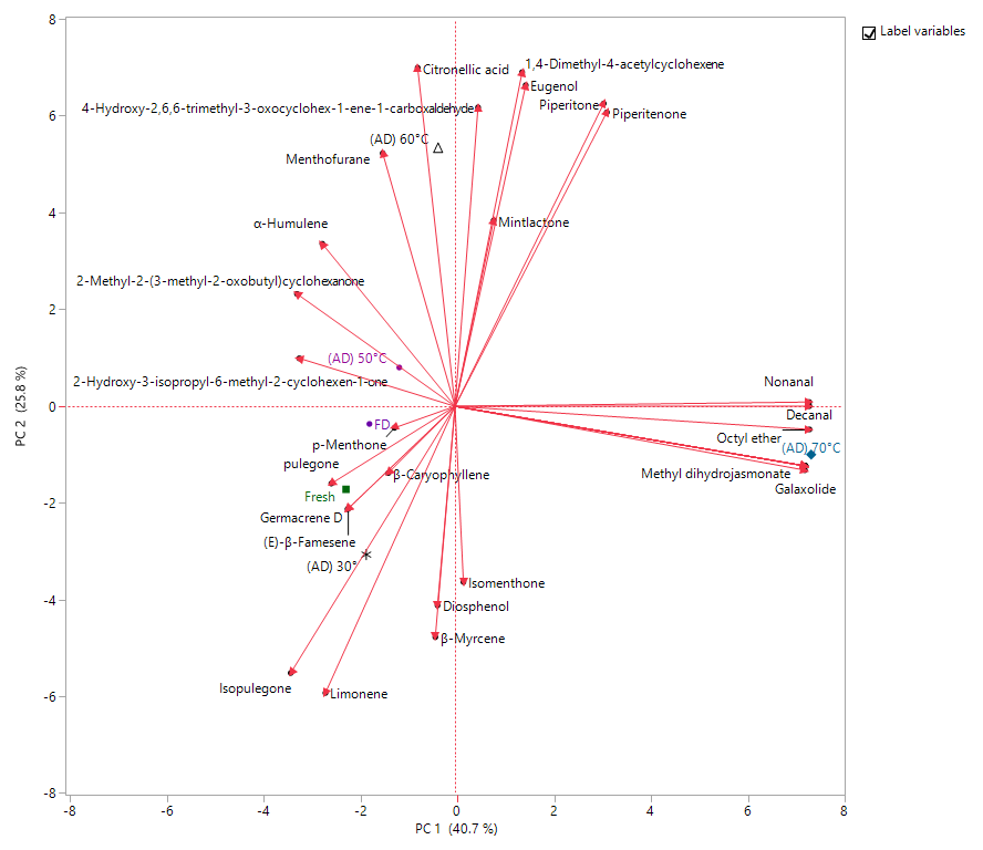
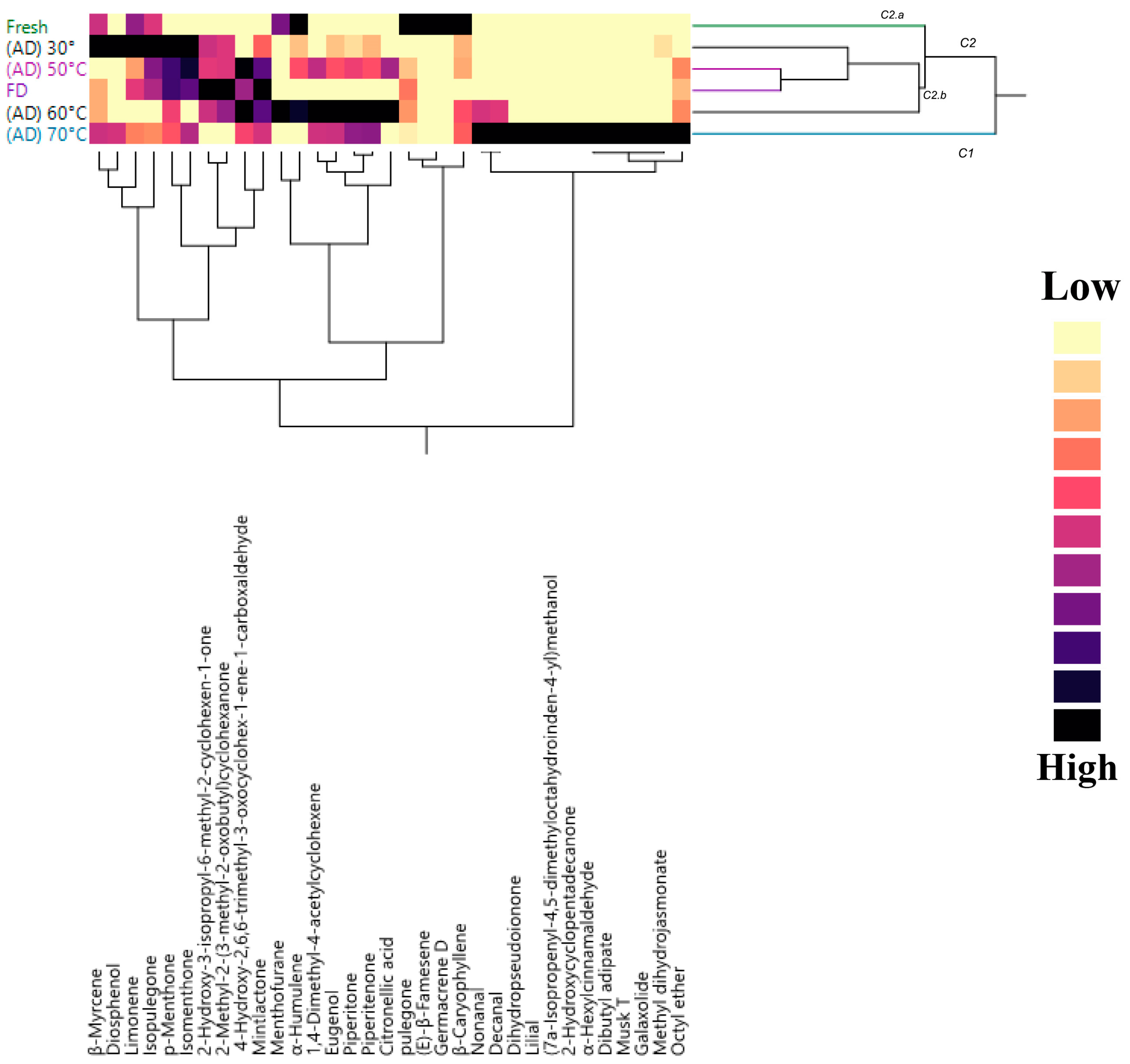
4. Volatilome Analyses

The chemical composition of volatiles present in fresh and treated flowers is reported in the Table 2. Overall, fifty-one compounds were identified with a total percentage of identification ranging from 97.5% (in FD samples) to 100% (in fresh flowers).
Oxygenated monoterpenes prevailed in all samples and they amounted for at least 67.1% in AD 70 °C to reach more than 90% in fresh flowers. In the fresh flowers, pulegone was the major compound (75.5%) followed by β-caryophyllene and menthone; the three compounds represented 90.3% of global identified fraction. Pulegone was a common compound among all samples where it dominated in AD 60 °C and AD 70 °C samples, while in the remaining ones was the second main constituent preceded only by menthone. Interesting to note that this latter compound was present in a much smaller amount in the fresh sample (5.2%) and increased exponentially to reach 50% of identified fraction in AD 30 °C. All flowers shared the other three constituents beside pulegone and menthone: limonene (ranged from 1.4% in AD 60 °C to 3.4% in AD 30 °C, respectively), isopulegone (from 1.3% in AD 60 °C to 3.4% in AD 30 °C, respectively). β-caryophyllene, initially the second main constituent in fresh samples, was detected in very low percentage with increasing the drying temperature (not exceeded 2% in AD 60 °C). This was an opposite behavior to that observed in the Thymus daenensis Celak, where β-caryophyllene increased enhancing the oven temperature [54]. The bivariate correlation in the cited compounds (Table S2) highlighted the absence of correlation between their amounts and the used temperature except for pulegone, which showed a statistically significant negative linear relationship (p < 0.05: greater the amount of pulegone is associated to lower temperature).
The fluctuation in the menthone and pulegone percentage may be due to their conversion into other compounds as mentioned by Asekun and coworkers [55]. They attributed the loss of these compounds (such as menthone, pulegone, and 1,8-cineole) in fresh and gently oven-dried samples of Mentha longifolia shoots to their vaporization or conversion into other compounds. Although drying, in general, may lead to changes in volatile composition and content, some temperatures allow better preservation of volatile constituents, whereas others may cause significant losses of volatiles.
Both freeze-drying and heat treatment with a temperature more or equal to 50 °C increased the non-terpene amount. Noteworthy is the presence of 4-hydroxy-2,6,6-trimethyl-3-oxo-1-cyclohexene-1-carboxaldehyde in most samples, except for fresh flowers and those treated with a low temperature (30 °C). The amount of this compound was around 5.0% in both AD 50 °C and AD 60 °C then to be halved in AD 70 °C, while in FD samples it amounted to 3.5% of the identified fraction. AD 70 °C flower was also characterized by the presence of good percentage of other non-terpenes such as octyl ether (6.6%) and methyl dihydrojasmonate (4.6%). These samples (AD 70 °C) were distinguished by the presence of galaxolide (3.2%), 2-hydroxycyclopentadecanone (1.8%), α-hexylcinnamaldehyde (1.0%), dibutyl adipate (1.0%), and musk T (1.0%). Even though 70 °C seems to induce the synthesis of these compounds, no correlation was observed between their percentages and the increasing of the temperature. The formation of new chemicals may take place [56] as shown in all treatments by Xing [57] and Díaz-Maroto [58]. This can be attributed to oxidations or to the breakdown of cell wall, or to the hydrolysis of glycosylated forms and thus to the release of several compounds. Concerning the flower volatile emission as a response to high temperature, no report was present in the literature. Only the presence of galaxoide was reported in the EO of Turkish Helichrysum plicatum subsp. polyphyllum and subsp. isauricum [59], while hedione naturally occurred in trace in several flowers [60]. AD 60 °C sample showed the highest number of identified compounds and it was noted by its high amount of eugenol (4.6%), piperitone (4.5%), 1,4-dimethyl-4-acetylcyclohexene (3.9%), mentofurane (3.3%), and piperitenone (2.1%). Only piperitone signaled a statistically positive correlation with the temperature effect (p < 0.05) (Table S2. The first two compounds of PCA analyses explained more than 66% of the variance (Figure 1).
PC1 (40.7%) axis was responsible for the segregation of the AD 70 °C flowers from the others due to its high amount in non-terpene compounds such as ester (methyl dihydrojasmonate (12)) and ether (galaxoide (17)). In the left high quadrant (negative PC1, positive PC2), AD 60 °C sample was placed in the borderline, while AD 50 °C almost overlapped PC 2 axis. In the opposite quadrant (negative plot in both axes) were gathered fresh with FD and AD 30 °C flowers; this behavior was especially due to the amount of isopulegone and limonene.
The two-way HCA analysis confirmed what was reported by PCA; in fact two main clusters were observed: C1, a homogeneous cluster formed by only AD 70 °C, and C2 englobed the rest of the samples. C2 is further divided into two subclusters: C2.a, homogeneous and included only fresh flowers, and C2.b, which on its own can be divided into 2 subgroups: one formed by AD 30 °C and the other one with FD and AD 50 °C. (Figure 2).
Only a few works on the spontaneous volatile emission from Agastache flowers were present in the literature and especially on the temperature effect and freeze-drying treatments. Both volatile organic compounds of fresh flowers and the EO composition of this studied species (Agastache aurantiaca (A. Gray) Lint & Epling) were reported by our team in a recent study [2], as well as only the essential oil composition of some species of this genus were previously investigated [30,61,62,63,64,65]. Drying method, a common technique to preserve the quality of herbs, impact directly in their quality [63]. Even though the eugenol was not present in both fresh and FD samples, this result confirmed what was reported by Ion [64], who underlined no statistical difference in its percentage among fresh and those processed by FD. The same authors concluded that although freeze-drying is one of the most recommended techniques for herb drying, significant changes can occur in the chemical composition of the EO from Ocimum basilicum and this result was confirmed by the present study. Of note, was the proximity between FD and AD 50 °C. The same observation was noted in the volatiles of tea flowers [65], where the authors deduced that the volatile from samples treated with 60 °C were close to those observed by the FD and thus recommended the AD method at 60 °C for 180 min. Başer and Buchbauer [66] claimed that monoterpenes vaporized more rapidly by increasing drying temperature, while Mashkani [54] reported that their contents significantly reduced by the increase of the oven temperature. This finding was in disagreement with the results reported herein where a very slight difference in the monoterpene percentage was observed in all samples except for AD 70 °C, where the difference was notable (a decrease of about 27%). Chua and coworkers [67] investigated the EO of Cassia alta, and showed the loss in volatile content after FD. They assumed that the loss could be due to the reduction of the pressure of drying chamber, although Antal reported in Mentha spicata that the reduction of pressure increased the release of volatile compounds [68]. This statement totally agrees with the results of the present work, where an increase of the number of identified fractions was noted in FD in comparison with fresh flowers.

5. Conclusions

Agastache aurantiaca is an appreciated ornamental plant for their color and aroma, and the flowers are consumed as food, therefore the conservation of their bioactive compounds and aromatic profile is crucial. The presented data showed that the freeze-drying technique is the best solution to prolong the shelf life of these flowers and to maintain high concentration of antioxidant compounds (e.g., phenolic compounds, flavonoids, anthocyanins, carotenoids). The air drying system at 30 °C is a time consuming method with a significant loss of sugars, probably due to the senescence process. The aromatic profile of different treated flowers showed the oxygenated monoterpene compounds as a major class. Pulegone is the main or one of the major constituents of all samples together with p-menthone. With the increasing of temperature, pulegone decreased and p-menthone increased.

Supplementary Materials

The following are available online at https://www.mdpi.com/article/10.3390/horticulturae7040083/s1. Table S1. Correlation between antioxidants content (total polyphenols, total flavonoids total anthocyanins, total carotenoids) in flowers of Agastache aurantiaca and their radical scavenger activity (DPPH-assay and FRAP-assay). Table S2: Bivariate correlation test in the main compounds.

Author Contributions

Conceptualization, L.P. (Laura Pistelli), L.P. (Luisa Pistelli), B.R.; methodology, I.M., B.N., R.D.; software, I.M., B.N.; validation, I.M., B.N., L.P. (Laura Pistelli) and L.P. (Luisa Pistelli); formal analysis, B.N., I.M., R.D.; investigation, B.N., I.M., R.D., G.G.; writing—original draft preparation, B.N., I.M.; writing—review and editing, I.M., B.N., B.R., L.P. (Laura Pistelli), L.P. (Luisa Pistelli); supervision, L.P. (Laura Pistelli), L.P. (Luisa Pistelli); project administration, B.R.; funding acquisition, B.R., L.P. (Laura Pistelli), L.P. (Luisa Pistelli). All authors have read and agreed to the published version of the manuscript.

Funding

“This research was funded by the INTERREG-ALCOTRA UE 2014-2020 Project “ANTEA”–Attività innovative per lo sviluppo della filiera transfrontaliera del fiore edule (n. 1139), grant number: CUP C12F17000080003.

Institutional Review Board Statement

Not applicable.

Informed Consent Statement

Not applicable.

Data Availability Statement

Data is contained within the article.

Acknowledgments

The authors thank Andrea Copetta, Sophie Descamps and Laurent Cambournac for the production and cultivation of plant material.

Conflicts of Interest

The authors declare no conflict of interest.

References

  1. Cunningham, E. What nutritional contribution do edible flowers make? J. Acad. Nutr. Diet. 2015, 115, 856. [Google Scholar] [CrossRef]
  2. Najar, B.; Marchioni, I.; Ruffoni, B.; Copetta, A.; Pistelli, L.; Pistelli, L. Volatilomic analysis of four edible flowers from Agastache Genus. Molecules 2019, 24, 4480. [Google Scholar] [CrossRef] [Green Version]
  3. Li, W.; Song, X.; Hua, Y.; Tao, J.; Zhou, C. Effects of different harvest times on nutritional component of herbaceous peony flower petals. J. Chem. 2020, 2020. [Google Scholar] [CrossRef]
  4. Drava, G.; Iobbi, V.; Govaerts, R.; Minganti, V.; Copetta, A.; Ruffoni, B.; Bisio, A. Trace elements in edible flowers from Italy: Further insights into health benefits and risks to consumers. Molecules 2020, 25, 2891. [Google Scholar] [CrossRef] [PubMed]
  5. Marchioni, I.; Najar, B.; Ruffoni, B.; Copetta, A.; Pistelli, L.; Pistelli, L. Bioactive compounds and aroma profile of some Lamiaceae edible flowers. Plants 2020, 9, 691. [Google Scholar] [CrossRef] [PubMed]
  6. Skrajda-Brdak, M.; Dąbrowski, G.; Konopka, I. Edible flowers, a source of valuable phytonutrients and their pro-healthy effects–A review. Trends Food Sci. Technol. 2020, 103, 179–199. [Google Scholar] [CrossRef]
  7. Mlcek, J.; Rop, O. Fresh edible flowers of ornamental plants–A new source of nutraceutical foods. Trends Food Sci. Technol. 2011, 22, 561–569. [Google Scholar] [CrossRef]
  8. Lucarini, M.; Copetta, A.; Durazzo, A.; Gabrielli, P.; Lombardi-Boccia, G.; Lupotto, E.; Santini, A.; Ruffoni, B. A Snapshot on Food Allergies: A Case Study on Edible Flowers. Sustainability 2020, 12, 8709. [Google Scholar] [CrossRef]
  9. Bourgaud, F.; Gravot, A.; Milesi, S.; Gontier, E. Production of plant secondary metabolites: A historical perspective. Plant Sci. 2001, 161, 839–851. [Google Scholar] [CrossRef]
  10. Yang, L.; Wen, K.S.; Ruan, X.; Zhao, Y.X.; Wei, F.; Wang, Q. Response of plant secondary metabolites to environmental factors. Molecules 2018, 23, 762. [Google Scholar] [CrossRef] [Green Version]
  11. Pires, T.C.; Barros, L.; Santos-Buelga, C.; Ferreira, I.C. Edible flowers: Emerging components in the diet. Trends Food Sci. Technol. 2019, 93, 244–258. [Google Scholar] [CrossRef]
  12. Lu, B.; Li, M.; Yin, R. Phytochemical content, health benefits, and toxicology of common edible flowers: A review (2000–2015). Crit. Rev. Food Sci. Nutr. 2016, 56, S130–S148. [Google Scholar] [CrossRef]
  13. Fernandes, L.; Casal, S.; Pereira, J.A.; Saraiva, J.A.; Ramalhosa, E. Edible flowers: A review of the nutritional, antioxidant, antimicrobial properties and effects on human health. J. Food Compos. Anal. 2017, 60, 38–50. [Google Scholar] [CrossRef]
  14. Grzeszczuk, M.; Stefaniak, A.; Meller, E.; Wysocka, G. Mineral composition of some edible flowers. J. Elem. 2018, 23, 151–162. [Google Scholar] [CrossRef]
  15. Takahashi, J.A.; Rezende, F.A.G.G.; Moura, M.A.F.; Dominguete, L.C.B.; Sande, D. Edible flowers: Bioactive profile and its potential to be used in food development. Food Res. Int. 2020, 129, 108868. [Google Scholar] [CrossRef]
  16. Khoo, H.E.; Azlan, A.; Tang, S.T.; Lim, S.M. Anthocyanidins and anthocyanins: Colored pigments as food, pharmaceutical ingredients, and the potential health benefits. Food Nutr. Res. 2017, 61, 1361779. [Google Scholar] [CrossRef] [Green Version]
  17. Zhu, C.; Bai, C.; Sanahuja, G.; Yuan, D.; Farré, G.; Naqvi, S.; Shi, L.; Capell, T.; Christou, P. The regulation of carotenoid pigmentation in flowers. Arch. Biochem. Biophys. 2010, 504, 132–141. [Google Scholar] [CrossRef] [PubMed]
  18. Hallmann, E. Quantitative and Qualitative Identification of Bioactive Compounds in Edible Flowers of Black and Bristly Locust and Their Antioxidant Activity. Biomolecules 2020, 10, 1603. [Google Scholar] [CrossRef]
  19. Landi, M.; Ruffoni, B.; Combournac, L.; Guidi, L. Nutraceutical value of edible flowers upon cold storage. Ital. J. Food Sci. 2017, 30, 1–18. [Google Scholar] [CrossRef]
  20. Fernandes, L.; Casal, S.; Pereira, J.A.; Malheiro, R.; Rodrigues, N.; Saraiva, J.A.; Ramalhosa, E. Borage, calendula, cosmos, Johnny Jump up, and pansy flowers: Volatiles, bioactive compounds, and sensory perception. Eur. Food Res. Technol. 2019, 245, 593–606. [Google Scholar] [CrossRef] [Green Version]
  21. Marchioni, I.; Pistelli, L.; Ferri, B.; Copetta, A.; Ruffoni, B.; Pistelli, L.; Najar, B. Phytonutritional content and aroma profile changes during postharvest storage of edible flowers. Front. Plant Sci. 2020, 11, 590968. [Google Scholar] [CrossRef] [PubMed]
  22. Orphanides, A.; Goulas, V.; Gekas, V. Drying technologies: Vehicle to high-quality herbs. Food Eng. Rev. 2016, 8, 164–180. [Google Scholar] [CrossRef]
  23. Pires, T.C.; Dias, M.I.; Barros, L.; Ferreira, I.C. Nutritional and chemical characterization of edible petals and corresponding infusions: Valorization as new food ingredients. Food Chem. 2017, 220, 337–343. [Google Scholar] [CrossRef] [Green Version]
  24. Garcìa, L.M.; Ceccanti, C.; Negro, C.; De Bellis, L.; Incrocci, L.; Pardossi, A.; Guidi, L. Effect of Drying Methods on Phenolic Compounds and Antioxidant Activity of Urtica dioica L. Leaves. Horticulturae 2021, 7, 10. [Google Scholar] [CrossRef]
  25. Zielińska, S.; Matkowski, A. Phytochemistry and bioactivity of aromatic and medicinal plants from the genus Agastache (Lamiaceae). Phytochem. Rev. 2014, 13, 391–416. [Google Scholar] [CrossRef] [Green Version]
  26. Duda, M.M.; Varban, D.I.; Muntean, S.; Moldovan, C.; Olar, M. Use of species Agastache foeniculum (Pursh) Kuntze. Hop Med. Plants 2014, 21, 52–54. [Google Scholar]
  27. Husti, A.; Cantor, M.; Buta, E.; Horţ, D. Current trends of using ornamental plants in culinary arts. ProEnvironment 2013, 6, 52–58. [Google Scholar]
  28. Myadelets, M.A.; Vorobyeva, T.A.; Domrachev, D.V. Composition of the essential oils of some species belonging to genus Agastache Clayton ex Gronov (Lamiaceae) cultivated under the conditions of the middle Ural. Chem. Sustain. Dev. 2013, 21, 397–401. [Google Scholar]
  29. Sanders, R.W. Taxonomy of Agastache section Brittonastrum (Lamiaceae-Nepeteae). Syst. Botany Monogr. 1987, 15, 1–92. [Google Scholar] [CrossRef]
  30. Kovalenko, N.A.; Supichenko, G.N.; Leontiev, V.N.; Shutova, A.G. Composition of essential oil of plants some species of the genus Agastache l. Introduced in Belarus. Proceedings of the National Academy of Sciences of Belarus. Biol. Ser. 2019, 64, 147–155. [Google Scholar] [CrossRef]
  31. Exner, J.; Ulubelen, A.; Mabry, T.J. Chemistry of Agastache. Part II. Flavonoids of Agastache aurantiaca. Rev. Latinoamer. Quim. 1981, 12, 37–38. [Google Scholar]
  32. Saito, N.; Harborne, J.B. Correlations between anthocyanin type, pollinator and flower color in the Labiatae. Phytochemistry 1992, 31, 3009–3015. [Google Scholar] [CrossRef]
  33. Holloway, P.S.; Matheke, G.E.; Hanscom, J.; Gardiner, A.; Hill, V.; Van Wyhe, E. Annual Flower Evaluations 2003; University of Alaska Fairbanks, Agricultural and Forestry Experiment Station, Georgeson Botanical Garden: Sitka, AK, USA, 2003; pp. 1–37. [Google Scholar]
  34. Lichtenthaler, H.K. Chlorophylls and carotenoids: Pigments of photosynthetic biomembranes. Methods Enzymol. 1987, 148, 350–382. [Google Scholar]
  35. Singleton, V.L.; Rossi, J.A. Colorimetry of total phenolics with phosphomolybdic-phosphotungstic acid reagents. Am. J. Enol. Viticult. 1965, 16, 144–158. [Google Scholar]
  36. Kim, D.; Jeong, S.; Lee, C. Antioxidant capacity of phenolic phytochemicals from various cultivars of plums. Food Chem. 2003, 81, 321–326. [Google Scholar] [CrossRef]
  37. Lee, J.; Durst, R.W.; Wrolstad, R.E.; Eisele, T.; Giusti, M.M.; Hach, J.; Hofsommer, H.; Koswig, S.; Krueger, D.A.; Kupina, S.; et al. Determination of total monomeric anthocyanin pigment content of fruit juices, beverages, natural colorants, and wines by the pH differential method: Collaborative study. J. AOAC Int. 2005, 88, 1269–1278. [Google Scholar] [CrossRef] [PubMed] [Green Version]
  38. Giusti, M.; Wrolstad, R.E. Characterization and Measurement of Anthocyanins by UV-Visible Spectroscopy. Curr. Protoc. Food Anal. Chem. 2001, F1.2.1–F1.2.13. [Google Scholar] [CrossRef]
  39. Brand-Williams, W.; Cuvelier, M.E.; Berset, C.L.W.T. Use of a free radical method to evaluate antioxidant activity. LWT Food Sci. Technol. 1995, 28, 25–30. [Google Scholar] [CrossRef]
  40. Szôllôsi, R.; Szôllôsi Varga, I. Total antioxidant power in some species of Labiatae (adaptation of FRAP method). Acta Biol. Szeged. 2002, 46, 125–127. [Google Scholar]
  41. Das, B.; Choudhury, B.; Kar, M. Quantitative estimation of changes in biochemical constituents of mahua (madhuca indica syn. bassia latifolia) flowers during postharvest storage. J. Food Process. Preserv. 2010, 34, 831–844. [Google Scholar] [CrossRef]
  42. Najar, B.; Nardi, V.; Cervelli, C.; Pistelli, L. Volatilome analyses of four South African Helichrysum spp. grown in Italy. Nat. Prod. Res. 2020. [Google Scholar] [CrossRef]
  43. Choi, S.W.; Park, J.H.; Lee, I.B. Process monitoring using a Gaussian mixture model via principal component analysis and discriminant analysis. Comput. Chem. Eng. 2004, 28, 1377–1387. [Google Scholar] [CrossRef]
  44. Yeo, H.J.; Park, C.H.; Park, Y.E.; Hyeon, H.; Kim, J.K.; Lee, S.Y.; Park, S.U. Metabolic profiling and antioxidant activity during flower development in Agastache rugosa. Physiol. Mol. Biol. Plants 2021, 27, 445–455. [Google Scholar] [CrossRef]
  45. Barani, Y.H.; Zhang, M.; Wang, B.; Devahastin, S. Influences of four pretreatments on anthocyanins content, color and flavor characteristics of hot-air dried rose flower. Drying Technol. 2020, 38, 1988–1995. [Google Scholar] [CrossRef]
  46. Xu, K.; Zhang, M.; Fang, Z.; Wang, B. Degradation and regulation of edible flower pigments under thermal processing: A review. CRC Rev. Food Sci. 2021, 61, 1038–1048. [Google Scholar] [CrossRef]
  47. Zhao, L.; Fan, H.; Zhang, M.; Chitrakar, B.; Bhandari, B.; Wang, B. Edible flowers: Review of flower processing and extraction of bioactive compounds by novel technologies. Food Res. Int. 2019, 126, 108660. [Google Scholar] [CrossRef] [PubMed]
  48. Lončarić, M.; Strelec, I.; Moslavac, T.; Šubarić, D.; Pavić, V.; Molnar, M. Lipoxygenase Inhibition by Plant Extracts. Biomolecules 2021, 11, 152. [Google Scholar] [CrossRef]
  49. Vamos-Vigyàzò, L. Polyphenol oxidase and peroxidase infruits and vegetables. CRC Rev. Food Sci. 1981, 15, 49–127. [Google Scholar] [CrossRef]
  50. Severini, C.; Baiano, A.; De Pilli, T.; Romaniello, R.; Derossi, A. Prevention of enzymatic browning in sliced potatoes by blanching in boiling saline solutions. LWT Food Sci. Technol. 2003, 36, 657–665. [Google Scholar] [CrossRef]
  51. Olley, C.M.; Joyce, D.C.; Irving, D.E. Changes in sugar, protein, respiration, and ethylene in developing and harvested Geraldton waxflower (Chamelaucium uncinatum) flowers. N. Z. J. Crop Hortic. 1996, 24, 143–150. [Google Scholar] [CrossRef] [Green Version]
  52. Rogers, H.J. From models to ornamentals: How is flower senescence regulated? Plant Mol. Biol. 2013, 82, 563–574. [Google Scholar] [CrossRef]
  53. Yangkhamman, P.; Tanase, K.; Ichimura, K.; Fukai, S. Depression of enzyme activities and gene expression of ACC synthase and ACC oxidase in cut carnation flowers under high-temperature conditions. Plant Growth Regul. 2007, 53, 155–162. [Google Scholar] [CrossRef]
  54. Mashkani, M.R.D.; Larijani, K.; Mehrafarin, A.; Badi, H.N. Changes in the essential oil content and composition of Thymus daenensis Celak. under different drying methods. Ind. Crops Prod. 2018, 112, 389–395. [Google Scholar] [CrossRef]
  55. Asekun, O.T.; Grierson, D.S.; Afolayan, A.J. Characterization of essential oils from Helichrysum odoratissimum using different drying methods. J. Appl. Sci. 2007, 7, 1005–1008. [Google Scholar] [CrossRef]
  56. Chua, L.Y.W.; Chong, C.H.; Chua, B.L.; Figiel, A. Influence of Drying Methods on the Antibacterial, Antioxidant and Essential Oil Volatile Composition of Herbs: A Review. Food Bioprocess Technol. 2019, 12, 450–476. [Google Scholar] [CrossRef]
  57. Xing, Y.; Lei, H.; Wang, J.; Wang, Y.; Wang, J.; Xu, H. Effects of different drying methods on the total phenolic, rosmarinic acid and essential oil of purple perilla leaves. J. Essent. Oil-Bearing Plants 2017, 20, 1594–1606. [Google Scholar] [CrossRef]
  58. Díaz-Maroto, M.C.; Palomo, E.S.; Castro, L.; González Viñas, M.A.; Pérez-Coello, M.S. Changes produced in the aroma compounds and structural integrity of basil (Ocimum basilicum L) during drying. J. Sci. Food Agric. 2004, 84, 2070–2076. [Google Scholar] [CrossRef]
  59. Öztürk, B.; Özek, G.; Özek, T.; Baser, K.H.C. Chemical diversity in volatiles of Helichrysum plicatum DC. subspecies in Turkey. Rec. Nat. Prod. 2014, 8, 373–384. [Google Scholar]
  60. Chapuis, C. The Jubilee of methyl jasmonate and Hedione. Helv. Chim. Acta 2012, 95, 1479–1511. [Google Scholar] [CrossRef]
  61. Kovalenko, N.N.; Supichenko, G.N.; Akhramovich, T.T.; Shutova, A.G.; Léontiev, V.N. Antibacterial activity of essential oil of Agastache aurantiaca. Chem. Plant Raw Mater. 2018, 2, 63–70. [Google Scholar] [CrossRef]
  62. Yamani, H.; Mantri, N.; Morrison, P.D.; Pang, E. Analysis of the volatile organic compounds from leaves, flower spikes, and nectar of Australian grown Agastache rugosa. BMC Complement. Altern. Med. 2014, 14, 1–6. [Google Scholar] [CrossRef] [PubMed] [Green Version]
  63. Pop, A.; Muste, S.; Păucean, A.; Chiș, M.S.; Man, S.; Salanță, L.; Romina Marc, G.M. A Review of the Drying and Storage Effect on Some Aromatic and Medicinal Plants. Hop Med. Plants 2020, 28, 142–149. [Google Scholar]
  64. Ion, V.A.; Nicolau, F.; Petre, A.; Bujor, O. Variation of bioactive compounds in organic Ocimum basilicum L. during freeze-drying processing. Sci. Papers Ser. B Hortic. 2020, LXIV, 397–404. [Google Scholar]
  65. Shi, L.; Gu, Y.; Wu, D.; Wu, X.; Grierson, D.; Tu, Y.; Wu, Y. Hot air drying of tea flowers: Effect of experimental temperatures on drying kinetics, bioactive compounds and quality attributes. Int. J. Food Sci. Technol. 2019, 54, 526–535. [Google Scholar] [CrossRef]
  66. Başer, K.H.C.; Buchbauer, G. Handbook of Essential Oils: Science, Technology, and Applications, 2nd ed.; CRC Press Taylor Francis Group: Boca Raton, FL, USA, 2015. [Google Scholar] [CrossRef]
  67. Wen Chua, L.Y.; Chua, B.L.; Figiel, A.; Chong, C.H.; Wojdyło, A.; Szumny, A.; Lech, K. Characterisation of the convective hot-air drying and vacuum microwave drying of Cassia alata: Antioxidant activity, essential oil volatile composition and quality studies. Molecules 2019, 24, 1625. [Google Scholar] [CrossRef] [Green Version]
  68. Antal, T.; Figiel, A.; Kerekes, B.; Sikolya, L. Effect of drying methods on the quality of the essential oil of spearmint leaves (Mentha spicata L.). Dry. Technol. 2011, 29, 1836–1844. [Google Scholar] [CrossRef]
Figure 1. Scatter plot of the principal component analysis (PCA) of the volatile organic compounds (VOCs) of A. aurantiaca ‘Apricot Sprite’ flowers. Horticulturae 07 00083 i001 Fresh Flowers, Horticulturae 07 00083 i002 Freeze Drying (FD), Horticulturae 07 00083 i003 (AD) 30°C, Horticulturae 07 00083 i004 (AD) 50 °C, Horticulturae 07 00083 i005 (AD) 70 °C.
Horticulturae 07 00083 g001
Figure 2. Dendrogram of cluster hierarchical analysis performed on VOCs from A. aurantiaca ‘Apricot Sprite’ flowers.
Horticulturae 07 00083 g002
Table 1. Determination of metabolites in A. aurantiaca flowers subjected to different treatments. Data are presented as means ± standard error (SE, n = 3). Abbreviations: FW = fresh weight; DW = dry weight; GAE—gallic acid equivalents; CE—± catechin equivalents; C3G—cyanidin-3-O-glucoside equivalents; GLU—glucose. sig.= significant post hoc test at p < 0.05.
Fresh
Flowers 1
Freeze-Drying (FD)Air Dried
(AD) 30 °C
Air Dried
(AD) 50 °C
Air Dried
(AD) 60 °C
Air Dried
(AD) 70 °C
Total Carotenoids
µg/g DW
125.93 ± 6.73222.49 ± 4.88 a177.83 ± 2.64 c86.77 ± 0.46 e187.07 ± 3.68 b146.05 ± 1.8 d
Total Anthocyanins
µg C3G/g DW
55.80 ± 2.51366.65 ± 28.32 a185.31 ± 42.2 b360.96 ± 58.65 ab422.56 ± 15.91 a245.7 ± 19.74 b
Total phenolics (TPC)
mg GAE/g DW
3.33 ± 0.1232.58 ± 2.01 ab34.26 ± 1.38 a24.60 ± 1.40 b25.84 ± 1.90 b26.07 ± 1.38 b
Total flavonoids (TFC)
mg CE/g DW
1.768 ± 0.0329.77 ± 0.58 a30.93 ± 0.56 a22.66 ± 0.67 b23.35 ± 0.81 b22.62 ± 0.21 b
Total soluble sugars (TSS) mg GLU/g DW26.12 ± 0.37358.91 ± 5.55 c212.12 ± 2.96 d512.18 ± 2.68 a461.31 ± 4.11 b341.99 ± 4.77 c
FRAP activity
µmolFeSO4/g DW
19.33 ± 0.07403.4 ± 21.78 ab458.79 ± 12.88 a281.75 ± 17.62 c306.84 ± 24.77 c318.53 ± 4.63 bc
DPPH activity
mg TEAC/g DW
1.49 ± 0.0319.4 ± 0.48 b23.26 ± 0.52 a14.87 ± 0.05 c17.88 ± 0.81 b17.67 ± 0.01 b
1 fresh flower’s data are reported as g FW−1 instead of g DW−1.
Table 2. Chemical composition of the volatile organic compounds (VOCs) from A. aurantiaca ‘Apricot Sprite’ flowers. Headspace–Solid Phase Micro-Extraction (HS-SPME) was performed on Gas Chromatography–Mass Spectrometry (GC–MS) with DB-5 capillary column. Data represent mean values of relative percentage (n = 3, ±SD).
CompoundsClassLRIFresh(AD) 30 °C(AD) 50 °C(AD) 60 °C(AD) 70 °CFD
13-Methylcyclohexanonent9580.1 ± 0.010.2 ± 0.03
2Sabinenemh9740.1 ± 0.01
31-Octen-3-olnt9800.2 ± 0.010.1 ± 0.00
43-Octanonent9860.2 ± 0.010.3 ± 0.08
56-Methyl-5-heptene-2-onent9860.2 ± 0.05
6β-Myrcenemh9910.6 ± 0.020.9 ± 0.140.1 ± 0.020.3 ± 0.070.1 ± 0.01
7Limonenemh10302.5 ± 0.033.4 ± 0.401.7 ± 0.041.4 ± 0.481.8 ± 0.932.1 ± 0.48
8Benzyl alcoholnt10360.4 ± 0.25
9Nonanalnt11040.1 ± 0.010.5 ± 0.16
10p-Menthoneom11545.2 ± 0.0650.0 ± 0.3943.3 ± 0.8928.9 ± 1.2725.7 ± 0.8843.4 ± 2.09
11Isomenthoneom11644.8 ± 0.304.4 ± 0.112.9 ± 0.133.7 ± 0.13
12Menthofuraneom11651.7 ± 0.603.3 ± 0.44
13Isopulegoneom11772.3 ± 0.293.4 ± 0.172.7 ± 0.031.3 ± 0.781.8 ± 0.092.5 ± 0.14
14Verbenoneom12050.2 ± 0.020.3 ± 0.130.3 ± 0.05 0.3 ± 0.08
15Decanalnt12060.3 ± 0.071.6 ± 0.62
16(−)-trans-Isopiperitenolom12100.10 ± 0.03
171,4-Dimethyl-4-acetylcyclohexenent12261.3 ± 0.113.9 ± 0.971.1 ± 0.13
182-Hydroxycineoleom12280.2 ± 0.03
19cis-Pulegone Oxideom12300.3 ± 0.030.2 ± 0.05
20Pulegoneom123775.5 ± 1.8530.2 ± 0.2332.6 ± 0.0134.5 ± 1.6630.8 ± 0.5335.8 ± 1.39
21Piperitoneom12530.2 ± 0.001.2 ± 0.064.5 ± 0.552.3 ± 0.16
222-Hydroxy-3-isopropyl-6-methyl-2-cyclohexen-1-onent12740.8 ± 0.010.7 ± 0.010.8 ± 0.06 2.2 ± 0.00
231-Cyclohexene-1-carboxaldehyde, 4-hydroxy-2,6,6-trimethyl-3-oxo-nt13025.6 ± 0.045.7 ± 0.732.4 ± 0.423.5 ± 0.12
242-Hydroxypiperitoneom13022.0 ± 0.060.4 ± 0.04
25Undecanalnt13070.2 ± 0.05
262-Methyl-2-(3-methyl-2-oxobutyl)cyclohexanonent13090.4 ± 0.020.4 ± 0.040.6 ± 0.091.1 ± 0.07
27Citronellic acid)nt13140.3 ± 0.040.8 ± 0.06
28Piperitenoneom13400.2 ± 0.010.5 ± 0.032.1 ± 0.171.1 ± 0.05
29Menthofurolactoneom13540.1 ± 0.08
30Eugenolpp13570.3 ± 0.030.9 ± 0.044.6 ± 0.421.3 ± 0.04
311,2-Dimethyl-1-cyclodecenent13600.3 ± 0.04
328-Methyl-6-nonenoic acidnt13730.1 ± 0.04
33cis-trans-Nepetalactoneom13770.1 ± 0.07
34β-Caryophyllenesh14199.6 ± 1.510.9 ± 0.121.0 ± 0.082.0 ± 0.311.8 ± 0.160.1 ± 0.06
35Dihydropseudoiononent14560.7 ± 0.03
36α-Humulenesh14561.0 ± 0.320.1 ± 0.010.3 ± 0.010.9 ± 0.11
37(E)-β-Famesenesh14570.6 ± 0.32
382-Hydroxy-4,5-dimethylacetophenonent14760.3 ± 0.01
39Germacrene Dsh14811.0 ± 0.71
40Mintlactoneom15000.4 ± 0.040.8 ± 0.060.8 ± 0.040.6 ± 0.031.0 ± 0.18
41Isomintlactoneom15310.1 ± 0.010.2 ± 0.000.1 ± 0.08-0.4 ± 0.09
42Lilialpp15340.7 ± 0.16
43Caryophyllene oxideos15810.10 ± 0.010.1 ± 0.030.10.01
44Hedionent16490.1 ± 0.014.6 ± 0.59
45(7a-Isopropenyl-4,5-dimethyloctahydroinden-4-yl)methanolnt16590.7 ± 0.16
46Octyl ethernt16590.7 ± 0.350.7 ± 0.086.6 ± 0.170.4 ± 0.05
47α-Hexylcinnamaldehydepp17501.0 ± 0.10
48Dibutyl adipatent17661.0 ± 0.13
49Dodecahydro-3a,6,6,9a-tetramethylnaphtho[2, 1-β]furanos17660.2 ± 0.03
502-Hydroxycyclopentadecanonent18461.8 ± 0.25
51Galaxolident18513.2 ± 0.71
52Musk Tos19891.0 ± 0.07
Number of identified compounds 102321302919
Class of compounds Fresh(AD) 30 °C(AD) 50 °C(AD) 60 °C(AD) 70 °CFD
Monoterpene hydrocarbons (mh) 3.1 ± 0.054.4 ± 0.571.7 ± 0.041.5 ± 0.362.1 ± 0.132.2 ± 0.37
Oxygenated monoterpenes (om) 84.7 ± 2.8091.5 ± 0.4088.5 ± 0.8176.1 ± 1.6867.1 ± 0.5587.3 ± 0.50
Sesquiterpene hydrocarbons (sh) 12.2 ± 2.861.0 ± 0.131.3 ± 0.102.9 ± 0.421.8 ± 0.160.1 ± 0.01
Oxygenated sesquiterpenes (os) 0.1 ± 0.010.1 ± 0.001.2 ± 0.160.1 ± 0.07
Phenylpropanoids (pp) 0.3 ± 0.030.9 ± 0.044.6 ± 0.421.7 ± 0.25
Non-terpene derivatives (nt) 1.8 ± 0.048.0 ± 0.4813.9 ± 1.9724.4 ± 0.547.8 ± 0.68
Total identified 100 ± 0.0099.1 ± 0.3399.5 ± 0.2799.1 ± 0.6298.3 ± 0.7497.5 ± 0.37
Publisher’s Note: MDPI stays neutral with regard to jurisdictional claims in published maps and institutional affiliations.

