Article

The Bacterial Soft Rot Pathogens, Pectobacterium carotovorum and P. atrosepticum, Respond to Different Classes of Virulence-Inducing Host Chemical Signals

1
Department of Agricultural and Environmental Sciences, 3500 John A. Merritt Blvd, Campus Box 9543, Tennessee State University, Nashville, TN 37209, USA
2
Department of Biological, Physical, and Human Sciences, Freed-Hardeman University, Henderson, TN 38340, USA
*
Author to whom correspondence should be addressed.
Present Address: Research Department, Quality Control Company (Ghana Cocoa Board), P.O. Box 247 Tema, Ghana.
Horticulturae 2020, 6(1), 13; https://doi.org/10.3390/horticulturae6010013
Submission received: 21 November 2019 / Revised: 23 January 2020 / Accepted: 30 January 2020 / Published: 10 February 2020
(This article belongs to the Special Issue Postharvest Pathogens and Disease Management of Horticultural Crops)

Abstract

:
Soft rot bacteria of the Pectobacterium and Dickeya genera are Gram-negative phytopathogens that produce and secrete plant cell wall-degrading enzymes (PCWDE), the actions of which lead to rotting and decay of their hosts in the field and in storage. Host chemical signals are among the factors that induce the bacteria into extracellular enzyme production and virulence. A class of compounds (Class I) made up of intermediate products of cell wall (pectin) degradation induce exoenzyme synthesis through KdgR, a global negative regulator of exoenzyme production. While the KdgR mutant of P. carotovorum is no longer inducible by Class I inducers, we demonstrated that exoenzyme production is induced in this strain in the presence of extracts from hosts including celery, potato, carrot, and tomato, suggesting that host plants contain another class of compounds (Class II inducers) different from the plant cell wall-degradative products that work through KdgR. The Class II inducers are thermostable, water-soluble, diffusible, and dialysable through 1 kDa molecular weight cut off pore size membranes, and could be a target for soft rot disease management strategies.

1. Introduction

Pectobacterium species are Gram-negative plant pathogenic bacteria belonging to the family Pectobacteriaceae. Together with Dickeya species, these bacteria form the major pathogenic agents of diseases such as blackleg, aerial stem rot, and soft rot in potato, and vegetables such as celery, carrot, tomato, and a number of crops in the field and in storage worldwide [1,2,3]. The virulence and pathogenicity of these bacteria rely on their ability to produce and secret copious amounts of a wide range of extracellular plant cell wall-degrading enzymes (PCWDE, exoenzymes) including pectate lyases (Pel), polygalacturonases (Peh), proteases (Prt), and cellulases (Cel), which cause extensive tissue maceration, rotting, and subsequent death of the whole plant [4,5].
Exoenzyme production and pathogenicity in soft rot Enterobacteriaceae are coordinately regulated by transcriptional and posttranscriptional mechanisms involving a complex network of positive and negative regulatory proteins. This regulation, in turn, is influenced by physiological and environmental factors including chemical signals produced by both the pathogen and the host [6,7,8,9,10,11,12,13,14,15].
An example of a chemical signal produced by the bacteria—that are required for pathogenesis with a profound effect on the expression of virulence—is the diffusible quorum-sensing signal molecules N-acyl homoserine lactones (AHLs) [16,17,18]. Coupled with the posttranscriptional Rsm regulatory system in Pectobacterium, the quorum-sensing signal functions mostly as an antagonist to ExpR1 and ExpR2, which are powerful transcriptional activators of rsmA (homologue of E. coli csrA). RsmA acts by promoting the degradation of target transcripts such as those for cell wall-degrading enzymes [19]. The interaction of AHL molecule with ExpR proteins inactivates the proteins and stops the transcriptional activation of rsmA. The quorum-sensing system has such a strong control on virulence that AHL nonproducing mutants are completely attenuated in virulence [11,17,18,20], and the expression of AHL-degrading enzyme in the host completely protects the host against infection by wild-type AHL-producing strains [21]. In P. atrosepticum SCRI 1043, as much as 26% of the genes are under quorum-sensing control [22].
Pectin, specifically, its demethylated derivative, polygalacturonic acid (PGA or pectate) and its degradation intermediates, also induce exoenzyme production in Pectobacterium and Dickeya species [23,24,25]. The pectic degradative derivatives, 2-keto-3-deoxygluconate (KDG), 2, 5-diketo-3-deoxygluconate (DKII), and 5-keto-4-deoxyuronate (DKI) are the specific intracellular inducers of pectin and galacturonate catabolism [26]. KdgR is a repressor protein that negatively regulates the expression of genes involved in pectin and galacturonate catabolism [7,27] by binding to the operators of the genes it regulates [28]. In the presence of PGA or its degradation intermediates (we have designated these as Class I inducers), KdgR is disengaged from the respective operators and binds these inducers instead. This derepression allows the transcription to occur of the previously repressed genes and the subsequent production of exoenzymes, leading to host tissue maceration. KdgR binding sequences have been identified in the upstream regulatory regions of genes for exoenzymes and the regulatory RsmB RNA [27,28].
Besides pectate and its catabolite intermediates (Class I inducers), extracts from potato, celery, and carrot, or plant-derived compounds such as o-coumaric acid and trans-cinnamic acid, also regulate the expression of virulence genes in Pectobacterium and Dickeya species [23,24,29,30,31,32,33]. Due to their ability to induce Pel isozymes which are not inducible by pectate catabolism intermediates, the extracts from Chrysanthemum were thought to contain a unique class of chemical inducers [34]. The chemicals in these plant extracts could thus form another class of inducers of exoenzymes and virulence (we call these Class II inducers). There have been previous attempts to identify the inducer from carrots [24] and potato [34], but generally, not enough research attention has been given to this class of inducers and the bacterial factors that mediate their effect. For example, the questions of whether the compound(s) from different hosts are the same, and whether they induce exoenzyme synthesis through KdgR, the regulator that mediates the action of Class I inducers, have not been addressed. The response of a kdgR mutant to plant extracts containing these inducers should give insight into the nature of this interaction, as well as provide evidence for or against the existence of a separate class of compounds (Class II) that induce exoenzyme production through a mechanism which is possibly different from that of Class I inducers.
The goal of this study was to investigate the interaction between Class II inducers and Pectobacterium species by determining if the Class II inducers work through KdgR, and to further characterize the nature and properties of these host molecules. We report here the nature of the response of the soft rot pathogens P. carotovorum and P. atrosepticum to Class II inducers from a range of plant hosts, and show that, through their action in KdgR strains of Pectobacterium carotovorum, Class I and Class II inducers are not the same. We also present some general physical and chemical properties of Class II inducers.

2. Materials and Methods

2.1. Bacterial Strains, Plasmids, Media and Growth Conditions

Strains Ecc71 and SCRI1043 are wild-types of P. carotovorum and P. atrosepticum, respectively, and have been described previously [35,36]. The strain AC5073 is a kdgR mutant of Ecc71 [27,37] and KD100 is spontaneous nalidixic acid resistant derivative of AC5006 [38]. The pel1-lacZ fusion plasmid pAKC1031 [27] was used to monitor transcription from the pel1 promoter by β-galactosidase activity.
Cultures of Pectobacterium strains were grown in minimal salts plus sucrose medium (MM) with or without plant extracts or polygalacturonic acid (PGA) at 28 °C or in Luria-Bertani medium (LB). Unless otherwise indicated, chemical reagents were obtained from Fisher Scientific Co (Suwannee, GA, USA). Polygalacturonic acid was obtained from Pfaultz and Bauer (Waterbury, CT). The components of MM have been described [39] and were,: 2 g/L KH2PO4; 7 g/L K2HPO4; 0.1 g/L MgSO4.7H2O; 1 g/L (NH4)2SO4; and 0.2% (w/v) sucrose. Minimal media were supplemented with host extract or PGA (MM+PGA; 0.2% (v/v) as needed. Where necessary, media were also supplemented with the following; 50 µg/mL 5-bromo-4-chloro-3-indolyl-β-D-galacto-pyranoside (X-Gal); 50 µg/mL kanamycin (Km); 50 µg/mL nalidixic (Nal); 100 µg/mL spectinomycin (Sp); and 10 µg/mL tetracycline (Tc). Solid media were solidified by the addition of 1.5% (w/v) agar. Culture media were inoculated with bacterial suspensions prepared from overnight agar cultures (A600 = 0.1) and grown with shaking at 200 rpm for 14–16 h in test tubes or to a specified Klett unit in Klett culture flasks.

2.2. Preparation of Crude Plant Extracts

All extracts used in this study were prepared from host plants (Table 1) obtained from local supermarkets (Aldi, Walmart or Kroger). The cultivars of the crops were not available, but for all these experiments, we observed that any produce obtained from any local store worked to induce virulence, regardless of where and when we obtained them. Thus, we bought what was available in the store. The extracts were obtained by two methods. Method one was previously described by Murata et al. [31]. Briefly, 100 g sample of each host was chopped and put in 100 mL water and autoclaved. After autoclaving, the extract was centrifuged under sterile conditions and used to supplement the media. This type of extract is referred to as cut and autoclaved (CA). In the second method, the plant parts were thoroughly washed in running distilled water and extracts were obtained with a juicer. The resulting juices were clarified by centrifugation and part of the supernatant was sterilized by filtration through a 0.2 µm filter and referred to as filtered extract (FE). The other part of the resulting juice was autoclaved, clarified as described above, and referred to as autoclaved extract (AE). All extracts were stored at −80 °C.

2.3. Treatment of Extracts for Enzyme Assays

To determine the optimum concentration of AE or FE required to induce Pel production, bacteria were grown in MM supplemented with different concentrations of autoclaved or filtered extracts, i.e., (5 to 93%) of celery (CE) or potato (PE) or sterile PGA (0.1–4.0%), respectively. For pH shock experiments using FE, the pH of celery or potato extract was adjusted with HCl or NaOH solutions to pH 1.0 (acid shock) or pH 13.0 (alkaline shock), incubated at 28 °C for 15 min, and the pH was readjusted back to the original values (6.19 for CE; 6.25 for PE). The shocked extracts were filter-sterilized through a 0.2 µm filter before supplementing into MM. To study the effect of medium pH on Pel induction by an inducer, MM + CE or MM + PE were adjusted to a pH gradient (range) of 5.0–10.0. The treated media were filter-sterilized before bacterial inoculation.

2.4. Dialysisof Extracts

Extracts were dialyzed through a membrane with a molecular weight cut-off (MWCO) of 6 kDa or 1 kDa against 10 mM phosphate buffer or distilled water with stirring at 4 °C. There were at least two buffer changes. Extracts that remained in the bags (dialyzed extract) were filter-sterilized through a 0.2 µm filter. In reverse dialysis, dialysis tubing with a MWCO, 1 kDa was filled with 100 mL sterile distilled water and then immersed in 900 mL of filtered CE. After 24 h of dialysis with stirring, different concentrations of the contents of the dialysis bags (reverse dialyzed extract) were supplemented into MM salts medium.

2.5. Enzyme Assays

Quantitative Pel activities were determined as described in [38,40], i.e., in a reaction volume of 0.3 mL in a 96-well quartz microtiter plate. The rate of product accumulation (Vmax in units/min) at 30 °C was measured at A235 by a Spectra Max M5 microtiter plate reader (Molecular Devices, Sunnyvale, CA, USA). Pectate lyase activity was expressed as Vmax/mL/A600. Quantitative protease activity was also measured as described in [38,41], using 1% Azocasein as the substrate. β-Galactosidase activity was assayed according to Miller [42] and expressed in Miller Units.

2.6. Statistical Analyses

Analysis of variance (ANOVA) was used to compare mean exoenzyme levels produced by Pectobacterium with the analysis toolpak in Microsoft Excel and the R statistical package. Means were described as significant when P < 0.05. Student’s t-test was used to compare Pel levels in extract-supplemented media to that of MM, and the levels of Pel in media with dialyzed extract in comparison with media supplemented with undialyzed extract.

3. Results

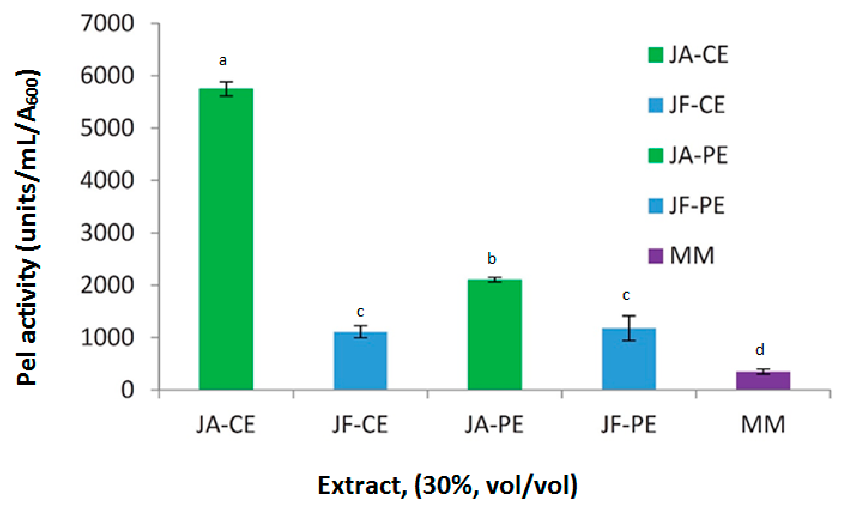
3.1. Survey of Extracts from Different Host Plants for Inducer Activity

To determine how widely distributed the inducer(s) might be among different host plant species with filtered extracts (FE) from ten hosts of Pectobacterium, the levels of extracellular pectate lyase (Pel) produced by bacteria growing in MM were compared to that of the same bacterial strains growing in the same medium supplemented with cut and autoclaved (CA) extract from celery, potato, carrot, and tomato. Pectate lyase activity measured from the culture supernatants showed significant increases (Student’s t-test at P < 0.05), i.e., 17-fold to 54-fold over MM in P. carotovorum Ecc71 and 13-fold to 58-fold over MM in P. atrosepticum SCRI1043 (Table 1). This indicates that the extracts from these hosts contained inducer activities in different concentrations, or that the inducer(s) in extracts from the different hosts induced Pel activities to different degrees in the two bacterial strains.
Following the observation that many hosts produced the inducer, the optimum concentration of juiced autoclaved extract (AE) and filtered extract (FE) suitable for induction of Pel in Pectobacterium strains were determined. Pectate lyase activity was measured in P. carotovorum Ecc71 and in P. atrosepticum SCRI1043 grown in MM supplemented with varying concentrations of filter-sterilized or autoclaved extract of celery or potato. Pectate lyase and β-galactosidase activities of test tube-grown cultures were also assayed in P. carotovorum strain KD100 carrying the pel1-lacZ plasmid. Optimal concentrations of the host extracts were determined for the induction of enzyme production and β-galactosidase activities. Based on these experiments, MM supplemented with 30% potato extract (PE), 30% celery extract (CE) or 0.2% PGA were chosen for use in subsequent experiments (data not shown).

3.2. Induction of Pel Activity in a KdgR Mutant by Host Extracts and PGA

We compared the responses of a kdgR mutant of P. carotovorum, (AC5073) and its parent, Ecc71, to host extracts by growing the two strains in MM supplemented with PGA (0.2%, w/v), filtered CE or PE (30%, v/v) in culture flasks. In the wild-type Ecc71, the addition of each of the three extracts, i.e., PGA, celery extract (CE), or potato extract (PE), alone to the basal medium increased Pel activity by 13-fold, 40-fold, and 102-fold, respectively (Table 2). There was a slightly higher induction when PGA + PE (117-fold) was added to the basal medium. Thus, potato extract and PGA had a synergistic effect on induction of Pel production in Ecc71 suggesting they have different constituents. Similarly, in the KdgR mutant, induction of Pel production by the addition of CE or PE to the basal medium was approximately 18-fold. Unlike the parent, Pel production was not induced when the KdgR mutant was grown in MM + PGA (Table 2) or when PGA was added to CE or PE. Pel activities by the KdgR mutant were higher than in the wild-type strain in all of the media except MM + PGA (Table 2). Moreover, the absence of any apparent increase in Pel production by the mutant when PGA was added to the extracts strongly suggests that the inducer(s) in the extracts (not PGA and its degradation intermediates) was/were responsible for the Pel induction in the mutant.

3.3. Temperature Stability of the Inducer

To learn about some of the physical properties of the inducing compounds, the temperature stability of the inducer was tested. Pectate lyase production by Ecc71 grown in medium supplemented (30%, v/v) with autoclaved extracts of celery was significantly higher (P < 0.05) than that from unautoclaved extract at same concentration (Figure 1). The original inducing activity of the extract was fully retained after freezing the extract at −80 °C for up to 6 months (data not shown). Thus, the inducing factor(s) was not destroyed by either heating during the autoclaving process or by freezing, and could be described as thermostable and freeze-tolerant. Since making this observation, our extract is routinely stored at −20 °C.

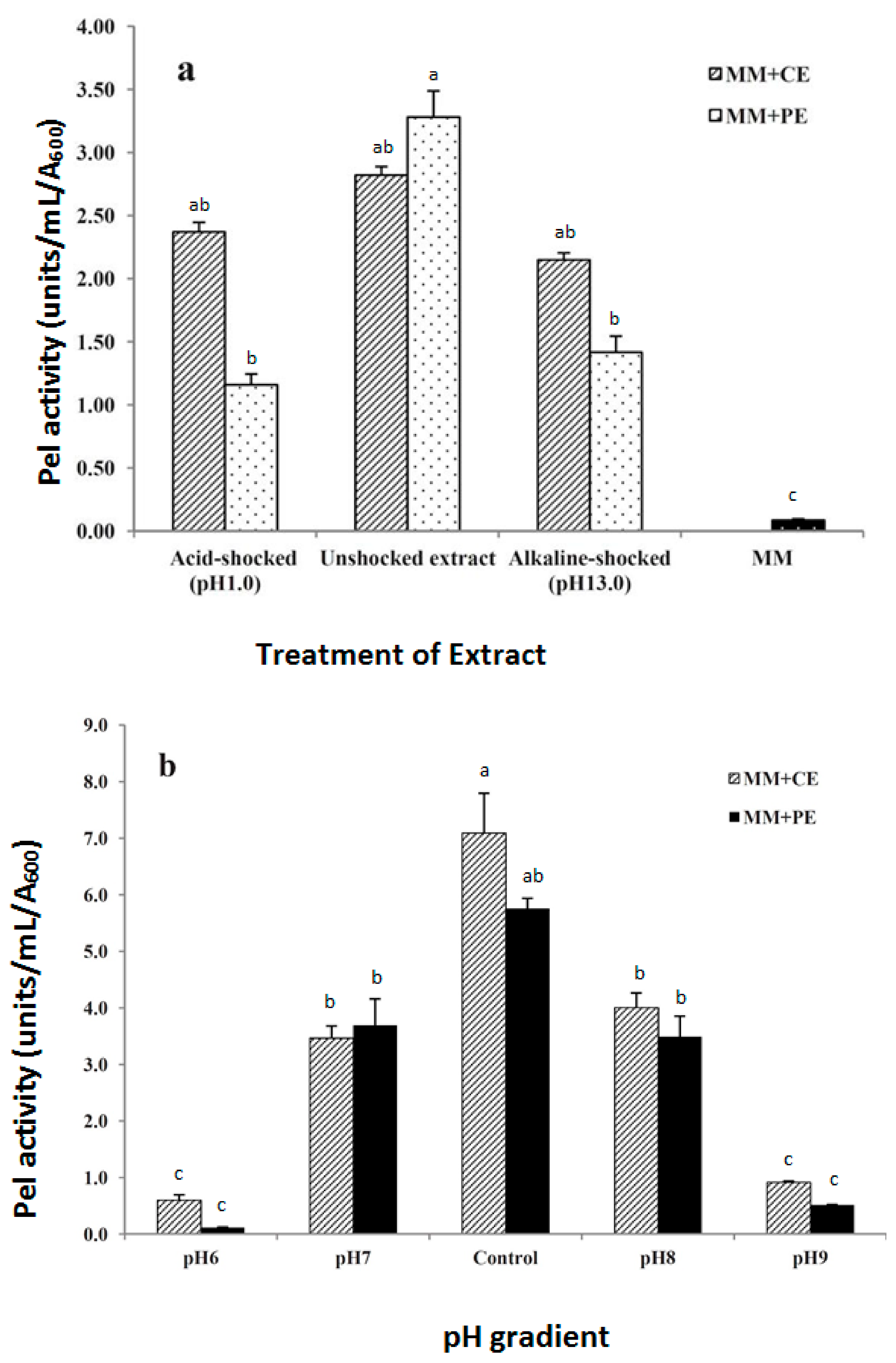
3.4. Effect of pH on Inducer and Induction

The induction properties of the extracts were significantly reduced (P < 0.05) by the pH-shock treatments (Figure 2a). In both low and high pH-shock treatments and for both celery and potato extracts, however, enzyme production levels remained significantly higher than in MM. This suggested that the pH shock could have produced a change in the inducer molecule(s) that in some way affected its induction ability. Growing bacteria in media with a pH gradient showed that Pel induction by the extracts was pH-dependent. In MM + CE medium, induction of Pel in P. carotovorum strain KD100 dropped from 7.1-fold at pH 7.4 to 0.6-fold at both pH 6.0 and pH 9.0 (Figure 2b). The same trend was observed in MM+PE, but interestingly, bacterial growth was not visibly affected at these pH conditions. These results suggest that the induction of exoenzyme production could only take place under optimal pH.

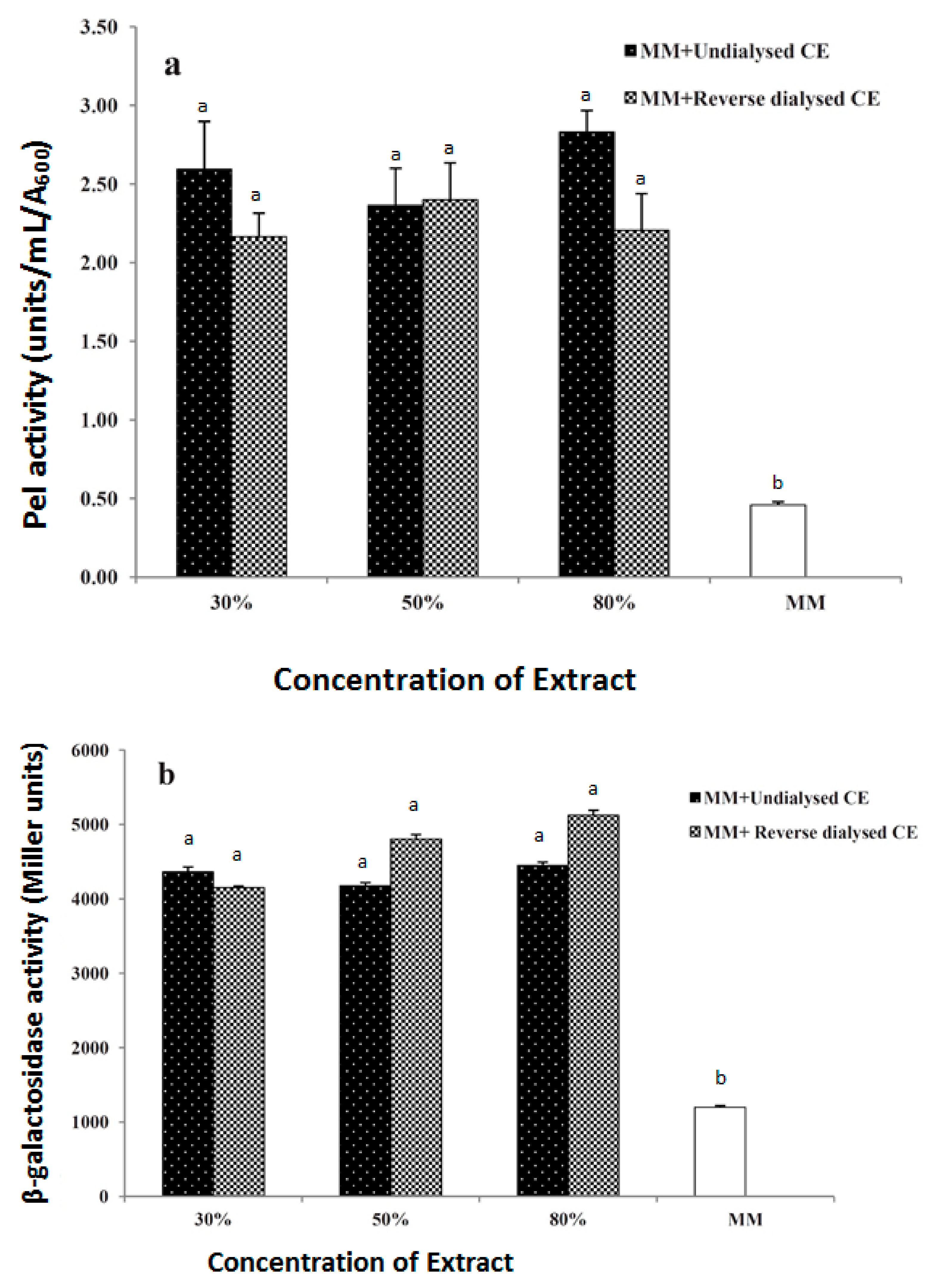
3.5. Dialysis of the Inducer

The activities of Pel and Prt of bacteria grown in media supplemented with autoclaved (CA) extracts from potato tuber, celery petioles, tomato fruits, and carrot roots were significantly reduced (P < 0.05) in media supplemented with dialyzed (MWCO, 1 kDA) compared to activities in media with undialyzed extracts (Table 3). Similar reductions in Pel activity and pel1 expression were observed in assays of KD100/pel1-lacZ grown in increasing concentrations of dialyzed (MWCO, 1 kDa) filtered CE. The level of Pel activity in the presence of the dialyzed extract was similar to that produced in MM alone. This indicated that the inducer molecule(s) in the extract dialyzed out of the bag and therefore induction of Pel production was reduced or eliminated due to the lower diluted concentration of of the inducer.
In a reverse experiment, the inducer from the extract was dialyzed into water in a dialysis bag, and the water with the inducer molecule(s) dialyzed into it was tested for induction of both Pel and pel1-lacZ activities. Both the undialyzed and reverse dialyzed, filtered CE induced Pel activities by approximately the same level (5.3-fold) over MM at all levels of extract concentration (Figure 3a). Similarly, both extracts induced pel1 expression by approximately 3.8-fold over MM in the lac strain of P. carotovorum strain KD100 (Figure 3b). This implied that in the reverse dialyzed extract, the inducer molecule(s) dialyzed out from the CE into the distilled water; its presence in the latter was ascertained by the induction of enzyme production in the assays. These data suggest that the inducer molecule(s) is/are small, diffusible, and water-soluble, with a molecular weight of less than 1 kDa.

4. Discussion

In this study, we present evidence that a second class of signal molecules (Class II) in plant host extracts induces pectate lyase and protease activities in Pectobacterium, and that these Class II inducers are different from pectin degradation products or Class I inducers. Working with PE and P. atrosepticum SCRI1043, Tarasova and colleagues even eliminated the possibility of the inducer being an AHL-like metabolite [34]. Our data showed that the addition of CE or PE to PGA further stimulated the induction of Pel synthesis in the wild-type; this was in agreement with an earlier report with carrot extracts using the D. dadantii strain 3937 [23]. In contrast with the findings with carrot extracts and D. dadantii, [7] the addition of PGA to CE or PE resulted in higher induction in P. carotovorum. In the presence of class II inducers (CE or PE), induction still occurred in the KdgR mutant as in the wild-type, but the mutant failed to respond to PGA. In the KdgR mutant, any induction of Pel production had to be by a class of molecules different from products of pectin degradation, since class I inducers act through KdgR and kdgR strains are no longer inducible by these inducers. This suggests that there is a different class of compound(s) (Class II) present in the extracts which is responsible for inducing Pel production and these are different from KdgR-mediated Class I compounds reported earlier [26]. Based on the above observation, we believe that it is not likely that the Class II inducer(s) in CE or PE work through the KdgR regulatory system, because even in the absence of KdgR, the Class II inducers were capable of increasing Pel production.
Consistent with the KdgR mechanism of action in both Pectobacterium and Dickeya, we expected the KdgR mutant to be derepressed, since it no longer had the repressive action of KdgR. The higher Pel production in the kdgR strain in the presence of CE and PE and its lack of response to PGA suggests that there are other signal molecules in the extract different from PGA and its degradation intermediates and these might have induced Pel activity through other pathways different from the KdgR system.
Our findings raise the interesting question of which regulators mediate the action of Class II inducers. In addition to host signals, pathogenesis in soft rot Pectobacteriaceae is also controlled by numerous bacterial signals and regulators. Many of these regulators exert their effect on the production of plant cell wall-degrading enzymes that make up the major pathogenicity factors of these bacteria. Thus, in the absence of KdgR, there is still a long list of regulators that could mediate the effect of Class II signals. Among the candidates are regulators such as HexA or its homologue, PecT, LysR-type regulators, which negatively control the expression of pectinase genes in P. carotovorum and D. didantii,, respectively [43,44,45,46]. Other regulators such as the KdgR-like protein, RexZ, act as positive regulators of exoenzyme production including Pel, Cel and Prt in P. carotovorum [47]. The relationship between host signal molecules and these regulators is, however, not known. Further studies are needed to investigate the response of these regulators to host extracts and elucidate the mechanisms by which host signals induce exoenzyme production.
This study revealed that there are optimum, as well as inhibitory, concentrations of host extracts as it relates to the induction of enzyme activity. Reduction in exoenzyme production due to inducer concentrations above a threshold could be partly due to the presence of inhibitors of exoenzyme induction in the extracts and their effects at higher concentrations. In a similar study using pelA::uidA fusion in D. dadantii [24], there was a decrease in Pel induction ratio with increasing carrot extract concentration after a maximum threshold. We think that above a certain threshold concentration of inducers in the culture medium, the bacteria lose their capacity to synthesize enzymes, as has been hypothesized by others [48], even though bacterial growth is not affected.
The chemical identity of inducers in the extracts from the six plant families and ten species that we surveyed could be the same or different. Since different plants have different chemical compositions and may vary in the amounts and the kinds of inducer(s) and inhibitors they contain, the observed variability in the level of exoenzyme production induced by these extracts is expected (Table 1), and has also been previously reported [49]. Our preliminary probe into the physical properties of the inducer(s) has revealed some general properties of the inducers. Forward and reverse dialysis experiments using CE show that Class II inducers in CE are small diffusible hydrophilic molecule(s) with a size, i.e., possibly less than 1 kDa. An inducer of PelA-LacZ activity extracted from carrot also had similar size range [24], as did the inducer of enzymes from PE [34]. Those inducers could be the same or related to the compounds from celery and potato extracts that we report on here. Our experiments revealed that the inducing molecule is diffusible, thermostable, freeze-tolerant, pH-sensitive, water-soluble, and small, i.e., with a molecular mass of less than 1 kDa. Further studies are underway to determine the chemical identity of the inducer(s) in these extracts.

Author Contributions

Conceptualization, C.K.D.; methodology, P.A.A., C.M.K., M.N.K., C.K.D., validation, P.A.A., C.M.K., M.N.K., data analysis, P.A.A., C.M.K., M.N.K.; resources, C.K.D.; writing—original draft preparation, P.A.A.; writing—review and editing, P.A.A., C.M.K., M.N.K., C.K.D., supervision, C.K.D.; project administration, C.K.D.; funding acquisition, C.K.D. All authors have read and agreed to the published version of the manuscript.

Funding

This research was funded by National Institute of Food and Agriculture of USDA grant number TENX-0004-GFSH.

Acknowledgments

We thank A. Chatterjee for bacterial strains and plasmids and K. Dzantor for reviewing the manuscript. The first three authors were supported by graduate assistantships from the Departments of Biological, Agricultural and Environmental Sciences.

Conflicts of Interest

The authors declare no conflict of interest.

References

  1. Perombelon, M.C.M.; Kelman, A. Ecology of the Soft Rot Erwinias. Annu. Rev. Phytopathol. 1980, 18, 361–388. [Google Scholar] [CrossRef]
  2. Perombelon, M.C.M.; Hyman, L.J. Serological methods to quantify potato seed contamination by Erwinia carotovora subspatroseptica. Bull. OEPP 1995, 25, 195–202. [Google Scholar] [CrossRef]
  3. Perombelon, M.C.M. Potato diseases caused by soft rot erwinias: An overview of pathogenesis. Plant Pathol. (Oxford) 2002, 51, 1–12. [Google Scholar]
  4. Barras, F.; Van Gijsegem, F.; Chatterjee, A.K. Extracellular enzymes and pathogenesis of soft-rot Erwinia. Annu. Rev. Phytopathol. 1994, 32, 201–234. [Google Scholar] [CrossRef]
  5. Toth, I.K.; Bell, K.S.; Holeva, M.C.; Birch, P.R. Soft rot erwiniae: From genes to genomes. Mol. Plant Pathol. 2003, 4, 17–30. [Google Scholar] [CrossRef] [PubMed]
  6. Collmer, A.; Keen, N.T. The role of pectic enzymes in plant pathogenesis. Annu. Rev. Phytopathol. 1986, 24, 383–409. [Google Scholar] [CrossRef]
  7. Hugouvieux-Cotte-Pattat, N.; Condemine, G.; Nasser, W.; Reverchon, S. Regulation of pectinolysis in Erwinia chrysanthemi. Annu. Rev. Microbiol. 1996, 50, 213–257. [Google Scholar] [CrossRef]
  8. Toth, I.K.; Birch, P.R. Rotting softly and stealthily. Curr. Opin. Plant Biol. 2005, 8, 424–429. [Google Scholar] [CrossRef] [PubMed]
  9. Brencic, A.; Winans, S.C. Detection of and response to signals involved in host-microbe interactions by plant-associated bacteria. Microbiol. Mol. Biol. Rev. 2005, 69, 155–194. [Google Scholar] [CrossRef] [PubMed] [Green Version]
  10. Cui, Y.; Chatterjee, A.; Liu, Y.; Dumenyo, C.K.; Chatterjee, A.K. Identification of a global repressor gene, rsmA, of Erwinia carotovora subsp. carotovora that controls extracellular enzymes, N-(3-oxohexanoyl)-L-homoserine lactone, and pathogenicity in soft-rotting Erwinias. J. Bacteriol. 1995, 177, 5108–5115. [Google Scholar] [CrossRef] [Green Version]
  11. Chatterjee, A.; Cui, Y.; Liu, Y.; Dumenyo, C.K.; Chatterjee, A.K. Inactivation of rsmA leads to overproduction of extracellular pectinases, cellulases, and proteases in Erwinia carotovora subsp. carotovora in the absence of the starvation/cell density-sensing signal, N-(3-oxohexanoyl)-L-homoserine lactone. Appl. Environ. Microbiol. 1995, 61, 1959–1967. [Google Scholar] [CrossRef] [PubMed] [Green Version]
  12. Liu, Y.; Cui, Y.; Mukherjee, A.; Chatterjee, A.K. Characterization of a novel RNA regulator of Erwinia carotovora sspcarotovora that controls production of extracellular enzymes and secondary metabolites. Mol. Microbiol. 1998, 29, 219–234. [Google Scholar] [CrossRef] [PubMed]
  13. Chatterjee, A.K.; Dumenyo, C.K.; Liu, Y.; Chatterjee, A. Erwinia: Genetics of pathogenicity factors. Encycl. Microbiol. 2000, 2, 236–260. [Google Scholar]
  14. Cui, Y.; Chatterjee, A.; Chatterjee, A.K. Effects of the two-component system comprising GacA and GacS of Erwinia carotovora subsp. carotovora on the production of global regulatory rsmB RNA, extracellular enzymes, and harpinEcc. Mol. Plant Microbe Interact. 2001, 14, 516–526. [Google Scholar] [CrossRef] [PubMed] [Green Version]
  15. Cui, Y.; Chatterjee, A.; Hasegawa, H.; Dixit, V.; Leigh, N.; Chatterjee, A.K. ExpR, a LuxR homolog of Erwinia carotovora subsp. carotovora, activates transcription of rsmA, which specifies a global regulatory RNA-binding protein. J. Bacteriol. 2005, 187, 4792–4803. [Google Scholar] [CrossRef] [PubMed] [Green Version]
  16. Charkowski, A.; Blanco, C.; Condemine, G.; Expert, D.; Franza, T.; Hayes, C.; Hugouvieux-Cotte-Pattat, N.; Solanilla, E.L.; Low, D.; Moleleki, L.; et al. The role of secretion systems and small molecules in soft-rot Enterobacteriaceae pathogenicity. Annu. Rev. Phytopathol. 2012, 50, 425–449. [Google Scholar] [CrossRef] [Green Version]
  17. Pirhonen, M.; Flego, D.; Heikinheimo, R.; Palva, E.T. A small diffusible signal molecule is responsible for the global control of virulence and exoenzyme production in the plant pathogen Erwinia carotovora. EMBO J. 1993, 12, 2467–2476. [Google Scholar] [CrossRef]
  18. Jones, S.; Yu, B.; Bainton, N.A.; Birdsall, M.; Bycroft, B.W.; Chhabra, S.R.; Cox, A.J.; Golby, P.; Reeves, P.J.; Stephens, S. The lux autoinducer regulates the production of exoenzyme virulence determinants in Erwinia carotovora and Pseudomonas aeruginosa. EMBO J. 1993, 12, 2477–2482. [Google Scholar] [CrossRef]
  19. Cui, Y.; Chatterjee, A.; Hasegawa, H.; Chatterjee, A.K. Erwinia carotovora subspecies produce duplicate variants of ExpR, LuxR homologs that activate rsmA transcription but differ in their interactions with N-acylhomoserine lactone signals. J. Bacteriol. 2006, 188, 4715–4726. [Google Scholar] [CrossRef] [Green Version]
  20. Jones, D.L.; Nguyen, C.; Finlay, R.D. Carbon flow in the rhizosphere: Carbon trading at the soil–root interface. Plant Soil 2009, 321, 5–33. [Google Scholar] [CrossRef]
  21. Dong, Y.H.; Xu, J.L.; Li, X.Z.; Zhang, L.H. AiiA, an enzyme that inactivates the acylhomoserine lactone quorum-sensing signal and attenuates the virulence of Erwinia carotovora. Proc. Natl. Acad. Sci. USA 2000, 97, 3526–3531. [Google Scholar] [CrossRef] [PubMed]
  22. Liu, H.; Coulthurst, S.J.; Pritchard, L.; Hedley, P.E.; Ravensdale, M.; Humphris, S.; Burr, T.; Takle, G.; Brurberg, M.B.; Birch, P.R.; et al. Quorum sensing coordinates brute force and stealth modes of infection in the plant pathogen Pectobacterium atrosepticum. PLoS Pathog 2008, 4, e1000093. [Google Scholar] [CrossRef] [PubMed]
  23. Hugouvieux-Cotte-Pattat, N.; Dominguez, H.; Robert-Baudouy, J. Environmental conditions affect transcription of the pectinase genes of Erwinia chrysanthemi 3937. J. Bacteriol. 1992, 174, 7807–7818. [Google Scholar] [CrossRef] [PubMed] [Green Version]
  24. Bourson, C.; Favey, S.; Reverchon, S.; Robert-Baudouy, J. Regulation of the expression of a pelA:uidA fusion in Erwinia chrysanthemi and demonstration of the synergistic action of plant extract with polygalacturonate on pectate lyase synthesis. J. Gen. Microbiol. 1993, 139, 1–9. [Google Scholar] [CrossRef] [Green Version]
  25. Matsumoto, H.; Jitareerat, P.; Baba, Y.; Tsuyumu, S. Comparative study of regulatory mechanisms for pectinase production by Erwinia carotovora subsp. carotovora and Erwinia chrysanthemi. Mol. Plant Microbe Interact. 2003, 16, 226–237. [Google Scholar] [CrossRef] [Green Version]
  26. Condemine, G.; Hugouvieux-Cotte-Pattat, N.; Robert-Baudouy, J. Isolation of Erwinia chrysanthemi kduD mutants altered in pectin degradation. J. Bacteriol. 1986, 165, 937–941. [Google Scholar] [CrossRef] [Green Version]
  27. Liu, Y.; Jiang, G.; Cui, Y.; Mukherjee, A.; Ma, W.L.; Chatterjee, A.K. kdgREcc negatively regulates genes for pectinases, cellulase, protease, HarpinEcc, and a global RNA regulator in Erwinia carotovora subsp. carotovora. J. Bacteriol. 1999, 181, 2411–2421. [Google Scholar] [CrossRef] [Green Version]
  28. Nasser, W.; Reverchon, S.; Condemine, G.; Robert-Baudouy, J. Specific interactions of Erwinia chrysanthemi KdgR repressor with different operators of genes involved in pectinolysis. J. Mol. Biol. 1994, 236, 427–440. [Google Scholar] [CrossRef]
  29. Mattinen, L.; Nissinen, R.; Riipi, T.; Kalkkinen, N.; Pirhonen, M. Host-extract induced changes in the secretome of the plant pathogenic bacterium Pectobacterium atrosepticum. Proteomics 2007, 7, 3527–3537. [Google Scholar] [CrossRef]
  30. Mattinen, L.; Somervuo, P.; Nykyri, J.; Nissinen, R.; Kouvonen, P.; Corthals, G.; Auvinen, P.; Aittamaa, M.; Valkonen, J.P.; Pirhonen, M. Microarray profiling of host-extract-induced genes and characterization of the type VI secretion cluster in the potato pathogen Pectobacterium atrosepticum. Microbiology 2008, 154, 2387–2396. [Google Scholar] [CrossRef] [Green Version]
  31. Murata, H.; McEvoy, J.L.; Chatterjee, A.; Collmer, A.; Chatterjee, A.K. Molecular cloning of an aepA gene that activates production of extracellular pectolytic, cellulolytic, and proteolytic enzymes in Erwinia carotovora subsp. carotovora. Mol. Plant Microbe Interact. 1991, 4, 239–246. [Google Scholar] [CrossRef]
  32. Li, Y.; Peng, Q.; Selimi, D.; Wang, Q.; Charkowski, A.O.; Chen, X.; Yang, C.H. The plant phenolic compound p-coumaric acid represses gene expression in the Dickeya dadantii type III secretion system. Appl. Environ. Microbiol. 2009, 75, 1223–1228. [Google Scholar] [CrossRef] [PubMed] [Green Version]
  33. Kelemu, S.; Collmer, A. Erwinia chrysanthemi EC16 Produces a Second Set of Plant-Inducible Pectate Lyase Isozymes. Appl. Environ. Microbiol. 1993, 59, 1756–1761. [Google Scholar] [CrossRef] [PubMed] [Green Version]
  34. Tarasova, N.; Gorshkov, V.; Petrova, O.; Gogolev, Y. Potato signal molecules that activate pectate lyase synthesis in Pectobacterium atrosepticum SCRI1043. World J. Microbiol. Biotechnol. 2013, 29, 1189–1196. [Google Scholar] [CrossRef] [PubMed]
  35. Zink, R.T.; Kemble, R.J.; Chatterjee, A.K. Transposon Tn5 mutagenesis in Erwinia carotovora subsp. carotovora and E. carotovora subsp. atroseptica. J. Bacteriol. 1984, 157, 809–814. [Google Scholar] [CrossRef] [PubMed] [Green Version]
  36. Hinton, J.C.D.; Sidebotham, J.M.; Gill, D.R.; Salmond, G.P.C. Extracellular and periplasmic isoenzymes of pectate lyase from Erwinia carotovora subspecies carotovora belong to different gene families. Mol. Microbiol. 1989, 3, 1785–1795. [Google Scholar] [CrossRef]
  37. Murata, H.; Fons, M.; Chatterjee, A.; Collmer, A.; Chatterjee, A.K. Characterization of transposon insertion Out mutants of Erwinia carotovora subsp. carotovora defective in enzyme export and of a DNA segment that complements out mutations in E. carotovora subsp. carotovora, E. carotovora subsp. atroseptica, and Erwinia chrysanthemi. J. Bacteriol. 1990, 172, 2970–2978. [Google Scholar]
  38. Kersey, C.M.; Agyemang, P.A.; Dumenyo, C.K. CorA, the magnesium/nickel/cobalt transporter, affects virulence and extracellular enzyme production in the soft rot pathogen Pectobacterium carotovorum. Mol. Plant Pathol. 2012, 13, 58–71. [Google Scholar] [CrossRef]
  39. Chatterjee, A.K. Acceptance by Erwinia s of R plasmid R68.45 and its ability to mobilize the chromosome of Erwinia chrysanthemi. J. Bacteriol. 1980, 142, 111–119. [Google Scholar] [CrossRef] [Green Version]
  40. Zink, R.T.; Engwall, J.K.; McEvoy, J.L.; Chatterjee, A.K. recA is required in the induction of pectin lyase and carotovoricin in Erwinia carotovora subsp. carotovora. J. Bacteriol. 1985, 164, 390–396. [Google Scholar] [CrossRef] [Green Version]
  41. Braun, V.; Schmitz, G. Excretion of a protease by Serratia marcescens. Arch Microbiol. 1980, 124, 55–61. [Google Scholar] [CrossRef] [PubMed]
  42. Miller, J.H. Experiments in Molecular Genetics; Cold Spring Harbor Laboratory Press: Cold Spring Harbor, NY, USA, 1972; p. 468. [Google Scholar]
  43. Reverchon, S.; Nasser, W.; Robert-Baudouy, J. PecS: A locus controlling pectinase, cellulase and blue pigment production in Erwinia chrysanthemi. Mol. Microbiol. 1994, 11, 1127–1139. [Google Scholar] [CrossRef] [PubMed]
  44. Praillet, T.; Nasser, W.; Robert-Baudouy, J.; Reverchon, S. Purification and functional characterization of PecS, a regulator of virulence-factor synthesis in Erwinia chrysanthemi. Mol. Microbiol. 1996, 20, 391–402. [Google Scholar] [CrossRef] [PubMed]
  45. Castillo, A.; Reverchon, S. Characterization of the pecT control region from Erwinia chrysanthemi 3937. J. Bacteriol. 1997, 179, 4909–4918. [Google Scholar] [CrossRef] [PubMed] [Green Version]
  46. Mukherjee, A.; Cui, Y.; Ma, W.; Liu, Y.; Chatterjee, A.K. hexA of Erwinia carotovora sspcarotovora strain Ecc71 negatively regulates production of RpoS and rsmB RNA, a global regulator of extracellular proteins, plant virulence and the quorum-sensing signal, N-(3-oxohexanoyl)-l-homoserine lactone. Environ. Microbiol. 2000, 2, 203–215. [Google Scholar] [CrossRef] [PubMed]
  47. Thomson, N.R.; Nasser, W.; McGowan, S.; Sebaihia, M.; Salmond, G.P. Erwinia carotovora has two KdgR-like proteins belonging to the IciR family of transcriptional regulators: Identification and characterization of the RexZ activator and the KdgR repressor of pathogenesis. Microbiology 1999, 145, 1531–1545. [Google Scholar] [CrossRef] [PubMed] [Green Version]
  48. Kepseu, W.D.; Sepulchre, J.A.; Reverchon, S.; Nasser, W. Toward a quantitative modeling of the synthesis of the pectate lyases, essential virulence factors in Dickeya dadantii. J. Biol. Chem. 2010, 285, 28565–28576. [Google Scholar] [CrossRef] [Green Version]
  49. Tsuyumu, S.; Funamubo, T.; Hori, K.; Takikawa, Y.; Goto, M. Presence of DNA damaging agents in plants as the possible inducers of pectin lyases of soft-rot Erwinia. Jpn. J. Phytopathol. 1985, 51, 294–302. [Google Scholar] [CrossRef] [Green Version]
Figure 1. Levels of Pel produced by P. carotovorum strain KD100/pAKC1031 (pel1-lacZ) grown in MM supplemented with filtered and autoclaved extracts of celery (CE) and potato (PE). Cultures were grown overnight, and culture supernatants were collected and assayed for Pel activity. Bacterial growth was measured with A600. Pel activity was kinetically determined from culture supernatants at 235 nm and expressed as units/mL/A600. Means with different letters are significantly different by Student’s t-test at P < 0.05. Abbreviation: JA-CE, juice autoclaved CE; JF-CE, juice filtered CE; JA-PE, juice autoclaved PE; JF-PE, juice filtered PE.
Horticulturae 06 00013 g001
Figure 2. Effect of pH on Pel induction. (a) The pH of juiced extracts of celery (CE) and potato (PE) were adjusted to either pH 1.0 (acid-shocked) or pH 13.0 (alkaline-shocked) and incubated at 28 °C for 15 min. The pH was readjusted to the original pH (6.19 for CE; 6.25 for PE), filtered and used to supplement MM. (b) The pH of extract-supplemented media were adjusted to 6, 7, 8, or 9 and inoculated with bacterial. All experiments were performed in triplicate, and the mean values ±SD (indicated as error bars) are presented. Means with same letters are not significantly different by Student’s t-test at P < 0.05.
Horticulturae 06 00013 g002
Figure 3. Pectate lyase (a) and β-galactosidase (b) activities in cultures of Pectobacterium strain KD100 carrying pel1-lacZ fusion. Bacteria were grown in MM medium supplemented with undialyzed or reverse dialyzed extract (RDE). Pectate lyase activity, expressed as units/mL/A600, and β-galactosidase activity, expressed in Miller units, were determined from culture supernatant and cells of the test strain, respectively. All experiments were performed in triplicate, and the mean values ±SD (indicated as error bars) are presented. Means with different letters are significantly different by Student’s t-test at P < 0.05. At all concentrations, the level of induction by both dialyzed and reverse dialyzed extracts were significantly higher than minimal medium.
Horticulturae 06 00013 g003
Table 1. Induction of pectate lyase activity by filtered extracts from different hosts.
Pectate Lyase Activity zy
Source of ExtractEcc71SCRI1043
Apium graveolens (celery petiole)52.0 ± 2.962.9 ± 5.5
Brassica oleracea (cabbage leaf)32.9 ± 4.715.1 ± 2.1
Latuca sativa (lettuce leaf)22.2 ± 3.447.6 ± 9.7
Daucus carota (carrot root)45.3 ± 2.267.8 ± 9.6
Cucumis sativa (cucumber)23.1 ± 2.741.2 ± 4.2
Allium sepa (onion bulb)19.1 ± 2.314.5 ± 0.4
Solanum tuberosum (potato tuber)53.6 ± 3.237.4 ± 5.6
Solanum lycopersicon (tomato fruit)35.5 ± 3.523.7 ± 1.0
Caspicum annuum (bell pepper fruit)22.4 ± 2.142.7 ± 4.7
Brassica rapa (turnip root)53.3 ± 2.634.2 ± 3.9
Medium only1.00 ± 0.01.00 ± 0.00
z Bacterial growth was measured with A600. Pel activity was kinetically determined from culture supernatants at 235 nm and expressed as units/mL/A600. y Fold increase over minimal salts media. The values represent the mean ±SD of three independent measurements.
Table 2. Levels of pectate lyase produced by a KdgR mutant and parental strain in filtered extract-supplemented media.
Medium zPectate lyase activity wxy
Ecc71(KdgR+)AC5073 (KdgR)
MM0.03 ± 0.0090.33 ± 0.020
MM + PGA0.44 ± 0.017 *0.30 ± 0.019
MM + CE1.31 ± 0.102 **6.15 ± 0.276 **
MM + PE3.37 ± 0.165 **6.10 ± 0.640 **
MM + CE + PGA2.78 ± 0.088 **4.41 ± 0.140 **
MM + PE + PGA3.87 ± 0.104 **5.03 ± 0.131 **
z Media and supplements were: Minimal salts medium (MM); polygalacturonic acid (PGA); celery extract (CE), potato extract (PE). Bacteria were cultured with shaking at 28 °C and harvested at 250 (MM), 300 (MM+PGA), 350 (MM + CE), 400 (MM + PE), 350 (MM + PGA + CE) and 450 (MM + PGA + PE) Klett units. y Bacterial growth was measured with A600. Pel activity was kinetically determined from culture supernatants at 235 nm and expressed as units/mL/A600. x Data presented are the means ±SD of Pel activities from three independent experiments. w Significant over MM at P < 0.01 (**) or at P < 0.05 (*) by Student’s t-test.
Table 3. Pectate lyase and protease activities produced by Pectobacterium strains in media supplemented with dialyzed or undialyzed cut and autoclaved extracts from different hosts.
Strain Ecc71 zwStrain SCRI 1043 zw
Pectate Lyase activityyProtease Activity xPectate Lyase ActivityyProtease Activity x
Host ExtractUndialyzedDialyzedUndialyzedDialyzedUndialyzedDialyzedUndialyzedDialyzed
Carrot1.98 ± 0.0520.06 ± 0.006 **w0.36 ± 0.0070.08 ± 0.004 **2.14 ± 0.0330.097 ± 0.011 **0.36 ± 0.0030.072 ± 0.007 **
Tomato1.54 ± 0.0430.07 ± 0.001 **0.35 ± 0.0140.08 ± 0.017 **1.19 ± 0.0490.107 ± 0.008 **0.26 ± 0.0060.068 ± 0.005 **
Celery0.85 ± 0.0350.05 ± 0.005 **0.25 ± 0.0230.07 ± 0.008 *0.89 ± 0.0390.061 ± 0.005 **0.24 ± 0.0050.086 ± 0.092 *
Potato1.58 ± 0.0620.11 ± 0.002 **0.41 ± 0.0100.09 ± 0.007 **0.96 ± 0.0390.084 ± 0.006 **0.27 ± 0.0070.064 ± 0.003 **
MM0.02 ± 0.0030.05 ± 0.0020.05 ± 0.0080.06 ± 0.006
z Bacteria were grown overnight with shaking at 28 °C in MM supplemented with 30% (CA) extracts. y Bacterial growth was measured with A600. Pel activity was kinetically determined from culture supernatants at 235 nm and expressed as units/mL/A600. x Protease activity was spectrophometrically determined using azocasein as a substrate and expressed as units/mL/A600. w Data presented are the average (±SD) of three independent experiments. Means of dialyzed and undialyzed extracts were compared by Student’s t-test, with significance at P < 0.01 (**) or at P < 0.05 (*).

