Review

A Review on Flower Bulb Micropropagation: Challenges and Opportunities

by
Sara Yasemin
1,* and
Margherita Beruto
2
1
Department of Horticulture, Faculty of Agriculture, University of Siirt, Kezer, 56100 Siirt, Turkey
2
International Society for Horticultural Science, Ornamental Plant Division, 18038 Sanremo, Italy
*
Author to whom correspondence should be addressed.
Horticulturae 2024, 10(3), 284; https://doi.org/10.3390/horticulturae10030284
Submission received: 4 January 2024 / Revised: 28 February 2024 / Accepted: 13 March 2024 / Published: 15 March 2024

Abstract

:
This comprehensive review scrutinizes tissue culture and micropropagation methodologies in geophytes, focusing on bulbous plants. The examination encompasses key stages, including somatic embryogenesis, bulb growth, dormancy breaking, and planting. Studies underscore the pivotal role of plant growth regulators (PGRs) in plant regeneration and bulb growth. Bioreactor systems for healthy plant regeneration, rooting methods, acclimatization strategies, and considerations for ex vitro survival are elucidated. The review also delves into somaclonal variation dynamics and acknowledges the burgeoning field of gene editing, particularly Clustered Regularly Interspaced Short Palindromic Repeats (CRISPR) studies, as a promising avenue for enhancing valuable compound content in geophytes. In addition to addressing challenges in flower bulb micropropagation, this review briefly highlights emerging opportunities, including the potential integration of artificial intelligence (AI) to optimize culture conditions, predict growth parameters, and enhance efficiency in bulb production. The conclusion emphasizes the necessity of a multifaceted approach integrating biochemistry, physiology, and molecular biology to address existing challenges and improve tissue culture protocols for diverse geophyte species. This review article also intends to highlight how tissue culture techniques could contribute to the development and valorization of flower bulbs in today’s scenario of the ornamental industry.

1. Ornamental Geophyte History

For centuries, flower bulbs have captivated societies worldwide, drawing attention for their enticing fragrance, vibrant colors, and diverse blooming periods. These bulbs have held a significant place in various forms of art [1,2,3]. Greek philosophy dating back to 300 BC mentions the medicinal and consumable properties of flower bulbs like Crocus, Colchicum, and Gladiolus. Greek mythology also references bulbs such as Lilium, Hyacinthus, Crocus, Iris, and Narcissus [4,5]. Furthermore, Warren [6] reported that Anemone coronaria, Ranunculus asiaticus, and Lilies were shown in Knossos Palace in Crete, Greece, during the Bronze Age. Day [7] emphasized that the Crocus has been recognized in Minoan art as an important motif during the Aegean Bronze Age. Japan and China are known to have numerous wild Lilium species that are endemic to their regions. The United Kingdom is the origin of Narcissus pseudonarcissus, commonly known as the lenty lily, which serves as the ancestor for many cultivated daffodil varieties. The Persian-style gardens of the Taj Mahal, an iconic 17th-century monument, feature essential flower bulbs like Anemone, Ranunculus, Fritillaria, Iris, Lilium, Pancratium, and Gloriosa [8]. Flower bulbs such as Aztec lily, canna, dahlias, and tuberose are reported to have originated in the Americas [9]. In the 1180s, flower bulbs, particularly those with iris motifs, adorned the military uniforms of Italians from Florence-Tuscany and the French [10].
The worldwide spread of tulips has sparked numerous hypotheses. Originally found in the western regions of China, the Caucasus, Turkey, Iran, and Central Asia, tulips first appeared in Anatolia in 12th-century Konya motifs. The breeding of tulips began during the Ottoman Empire in the 16th century [11]. Tulip mania reached its peak between 1673 and 1736 during the Ottoman era, earning that period its name. The arrival of tulips in Europe has triggered several hypotheses. One suggests that Busbeck, an ambassador representing the Austro-Hungarian Empire in 1554, brought tulip bulbs from Istanbul to his friend Carolus Clusius in Austria. Another possibility involves a French physician named Belon, who traveled to the Near East in 1549 and visited Istanbul. Belon recorded in his memoirs that numerous foreigners journeyed to Istanbul by ship to acquire tulip bulbs, which he referred to as the “red lily”. These accounts shed light on the early introduction of tulips to Europe and the growing fascination with these captivating flowers. In the 17th century, tulip mania took hold in the Netherlands. The tulip, originally imported from Ottoman lands in the 16th century, became a highly sought-after commodity in the 18th century, primarily sourced from the Netherlands [11,12,13,14].
As mentioned by Rees [15], the habitats of the ancestors of some flower bulbs were reported by Bailey [16], namely Tulipa: in the Mediterranean and across Asia to Japan; Hyacinthus: in Greece, Syria, and Asia Minor; N. pseudonarcissus L.: from Sweden to Spain and Romania; Narcissus tazetta L.: from the Canary Islands to Japan; Narcissus poeticus L.: from France to Greece; Iris tingitana Boiss. & Reut.: in Morocco; Iris reticulata Bieb.: in the Caucasus; Iris xiphium L.: in Spain and North Africa; Freesia: in South Africa; Lilium longiflorum Thunb.: in Japan; and Lilium regale Wils.: in West China.
Over time, flower bulbs have been recognized for their unique characteristics and have been cultivated with consideration for their potential uses and economic and social values. They have transitioned from being sold as individual bulbs to being traded in bulk quantities and have found commercial applications as cut flowers and potted plants. By the conclusion of the twentieth century, the Netherlands had emerged as the leading global player in the flower bulb trade, asserting its dominance in this industry [10].

2. World Ornamental Plants Sector: Situation of Flower Bulbs in the Sector

The global production of ornamental plants has witnessed a widespread increase, playing a crucial role in boosting the economies of more than 50 countries. According to Gabellini and Scaramuzzi [17], the total worldwide cut flower and potted plant production is about 35.5 billion, with a cultivation area of 745,000 ha. Asia–Pacific is the largest region, covering about 79% of the total surface area dedicated to ornamentals. The European Union occupies about 10% of the world flower production surface [18], but thanks to its high productivity per hectare, the ornamental industry represents about 40% of the worldwide production (value of more than 11 billion EUR). The biggest producers are the Netherlands (32%), France (12%), Germany (12%), Spain (12%), and Italy (11%). Outside the EU, Mexico, Colombia, and Ecuador are important producer countries in Central America. Thanks to favorable climatic conditions, foreign investments, and relatively cheap labor, Latin American countries have become an important export region for both the American and European markets. In South America, Brazilian floriculture has developed in the past five years with a cultivated surface of about 15,600 ha and a production value of 1.7 billion EUR; the internal market absorbs most of this production [19]. Countries such as Kenya, Tanzania, Ethiopia, Uganda, and Zambia in Africa, especially in the equatorial belt, have boosted the production and export of cut flowers. Advantages such as the economy being based on agriculture, suitable climatic conditions, and cheap labor in African countries have led to the development of ornamental plant production. Production areas are generally managed by professional companies and consist of large-scale nurseries owned by foreign investors. The global trade volume of ornamental plants is expected to surpass 60 billion USD (about 54.6 billion EUR) in 2027 at a CAGR of 8.8% (source: Flower and Ornamental Plant Global Market Report; https://www.thebusinessresearchcompany.com/report/flower-and-ornamental-plant-global-market-report, accessed on 25 February 2024), with a particular emphasis on markets with strong purchasing power, including the European Union, USA, and Japan. The Netherlands confirms its central and dominant role in the international trade of flowers and ornamental plants thanks to the combination of national production and re-exported products. Other important exporting countries in the world are Colombia, Kenya, Ecuador, and Israel. The EU is a net exporter of pot plants, conifers, and hardy perennial plants, bulbs, and corms, and a net importer of cut flowers and cut foliage. The most important European importing countries for flowers and potted plants are Germany, France, and the UK.
As the global economic situation improves and people want to spend more, the demand for high-quality products increases. This trend also applies to the global market for flowers and ornamental plants. Consequently, there is a growing demand for high-quality bouquets from different flower collections. Thanks to the huge diversity of the genera, flower bulbs can satisfy the need for new products; in addition, increasing research progress enables the establishment of a productive chain, enhancing the local flower industry. This can be of particular interest for countries that are aiming to diversify their production from the major popular floriculture products. An interesting example is provided by Turkey, which is very rich in natural resources of ornamental geophytes and quite recently started to develop a commercial production for flower bulbs with 51 ha dedicated (Turkish Statistical Institute, 2023).
Ornamental geophytes, also called flower bulbs, contribute significantly to the global ornamental industry and are used for commercial bulbs, cut flowers or potted flowering plant production, landscaping, and private gardening. In 2010, Benschop et al. [1] estimated the global value of the flower bulb industry at more than 1 billion USD, with perspectives of expansion all over the world. In 2022, the product category “Bulbs and Roots”, including dormant and in growth bulbs, tubers, roots, corms, crowns, and rhizomes, registered a global value of export of over 2 billion USD (about 1.82 billion EUR), with a slight decrease with respect to the year 2021 counterbalanced by a 4% increase in the period 2018–20 (source: Trade statistics for international business development; https://www.trademap.org/, accessed on 17 October 2023). In 2022, the EU exported a total of over 100 million EUR worth of orchid, hyacinth, narcissi, and tulip bulbs in growth or in flower; the imports were 30.9 million EUR. Of all EU countries, the Netherlands was responsible for 81% of all exports of these bulbs, and other exporter countries were Lithuania (7%), Poland (5%), Denmark (2%) and Latvia (2%). The majority of these exported bulbs are addressed to the European area (Switzerland, UK, Norway, Russia, and Ukraine; Eurostat database, 2023; https://ec.europa.eu/eurostat/web/products-eurostat-news/-/edn-20230406-1, accessed on 17 October 2023). Due to the versatility of the marketable ornamental geophytes and the evolving consumption trend, in recent years the ratio for bulb forcing and landscaping uses has varied in the different countries [1]; moreover, as with any other product of the floriculture industry, the ornamental geophytes have also had to adapt their quality to the “big-box” selling system, requiring new products, standard quality, availability of product quantity, consistency in price, and regular supply. Flower bulbs can address these challenges thanks to their great diversity in morphology, growth, and physiological responses to environmental factors, which allow for scheduled production. Moreover, the globalization of the horticulture trade has led to new production centers located in Latin America, Africa, and Asia, expanding the production of high-quality flower bulbs once limited in countries with temperate climates. Another important aspect that can enhance the value of flower bulbs in the global economic situation is the increasing use of these plants in landscaping or gardens to satisfy the new customer-driven approach in the ornamental industry [10]. On this basis, the current megatrend of ”neo-ecology” has also increased interest in natural products. Consequently, drugs from medicinal plants are more frequently included in clinical treatments, and many herbal extracts are authorized to be used in therapy [20]. Flower bulbs are well recognized for their utility as food or medicinal and aromatic plants [18,21,22,23,24]. The World Health Organization (WHO), aware of the fact that a poor scientific literature is addressed to evaluate medicinal herbs and that in most countries the market for herbal medicines is poorly regulated, published monographs where scientific information on the safety, efficacy, and quality control of widely used medicinal plants is provided [25,26,27,28,29]. Ornamental geophytes are considered in these monographs, and it can be envisaged that the number of flower bulbs used for the extraction of natural products will increase due to the worldwide floristic diversity of these plants and the various underground organs that can be used for the extraction of bioactive compounds, such as alkaloids. For example, it is reported that an increase in research activity is foreseen for Narcissus due to the increasing demand for galanthamine-based oral medicines and other alkaloids (the so-called Amaryllidaceae alkaloids) that promise to be of value to medicine [30].
Although ornamental geophytes consist of more than 800 different genera, the industry is still dominated by a few genera. Besides the genera that comprise most of the worldwide bulb production (Tulipa, Lilium, Narcissus, Gladiolus, Hyacinthus, Crocus, and Iris), there are other ornamental geophytes of economic importance in the cut flower industry (e.g., Freesia, Altroemeria, Hippeatrum, and Zantedeschia). Anemone and Ranunculus are two important genera for cut flower production under Mediterranean conditions.
In the following paragraphs, we want to review the most salient and recent findings for the micropropagation of flower bulb crops by providing examples of successful application of tissue culture techniques and outlining concluding remarks on the state of the art of flower bulb in vitro culture and future perspectives.

3. Methodology

The literature review on geophytes micropropagation followed the https://guides.library.uq.edu.au/research-techniques/literature-reviews#s-lg-box-16401198 (accessed on 4 February 2024) and Pautasso [31] rules guidelines to ensure a transparent and rigorous methodology. The review process began with a clearly defined research question focusing on tissue culture and micropropagation methodologies in ornamental geophytes, with a specific emphasis on bulbous plants. A comprehensive literature search was conducted across relevant databases, including Web of Science, Google Scholar, PubMed, ScienceDirect, and Scopus, using predetermined search terms.
The inclusion and exclusion criteria were established to filter studies based on their relevance and quality. Initial screening involved reviewing titles and abstracts, followed by a full-text assessment of potentially eligible articles. Data extraction included key information on somatic embryogenesis, bulb growth, dormancy breaking, planting, and other relevant aspects (Table 1). The quality of the included studies was assessed, and potential bias was considered during the analysis. The relevant literature was saved in the paper management system, Endnote Library. The flow diagram presented in Table 2 illustrates the study selection process, and the resulting evidence was synthesized and presented in accordance with Pautasso [31] guidelines. The gray literature not available in traditional channels and not represented in indexing sources addressed to commercial applications was not considered in this review article. The paper concluded with a discussion on the challenges and opportunities in geophyte micropropagation, emphasizing the necessity of a multidisciplinary approach integrating biochemistry, physiology, and molecular biology to advance tissue culture protocols for diverse geophyte species.

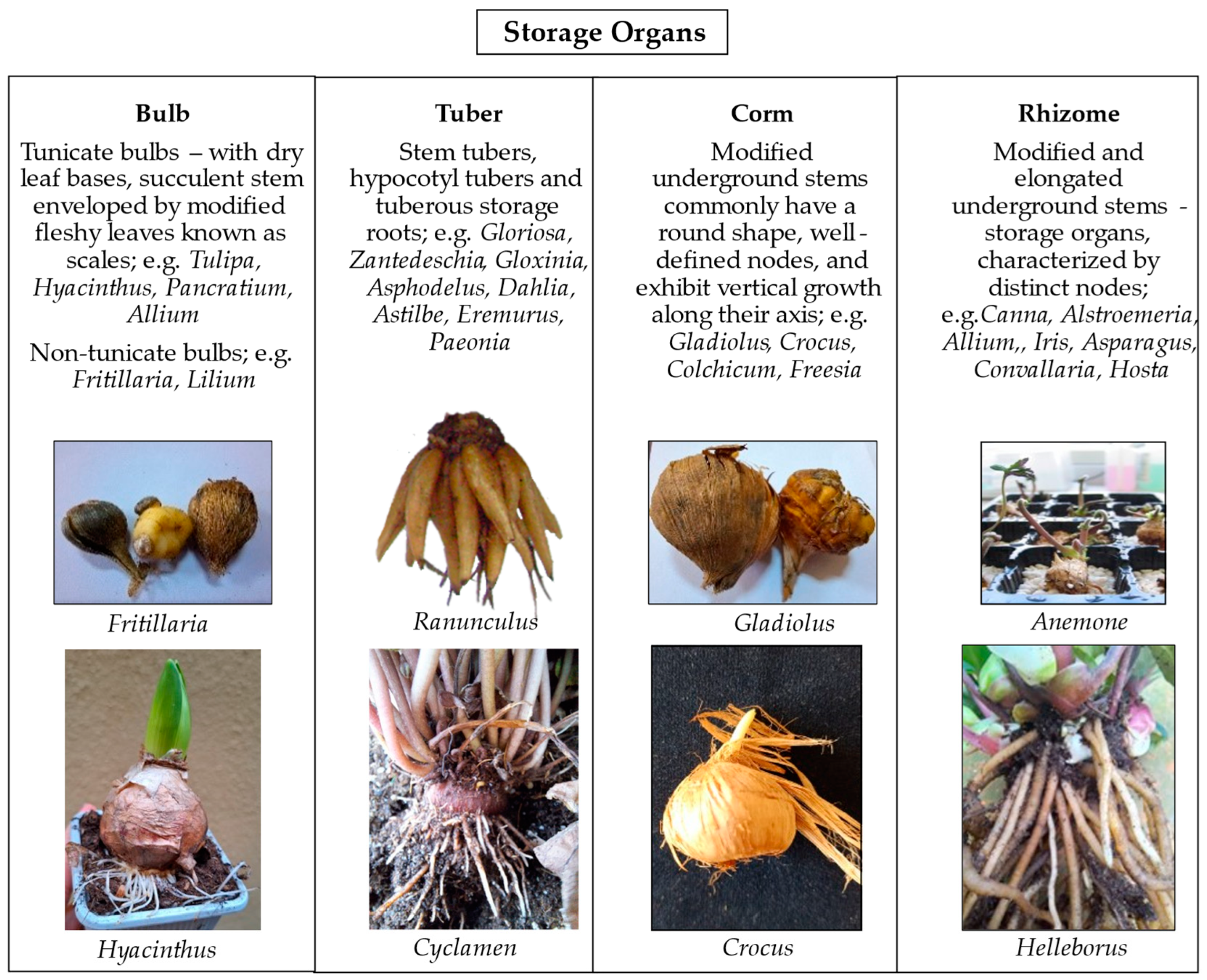
4. Flower Bulbs: Traditional Propagation and Challenges

Flower bulbs are geophytes with storage organs that are of several kinds, morphologically and physiologically (Figure 1). These storage organs can allow the plant to survive unfavorable periods (whether too dry, too hot, or too cold) [15,32,33]. Thanks to the nutrient reserves within these modified structures, plant viability and further plant development are ensured despite the severe external conditions [34,35]. Prior to the onset of favorable environmental conditions, the differentiation of new buds occurs, allowing for shoot development through the ongoing physiological processes in the underground organs. [18,33,36]. Bulbs go through distinct developmental phases: juvenile vegetative, adult vegetative, and reproductive. The transition to the adult vegetative phase is necessary for flower initiation, which occurs after several years in the juvenile phase for certain species like Tulipa and Narcissus. The shift to the reproductive phase is triggered by high temperatures, leading to flower bud formation. Dormancy follows, requiring a prolonged period of cold for dormancy release and preparation for spring growth. This life cycle is common among bulbous plants, including Tulipa, Crocus, and Hyacinthus [15,33,37].
Geophytes can be propagated through generative (seed production) and vegetative methods [18]. The commercial production of flower bulbs through seed propagation is not widely preferred due to several reasons [36,38]. Firstly, seed production could result in offspring that are not identical to the parent plant, leading to increased diversity. This lack of uniformity makes this kind of propagation unsuitable for commercial production, except for specific seed-raised crops. Furthermore, the seed of some bulb plants has limited viability, and precise requirements for germination could be necessary to produce seedlings. Another difficulty in applying seed multiplication is the extended juvenile phase, lasting up to 5–7 years before flowering, which could be faced by certain species [38]. Nevertheless, seed production is highly valuable in breeding programs, allowing for the development of new cultivars. Seed propagation is particularly advantageous if a high number of seeds can be obtained, the juvenile phase is relatively short, and variability is not a concern compared to vegetative propagation [32]. Another advantage of seed propagation is that in almost all cases, plants produced are free of virus infection; vice versa, through vegetative propagation methods, there is the risk of spreading pathogenic microorganisms and compromising production. Examples of geophytes that can be commercially propagated through seed production include Anemone, Allium, Begonia, Cyclamen, Freesia, Fritillaria, and Ranunculus [18,33,36,38,39].
New techniques and strategies have been developed to improve the seed set, increase seed germination, and shorten the time required to flower [40]. Interesting findings were presented by Anderson [41] for reducing the generation time in Lilium and Gladiolus (less than 1 year from seeding to flowering). His laboratory developed a selection strategy to reach rapid generation cycling (RGC) in geophytes. A “toothpicking” technique by selecting germinated seedlings having different colors based on the week of germination was proposed to select early flowering hybrids of Gladiolus. In addition, comprehensive selection work was undertaken for all the phases of the life cycle of geophytes to correlate the features with the early flowering. The author’s conclusions were that the geophytes breeding program can be shortened by selecting genotypes with good seed germination and seedlings with rapid leaf growth or with enhanced leaf unfolding rates, followed by further observations on the geophytic structures and contractile root generation, as well as the dormancy status and the subsequent stalk elongation and flowering.
In nature, the vegetative propagation of flower bulbs occurs through natural division. Natural division can be with daughter bulbs (tulip, crocus), offsets (hyacinths, muscari), bulblets (lily), bulbils (lily), cormels, rhizomes, branched rhizomes, and stolons (allium, oxalis) [32,38,42]. The multiplication rates of the natural division are quite low [15,18,32]. For this reason, it has been envisaged that flower bulbs need alternative propagation methods. Traditional propagation methods include scaling, twin scaling, chipping, scooping, and scoring, as well as stem and leaf cuttings [42]. Scaling is the method where individual scales are removed from the bulb to produce bulblets. Twin scaling, a variation of scaling, involves attaching two scales to a piece of the basal plate. Chipping, a mechanical technique similar to twin scaling, is employed to separate scales. Scooping involves removing the basal plate from the scales, while scoring entails cutting the basal plate. Additionally, stem and leaf cuttings can be used as propagation methods for certain bulbous plants. These techniques have been widely practiced by horticulturists and gardeners to propagate and expand their bulb collections, ensuring the continued availability and diversity of these plants [18,38,39]. It can also be considered a bulbous structure and propagation method. Tunicate bulbs can be multiplied using techniques such as offsets, scoring, scooping, coring, sectioning, and cottage. Non-tunicate bulbs, on the other hand, can be propagated through scaling after they have bloomed. Corms can be multiplied using cormels, which are small corms that develop between the old and new corms. It typically takes one to two years of growth for cormels to reach the size at which they can produce flowers. Tubers can be propagated by either planting the entire tuber or dividing it into sections, ensuring each section has at least one bud (eye). Rhizomes are divided into sections, each containing at least one shoot bud or active shoot. Some rhizomes have roots attached to their bottoms and can be individually planted in containers. Rhizomes can also be planted in nursery beds and used as a source of bare-root stock for planting or for cultural purposes such as basketry. Crown division is another method of propagation that differs slightly from dividing rhizomes [32,43]. However, these methods also present notable limitations. Firstly, generating a substantial number of plants within a practical time frame can be challenging. Secondly, the propagation process carries a significant risk of disease transmission. Tissue culture techniques offer effective solutions to conventional challenges in flower bulb propagation, and this is explained in Section 5.

5. Micropropagation

Micropropagation is a widely recognized method of vegetative propagation that allows for the production of numerous offspring plants. Podwyszyńska et al. [44] reported that the global annual production of ornamental plants through in vitro cultures has surged from 800 million to 2 billion in the last decade. According to a recent research report, the micropropagation market size is expected to rise by 2030, reaching a value of 2.8 billion USD (about 2.5 billion EUR; report “Micropropagation Market-Global Outlook and Forecast 2022–2027”-https://www.reportlinker.com/p06313600/?utm_source=GNW, accessed on 25 February 2024). Europe is one of the most prominent geographical segments in the global micropropagation market, with 140 commercial in vitro laboratories for which the production of ornamental plants is pivotal [44].
The micropropagation process involves multiplying plants under sterile conditions on a nutrient culture medium with a known composition and optimal artificial culture conditions [45]. Micropropagation represents an intricate plant multiplication technique involving substantial capital investment, significant operational costs, and a need for specialized expertise [46,47,48]. However, tissue culture methods offer various benefits, particularly in micropropagation, which is employed to multiply diverse plants, including genetically modified or conventionally bred ones. It proves useful for producing plantlets from seedless or challenging-to-reproduce plants economically and quickly, significantly reducing the time needed for abundant plantlet production [36].

5.1. In Vitro Regeneration Pathways

In vitro plant regeneration is a process where explants, through cell division and differentiation, give rise to the formation of organs and tissues [49]. This comprehensive process involves the regeneration of an entire plant from various sources, including adult tissues, organs, unorganized calli, or even a single cell [50,51].
Somatic embryogenesis is a remarkable process in which plant somatic cells undergo dedifferentiation, acquire embryonic cell characteristics, and subsequently, through embryonic development, form complete plants [52,53,54]. This process highlights the totipotency of plant cells, demonstrated by the formation of embryogenic calli [51,55]. It involves the development of structures resembling zygotic embryos from cells that are not associated with zygotes or the original tissue, closely mirroring the characteristic stages observed during zygotic embryogenesis [54]. Somatic embryos can differentiate through two pathways: direct somatic embryogenesis, where they differentiate directly from explants without the formation of a callus, and indirect somatic embryogenesis, where differentiation occurs after passing through a callus stage [56]. Somatic embryogenesis is favored for mass propagation due to its higher proliferation rate, ease of liquid culture medium utilization, efficient handling of numerous embryos, and greater adaptability for bioreactors [36,49].
Freshly formed structures like shoots, roots, or embryos can emerge on plant tissues without preexisting meristems. These recently developed organs are termed adventive or adventitious, and this initiation of a novel structure and organization is termed organogenesis [57,58]. Organogenesis involves the perception of phytohormones, differentiation of specialized cells to gain organogenic competence, cell division regulation, cell expansion, and the overall patterning of the organ [59,60,61]. The presence or absence of the callus stage results in two distinct developmental stages. The developmental stage that includes the callus phase is called indirect organogenesis. In indirect organogenesis, the likelihood of observing somaclonal variation is higher. The developmental stage that occurs without the callus phase is termed direct organogenesis [62,63]. The first step in the propagation of plant material using the organogenesis technique is the selection of the explant source from the donor plant. Usually, embryos, seedling parts, apical meristems (root or shoot tips), primordial organs, protoplasts, and young flower buds are used [58,61].
Many factors can affect the efficiency of the regeneration pathway, such as explant type, genotype, concentration, and type of PGRs, regeneration medium, other chemicals that indirectly affect plant growth, stress conditions, subculture, electrical excitation, or gene expression modification [51,52,53,64,65]. The shoot regeneration capacity depends on the establishment of a complex process involving the endogenous cytokinin metabolism of tissues and how the explants react to the exogenously supplied plant regulators in the culture medium [60]. Consequently, many factors should be considered when an explant is chosen to initiate an in vitro culture. Beside the medium composition and the culture conditions, the physiological and ontogenic age of the tissues, the season in which the explant is taken, the size and location of the explant, and the quality of the donor plant are key factors, which affect the explant viability and the degree of contamination in in vitro culture [51]. In addition, explant age and origin, as well as the period of the year in which the in vitro culture is initiated, affect the phenolic content of the tissues, which, in turn, can cause browning and subsequent death of tissues [66]. Figure 2 shows the aforesaid regeneration pathways.

5.2. Micropropagation of Flower Bulbs

Micropropagation of ornamental plants has been studied since the 1950s [67,68]. The impact of this technology on flower bulbs is also testified to by the literature cited in this review. Micropropagation involves axillary bud development through organogenesis, adventitious shoot formation, and somatic embryogenesis [69].
Plant biotechnology significantly contributes to the production of ornamental geophytes through various approaches. These methods focus on propagating specific genotypes, acquiring virus-free plant material, and supporting breeding and crop improvement programs [18,36,69,70]. Techniques such as callus culture, embryo rescue, in vitro pollination, somatic hybridization, induction of somaclonal variation, protoplast culture, synthetic seed production, in vitro ploidy manipulation, genetic transformation, gene mapping, and DNA fingerprinting play essential roles [22,69,71]. In vitro pollination and fertilization, embryo, ovary, ovule, anther, and pollen culture have been reported for some geophytes such as Lilium, Cyclamen, Anemone, Ranunculus, and Amaryllis [72,73,74,75,76,77,78,79,80]. Further scientific inquiry is warranted to evaluate the feasibility and potential applicability of protoplast culture and somatic cell hybridization techniques in the context of geophytes such as Crocus cancellatus as studied by Karamian and Ebrahimzadeh [81]. Recently, Koetle et al. [82] reported about the procedures using Agrobacterium strains to transform into geophytes such as Crocus, Allium, Agapanthus, Lilium, Tulip, Gladioulus, Hyacinthus, and Narcissus [83,84,85,86,87,88,89,90,91,92].
Micropropagation systems were developed for major bulbous plants, but commercial propagation was achieved only for a few bulbous plants, such as Lily and Zantadeschia [36]. For other ornamental geophytes, more efforts are necessary to reach commercial applications. Nevertheless, it is well recognized that the availability of proper tissue culture protocols would enable the production of high-quality stock plant material that is virus-free and the advancement of breeding programs or the propagation of selected genotypes.

5.3. The Key Factors Affecting Micropropagation of Flower Bulbs

Establishing a successful micropropagation protocol is a complex task, influenced by various interacting factors, including plant materials, culture conditions, and culture media ingredients. Despite the development of micropropagation protocols for many flower bulb crops, challenges hinder commercial viability. Key limitations include the high cost of tissue-culture plantlets, inefficiencies in the initiation phase, low propagation rates for numerous genotypes, challenges during the acclimatization of ex vitro plantlets, and the occurrence of off-types.

5.3.1. Explant Choice

Thanks to its totipotent character, the potential for entire plant regeneration exists within every living plant cell. However, cells or tissues displaying active growth and robust physiological metabolism are commonly utilized as explants for the regeneration process in many studies [51]. A variety of tissue sources, including shoot tips (apical and axillary buds), bulb scales, leaves, stems, and different parts of the inflorescence, are utilized as explants in the initial stages of in vitro culture [38,93]. Below, in Section 5.4.2, some examples of the establishment of an in vitro culture for flower bulbs are provided.
As previously explained (Section 5.1), the selection and preparation of suitable explants play a crucial role in successful in vitro culture. Factors such as the regeneration capacity, physiological state, and hormonal balance of the donor plant, the origin, age, and size of the explants, their polarity, and pre-treatment methods are key considerations for the successful initiation of ornamental bulbous plants [94]. The induction of in vitro bulblets is primarily influenced by the temperatures and storage durations of the mother plant. Yasemin et al. [95] emphasized the significance of explant types regarding factors such as callus color, callus hardness, callus fragility, callus formation rate, embryogenic callus rate, and callus growth rate of Pancratium maritimum.

5.3.2. Culture Medium

The composition of the culture medium is a critical factor influencing the growth and morphogenesis of plant tissues in culture. The Murashige and Skoog (1962; MS) [96] formulation is widely used, initially designed to support the optimal growth of tobacco calli through extensive dose–response curve studies for essential minerals. Furthermore, N6 [97], Woody Plant Medium (WPM) [98], and B5 [99] are used as culture media in tissue culture [51]. Plant tissue and cell culture media are generally made up of some or all of the following components: macronutrients, micronutrients, sugar(s), vitamins, amino acids or other nitrogen supplements, other undefined supplements, solidifying agents or support systems, and growth regulators [100]. Undefined elements can be coconut milk, meat, malt, yeast extracts, potato extracts, juices, pulps, fruit extracts, plant/plant pars/seedling extracts. Osmotic balance, pH, and buffers of the culture medium also affect the target in the tissue culture [101]. Elicitors, such as chitosan, aminolevulinic acid (ALA), alginate (ALG), N-acetylglucosamine (NAG), salicylic acid (SA), hyaluronic acid (HA), silver nitrate (AgNO3), jasmonic acid (JA), methyl jasmonate (MeJA), phloroglucinol (PG), and pectin, are extensively employed to induce secondary metabolites in plant tissue culture [71]. Methyl-Jasmonate (MeJA) and various polyamines also prove advantageous in tulip tissue culture by promoting efficient bulb formation during micropropagation [102]. Moreover, the medium’s physico-chemical properties can alter the nutrient status of microshoots. The selection of the gelling agent is a crucial factor in the process of in vitro plant regeneration. The medium should be sufficiently firm to support explants, avoiding excessive rigidity that may hinder proper contact. The phenomenon of hyperhydricity, linked to agar, is also a recognized concern [103]. The experiments carried out over a ten-year period, highlighted that a relationship can be found between gel properties and the biological performance of in vitro Ranunculus shoots, and we correlate it with the different water and nutrient availability created in the different gelled media [104]. Moreover, our studies identified the concentration and diffusion through the gel of major phenolic impurities that can be found in the agar powder used to jellify the media. These findings proved that agar impurities can greatly affect the growth and multiplication of microshoots (Figure 3). Therefore, attention should be paid to choosing the agar brand and concentration and, even more importantly, creating a standardized method for the preparation of the gelled media.
The use of solidified media in micropropagation offers advantages, including easy visibility and recovery of small explants, maintained explant orientation, improved aeration without special measures, and orderly growth of shoots and roots. In contrast, liquid media can cause disoriented growth and difficulties in shoot separation. However, there are drawbacks to semi-solid media, such as the presence of the above-said inhibitory substances in agars, slow growth rates, and limited diffusion of toxic exudates. Poor oxygen diffusion to developing roots is a concern, even in aerated liquid media. Adherence of gel to roots can pose issues during plantlet transfer to soil, and cleaning containers for re-use becomes time-consuming with semi-solid media [105]. Different plants have varying nutritional requirements, affecting optimal growth and morphogenesis. Tissues from different plant parts may have unique needs for satisfactory growth [51].

5.3.3. Environmental Conditions

Temperature and light have key roles in micropropagation. Under in vitro conditions, younger tissues typically import sucrose and have limited photosynthesis. However, light (color and intensity) influences plant growth, metabolism, and organogenesis and has proved to be crucial for the in vitro regeneration of the storage organs of geophytes [38,70,71]. White, fluorescent light (350~750 nm) is conventionally used in in vitro culture but has drawbacks like high electricity consumption and uneven radiation. Monochromatic LEDs with specific wavelengths are now widely used for more energy-efficient and controlled in vitro plant propagation [71,106]. Phytochrome governs the plant’s light response, influencing organogenesis induction and direction. Blue light and its receptor, cryptochrome, play a role in storage organ formation [107]. Examining light quality effects during adventitious organogenesis in bulbous plants, researchers utilized monochromatic fluorescent lamps. The results revealed that in in vitro cultures of Hyacinthus orientalis, blue light stimulates adventitious shoot development, while red light encourages the formation of adventitious bulbs [108]. Studies have explored the impact of light on plant growth and development parameters in various lily species and cultivars during in vitro organogenesis. [109,110,111]. According to Bach and Sochacki [38], the choice of light may interact with tissue cytokinin metabolism, potentially influencing in vitro tissue dormancy and subsequent development. While light is crucial for typical green shoot and plantlet growth, unorganized cell and tissue cultures can thrive without it. Surprisingly, darkness may enhance growth and morphogenesis, with brief dark periods observed to promote shoot morphogenesis in certain cases [105]. Embryo explants from P. maritimum were cultivated under both photoperiod and dark conditions. The results, based on unpublished data from Yasemin S., indicate that the dark condition was more successful in generating calli, and Figure 4 illustrates the outcomes after one month of incubation.
Temperature influences both growth rates and the transition between vegetative and reproductive phases in plant development. In flower bulbs undergoing in vitro culture, temperature is a vital factor influencing regeneration, bulbing, and dormancy. For temperate ornamental geophytes, lower temperatures mimic winter, prompting dormancy. Under in vitro conditions, reduced temperatures induce storage organs to withstand unfavorable growth conditions [67]. Temperature is another key factor that is able to influence the regeneration, bulbing, and dormancy of flower bulbs during in vitro culture [112]. In vitro-regenerated bulblets necessitate dormancy breaking to facilitate ongoing development. A cold treatment was proven to be effective for bulblet initiation and for breaking dormancy in the regenerated bulblets [38].

5.4. Stages of the Micropropagation of the Flower Bulbs

Many scientific papers have been published on flower bulbs, highlighting the discrepancy between the potentiality of the micropropagation technique and its practical application. Next, we would like to analyze the most important findings related to the major steps in micropropagation of flower bulb crops, which are: preparation of mother plants, initiation, multiplication, bulbing/bulb growth, dormancy breaking, and planting. Dormancy breaking is a different step from the other herbaceous and woody plants [18,36].

5.4.1. Stage 0: Preparation of Mother Stock Plant Material

Happy plants make happy plantlets. Stage 0 in ornamental geophyte propagation represents the initial and crucial phase dedicated to ensuring the selection and cultivation of healthy plants [47]. This pivotal stage aims to pre-process the chosen specimens to prevent contamination, ultimately fostering the development of thriving plantlets [42]. Key pre-treatments, such as regulating humidity, irrigation methods, temperature, and light exposure, are implemented to curtail the potential source of contamination and maintain the genetic integrity of the desired traits. Additionally, the duration of storage impacts the formation of bulb-like structures in geophytes, further emphasizing the significance of meticulous handling during this phase [18,46,70,113,114,115,116].
To initiate the in vitro culture of tulip, Podwyszyńska and Sochacki [117] selected healthy, true-to-type, and virus-free plants during their flowering phase in spring. After having carried out ELISA tests to verify the virus-free status of their plants, they harvested the bulbs in June and stored them at 17–20 °C until October, followed by dry cooling at 5 °C. The selected healthy bulbs (the recommended bulb size is 10 cm in diameter) were then planted in pots with a fungicide (Captan)-soaked perlite substrate and forced in darkness at 9 °C for 5–14 days prior to initiating the in vitro culture. The effect of a bulb pre-treatment at 5 ± 2 °C for a period of 3–4 weeks was also positively referred by Muraseva and Novikova [118], who developed an efficient protocol for in vitro propagation from bulb scale explants of Fritillaria ruthenica Wikstr., an endangered and rare species. In other cases, natural conditions made it advisable to prepare the mother plants. Kumar et al. [119] successfully established an efficient plant regeneration system for Lachenalia. viridiflora via somatic embryogenesis by growing the mother stock plants in greenhouses under controlled conditions that closely resembled the natural habitat of this plant species, which is collected from a specific sub-population in St. Helena Bay, Western Cape, South Africa, credited to Prof. Graham Duncan. Around 10 mature bulbs of L. viridiflora were carefully selected and planted in terracotta pots with a standardized diameter of 200 mm. The potted plants were then nurtured under ambient temperature conditions and exposed to the natural photoperiod in the controlled environment of the University of KwaZulu-Natal Botanical Gardens’ greenhouse. The researchers took special care to ensure regular watering and maintained a weed-free environment to promote optimal growth and development, mimicking the plants’ natural conditions as closely as possible. Mirici et al. [120] studied in vitro bulblet regeneration from fresh bulb scale and immature embryos of the endangered geophyte Sternbergia fischeriana. Before explant sterilization, the collected bulbs were subjected to a drying process in the dark at room temperature for a duration of 6 weeks.
In conclusion, it could be argued that closed environments, such as glasshouses and tunnels, are high-quality places for plant cultivation with a reduced risk of contamination. Treating plants with fungicides and insecticides before obtaining plant material helps to decrease the risk of contamination. Various practices are available to enhance in vitro development, altering the physiological state of the parent plant from which explants will be taken. These practices include implementing long-day treatments, cultivating under red light, maintaining bulbous plants at low temperatures, and applying PGRs to the leaves [48]. By focusing on the careful selection and pre-processing of mother plants, stage 0 lays the foundation for a successful propagation process, leading to the cultivation of healthy ornamental geophytes [47].

5.4.2. Stage 1: Establishment of Aseptic Culture

In this stage, the selected explants are excised from the stock plant material and surface sterilized with certain chemicals before they are inoculated in the culture medium. Surface sterilization of plant explants for successful establishment in in vitro conditions, particularly in the context of plant tissue culture, is very important. The primary goal is to eliminate microbial contamination, both exogenous and endogenous, without causing harm to the plant tissues. The success of the sterilization process is crucial for the subsequent stages of tissue culture [121,122,123,124]. Various chemicals, such as antibiotics, fungicides, sodium hypochlorite, mercuric chloride, ethanol, hydrogen peroxide, calcium hypochlorite, and silver nitrate, are commonly used for surface sterilization. The concentration and duration of the disinfection process need to be carefully considered to achieve effective sterilization without compromising the viability and regeneration ability of the explants. The choice of disinfectants depends on the specific plant species and type of explant. The goal is to create aseptic conditions for plant tissue culture by addressing the challenges associated with microbial contamination and maintaining the health of the explants [122,125]. The use of seeds as an initial material in in vitro studies is advantageous because it decreases the risk of contamination. After germination of the seeds, in vitro seedling parts can be used as explants for some research [124,126,127]. Yasemin et al. [124] conducted the surface sterilization experiments for the seeds of P. maritimum. They used 70% ethanol and washed the seed with distilled water, followed by treatment with different concentrations of sodium hypochlorite (NaOCl 1.6% and 3.2%) at different durations (15, 20, and 25 min). High NaOCl and long durations had negative effects on the seeds viability, germination, and plant formation. Kumar et al. [119] described the successful surface sterilization process for L. viridiflora leaves. The leaves were treated on a laminar flow bench using 70% ethanol (v/v) for 60 s, followed by immersion in 2% sodium hypochlorite (NaOCl) for 10 min. To aid in the process, a few drops of Tween 20 were added as a surfactant. After sterilization, the leaves were rinsed three times with sterile distilled water.
Unfortunately, the contamination issue represents a substantial concern for the flower bulb tissue culture, particularly when underground plant tissues are used to initiate the culture. Although surface sterilization is successful, the plant tissues can be contaminated due to endogenous microorganisms in the bulbs, and this could also appear in further culture cycles [36]. Therefore, a thorough sterilization process is indispensable to eliminate contaminants from the geophytic storage organ explants. In the study conducted by Lagram et al. [128], a protocol was employed for the preparation of saffron daughter corms (Crocus sativus L.). The corms were subjected to a series of treatments, including a 30 min immersion in tap water, gentle brushing with Tween-20, a 1 min exposure to 80% ethanol, a 20 min treatment with freshly prepared mercuric chloride (HgCl2) at a concentration of 0.2%, and, finally, rinsing four times for 4 min each with sterile distilled water. Sochacki and Orlikowska [129], in their studies aimed at improving the micropropagation protocol for two Narcissus cultivars, tested several pre-treatments of bulbs, including the use of fungicides and hot water treatment (44.4 °C for 3 h), followed by a disinfection procedure with HgCl2 and chloramine T. By using these methods, they were successful in reducing the initial contamination to 14–17%, and they showed that different genotypes could differently react to the same treatment.
Seeds of P. maritimum were used as explants in some studies, and surface sterilization was successful [95,124,126,130]. However, surface sterilization of the flower bulbs is not quite easy. According to studies conducted by Yasemin S (unpublished data), Gladioulus corms and Fritillaria bulbs were washed under tap water, then treated with 70% ethanol and 2.5% NaOCl and washed by sterile distilled water (three times). Unfortunately, all the explants were contaminated due to the fact that the mother plant was already not healthy. This could show the importance of mother plant material selection. Some explant photos are shown in Figure 5.
Evaluating the negative impacts of sterilizing agents on plant tissues is crucial, with explant survival data serving as a key parameter. Rafiq et al. [131] and Rather et al. [132] indicate that combined chemicals were more effective for the achievement of disinfection, but the use of combined sterilants lead to a reduction in explant survival. Rafiq et al. [131] experimented with diverse sterilization approaches, incorporating NaOCl (1%) and HgCl2 (0.1%) at varying durations (10 and 20 min), along with combinations involving 70% ethyl alcohol and carbendazim (200 ppm). The explants were bulb scale-base and bulb scale-tip from the oriental hybrid Lilium cv. Ravenna. The most effective surface sterilization method entailed treating healthy bulb scales with carbendazim (200 ppm) for 30 min, followed by 0.1% HgCl2 for 10 min, and then 70% ethyl alcohol for 30 s. Farooq et al. [133] used bulb scales and young leaves to initiate the culture of Lilium LA hybrids ‘Indian Summerset’ and ‘Nashville’. A pre-treatment of both explants has been made by washing the tissues in a water solution containing Tween-20 and the fungicide carbendezim. Afterwards, the surface sterilization of the explants was performed using carbendenzim, HgCl2, and ethyl alcohol at different concentrations and durations. The highest surface sterilization success for bulb scales (Indian Summerset: 85.41%, Nashwille: 89.58%) was higher in the combinational sterilant. Chib et al. [84] also detailed an optimized two-step surface sterilization method for C. sativus L. This involved separately employing 0.1% HgCl2 and 4% NaOCl, leading to effective asepsis and a noteworthy 86% survival rate of explants. The disinfection of corms was successfully achieved through this process. Application of antioxidants, such as ascorbic acid and citric acid, can effectively prevent browning caused by oxidation. Furthermore, some additives, such as polyvinylpyrrolidone (PVP), charcoal, and fungicides, can also prevent the browning of explants and endogenous contamination risks. Appleton et al. [134] performed in vitro regeneration of Hypoxis colchicifolia. When establishing in vitro cultures, they have experimented with PVP, activated charcoal, ascorbic acid, citric acid to inhibit browning, and benomyl solutions as fungicides to inhibit contamination. They have solved the browning problem with PVP and partially solved the contamination problem with benomyl. However, these applications do not create the same response in every plant or plant part. This should be analyzed and optimized for each plant. Devi et al. [135] have used charcoal and ascorbic acid to reduce the phenolic exudates, but charcoal inhibited tissue growth, and ascorbic acid caused somatic embyo death in saffron (C. sativus L.).
In general, young tissues and organs have a higher regeneration capacity than older ones. Apical and axillary buds from tubers or bulbs can be used to initiate the in vitro culture. Furthermore, adventitious buds are generally induced by bulb scales or flower stems. Twin scales are useful explants, although the use of underground storage organs leads to serious contamination problems during the initiation and further multiplication phases. Flower stems have the advantage of having low endogenous contamination rates compared to other tissues [18,24,36]. Vegetative segments of plants often regenerate more easily in vitro than generative ones. Rafiq et al. [131] used basal and tip bulb scales to micropropagate the oriental hybrid Lilium cv. Ravenna. They also found that the explant survival rate of basal scales was higher than that of tip bulb scales. Lapiz-Culqui et al. [136] and Patil et al. [137] used the bulb scales to cultivate different Lilium cultivars. Youssef et al. [138] used the leaf as an explant in their study to increase Lilium bulb number and size. Ozel et al. [139] used a twin-scale bulb explant to micropropagate the endemic Muscari muscarimi. Twin scales were also used by Santos et al. [140] to propagate Narcissus asturiensis and Kukulczanka et al. [141] to propagate Fritilaria melagris through the same system. Kumar et al. [142] used the bulb scales of the critically endangered Fritillaria roylei for in vitro culture. Sevindik and Mendi [143], Taheri-Dehkordi et al. [144] used the corms as explants to propagate the C. sativus L. Furthermore, Slimani et al. [145] informed us that the vegetative apices, apical and axillary buds, meristematic zone, segments, leaves, ovaries, protoplasts, corms, and roots are useful to induce the somatic embryogenesis of C. sativus L.
Success in this initiation stage can be influenced by environmental factors encompassing nutrient composition, PGRs, light exposure, temperature, atmospheric composition, and various culture methodologies [18]. Devi et al. [135] reported that the dark conditions were effective for somatic embryo proliferation in saffron.

5.4.3. Stage 2: Multiplication

The process of multiplication, which is a continuation of in vitro initiation, constitutes one of the most critical stages in micropropagation. As mentioned in previous stages, leaves, inflorescences, and bulbous structures (such as bulbs, corms, tubers, etc.) are utilized for multiplication [53,69]. The selection of the medium to be used (solid, liquid, semi-solid, or media types MS, B5, WPM, etc.) is crucial for achieving successful responses, in addition to the use of different explants. The optimization of factors such as salt mixtures (essential macro-micro elements, carbon sources, vitamins), sugar derivatives, ratios, types, and concentrations of PGRs, light, temperature, and inductive agents is necessary [69,70]. The concentration of the PGRs (e.g., cytokinin) is critical, as it can simultaneously promote multiplication and development while causing adverse effects [146,147,148,149]. Each step implemented here will influence both multiplication and subsequent planting stages; hence, optimal requirements need to be determined. Unfortunately, due to the variability in results obtained for each species and even within varieties, specific optimization efforts are required for each plant. This situation poses one of the challenges of tissue culture. Additionally, high production costs add to these challenges [150].
To achieve effective results at this stage, understanding the plant’s physiology and mimicking its natural requirements based on the choices made during this phase can be impactful. The selections made during this stage lead to different differentiations according to the meristematic zones that will be formed as a result of stimulations in explants (somatic tissues) [69]. In the regeneration stage, unipolar meristems give rise to shoots or roots, which is organogenesis. Bipolar meristems, on the other hand, lead to the development of a complete plant or microcorm, representing somatic embryogenesis [151].
PGRs can stimulate or inhibit the in vitro development of geophytes. Sochacki et al. [152] aimed at evaluating the influence of PGRs on in vitro shoot multiplication of Tulipa L. ‘Heart of Warsaw’, and they yielded the best performance (9.14 shoots/clump) when MS medium was supplemented with N6-(-isopentyl)adenine (2iP) 0.1 mg/L, 1-Naphthaleneacetic acid (NAA) 0.1 mg/L, and meta-topolin (mT) 5.0 mg/L. Additionally, this study showed that the type of carbohydrate added to the nutrient solution had a significant effect on the shoot proliferation of the tulip cultivar; the highest multiplication of the shoots (number of shoots for one starting clump) was achieved with sucrose (34.33 shoots/per clump) and glucose (35.88 shoots/per clump) as carbohydrates. Lagram et al. [128], used excised mother corm buds under different 6-benzyl aminopurine (BAP), 2,4-Dichlorophenoxy acetic acid (2.4-D) and NAA concentrations in MS medium. The highest bud sprouting (96.67%) and shoot growth (8.87 cm) were obtained from 1 mg/L BAP and 1 mg/L NAA. They also evaluated adventitious shoot formation in the explants. The highest adventitious shoot regeneration (80%) was obtained from 0.5 mg/L NAA and 2.75 mg/L BAP. Recently, micropropagation via organogenesis has been on the rise. Successful adventitious bud regenerations were obtained in Lilium, Narcissus, Sternbergia, Hippeastrum, Firttilaria, Muscari, Tulip, Iris, Lachenelia, and Hyacinthus [111,112,118,120,138,139,153,154,155,156,157,158,159,160,161,162]. As seen in Figure 6, organogenic structures were obtained from bulb scales in P. maritimum.
Slimani et al. [145] explained the importance of somatic embyrogenesis for C. sativus L. to propagate healthy corms. MS, LS, and B5 media were supplemented with the growth regulators BA, NAA, Kinetin (Kn), Thidiazuron (TDZ), and 2.4 D tested at different concentrations. Ebrahimzadeh et al. [163], obtained somatic embryos by using meristem explants of C. sativus L. cultured in LS culture medium, which included 2 µM benzyladenine (BA) and 2 µM NAA. Sheibani et al. [164] used corm explants in MS medium supplemented with TDZ (0, 0.1, 0.25, and 0.5 mg/L), and they obtained somatic embryos from 0.5 mg/L TDZ. Marković et al. [165] delved into the intricate world of somatic embryogenesis in F. meleagris, emphasizing the crucial role of PGRs in this process. Their study illuminated that the medium without PGRs exhibited exceptional efficiency throughout the experiment, showcasing a robust somatic embryogenic response. Moreover, the medium with lower concentrations of PGRs also proved to be conducive to somatic embryogenesis. The pinnacle of morphogenetic success was achieved in a BAP/2,4-D-containing medium with the lowest PGR concentrations. This insight into the optimal conditions for somatic embryogenesis opens avenues for enhanced propagation strategies, potentially revolutionizing the large-scale production of F. meleagris. Kocak et al. [166] investigated somatic embryogenesis potential in various explants (ovules, divided ovary parts, leaves, and petiole segments) from 15 different genotypes of the wild species Cyclamen persicum Mill. The explants were cultured on a medium with specific PGRs to induce embryogenic callus. The study found significant variations in embryogenic potential among explants and genotypes. While petiole explants produced the most callus, ovary explants were most efficient in forming somatic embryos. Recently, an efficient method for tulip regeneration via SE was developed [167,168]. Some reports on SE are also available by using leaf, petiole, ovary, anther, roots, and aseptic seedling tissues as explants in cyclamen [166,169,170,171,172,173,174]. Aseptic seedling tissues (cotyledons, petioles, tubers, and roots) have also been used as explants to initiate SE culture in many medicinal and endemic geophytes (e.g., Crocus, Iris, Hypoxis, Colchicum, etc.) [135,143,163,164,175,176,177,178]. Some embryo-like structures from P. maritimum bulb explants are shown in Figure 7A–C. Moreover, embryogenic callus and regeneration in R. asiaticus were shown in Figure 7D,E. Thalamus-derived calli in R. asiaticus L. have been initiated on MS medium containing the growth regulator 2,4-D and cytokinins (BA and kinetin), according to Beruto et al. [179].

5.4.4. Stage 3: Bulb Growth

Following the multiplication phase, in non-bulbous plants, the optimization of media plays a crucial role in enhancing the successful rooting of developed shoots. However, this phenomenon is not uniformly applicable to geophytes, where the acquisition of storage organs proves to be challenging and time-intensive [152]. This phase in geophytes is directed towards the acquisition of shoots, primordia, and bulbs (Figure 8).
In some plant species, the sequence involves the initial rooting of shoots followed by subsequent bulb formation, while in others, bulbs are generated directly [18]. The induction of bulb formation occurs through diverse mechanisms. Investigations into bulb formation have explored the influence of various sugar types and concentrations, PGRs, light quality, temperature, and distinct tissue culture conditions (solid, liquid, medium, and bioreactor utilization). Sochacki et al. [152] conducted an investigation on the bulb formation of previously multiplied Tulipa L. ‘Heart of Warsaw’ plantlets, examining various carbohydrate types, different phase media, and PGRs. The highest bulb formation (28.00 bulbs) and the most mature bulbs (14.50 bulbs) were achieved in a two-phase (liquid) system with 1 mg/L PBZ and glucose. Pałka et al. [111] utilized bulb scales of Lilium candidum as explants in in vitro MS media and assessed the impact of different light qualities (spectral compositions of red (100%), blue (100%), and red and blue light (RB ratio 7:3), RB light was mixed in equal proportions (50%) with green (RBG), yellow (RBY), UV (RBUV), and far-red (RBfR) light, white LED (Wled), fluorescent lamp light (Fl), and darkness (D)) on bulb formation without the use of PGRs. The study resulted in a bulb formation rate of 79–100% across all explants. The best results in terms of bulb number were obtained from Fl (16.3), RBY (13.33), and RBfR (13.33), B (12.69), RB (12.63), and Wled (14.36) treatments. The lowest rate (3.00) was observed in plants kept in darkness. The largest bulb diameter (5.41 mm) and the highest photosynthetic pigment content were obtained from the RBG treatment. Darkness and red light induced etiolation. Additionally, soluble sugars in bulbs were stimulated by darkness and blue light. Lagram et al. [128] used different sucrose, IBA, and NAA concentrations on corm production and root regeneration in ½ MS. Furthermore, they considered the photoperiod effect (16 h/8 h and dark conditions) on the parameters. They obtained the best results in ½ MS, 6% sucrose, 1 mg/L NAA, and dark conditions with 100% corm production, 7.9 g mini-corm weight, 93.8% root formation, and a root number of 14.9 per mini-corm. In the realm of bulb growth in F. meleagris, Marković et al. [165] unraveled key factors influencing the development of bulbs in vitro. Their investigation revealed that bulbing ability remained relatively high in the absence of PGRs and, for the most part, was unaffected by varying concentrations of the tested PGRs. Notably, the study identified that the lowest cytokinin concentration, in combination with low auxin, significantly enhanced bulb formation when the cultures were transferred to a higher temperature. This finding underscores the importance of temperature modulation and specific PGR combinations for optimizing bulb growth in F. meleagris. Understanding these dynamics can contribute to refining propagation methods and accelerating the scale-up of bulb production for this species. Azeri and Öztürk [180] investigated the most effective hormone treatment to induce and produce Lilium monodelphum M. Bieb, var. Armenum in tissue culture rapidly and efficiently. Optimal bud regeneration (11.67) was achieved in a medium containing 3.0 mg/L TDZ, 0.25 mg/L NAA, and 0.1 mg/L GA3. Additionally, the highest microbulb formation (15.83) was observed in a medium comprising 2.0 mg/L PAC, 0.2 mg/L NAA, and 0.1 mg/L GA3.
In recent years, the use of bioreactor systems has shown an increase in healthy plant regeneration. Studies on the regeneration of bulbs, corms, rhizomes, microtubers, shoots, and subsequent rooting, as well as somatic embryogenesis, have been on the rise [181]. In their published article, Murthy et al. [181] indicated the proliferation of Lilium hybrids and Allium sativum bulbs, C. sativus and Alocasia amazonica corms, microtubers of Solanum tuberosum varieties, and rhizomes of Cymbidium sinense using various bioreactor systems [110,182,183,184,185,186,187,188,189,190,191,192,193,194]. According to Murthy et al. [181] the selection of an appropriate bioreactor system is crucial due to various factors (design, principle, inoculation density, aeration, temperature, light intensity, etc.) that influence the regeneration of propagules. To minimize losses of small bulblets and prevent dormancy, direct transplantation of plantlets is often preferred.
Rooting can be improved through various methods, such as adding auxin and/or activated charcoal (AC) to the culture medium, adjusting the auxin-to-cytokinin ratio, and using half-strength salts and sucrose. In the study by Azeri and Öztürk [180], microbulbs were subjected to rooting media with indole 3-butyric acid (IBA 0.5 and 1.0 mg/L). Successful plant development, with root numbers of 6.4 and 5.9, respectively, was observed in media containing 0.5 and 1 mg/L IBA.
For successful acclimatization, hardening is essential. This process enhances tolerance to moisture stress and prevents hyperhydricity [18]. In Rafiq et al. [131] study on Oriental Lilium hybrid cv. Ravenna, varying combinations of IBA and NAA significantly influenced microshoot rooting behavior. IBA outperformed NAA, with the highest rooting (92.71%) and primary root characteristics observed in a medium supplemented with 1.50 mg/L IBA. Explants showed differences, with basal scale segments exhibiting the highest rooting (92.71%), percentage root number/shoot (10.02), and length of primary roots (2.17 cm), while tip scale segments had the lowest (77.55%, 9.08, 1.65 cm). Notably, primary hardening influenced plantlet survival, with the highest ex vitro survival (98.96%) in plantlets from IBA (1.5 mg/L)-fortified media, particularly in basal scale segments. Rooted plantlets were hardened in media containing perlite and vermiculite (1:1). This suggests that IBA-treated cultures resulted in superior ex vitro survival, possibly due to enhanced rooting characteristics such as root number and length.
Direct bulblet induction in certain genotypes can provide several advantages, including the elimination of in vitro rooting, prevention of hyperhydricity, avoidance of the need for hardening, increased survival rates, and a shortened bulb production period. Conditions conducive to bulb formation involve high sucrose concentrations, the application of plant growth retardants, exposure to low temperatures, and the utilization of aged shoots. It’s worth noting that in vitro formed bulblets may experience spontaneous dormancy, as reported by Kim and De Hertogh [18]. In the research conducted by Chib et al. [84], they experimented with a high sucrose concentration combined with PGRs to enhance efficiency over a 90-day timeframe. The most favorable outcomes were observed when using a combination of MS medium, TDZ, IAA, activated charcoal, and 4% sucrose, resulting in a notable 68% efficiency. Lower sucrose concentrations led to sluggish growth, while concentrations exceeding 4% resulted in cell death, as evidenced by the blackening of the callus. Different sucrose, BA, and 2,4D concentrations in MS media affected the growing bulbs in in vitro conditions for P. maritimum [95].

5.4.5. Stage 4: Dormancy Breaking

Some geophytes produce new buds that enable vegetative propagation through their underground storage organs [195,196]. After an active growth and flowering period, senescence of aboveground tissues is followed by root senescence, leading the plant into a dormant phase with no visible organogenesis. To survive under unfavorable environmental conditions that are not conducive to their development and proliferation, these organs enter a state of dormancy. Seeds, apical and vegetative buds, floral buds, bulbs, corms, and tubers can enter dormancy. Most geophytes show dormancy as an integral part of their life cycle [197]. Dormancy is characterized by the inability to initiate growth from meristems under favorable conditions. In geophytes, dormancy can manifest in three different types: endodormancy (internal inability to grow), ecodormancy (environmental conditions), and paradormancy (apical dominance, hormonal status, metabolite-sugar levels) [196,198,199,200]. When evaluating the concepts of dormancy and true dormancy, it is important to note that true dormancy persists until dormancy is completely terminated, even if favorable environmental conditions are present. Unlike many other plants, geophytes do not exhibit true dormancy, as activities continue even during dormant phases. Most bulbous structures obtained through tissue culture display dormancy. During in vitro regeneration, bulbs and other storage organs such as tubers and corms typically undergo dormancy, similar to their behavior in natural conditions. The level of dormancy may vary depending on factors such as sucrose concentration, age of the bulb, and environmental conditions [18,116,201]. The growth and sprouting of geophytes in vitro are influenced by dormancy, bulb size, and maturity [202]. Therefore, an efficient in vitro protocol, including dormancy release, is crucial for the rapid, efficient, and valid commercial exploitation of all horticultural geophytes. To overcome this constraint, in some Fritillaria species, different temperature regimes were tested under in vitro conditions. Kizil and Khawar [158] found the bulblet diameter increased on MS medium with 50 mg/L sucrose after 30 days at 4 °C. Successful rooting of Fritillaria bulblets was achieved on MS medium with 0.5 mg/L NAA. Marković et al. [165] carried out bulb-scale culture with different PGR combinations for four weeks at 7 °C in F. meleagris. They found that in the control medium (PGR-free medium), shoots per explant were higher and reached a maximum at the end of the chilling. In the study conducted by Carasso and Mucciarelli [203], seeds of F. tubiformis Gren. & Godr were chilled at 4 °C for 30, 60, and 90 days in 1% agar medium. At the end of cold stratification, seeds were disinfected, and immature zygotic embryos were rescued from seed coats. The highest number of somatic embryos occurred in zygotic embryos subjected to 30 days of cold stratification on MS medium supplemented with 8.88 µM BA and 2.68 µM NAA. Successful conversion of somatic embryos into bulblets necessitated transfer to a maturation medium supplemented with 4% (w/v) sucrose. In the presence of 4.92 µM IBA, bulblets sprouted and developed roots, leading to the establishment of newly formed plants suitable for pot cultivation. Çakmak et al. [162] obtained the F. persica in vitro bulblets from MS medium containing 20 g/L sucrose. They found major constraints during the acclimatization stage unless a cold treatment (4 °C for 2 months) was applied for dormancy breaking of bulblets.
Bulblets and other storage organs produced in vitro are easily handled, transported, and stored, which makes them preferable propagation plant material. The dormancy period facilitates global commercial handling, and, consequently, the precise regulation of geophyte dormancy and geophyte dormancy release becomes imperative for the efficient management of their production, shipping, and utilization [199].

5.4.6. Stage 5: Ex Vitro Acclimatization and Growth

Transplanting ex vitro plantlets to greenhouse conditions requires specific care, a gradual lowering in air humidity, and a progressive adaptation to high light levels [18]. However, bulblets and other storage organs produced in vitro do not require an extensive acclimatization procedure after transfer to soil. In the last step of micropropagation, in vitro bulblets are produced for many flower bulbs: Hippeastrum [157], Hyacinthus [204], Iris [205], Lilium [138], Muscari [139], Narcissus [206], and Tulip [207]. In some cases, such as lily and hyacinth, in vitro bulblets are formed under the normal tissue culture conditions used in the final stage of micropropagation; in other cases, a specific bulb-inducing treatment should be performed (e.g., in tulip and iris). The size and weight of the bulblets produced in vitro greatly can affect their further performance under in vivo conditions [208]. In addition, it was found that sufficiently large lily bulblets were able to sprout with a stem instead of a rosette, enhancing in vivo growth [209]; this was related to a switch in ontogenetic development from juvenile to adult vegetative status. The in vitro-developed bulblets of F. persica were transplanted under in vivo conditions according to a two-step procedure. Ex vitro bulblets were first transferred in trays containing compost and placed in growth cabinets under a 16 h light photoperiod at 23 °C and 90% humidity over a 2-week period. Following this, the bulbs were moved to in vivo conditions, where satisfactory survival and noteworthy flowering were observed [162]. In the study by Azeri and Öztürk [180], microbulbs were subjected to rooting media with IBA (0.5 and 1.0 mg/L). Successful plant development with root lengths of 6.4 and 5.9, respectively, was observed in media containing 0.5 and 1 mg/L IBA. All plantlets obtained through tissue culture demonstrated survival when transferred to the soil.
The different substrates and their mixtures used during the acclimatization could affect the success of this stage. However, in the literature, we have several successful examples. Yasemin et al. [95] transferred the P. maritimum plantlets into sand, sand:peat (1:1), and peat, and the survival rates were more than 90%. Rafiq et al. [131] transferred the rooted plantlets into media containing perlite and vermiculite (1:1). The survival rates were more than 80%. Fritillaria ruthenica was acclimatized to a coconut fiber and sand (3:1) mixture in a greenhouse. The survival rate was found to be 72% [118]. In Cyclamen, İzgü et al. [169] obtained embryogenic-like structures (ELSs), which further germinated on PGR-free culture initiation medium (CIM). Developed plantlets were transplanted to pots with peat and sand, and successful acclimatization was achieved, with survival rates of 70%, 63%, 54%, and 25% for C. mirabile, C. pseudibericum, C. cilicium, and C. parviflorum, respectively.

6. Somaclonal Variation

In vitro isolation and culture of explants often lead to callus formation, influenced by species and a high auxin-to-cytokinin ratio. Continuous callus culture may result in genetic variation and the loss of regeneration potential over generations. Callus cultures in geophytes have been used for plant regeneration, cell suspension production, and isolating somaclonal variants [18,210]. The evaluation of somaclonal variation is crucial for ensuring the success of in vitro propagation to achieve true-to-type clones. While somaclonal variations may pose challenges in clonal propagation, they can be advantageous in breeding programs [211]. Van Harmelen et al. [212] determined somaclonal variations from callus derived from bulb scales of L. longliflorum. The callus was kept for 3 years at 20 °C in the dark, and after this period, regeneration was detected. Their findings showed mutations in the regenerated plants, such as dwarf plants, malformation of the leaves, and male sterility. The use of molecular markers is one of the most effective strategies for monitoring somaclonal variations, and ISSR markers have been used in many studies [213,214,215]. Memon et al. [216] detected somaclonal variation among in vitro propagated cormels of gladiolus using RAPD and ISSR molecular markers. The observed variations had varying degrees in the mother cormels and were evident across different varieties of gladiolus. Asadi et al. [217] found that indirect embryogenesis resulted in somaclonal variation, while direct embryogenesis produced uniform plants in Galanthus transcaucasicus. Higher NAA concentrations increased somaclonal variation, with the highest observed at the maximum NAA concentration. ISSR analysis showed no somaclonal variation in plants at low BA and NAA concentrations, but significant variation occurred at high NAA concentrations. Kritskaya et al. [146] investigated the somaclonal variations in Tulipa suaveolens with ISSR markers. They obtained bulblets through direct organogenesis, showing morphological similarities with intact plants. However, ISSR analysis detected a notable level of somaclonal variability, ranging from 13.9% to 15.8%.

7. New Approaches and Future Perspectives for Flower Bulb Micropropagation

As presented in this chapter, all micropropagation stages of the flower bulbs need optimization. Each stage depends on variable factors such as plant genotype, culture medium, different types and concentrations of plant growth regulators (PGRs), etc. The optimization of a tissue culture medium for specific purposes involves numerous components and combinations, requiring considerable time and expertise. Integrating artificial intelligence (AI) into the micropropagation of plants proves to be a promising approach to overcoming the challenges associated with in vitro culture. AI models and optimization algorithms have emerged as effective tools for addressing this complexity [218,219]. AI models started to be applied to increase the efficiency of tissue culture studies such as in vitro sterilization, callus induction, shoot multiplication, organogenesis, somatic embryogenesis, rooting, acclimatization stages, and in vitro haploid production. By categorizing the diverse data derived from plant tissue culture, including binary inputs (e.g., non-embryogenic/embryogenic callus), discrete variables (e.g., the number of roots, shoots, and embryos), continuous variables (e.g., length of shoots or roots, and callus weight), time-series data, temporal data, fuzzy inputs (e.g., the degree of vitrification, callus color, and the developmental stages of embryos), and categorical variables (e.g., the type of reaction, or the type of phytohormones and carbohydrate sources), AI facilitates a comprehensive understanding of the interactive nature of these variables [218]. These computational models have found application across various plant species, and their potential efficacy extends to flower bulbs. Notably, an investigation into the capacity of microshoots to form corms in Gladiolus hybridus employed self-organizing mapping and artificial neural network (ANN) models [220]. The utilization of these models underscores the versatility of AI in advancing micropropagation techniques specific to flower bulbs, exemplifying its potential to contribute significantly to the optimization and enhancement of bulbous plant propagation methodologies. However, despite their potential, the use of AI and OA in plant tissue culture processes could be limited due to complex definitions and computational algorithms [218]. Advances in biotechnology should be coupled with advances in AI to reach the goal of new impacts in tissue culture processes.
Like in other plants, gene editing studies are conducted in geophytes as well. In the context of ongoing advancements in genetic engineering, the significance of tissue culture in relation to gene editing is particularly evident [221] Particularly in geophytes of economic importance with high medicinal and aromatic values, we can come across genetic editing studies aimed at increasing the content of important compounds in storage organs or plant parts. Ornamental geophytes, with large genomes, pose research complexities. Kamo et al. [210] emphasized that the application of modern biotechnological approaches to geophytes faces challenges in developing transgenic varieties. Issues include the lack of efficient transformation systems and difficulties in gene integration into specific genome regions. The initial phase in achieving the successful development of genetically modified plants involves creating a straightforward, effective, and practical protocol for transferring and integrating recombinant DNA molecules into host cells. The challenges associated with genetic transformation in plants, such as the resistance of cells to in vitro culture and the genotype-dependent nature of traditional methods, underscore the need for efficient gene delivery systems. The utilization of advanced techniques like pollen transfection, as demonstrated in a study by [222], represents a breakthrough in achieving large-scale, fast, and efficient transfection in lilies. Subsequently, the second step involves regenerating the recombinant cells into mature plants. Researchers have explored various modifications in inoculation, co-cultivation, and regeneration media to devise an efficient gene transformation protocol [223]. The integration of the CRISPR/Cas9 system further amplifies the potential for precise gene editing, allowing for site-directed mutagenesis, sequence insertion, and modulation of gene expression. Protoplasts, an essential component of plant tissue culture, play a crucial role in studying various aspects of plant development, physiology, and genetics. The integration of CRISPR/Cas9 technology into protoplast-based systems highlights the convergence of molecular biology tools with tissue culture methods, offering unprecedented opportunities for the study of molecular mechanisms and the advancement of plant breeding techniques [221]. This approach also touches geophytes, even though geophytes have a lot of challenges. For example, C. sativus contains numerous aromatic and volatile compounds. Genetic editing has been targeted to enhance the content of crocin, picrocrocin, and safranal apocarotenoids in this plant, as they are responsible for color, bitter taste, and aroma. Preliminary CRISPR studies have been conducted for this purpose [86]. Biotechnological research on geophytes contributes to expanding our understanding of plant biology. Using the CRISPR method, it is possible to enhance and replicate many important medicinal and aromatical contents of geophytes, overcome difficulties in their multiplication, and increase tolerance to biotic and abiotic stresses.

8. Conclusions

Tissue culture techniques can reply to the requirements of present-day floriculture, asking for novelties and good quality of the starting plant material to introduce in the productive flow. Tissue culture has found great application in the flower bulb industry, and many new methods of micropropagation have been developed over the last several decades. Many research papers are aimed at describing micropropagation protocols for specific species, but, as deduced from our review, there is a need for a multifunctional approach able to face the important challenges that are still present. Aside from the insights on biochemistry and physiology, molecular biology and artificial intelligence could be of great help in advancing the establishment of valuable tissue culture protocols for new genera/species.

Author Contributions

Conceptualization, S.Y. and M.B.; methodology, S.Y. and M.B.; investigation, S.Y. and M.B.; resources, S.Y. and M.B.; data curation, S.Y. and M.B.; writing—original draft preparation, S.Y. and M.B.; writing—review and editing, S.Y. and M.B.; visualization, S.Y. and M.B.; supervision, M.B. All authors have read and agreed to the published version of the manuscript.

Funding

This research received no external funding.

Data Availability Statement

No new data were created or analyzed in this study. Data sharing is not applicable to this article.

Conflicts of Interest

The authors declare no conflict of interest.

References

  1. Benschop, M.; Kamenetsky, R.; Le Nard, M.; Okubo, H.; De Hertogh, A. 1 The global flower bulb industry: Production, utilization, research. Hortic. Rev. 2010, 36, 1–115. [Google Scholar]
  2. Correvon, H.; Massé, H. Les Iris Dans les Jardins; Aux Jardins Correvon: Geneva, Switzerland, 1907; p. 214. [Google Scholar]
  3. Reynolds, M.; Meachem, W. The Garden Bulbs of Spring; Funk & Wagnalls: Madison, WI, USA, 1967; p. 237. [Google Scholar]
  4. Doerflinger, F. The Bulb Book; David & Charles: Newton Abbot, UK, 1973; p. 309. [Google Scholar]
  5. Margaris, N. Flowers in Greek mythology. In Proceedings of the IV International Symposium on New Floricultural Crops 541, Crete, Greece, 22–27 May 1999; pp. 23–29. [Google Scholar]
  6. Warren, P. The fresco of the garlands from Knossos. Bull. Corresp. Hellénique 1985, 11, 187–208. [Google Scholar] [CrossRef]
  7. Day, J. Crocuses in context: A diachronic survey of the crocus motif in the Aegean Bronze Age. Hesperia J. Am. Sch. Class. Stud. Athens 2011, 80, 337–379. [Google Scholar]
  8. Janick, J.; Kamenetsky, R.; Puttaswamy, S.H. Horticulture of the Taj Mahal: Gardens of the imagination. Chron. Hortic. 2010, 50, 30–33. [Google Scholar]
  9. Bryan, J.E. Bulbs; Timber Press, Inc.: Portland, OR, USA, 1989; p. 451. [Google Scholar]
  10. De Hertogh, A.; Schepeen, J.; Kamenetsky, R.; Le Nard, M.; Okubo, H. The Globalization of Flower Bulb Industry. In Ornamental Geophytes: From Basic Science to Sustainable Production; Kamenetsky, R., Hiroshi, O., Eds.; CRC Press: Boca Raton, FL, USA, 2013; pp. 1–16. [Google Scholar]
  11. Pavord, A. The Tulip: The Story of a Flower That Has Made Men Mad; Bloomsbury Publishing: London, UK, 1999; p. 480. [Google Scholar]
  12. Marasek-Ciolakowska, A.; Sochacki, D.; Marciniak, P. Breeding Aspects of Selected Ornamental Bulbous Crops. Agronomy 2021, 11, 1709. [Google Scholar] [CrossRef]
  13. Orlikowska, T.; Podwyszyńska, M.; Marasek-Ciołakowska, A.; Sochacki, D.; Szymański, R. Tulip. In Ornamental Crops; Springer: Cham, Switzerland, 2018; pp. 769–802. [Google Scholar] [CrossRef]
  14. Saraç, Y.; Balkaya, A.; Deligöz, İ. Lale Islahı. In Süs Bitkileri Islahı (Türler); Kazaz, S., Mendi, Y.Y., Eds.; Gece Publishing: Ankara, Turkey, 2021; pp. 375–414. [Google Scholar]
  15. Rees, A. The physiology of ornamental bulbous plants. Bot. Rev. 1966, 32, 1–23. [Google Scholar] [CrossRef]
  16. Bailey, L.H. Manual of Cultivated Plants—Most Commonly Grown in the Continental United States and Canada, 2nd ed.; Macmillan Company: New York, NY, USA, 1949; ISBN 0025055208. [Google Scholar]
  17. Gabellini, S.; Scaramuzzi, S. Evolving consumption trends, marketing strategies, and governance settings in ornamental horticulture: A grey literature review. Horticulturae 2022, 8, 234. [Google Scholar] [CrossRef]
  18. Kim, K.-W.; De Hertogh, A. Tissue culture of ornamental flowering bulbs (geophytes). Hortic. Rev. 1997, 18, 87–169. [Google Scholar]
  19. Pizano, M. Innovation and sustainability in South American floriculture. In Proceedings of the XXXI International Horticultural Congress (IHC2022): International Symposium on Innovations in Ornamentals: From Breeding to Market, Angers, France, 14–20 August 2022; pp. 77–84. [Google Scholar] [CrossRef]
  20. Sener, B.; Sevim, D. Discovery of bioactive drug candidates from some Turkish medicinal plants-neuroprotective potential of Iris pseudacorus L. Pure Appl. Chem. 2020, 92, 1175–1179. [Google Scholar] [CrossRef]
  21. Janowska, B.; Andrzejak, R. Plant growth regulators for the cultivation and vase life of geophyte flowers and leaves. Agriculture 2023, 13, 855. [Google Scholar] [CrossRef]
  22. Kocak, M.; Sevindik, B.; Izgu, T.; Tutuncu, M.; Mendi, Y.Y. Synthetic seed production of flower bulbs. In Synthetic Seeds: Germplasm Regeneration, Preservation and Prospects; Springer: Cham, Switzerland, 2019; pp. 283–299. [Google Scholar] [CrossRef]
  23. Seyidoglu, N.; Zencirkiran, M.; Ayasligil, Y. Position and application areas of geophytes within landscape design. Afr. J. Agric. Res. 2009, 4, 1351–1357. [Google Scholar]
  24. Ziv, M.; Lilien-Kipnis, H. Bud regeneration from inflorescence explants for rapid propagation of geophytes in vitro. Plant Cell Reports 2000, 19, 845–850. [Google Scholar] [CrossRef]
  25. WHO. Monographs on Selected Medicinal Plants Volume 1; World Health Organization: Geneva, Switzerland, 1999; 297p, Available online: https://www.who.int/publications/i/item/9241545178 (accessed on 18 February 2024).
  26. WHO. Monographs on Selected Medicinal Plants Volume 2; World Health Organization: Geneva, Switzerland, 2002; 375p, Available online: https://iris.who.int/bitstream/handle/10665/42052/9241545372.pdf?sequence=2&isAllowed=y (accessed on 18 February 2024).
  27. WHO. Monographs on Selected Medicinal Plants Volume 3; World Health Organization: Geneva, Switzerland, 2007; 390p, Available online: https://iris.who.int/bitstream/handle/10665/42052/9789241547024_eng.pdf?sequence=3&isAllowed=y (accessed on 18 February 2024).
  28. WHO. Monographs on Selected Medicinal Plants Volume 4; World Health Organization: Geneva, Switzerland, 2009; 456p, Available online: https://iris.who.int/bitstream/handle/10665/42052/9789241547055_eng.pdf?sequence=4&isAllowed=y (accessed on 18 February 2024).
  29. WHO. Monographs on Medicinal Plants Commonly Used in the Newly Independent States (NIS); World Health Organization: Geneva, Switzerland, 2010; 441p, Available online: https://www.e-lactancia.org/media/papers/0-Fitoterapia-Plantas-WHO-O5NIS_2010.pdf (accessed on 18 February 2024).
  30. Das, R.; Barman, A.; Ray, S. Traditional knowledge, phytochemistry, and pharmacological properties of the African genus Scadoxus (Amaryllidaceae). South Afr. J. Bot. 2022, 151, 565–577. [Google Scholar] [CrossRef]
  31. Pautasso, M. Ten simple rules for writing a literature review. PLoS Comput. Biol. 2013, 9, e1003149. [Google Scholar] [CrossRef]
  32. Rees, A.R. Ornamental Bulbs, Corms and Tubers; CABI International: Wallingford, UK, 1992; Volume 1, p. 220. [Google Scholar]
  33. Kamenetsky, R. Biodiversity of geophytes, phytogeography, morphology and survival strategies. In Ornamental Geophytes: From Basic Science to Sustainable Production; Kamenetsky, R., Okubo, H., Eds.; CRC Press: Boca Raton, FL, USA, 2013; pp. 57–76. [Google Scholar]
  34. Dafni, A.; Cohen, D.; Noy-Mier, I. Life-cycle variation in geophytes. Ann. Mo. Bot. Gard. 1981, 68, 652–660. [Google Scholar] [CrossRef]
  35. Woodhead, T.W. Ecology of woodland plants in the neighbourhood of Huddersfield. Bot. J. Linn. Soc. 1906, 37, 333–406. [Google Scholar] [CrossRef]
  36. De Klerk, G. Micropropagation of bulbous crops: Technology and present state. Floric. Ornam. Biotechnol. 2012, 6, 1–8. [Google Scholar]
  37. Leeggangers, H.C.F.; Moreno-Pachon, N.; Gude, H.; Immink, R.H. Transfer of knowledge about flowering and vegetative propagation from model species to bulbous plants. Int. J. Dev. Biol. 2013, 57, 611–620. [Google Scholar] [CrossRef] [PubMed]
  38. Bach, A.; Sochacki, D. Propagation of Ornamental Geophytes:Physiology and Management Systems. In Ornamental Geophytes from Basic Science to Sustainable Production; Kamenetsky, R., Okubo, H., Eds.; CRC Press: Boca Raton, FL, USA, 2013; pp. 261–286. [Google Scholar]
  39. Ulus, A.; Seyidoğlu, N. Bazi Doğal Geofitlerin Doku Kültürü İle Üretimi. J. Fac. For. Istanb. Univ. 2006, 56, 71–80. [Google Scholar] [CrossRef]
  40. Baskin, C.C.; Baskin, J.M. Seeds: Ecology, Biogeography, and, Evolution of Dormancy and Germination; Academic Press: London, UK, 1998; p. 666. [Google Scholar]
  41. Anderson, N. Selection tools for reducing generation time of geophytic herbaceous perennials. In Proceedings of the XIII International Symposium on Flower Bulbs and Herbaceous Perennials 1237, Seoul, Republic of Korea, 1–3 May 2019; pp. 53–66. [Google Scholar] [CrossRef]
  42. Langens-Gerrits, M.M.; De Klerk, G.-J.M. Micropropagation of flower bulbs: Lily and Narcissus. In Plant Cell Culture Protocols; Humana Press: Totowa, NJ, USA, 1999; pp. 141–147. [Google Scholar] [CrossRef]
  43. Luna, T. Vegetative propagation. In Nursery Manual for Native Plants: A Guide for Tribal Nurseries; Dumroese, R.K., Luna, T., Landis, T.D., Eds.; U.S. Department of Agriculture, Forest Service: Washington, DC, USA, 2009; Volume 1, pp. 153–175. [Google Scholar]
  44. Podwyszyńska, M.; Orlikowska, T.; Trojak-Goluch, A.; Wojtania, A. Application and Improvement of In Vitro Culture Systems for Commercial Production of Ornamental, Fruit, and Industrial Plants in Poland. Acta Soc. Bot. Pol. 2022, 91, 914. [Google Scholar] [CrossRef]
  45. Hartmann, H.T.; Kester, D.E. Plant Propagation: Principles and Practice; Soil Science: New York, NY, USA, 1959; p. 662. [Google Scholar]
  46. Debergh, P.; Maene, L. A scheme for commercial propagation of ornamental plants by tissue culture. Sci. Hortic. 1981, 14, 335–345. [Google Scholar] [CrossRef]
  47. Debergh, P.C.; Read, P. Micropropagation. In Micropropagation: Technology and Application; Debergh, P.C., Zimmerman, R.H., Eds.; Springer: Cham, Switzerland, 1991; pp. 1–13. [Google Scholar] [CrossRef]
  48. Ozzambak, M.E.; Senel, U.; Gulden, H.; Zeybekoglu, E.; Baser, S. Mikroçoğaltım teknikleri. In Süs Bitkileri Islahı (Klasik ve Biyoteknolojik Yöntemler); Yalcin Mendi, Y.K.S., Ed.; Gece Publishing: Ankara, Turkey, 2021; Volume 341–360. [Google Scholar]
  49. Bidabadi, S.S.; Jain, S.M. Cellular, molecular, and physiological aspects of in vitro plant regeneration. Plants 2020, 9, 702. [Google Scholar] [CrossRef] [PubMed]
  50. Ikeuchi, M.; Ogawa, Y.; Iwase, A.; Sugimoto, K. Plant regeneration: Cellular origins and molecular mechanisms. Development 2016, 143, 1442–1451. [Google Scholar] [CrossRef] [PubMed]
  51. Long, Y.; Yang, Y.; Pan, G.; Shen, Y. New insights into tissue culture plant-regeneration mechanisms. Front. Plant Sci. 2022, 13, 926752. [Google Scholar] [CrossRef]
  52. Von Arnold, S. Somatic embryogenesis. In Plant Propagation by Tissue Culture, 3rd ed.; George, E.F., Hall, M.A., de Klerk, G.J., Eds.; Springer: Dordrecht, The Netherlands, 2008; Volume 1, pp. 335–354. [Google Scholar]
  53. Williams, E.; Maheswaran, G. Somatic embryogenesis: Factors influencing coordinated behaviour of cells as an embryogenic group. Ann. Bot. 1986, 57, 443–462. [Google Scholar] [CrossRef]
  54. Yang, X.; Zhang, X. Regulation of Somatic Embryogenesis in Higher Plants. Crit. Rev. Plant Sci. 2010, 29, 36–57. [Google Scholar] [CrossRef]
  55. Zimmerman, J.L. Somatic embryogenesis: A model for early development in higher plants. Plant Cell 1993, 5, 1411. [Google Scholar] [CrossRef] [PubMed]
  56. Sharp, W.; Sondahl, M.; Caldas, L.; Maraffa, S. The physiology of in vitro asexual embryogenesis. Hortic. Rev. 1980, 2, 268–310. [Google Scholar] [CrossRef]
  57. Green, P.B. Organogenesis-a biophysical view. Annu. Rev. Plant Physiol. 1980, 31, 51–82. [Google Scholar] [CrossRef]
  58. Hicks, G.S. Patterns of organ development in plant tissue culture and the problem of organ determination. Bot. Rev. 1980, 46, 1–23. [Google Scholar] [CrossRef]
  59. Irish, V.F. The Arabidopsis petal: A model for plant organogenesis. Trends Plant Sci. 2008, 13, 430–436. [Google Scholar] [CrossRef] [PubMed]
  60. Sugiyama, M. Organogenesis in vitro. Curr. Opin. Plant Biol. 1999, 2, 61–64. [Google Scholar] [CrossRef]
  61. Tutuncu, M.; Basar, S.; Yalcin Mendi, N.Y. Organogenesis. In Süs Bitkileri Islahı (Klasik ve Biyoteknolojik Yöntemler); Yalcin Mendi, Y.K.S., Ed.; Gece Publishing: Ankara, Turkey, 2021; pp. 361–392. [Google Scholar]
  62. Hnatuszko-Konka, K.; Gerszberg, A.; Weremczuk-Jeżyna, I.; Grzegorczyk-Karolak, I. Cytokinin signaling and de novo shoot organogenesis. Genes 2021, 12, 265. [Google Scholar] [CrossRef] [PubMed]
  63. Schwarz, O.J.; Beaty, R.M. Organogenesis. In Plant Tissue Culture Concepts and Laboratory Exercises; Routledge: Abingdon, UK, 2018; pp. 125–138. [Google Scholar] [CrossRef]
  64. Sevindik, B.; Tutuncu, M.; Evci Çürük, P.; Mendi, Y.Y. Somatik embryogenesis. In Süs Bitkileri Islahı (Klasik ve Biyoteknolojik Yöntemler); Yalcin Mendi, Y.K.S., Ed.; Gece Publishing: Ankara, Turkey, 2021; pp. 413–440. [Google Scholar]
  65. Von Arnold, S.; Sabala, I.; Bozhkov, P.; Dyachok, J.; Filonova, L. Developmental pathways of somatic embryogenesis. Plant Cell Tissue Organ. Cult. 2002, 69, 233–249. [Google Scholar] [CrossRef]
  66. Martini, A.N.; Papafotiou, M.; Vemmos, S.N. Season and explant origin affect phenolic content, browning of explants, and micropropagation of × Malosorbus florentina (Zucc.) Browicz. HortScience 2013, 48, 102–107. [Google Scholar] [CrossRef]
  67. Ascough, G.D.; van Staden, J.; Erwin, J.E. In Vitro Storage Organ Formation of Ornamental Geophytes. Hortic. Rev. 2008, 34, 417–445. [Google Scholar]
  68. Robb, S.M. The Culture of Excised Tissue 3 Lilium speciosum Thun. J. Exp. Bot. 1957, 8, 348–352. [Google Scholar] [CrossRef]
  69. Ziv, M. The Contribution Of Biotechnology To Breeding, Propagation And Disease Resistance In Geophytes. In Proceedings of the VII International Symposium on Flowerbulbs, Herzliya, Israel, 1 December 1997. [Google Scholar] [CrossRef]
  70. Hussey, G. The application of Tissue Culture to the Vegetative Propagation of Plants; Science Progress: Norwich, UK, 1978; pp. 185–208. [Google Scholar]
  71. Mehbub, H.; Akter, A.; Akter, M.A.; Mandal, M.S.H.; Hoque, M.A.; Tuleja, M.; Mehraj, H. Tissue Culture in Ornamentals: Cultivation Factors, Propagation Techniques, and Its Application. Plants 2022, 11, 3208. [Google Scholar] [CrossRef]
  72. Dhooghe, E.; Reheul, D.; Van Labeke, M.C. Overcoming Pre-Fertilization Barriers in Intertribal Crosses between Anemone coronaria L. and Ranunculus asiaticus L. Horticulturae 2021, 7, 529. [Google Scholar] [CrossRef]
  73. Sevindik, B.; İzgü, T.; Tütüncü, M.; Çürük, P.; Sarı, N.; Mendi, Y.Y. Double-haploid plant production through anther and ovule culture of wild Cyclamen persicum Mill. and Melody F1 cyclamen cultivar. Vitr. Cell. Dev. Biol. Plant 2023, 59, 711–723. [Google Scholar] [CrossRef]
  74. Sevindik, B.; İzgü, T.; Tütüncü, M.; Çürük, P.; Söğüt, Z.; Mendİ, Y.Y. Effects of different plant growth regulators on ovule culture of Turkish Cyclamen persicum Mill. and commercial variety "Melody F1". Alatarım 2017, 16, 1–9. [Google Scholar]
  75. Tütüncü, M.; Mendi, Y.Y. Effect of pollination with gamma irradiated pollen on in vitro regeneration of ovule culture in Cyclamen. Turk. J. Agric. Food Sci. Technol. 2022, 10, 2415–2420. [Google Scholar] [CrossRef]
  76. Van Creij, M.; Kerckhoffs, D.; De Bruijn, S.; Vreugdenhil, D.; Van Tuyl, J. The effect of medium composition on ovary-slice culture and ovule culture in intraspecific Tulipa gesneriana crosses. Plant Cell Tissue Organ. Cult. 2000, 60, 61–67. [Google Scholar] [CrossRef]
  77. Van Tuyl, J.; Van Creij, M.; Van Dien, M. In vitro pollination and ovary culture as a breeding tool in wide hybridization of Lilium and Nerine. In Proceedings of the VI International Symposium on Flower Bulbs 325, Skiernieweice, Poland, 12 May 1992; pp. 461–466. [Google Scholar] [CrossRef]
  78. Van Tuyl, J.M.; Van Diën, M.P.; Van Creij, M.G.M.; Van Kleinwee, T.C.M.; Franken, J.; Bino, R.J. Application of in vitro pollination, ovary culture, ovule culture and embryo rescue for overcoming incongruity barriers in interspecific Lilium crosses. Plant Sci. 1991, 74, 115–126. [Google Scholar] [CrossRef]
  79. Wang, J.; Huang, L.; Bao, M.-z.; Liu, G.-f. Production of interspecific hybrids between Lilium longiflorum and L. lophophorum var. linearifolium via ovule culture at early stage. Euphytica 2009, 167, 45–55. [Google Scholar] [CrossRef]
  80. Tütüncü, M.; Mendi, Y. Evaluation of pollen tube growth and fertilization via histological analysis in Cyclamen persicum. In Proceedings of the XXVI International Eucarpia Symposium Section Ornamentals: Editing Novelty 1283, Erfurt, Germany, 1–4 September 2019; pp. 21–26. [Google Scholar] [CrossRef]
  81. Karamian, R.; Ebrahimzadeh, H. Plantlet regeneration from protoplast-derived embryogenic calli of Crocus cancellatus. Plant Cell Tissue Organ. Cult. 2001, 65, 115–121. [Google Scholar] [CrossRef]
  82. Koetle, M.; Finnie, J.; Balázs, E.; Van Staden, J. A review on factors affecting the Agrobacterium-mediated genetic transformation in ornamental monocotyledonous geophytes. South Afr. J. Bot. 2015, 98, 37–44. [Google Scholar] [CrossRef]
  83. Babu, P.; Chawla, H. In vitro regeneration and Agrobacterium mediated transformation in gladiolus. J. Hortic. Sci. Biotechnol. 2000, 75, 400–404. [Google Scholar] [CrossRef]
  84. Chib, S.; Thangaraj, A.; Kaul, S.; Dhar, M.K.; Kaul, T. Development of a system for efficient callus production, somatic embryogenesis and gene editing using CRISPR/Cas9 in Saffron (Crocus sativus L.). Plant Methods 2020, 16, 1–10. [Google Scholar] [CrossRef]
  85. Cohen, A.; Meredith, C.P. Agrobacterium-mediated transformation of Lilium. In Proceedings of the VI International Symposium on Flower Bulbs 325, Skierniewice, Poland, 12 May 1992; pp. 611–618. [Google Scholar] [CrossRef]
  86. Hoshi, Y.; Kondo, M.; Mori, S.; Adachi, Y.; Nakano, M.; Kobayashi, H. Production of transgenic lily plants by Agrobacterium-mediated transformation. Plant Cell Rep. 2004, 22, 359–364. [Google Scholar] [CrossRef]
  87. Kamo, K.; Jordan, R.; Guaragna, M.A.; Hsu, H.-t.; Ueng, P. Resistance to Cucumber mosaic virus in Gladiolus plants transformed with either a defective replicase or coat protein subgroup II gene from Cucumber mosaic virus. Plant Cell Rep. 2010, 29, 695–704. [Google Scholar] [CrossRef]
  88. Kondo, T.; Hasegawa, H.; Suzuki, M. Transformation and regeneration of garlic (Allium sativum L.) by Agrobacterium-mediated gene transfer. Plant Cell Rep. 2000, 19, 989–993. [Google Scholar] [CrossRef]
  89. Lu, G.; Zou, Q.; Guo, D.; Zhuang, X.; Yu, X.; Xiang, X.; Cao, J. Agrobacterium tumefaciens-mediated transformation of Narcissus tazzeta var. chinensis. Plant Cell Rep. 2007, 26, 1585–1593. [Google Scholar] [CrossRef]
  90. Popowich, E.; Firsov, A.; Mitiouchkina, T.; Filipenya, V.; Dolgov, S.; Reshetnikov, V. Agrobacterium-mediated transformation of Hyacinthus orientalis with thaumatin II gene to control fungal diseases. Plant Cell Tissue Organ. Cult. 2007, 90, 237–244. [Google Scholar] [CrossRef]
  91. Suzuki, S.; Supaibulwatana, K.; Mii, M.; Nakano, M. Production of transgenic plants of the Liliaceous ornamental plant Agapanthus praecox ssp. orientalis (Leighton) Leighton via Agrobacterium-mediated transformation of embryogenic calli. Plant Sci. 2001, 161, 89–97. [Google Scholar] [CrossRef]
  92. Wilmink, A.; Van de Ven, B.; Dons, J. Expression of the GUS-gene in the monocot tulip after introduction by particle bombardment and Agrobacterium. Plant Cell Rep. 1992, 11, 76–80. [Google Scholar] [CrossRef]
  93. Van Aartrijk, J.; Van der Linde, P. In vitro propagation of flower-bulb crops. In Proceedings of the Tissue culture as a plant production system for horticultural crops: Conference on Tissue Culture as a Plant Production System for Horticultural Crops, Beltsville, MD, USA, 20–23 October 1985; pp. 317–331. [Google Scholar] [CrossRef]
  94. Preece, J. Stock plant physiological factors affecting growth and morphogenesis. In Plant Propagation by Tissue Culture. Volume 1: The Background; Springer: Dordrecht, The Netherlands, 2008; pp. 403–422. [Google Scholar] [CrossRef]
  95. Yasemin, S.; Koksal, N.; Buyukalaca, S. Indirect organogenesis and in vitro bulb formation of Pancratium maritimum. Plant Cell Tissue Organ Cult. (PCTOC) 2023, 154, 713–727. [Google Scholar] [CrossRef]
  96. Murashige, T. A revised medium for rapid growth and bioassay with tobacco tissue culture. Physiol. Plant 1962, 15, 473–497. [Google Scholar] [CrossRef]
  97. Chu, C. Establishment of an efficient medium for another culture of rice through comparative experiments on the nitrogen sources. Sci. Sin. 1975, 18, 223–231. [Google Scholar]
  98. Lloyd, G.; McCown, B. Commercially-feasible micropropagation of mountain laurel, Kalmia latifolia, by use of shoot-tip culture. Comb. Proc. Int. Plant Propagators Soc. 1980, 30, 421–427. [Google Scholar]
  99. Gamborg, O.L.c.; Miller, R.A.; Ojima, K. Nutrient requirements of suspension cultures of soybean root cells. Exp. Cell Res. 1968, 50, 151–158. [Google Scholar] [CrossRef]
  100. George, E.; de Klerk, G. The components of plant tissue culture media I: Macro-and micro-nutrients. In Plant Propagation by Tissue Culture. Volume 1: The Background; Springer: Dordrecht, The Netherlands, 2008; pp. 65–113. [Google Scholar] [CrossRef]
  101. Thorpe, T.; Stasolla, C.; Yeung, E.; de Klerk, G.; Roberts, A.; George, E. The components of plant tissue culture media II: Organic additions, osmotic and pH effects, and support systems. In Plant Propagation by Tissue Culture. Volume 1: The Background; Springer: Dordrecht, The Netherlands, 2008; pp. 115–173. [Google Scholar] [CrossRef]
  102. Podwyszyńska, M.; Kosson, R.; Treder, J. Polyamines and methyl jasmonate in bulb formation of in vitro propagated tulips. Plant Cell Tissue Organ Cult. (PCTOC) 2015, 123, 591–605. [Google Scholar] [CrossRef]
  103. Debergh, P.C. Effects of agar brand and concentration on the tissue culture medium. Physiol. Plant. 1983, 59, 270–276. [Google Scholar] [CrossRef]
  104. Beruto, M.; Curir, P.; Debergh, P. Influence of agar on in vitro cultures: II. Biological performance of Ranunculus on media solidified with three different agar brands. Vitr. Cell. Dev. Biol. Plant 1999, 35, 94–101. [Google Scholar] [CrossRef]
  105. George, E.F.; Hall, M.A.; De Klerk, G.-J. Plant Propagation by Tissue Culture: The Background, 3rd ed.; Springer: Dordrecht, The Netherlands, 2008. [Google Scholar] [CrossRef]
  106. Bornwaßer, T.; Tantau, H.-J. Evaluation of LED lighting systems in in vitro cultures. In Proceedings of the VII International Symposium on Light in Horticultural Systems 956, Wageningen, The Netherlands, 15–18 October 2012; pp. 555–562. [Google Scholar] [CrossRef]
  107. Pundir, R.K.; Pathak, A.; Upadhyaya, D.C.; Muthusamy, A.; Upadhyaya, C.P. Red and Blue Light-Emitting Diodes Significantly Improve Tuberization of Potato (L.). J. Hortic. Res. 2021, 29, 95–108. [Google Scholar] [CrossRef]
  108. Bach, A.; Świderski, A. The effect of light quality on organogenesis of Hyacinthus orientalis L. in vitro. Acta Biol. Cracoviensia Ser. Bot. 2000, 42, 115–120. [Google Scholar]
  109. Bakhshaie, M.; Khosravi, S.; Azadi, P.; Bagheri, H.; van Tuyl, J.M. Biotechnological advances in Lilium. Plant Cell Rep. 2016, 35, 1799–1826. [Google Scholar] [CrossRef] [PubMed]
  110. Lian, M.; Chakrabarty, D.; Paek, K. Growth and uptake of sucrose and mineral ions by bulblets of Lilium oriental hybrid ‘Casablanca’during bioreactor culture. J. Hortic. Sci. Biotechnol. 2002, 77, 253–257. [Google Scholar] [CrossRef]
  111. Pałka, P.; Cioć, M.; Hura, K.; Szewczyk-Taranek, B.; Pawłowska, B. Adventitious organogenesis and phytochemical composition of Madonna lily (Lilium candidum L.) in vitro modeled by different light quality. Plant Cell Tissue Organ Cult. (PCTOC) 2023, 152, 99–114. [Google Scholar] [CrossRef]
  112. Van Aartrijk, J.; Blom-Barnhoorn, G.J. Adventitious Bud Formation from Bulb-scale Explants of Lilium speciosum Thunb. in vitro Interacting effects of NAA, TIBA, wounding, and temperature. J. Plant Physiol. 1984, 116, 409–416. [Google Scholar] [CrossRef]
  113. Amaki, W.; Shinohara, Y.; Hayata, Y.; Sano, H.; Suzuki, Y. Effects of bulb desiccation and storage on the in vitro propagation of hyacinth. Sci. Hortic. 1984, 23, 353–360. [Google Scholar] [CrossRef]
  114. Gavinlertvatana, P.; Read, P.E.; Wilkins, H.; Heins, R. Influence of Photoperiod and Daminozide Stock Plant Pretreatments on Ethylene and CO2 Levels and Callus Formation from Dahlia Leaf Segment Cultures1. J. Am. Soc. Hortic. Sci. 1979, 104, 849–852. [Google Scholar] [CrossRef]
  115. Hosoki, T.; Sagawa, Y. Clonal Propagation of Ginger (Zingiber officinale Roscoe) through Tissue Culture1. HortScience 1977, 12, 451–452. [Google Scholar] [CrossRef]
  116. Stimart, D.P.; Ascher, P.D. Developmental Responses of Lilium longiflorum Bulblets to Constant or Alternating Temperatures in Vitro1. J. Am. Soc. Hortic. Sci. 1981, 106, 450–454. [Google Scholar] [CrossRef]
  117. Podwyszyńska, M.; Sochacki, D. Micropropagation of tulip: Production of virus-free stock plants. In Protocols for In Vitro Propagation of Ornamental Plants; Humana Press: Totowa, NJ, USA, 2010; pp. 243–256. [Google Scholar] [CrossRef]
  118. Muraseva, D.S.; Novikova, T.I. Efficient protocol for in vitro propagation from bulb scale explants of Fritillaria ruthenica Wikstr. (Liliaceae), a rare ornamental species. Rendiconti Lincei. Sci. Fis. Nat. 2018, 29, 491–497. [Google Scholar] [CrossRef]
  119. Kumar, V.; Moyo, M.; Van Staden, J. Enhancing plant regeneration of Lachenalia viridiflora, a critically endangered ornamental geophyte with high floricultural potential. Sci. Hortic. 2016, 211, 263–268. [Google Scholar] [CrossRef]
  120. Mirici, S.; Parmaksız, İ.; Özcan, S.; Sancak, C.; Uranbey, S.; Sarıhan, E.O.; Gümüşcü, A.; Gürbüz, B.; Arslan, N. Efficient in vitro bulblet regeneration from immature embryos of endangered Sternbergia fischeriana. Plant Cell Tissue Organ. Cult. 2005, 80, 239–246. [Google Scholar] [CrossRef]
  121. Buckley, P.M.; Reed, B.M. 045 Antibiotic susceptibility of plant-associated bacteria. HortScience 1994, 29, 434. [Google Scholar] [CrossRef]
  122. George, E.F. Plant Propagation by Tissue Culture Part 1. The Technology; CABI: Wallingford, UK, 1993. [Google Scholar]
  123. Silva, J.A.T.d.; Winarto, B.; Dobránszki, J.; Zeng, S. Disinfection procedures for propagation of. Folia Hortic. 2015, 27, 3–14. [Google Scholar] [CrossRef]
  124. Yasemin, S.; Köksal, N.; Büyükalaca, S. Effects of disinfection conditions and culture media on in vitro germination of sea daffodil (Pancratium maritimum). J. Biol. Environ. Sci. 2018, 34, 13–22. [Google Scholar]
  125. Barrett, C.; Cassells, A.C. An evaluation of antibiotics for the elimination of Xanthomonas campestris pv. pelargonii (Brown) from Pelargonium x domesticum cv.‘Grand Slam’explants in vitro. Plant Cell Tissue Organ. Cult. 1994, 36, 169–175. [Google Scholar] [CrossRef]
  126. Alexopoulos, A.A.; Mavrommati, E.; Kartsonas, E.; Petropoulos, S.A. Effect of Temperature and Sucrose on In Vitro Seed Germination and Bulblet Production of Pancratium maritimum L. Agronomy 2022, 12, 2786. [Google Scholar] [CrossRef]
  127. Nikopoulos, D.; Alexopoulos, A.A. In vitro propagation of an endangered medicinal plant: Pancratium maritimum L. J. Food Agric. Environ. 2008, 6, 393–398. [Google Scholar]
  128. Lagram, K.; El Merzougui, S.; Boudadi, I.; Ben El Caid, M.; El Boullani, R.; El Mousadik, A.; Serghini, M.A. In vitro shoot formation and enrooted mini-corm production by direct organogenesis in saffron (Crocus sativus L.). Vegetos 2023, 1–6. [Google Scholar] [CrossRef]
  129. Sochacki, D.; Orlikowska, T. The obtaining of narcissus plants free from potyviruses via adventitious shoot regeneration in vitro from infected bulbs. Sci. Hortic. 2005, 103, 219–225. [Google Scholar] [CrossRef]
  130. Redhwan, A.; Acemi, A.; Özen, F. Effects of plant growth regulators on in vitro seed germination, organ development and callogenesis in Pancratium maritimum L. Plant Cell Tissue Organ Cult. (PCTOC) 2023, 154, 97–110. [Google Scholar] [CrossRef]
  131. Rafiq, S.; Rather, Z.; Bhat, R.A.; Nazki, I.; Al-Harbi, M.S.; Banday, N.; Farooq, I.; Samra, B.N.; Khan, M.; Ahmed, A.F. Standardization of in vitro micropropagation procedure of Oriental Lilium Hybrid Cv.‘Ravenna’. Saudi J. Biol. Sci. 2021, 28, 7581–7587. [Google Scholar] [CrossRef]
  132. Rather, Z.; Nazki, I.; Qadri, Z.; Mir, M.; Bhat, K.; Hussain, G. In vitro propagation of herbaceous peony (Paeonia lactiflora Pall.) cv. Sara Bernhardt using shoot tips. Indian J. Hortic. 2014, 71, 385–389. [Google Scholar]
  133. Farooq, I.; Banday, N.; Nazki, I.; Malik, A.A.; Khan, F.; Mir, M.; Yaseen, T. In vitro sterilization of Lilium LA Hybrids “Indian Summerset” and “Nashville” as influenced by different sterilant combinations. Pharma Innov. J. 2022, 11, 1795–1798. [Google Scholar]
  134. Appleton, M.; Ascough, G.; Van Staden, J. In vitro regeneration of Hypoxis colchicifolia plantlets. South Afr. J. Bot. 2012, 80, 25–35. [Google Scholar] [CrossRef]
  135. Devi, K.; Sharma, M.; Ahuja, P. Direct somatic embryogenesis with high frequency plantlet regeneration and successive cormlet production in saffron (Crocus sativus L.). South Afr. J. Bot. 2014, 93, 207–216. [Google Scholar] [CrossRef]
  136. Lapiz-Culqui, Y.K.; Meléndez-Mori, J.B.; Mállap-Detquizán, G.; Tejada-Alvarado, J.J.; Vilca-Valqui, N.C.; Huaman-Human, E.; Oliva, M.; Goñas, M. In Vitro Bulbification of Five Lily Varieties: An Effective Method to Produce Quality Seeds and Flowers. Int. J. Agron. 2022, 2022, 8775989. [Google Scholar] [CrossRef]
  137. Patil, A.M.; Gunjal, P.P.; Das, S. In vitro micropropagation of Lilium candidum bulb by application of multiple hormone concentrations using plant tissue culture technique. Int. J. Res. Appl. Sci. Biotechnol. 2021, 8, 244–253. [Google Scholar] [CrossRef]
  138. Youssef, N.M.; Shaaban, S.A.; Ghareeb, Z.F.; Taha, L.S. In vitro bulb formation of direct and indirect regeneration of Lilium orientalis cv. “Starfighter” plants. Bull. Natl. Res. Cent. 2019, 43, 211. [Google Scholar] [CrossRef]
  139. Ozel, C.A.; Khawar, K.M.; Unal, F. Factors affecting efficient in vitro micropropagation of Muscari muscarimi Medikus using twin bulb scale. Saudi J. Biol. Sci. 2015, 22, 132–138. [Google Scholar] [CrossRef] [PubMed]
  140. Santos, A.; Fidalgo, F.; Santos, I.; Salema, R. In vitro bulb formation of Narcissus asturiensis, a threatened species of the Amaryllidaceae. J. Hortic. Sci. Biotechnol. 2002, 77, 149–152. [Google Scholar] [CrossRef]
  141. Kukulczanka, K.; Kromer, K.; Czastka, B. Propagation of Fritillaria meleagris L. through tissue culture. In Proceedings of the III International Symposium on Growth Regulators in Ornamental Horticulture 251, Skierniewice, Poland, 5–10 September 1988; pp. 147–154. [Google Scholar] [CrossRef]
  142. Kumar, P.; Partap, M.; Ashrita; Rana, D.; Kumar, P.; Warghat, A.R. Metabolite and expression profiling of steroidal alkaloids in wild tissues compared to bulb derived in vitro cultures of Fritillaria roylei—High value critically endangered Himalayan medicinal herb. Ind. Crops Prod. 2020, 145, 111945. [Google Scholar] [CrossRef]
  143. Sevindik, B.; Mendi, Y.Y. Somatic embryogenesis in Crocus sativus L. In In Vitro Embryogenesis in Higher Plants; Humana Press: New York, NY, USA, 2016; pp. 351–357. [Google Scholar] [CrossRef]
  144. Taheri-Dehkordi, A.; Naderi, R.; Martinelli, F.; Salami, S.A. A robust workflow for indirect somatic embryogenesis and cormlet production in saffron (Crocus sativus L.) and its wild allies; C. caspius and C. speciosus. Heliyon 2020, 6, e05841. [Google Scholar] [CrossRef] [PubMed]
  145. Slimani, C.; El Goumi, Y.; Chaimae, R.; El Ghadraoui, L.; Benjelloun, M.; Lazraq, A. Micropropagation and potential of bioactive compounds of saffron (Crocus sativus L.) for nutrition and health. Not. Sci. Biol. 2022, 14, 11278. [Google Scholar] [CrossRef]
  146. Kritskaya, T.; Kashin, A.; Kasatkin, M.Y. Micropropagation and Somaclonal Variation of Tulipa suaveolens (Liliaceae) in vitro. Russ. J. Dev. Biol. 2019, 50, 209–215. [Google Scholar] [CrossRef]
  147. Pierik, R.; Sprenkels, P.; Van Der Harst, B.; Van Der Meys, Q. Seed germination and further development of plantlets of Paphiopedilum ciliolare Pfitz. in vitro. Sci. Hortic. 1988, 34, 139–153. [Google Scholar] [CrossRef]
  148. Tubić, L.; Savić, J.; Mitić, N.; Milojević, J.; Janošević, D.; Budimir, S.; Zdravković-Korać, S. Cytokinins differentially affect regeneration, plant growth and antioxidative enzymes activity in chive (Allium schoenoprasum L.). Plant Cell Tissue Organ Cult. (PCTOC) 2015, 124, 1–14. [Google Scholar] [CrossRef]
  149. Ziv, M. Morphogenesis of gladiolus buds in bioreactors—Implication for Scaled-up Propagation of Geophytes. In Progress in Plant Cellular and Molecular Biology. Current Plant Science and Biotechnology in Agriculture; Nijkamp, H.J.J., Van Der Plas, L.H.W., Van Aartrijk, J., Eds.; Springer: Dordrecht, The Netherlands, 1990; Volume 9. [Google Scholar] [CrossRef]
  150. Pence, V.C. Evaluating costs for the in vitro propagation and preservation of endangered plants. Vitr. Cell. Dev. Biol. Plant 2011, 47, 176–187. [Google Scholar] [CrossRef]
  151. Backs-Hüsemann, D.; Reinert, J. Embryobildung durch isolierte Einzelzellen aus Gewebekulturen von Daucus carota. Protoplasma 1970, 70, 49–60. [Google Scholar] [CrossRef]
  152. Sochacki, D.; Marciniak, P.; Ciesielska, M.; Zaród, J.; Sutrisno. The Influence of Selected Plant Growth Regulators and Carbohydrates on In Vitro Shoot Multiplication and Bulbing of the Tulip (Tulipa L.). Plants 2023, 12, 1134. [Google Scholar] [CrossRef] [PubMed]
  153. Plessner, O.; Ziv, M.; Negbi, M. In vitro corm production in the saffron crocus (Crocus sativus L.). Plant Cell Tissue Organ. Cult. 1990, 20, 89–94. [Google Scholar] [CrossRef]
  154. Santos, J.; Santos, I.; Salema, R. In vitro production of bulbs of Narcissus bulbocodium flowering in the first season of growth. Sci. Hortic. 1998, 76, 205–217. [Google Scholar] [CrossRef]
  155. Wu, X.Q.; Liao, Z.P.; Luo, P.; Luan, A.Y.; Zeng, L.H. In vitro induction and regeneration of callus from three inflorescence organs of Chinese narcissus (Narcissus tazetta L. var. chinensis Roem). J. Hortic. Sci. Biotechnol. 2014, 89, 619–624. [Google Scholar] [CrossRef]
  156. Bahr, L.R.; Compton, M.E. Competence for in vitro bulblet regeneration among eight Lilium genotypes. HortScience 2004, 39, 127–129. [Google Scholar] [CrossRef]
  157. Sultana, J.; Sutlana, N.; Siddique, M.; Islam, A.; Hossain, M.; Hossain, T. In vitro bulb production in Hippeastrum (Hippeastrum hybridum). J. Cent. Eur. Agric. 2010, 11, 469–474. [Google Scholar]
  158. Kizil, S.; Khawar, K.M. The effects of plant growth regulators and incubation temperatures on germination and bulb formation of Fritillaria persica L. Propag. Ornam. Plants 2014, 14, 133–138. [Google Scholar]
  159. Stanišić, M.; Raspor, M.; Ninković, S.; Milošević, S.; Ćalić, D.; Bohanec, B.; Trifunović, M.; Petrić, M.; Subotić, A.; Jevremović, S. Clonal fidelity of Iris sibirica plants regenerated by somatic embryogenesis and organogenesis in leaf-base culture—RAPD and flow cytometer analyses. South Afr. J. Bot. 2015, 96, 42–52. [Google Scholar] [CrossRef]
  160. Maślanka, M.; Mazur, J.; Kapczyńska, A. In Vitro Organogenesis of Critically Endangered Lachenalia viridiflora. Agronomy 2022, 12, 475. [Google Scholar] [CrossRef]
  161. Doğan, S.; Çağlar, G.; Palaz, E.B. The effect of different applications on in vitro bulb development of an endemic hyacinth plant (Hyacinthus orientalis L. subsp. chionophyllus Wendelbo) grown in Turkey. Turk. J. Agric. Food Sci. Technol. 2020, 8, 1713–1719. [Google Scholar] [CrossRef]
  162. Çakmak, D.; Karaoglu, C.; Aasim, M.; Sancak, C.; Özcan, S. Advancement in protocol for in vitro seed germination, regeneration, bulblet maturation, and acclimatization of Fritillaria persica. Turk. J. Biol. 2016, 40, 878–888. [Google Scholar] [CrossRef]
  163. Ebrahimzadeh, H.; Karamian, R.; Nouri, D.M. Somatic Embryogenesis and Regeneration of Plantlet in Saffron, Crocus sativus L. 2000. Available online: https://www.sid.ir/paper/536838/en (accessed on 3 January 2024).
  164. Sheibani, M.; Nemati, S.H.; Davarinejad, G.H.; Azghandi, A.V.; Habashi, A.A. Induction Of Somatic Embryogenesis In Saffron Using Thidiazuron (TDZ). In Proceedings of the ISHS Acta Horticulturae 739: II International Symposium on Saffron Biology and Technology, Masshad, Iran, 28 October 2006; pp. 259–267. [Google Scholar] [CrossRef]
  165. Marković, M.; Trifunović-Momčilov, M.; Radulović, O.; Paunović, D.M.; Antonić Reljin, D.D.; Uzelac, B.; Subotić, A. The Effects of Different Auxin–Cytokinin Combinations on Morphogenesis of Fritillaria meleagris Using Bulb Scale Sections In Vitro. Horticulturae 2023, 9, 910. [Google Scholar] [CrossRef]
  166. Kocak, M.; Izgu, T.; Sevindik, B.; Tutuncu, M.; Curuk, P.; Simsek, O.; Aka Kacar, Y.; Teixeira da Silva, J.A.; Yalcin Mendi, Y. Somatic embryogenesis of Turkish Cyclamen persicum Mill. Scientia Horticulturae 2014, 172, 26–33. [Google Scholar] [CrossRef]
  167. Bach, A.; Ptak, A. Somatic embryogenesis and plant regeneration from ovaries of Tulipa gesneriana L. in in vitro cultures. Acta Hortic. 2001, 391–394. [Google Scholar] [CrossRef]
  168. Podwyszyńska, M.; Marasek-Ciolakowska, A. Micropropagation of tulip via somatic embryogenesis. Agronomy 2020, 10, 1857. [Google Scholar] [CrossRef]
  169. İzgü, T.; Sevindik, B.; Çürük, P.; Şimşek, Ö.; Aka Kaçar, Y.; Teixeira da Silva, J.A.; Yalçın Mendi, Y. Development of an efficient regeneration protocol for four Cyclamen species endemic to Turkey. Plant Cell Tissue Organ Cult. (PCTOC) 2016, 127, 95–113. [Google Scholar] [CrossRef]
  170. Kiviharju, E.; Tuominen, U.; Törmälä, T. The effect of explant material on somatic embryogenesis of Cyclamen persicum Mill. Plant Cell Tissue Organ. Cult. 1992, 28, 187–194. [Google Scholar] [CrossRef]
  171. Otani, M.; Shimada, T. Somatic embryogenesis and plant regeneration from Cyclamen persicum Mill. leaf cultures. Plant Tissue Cult. Lett. 1991, 8, 121–123. [Google Scholar] [CrossRef]
  172. Takamura, T.; Miyajima, I.; Matsuo, E. Somatic embryogenesis of Cyclamen persicum Mill. ‘Anneke’from aseptic seedlings. Plant Cell Rep. 1995, 15, 22–25. [Google Scholar] [CrossRef] [PubMed]
  173. Wicart, G.; Mouras, A.; Lutz, A. Histological study of organogenesis and embryogenesis in Cyclamen persicum Mill. tissue cultures: Evidence for a single organogenetic pattern. Protoplasma 1984, 119, 159–167. [Google Scholar] [CrossRef]
  174. Fersing, J.; Mouras, A.; Lutz, A. First stage in the industrial vegetative propagation of Cyclamen by in vitro culture. Pepinier. Hortic. Maraichers 1982, 224, 27–30. [Google Scholar]
  175. Kumari, A.; Baskaran, P.; Van Staden, J. In vitro propagation via organogenesis and embryogenesis of Cyrtanthus mackenii: A valuable threatened medicinal plant. Plant Cell Tissue Organ Cult. (PCTOC) 2017, 131, 407–415. [Google Scholar] [CrossRef]
  176. Nautiyal, A.; Ramlal, A.; Agnihotri, A.; Rashid, A. Stress-induced somatic embryogenesis on seedlings of Azadirachta indica A. Juss. by thidiazuron and its inhibition by ethylene modulators. Plant Cell Tissue Organ Cult. (PCTOC) 2023, 153, 357–366. [Google Scholar] [CrossRef]
  177. Yu, Y.; Qin, W.; Li, Y.; Zhang, C.; Wang, Y.; Yang, Z.; Ge, X.; Li, F. Red light promotes cotton embryogenic callus formation by influencing endogenous hormones, polyamines and antioxidative enzyme activities. Plant Growth Regul. 2019, 87, 187–199. [Google Scholar] [CrossRef]
  178. Mendi, N.Y.Y.; Özdemir, Ş.; Ugur, S.; Sevindik, B.; İzgu, T.; Kaya, E.; Tutuncu, M.; Çuruk, P.; Şimsek, Ö.; Koyuncu, O. In vitro Regeneration and Synthetic Seed Production of Colchicum cilicicum Grown Naturally in Turkey. Alatarım 2022, 21, 18–26. [Google Scholar]
  179. Beruto, M.; Curir, P.; Debergh, P. Callus growth and somatic embryogenesis in thalamus tissue of Ranunculus asiaticus L. cultivated in vitro: Cytokinin effect and phenol metabolism. Vitr. Cell. Dev. Biol. Plant 1996, 32, 154–160. [Google Scholar] [CrossRef]
  180. Azeri, F.N.; Öztürk, G. Microbulb and plantlet formation of a native bulbous flower, Lilium monodelphum M. Bieb, var. Armenum, through tissue culture propagation. Biotechnol. Rep. 2021, 32, e00665. [Google Scholar] [CrossRef]
  181. Murthy, H.N.; Joseph, K.S.; Paek, K.Y.; Park, S.Y. Bioreactor systems for micropropagation of plants: Present scenario and future prospects. Front. Plant Sci. 2023, 14, 1159588. [Google Scholar] [CrossRef]
  182. Akita, M.; Takayama, S. Induction and development of potato tubers in a jar fermentor. Plant Cell Tissue Organ. Cult. 1994, 36, 177–182. [Google Scholar] [CrossRef]
  183. Dewir, Y.H.; Alsadon, A.; Al-Aizari, A.A.; Al-Mohidib, M. In vitro floral emergence and improved formation of saffron daughter corms. Horticulturae 2022, 8, 973. [Google Scholar] [CrossRef]
  184. Gao, R.; Wu, S.-Q.; Piao, X.-C.; Park, S.-Y.; Lian, M.-L. Micropropagation of Cymbidium sinense using continuous and temporary airlift bioreactor systems. Acta Physiol. Plant. 2014, 36, 117–124. [Google Scholar] [CrossRef]
  185. Hao, Z.; Ouyang, F.; Geng, Y.; Deng, X.; Hu, Z.; Chen, Z. Propagation of potato tubers in a nutrient mist bioreactor. Biotechnol. Tech. 1998, 12, 641–644. [Google Scholar] [CrossRef]
  186. Jiménez, E.; Pérez, N.; de Feria, M.; Barbón, R.; Capote, A.; Chávez, M.; Quiala, E.; Pérez, J.C. Improved production of potato microtubers using a temporary immersion system. Plant Cell Tissue Organ. Cult. 1999, 59, 19–23. [Google Scholar] [CrossRef]
  187. Jo, U.; Murthy, H.; Hahn, E.; Paek, K. Micropropagation of Alocasia amazonica using semisolid and liquid cultures. Vitr. Cell. Dev. Biol. Plant 2008, 44, 26–32. [Google Scholar] [CrossRef]
  188. Kim, E.; Hahn, E.; Murthy, H.; Paek, K. Enhanced shoot and bulblet proliferation of garlic (Allium sativum L.) in bioreactor systems. J. Hortic. Sci. Biotechnol. 2004, 79, 818–822. [Google Scholar] [CrossRef]
  189. Lian, M.L.; Piao, X.C.; Park, S.Y. Mass Production of Lilium Bulblets in Bioreactors. In Production of Biomass and Bioactive Compounds Using Bioreactor Technology; Paek, K.Y., Murthy, H., Zhong, J.J., Eds.; Springer: Dordrecht, The Netherlands, 2014; pp. 389–415. [Google Scholar] [CrossRef]
  190. Lian, M.; Chakrabarty, D.; Paek, K. Bulblet formation from bulbscale segments of Lilium using bioreactor system. Biol. Plant. 2003, 46, 199–203. [Google Scholar] [CrossRef]
  191. Lim, S.; Seon, J.; Paek, K.; Han, B.; Son, S. Development of pilot scale process for mass production of Lilium bulblets in vitro. In Proceedings of the International Symposium on Biotechnology of Tropical and Subtropical Species Part 2 461, Brisbane, QLD, Australia, 29 September–3 October 1997; pp. 237–242. [Google Scholar] [CrossRef]
  192. Piao, X.C.; Chakrabarty, D.; Hahn, E.J.; Paek, K.Y. A simple method for mass production of potato microtubers using a bioreactor system. Curr. Sci. 2003, 84, 1129–1132. [Google Scholar]
  193. Teisson, C.; Alvard, D. In vitro production of potato microtubers in liquid medium using temporary immersion. Potato Res. 1999, 42, 499–504. [Google Scholar] [CrossRef]
  194. Yu, W.-C.; Joyce, P.; Cameron, D.; McCown, B. Sucrose utilization during potato microtuber growth in bioreactors. Plant Cell Rep. 2000, 19, 407–413. [Google Scholar] [CrossRef]
  195. Raunkiaer, C. The Life Forms of Plants and Statistical Plant Geography; Being the Collected Papers of C. Raunkiaer; CABI: Wallingford, UK, 1934. [Google Scholar]
  196. Sheikh, F.R.; Jose-Santhi, J.; Kalia, D.; Singh, K.; Singh, R.K. Sugars as the regulators of dormancy and sprouting in geophytes. Ind. Crops Prod. 2022, 189, 115817. [Google Scholar] [CrossRef]
  197. Borochov, A.; Spiegelstein, H.; Weiss, D. Dormancy and storage of geophytes. In Proceedings of the VII International Symposium on Flowerbulbs 430, Herzliya, Israel, 10 March 1996; pp. 405–410. [Google Scholar] [CrossRef]
  198. Marković, M.; Trifunović Momčilov, M.; Uzelac, B.; Jevremović, S.; Subotić, A. Bulb Dormancy In Vitro—Fritillaria meleagris: Initiation, Release and Physiological Parameters. Plants 2021, 10, 902. [Google Scholar] [CrossRef] [PubMed]
  199. Zhao, Y.; Liu, C.; Sui, J.; Liang, J.; Ge, J.; Li, J.; Pan, W.; Yi, M.; Du, Y.; Wu, J. A wake-up call: Signaling in regulating ornamental geophytes dormancy. Ornam. Plant Res. 2022, 2, 8. [Google Scholar] [CrossRef]
  200. Lang, G.A.; Early, J.D.; Martin, G.C.; Darnell, R.L. Endo-, para-, and ecodormancy: Physiological terminology and classification for dormancy research. HortScience 1987, 22, 371–377. [Google Scholar] [CrossRef]
  201. Takayama, S.; Misawa, M. Differentiation in Lilium bulbscales grown in vitro. Effects of activated charcoal, physiological age of bulbs and sucrose concentration on differentiation and scale leaf formation in vitro. Physiol. Plant. 1980, 48, 121–125. [Google Scholar] [CrossRef]
  202. Langens-Gerrits, M.M.; Miller, W.B.; Croes, A.F.; De Klerk, G.-J. Effect of low temperature on dormancy breaking and growth after planting in lily bulblets regenerated in vitro. Plant Growth Regul. 2003, 40, 267–275. [Google Scholar] [CrossRef]
  203. Carasso, V.; Mucciarelli, M. In vitro bulblet production and plant regeneration from immature embryos of Fritillaria tubiformis Gren. & Godr. Propag. Ornam. Plants 2014, 14, 101–111. [Google Scholar]
  204. Santos, A.; Fidalgo, F.; Santos, I. In vitro propagation of Hyacinthus orientalis cv. Jan Boss from bulb twin-scale explants. Floric. Ornam. Plant Biotechnol. 2006, 2, 561–563. [Google Scholar]
  205. Nower, A. Micropropagation and Bulblet Formation In vitro of Dutch Iris cv. Blue Magic. J. Product. Dev. 2007, 12, 329–340. [Google Scholar] [CrossRef]
  206. Staikidou, I.; Watson, S.; Harvey, B.M.; Selby, C. Narcissus bulblet formation in vitro: Effects of carbohydrate type and osmolarity of the culture medium. Plant Cell Tissue Organ. Cult. 2005, 80, 313–320. [Google Scholar] [CrossRef]
  207. Podwyszyńska, M.; Ross, H. Formation of tulip bulbs in vitro. Acta Hortic. 2003, 616, 413–419. [Google Scholar] [CrossRef]
  208. Islam, M.S.; Roni, M.Z.K.; Shimasaki, K. Factors affecting bulblet growth of ‘Lilium sp. in vitro’ and ‘in vivo’. Plant Omics 2017, 10, 263–268. [Google Scholar] [CrossRef]
  209. Langens-Gerrits, M.; De Klerk, G.J.; Croes, A. Phase change in lily bulblets regenerated in vitro. Physiol. Plant. 2003, 119, 590–597. [Google Scholar] [CrossRef]
  210. Kamo, K.; Roh, M.; Blowers, A.; Smith, E.; Van Eck, J. Transgenic Gladiolus. In Transgenic Crops III. Biotechnology in Agriculture and Forestry; Bajaj, Y.P.S., Ed.; Springer: Berlin/Heidelberg, Germany, 2001; Volume 48. [Google Scholar] [CrossRef]
  211. Van Tuyl, J.M.; Arens, P.; Marasek-Ciolakowska, A. Breeding and genetics of ornamental geophytes. In Ornamental Geophytes: From Basic Science to Sustainable Horticultural Production, 1st ed.; Kamenetsky, R., Okubo, H., Eds.; CRC Press: Boca Raton, FL, USA, 2013; pp. 131–158. [Google Scholar]
  212. Van Harmelen, M.; Löffler, H.; Van Tuyl, J. Somaclonal variation in lily after in vitro cultivation. In Proceedings of the VII International Symposium on Flowerbulbs 430, Herzliya, Israel, 10 March 1996; pp. 347–350. [Google Scholar] [CrossRef]
  213. Abdolinejad, R.; Shekafandeh, A.; Jowkar, A.; Gharaghani, A.; Alemzadeh, A. Indirect regeneration of Ficus carica by the TCL technique and genetic fidelity evaluation of the regenerated plants using flow cytometry and ISSR. Plant Cell Tissue Organ Cult. (PCTOC) 2020, 143, 131–144. [Google Scholar] [CrossRef]
  214. Konar, S.; Adhikari, S.; Karmakar, J.; Ray, A.; Bandyopadhyay, T.K. Evaluation of subculture ages on organogenic response from root callus and SPAR based genetic fidelity assessment in the regenerants of Hibiscus sabdariffa L. Ind. Crops Prod. 2019, 135, 321–329. [Google Scholar] [CrossRef]
  215. Vitamvas, J.; Viehmannova, I.; Cepkova, P.H.; Mrhalova, H.; Eliasova, K. Assessment of somaclonal variation in indirect morphogenesis-derived plants of Arracacia xanthorrhiza. Pesqui. Agropecuária Bras. 2019, 54, e00301. [Google Scholar] [CrossRef]
  216. Memon, N.; Yasmin, A.; Pahoja, V.; Hussain, Z.; Ahmad, I. In vitro regeneration of gladiolus propagules. J. Agr. Technol. 2012, 8, 2331–2351. [Google Scholar]
  217. Asadi, N.; Zarei, H.; Hashemi-Petroudi, S.H.; Mousavizadeh, S.J. Micropropagation and assessment of somaclonal variation in Galanthus transcaucasicus in vitro plantlets. Ornam. Hortic. 2021, 27, 505–515. [Google Scholar] [CrossRef]
  218. Hesami, M.; Jones, A.M.P. Application of artificial intelligence models and optimization algorithms in plant cell and tissue culture. Appl. Microbiol. Biotechnol. 2020, 104, 9449–9485. [Google Scholar] [CrossRef] [PubMed]
  219. Kucukrecep, A.; Tekdal, D. Machine Learning Applications for Plant Biotechnology: Modeling of The Plant Tissue Culture Procedures with Artificial Neural Networks. Kadirli Uygulamalı Bilim. Fakültesi Derg. 2022, 2, 306–315. [Google Scholar]
  220. Prasad, V.; Gupta, S.D. Photometric clustering of regenerated plants of gladiolus by neural networks and its biological validation. Comput. Electron. Agric. 2008, 60, 8–17. [Google Scholar] [CrossRef]
  221. Loyola-Vargas, V.M.; Avilez-Montalvo, R.N. Plant Tissue Culture: A Battle Horse in the Genome Editing Using CRISPR/Cas9. In Plant Cell Culture Protocols; Loyola-Vargas, V.M., Ochoa-Alejo, N., Eds.; Springer: New York, NY, USA, 2018; pp. 131–148. [Google Scholar] [CrossRef]
  222. Zhang, M.; Ma, X.; Jin, G.; Han, D.; Xue, J.; Du, Y.; Chen, X.; Yang, F.; Zhao, C.; Zhang, X. A Modified Method for Transient Transformation via Pollen Magnetofection in Lilium Germplasm. Int. J. Mol. Sci. 2023, 24, 15304. [Google Scholar] [CrossRef] [PubMed]
  223. Abbasi, H.; Naderi, R.; Kafi, M.; Azadi, P.; Shakh-Asadi, M.; Okazaki, K. Effect of ‘Chloroxynil’ on Agrobacterium-mediated transformation efficiency of Lilium cv ‘Manissa’. Sci. Hortic. 2020, 271, 109404. [Google Scholar] [CrossRef]
Figure 1. Types of storage organs in ornamental geophytes [32,33].
Horticulturae 10 00284 g001
Figure 2. In vitro regeneration pathways.
Horticulturae 10 00284 g002
Figure 3. Biological performance of Ranunculus shoots cultured in vitro on basal MS medium gelled with different agar brands. (A) agar Oxoid (OX; cd L13(3)), 8 g/L; (B) agar Roth (RT; cd 4508), 8 g/L; (C) agar OX (8 g/L) supplemented with the impurities collected from dialysis of 8 g of RT agar powder; (D) washed agar RT, 18 g/L. It is possible to see that the gelling agent is responsible for quite different performance, with the growth on OX gels being satisfactory (A), while poor development and hyperhydricity were observed when RT gels were used (B). Removing the impurities from RT agar and increasing the agar concentration to reach a good gel firmness also allowed for good growth on RT gels (D). Vice versa, adding the RT impurities to OX gels decreased the quality of the microshoots (C).
Horticulturae 10 00284 g003
Figure 4. Embryo cultures of P. maritimum under 16/8 h photoperiod (A) and dark (B) conditions after one month of incubation.
Horticulturae 10 00284 g004
Figure 5. Different explant examples from in vitro propagation of flower bulbs: sea daffodil seeds (A), Gladioulus corm (B), and Fritillaria bulbs (C).
Horticulturae 10 00284 g005
Figure 6. Organogenic structure, leaf, and bulb growth from P. maritimum callus derived from bulb scales.
Horticulturae 10 00284 g006
Figure 7. Embryo like structures (ELS) in the callus of P. maritimum (AC) and the embryogenic callus and regeneration in R. asiaticus (D,E).
Horticulturae 10 00284 g007
Figure 8. Bulbs formation from seeds and callus derived from bulb scales in P. maritimum.
Horticulturae 10 00284 g008
Table 1. The determined flow before starting to write the literature review.
InputsOutputs
Research QuestionMicropropagation methodologies, challenges, and opportunities in geophytes, flower bulbs
DatabasesWeb of Science, Google Scholar, PubMed, ScienceDirect, Scopus
SectioningThe basic flow (history, economy, flower bulb propagation and challenges, micropropagation, flower bulb micropropagation, stages, challenges, opportunities, conclusion) was determined for the literature review, and titles were added
Searching For each section, keywords were determined
Screening literatureFirst, titles and abstracts; selecting the quality and relevance
Second, full text assessment
Paper ManagementEndnote
Table 2. The keywords used for relevant and quality literature search.
SectionsKeywords
HistoryGeophytes history
Flower bulbs history
World Ornamental Plants Sector–Flower Bulbs situation in the sectorFlower bulbs production rate
Flower bulbs marketing
Flower bulbs sector
Ornamental plants sector
Ornamental industry
Flower market
Ornamental plants sector reports
Flower bulb propagation and challengesGeophytes propagation
Flower bulbs propagation
Flower bulbs traditional propagation
Storage organs
Flower bulbs seed propagation
Flower bulbs vegetative propagation
Flower bulbs micropropagation
MicropropagationMicropropagation
Somatic embryogenesis
Organogenesis
Plant regeneration
Factors affecting plant regeneration
Explant choice for plant regeneration
Culture medium for plant regeneration
Environmental conditions for plant regeneration
Micropropagation of Flower BulbsGeophytes micropropagation
Flower bulbs micropropagation
Flower bulbs tissue culture
Flower bulbs in vitro culture
Flower bulbs micropropagation stages
Flower bulbs plant material preparation
Flower bulbs mother stock material
Flower bulbs surface sterilization
Flower bulbs disinfection
Flower bulbs multiplication
Flower bulbs dormancy breaking
Flower bulbs acclimatization
Flower bulbs planting
Tissue culture and artificial intelligence
Somaclonal variationFlower bulbs somaclonal variation
Geophytes somaclonal variation
ConclusionFlower bulbs genome editing
Flower bulbs CRISPR
Flower bulbs
Disclaimer/Publisher’s Note: The statements, opinions and data contained in all publications are solely those of the individual author(s) and contributor(s) and not of MDPI and/or the editor(s). MDPI and/or the editor(s) disclaim responsibility for any injury to people or property resulting from any ideas, methods, instructions or products referred to in the content.

