Article

Biological Detoxification of the Inhibitors in Corncob Acid Hydrolysate Using Aspergillus niger

1
Shanxi Key Laboratory of Chemical Product Engineering, College of Chemical Engineering and Technology, Taiyuan University of Technology, Taiyuan 030024, China
2
Institute of Nuclear and New Energy Technology, Tsinghua University, Beijing 100084, China
*
Authors to whom correspondence should be addressed.
Fermentation 2023, 9(9), 854; https://doi.org/10.3390/fermentation9090854
Submission received: 1 September 2023 / Revised: 14 September 2023 / Accepted: 15 September 2023 / Published: 19 September 2023
(This article belongs to the Special Issue Lignocellulosic Biorefineries and Downstream Processing)

Abstract

:
The biological detoxification of lignocellulose hydrolysate is an effective method through which to enhance microbial fermentation efficiency. In this study, an inhibitor-tolerant strain of A. niger (Aspergillus niger) was used for the biological detoxification of corncob hydrolysate. The results showed that A. niger M13 can tolerate a concentration of at least 7.50 ± 0.19 g/L of acetic acid, 1.81 ± 0.13 g/L of furfural, and 1.02 ± 0.10 g/L of HMF (5-Hydroxymethylfurfural). The spores had a higher detoxification efficiency than the mycelial pellets with a detoxification rate of 0.1566 g/L/h, 0.1125 g/L/h, and 0.015 g/L/h for acetic acid, furfural, and HMF, respectively. The cell preferentially consumed furfural, then HMF, before simultaneously degrading acetic acid and glucose. A. niger M13 spores could accumulate small amounts of citric acid directly from undetoxified hydrolysate at a concentration of about 6 g/L. Therefore, A. niger M13 can serve as an excellent biological detoxification strain and a potential citric acid fermenting strain when using undetoxified lignocellulosic hydrolysates.

1. Introduction

Lignocellulose is the most abundant renewable biomass resource on Earth [1]. The reasonable and high-value utilization of lignocellulose resources can greatly alleviate the problems of resource scarcity, severe pollution, and the greenhouse effect. Lignocellulose constitutes the cell wall of plants and protects cells. It is mainly composed of cellulose, hemicellulose, and lignin, along with a small amount of pectin, nitrogen-containing compounds, and inorganic components [2]. The pretreated lignocellulose is hydrolyzed to produce monosaccharide hydrolysates, which are primarily composed of glucose and xylose. The efficient utilization of glucose and xylose by microorganisms is the key to the fermentation of lignocellulosic raw materials. However, glucose and other sugar substances, as well as other various single-ring aromatic compounds derived from the degradation of lignin, will undergo a variety of forms of decomposition and oxidation reactions at high temperatures, thus producing by-products that inhibit the growth and metabolism of microorganisms. These by-products suppress microbial cell growth and product formation by inhibiting aerobic respiration, thereby increasing cell membrane permeability and disrupting enzyme activity [3]. Thus, detoxification before fermentation is a necessary step through which to improve the fermentability of hydrolysates. Currently, the main physical and chemical methods include activated carbon adsorption [4], Ca(OH)2 neutralization [5], ion exchange [6], or combinations of these methods [7]. The use of physical and chemical detoxification methods has insurmountable drawbacks: single methods cannot remove inhibitory substances with different physical and chemical properties; combining multiple physical and chemical methods significantly increases the cost of detoxification, and complex detoxification processes become impractical for production. In addition, certain physical and chemical detoxification methods also produce new pollutants. In contrast, biological detoxification can treat lignocellulose hydrolysates with little or no wastewater and waste production, and it has the advantages of simplicity, efficiency, economy, and environmental friendliness [8].
Biodetoxification is a method that relies on microorganisms to degrade the toxins as part of their normal metabolism by secreting peroxidase or laccase enzymes into the hydrolysate [9]. Currently, certain strains have demonstrated effective tolerance and detoxification capabilities against inhibitors derived from lignocellulose. The Kuthia huakuii LAM0618 strain can convert vanillin, eugenal, and 4-HBA from corn stalks that are pretreated with NH3/H2O2 into less toxic acids [10]. Rhodococcus opacus DSM 1069 and PD630 can transform lignin-derived aromatic compounds into microbial oils under nitrogen restriction [11]. Clostridium acetobutylicum ATCC 824 converts furfural/HMF to furfural and 2,5-bis-hydroxymethylfuran, which are less toxic [12].
A. niger is a commonly found strain of fungi belonging to the filamentous fungi genus Aspergillus, and it is widely used in the industrial enzyme preparation field due to its high yield, wide application range, and good safety. The enzyme preparations produced with A. niger include more than 30 different types of enzymes, such as amylase [13,14], starch enzyme [15], tannase [16,17,18], cellulase [19,20], hemicellulose [21,22], and pectinase [23]. In addition, A. niger has played a significant role in industrial fermentation and has received increasing attention in the liquefaction, alcohol, and glucose manufacturing industries [24]. It can decompose organic matter to produce various organic acids, including citric acid [25,26], tartaric acid [27], and gallic acid [16]. Our group obtained a strain of A. niger, designated as M13, which is resistant to inhibitors and can grow in the undetoxified acid hydrolysates from corn straw. We have further evaluated the potential of A. niger M13 for the biological detoxification of lignocellulose hydrolysates by investigating its degradation patterns in the major by-products in lignocellulose hydrolysates such as acetic acid, furfural, and HMF. The detoxification method was applied to various lignocellulose feedstocks and was found to work perfectly. The detoxification strain in this work provided an effective detoxification method for the utilization of lignocellulose with higher industrial application potential than those found in the previously used methods.

2. Materials and Methods

2.1. The Strain

A. niger M13 was screened and stored by our laboratory. The ITS region sequence of A. niger M13 has been deposited to the GenBank, and the accession number is OR481113.

2.2. Media

2.2.1. Potato Dextrose Agar Slant Culture Medium

A potato dextrose agar slant culture medium was prepared according to the instructions provided by Solarbio Biotechnology Co., Ltd., Beijing, China. The potato dextrose agar slant culture medium was prepared by dissolving potato dextrose agar powder in an appropriate amount of distilled water. The mixture was heated until it boiled completely, and then 5 mL of it was distributed into 18 cm tubes. The tubes were sealed and sterilized at 115 °C in a high-pressure steam sterilizer for 20 min. The tubes were then titled at a 15° angle and allowed to cool down until the agar solidified, thus resulting in a PDA-slanted medium.

2.2.2. Bran Seed Culture Medium

The bran seed culture medium was prepared according to the instructions provided by Solarbio Biotechnology Co., Ltd., Beijing, China. A total of 22.5 g of bran was mixed with 22.5 g of distilled water and the mixture was then transferred to a 500 mL conical flask that was wrapped with gauze and parchment paper. The flask was sterilized at 121 °C for 60 min. After sterilization, the bran tends to clump due to its starch content. Therefore, the bran should be left to cool and any clumps should be broken up before use.

2.2.3. Liquid Culture Medium

A liquid culture medium was used to prepare A. niger mycelium balls. The formula of liquid culture medium was as follows: glucose 80 g/L; KH2PO4 2.5 g/L; NH4Cl 2.5 g/L; MgSO4·7H2O 0.25 g/L; CuSO4·5H2O 0.236 mg/L; ZnSO4·7H2O 1.1 mg/L; FeSO4·7H2O 6.45 mg/L; and MnCl2 3.6 mg/L. The liquid culture medium was divided into 500 mL triangular bottles with a liquid filling of 50 mL; these were placed in a high-pressure sterilization pot and then sterilized at 115 °C for 15 min.

2.2.4. Fermentation Medium

A fermentation medium was used to study the degradation of a single inhibitor by A. niger M13. The formula of fermentation medium was as follows: glucose 150 g/L; KH2PO4 2.5 g/L; NH4Cl 2.5 g/L; MgSO4·7H2O 0.25 g/L; CuSO4·5H2O 0.236 mg/L; ZnSO4·7H2O 1.1 mg/L; FeSO4·7H2O 6.45 mg/L; and MnCl2 3.6 mg/L. The fermentation medium was divided into 500 mL triangular bottles with a liquid filling of 50 mL; these were placed in a high-pressure sterilization pot and then sterilized at 115 °C for 15 min.

2.2.5. Lignocellulose Dilute Acid Hydrolysate Medium

Corn cob, corn straw, and bagasse were purchased from Jiangsu Lianyungang Surui Straw Processing Plant, China. EFB (palm oil empty fruit bundle) was provided by the Malaysian Palm Oil Board. Before pretreatment, the four types of lignocellulosic raw materials were crushed and sieved through a 60–80 mesh sieve, which were then dried at 105 °C.
The pretreatment of lignocellulose involved adding 1.5% dilute sulfuric acid and 0.5% phosphoric acid with a solid-to-liquid ratio of 1:3. The mixture was stirred thoroughly and kept at 130 °C for 60 min. To increase the solid-to-liquid ratio to 1:5, the residual corncob was washed with distilled water after cooling. Filtering the mixture yielded a lignocellulose hydrolysate acid solution [28]. After pretreatment, the solid residue rates of corn cob, corn straw, bagasse, and EFB were 65.1%, 68.3%, 66.5%, and 71.2%, respectively. The dilute acid hydrolysate of lignocellulose contains glucose, xylose, arabinose, furfural, HMF, and acetic acid.
After adjusting the pH of the lignocellulose dilute acid hydrolysate to 6.5, KH2PO4 (2.5 g/L), NH4Cl (2.5 g/L), MgSO4·7H2O (0.25 g/L), CuSO4·5H2O (0.236 mg/L), ZnSO4·7H2O (1.1 mg/L), FeSO4·7H2O (6.45 mg/L), and MnCl2 (3.6 mg/L) were added. The mixture was then sterilized by membrane filtration.

2.3. Preparation of the A. niger M13 Spore Solution

An A. niger M13 spore suspension was obtained by washing the spores in a PDA medium for 5–6 days with sterile water. Then, 1 mL of the spore suspension was added to a triangular flask that contained a bran culture medium, which was incubated at 30 °C for 24 h. The bran culture medium was inverted, and the incubation continued with inversion every 12 h until 72 h was reached. By the 72 h mark, the entire triangular flask was filled with the A. niger M13 culture and no further inversions were performed. The culture was then incubated until the spores matured at approximately 10 days. A total of 200 mL of a 0.2% Tween-80 solution was sterilized and added to the triangular flask, which was then shaken thoroughly and filtered through a gauze mesh. The spores on the bran were washed with 200 mL of the Tween solution until the bran was white. The suspension of the washed spores was stirred evenly on a magnetic stirrer and counted. Each milliliter of the spore suspension contains 0.905 × 10⁸ spores.

2.4. Degradation Patterns of A. niger M13 Mycelium Balls in Response to a Single Inhibitor

To investigate their effects, acetic acid, furfural, and HMF were added to the fermentation medium at concentrations of 0, 2, 4, 6, and 8 g/L for the acetic acid; 0, 0.5, 1, 1.5, and 2 g/L for the furfural; and 0, 0.3, 0.6, 0.9, and 1.2 g/L for the HMF. The M13 spore suspension (1 mL) was inoculated into the liquid medium, and the fermentation was carried out at 30 °C and 220 r/min for 24 h. The fermented liquid medium after 24 h of cultivation was inoculated into the fermentation medium at 10%, and the fermentation was carried out at 30 °C and 220 r/min.

2.5. Researching the Biodetoxification of Corncob Dilute Acid Hydrolysates by A. niger M13

The spore solution of 1 mL of A. niger M13 was inoculated into a liquid culture medium and incubated on a shaker at 30 °C and 220 r/min for 24 h. Then, the seed liquid containing fungal mycelium balls was inoculated with different inoculation amounts into a corncob dilute acid hydrolysis liquid culture medium.
A. niger M13 spore suspension was inoculated with varying amounts into a 20 mL corncob dilute acid hydrolysis medium in triangular flasks. The flasks were then incubated on a shaking platform at 220 r/min and 30 °C to investigate the spores’ detoxification efficiency in the hydrolysis medium.

2.6. Analytical Methods

The concentrations of the substances were determined using high-performance liquid chromatography (HPLC) with an Aminex® HPX-87H column (Bio-Red, Honduras, CA, USA), a Shimadzu LC-20A device, and a mobile phase of 0.005 mol/L H2SO4 at a flow rate of 0.8 mL/min. The column temperature was set to 65 °C. The injection volume was 20 μL. Glucose, xylose, arabinose, acetic acid, and citric acid were detected using the RID-10A detector, while furfural and HMF were detected using the SPD-20A detector with a wavelength of 280 nm.
Biomass measurement was conducted as follows: centrifuge 500 µL of bacterial liquid at 10,625× g for 5 min and remove the supernatant. Wash the bacterial cells twice with 1 mL of sterile water, collect them by centrifugation, and then dry them to a constant weight at 50 °C.

3. Results and Discussion

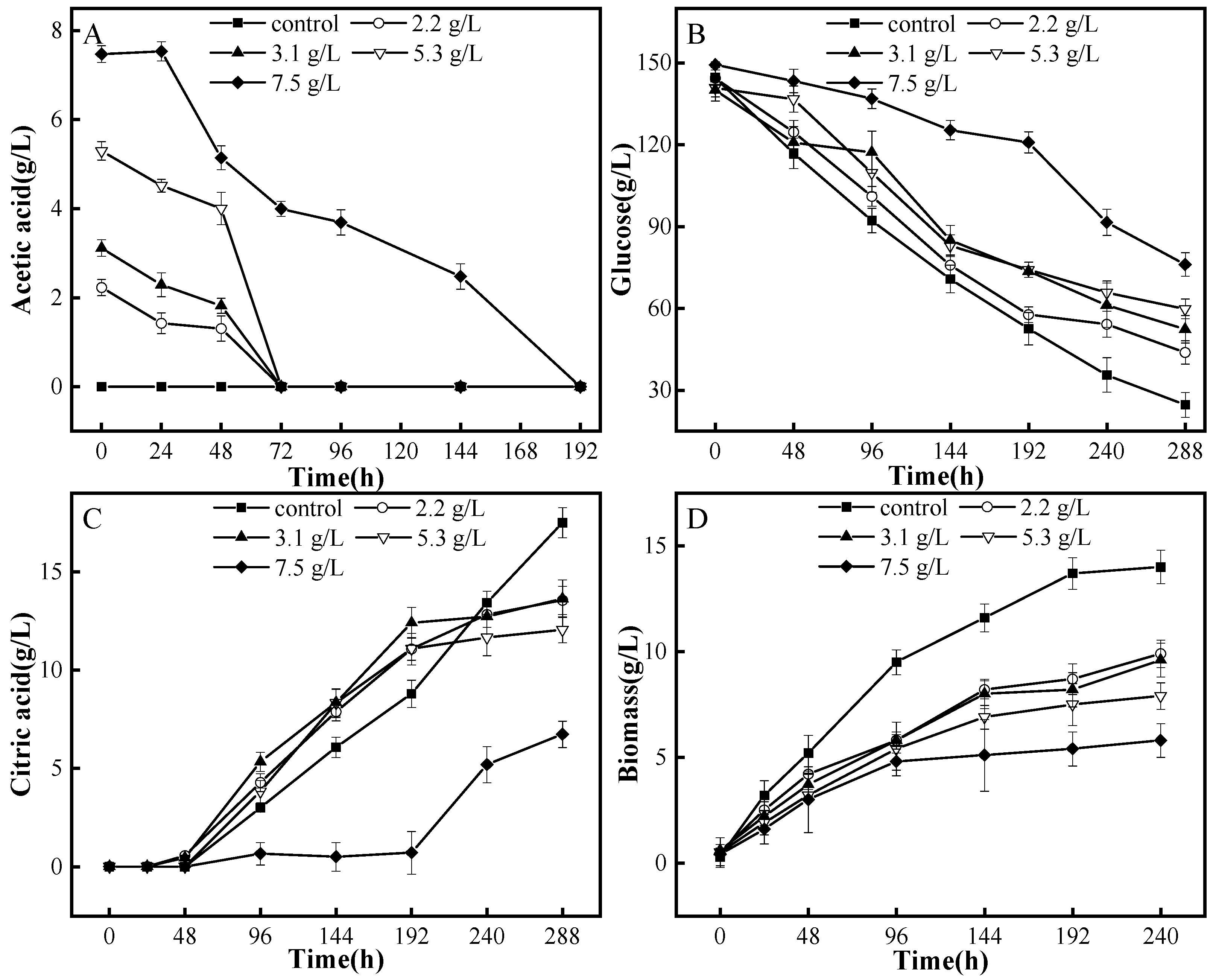
3.1. Degradation Patterns of A. niger M13 on Acetic Acid

Acetic acid makes the fermentation broth acidic. It can inhibit the growth of fermentation microorganisms. Undissociated organic acid molecules are lipid-soluble and can freely diffuse and penetrate into cells. They further dissociate into H+ in the cells, thus affecting the intracellular acid–base balance. To maintain a stable intracellular pH, microorganisms have to actively transport H+ from inside the cell to the outside, which is an energy-consuming process. This process requires a large amount of intracellular ATP consumption, thereby leading to a lack of ATP and an inhibition of normal microbial growth. In severe cases, it can even cause cell apoptosis [29]. The effects of different initial concentrations of acetic acid on the growth and fermentation of A. niger M13 are shown in Figure 1. When the initial concentration of acetic acid is less than 5.3 g/L, M13 consumes acetic acid in the fermentation broth within 72 h, with the highest consumption rate occurring in the 48–72 h period. When the initial concentration of acetic acid is around 7.5 g/L, M13 completely consumes the acetic acid in 192 h. As shown in Figure 1B—under the conditions of fermentation for 12 days with acetic acid concentrations of 2.2, 3.1, 5.3, and 7.5 g/L—the utilization rate of glucose decreased by 16.2%, 26.9%, 32.4%, and 39.0%, respectively, compared with the control group. The production efficiency of citric acid was slower and the biomass concentration of M13 was lower in the presence of acetic acid. Figure 1C,D show that, after 48–72 h, the rate of citric acid production increases as the amount of acetic acid is rapidly depleted. In the control group, fermentation for 12 days resulted in the production of approximately 17.50 g/L of citric acid, with the highest biomass concentration being 14 g/L. Citric acid yields in the medium with initial acetic acid concentrations of 2.2, 3.1, 5.3, and 7.5 g/L were 13.55, 13.63, 12.05, and 6.73 g/L, respectively. Compared to the control group, the citric acid yield was reduced by 22.6%, 22.1%, 31.1%, and 61.5%, respectively. The ability of M13 fermentation to produce citric acid gradually decreased with an increase in the initial acetic acid concentration, along with a gradual decrease in biomass concentration. In the medium with an initial acetic acid concentration of 7.5 g/L, the biomass concentration of M13 was the lowest at approximately 5.8 g/L, which is only 55.8% of the biomass concentration in the control group. This severe inhibition of citric acid fermentation in M13 was accompanied by the complete degradation of acetic acid after 8 days of culture, as well as the subsequent initiation of citric acid fermentation. Interestingly, when the initial acetic acid concentration was below 5.3 g/L, the rate of citric acid production was slightly higher during the mid-fermentation period compared to the control group. This may be due to the concurrent consumption of acetic acid and glucose by the cells in order to accumulate citric acid. The inhibitory effect of acetic acid on strain growth resulted in a decrease in citric acid yield.
The concentration of acetic acid in the hydrolysate of lignocellulosic biomasses is usually between 4–10 g/L [30], and it inhibits the growth and fermentation of A. niger. Compared to other microorganisms, A. niger has a lower tolerance for acetic acid. For instance, in the presence of 4 g/L of acetic acid, the ethanol production rate of C. glycerinogenes UG-21 is 2.37 g/L/h, and increasing the concentration of acetic acid to 6 g/L has almost no effect on its ethanol production [31]. This is because the inhibitory effect of acetic acid comes from undissociated molecules, which becomes more intense when the pH of the fermentation broths decreases due to the accumulation of organic acids.

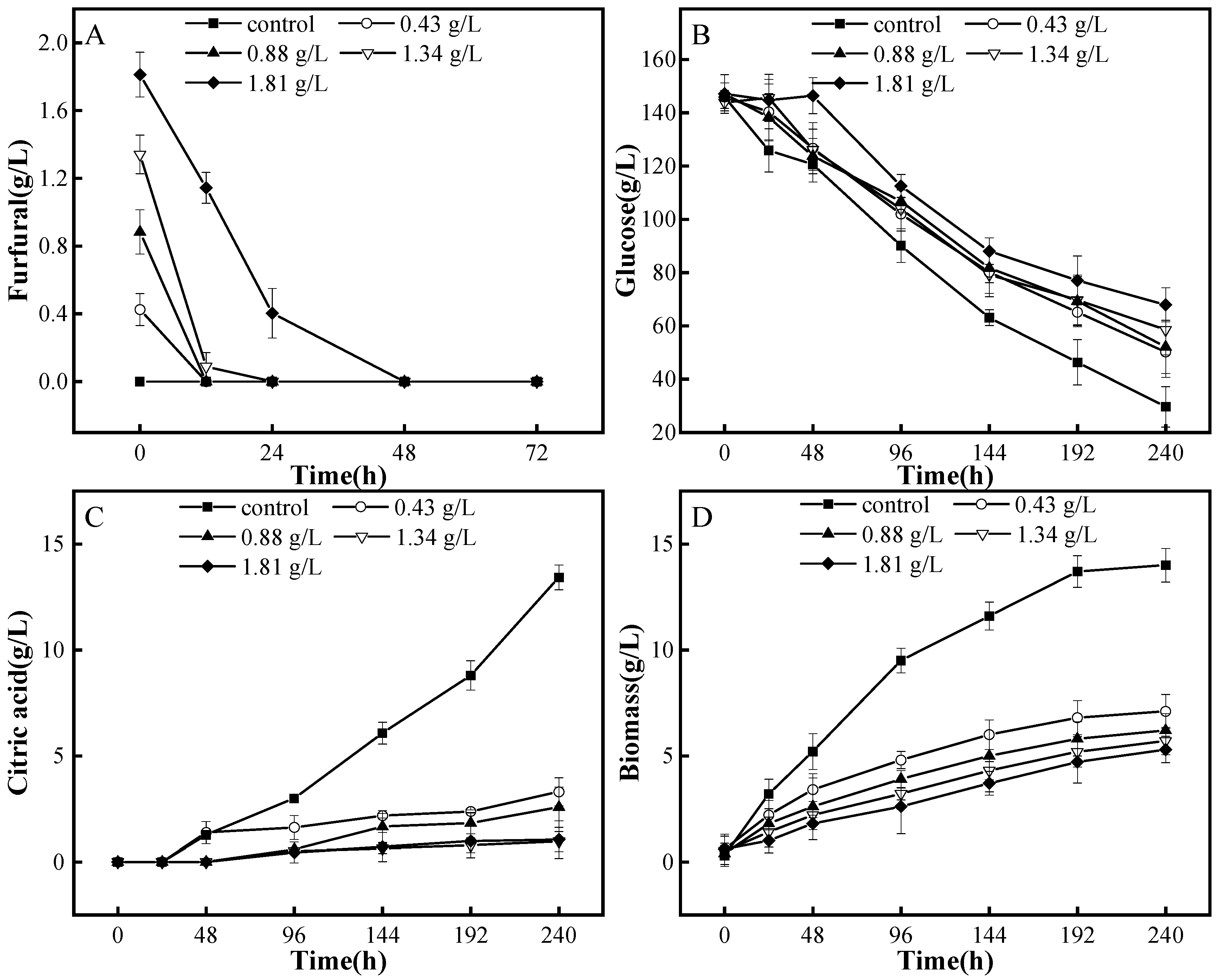
3.2. Degradation Patterns of A. niger M13 on Furfural

Furfural can destroy cell membranes and DNA, thereby inhibiting sugar fermentation and the TCA cycle [32]. Furfural degradation also consumes NADPH, which is necessary for cell biosynthetic reactions [33]. Figure 2 shows the effect of different initial concentrations of furfural on the growth and fermentation of an A. niger M13 strain. During the initial fermentation stage, M13 can completely consume furfural with concentrations ranging from 0.43 to 1.81 g/L in the medium. When the initial furfural concentration is below 1.34 g/L, M13 can consume most of the furfural within 12 h. However, it takes 48 h for cells to completely consume 1.81 g/L of furfural (Figure 2A). Figure 2A,B also show that M13 hardly consumes glucose when furfural is present in the medium. During the 10-day fermentation period in media containing 0.43, 0.88, 1.34, and 1.81 g/L of furfural, the glucose utilization rates decreased by 17.7%, 18.4%, 26.6%, and 31.9%, respectively, compared to the control group (Figure 2B). Furfural has a strong inhibitory effect on the growth of M13 and citric acid accumulation, and citric acid cannot be detected until furfural is completely consumed. Compared with the control group, when the initial furfural concentration is 0.43 g/L, the citric acid yield decreases by 61.3% (Figure 2C). As the initial furfural concentration increases, cell growth is severely inhibited. Figure 2D shows that the maximum biomass concentration of cells in the control group is 10.4 g/L. With the increase of initial furfural concentration, the biomass concentration of cells gradually decreases. When the initial furfural concentration is 1.81 g/L, the biomass concentration of the M13 strain is only 49.0% of that in the control group. In summary, furfural strongly inhibits cell growth and citric acid accumulation. Even after furfural is completely degraded, cell growth and citric acid production rates cannot be restored.

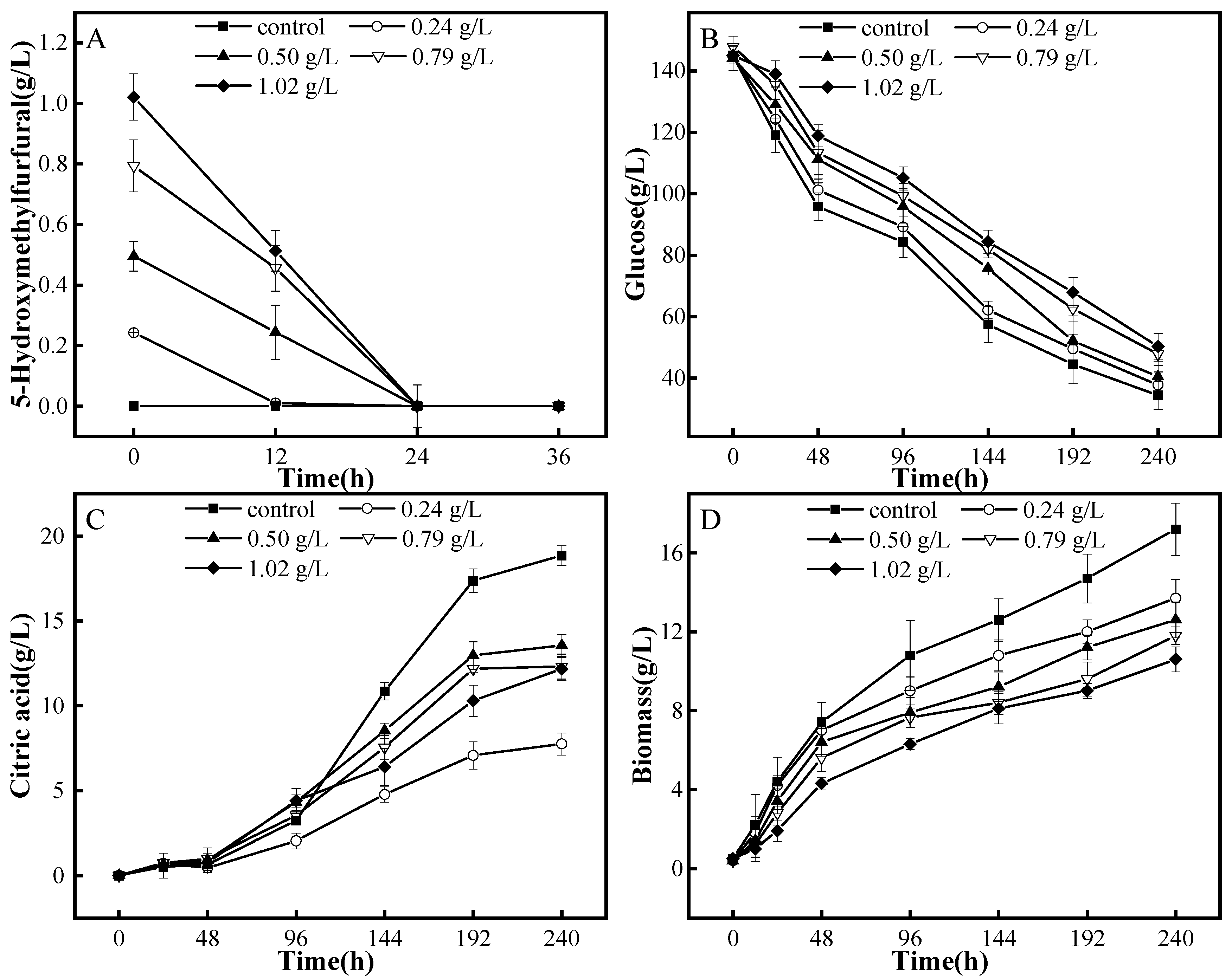
3.3. Degradation Patterns of A. niger M13 on HMF

HMF exhibited similar inhibitory effects as furfural on microorganisms by disrupting their cell membrane, thereby increasing membrane permeability and inhibiting microbial growth. However, according to certain reports, Saccharomyces cerevisiae and Pichia stipitis can largely reduce HMF into 2,5-dihydroxymethylfuran, which has a lower toxicity, when under the action of dehydrogenases. Furthermore, the continuous accumulation of 2,5-dihydroxymethylfuran does not affect cell growth and ethanol production [34]. In this study, A. niger was able to degrade HMF. As the initial concentration of HMF in the medium increased from 0.24 g/L to 1.02 g/L, the degradation rate of HMF increased (Figure 3A). Unlike the situation in the presence of furfural, which is when HMF was present, the cells could consume glucose and HMF together, and only the rate of glucose consumption was relatively slow during the early stage of fermentation. After 48 h of fermentation, the HMF was completely consumed, and the rate of glucose consumption was almost the same as that in the control group. Before the HMF was completely consumed, A. niger hardly produced citric acid. However, it was found that furfural had a significantly greater inhibitory effect on cell growth and citric acid fermentation than HMF, as shown by the comparison of Figure 2 and Figure 3.

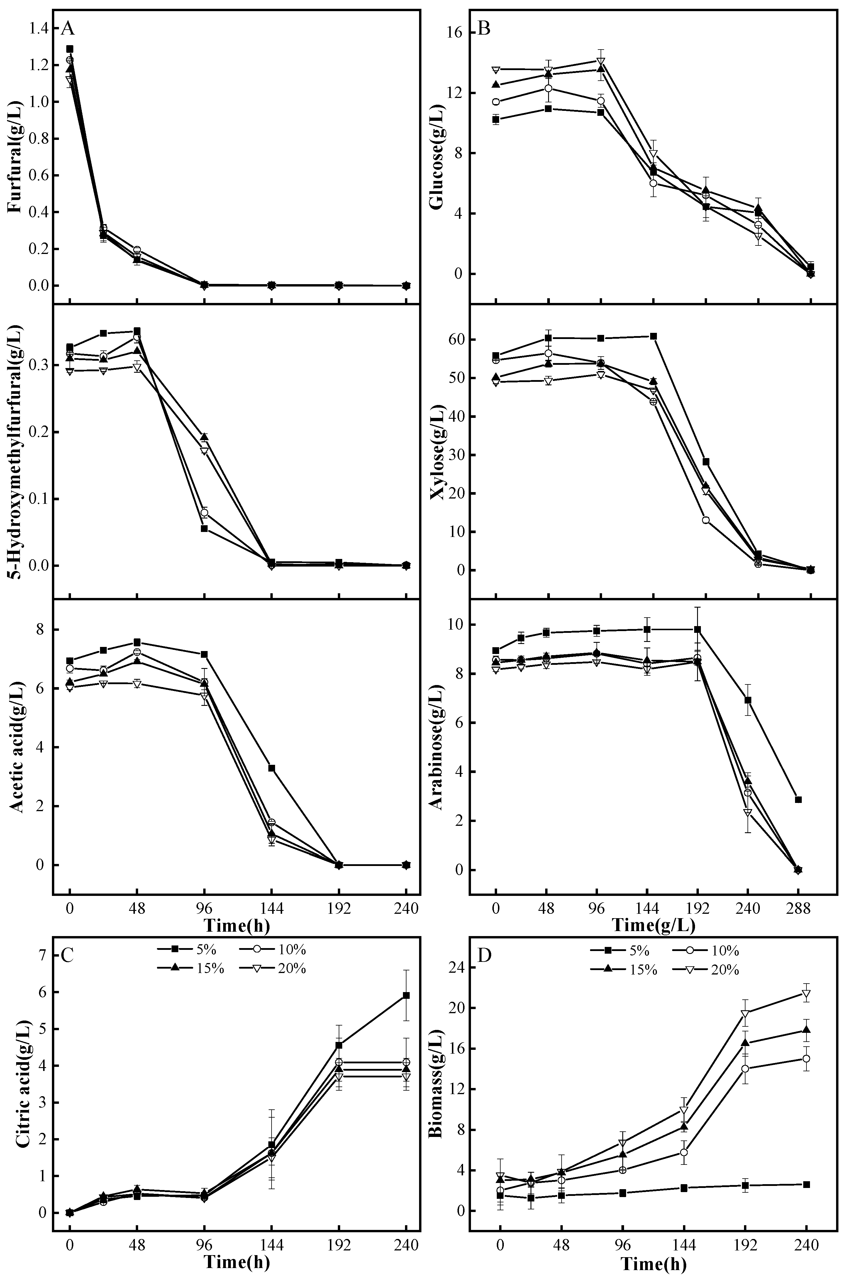
3.4. Detoxification and Fermentation of Corncob Dilute Acid Hydrolysate by A. niger M13

3.4.1. A. niger M13 Mycelium Biodetoxification of Corncob Dilute Acid Hydrolysate

A M13 mycelium was inoculated into a corncob dilute acid hydrolysate medium at different inoculum levels (Figure 4). The M13 mycelium degraded significant amounts of acetic acid, furfural, and HMF. However, different broth volumes did not have significant variations in terms of the detoxifying inhibitors in the corncob dilute acid hydrolysate (Figure 4). The M13 mycelium completely degraded furfural within 96 h, HMF in 144 h, and acetic acid in 192 h, while the detoxification rate was 100% for these inhibitors in the hydrolysate (Figure 4A). Under the presence of these three inhibitors simultaneously, M13 mycelial pellets preferred to degrade furfural and HMF, but then moved on to acetic acid after the furfural was completely degraded (Figure 4A). Meanwhile, within the first 96 h of inoculation, the M13 mycelial pellets did not use any of the carbon sources in the media. Glucose was consumed after 96 h, which was followed by xylose and then arabinose (Figure 4B). In the presence of furfural, the A. niger M13 mycelial pellets barely produced citric acid, thus indicating that furfural prevented the production of citric acid during fermentation. The rate of citric acid production increased substantially from the moment furfural was completely degraded, resulting in the maximum of a 6.0 g/L citric acid yield (Figure 4C). Table S1 shows the specific component contents of the corncob dilute acid hydrolysate before and after biodetoxification and fermentation when using M13 mycelium.

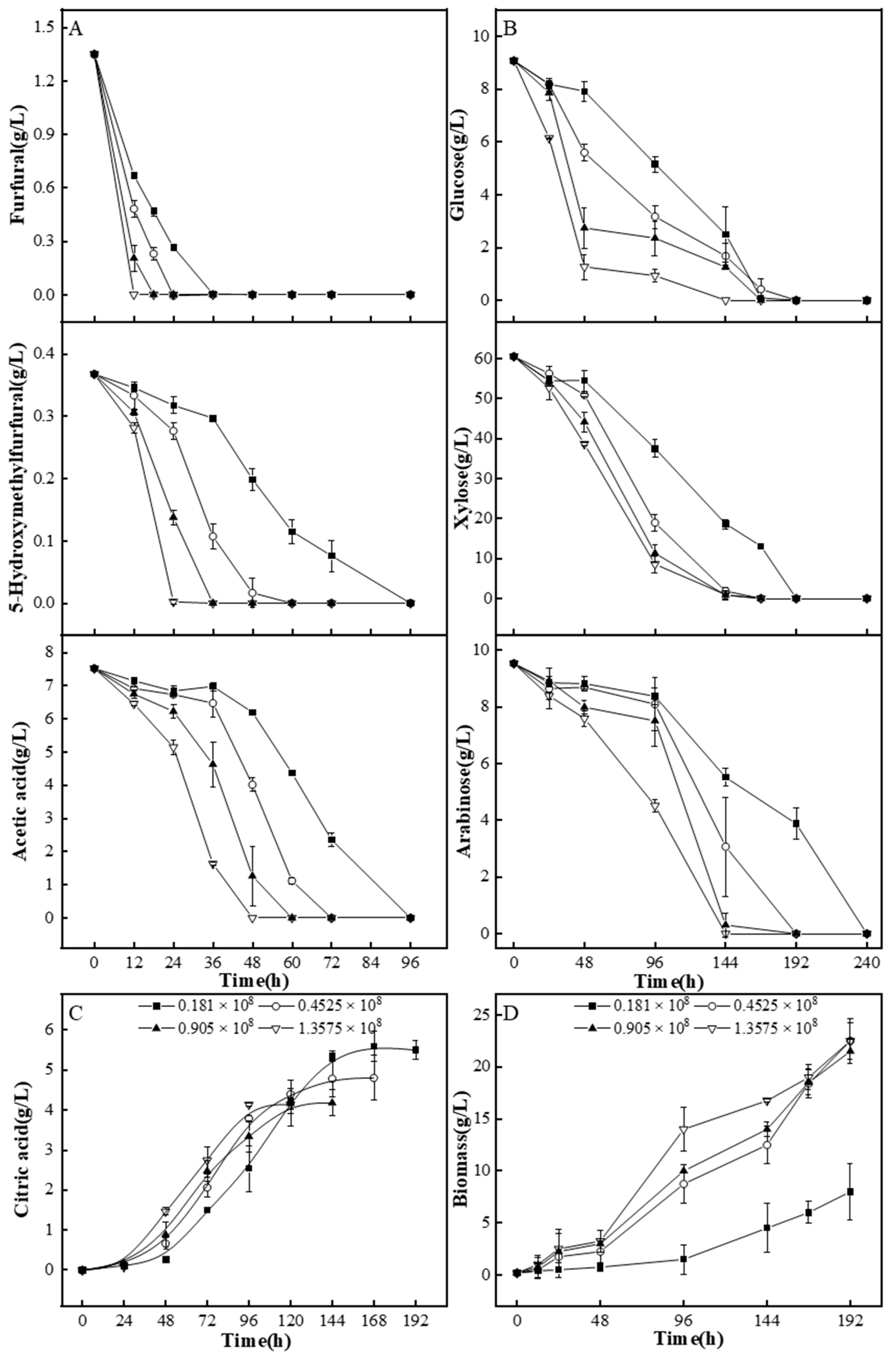
3.4.2. A. niger M13 Spores Biodetoxification of Corncob Dilute Acid Hydrolysate

To investigate the effect of different spore inoculum sizes on detoxification efficiency, 0.2, 0.5, 1.0, and 1.5 mL of M13 spore solutions were inoculated into a 20 mL maize cob dilute acid hydrolysate medium, which contained 0.181 × 10⁸, 0.4525 × 10⁸, 0.905 × 10⁸, and 1.3575 × 10⁸ spores, respectively.
As shown in Figure 5, the M13 spores significantly degraded acetic acid, furfural, and HMF in the corncob dilute acid hydrolysate, and the removal efficiency of the inhibitory substances increased significantly with increasing inoculum size. Particularly, with an inoculum size of 1.358 × 10⁸ spores, the M13 completely degraded 1.35 g/L of furfural in 12 h, 0.37 g/L HMF in 24 h, and 7.5 g/L acetic acid in 48 h, thus achieving a removal rate of 100% for the three inhibitory substances. M13 spores preferentially degraded furfural, followed by HMF, and, lastly, acetic acid. In addition, the acetic acid consumption occurred simultaneously with the sugar consumption. When inhibitory substances were present in the medium, A. niger M13 barely produced citric acid. However, once inhibitory substances were completely degraded, the production rate of citric acid by A. niger M13 significantly increased. At an inoculum size of 1.3575 × 10⁸ spores, 4.14 g/L of citric acid was produced. Interestingly, in contrast to the lowest inoculum size condition (which yielded 5.5 g/L of citric acid), it is possible that, in the cases where a larger inoculum size was used, while a significant amount of carbon sources were consumed by A. niger for cell growth and inhibitory substance degradation, there was an insufficient carbon source remaining for citric acid production during fermentation (Figure 5C,D). Table S2 shows the specific component contents of corncob dilute acid hydrolysate before and after biodetoxification and fermentation when using M13 spores.
After pretreatment with diluted acid, we conducted a biological detoxification study using A. niger M13 spores with some lignocellulosic raw materials (Table 1), the main components of which are listed in Table 2. After 24 h of detoxification, the furfural and HMF in the hydrolysate were completely removed, and about 32% of the acetic acid was removed (Table S3). For fermentation systems that use acetic acid and sugar as substrates, A. niger M13 spores are preferred for detoxification as it only takes them 24 h to remove furfural and HMF. In addition, studies have shown that a small amount of acetic acid has a promoting effect on microbial fermentation. Wu et al. found that, as the concentration of acetic acid in the medium increased from 0 to 30 g/L, the yield and productivity of 2,3-butanediol gradually increased, and the yield of 2,3-butanediol reached 0.4 g/g at a 30 g/L acetic acid concentration [35]. Qian et al. used Yarrowia lipolytica W29 to produce citric acid and malic acid in crude glycerol. The maximum concentration of citric acid was 9.87 g/L with the addition of a 10 g/L acetic acid salt solution in 50 g/L of glycerol, which was 14.7% higher than that without an acetic acid salt solution [36]. After 48 h of detoxification, furfural, HMF, and acetic acid were completely removed from the lignocellulosic dilute acid hydrolysate, which is suitable for fermentation systems that use sugar as a substrate. Using A. niger M13 spores for detoxification is a highly feasible option. These spores can completely remove the main inhibitory substance from the hydrolysate within 1–2 days while retaining the carbon source for subsequent fermentation production.
Currently, many bacteria and fungi have been identified as having the ability to degrade and inhibit the growth of the inhibitors in hydrolysates (Table 3). Issatchenkia orientalis S-7 is capable of degrading both acetic acid and furfural in hemicellulose hydrolysate. After 80 h of cell culture, the removal rates of acetic acid and furfural were 100% when the hydrolysate contained 4 g/L of acetic acid and 0.4 g/L of furfural [37]. Fonseca et al. used Issatchenkia occidentalis (CCTCC M 206097) to detoxify the hydrolysate of sugarcane bagasse. The detoxification efficiency within 24 h was 66.7% for eugenol, 73.3% for ferulic acid, 62% for furfural, and 85% for HMF [38]. Aspergillus nidulans FLZ10 was able to detoxify steam-exploded corn stover fiber with removal rates of 75% formic acid, 54% acetic acid, and 100% of both HMF and furfural after 72 h of cultivation [39]. Compared to the strains currently used for biodegradation, A. niger M13 spores can degrade inhibitors in hydrolysate with a removal rate of 100% within 48 h. In addition, they can maintain a high removal rate, even in the presence of higher levels of inhibitors, at rates of 0.1566 g/L/h for acetic acid, 0.1125 g/L/h for furfural, and 0.015 g/L/h for HMF. The order of inhibitor degradation by A. niger M13 is furfural, HMF, and then acetic acid. Therefore, for fermentation systems that use acetic acid and sugars as substrates, M13 spores can be used to remove furfural and HMF as the priority.

4. Conclusions

The effective removal of fermentation inhibitors produced during lignocellulosic pretreatment is a necessary step in utilizing lignocellulosic feedstocks for bioconversion. This study investigated the degradation patterns of A. niger M13 toward the three main inhibitors. A. niger M13 was able to degrade acetic acid, furfural, and HMF at concentrations of at least 7.5 g/L, 1.81 g/L, and 1.02 g/L, respectively. The time required to degrade all inhibitors increased with their increasing concentrations. When the A. niger M13 spores and mycelial pellets were separately inoculated into a corn cob acid hydrolysate medium, both demonstrated a 100% removal rate toward the three main inhibitors in the hydrolysate. The spores, when compared to mycelial pellets, were found to have a higher removal efficiency for the inhibitors with detoxification rates of 0.1566 g/L/h, 0.1125 g/L/h, and 0.015 g/L/h for acetic acid, furfural, and HMF, respectively. The A. niger M13 can effectively detoxify various acidic hydrolysates from different sources. In conclusion, biodetoxification by A. niger M13 provides a fast and efficient biodetoxification method for removing the inhibitors generated during intensive lignocellulose pretreatment, and its advantages allow it to be usable for potential industrial applications.

Supplementary Materials

The following supporting information can be downloaded at: https://www.mdpi.com/article/10.3390/fermentation9090854/s1, Table S1: The composition content of corn cob dilute acid hydrolysate before and after the biological detoxification using various quantities of A. niger M13 mycelial pellets; Table S2: The composition content of corn cob dilute acid hydrolysate before and after the biological detoxification using various quantities of A. niger M13 spores; Table S3: The sugar and inhibitor concentration in corncob dilute acid hydrolysate within 24 h of biological detoxification.

Author Contributions

Conceptualization, J.Y. and J.W.; Data curation, J.Y.; Formal analysis, J.Y., B.L., H.L., J.W. and J.Z.; Funding acquisition, J.W.; Investigation, J.Y., J.W. and J.Z.; Methodology, J.Y., C.W., Y.L., H.L., J.W. and J.Z.; Project administration, J.W.; Resources, J.W. and J.Z.; Supervision, Y.X., J.W. and J.Z.; Validation, J.Y., C.W., Y.L., B.L., Y.X. and J.W.; Writing—original draft, J.Y.; Writing—review & editing, J.Y., J.W. and J.Z. All authors have read and agreed to the published version of the manuscript.

Funding

This work was funded by the Basic Research Program (Free Exploration) Project of Shanxi Province (No. 20210302124464) and the Project (No. RH2100001380) of Tsinghua University.

Institutional Review Board Statement

Not applicable.

Informed Consent Statement

Not applicable.

Data Availability Statement

The data are available from the corresponding authors upon request.

Conflicts of Interest

The authors declare no conflict of interest.

References

  1. Ravindran, R.; Jaiswal, A.K. A comprehensive review on pre-treatment strategy for lignocellulosic food industry waste: Challenges and opportunities. Bioresour. Technol. 2016, 199, 92–102. [Google Scholar] [CrossRef] [PubMed]
  2. Hu, Q.; Xiong, X.; Tan, L.; Su, X.; He, Y.; Liu, X.; Yi, J. Advances in Pretreatment Technologies of Lignocellulosic Biomass. Chin. Agric. Sci. Bull. 2011, 27, 1–7. [Google Scholar]
  3. Zhuang, J.; Lin, L.; Pang, C.; Liu, Y.; Sun, Y. Research advances in detoxification of lignocellulose hydrolysates-making. Mod. Chem. Ind. 2009, 29, 19–23. [Google Scholar]
  4. Sene, L.; Arruda, P.V.; Oliveira, S.M.M.; Felipe, M.G.A. Evaluation of sorghum straw hemicellulosic hydrolysate for biotechnological production of xylitol by Candida guilliermondii. Braz. J. Microbiol. 2011, 42, 1141–1146. [Google Scholar] [CrossRef]
  5. Cheng, K.K.; Zhang, J.A.; Ling, H.Z.; Ping, W.X.; Huang, W.; Ge, J.P.; Xu, J.M. Optimization of pH and acetic acid concentration for bioconversion of hemicellulose from corncobs to xylitol by Candida tropicalis. Biochem. Eng. J. 2009, 43, 203–207. [Google Scholar] [CrossRef]
  6. Canilha, L.; De Almeida E Silva, J.B.; Solenzal, A.I.N. Eucalyptus hydrolysate detoxification with activated charcoal adsorption or ion-exchange resins for xylitol production. Process. Biochem. 2004, 39, 1909–1912. [Google Scholar] [CrossRef]
  7. Cheng, Z.; Yang, R.D.; Liu, X.; Liu, X.A.; Chen, H. Green synthesis of bacterial cellulose via acetic acid pre-hydrolysis liquor of agricultural corn stalk used as carbon source. Bioresour. Technol. 2017, 234, 8–14. [Google Scholar] [CrossRef]
  8. Zhang, C.; Zhang, T.; Jiang, B.; Mu, W.; Miao, M. Research advances in bioconversion of xylitol. Food Ferment. Ind. 2016, 42, 288–294. [Google Scholar]
  9. Wierckx, N.; Koopman, F.; Bandounas, L.; De Winde, J.H.; Ruijssenaars, H.J. Isolation and characterization of Cupriavidus basilensis HMF14 for biological removal of inhibitors from lignocellulosic hydrolysate. Microb. Biotechnol. 2010, 3, 336–343. [Google Scholar] [CrossRef]
  10. Xie, Y.; Hu, Q.; Feng, G.; Jiang, X.; Hu, J.; He, M.; Hu, G.; Zhao, S.; Liang, Y.; Ruan, Z.; et al. Biodetoxification of phenolic inhibitors from lignocellulose pretreatment using Kurthia huakuii LAM0618(T) and subsequent lactic acid fermentation. Molecules. 2018, 23, 2626. [Google Scholar] [CrossRef]
  11. Kosa, M.; Ragauskas, A.J. Biotransformation of lignin model compounds with oleaginous Rhodococci. Appl. Microbiol. Biotechnol. 2019, 152, 199–212. [Google Scholar]
  12. Zhang, Y.; Han, B.; Ezeji, T.C. Biotransformation of furfural and 5-hydroxymethyl furfural (HMF) by Clostridium acetobutylicum ATCC 824 during butanol fermentation. New Biotechnol. 2012, 29, 345–351. [Google Scholar] [CrossRef] [PubMed]
  13. Sun, H.Y.; Zhao, P.J.; Peng, M. Application of maltitol to improve production of raw starch digesting glucoamylase by Aspergillus niger F-08. World J. Microbiol. Biotechnol. 2008, 24, 2613–2618. [Google Scholar] [CrossRef]
  14. Ekedegba, F.E.; Ogbonna, A.I.; Nwibari, B.M.W.; Okoye, C.T.; Ogbonna, U.S.A.; Onyimba, I.A.; Madu, J.M.; Njoku, A.I. Optimization of amylase production in three fungal species. Asian J. Biochem. Genet. Mol. Biol. 2022, 12, 1–9. [Google Scholar] [CrossRef]
  15. Priyanka, U.; Lens, P.N.L. Enhanced production of amylase, pyruvate and phenolic compounds from glucose by light-driven Aspergillus niger-CuS nanobiohybrids. J. Chem. Technol. Biotechnol. 2023, 98, 602–614. [Google Scholar] [CrossRef] [PubMed]
  16. Cabezudo, I.; Galetto, C.S.; Romanini, D.; Furlan, R.L.E.; Meini, M.R. Production of gallic acid and relevant enzymes by Aspergillus niger and Aspergillus oryzae in solid-state fermentation of soybean hull and grape pomace. Biomass Convers. Bior. 2022, 9. [Google Scholar] [CrossRef]
  17. Liu, F.L.; Wang, B.; Ye, Y.R.; Pan, L. High level expression and characterization of tannase tan7 using Aspergillus niger SH-2 with low-background endogenous secretory proteins as the host. Protein Expr. Purif. 2018, 144, 71–75. [Google Scholar] [CrossRef]
  18. Xiao, T.T.; Hu, N.; Feng, Z.J.; Wu, X.Y.; Qiu, S.Y. Tannase production via Aspergillus niger B1401-mediated solid fermentation of tea-processed waste. Food Res. Dev. 2021, 42, 104–111. [Google Scholar]
  19. Liu, H.; Chen, H.; Zhou, X.; Tian, X.; Ma, L.; Hu, W.; Yan, Y.; Han, L. Heavy Ion Irradiation Combined with alpha-cellulose adaptive evolution to improve cellulase activity of Aspergillus niger. Nucl. Phys. Rev. 2022, 39, 382–390. [Google Scholar]
  20. Ma, X.Y.; Gao, M.; Wang, N.H.; Liu, S.; Wang, Q.H.; Sun, X.H. Lactic acid production from co-fermentation of food waste and spent mushroom substance with Aspergillus niger cellulase. Bioresour. Technol. 2021, 337, 125365. [Google Scholar] [CrossRef]
  21. Desai, D.I.; Iyer, B.D. Utilization of Corn Cob Waste for Cellulase-Free Xylanase Production by Aspergillus niger DX-23: Medium Optimization and Strain Improvement. Waste Biomass Valori. 2017, 8, 103–113. [Google Scholar] [CrossRef]
  22. Dias, L.M.; Dos, S.B.V.; Albuquerque, C.J.B.; Baeta, B.E.L.; Pasquini, D.; Baffi, M.A. Biomass sorghum as a novel substrate in solid-state fermentation for the production of hemicellulases and cellulases by Aspergillus niger and A. fumigatus. J. Appl. Microbiol. 2018, 124, 708–718. [Google Scholar] [CrossRef] [PubMed]
  23. Cao, K.; Li, Y. Pectinase production by Aspergillus niger solid state fermentation with grape pomace. China Brew. 2015, 34, 83–86. [Google Scholar]
  24. Jin, M.; Li, J.; Zhu, F.; Liu, C. Advances in metabolomics of Aspergillus niger. Food Eng. 2017, 4, 1–4+37. [Google Scholar]
  25. Zhou, P.P.; Meng, J.; Bao, J. Fermentative production of high titer citric acid from corn stover feedstock after dry dilute acid pretreatment and biodetoxification. Bioresour. Technol. 2017, 224, 563–572. [Google Scholar] [CrossRef]
  26. Jin, S.; Sun, F.X.; Hu, Z.J.; Liu, L.; Li, J.H.; Du, G.C.; Li, Y.R.; Shi, G.Y.; Chen, J. Improving Aspergillus niger seed preparation and citric acid production by morphology controlling-based semicontinuous cultivation. Biochem. Eng. J. 2021, 174, 108102. [Google Scholar] [CrossRef]
  27. Zhong, P.S.; Zheng, L.F.; Yang, Y.L.; Zhou, Y.T.; Liu, X.Y.; Yang, Q.; Ren, J.L. Piezoelectric sensing of glucose oxidase activity of Aspergillus niger spores pretreated by different methods. Food Chem. 2022, 370, 130901. [Google Scholar] [CrossRef]
  28. Wu, J.; Cheng, K.; Wang, G.; Li, W.; Feng, J.; Zhang, J. Analysis of acetic acid, furfural and 5-hydroxymethylfurfural affecting 2, 3-butanediol production using Klebsiella oxytoca. J. Chem. Technol. Biotechnol. 2013, 88, 2239–2243. [Google Scholar] [CrossRef]
  29. Palmqvist, E.; Grage, H.; Meinander, N.Q.; Hahn-Hagerdal, B. Main and interaction effects of acetic acid, furfural, and p-hydroxybenzoic acid on growth and ethanol productivity of yeasts. Biotechnol. Bioeng. 1999, 63, 46–55. [Google Scholar] [CrossRef]
  30. Guo, H.L.; Zhao, Y.; Chang, J.S.; Lee, D.J. Inhibitor formation and detoxification during lignocellulose biorefinery: A review. Bioresour. Technol. 2022, 361, 127666. [Google Scholar] [CrossRef]
  31. Zhao, M.L.; Shi, D.C.; Lu, X.Y.; Zong, H.; Zhuge, B.; Ji, H. Ethanol fermentation from non-detoxified lignocellulose hydrolysate by a multi-stress tolerant yeast Candida glycerinogenes mutant. Bioresour. Technol. 2019, 273, 634–640. [Google Scholar] [CrossRef]
  32. Wu, L.M.; Zhou, Y.H.; Chen, H.S.; Zhang, H.R. Biodegradation of the microbial metabolic inhibitors of lignocellulose hydrolyzed by dilute acid. Food Ferment. Ind. 2012, 38, 164–169. [Google Scholar]
  33. Seo, H.M.; Jeon, J.M.; Lee, J.H.; Song, H.S.; Joo, H.B.; Park, S.H.; Choi, K.Y.; Kim, Y.H.; Park, K.; Ahn, J.; et al. Combinatorial application of two aldehyde oxidoreductases on isobutanol production in the presence of furfural. J. Ind. Microbiol. Biotechnol. 2016, 43, 37–44. [Google Scholar] [CrossRef] [PubMed]
  34. Liu, Z.L.; Slininger, P.J.; Dien, B.S.; Berhow, M.A.; Kurtzman, C.P.; Gorsich, S.W. Adaptive response of yeasts to furfural and 5-hydroxymethylfurfural and new chemical evidence for HMF conversion to 2,5-bis-hydroxymethylfuran. J. Ind. Microbiol. Biotechnol. 2004, 31, 345–352. [Google Scholar] [CrossRef]
  35. Wu, J.; Cheng, K.; Li, W.; Feng, J.; Zhang, J.A. Effect of acetic acid, furfural and 5-hydroxymethylfurfural on production of 2,3-butanediol by Klebsiella oxytoca. Chin. J. Biotechnol. 2013, 29, 350–357. [Google Scholar]
  36. Qian, X.; Xu, N.; Jing, Y.; Song, M.; Zhou, J.; Dong, W.; Xin, F.; Zhang, W.; Jiang, M.; Ochsenreither, K. Valorization of Crude Glycerol into Citric Acid and Malic Acid by Yarrowia lipolytica. Ind. Eng. Chem. Res. 2020, 59, 17165–17172. [Google Scholar] [CrossRef]
  37. Qin, X.; Zhang, H.; Cai, A.; Zhou, Y.; Chen, H. Biodegradation of Xylitol Fermentation Inhibitors by Issatchenkia orientalis. Guangxi Sci. 2010, 17, 358–362. [Google Scholar]
  38. Fonseca, B.G.; Moutta, R.D.; Ferraz, F.D.; Vieira, E.R.; Nogueira, A.S.; Baratella, B.F.; Rodrigues, L.C.; Zhang, H.R.; Da Silva, S.S. Biological detoxification of different hemicellulosic hydrolysates using Issatchenkia occidentalis CCTCC M 206097 yeast. J. Ind. Microbiol. Biotechnol. 2011, 38, 199–207. [Google Scholar] [CrossRef]
  39. Yu, Y.L.; Feng, Y.J.; Xu, C.; Liu, J.; Li, D.M. Onsite bio-detoxification of steam-exploded corn stover for cellulosic ethanol production. Bioresour. Technol. 2011, 102, 5123–5128. [Google Scholar] [CrossRef]
  40. Zhang, J.; Zhu, Z.N.; Wang, X.F.; Wang, N.; Wang, W.; Bao, J. Biodetoxification of toxins generated from lignocellulose pretreatment using a newly isolated fungus, Amorphotheca resinae ZN1, and the consequent ethanol fermentation. Biotechnol. Biofuels. 2010, 3, 15. [Google Scholar] [CrossRef]
Figure 1. Effects of acetic acid on the growth and metabolism of A. niger M13. (A) Degradation of acetic acid at different initial concentrations by M13. (B) Sugar consumption cultured at different initial concentrations of acetic acid. (C) Citric acid production at different initial concentrations of acetic acid. (D) Biomass of M13 cultured at different initial acetic acid concentrations.
Fermentation 09 00854 g001
Figure 2. Effects of furfural on the growth and metabolism of A. niger M13. (A) Degradation of furfural at different initial concentrations by M13. (B) Sugar consumption cultured at different initial furfural concentrations. (C) Citric acid production of M13 at different initial furfural concentrations. (D) Biomass of M13 cultured at different initial furfural concentrations.
Fermentation 09 00854 g002
Figure 3. Effects of HMF on the growth and metabolism of A. niger M13. (A) Degradation of HMF at different initial concentrations by M13. (B) Sugar consumption of M13 at different initial concentrations of HMF. (C) Citric acid yield of M13 at different initial HMF concentrations. (D) Biomass changes of M13 at different initial HMF concentrations.
Fermentation 09 00854 g003
Figure 4. Biological detoxification of A. niger M13 mycelium pellets in the corncob dilute acid hydrolysate. (A) Degradation rules of different inoculated M13 mycelium pellets on the inhibitors in the corncob dilute acid hydrolysate. (B) Consumption of glucose in the corncob dilute acid hydrolysate by M13 mycelium pellets with different inoculum amounts. (C) Citric acid fermentation of the M13 mycelium pellets with different inoculum amounts in the corncob dilute acid hydrolysate. (D) Biomass of the M13 mycelium pellets with different inoculum amounts in corncob dilute acid hydrolysate. (—■—: 5%; —○—: 10%; —▲—: 15%; —▽—: 20%).
Fermentation 09 00854 g004
Figure 5. Biological detoxification of the A. niger M13 spores in the corncob dilute acid hydrolysate. (A) Degradation of inhibitory substances in the corncob dilute acid hydrolysate by different inoculated M13 spores. (B) Consumption of glucose in the corncob dilute acid hydrolysate by M13 spores at different inoculations. (C) Citric acid fermentation of the M13 spores with different inoculum amounts in corncob dilute acid hydrolysate. (D) Changes in biomass of the M13 spores in the corncob dilute acid hydrolysate with different inoculated amounts.
Fermentation 09 00854 g005
Table 1. Component content of lignocellulose dilute acid hydrolysate after detoxification.
24 h Removal Rate24 h Total Sugar Retention Rate48 h Removal Rate48 h Total Sugar Retention Rate
FurfuralHMFAcetic AcidFurfuralHMFAcetic Acid
Corn cob100%100%31.5%100%100%100%100%97.5%
Corn stalk100%100%38.0%98.9%100%100%100%86.8%
Bagasse100%100%26.9%97.9%100%100%100%95.7%
EFB100%100%34.3%85.0%100%100%100%48.0%
Table 2. Component content of lignocellulose dilute acid hydrolysate before detoxification.
Furfural (g/L)HMF (g/L)Acetic Acid (g/L)Total Sugar (g/L)
Corn cob1.350.377.5079.06
Corn stalk0.950.404.1655.23
Bagasse1.240.486.7167.81
EFB1.510.477.7341.89
Table 3. A comparison of the biodegradation rates among different bacterial strains is presented.
StrainConcentration (g/L)Degradation RateDetoxification Rate (g/L/h)References
FurfuralHMFAcetic AcidFurfuralHMFAcetic AcidFurfuralHMFAcetic Acid
Issatchenkia orientalis S-70.4--4.0100%--100%0.005--0.05[37]
Issatchenkia occidentalis CCTCC M 2060970.0160.023.3100%100%6.1%0.000330.0004170.00280[38]
Aspergillus nidulans FLZ101.873 × 10−62.411 × 10−51.868 × 10−3100%100%53.6%2.601 × 10−83.349 × 10−71.391 × 10⁻⁵[39]
Amorphotheca resinae ZN10.82.274.0100%100%35.0%0.16670.02360.01167[40]
A. niger M131.350.377.5100%100%100%0.11250.0150.1566This study
Disclaimer/Publisher’s Note: The statements, opinions and data contained in all publications are solely those of the individual author(s) and contributor(s) and not of MDPI and/or the editor(s). MDPI and/or the editor(s) disclaim responsibility for any injury to people or property resulting from any ideas, methods, instructions or products referred to in the content.

