Article

The Bacterial Microbiota of Artisanal Cheeses from the Northern Caucasus

by
Tatiana V. Kochetkova
1,*,
Ilya P. Grabarnik
2,
Alexandra A. Klyukina
1,
Kseniya S. Zayulina
1,
Liliya A. Gavirova
3,
Polina A. Shcherbakova
3,
Gennady S. Kachmazov
4,
Andrey I. Shestakov
3,
Ilya V. Kublanov
1 and
Alexander G. Elcheninov
1
1
Winogradsky Institute of Microbiology, Federal Research Center of Biotechnology of the Russian Academy of Sciences, Moscow 117312, Russia
2
Applied Genomics Laboratory, SCAMT Institute, ITMO University, Saint Petersburg 197101, Russia
3
Faculty of Biology, Lomonosov Moscow State University, Moscow 119234, Russia
4
Department of Food Technology, Faculty of Chemistry, North Ossetian State University after K.L. Khetagurov, Vladikavkaz 362025, Russia
*
Author to whom correspondence should be addressed.
Fermentation 2023, 9(8), 719; https://doi.org/10.3390/fermentation9080719
Submission received: 30 June 2023 / Revised: 26 July 2023 / Accepted: 27 July 2023 / Published: 29 July 2023

Abstract

:
In this study, we used culture-independent analysis based on 16S rRNA gene amplicons and metagenomics to explore in depth the microbial communities and their metabolic capabilities of artisanal brine cheeses made in the North Caucasus. Additionally, analysis of organic acid profiles was carried out for cheese characterization. Twelve cheese samples (designated as 05SR–16SR) from various artisanal producers were taken from five different villages located in Northern Ossetia–Alania (Russia). These cheeses were made using methods based on cultural traditions inherited from previous generations and prepared using a relatively uncontrolled fermentation process. The microbial diversity of Caucasus artisanal cheeses was studied for the first time. The results showed a diverse composition in all cheeses, with Bacillota (synonym Firmicutes) (9.1–99.3%) or Pseudomonadota (synonym Proteobacteria) (0.2–89.2%) prevalence. The microbial communities of the majority of the studied cheeses were dominated by lactic acid bacteria (LAB) genera, like Lactococcus (10.3–77.1% in 07SR, 09SR, 10SR, 11SR, 13SR, 15SR, 16SR), Lactobacillus (54.6% in 09SR), Streptococcus (13.9–93.9% in 11SR, 13SR, 14SR, 15SR), Lactiplantibacillus (13.4–30.6% in 16SR and 07SR) and Lentilactobacillus (5.9–14.2% in 09SR, 10SR and 13SR). Halophilic lactic acid bacteria belonging to the Tetragenococcus genus accounted for 7.9–18.6% in 05SR and 06SR microbiomes. A distinctive feature of Ossetia cheese microbiomes was the large variety of halophilic proteobacteria, and in some cheeses they prevailed, e.g., Chromohalobacter (63–76.5% in 05SR and 06SR), Psychrobacter (10–47.1% in 08SR, 11SR, 12SR), Halomonas (2.9–33.5% in 06SR, 08SR, 11SR and 12SR), Marinobacter (41.9% in 12SR) or Idiomarina (2.9–14.4% in 06SR, 08SR and 11SR samples). Analysis of the genomes assembled from metagenomes of three cheeses with different bacterial composition revealed the presence of genes encoding a variety of enzymes, involved in milk sugar, proteins and lipid metabolism in genomes affiliated with LAB, as well as genes responsible for beneficial bioamine and bacteriocin synthesis. Also, most of the LAB did not contain antibiotic resistance genes, which makes them potential probiotics, so highly demanded nowadays. Analysis of the genomes related to halophilic proteobacteria revealed that they are not involved in milk fermentation; however, the search for “useful” genes responsible for the synthesis of beneficial products/metabolites was partially positive. In addition, it has been shown that some halophiles may be involved in the synthesis of inappropriate bioactive components. The results obtained by culture-independent analyses confirm the importance of using such techniques both to clarify the quality and health-promoting properties of the product, and to look for probiotic strains with specified unique properties. This study has shown that traditional dairy foods may be a source of such beneficial strains.

1. Introduction

Cheese has always been valued by men both as an everyday food and as a gourmet dish. Cheese is mentioned as a major component of the daily diet of the Babylonian dynasty. In the Roman Empire, cheese was an integral part of the feasts of the patricians. In ancient Greece, cheese making was as well-known as in our age. It is proved by Aristotle’s treatise, where the processes of milk coagulation and cheese making technique are described. Nowadays cheese is a traditional product widely consumed around the world. It is high in protein (up to 25%), milk fat (up to 60%) and minerals (up to 3.5%, not including table salt). Proteins in cheese are better assimilated than proteins in other dairy products. It is also consumed by people with lactose intolerance because some amount of the lactose present in milk is hydrolyzed during fermentation, and the rest is processed into whey during the production of cheese [1].
Traditional products (including cheeses) are frequently consumed, usually passed down from one generation to the next, are prepared precisely defined according to its gastronomic heritage, with little or no processing/manipulation and are associated with a particular locality, region or country [2]. It is not a secret that the microbiota common to these foods are linked to health-promoting and beneficial properties for humans, such as up-regulation of the immune system, strengthening the intestinal–brain barrier, minimization of carcinogenic effects and more [3]. It was revealed that lactic acid bacteria species, isolated from cheeses, could be producers of such bioactive molecules or enzymes as peptidases and lipases [4], vitamins [3], GABA (γ-aminobutyric acid) [5], antibacterial peptides and peptides inhibiting the increase of blood pressure [6]. Microorganisms isolated from artisanal cheeses secrete metabolites with anti-cancer effects [7,8,9], exhibit antimicrobial activity against pathogenic bacteria [10], participate in lowering cholesterol levels [11], enhance the immune system by increasing the level of IgA and T helper cells [7], etc. On the other hand, we must not forget about foodborne infection and foodborne intoxication, caused by “unwanted” microbiota, which can occur for various reasons (disruption or low hygienic level of the production process or transportation, storage conditions and so on) in traditional dairy foods, including cheeses [12,13].
Ultimately, the identification of microbial communities that contribute significantly to the quality, safety, beneficial properties and flavor characteristics of cheese and their potential functional properties is of high importance. The cheese microbiome is a dynamic community that can change throughout ripening, and factors such as raw materials, aging, production process, storage conditions and specific product characteristics affect its diversity. The next-generation sequencing (NGS) technologies have significantly enhanced the capability to characterize and describe the cheese microbiome compared to commonly used culture-dependent methods. A lot of work on microbiota of commercial and artisanal cheeses has already been carried out all over the world using NGS (see reviews [14,15]). Studies devoted to microbiome analysis of cheese, produced in Russia, with its huge diversity of ethnic groups and, therefore, their own traditions in preparing dairy products, remains extremely scarce [16,17,18,19].
The Caucasus is a geographic mountainous region located on the border between Europe and Asia, covering the territory of Russia, Georgia, Azerbaijan and Armenia. This territory has long been famous for its ancient traditions in the preparation of dairy products. A distinctive feature of the Caucasian cuisine is the frequent and abundant inclusion of cheeses. Exclusively brine-type cheeses are in usage. The region of North Ossetia–Alania, located in the North Caucasus, occupies an area of 8000 km2 and is characterized by a moderate continental climate and a long growing season, which allows obtaining high-quality milk from farm animals. The climatic zone of North Ossetia, softened by the proximity of the mountains, with mild winters and long rainy summers can affect the ripening of cheese, as this process proceeds at environmental conditions. Traditional cheese in North Ossetia is made by adding rennet extract (prepared from veal’s stomach chopped into pieces and steeped for 2–3 days in acidified cheese whey) to raw milk (sheep’s, goat’s or cow’s) and incubation of this mix at room temperature. After full coagulation, the cheese mass is left to stand in the tank to remove the whey, and after the formed cheese is dry, salted by rubbing the surface (young soft cheese). For longer storage, cheese is consistently salted and dried, repeating these procedures, and then placed in a concentrated brine solution (approximately 1 kg of salt per liter of water). In this form the cheese can mature for 3–12 months (semi-hard cheese) and even up to 1–1.5 years (hard cheese). Studies on the microbial composition of such traditional fermented foods have never been documented.
The aim of this work is to explore the prokaryote diversity of home-made authentic Ossetia cheeses, as well as to investigate the gene sets related with metabolic activities and probiotic functions that could be involved in their sensory profile, safety and beneficial properties. In this study we used culture-independent NGS analysis based on 16S rRNA gene amplicons of bacterial communities of 12 traditional cheeses and metagenomics of three of them.

2. Materials and Methods

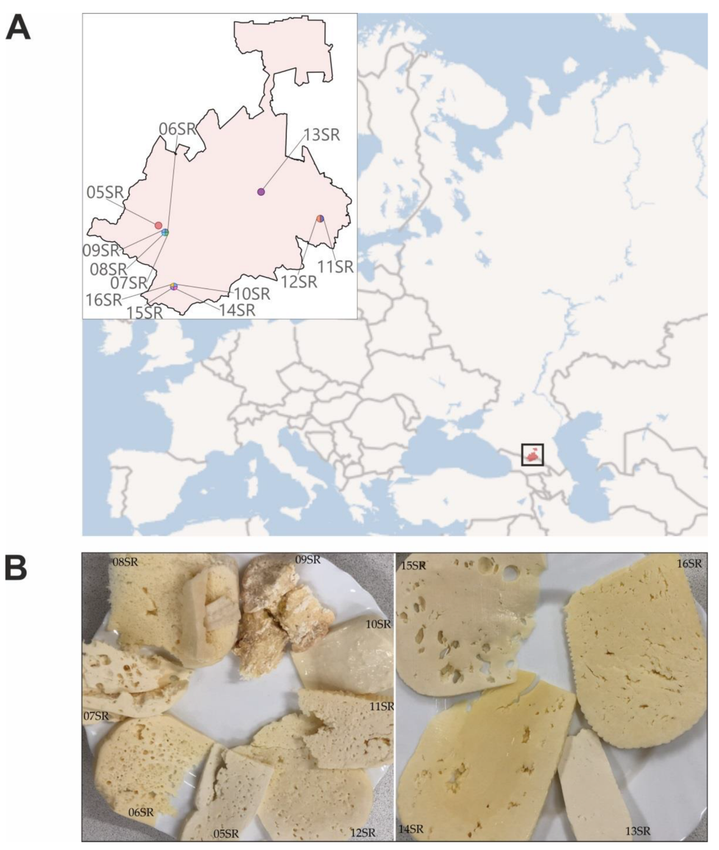
2.1. Sample Collection and DNA Extraction

Cheeses prepared from cow’s milk by traditional methods were sampled in local markets and houses in villages and towns of various districts of North Ossetia–Alania in spring 2022 (Table 1). Cheese wheels were transported to the laboratory at 4 °C. For DNA fixation, 2 mL of product (a 20 mL syringe with the front end cut off was used to sample 20 mL of cheese, a cylinder was cut out, capturing both the outside and the inside of the piece of cheese, then the cylinder was pressed through a metal net with a hole size of 0.8 × 0.5 mm, 2 mL was taken from this sample) were mixed with 2 mL of fixing buffer (100 mM EDTA, 100 mM Tris-HCl, 150 mM NaCl; pH 8.2). DNA extraction and all other manipulations were carried out within 7 days after sampling. Fixed product samples were centrifuged at 18,000× g for 20 min. Total DNA was extracted from the pellets using DNeasy PowerLyzer Microbial Kit (Qiagen, Hilden, Germany) and FastPrep-24™ 5G grinder (MP Bio, Irvine, CA, USA) according to the manufacturer’s instructions. DNA concentrations were detected by Qubit 2.0 (Invitrogen/Life Technologies, Vallejo, CA, USA). DNA was stored at −20°C.

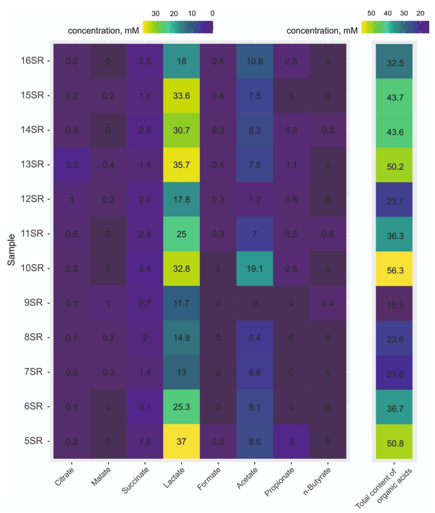
2.2. Organic Acids Analysis

Sample preparation and organic acids analysis were performed as described previously [19]. The pH value of the samples was measured using pH indicator strips (pH-fix 0.0–6.0, Macherey-Nagel, Dueren, Germany).

2.3. Library Preparation and Sequencing

For amplicon-based library preparation the V4 hyper-variable region of the 16S rRNA gene was amplified with a pair of primers 515F (5′-GTGBCAGCMGCCGCGGTAA-3′) [20] and Pro-mod-805R (5′-GGACTACHVGGGTWTCTAAT-3′) [21], and then adaptors and dual indices were added in a secondary amplification as described previously [22]. The libraries were sequenced using the MiSeq system (Illumina, San Diego, CA, USA). The libraries were prepared and sequenced in two technical replicates for each sample.
Preparation of DNA libraries for shotgun metagenomic sequencing was carried out using KAPA HyperPlus kit (KAPA, Wilmington, MA, USA) according to manufacturer recommendations. The manipulations include enzymatic fragmentation of DNA which resulted in fragments with length of 500–700 bp, ends polishing and A-tailing, ligation of specific adapters for sequencing (Nextera Mate Pair Library Prep Kit, Illumina, San Diego, CA, USA) as well as amplification of obtained libraries. Metagenomic sequencing was performed using Illumina NovaSeq 6000 platform (Illumina, San Diego, CA, USA).

2.4. Bioinformatics and Statistical Analysis

For amplicon sequences adapter trimming and demultiplexing were performed as described earlier [23]. The obtained reads were filtered and processed using dada2 package v.1.14.1 [24] (parameters: truncLen = 220, maxN = 0, maxEE = 2, truncQ = 2) resulting in identification of the amplicon sequence variants (ASV). Taxonomic assignment of ASV was performed using dada2 package v.1.14.1 with native Bayesian classifier [25] and Silva 138.1 database [26]. Biodiversity indexes such as Shannon [27], InvSimpson [28] and Chao1 [29] indexes were calculated using the phyloseq v.1.3 package [30]. To estimate the dissimilarity of the microbial composition of cheeses (i.e., the beta-diversity), a non-metric multidimensional scaling (NMDS) was performed as an ordination method based on ASV summarized table and the Bray–Curtis dissimilarity indices using phyloseq and vegan v.2.6.-4 packages (accessed on 23 June 2023 https://CRAN.R-project.org/package=vegan). Visualization of the results was performed with the ggplot2 package (accessed on 23 June 2023 https://ggplot2.tidyverse.org).

2.5. Metagenome Assembly and Dominant Genomes Reconstruction

Raw sequences of three cheese samples’ (06SR, 07SR, 13SR) metagenomes were filtered using trim tool (quality limit = 0.03, maximum ambiguous nucleotides = 2, and minimum length = 80) in CLC Genomic Workbench v.10 software (Qiagen). Prepared high-quality reads were assembled using the metaSPAdes v3.15.5 [31]. Contigs with length less than 500 bp were eliminated from the assemblies. To generate metagenome-assembled genomes (MAGs) from metagenomes, and assembled contigs were processed using the metaWRAP v1.3.2 standard pipeline [32]. The metaWRAP::Binning module was used to reconstruct bins from contigs and mapped reads by three independent binning tools (MaxBin2 [33], metaBAT2 [34] and CONCOCT [35]) with default settings. In the metaWRAP::Bin_refinement module multiple combinations of obtained bins were compared, filtered by completeness (-c 80; more than 80%) and contamination (-x 10; less than 10%) estimation with CheckM v1.1.6 [36] and consolidated to the refined bin set. Abundance and statistical calculations were performed in the metaWRAP::Quant_bins module by Salmon v1.10.1 [37]. Taxonomic classification of MAGs was performed using the GTDB-Tk v2.1.1 [38] with GTDB_r207 database as reference.

2.6. Gene Prediction and Functional Annotation

Functional analysis was performed using a set of MAGs from three cheese samples. Gene calling was performed using Prodigal v.2.6.3 [39] and Prokka v1.12 [40] was used for rapid genome-wide annotation.
Carbohydrate-active enzymes (CAZymes) genes were searched in MAGs using dbCAN v.4 [41] with hmmer tool [42]. To found putative proteases blast search was performed using in silico translated MAGs as queries and MEROPS_scan [43] with an e-value threshold of 1 × 10⁻⁵; positive hits were analyzed with SignalP v.6.0 [44] to identify extracellular enzymes. Putative esterase was identified in translated MAGs using blast search against a database of bacterial lipolytic enzymes [45] with an e-value threshold of 1 × 10⁻¹⁰ and minimal coverage of 30%.
Antimicrobial resistance genes in MAGs were determined using the ABRicate tool (accessed on 20 June 2023 https://github.com/tseemann/abricate). Each sample bin set was screened using the CARD database [46] with compared regions’ minimal identity of 80% and minimal coverage of 90%. Feature annotation of MAG sequences produced by Prokka and genomes in fasta format were used for biosynthetic gene clusters (BGC) identification by antiSMASH bacterial version 7.0 [47]. The search for bacteriocins among secondary metabolites identified by antiSMASH was carried out by selecting gene clusters containing genes with the corresponding annotation in the Pfam and TIGRFam databases and their further annotation using NCBI BlastP analysis.
Genes encoding enzymes involved in biogenic amines and GABA production were searched using blast with characterized enzymes as queries and translated MAGs as databases (e-value of threshold 1 × 10⁻⁵). Functions of positive hits were checked with blast against the SwissProt database [48].

2.7. Data Availability

All sequencing data used for 16S rRNA amplicon analysis were deposited into the NCBI SRA database under BioProject number PRJNA789261 (Table S1). Metagenomic sequences are available in the Genbank database under accession numbers JARIFJ000000000, JARIFN000000000 and JARIFP000000000 within Bioproject PRJNA907749.

3. Results

We analyzed 12 samples of home-made cheeses from different districts of North Ossetia-Alania (Table 1, Figure 1).

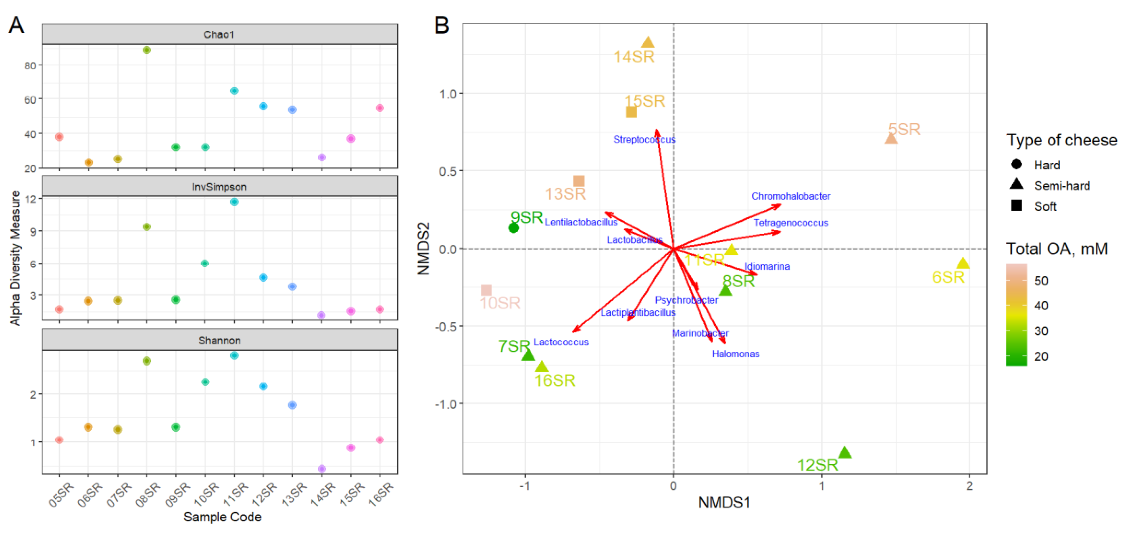
3.1. Organic Acids Content

The profile of organic acids (OA), contained in the cheeses, include formate, acetate, propionate, n-butyrate, lactate, succinate, malate and citrate. The major organic acid in all samples was lactate (Figure 2) with concentration varying from 11.7 (in 09SR sample) to 37 mM (in 05SR). Acetate was detected in all cheeses, except sample 09SR, with concentration from 1.2 mM (for 12 SR) to 19.1 (for 10SR). Other volatile fatty acids like formate, propionate and n-butyrate were identified for all samples in negligible concentrations from 0.3 to 1.1 mM, except sample 05SR, which contained 3 mM of propionate. Succinate was measured for all samples and its concentration varied from 1.4 mM (for 05SR, 07SR and 13SR samples) to 3.4 mM (10SR), as well as citrate, its concentration varied from 0.1 mM (09SR) to 3.3 mM (13SR). Small amounts of malate were detected in six samples from 0.2 mM (12SR and 15SR) to 1 mM (09SR). The total concentration of all OA varied greatly among the samples—from 15.9 mM (for 09SR) to 56.3 (10SR). pH values of cheeses were in a narrow range from 4.5 to 5.5.

3.2. Sequencing, Taxonomic Annotation and Genome Reconstruction

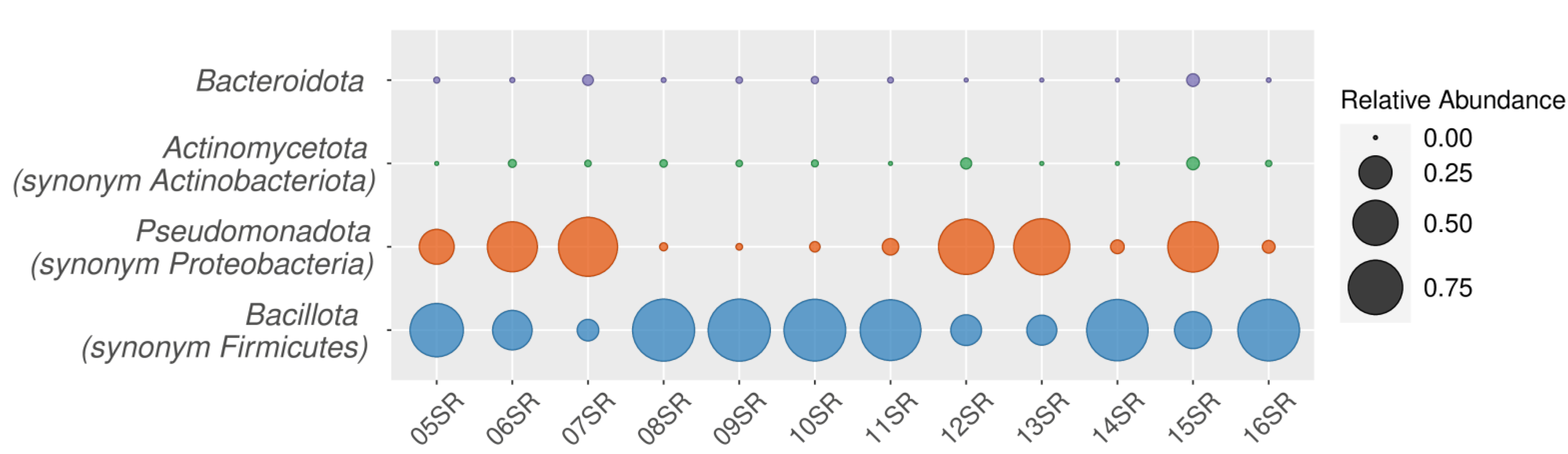
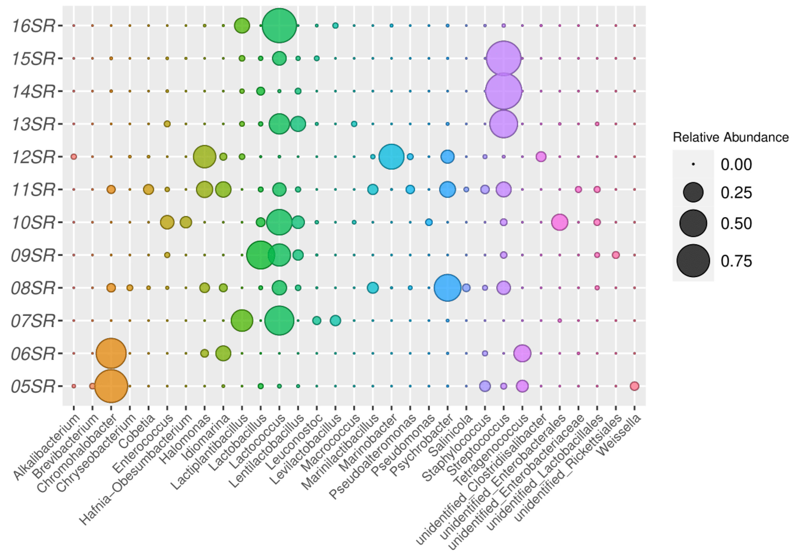
3.2.1. Taxonomic Analysis by Amplicons of V4 Region of the 16S rRNA Gene Sequencing

A total of 279,232 reads with an average length of 250 bp were obtained from sequencing of 12 samples. After filtering, denoising and chimera detection 179,320 reads were retained representing 288 unique sequences. The obtained ASV were assigned to more than one hundred genera (145 unique genera + 37 NA) within 25 phyla, but 98.9% of the total number of sequences were affiliated to Firmicutes and Proteobacteria (synonymes Bacillota and Pseudomonadota, respectively, according to GTDB database, accessed on 29 June 2023 https://gtdb.ecogenomic.org/) (Figure 3).
To estimate overall diversity in all samples analyzed the alpha-diversity indexes were calculated for each sample. According to the Shannon Index, cheeses 11SR and 08SR possessed the highest biodiversity in comparison with other studied products (Figure 4A). The maximum value of 2.80 was observed for the 11SR sample. Indeed, this cheese had the highest microbial composition and was presented by Streptococcus (14% of the total community), Lactococcus (10%), Idiomarina (14.4%), Halomonas (16%), Psychrobacter (15%), Marinilactibacillus (5.5%), Cobetia (5.3%), Chromohalobacter (3.1%), Pseudoalteromonas (4%), Staphylococcus (3.5%), Lentilactobacillus (0.9%), Lactobacillus (0.8%), Salinicola (0.7%) and Enterococcus (0.5%) species (Figure 5). The sample 08SR had very similar diversity, however in other proportions. Samples 05SR-07SR, 09SR, 14SR-16SR had lower biodiversity levels with 0.45–1.31 values of Shannon index. The dominant genera in these samples were: Chromohalobacter (05SR, 06SR), Lactococcus (07SR, 09SR, 16SR), Lactobacillus (09SR), Streptococcus (14SR, 15SR), Lactiplantibacillus (07SR) and Tetragenococcus (06SR). The middle index values were observed for 10SR (2.24), 12SR (2.15) and 13SR (1.76) samples. The microbial composition of the sample 05SR was relative to 06SR — dominance of Chromohalobacter species (77%), followed by Tetragenococcus (8%), Staphylococcus (6%), Streptococcus (1.4%), Weissella (3.6%) and Lactobacillus (0.9%). Streptococcus was the dominant genus in cheeses 13SR (53%), 14SR (94%) and 15SR (83%). The community of the sample 16SR consisted of only Lactococcus (77%), Lactiplantibacillus (13.4%) and Streptococcus (0.2%) representatives. The analysis of the diversity based on Chao1 and the inverse Simpson indexes revealed relatively similar trends. The Chao1 indexes varied from 23 (06SR) to 89 (08SR), while inverse Simpson values—from 1.18 (14SR) to 11.67 (11SR).
Within the non-dominant population determined by read abundance of 16S rRNA gene marker, bacteria related to food sanitary quality of less than 3.3% of total richness, were identified: Escherichia-Shigella (less than 0.5%), Klebsiella (less than 0.9%), Acinetobacter (less than 3.3%), Raoultella (0.2%), Chryseobacterium (1.4%), Rothia (0.3%), Ralstonia (0.1%), etc. There were no sequences with annotation related to pathogenic bacteria such as Salmonella, Listeria, Yersinia, Mycobacterium, etc. (Table S2).
The comparison of the microbiomes of different cheeses was performed with NMDS using Bray–Curtis dissimilarity indices based on an ASV table. The microbial components of each sample (ASV) determine the spatial distribution of points, i.e., the biodiversity of the products. The cheese samples were evenly distributed across the space of the NMDS plot (Figure 4B), which may indicate a wide diversity of microbiome composition. However, reflection of the metadata on the points makes it possible to evaluate the general patterns of sample characteristics depending on the similarity of the microbial community: cheeses with a low concentration of organic acids (06SR, 07SR, 08SR, 11SR, 12SR, 16SR) are located in the central and lower parts of the graph, where Lactococcus, Halomonas, Idiomarina, Marinobacter and Lactiplantibacillus domination was observed. The same group covers almost the entire set of semi-hard type cheeses, except for 05SR and 14SR. The opposite group of samples in the upper half of the plot (05SR, 13SR, 14SR, 15SR) has a high concentration of organic acids, includes an equal amount of soft and semi-hard type cheeses and has Streptococcus, Lentilactobacillus, Chromohalobacter and Tetragenococcus as the dominant bacteria. Samples 09SR and 10SR did not fit into groups formed on the plot with their characteristics. The small distance between the points may indicate a significant similarity of samples 07SR and 16SR, as well as 08SR and 11SR. The distance between samples 05SR, 06SR and 12SSR, which have different compositions of the microbiomes with the dominance of Chromohalobacter, Tetragenococcus, Halomonas and Idiomarina, was also noticeable.

3.2.2. Metagenomic Assembly, Gene Prediction and Functional Annotation

Metagenomes of three cheese samples 06SR, 07SR and 13SR were used for assembling and reconstructing bacterial MAGs. The metagenomes assembly sizes for samples were 26.53, 26.16 and 45.75 Mb respectively as shown in Table 2. Mapping reads back to contigs (with length ≥ 500 bp), 98.72%, 96.83% and 68.15% of the total reads were recruited with mean coverage 423×, 300× and 114× respectively. After metaWRAP binning module with CONCOCT, MetaBAT2 and MaxBin2 usage, the total number of primary 203 bins were generated, number of which decreased after filtering by completeness and contamination level and refinement algorithm to final 21 MAGs (5, 6 and 10, respectively). Characteristics of MAGs as size (1.52 to 6.06 Mb), completeness and contamination percentage (average 95.98% and 1%, respectively), GC content, number of scaffolds and N50 are summarized in Table 3.
After taxonomical classification, four assembled bacterial genomes were identified as Streptococcus, two MAGs of Lactococcus and Lactiplantibacillus, single genomes belonging to Idiomarina, Halomonas, Chromohalobacter, Tetragenococcus, Staphylococcus, Leuconostoc, Pseudomonas, Enterobacter, Lentilactobacillus, Enterococcus, Macrococcus and Pediococcus.
Species assignment using ANI (average nucleotide index, 95% as reference radius) resulted in the detection of two possible new species of Idiomarina and Halomonas genera that were not assigned with a sufficient level of confidence, whereas the remaining 19 MAGs were classified up to species level (Table 3). The relative representation of individual assembled MAGs in the three metagenomes is shown in Figure S1.

Search for Bacteriocin Gene Clusters

During the search for BGC genes with antiSMASH in in silico translated MAGs obtained from three metagenomes, 86 genes from 25 gene clusters were identified, including 63 genes within 11 regions annotated as bacteriocin synthases or involved in bacteriocin synthesis (Table S3). The distribution of BGCs corresponds to the microbiological diversity: the most common clusters are class II/IIb bacteriocins of the blp family (13SR_bin6, 13SR_bin8, 13SR_bin10) and the lactococcin family (07SR_bin3, 13SR_bin5). For some MAGs (07SR_bin6), the complete structure of the bacteriocin synthesis cluster can be observed: ABC transporter, secretion protein, CPBP family intramembrane metalloprotease, response and transcriptional regulator, bacteriocin immunity protein and two to six subunits of bacteriocin protein [49]. In sample 07SR, the Linocin_M18 gene was found in the genome of Levilactobacillus brevis (07SR_bin4), the plantaricin synthesis genes were found in the Lactiplantibacillus plantarum genome (07SR_bin6). In samples 07SR and 13SR fragments of lanthionine-containing bacteriocin were found as Lanthipeptide Class IV clusters (07SR_bin4, 13SR_bin10). Numerous genomes (06SR_bin2, 06SR_bin3, 06SR_bin5, 07SR_bin1, 07SR_bin5) contain the YcaO domain gene that is involved in microcin antibiotics synthesis as azoline-forming protein [50].

Search for Genes of Antibiotic Resistance

Search of antibiotic resistance potential in microbiome components resulted in detection of 24 genes coding resistance-providing products in seven MAGs, as shown in Table S4. Most of the antibiotic resistance (ABR) genes contain 07SR_bin1: oqxB and oqxA encode subunits of the RND efflux pump that confers resistance to fluoroquinolone and several other antibiotics, marA and crp encode efflux pump regulator proteins in antibiotic stress (cephalosporin, glycylcycline, fluoroquinolone, macrolide, penam, tetracycline and other), bacA translation product confers resistance to bacitracin, acrD encodes aminoglycoside efflux pump, ACT-23 gene is beta-lactamase, fosA2 gene product provides fosfomycin resistance, msbA codes ABC transporter which gives nitroimidazole resistance, emrB is a translocase gene with concomitant fluoroquinolone resistance and baeR encodes response regulator for aminocoumarin and aminoglycoside resistance. Only two ABR genes contained 07SR_bin2 (lmrD for lincosamide and tetS for tetracycline). Inner membrane transporter MexF which confers diaminopyrimidine, fluoroquinolone and phenicol resistance was found in 06SR_bin5. Two close genes from the MFS efflux pump family and lincosamide resistance lmrP and lmrD were detected in 13SR_bin1 and 13SR_bin5 respectively, last MAG also contains ANT(6)-la that is aminoglycoside nucleotidyltransferase. Five genes are part of 13SR_bin7 genome: emeA as a multidrug efflux pump gene (acridine dye resistant), lsaA that encodes ABC-F protein (provides resistance to lincosamide, macrolide, oxazolidinone and others), efrA and efrB encode the subunits of efflux pump (fluoroquinolone, macrolide, rifamycin resistance) and dfrE encodes dihydrofolate reductase that confers diaminopyrimidine resistance. Summary, in 7 out of 21 MAGs most often are genes of resistance to fluoroquinolone (n = 9), tetracycline (n =5) and macrolide (n = 5) antibiotics.

Synthesis of Biogenic Amines and Some Non-Proteinogenic Amino Acids

Several bioactive amines as well as GABA can be produced by microbial communities of studied cheeses (Table S5). Genes of glutamate decarboxylase, which is responsible for GABA synthesis, were found in 06SR_bin2, four MAGs of 07SR (07SR_bin3, 07SR_bin4, 07SR_bin5 and 07SR_bin6), two MAGs of 13SR (13SR_bin3 and 13SR_bin5). Tyrosine decarboxylase which converts tyrosine to tyramine was encoded only in two MAGs (07SR_bin4, 13SR_bin7). Probably, these enzymes can also participate in beta-phenylethylamine production. No genes encoding histidine decarboxylase (responsible for histamine formation) were found. Moreover, some MAGs possessed genes of enzymes involved in putrescine formation. Putrescine production pathways in 06SR_bin2 and 13SR_bin6 include arginine decarboxylase, agmatine deaminase and N-carbamoylputrescine amidase while in 06SR_bin5 and 13SR_bin4—arginine decarboxylase and agmatine ureohydrolase. MAG 07SR_bin5 had genes encoding enzymes of both these variants. Ornithine decarboxylase genes were identified in 07SR_bin1 and 13SR_bin9.

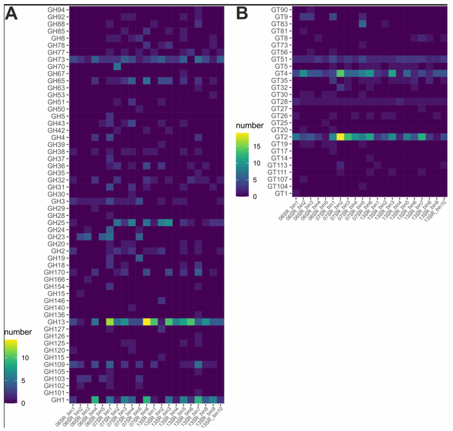
CAZymes, Esterases and Proteases

All studied MAGs possessed CAZymes genes (glycosidases, polysaccharide lyases, carbohydrate esterases, glycosyltransferases and carbohydrate-binding modules) but their numbers varied greatly depending on cheese sample. There were 29–49 genes in MAGs of cheese 06SR, from 66 to 111 in 07SR, and from 22 to 80 in 13SR (Table S6). Sets of glycoside hydrolases (GH) and glycosyltransferase (GT) were in the focus because these enzymes are main actors in polysaccharide decomposition and synthesis, respectively. Most universal GH found in most MAGs belonged to GH1, GH13 and GH73 (Figure 6). The first one contains enzymes with different activities including beta-galactosidases which hydrolase lactose—milk sugar. Enzymes of the GH13 family could be active against exogenous or endogenous storage alpha-glucans. In the GH73 family there is a single main activity, peptidoglycan hydrolase, so they can either in cell wall transformation during cell growth or in microbial interactions (e.g., inhibition of growth of competitors). The majority of enzymes belonging to GH2, GH8, GH25, GH65 and GH77 were encoded in MAGs obtained from 07SR and 13SR while glycosidases from GH3, GH23 and GH103 in 06SR. Among GT there were families with high abundance in all studied MAGs (GT2, GT4, GT28, GT51) but some families were sample-specific: genes of GT5 and GT35 enzymes were found only in MAGs from 07SR and 13SR, while GT9, GT19 and GT30 were found in MAGs from 06SR and 07SR.
Genes of putative proteases were identified in all tested MAGs. Moreover, some of them have signal peptides and can be exported outside the cells or anchored at the cell surface (Table S7). Numbers of encoded extracellular proteases were 4–49, 2–41 and 1–18 in MAGs of 06SR, 07SR and 13SR, respectively. The most dominant protease families were C40, S11, S12 and M23B (found only in 06SR and 07SR).
Homologs of lipolytic enzymes affiliated to some families according to [45] were found in all studied metagenomes (Figure S2, Table S8). In total, sixteen putative esterases were encoded on 06SR: there were enzymes relatively close to Family_4 (only in 06SR_bin4), Family_6, Family_11, Family_13, Family_24, Family_32 (only in 06SR_bin5), Family_33 (only in 06SR_bin1) and Family_34. MAGs obtained from 07SR metagenome contained 24 genes of putative esterases which belong to Family_1.1 (only in 07SR_bin5), Family_1.8 (only in 07SR_bin1), Family_4, Family_6 (07SR_bin5), Family_8 (07SR_bin5), Family_10 (07SR_bin5), Family_11, Family_13, Family_15 (07SR_bin5), Family_23 (07SR_bin5), Family_29 (07SR_bin5), Family_33 and Family_34 (07SR_bin5). Twenty genes encoding esterases were found in MAGs of the 13SR metagenome. These enzymes were homologous to representatives of Family_1.10 (13SR_bin2), Family_4, Family_11 (13SR_bin3), Family_13, Family_15 (13SR_bin4), Family_31 (13SR_bin4), Family_33 and Family_35 (13SR_bin8). Some of the listed enzymes could participate in production of flavor-forming compounds.

4. Discussion

Fermented foods usually contain microorganisms with Generally Recognized as Safe (GRAS) status which can produce a number of beneficial by-products/metabolites. The role of probiotics in maintaining human gut health has been largely documented [1,3,51]. Artisanal dairy products may be a source of new beneficial microorganisms that are able to limit unusual inflammatory responses and metabolic abnormalities [3]. Ethnic fermented foods have traditionally been prepared by native populations and have preserved unique microbiota over the centuries. To use these distinctive microorganisms for human health improvement, it is crucial to understand the probiotic properties of the bacteria, as well as the composition of the community itself.
The history of Ossetia cheese goes back to the Scythian–Alanian times. Since ancient times, in the conditions of mountain farming, it was one of the main food products. And the recipe for making cheese has not changed much since then. The preparation of traditional Caucasian cheeses involves manual steps, such as whey draining, stomach cutting, curd stirring for salting and contact with salt, and the latter could be important microbial sources, especially halophilic bacteria [14]. Considering that examined cheeses are made from raw milk, the bacterial microbiota is expected to be highly diverse [52]. In addition, milk in different regions may contain different levels of micronutrients, proteins, lactose and fats, depending on regional conditions and the season of harvesting, all of these affect the composition of the community and their functions. Taxonomic annotation made by NGS analysis of 16S rRNA gene amplicons revealed that the microbial diversity of analyzed Caucasus cheeses did not depend on the place of origin (Figure 4B). Representatives of the genus Lactococcus, Streptococcus, Lactobacillus and Lactiplantibacillus belong to the dominant population. Although a few ASVs related to spoilage microorganisms were sporadically detected (e.g., Brochothrix spp., Serratia spp., etc.), according to the results of Illumina sequencing, no major foodborne pathogens (e.g., Listeria monocytogenes or Salmonella spp.) occurred in all analyzed samples, thus suggesting that relatively good hygiene practices had been followed during cheese production.
The dominance of specific LAB slightly correlated with the age of cheeses analyzed (in our case, with the time of ripening in salt brine) (Figure 4B). For example, Streptococcus and Lactococcus species dominated in young soft cheeses, Lactobacillus representatives were detected in significant amounts only in the hard cheese. The same correlation was observed for OA profile—hard cheese contained low amounts of OA, while semi-hard and soft cheeses were rich in organic acids, suggesting the intensive metabolic processes of LAB genera presented there (Figure 2). Concentrations of lactate, acetate, propionate, butyrate, succinate and citrate were similar to values, estimated for cheeses of the same stages of ripening [53]. Lactate and acetate were the major products of fermentation followed by succinate and citrate, the latter was regarded as a substrate for succinate production by lactobacilli [54,55]. The youngest cheese in our analysis (13SR), which was produced in the farthest region, contained the relatively significant concentrations of citrate, malate and propionate, likely due to the quality of the milk used or time of ripening [56,57].
The dominance of lactococci in non-mature, and especially in young cheeses, was an expected result, according to the relevant literature [14,17,58,59,60,61,62]. Lactobacillus species, on the other hand, begin to dominate in long-ripening cheeses [14,58,63]. Most of the other LAB genera detected in this work as dominant and subdominant (Streptococcus, Lactiplantibacillus, Lentilactobacillus, Levilactobacillus, Leuconostoc) have often been described as a part of microbiota of spontaneously ripened cheese [14,16,60,61,62,64].
In genomes of Lactococcus lactis, detected in 07SR and 13SR cheeses, genes encoding enzymes, involved in the degradation of carbohydrates present in milk (like GH1, GH2, GH13), as well as those responsible for the synthesis of polysaccharides (GT2) that could influence the cheese texture, were found (Figure 6). In addition, lactococci actively hydrolyze milk proteins, producing flavor compounds, medium- and small-sized peptides as well as free amino acids with a high impact on flavor [65], and in our research we also identified some peptidases that could play a role in hydrolysis of milk proteins during the fermentation process. Together with Lc. lactis, Lactiplantibacillus plantarum and Levilactobacillus brevis, detected in 07SR cheese, are actively involved in milk sugars fermentation, due to the large variety and number of GH enzymes in their genomes. According to the literature, besides the carboxylic acid content of cheese, these LAB species also contribute to the amino acid profile [66]. The largest quantity of GTs, the main enzymes involved in polysaccharide synthesis (especially, GT2 family), was found in Leuconostoc mesenteroides genome from 07SR cheese, that could produce exopolysaccharides (e.g., an indigestible α-glucan) with substantial host metabolic benefits [67].
Metagenomic analysis revealed the dominance of Streptococcus genera (Streptococcus thermophilus, S. parasuis, S. macedonicus and S. dysgalactiae) in the 13SR cheese microbiome. It is believed that Streptococcus representatives are part of the primary starters, and their source may be not only milk, but also whey and veal stomachs used for cheese making [52,68]. S. thermophilus causes rapid acidification of milk through the production of lactic acid, proteolytic and urease activities [69]. Our metagenome analysis also revealed a large number of genes encoding GHs in Streptococcus MAGs and no or poor representation of lipolytic enzymes (Figure 6 and Figure S2). It may indicate that these bacteria are not involved in fatty acid production from triglycerides, and most likely participate in the hydrolysis of milk sugars. Apparently, the lipolysis of milk fats in this cheese is responsible for Lactobacillaceae representatives and Macrococcus caseolyticus [70]. Another feature of 13SR cheese was the presence of Lentilactobacillus kefiri, that is commonly associated with kefir beverage, but is also detected in cheeses of different origin [61,71]. The genome of L. kefiri possessed GH’s genes (encoding GH127, GH146 and GH39), that are practically absent in other genomes analyzed in this work and are involved in L-arabinose production (Figure 6). L-arabinose is a five-carbon sugar that is metabolized in bacteria to ethanol [72], which can affect the taste of the cheese.
LAB can produce a huge number of biologically active compounds, such as short-chain fatty acids, conjugated linoleic acid, vitamins, different peptides with antimicrobial activities, bioamines, 1,2-propanediol and much more [3]. In this work we explored the assembled genomes for the presence of genes involved in the bacteriocins and bioamines synthesis. Bacteriocins are proteins with antimicrobial activity and differ from other antibiotics in that they are all synthesized by ribosomes and that they are quite specific and often active against closely related microorganisms. Bacteriocins of lactic acid bacteria are often characterized by resistance to high temperatures and low pH but are degraded by digestive enzymes. That is why they are considered as beneficial and safe for humans and are of particular interest for applications such as food preserving agents [73]. In metagenomes of 07SR and 13SR cheeses we found that dominant LAB may be producers of bacteriocins (lactococcin A, B, plantaricin, linocin, lanthipeptide class IV, lactobin A and lactocin F) (Table S3). Moreover, genes for the synthesis of several peptides with antimicrobial activity were found in some of their genomes (Lc. lactis—lactococcin A and B, L. brevis—linocin M18 and lanthipeptide class IV, L. plantarum—lactococcin A and plantaricin EF, S. thermophilus—lactacin F, lanthipeptide class IV and unidentified bacteriocin Bpl family class II). The identification of genes encoded linocin and lanthipeptide in the L. brevis genome (from 07SR sample) is particularly noteworthy, since these bacteriocins have a broad antibacterial activity (unlike lactococcins and plantaricin) and even antifungal and antiviral activity by the latter [74,75].
Biogenic amines and non-proteinogenic amino acids are often the result of the bacterial decarboxylative activity towards free amino acids in food and LAB are strong amine producers. Bioamines were of our interest because of their adverse effects on human health and the large production of these molecules specifically in cheeses. In our metagenome analysis we identified genes encoding enzymes that catalyze the production of two different bioamines (tyramine and putrescine) as well as GABA. GABA is a product of glutamate decarboxylation and is the only bioamine with a really positive effect on human health, due to its relaxing effect on muscles and its overall beneficial influence on the nervous system [76]. Tyramine is the most frequently found bioamine in cheese and acts both on the vascular level (hypertension, vasoconstriction) and on the central nervous system. It can cause not only headaches, but it can also lead to serious health consequences [77]. Putrescine is one of the most common biogenic amines found in food, with higher concentrations in fermented dairy products such as cheese and fish [78]. It is greatly responsible for the unpleasant smell of unripe foods [79]. The risk effect depends on its concentration; however, there is a lack of regulation concerning safe putrescine limits in food. We found that all the dominant bacteria in the microbiome of cheese 07SR (Lc. lactis, L. brevis and L. plantarum) are producers of GABA, which significantly increases the beneficial quality of this product. Also, Pseudomonas sp., found in this community in minor proportions, is also capable of producing this useful bioamine (Table S5). However, it also contains the putrescine synthesis genes, as does the other minor component—Enterobacter hormaechei, the origin of which may be related to the cow’s stomach, used for cheese preparation [80]. Only two genes of glutamate decarboxylase were found in the 13SR metagenome (L. plantarum and Lc. lactis), despite the greater diversity of LAB in this community. Moreover, some representatives of Streptococcus genus, Pediococcus parvulus, M. caseolyticus and Enterococcus faecalis have been shown to be sources of putrescine and tyramine.
The latter one as well as E. hormaechei (from 07SR sample) possessed the highest number of antibiotic resistance-related genes in their assembled genomes among all MAGs in this work (Table S4), including ABR genes against fluoroquinolone, cephalosporin, macrolide, tetracycline etc. The detection of antibiotic resistance in dairy microflora is related to food safety issues, because the emergence and spread of antibiotic resistance is a serious problem at the present time. All this, together with the properties described above for these species, suggests against such bacteria presence in the food. As for the other components of the microbiome analyzed, the Leu. mesenteroides MAG (from 07SR) contained ABR genes conferring resistance to lincosamide and tetracycline, Lc. lactis (from 13SR) possessed genes from lincosamide resistance, and the S. dysgalactiae genome (also from 13SR cheese) carried genes of efflux pump belonging to the major facilitator superfamily (MFS) family of transporters that provided resistance to multiple dissimilar drugs [81]. The other LAB that made up the microbiomes of cheeses did not contain ABR genes, thus complying with one of the safety requirements for LAB used as food starters or probiotics.
The other distinctive feature was revealed during Ossetia cheese analysis. Samples 05SR and 06SR contained halophilic bacteria as the predominant majority of the community that is not commonly associated with the cheese environment [14]. Representatives of Chromohalobacter japonicus were prevalent in these two cheeses. Idiomarina, Halomonas and Tetragenococcus were detected among other dominant genera (Figure 5). These species are commonly associated with salt fermented liquid products of vegetable origin, popular in Asian cuisine [82,83]. Chromohalobacter species are often detected in salty environments, including foods, not only fermented [84], and in brines, used for cheese ripening [85]. Tetragenococcus representatives also may dominate in cheese brines, as was demonstrated for artisanal Belgian cheese production [86]. Other halophilic (or halotolerant) genera, such as Halomonas, Idiomarina, Marinobacter and Psychrobacter constituted a significant proportion, but not a prevailing one, in some analyzed cheese samples too (08SR, 11SR and 12SR). These bacteria are often found in saline habitats, including fermented seafood [87,88,89], and also in sub-dominant or minor quantities in soft and semi-hard cheeses [64,90,91]. The origin of halophilic species in the cheeses studied is difficult to explain based on the results obtained in this paper. Some authors believe that raw milk microbiota could contain some halophilic bacteria [92] or salt used for brine preparation and for rubbing the cheese surface may be a source of halophiles [86,92,93]. In any case, the question remains open—why do the halophiles we discovered dominate some cheese microbiomes: are they involved in milk fermentation and cheese maturation or are they simply displacing lactic acid bacteria over time, winning competition in salty conditions?
Halophilic and halotolerant bacteria capable of lactic acid fermentation are detected and functionate in dairy products, including cheese, called HALAB, and belong to the phylum Bacillota [93,94,95]. It is known that bacteria of the Tetragenococcus genus, considered as HALAB, are often involved in the fermentation of sea foods as dominant microbes [96]. But all other predominant halophilic bacteria we detected in cheese samples are aerobes and belong to the phylum Pseudomonadota and their value in the fermentation of milk, as well as their potential probiotic properties, remain unknown.
A metagenomic analysis of the 06SR cheese was performed to clarify the role of halophilic bacteria in cheese ripening and to evaluate their possible beneficial impact. Analysis of CAZymes genes revealed that only Tetragenococcus halophilus and Staphylococcus equorum genomes possess a wide range of glycosidases involved in the hydrolysis of various sugars that may be involved in the fermentation of milk, most notably GH1 (Figure 6). The visible proportion of ASVs related to Staphylococcus genus in cheeses 05 and 06 is most likely due to environmental contamination. Usually, if these bacteria are found in cheeses, they are localized on the outside of cheeses (rinds) of long maturation (more than 4–6 months) [97], as in our case (Table 1). Genomes related to Chromohalobacter, Halomonas and Idiomarina genera contained mainly GHs genes, involved in the lysis of peptidoglycans of the bacterial cell wall (GH103, GH23, GH24, GH73). This means that these microorganisms were not involved in lactose fermentation. However, the high values of lactate and acetate in 05SR and 06SR cheeses indicate an active fermentation process, which appears to be carried out only by representatives of T. halophilus, streptococci (in the case of 05SR cheese) and other LAB presented in minor proportions (Figure S1). As for the synthesis of bioactive molecules by halophilic bacteria, no definite evidence was found for the ability to synthesize bacteriocins by any of the components. Only genes encoding proteins potentially involved in microcin synthesis (Table S3) have been found in the genomes of Chromohalobacter, Halomonas and Idiomarina genera, but their antibacterial capabilities remain enigmatic [98]. At the same time, putrescine genes were detected in three genomes from the 06SR cheese sample (Staphylococcus equorum, C. japonicus and Idiomarina sp.), and GABA gene only in C. japonicus MAG. All of the above points against the beneficial effects of the dominance of halophiles in cheeses (except of HALAB strains of T. halophilus, that can be used as a health-promoting probiotic [99,100]), and the use of such products in food is rather undesirable.

5. Conclusions

Artisanal Caucasus cheeses coagulated with animal rennet represent a source of still-undiscovered microbial diversity, and the present study contributes to increase the knowledge of the microbial species naturally occurring in dairy products. In this study, in addition to lactic acid bacteria species that contribute to fermentation and ripening, other unexpected species of halophilic bacteria were also found and even dominated in some of them. Metagenome analysis revealed that halophiles (except for T. halophilus) were not involved in milk fermentation and they most likely cannot be considered as probiotic because they do not contain the genes responsible for the synthesis of beneficial products/metabolites, but instead they may be involved in the synthesis of inappropriate ones, such as bioamines. In contrast, analysis of the genomes of most LAB revealed the presence of genes encoding beneficial properties for humans. This also includes the absence of antibiotic resistance genes in most LAB, detected in analyzed cheeses. Thus, the use of NGS technologies makes it possible to relatively quickly assess the microbial composition and functionality of the fermented dairy products, which can contribute to the targeted exploration of selected strains with specific probiotic properties, isolated from the products.

Supplementary Materials

The following supporting information is available online at https://www.mdpi.com/article/10.3390/fermentation9080719/s1, Table S1. Accession numbers of SRA used in this work. Table S2. Relative abundance of 182 taxa identified in 288 amplicon sequence variants. Table S3. Bacteriocin gene clusters. Table S4. Genes of antimicrobial resistance found in MAGs. Table S5. Enzymes involved in biogenic amines and GABA biosynthesis. Table S6. CAZymes encoded in MAGs. Table S7. Extracellular proteases encoded in MAGs. Table S8. Esterases genes found in MAGs obtained from metagenomes of three cheeses (06SR, 07SR, 13SR). Figure S1. Relative abundance of MAGs in metagenome samples. As a measurement of relative abundance here used normalized GCPM (gene copies per million reads), obtained by estimation of the abundance of individual bins and compiling average values for samples. Figure S2. Putative esterases encoded in MAGs obtained from three cheese metagenomes (06SR, 07SR and 13SR).

Author Contributions

Sampling, A.I.S. and G.S.K.; sample processing for OA analysis, K.S.Z., L.A.G. and P.A.S.; HPLC OA analysis, K.S.Z.; DNA isolation and sequencing, A.A.K., P.A.S. and K.S.Z.; bioinformatic analysis, I.P.G., I.V.K. and A.G.E.; writing—original draft preparation, T.V.K., K.S.Z., I.P.G. and A.G.E.; figures preparation, A.G.E., K.S.Z. and I.P.G.; writing—review and editing, T.V.K., I.P.G., A.A.K. and A.G.E.; supervision, T.V.K. All authors have read and agreed to the published version of the manuscript.

Funding

This work was supported by the Ministry of Science and Higher Education of the Russian Federation in the framework of the Federal scientific–technical programme of the genetic technologies development for 2019–2027 (Agreement No. 075-15-2021-1401, 3 November 2021).

Institutional Review Board Statement

Not applicable.

Informed Consent Statement

Not applicable.

Data Availability Statement

Conflicts of Interest

The authors declare no conflict of interest. The funders had no role in the design of the study; in the collection, analyses, or interpretation of data; in the writing of the manuscript, or in the decision to publish the result.

References

  1. Marco, M.L.; Heeney, D.; Binda, S.; Cifelli, C.J.; Cotter, P.D.; Foligné, B.; Gänzle, M.; Kort, R.; Pasin, G.; Pihlanto, A.; et al. Health benefits of fermented foods: Microbiota and beyond. Curr. Opin. Biotechnol. 2017, 44, 94–102. [Google Scholar] [CrossRef]
  2. Guerrero, L.; Guàrdia, M.D.; Xicola, J.; Verbeke, W.; Vanhonacker, F.; Zakowska-Biemans, S.; Sajdakowska, M.; Sulmont-Rossé, C.; Issanchou, S.; Contel, M.; et al. Consumer-driven definition of traditional food products and innovation in traditional foods. A qualitative cross-cultural study. Appetite 2009, 52, 345–354. [Google Scholar] [CrossRef] [PubMed]
  3. Ghosh, T.; Beniwal, A.; Semwal, A.; Navani, N.K. Mechanistic Insights Into Probiotic Properties of Lactic Acid Bacteria Associated With Ethnic Fermented Dairy Products. Front. Microbiol. 2019, 10, 502. [Google Scholar] [CrossRef] [PubMed] [Green Version]
  4. Guldfeldt, L.U.; Sørensen, K.I.; Strøman, P.; Behrndt, H.; Williams, D.; Johansen, E. Effect of starter cultures with a genetically modified peptidolytic or lytic system on Cheddar cheese ripening. Int. Dairy J. 2001, 11, 373–382. [Google Scholar] [CrossRef]
  5. Siragusa, S.; De Angelis, M.; Di Cagno, R.; Rizzello, C.G.; Coda, R.; Gobbetti, M. Synthesis of γ-Aminobutyric Acid by Lactic Acid Bacteria Isolated from a Variety of Italian Cheeses. Appl. Environ. Microbiol. 2007, 73, 7283–7290. [Google Scholar] [CrossRef] [Green Version]
  6. Minervini, F.; Algaron, F.; Rizzello, C.G.; Fox, P.F.; Monnet, V.; Gobbetti, M. Angiotensin I-Converting-Enzyme-Inhibitory and Antibacterial Peptides from Lactobacillus helveticus PR4 Proteinase-Hydrolyzed Caseins of Milk from Six Species. Appl. Environ. Microbiol. 2003, 69, 5297–5305. [Google Scholar] [CrossRef] [Green Version]
  7. de Moreno de LeBlanc, A.; Matar, C.; LeBlanc, N.; Perdigón, G. Effects of milk fermented by Lactobacillus helveticusR389 on a murine breast cancer model. Breast Cancer Res. 2005, 7, R477. [Google Scholar] [CrossRef] [PubMed]
  8. Haghshenas, B.; Nami, Y.; Abdullah, N.; Radiah, D.; Rosli, R.; Khosroushahi, A.Y. Anticancer impacts of potentially probiotic acetic acid bacteria isolated from traditional dairy microbiota. LWT-Food Sci. Technol. 2015, 60, 690–697. [Google Scholar] [CrossRef]
  9. Saber, A.; Alipour, B.; Faghfoori, Z.; Yari Khosroushahi, A. Cellular and molecular effects of yeast probiotics on cancer. Crit. Rev. Microbiol. 2017, 43, 96–115. [Google Scholar] [CrossRef]
  10. Martín, I.; Rodríguez, A.; Alía, A.; Martínez-Blanco, M.; Lozano-Ojalvo, D.; Córdoba, J.J. Control of Listeria monocytogenes growth and virulence in a traditional soft cheese model system based on lactic acid bacteria and a whey protein hydrolysate with antimicrobial activity. Int. J. Food Microbiol. 2022, 361, 109444. [Google Scholar] [CrossRef]
  11. Tulumoğlu, Ş.; Kaya, H.İ.; Şimşek, Ö. Probiotic characteristics of Lactobacillus fermentum strains isolated from tulum cheese. Anaerobe 2014, 30, 120–125. [Google Scholar] [CrossRef] [PubMed]
  12. Chaidoutis, E.; Keramydas, D.; Papalexis, P.; Migdanis, A.; Migdanis, I.; Lazaris, A.; Kavantzas, N. Foodborne botulism: A brief review of cases transmitted by cheese products (Review). Biomed. Rep. 2022, 16, 41. [Google Scholar] [CrossRef]
  13. Ferrante, M.C.; Mercogliano, R. Focus on histamine production during cheese manufacture and processing: A review. Food Chem. 2023, 419, 136046. [Google Scholar] [CrossRef]
  14. Kamilari, E.; Tomazou, M.; Antoniades, A.; Tsaltas, D. High Throughput Sequencing Technologies as a New Toolbox for Deep Analysis, Characterization and Potentially Authentication of Protection Designation of Origin Cheeses? Int. J. Food Sci. 2019, 2019, 5837301. [Google Scholar] [CrossRef] [Green Version]
  15. Nam, J.H.; Cho, Y.S.; Rackerby, B.; Goddik, L.; Park, S.H. Shifts of microbiota during cheese production: Impact on production and quality. Appl. Microbiol. Biotechnol. 2021, 105, 2307–2318. [Google Scholar] [CrossRef]
  16. Yu, J.; Wang, H.M.; Zha, M.S.; Qing, Y.T.; Bai, N.; Ren, Y.; Xi, X.X.; Liu, W.J.; Menghe, B.L.G.; Zhang, H.P. Molecular identification and quantification of lactic acid bacteria in traditional fermented dairy foods of Russia. J. Dairy Sci. 2015, 98, 5143–5154. [Google Scholar] [CrossRef] [PubMed] [Green Version]
  17. Jin, H.; Mo, L.; Pan, L.; Hou, Q.; Li, C.; Darima, I.; Yu, J. Using PacBio sequencing to investigate the bacterial microbiota of traditional Buryatian cottage cheese and comparison with Italian and Kazakhstan artisanal cheeses. J. Dairy Sci. 2018, 101, 6885–6896. [Google Scholar] [CrossRef] [PubMed] [Green Version]
  18. Leech, J.; Cabrera-Rubio, R.; Walsh, A.M.; Macori, G.; Walsh, C.J.; Barton, W.; Finnegan, L.; Crispie, F.; O’Sullivan, O.; Claesson, M.J.; et al. Fermented-Food Metagenomics Reveals Substrate-Associated Differences in Taxonomy and Health-Associated and Antibiotic Resistance Determinants. mSystems 2020, 5, 10-1128. [Google Scholar] [CrossRef]
  19. Kochetkova, T.V.; Grabarnik, I.P.; Klyukina, A.A.; Zayulina, K.S.; Elizarov, I.M.; Shestakova, O.O.; Gavirova, L.A.; Malysheva, A.D.; Shcherbakova, P.A.; Barkhutova, D.D.; et al. Microbial Communities of Artisanal Fermented Milk Products from Russia. Microorganisms 2022, 10, 2140. [Google Scholar] [CrossRef]
  20. Hugerth, L.W.; Wefer, H.A.; Lundin, S.; Jakobsson, H.E.; Lindberg, M.; Rodin, S.; Engstrand, L.; Andersson, A.F. DegePrime, a Program for Degenerate Primer Design for Broad-Taxonomic-Range PCR in Microbial Ecology Studies. Appl. Environ. Microbiol. 2014, 80, 5116–5123. [Google Scholar] [CrossRef] [Green Version]
  21. Merkel, A.Y.; Tarnovetskii, I.Y.; Podosokorskaya, O.A.; Toshchakov, S.V. Analysis of 16S rRNA Primer Systems for Profiling of Thermophilic Microbial Communities. Microbiology 2019, 88, 671–680. [Google Scholar] [CrossRef]
  22. Gohl, D.M.; Vangay, P.; Garbe, J.; MacLean, A.; Hauge, A.; Becker, A.; Gould, T.J.; Clayton, J.B.; Johnson, T.J.; Hunter, R.; et al. Systematic improvement of amplicon marker gene methods for increased accuracy in microbiome studies. Nat. Biotechnol. 2016, 34, 942–949. [Google Scholar] [CrossRef] [Green Version]
  23. Vortsepneva, E.; Chevaldonné, P.; Klyukina, A.; Naduvaeva, E.; Todt, C.; Zhadan, A.; Tzetlin, A.; Kublanov, I. Microbial associations of shallow-water Mediterranean marine cave Solenogastres (Mollusca). PeerJ 2021, 9, e12655. [Google Scholar] [CrossRef]
  24. Callahan, B.J.; McMurdie, P.J.; Rosen, M.J.; Han, A.W.; Johnson, A.J.A.; Holmes, S.P. DADA2: High-resolution sample inference from Illumina amplicon data. Nat. Methods 2016, 13, 581–583. [Google Scholar] [CrossRef] [PubMed] [Green Version]
  25. Wang, Q.; Garrity, G.M.; Tiedje, J.M.; Cole, J.R. Naïve Bayesian Classifier for Rapid Assignment of rRNA Sequences into the New Bacterial Taxonomy. Appl. Environ. Microbiol. 2007, 73, 5261–5267. [Google Scholar] [CrossRef] [Green Version]
  26. Quast, C.; Pruesse, E.; Yilmaz, P.; Gerken, J.; Schweer, T.; Yarza, P.; Peplies, J.; Glöckner, F.O. The SILVA ribosomal RNA gene database project: Improved data processing and web-based tools. Nucleic Acids Res. 2012, 41, D590–D596. [Google Scholar] [CrossRef]
  27. Shannon, C.E. A Mathematical Theory of Communication. Bell Syst. Tech. J. 1948, 27, 379–423. [Google Scholar] [CrossRef] [Green Version]
  28. Simpson, E.H. Measurement of Diversity. Nature 1949, 163, 688. [Google Scholar] [CrossRef]
  29. Chao, A. Nonparametric estimation of the number of classes in a population. Scand. J. Stat. 1984, 11, 265–270. [Google Scholar]
  30. McMurdie, P.J.; Holmes, S. phyloseq: An R Package for Reproducible Interactive Analysis and Graphics of Microbiome Census Data. PLoS ONE 2013, 8, e61217. [Google Scholar] [CrossRef] [PubMed] [Green Version]
  31. Nurk, S.; Meleshko, D.; Korobeynikov, A.; Pevzner, P.A. metaSPAdes: A new versatile metagenomic assembler. Genome Res. 2017, 27, 824–834. [Google Scholar] [CrossRef] [PubMed] [Green Version]
  32. Uritskiy, G.V.; DiRuggiero, J.; Taylor, J. MetaWRAP—A flexible pipeline for genome-resolved metagenomic data analysis. Microbiome 2018, 6, 158. [Google Scholar] [CrossRef] [PubMed] [Green Version]
  33. Wu, Y.-W.; Simmons, B.A.; Singer, S.W. MaxBin 2.0: An automated binning algorithm to recover genomes from multiple metagenomic datasets. Bioinformatics 2016, 32, 605–607. [Google Scholar] [CrossRef] [PubMed]
  34. Kang, D.D.; Li, F.; Kirton, E.; Thomas, A.; Egan, R.; An, H.; Wang, Z. MetaBAT 2: An adaptive binning algorithm for robust and efficient genome reconstruction from metagenome assemblies. PeerJ 2019, 7, e7359. [Google Scholar] [CrossRef]
  35. Alneberg, J.; Bjarnason, B.S.; de Bruijn, I.; Schirmer, M.; Quick, J.; Ijaz, U.Z.; Lahti, L.; Loman, N.J.; Andersson, A.F.; Quince, C. Binning metagenomic contigs by coverage and composition. Nat. Methods 2014, 11, 1144–1146. [Google Scholar] [CrossRef]
  36. Parks, D.H.; Imelfort, M.; Skennerton, C.T.; Hugenholtz, P.; Tyson, G.W. CheckM: Assessing the quality of microbial genomes recovered from isolates, single cells, and metagenomes. Genome Res. 2015, 25, 1043–1055. [Google Scholar] [CrossRef] [Green Version]
  37. Patro, R.; Duggal, G.; Love, M.I.; Irizarry, R.A.; Kingsford, C. Salmon provides fast and bias-aware quantification of transcript expression. Nat. Methods 2017, 14, 417–419. [Google Scholar] [CrossRef] [Green Version]
  38. Chaumeil, P.-A.; Mussig, A.J.; Hugenholtz, P.; Parks, D.H. GTDB-Tk v2: Memory friendly classification with the genome taxonomy database. Bioinformatics 2022, 38, 5315–5316. [Google Scholar] [CrossRef]
  39. Hyatt, D.; Chen, G.-L.; LoCascio, P.F.; Land, M.L.; Larimer, F.W.; Hauser, L.J. Prodigal: Prokaryotic gene recognition and translation initiation site identification. BMC Bioinform. 2010, 11, 119. [Google Scholar] [CrossRef] [Green Version]
  40. Seemann, T. Prokka: Rapid prokaryotic genome annotation. Bioinformatics 2014, 30, 2068–2069. [Google Scholar] [CrossRef] [Green Version]
  41. Zhang, H.; Yohe, T.; Huang, L.; Entwistle, S.; Wu, P.; Yang, Z.; Busk, P.K.; Xu, Y.; Yin, Y. dbCAN2: A meta server for automated carbohydrate-active enzyme annotation. Nucleic Acids Res. 2018, 46, W95–W101. [Google Scholar] [CrossRef] [PubMed] [Green Version]
  42. Mistry, J.; Finn, R.D.; Eddy, S.R.; Bateman, A.; Punta, M. Challenges in homology search: HMMER3 and convergent evolution of coiled-coil regions. Nucleic Acids Res. 2013, 41, e121. [Google Scholar] [CrossRef] [Green Version]
  43. Rawlings, N.D.; Barrett, A.J.; Finn, R. Twenty years of the MEROPS database of proteolytic enzymes, their substrates and inhibitors. Nucleic Acids Res. 2016, 44, D343–D350. [Google Scholar] [CrossRef] [Green Version]
  44. Teufel, F.; Almagro Armenteros, J.J.; Johansen, A.R.; Gíslason, M.H.; Pihl, S.I.; Tsirigos, K.D.; Winther, O.; Brunak, S.; von Heijne, G.; Nielsen, H. SignalP 6.0 predicts all five types of signal peptides using protein language models. Nat. Biotechnol. 2022, 40, 1023–1025. [Google Scholar] [CrossRef]
  45. Hitch, T.C.A.; Clavel, T. A proposed update for the classification and description of bacterial lipolytic enzymes. PeerJ 2019, 7, e7249. [Google Scholar] [CrossRef]
  46. Jia, B.; Raphenya, A.R.; Alcock, B.; Waglechner, N.; Guo, P.; Tsang, K.K.; Lago, B.A.; Dave, B.M.; Pereira, S.; Sharma, A.N.; et al. CARD 2017: Expansion and model-centric curation of the comprehensive antibiotic resistance database. Nucleic Acids Res. 2017, 45, D566–D573. [Google Scholar] [CrossRef] [PubMed]
  47. Blin, K.; Shaw, S.; Augustijn, H.E.; Reitz, Z.L.; Biermann, F.; Alanjary, M.; Fetter, A.; Terlouw, B.R.; Metcalf, W.W.; Helfrich, E.J.N.; et al. antiSMASH 7.0: New and improved predictions for detection, regulation, chemical structures and visualization. Nucleic Acids Res. 2023, 51, gkad344. [Google Scholar] [CrossRef]
  48. Boutet, E.; Lieberherr, D.; Tognolli, M.; Schneider, M.; Bansal, P.; Bridge, A.J.; Poux, S.; Bougueleret, L.; Xenarios, I. UniProtKB/Swiss-Prot, the Manually Annotated Section of the UniProt KnowledgeBase: How to Use the Entry View. In Plant Bioinformatics: Methods and Protocols; Humana Press: New York, NY, USA, 2016; pp. 23–54. [Google Scholar]
  49. Bogaardt, C.; van Tonder, A.J.; Brueggemann, A.B. Genomic analyses of pneumococci reveal a wide diversity of bacteriocins–including pneumocyclicin, a novel circular bacteriocin. BMC Genom. 2015, 16, 554. [Google Scholar] [CrossRef] [PubMed] [Green Version]
  50. Burkhart, B.J.; Schwalen, C.J.; Mann, G.; Naismith, J.H.; Mitchell, D.A. YcaO-Dependent Posttranslational Amide Activation: Biosynthesis, Structure, and Function. Chem. Rev. 2017, 117, 5389–5456. [Google Scholar] [CrossRef] [Green Version]
  51. Vandenplas, Y.; Huys, G.; Daube, G. Probiotics: An update. J. De Pediatr. 2015, 91, 6–21. [Google Scholar] [CrossRef] [Green Version]
  52. Cruciata, M.; Sannino, C.; Ercolini, D.; Scatassa, M.L.; De Filippis, F.; Mancuso, I.; La Storia, A.; Moschetti, G.; Settanni, L. Animal Rennets as Sources of Dairy Lactic Acid Bacteria. Appl. Environ. Microbiol. 2014, 80, 2050–2061. [Google Scholar] [CrossRef] [Green Version]
  53. Cao, W.; Aubert, J.; Maillard, M.-B.; Boissel, F.; Leduc, A.; Thomas, J.-L.; Deutsch, S.-M.; Camier, B.; Kerjouh, A.; Parayre, S.; et al. Fine-Tuning of Process Parameters Modulates Specific Metabolic Bacterial Activities and Aroma Compound Production in Semi-Hard Cheese. J. Agric. Food Chem. 2021, 69, 8511–8529. [Google Scholar] [CrossRef] [PubMed]
  54. Peaker, M.; Linzell, J.L. Citrate in milk: A harbinger of lactogenesis. Nature 1975, 253, 464. [Google Scholar] [CrossRef] [PubMed]
  55. Dudley, E.G.; Steele, J.L. Succinate production and citrate catabolism by Cheddar cheese nonstarter lactobacilli. J. Appl. Microbiol. 2005, 98, 14–23. [Google Scholar] [CrossRef] [PubMed]
  56. Sablé, S.; Cottenceau, G. Current Knowledge of Soft Cheeses Flavor and Related Compounds. J. Agric. Food Chem. 1999, 47, 4825–4836. [Google Scholar] [CrossRef]
  57. Bergamaschi, M.; Bittante, G. From milk to cheese: Evolution of flavor fingerprint of milk, cream, curd, whey, ricotta, scotta, and ripened cheese obtained during summer Alpine pasture. J. Dairy Sci. 2018, 101, 3918–3934. [Google Scholar] [CrossRef] [PubMed] [Green Version]
  58. De Filippis, F.; La Storia, A.; Stellato, G.; Gatti, M.; Ercolini, D. A Selected Core Microbiome Drives the Early Stages of Three Popular Italian Cheese Manufactures. PLoS ONE 2014, 9, e89680. [Google Scholar] [CrossRef]
  59. Terzić-Vidojević, A.; Veljović, K.; Tolinački, M.; Živković, M.; Lukić, J.; Lozo, J.; Fira, Đ.; Jovčić, B.; Strahinić, I.; Begović, J.; et al. Diversity of non-starter lactic acid bacteria in autochthonous dairy products from Western Balkan Countries-Technological and probiotic properties. Food Res. Int. 2020, 136, 109494. [Google Scholar] [CrossRef]
  60. Yang, C.; Zhao, F.; Hou, Q.; Wang, J.; Li, M.; Sun, Z. PacBio sequencing reveals bacterial community diversity in cheeses collected from different regions. J. Dairy Sci. 2020, 103, 1238–1249. [Google Scholar] [CrossRef]
  61. Cardinali, F.; Ferrocino, I.; Milanović, V.; Belleggia, L.; Corvaglia, M.R.; Garofalo, C.; Foligni, R.; Mannozzi, C.; Mozzon, M.; Cocolin, L.; et al. Microbial communities and volatile profile of Queijo de Azeitão PDO cheese, a traditional Mediterranean thistle-curdled cheese from Portugal. Food Res. Int. 2021, 147, 110537. [Google Scholar] [CrossRef]
  62. Yu, Z.; Peng, C.; Kwok, L.; Zhang, H. The Bacterial Diversity of Spontaneously Fermented Dairy Products Collected in Northeast Asia. Foods 2021, 10, 2321. [Google Scholar] [CrossRef] [PubMed]
  63. Rossi, F.; Gatto, V.; Sabattini, G.; Torriani, S. An assessment of factors characterizing the microbiology of Grana Trentino cheese, a Grana-type cheese. Int. J. Dairy Technol. 2012, 65, 401–409. [Google Scholar] [CrossRef]
  64. Escobar-Zepeda, A.; Sanchez-Flores, A.; Quirasco Baruch, M. Metagenomic analysis of a Mexican ripened cheese reveals a unique complex microbiota. Food Microbiol. 2016, 57, 116–127. [Google Scholar] [CrossRef] [PubMed]
  65. Surber, G.; Schäper, C.; Wefers, D.; Rohm, H.; Jaros, D. Exopolysaccharides from Lactococcus lactis affect manufacture, texture and sensory properties of concentrated acid milk gel suspensions (fresh cheese). Int. Dairy J. 2021, 112, 104854. [Google Scholar] [CrossRef]
  66. Pereira, C.I.; Neto, D.M.; Capucho, J.C.; Gião, M.S.; Gomes, A.M.P.; Malcata, F.X. How three adventitious lactic acid bacteria affect proteolysis and organic acid production in model Portuguese cheeses manufactured from several milk sources and two alternative coagulants. J. Dairy Sci. 2010, 93, 1335–1344. [Google Scholar] [CrossRef] [Green Version]
  67. Miyamoto, J.; Shimizu, H.; Hisa, K.; Matsuzaki, C.; Inuki, S.; Ando, Y.; Nishida, A.; Izumi, A.; Yamano, M.; Ushiroda, C.; et al. Host metabolic benefits of prebiotic exopolysaccharides produced by Leuconostoc mesenteroides. Gut Microbes 2023, 15, 2161271. [Google Scholar] [CrossRef] [PubMed]
  68. Aldrete-Tapia, A.; Escobar-Ramírez, M.C.; Tamplin, M.L.; Hernández-Iturriaga, M. High-throughput sequencing of microbial communities in Poro cheese, an artisanal Mexican cheese. Food Microbiol. 2014, 44, 136–141. [Google Scholar] [CrossRef]
  69. Iyer, R.; Tomar, S.K.; Singh, A.K. Response surface optimization of the cultivation conditions and medium components for the production of folate by Streptococcus thermophilus. J. Dairy Res. 2010, 77, 350–356. [Google Scholar] [CrossRef] [PubMed]
  70. Mazhar, S.; Kilcawley, K.N.; Hill, C.; McAuliffe, O. A Systems-Wide Analysis of Proteolytic and Lipolytic Pathways Uncovers The Flavor-Forming Potential of The Gram-Positive Bacterium Macrococcus caseolyticus subsp. caseolyticus. Front. Microbiol. 2020, 11, 1533. [Google Scholar] [CrossRef]
  71. Carasi, P.; Malamud, M.; Serradell, M.A. Potentiality of Food-Isolated Lentilactobacillus kefiri Strains as Probiotics: State-of-Art and Perspectives. Curr. Microbiol. 2022, 79, 21. [Google Scholar] [CrossRef]
  72. Ye, S.; Kim, J.; Kim, S.R. Metabolic Engineering for Improved Fermentation of L-Arabinose. J. Microbiol. Biotechnol. 2019, 29, 339–346. [Google Scholar] [CrossRef] [Green Version]
  73. Zacharof, M.P.; Lovitt, R.W. Bacteriocins Produced by Lactic Acid Bacteria a Review Article. APCBEE Procedia 2012, 2, 50–56. [Google Scholar] [CrossRef] [Green Version]
  74. Valdés-Stauber, N.; Scherer, S. Isolation and characterization of Linocin M18, a bacteriocin produced by Brevibacterium linens. Appl. Environ. Microbiol. 1994, 60, 3809–3814. [Google Scholar] [CrossRef] [PubMed]
  75. Ren, H.; Shi, C.; Bothwell, I.R.; van der Donk, W.A.; Zhao, H. Discovery and Characterization of a Class IV Lanthipeptide with a Nonoverlapping Ring Pattern. ACS Chem. Biol. 2020, 15, 1642–1649. [Google Scholar] [CrossRef] [PubMed]
  76. Pessione, E.; Cirrincione, S. Bioactive Molecules Released in Food by Lactic Acid Bacteria: Encrypted Peptides and Biogenic Amines. Front. Microbiol. 2016, 7, 876. [Google Scholar] [CrossRef] [Green Version]
  77. Pessione, E.; Pessione, A.; Lamberti, C.; Coïsson, D.J.; Riedel, K.; Mazzoli, R.; Bonetta, S.; Eberl, L.; Giunta, C. First evidence of a membrane-bound, tyramine and β-phenylethylamine producing, tyrosine decarboxylase in Enterococcus faecalis: A two-dimensional electrophoresis proteomic study. Proteomics 2009, 9, 2695–2710. [Google Scholar] [CrossRef]
  78. Scientific Opinion on risk based control of biogenic amine formation in fermented foods. EFSA J. 2011, 9, 2393. [CrossRef] [Green Version]
  79. Eller, K.; Henkes, E.; Rossbacher, R.; Höke, H. Amines, Aliphatic. In Ullmann’s Encyclopedia of Industrial Chemistry; Wiley-VCH Verlag GmbH & Co. KGaA: Weinheim, Germany, 2000. [Google Scholar]
  80. Zhong, H.; Zheng, N.; Wang, J.; Zhao, S. Isolation and pan-genome analysis of Enterobacter hormaechei Z129, a ureolytic bacterium, from the rumen of dairy cow. Front. Microbiol. 2023, 14, 1169973. [Google Scholar] [CrossRef]
  81. Nag, A.; Mehra, S. A Major Facilitator Superfamily (MFS) Efflux Pump, SCO4121, from Streptomyces coelicolor with Roles in Multidrug Resistance and Oxidative Stress Tolerance and Its Regulation by a MarR Regulator. Appl. Environ. Microbiol. 2021, 87, e022. [Google Scholar] [CrossRef]
  82. Mannaa, M.; Seo, Y.-S.; Park, I. Addition of Coriander during Fermentation of Korean Soy Sauce (Gangjang) Causes Significant Shift in Microbial Composition and Reduction in Biogenic Amine Levels. Foods 2020, 9, 1346. [Google Scholar] [CrossRef]
  83. Lülf, R.H.; Vogel, R.F.; Ehrmann, M.A. Microbiota dynamics and volatile compounds in lupine based Moromi fermented at different salt concentrations. Int. J. Food Microbiol. 2021, 354, 109316. [Google Scholar] [CrossRef] [PubMed]
  84. Sánchez-Porro, C.; Tokunaga, H.; Tokunaga, M.; Ventosa, A. Chromohalobacter japonicus sp. nov., a moderately halophilic bacterium isolated from a Japanese salty food. Int. J. Syst. Evol. Microbiol. 2007, 57, 2262–2266. [Google Scholar] [CrossRef] [Green Version]
  85. Haastrup, M.K.; Johansen, P.; Malskær, A.H.; Castro-Mejía, J.L.; Kot, W.; Krych, L.; Arneborg, N.; Jespersen, L. Cheese brines from Danish dairies reveal a complex microbiota comprising several halotolerant bacteria and yeasts. Int. J. Food Microbiol. 2018, 285, 173–187. [Google Scholar] [CrossRef]
  86. Vermote, L.; Verce, M.; De Vuyst, L.; Weckx, S. Amplicon and shotgun metagenomic sequencing indicates that microbial ecosystems present in cheese brines reflect environmental inoculation during the cheese production process. Int. Dairy J. 2018, 87, 44–53. [Google Scholar] [CrossRef]
  87. Yoon, J.-H.; Kang, K.H.; Park, Y.-H. Psychrobacter jeotgali sp. nov., isolated from jeotgal, a traditional Korean fermented seafood. Int. J. Syst. Evol. Microbiol. 2003, 53, 449–454. [Google Scholar] [CrossRef] [Green Version]
  88. Sitdhipol, J.; Visessanguan, W.; Benjakul, S.; Yukphan, P.; Tanasupawat, S. Idiomarina piscisalsi sp. nov., from fermented fish (pla-ra) in Thailand. J. Gen. Appl. Microbiol. 2013, 59, 385–391. [Google Scholar] [CrossRef] [Green Version]
  89. Lu, H.; Xing, P.; Zhai, L.; Phurbu, D.; Tang, Q.; Wu, Q. Halomonas tibetensis sp. nov., isolated from saline lakes on Tibetan Plateau. J. Microbiol. 2018, 56, 493–499. [Google Scholar] [CrossRef]
  90. Alegría, Á.; Szczesny, P.; Mayo, B.; Bardowski, J.; Kowalczyk, M. Biodiversity in Oscypek, a Traditional Polish Cheese, Determined by Culture-Dependent and -Independent Approaches. Appl. Environ. Microbiol. 2012, 78, 1890–1898. [Google Scholar] [CrossRef] [PubMed] [Green Version]
  91. Delcenserie, V.; Taminiau, B.; Delhalle, L.; Nezer, C.; Doyen, P.; Crevecoeur, S.; Roussey, D.; Korsak, N.; Daube, G. Microbiota characterization of a Belgian protected designation of origin cheese, Herve cheese, using metagenomic analysis. J. Dairy Sci. 2014, 97, 6046–6056. [Google Scholar] [CrossRef] [Green Version]
  92. Montel, M.-C.; Buchin, S.; Mallet, A.; Delbes-Paus, C.; Vuitton, D.A.; Desmasures, N.; Berthier, F. Traditional cheeses: Rich and diverse microbiota with associated benefits. Int. J. Food Microbiol. 2014, 177, 136–154. [Google Scholar] [CrossRef]
  93. Ishikawa, M.; Kodama, K.; Yasuda, H.; Okamoto-Kainuma, A.; Koizumi, K.; Yamasato, K. Presence of halophilic and alkaliphilic lactic acid bacteria in various cheeses. Lett. Appl. Microbiol. 2007, 44, 308–313. [Google Scholar] [CrossRef]
  94. Oren, A. Diversity of halophilic microorganisms: Environments, phylogeny, physiology, and applications. J. Ind. Microbiol. Biotechnol. 2002, 28, 56–63. [Google Scholar] [CrossRef] [PubMed]
  95. Kivistö, A.T.; Karp, M.T. Halophilic anaerobic fermentative bacteria. J. Biotechnol. 2011, 152, 114–124. [Google Scholar] [CrossRef]
  96. Wakinaka, T.; Iwata, S.; Takeishi, Y.; Watanabe, J.; Mogi, Y.; Tsukioka, Y.; Shibata, Y. Isolation of halophilic lactic acid bacteria possessing aspartate decarboxylase and application to fish sauce fermentation starter. Int. J. Food Microbiol. 2019, 292, 137–143. [Google Scholar] [CrossRef] [PubMed]
  97. Salazar, J.K.; Carstens, C.K.; Ramachandran, P.; Shazer, A.G.; Narula, S.S.; Reed, E.; Ottesen, A.S.K. Metagenomics of pasteurized and unpasteurized gouda cheese using targeted 16S rDNA sequencing. BMC Microbiol. 2018, 18, 189. [Google Scholar] [CrossRef]
  98. Collin, F.; Maxwell, A. The Microbial Toxin Microcin B17: Prospects for the Development of New Antibacterial Agents. J. Mol. Biol. 2019, 431, 3400–3426. [Google Scholar] [CrossRef]
  99. Kim, K.H.; Lee, S.H.; Chun, B.H.; Jeong, S.E.; Jeon, C.O. Tetragenococcus halophilus MJ4 as a starter culture for repressing biogenic amine (cadaverine) formation during saeu-jeot (salted shrimp) fermentation. Food Microbiol. 2019, 82, 465–473. [Google Scholar] [CrossRef] [PubMed]
  100. Kim, D.H.; Kim, S.-A.; Jo, Y.M.; Seo, H.; Kim, G.Y.; Cheon, S.W.; Yang, S.H.; Jeon, C.O.; Han, N.S. Probiotic potential of Tetragenococcus halophilus EFEL7002 isolated from Korean soy Meju. BMC Microbiol. 2022, 22, 149. [Google Scholar] [CrossRef]
Figure 1. Geographic location of sampling sites (A) and appearance of sampled cheeses (B) with designations.
Fermentation 09 00719 g001
Figure 2. The organic acids content in cheeses.
Fermentation 09 00719 g002
Figure 3. Composition of cheeses’ microbial communities at the phylum level. Only phyla containing genera, which made up ≥0.5% of the total microbial community for at least one sample, are shown.
Fermentation 09 00719 g003
Figure 4. Alpha- (A) and beta-diversity (B) metrics of analyzed cheeses. Alpha-diversity presented with the values of the Chao1, Shannon and inverse Simpson diversity indices. Cheese’s microbiomes dissimilarity displayed by NMDS plots (stress value = 0.0906). Color grading corresponds to the concentration of total organic acids (OA) for each product; the shape of points represents the time of cheese ripening in salt brine. Red vectors indicate the correlation of the dominant genera abundance with the axes of ordination and their statistical significance based on a permutation test (2000 permutations, p-value < 0.05), i.e., demonstrate the strength and direction (within the present coordinates) of influence of certain bacterial taxa on sample clustering.
Fermentation 09 00719 g004
Figure 5. Taxonomic assignment at the genus level for the 50 most abundant ASVs found in microbiomes of studied cheeses.
Fermentation 09 00719 g005
Figure 6. Numbers of glycoside hydrolases (A) and glycosyl transferases (B) encoded in MAGs obtained from metagenomes of three cheeses.
Fermentation 09 00719 g006
Table 1. Samples and types of artisanal cheeses, their location, pH and DNA analysis.
Sample
Designation
Type of CheeseTime of Ripening in Salt BrineDistrictGPSpHDNA, ng/μLDNA Analysis
05SRSH6 monthsIrafsky42.936421,
43.818312
5.038.6V4 16S
06SRSH12 monthsIrafsky42.906694,
43.857689
5.3104.3V4 16S
Metagenomics
07SRSH6 monthsIrafsky42.906694,
43.857689
5.092.1V4 16S Metagenomics
08SRSHNDIrafsky42.906694,
43.857689
5.09.9V4 16S
09SRH ND, but long dried on a shelfIrafsky42.906694,
43.857689
4.562.0V4 16S
10SRS Without ripeningAlagirsky42.674195
43.909169
5.556V4 16S
11SRSH12 monthsPrigorodny42.965969,
44.773623
5.362V4 16S
12SRSH12 monthsPrigorodny42.965969,
44.773623
5.397.6V4 16S
13SRS Without ripeningArdonsky43.081601,
44.424295
5.038V4 16S Metagenomics
14SRSH8 monthsAlagirsky42.674195
43.909169
5.521.6V4 16S
15SRS Without ripeningAlagirsky42.674195
43.909169
5.03.4V4 16S
16SRSH1 monthAlagirsky42.674195
43.909169
5.36.8V4 16S
SH—semi-hard cheese, H—hard cheese, S—soft cheese, ND—no data, V4 16S—NGS analysis of 16S rRNA gene amplicons of bacterial communities.
Table 2. General features of metagenomic assemblies.
SampleSize (Mb)ScaffoldsN50Mapped ReadsAverage Mean CoverageAssembly ID
13SR45.7525125495898.72%423GCA_029266255.1
06SR26.5312922757896.83%300GCA_029255955.1
07SR26.1677941590868.15%114GCA_029255935.1
Table 3. Metagenome-assembled genomes features and classification.
SampleBin #Compl., %Contamin., %GC, %Size, MbScaffoldsN50Species
06SRbin.51000.336472.7332169731Idiomarina sp.
bin.399.93060.42.775283372Halomonas sp.
bin.298.990.86260.93.378385018Chromohalobacter japonicus
bin.493.770.88335.62.2518217110Tetragenococcus halophilus
bin.192.732.5532.92.6527214553Staphylococcus equorum
07SRbin.21000.79337.62.179277980Leuconostoc mesenteroides
bin.399.620.37734.92.327345232Lactococcus lactis
bin.499.06045.52.6115462186Levilactobacillus brevis
bin.598.571.07558.26.0656517370Pseudomonas_E helleri
bin.696.292.77744.63.089755608Lactiplantibacillus plantarum
bin.186.922.06455.84.047206846Enterobacter hormaechei_A
13SRbin.299.25041.82.288036800Lentilactobacillus kefiri
bin.599.110.03434.82.2621719019Lactococcus lactis
bin.398.762.777452.8516825798Lactiplantibacillus plantarum
bin.1098.160.47838.91.979335113Streptococcus thermophilus
bin.696.170.18739.81.857838196Streptococcus parasuis
bin.794.83.3737.62.820019197Enterococcus faecalis
bin.494.270.55237.21.8314518105Macrococcus caseolyticus
bin.893.260.08437.51.67932698Streptococcus macedonicus
bin.191.480.75439.41.7818612108Streptococcus dysgalactiae
bin.984.431.13138.71.5213114625Pediococcus parvulus
Disclaimer/Publisher’s Note: The statements, opinions and data contained in all publications are solely those of the individual author(s) and contributor(s) and not of MDPI and/or the editor(s). MDPI and/or the editor(s) disclaim responsibility for any injury to people or property resulting from any ideas, methods, instructions or products referred to in the content.

