Article

Probiotic Properties of Lactic Acid Bacteria Isolated from the Spontaneously Fermented Soybean Foods of the Eastern Himalayas

by
Pynhunlang Kharnaior
and
Jyoti Prakash Tamang
*
Department of Microbiology, School of Life Sciences, Sikkim University, Tadong, Gangtok 737102, Sikkim, India
*
Author to whom correspondence should be addressed.
Fermentation 2023, 9(5), 461; https://doi.org/10.3390/fermentation9050461
Submission received: 8 April 2023 / Revised: 21 April 2023 / Accepted: 9 May 2023 / Published: 11 May 2023
(This article belongs to the Section Probiotic Strains and Fermentation)

Abstract

:
Spontaneously fermented soybean foods with sticky-textured and umami-flavor are popular delicacies of multi-ethnic communities of the Eastern Himalayas. Even though species of Bacillus have been reported earlier as pre-dominant bacteria, we hypothesized that some lactic acid bacteria (LAB) present in these unique soy-based foods may exhibit probiotic properties. Hence, the present study is aimed to evaluate some probiotic attributes of LAB. A total of 352 bacterial isolates from spontaneously fermented soybean foods of the Eastern Himalayas viz., kinema, grep-chhurpi, peha, peron namsing and peruñyaan were preliminarily screened for survival in low pH, bile salt tolerance, and cell surface hydrophobicity. Finally, eight probiotic LAB were selected and identified, based on the 16S rRNA gene sequencing, as Pediococcus acidilactici Ki20 and Enterococcus faecium Kn19 (isolated from kinema), E. faecalis Gc21 (grep-chhurpi), P. acidilactici Ph32 (peha), E. faecium Pn11 and E. faecalis Pn37 (peron namsing), E. lactis Py03, and E. lactis Py08 (peruñyaan). In vitro probiotic attributes, E. faecium Kn19 (73.67 ± 1.05) and P. acidilactici Ph32 (79.71 ± 0.13%) recorded higher survival ability in acid and bile salt test, respectively. Furthermore, attachment ability of isolates to hydrocarbons showed ≥80% adhesion property with E. faecalis Gc21 (90.50 ± 10.14%) marked the highest degree of hydrophobicity, and P. acidilactici Ki20 showed the higher auto-aggregation and co-aggregation property. LAB strains were able to produce antibacterial activity against pathogenic bacteria. Genetic screening revealed the presence of genes responsible for acid tolerance (groEl, clpL), bile salt tolerance (apf, bsh), adhesion (msa, mub1), and bacteriocin producing genes for pediocin (pedA, pedB) and enterocin (entA, entB). The present study highlighted the probiotic potentials of LAB strains isolated from Himalayan naturally fermented soybean foods that may be developed as a starter or co-starter culture for controlled and optimized fermentation of soybeans.

1. Introduction

Fermented foods have grown worldwide interest as functional foods, which enhanced and improved the availability of nutrients that gives additional value with health benefits to the consumers [1]. Several potential properties of fermented foods have increasing benefits due to the ability to enhance immunity, digestion, and lowering cholesterol levels [2]. In the Eastern Himalayas, numerous fermented products are available, and consumption of spontaneously fermented soybean foods is one of the robust dietary cultures of the Himalayan people living in Northeast India, Eastern Nepal, and Southern Bhutan [3]. Boiled soybean seeds are spontaneously fermented into sticky products with umami-flavor, which are known by different vernacular names in the Eastern Himalayas such as kinema, tungrymbai, hawaijar, bekang, aakhonii/axone, bemerthu, grep-chhurpi, peha, peron namsing, and peruñyaan [4,5,6,7]. Himalayan spontaneously fermented soybean foods harbour a diverse microbial community populated by bacteria with the dominance of Bacillus subtilis and another species viz. Bacillus velezensis, B. siamensis, B. tequilensis, B. safensis subsp. safensis, B. inaquosorum, B. halotolerans, B. glycinifermentans, B. cereus, B. licheniformis, B. thermoamylovorans, B. coagulans, B. circulans, and B. paralicheniformis [8,9,10,11,12,13]. Species of Bacillus in the Himalayan fermented soybean foods exhibit several biological functions such as the production of poly-glutamic acid [12,14], the production of untargeted metabolites [11], anti-thrombolytic property [4,7], the production of bio-peptides [15], and the enhancement of antioxidant properties [16,17].
Since soybeans are spontaneously fermented, the occurrence of a few species of lactic acid bacteria (LAB) in lower abundances during fermentation has also been reported, which included Enterococcus faecium, Lactiplantibacillus plantarum, and Limosilactobacillus fermentum in kinema [8,10,18,19,20]. LAB in fermented soybean products have been reported to play a crucial role during fermentation due to their antibacterial properties [21], production of protease [22], an increase in isoflavone content, antioxidant capacity [23], and their probiotic properties [24]. Several LAB species were reported with probiotic properties such as Lactococcus lactis from doenjang, Korean fermented soybean paste [25]; Lactobacillus rhamnosus and Lactobacillus casei from douchi, Chinese fermented soybean paste [26]; Leuconostoc mesenteroides, Lactiplantibacillus plantarum, Levilactobacillus brevis and Lactobacillus perolens from cheonggukjang, Korean fermented soybean food [27]; and Lactiplantibacillus plantarum from kinema, Himalayan fermented soybean food [20].
However, the probiotic property of LAB species in the Himalayan fermented soybean has not been assessed yet, except Lactiplantibacillus plantarum from kinema [20]. Hence, the present study is aimed to screen and identify the lactic acid bacteria in some spontaneously fermented soybean foods of the Eastern Himalayas viz. kinema, grep-chhurpi, peha, peron namsing, and peruñyaan, using 16S rRNA gene sequencing and evaluation of some probiotic attributes of isolates by in-vitro and genetic screening based on marker gene of certain characteristics.

2. Materials and Methods

2.1. Collection of Sample

A total of 52 samples of naturally fermented soybean foods were aseptically collected from different regions of the Eastern Himalayas. Six sample of kinema from Dharan, Nepal (Altitude = 371 m; Latitude = 26.8065° N; Longitude = 87.2846° E) and Ilam, Nepal (Altitude = 1205 m; Latitude = 26.9112° N; Longitude = 87.9237° E), six samples of kinema from Darjeeling hills, India (Altitude = 2042 m; Latitude = 27.0410° N; Longitude = 88.2663° E) and Sikkim, India (Altitude = 1650 m; Latitude = 27.3314° N; Longitude = 88.6138° E), six samples of kinema from Bhutan (Altitude = 417 m; Latitude = 26.9131° N; Longitude = 89.0836° E), four samples of grep-chhurpi from Tawang, Arunachal Pradesh in India (Altitude = 3048 m; Latitude = 27.5861° N; Longitude = 91.8594° E), ten samples of peha from Itanagar, Arunachal Pradesh (Altitude = 320 m; Latitude = 27.1719° N; Longitude = 93.7029° E), fourteen samples of peron namsing from Pasighat, Arunachal Pradesh (Altitude = 152 m; Latitude = 28.0632° N; Longitude = 95.3239° E), and six samples of peruñyaan from Ziro, Arunachal Pradesh (Altitude = 5538 m; Latitude = 27.6169° N; Longitude = 93.8392° E). The samples were collected in pre-sterile containers, sealed, labelled, and transported to the laboratory in an ice-box cooler, and stored in a deep freezer for subsequent analysis.

2.2. Microbiological Analysis

Ten grams of samples mixed with 90 mL of pre-sterilized physiological saline (0.85% NaCl) were homogenized using a stomacher blender (400, Seward, London, UK) and performed for a serial dilution (10−1 to 10−8). The sample mixture (1 mL) of each dilution was transferred to MRS (de Man-Rogosa-Sharpe) agar plates (GM641, HiMedia, Mumbai, India) containing 1% CaCO3 (GRM1044, HiMedia, Mumbai, India) and incubated under anaerobic condition for 24–48 h at 30 °C. After incubation, colonies were purified and proceeded for further analysis [28].

2.3. Preliminary Screening of Probiotic Isolates

2.3.1. Acid Tolerance Test

The overnight-grown isolates in MRS broth (GM369, HiMedia, Mumbai, India) were centrifuged at 8000× g for 5 min at 4 °C. The MRS broth was adjusted at pH 3.0 with 1N HCl [29] and the harvested cell pellets of isolates were resuspended in MRS broth with acidic pH and incubated at 30 °C for 24 h. The un-inoculated MRS broth served as control. The cell density of the isolates in MRS broth with acidic pH was observed at 600 nm using an Eppendorf BioSpectrometer (Model No. 6135 000 009, Hamburg, Germany). The minimum thresholds of ≥0.5 optical density for the survival of isolates in low pH were selected for further probiotic assessment [30].

2.3.2. Bile Salt Tolerance Test

Bile salt tolerance of isolates was carried out following the method described by Bao et al. [30]. Isolates inoculated in pre-sterilized MRS broth were incubated at 30 °C for 24 h. A cell density of 2% (±0.1) was transferred into MRS broth containing 0.3% ox-bile (CR010, HiMedia, Mumbai, India) with un-inoculated broth served as control, followed by incubation for 24 h at 30 °C. After incubation, a cell density was observed at 600 nm using an Eppendorf BioSpectrometer (Model No. 6135 000 009, Hamburg, Germany). The minimum thresholds of ≥0.5 optical density for the survival of isolates in bile salts were selected for further probiotic assessment [30].

2.3.3. Assessment of Cell Surface Hydrophobicity

The adhesive ability of isolates to the epithelial cells was performed using the method of Nath et al. [31]. A 24 h fresh culture broth was centrifuged at 8000× g for 5 min at 4 °C, and cell pellets were collected and washed twice using Phosphate Buffered Saline (PBS) buffer (M1452, HiMedia, Mumbai, India). Cell pellets were re-suspended in PBS buffer and measured the optical density at 600 nm using an Eppendorf BioSpectrometer (Model No. 6135 000 009, Hamburg, Germany) maintaining 1.0 as initial absorbance (AInitial). Thereafter, 3 mL of cell suspensions were mixed with 1 mL of hydrocarbons [n-hexadecane (RM2238, HiMedia, Mumbai, India) and toluene (AS072, HiMedia, Mumbai, India)] and vortexed for 2 min, followed by incubation at room temperature and kept undisturbed for 1 h. After incubation, the aqueous phase was removed to measure the final absorbance (AFinal) at 600 nm. The cell surface hydrophobicity rate was calculated using the formula:
Cell Surface Hydrophobicity (%) = [(AInitial − AFinal)/AFinal] × 100

2.4. Genotypic Identification

2.4.1. Genomic DNA Extraction

The genomic DNA of isolates was extracted following the method described by Shangpliang and Tamang [28]. Briefly, an 18–24 h fresh culture was transferred into a 2 mL microcentrifuge tube and centrifuged at 8000× g for 5 min. The cell pellet was then collected and washed twice with sterile 0.5 M sodium chloride (GRM031, HiMedia, Mumbai, India), followed by immediate rinsing with sterile deionized water. Further, the cell pellet was resuspended with 500 µL of 1X TE buffer (pH 8), followed by the addition of 10-µL lysozyme (20 mg/mL) (MB098, HiMedia, Mumbai, India) to the solution. Additionally, the cell suspension was incubated for 30 min at 37 °C for enzyme activation followed by heating at 98 °C for 15 min. The supernatant was collected in a sterile microcentrifuge tube after centrifugation at 10,000× g for 10 min at 4 °C. Lastly, DNA was quantified using Eppendorf BioSpectrometer (Model No. 6135 000 009, Hamburg, Germany). DNA purity with an absorbance (A260/A280) of 1.8 to 2.2 was carried out for polymerase chain reaction (PCR).

2.4.2. PCR Amplification

A 50-μL volume of PCR amplification was performed using a reaction containing GoTaq® Green Master Mix (M7122, Promega, WI, USA), 30–50 ng of DNA template and 27F 5′-AGAGTT TGATCATGGCTCAG-3′; 1492R 5′-GTTACCTTGTTA CGACTT-3′ [32]. VeritiTM Thermal cycler (4375305, Applied Biosystems, Thermo Fisher Scientific, Carlsbad, CA, USA) was used for PCR amplification using the following conditions: initial denaturation of PCR amplification (94 °C for 5 min), followed by 30 cycles denaturation (94 °C for 1 min), annealing process (55 °C for 1 min) and elongation (72 °C for 1.5 min), respectively. Lastly, PCR amplification was set for continuation of elongation at 72 °C for up to 10 min followed by a stoppage at 4 °C.

2.4.3. Purification of PCR

Amplified PCR products were purified following the method described by Shangpliang and Tamang [28]. PCR amplicons were mixed with 0.6 volumes of polyethylene glycol-sodium chloride (PEG-NaCl) and incubated at 37 °C for 30 min, followed by centrifugation at 10,000× g for 30 min at 4 °C. The DNA (cell pellet) was collected, washed twice with 70% freshly prepared ethanol, and air dried. Finally, the purified DNA pellet was resuspended with 30 µL nuclease-free water and checked the quality by agarose gel electrophoresis (0.8%) and visualized using Gel Doc™ EZ Imager (Model No. EZ735BR06435, BioRad, Hercules, CA, USA).

2.4.4. 16S rRNA Gene Sequencing

The purified PCR amplicon was prepared for sequencing library preparation. A set of primers including both 27F 5′-AGAGTTTGATCATGGCTCAG-3′ forward and 1492R 5′-GTTACCTTGTTACGACTT-3′ reverse primers [32] were used and prepared with two sets of separate sequencing reaction for forward and reverse primers, respectively. A final reaction volume of 50 μL containing 0.2 μM primer, 0.2 mM dNTPs (dATPs, dTTPs, dGTPs, dCTPs), 2.0 mM MgCl2, 0.5 mg/mL, and 0.04 U/μL Taq DNA polymerase. Further, the PCR reaction was prepared under the following conditions initiated by initial denaturation (95 °C for 10 min), denaturation process for 35 cycles (95 °C for 1 min), annealing process (40 °C for 2 min), elongation (72 °C for 1 min) and further elongation process at 72 °C for 10 min. Lastly, an automated DNA analyzer (ABI 3730XL Capillary Sequencers, Applied Biosystems, Foster City, CA, USA) was used for sequencing the library-prepared DNA.

2.5. In Vitro Screening of Probiotic Properties

2.5.1. Survival to Acid and Bile Salt

The acid and bile survival properties of isolates were determined by following the method described by Mallappa et al. [33]. The culture was harvested by centrifugation at 8000× g for 5 min at 4 °C. The cell pellets were resuspended in pre-adjusted MRS broth (pH 3) and 0.3% bile salt, respectively, followed by incubation for 3 h at 37 °C. The samples before and after incubation were plated on MRS agar and incubated at 37 °C for 48 h to determine the survival rate after exposure.

2.5.2. Auto-Aggregation and Co-Aggregation Assays

Auto-aggregation and co-aggregation of isolates were determined following the method described by Li et al. [34]. In auto-aggregation, overnight culture incubated at 37 °C using MRS broth was harvested by centrifugation at 5000× g for 15 min. The cell pellets were washed three times using PBS buffer (pH 7.2) and resuspended in 2 mL PBS solution adjusting the cell density to 0.1 ± 0.05 at 600 nm using an Eppendorf BioSpectrometer (Model No. 6135 000 009, Hamburg, Germany) and further incubated at 37 °C for 3 h. After incubation, 100 μL of the upper part of the bacterial cell suspension was collected and observed the final absorbance (ODA600). The auto-aggregation percentage was determined using the following equation:
Auto-aggregation (%) = [(AInitial − AFinal)/AInitial] × 100
where, AInitial denotes the absorbance at time = 0, and AFinal denotes the absorbance at time = 3 h.
For co-aggregation, isolates were tested for their ability to adhere to pathogenic strains such as Escherichia coli KL96 MTCC (Microbial Type Culture Collection, Chandigarh, India) 1583, Salmonella enterica subsp. enterica ser. typhimurium MTCC 3223, Staphylococcus aureus subsp. aureus MTCC 740, and Bacillus cereus MTCC 1272. Briefly, an overnight freshly prepared bacterial cell suspension was centrifuged at 5000× g for 15 min for both LAB and pathogens. The cell pellets were collected, washed three times with PBS buffer, resuspended in 2 mL (PBS), and adjusted to an optical density of 0.1 ± 0.05 at 600 nm using an Eppendorf BioSpectrometer (Model No. 6135 000 009, Hamburg, Germany). The cell suspensions of an equal volume of isolates and pathogenic strains were mixed and incubated at 37 °C for 3 h. After incubation, the absorbance of the bacterial cell suspension mixture was determined at 600 nm and the co-aggregation rate (%) was calculated as follows:
Co-aggregation percentage (%) = [(ALAB + APathogen) − AMix]/(ALAB + APathogen) × 100

2.5.3. Resistance to Lysozyme

The resistivity of isolates against lysozyme was performed following the method of Vera-Pingitore et al. [35]. An overnight fresh culture was harvested by centrifugation at 8000× g for 10 min to obtain the cell pellets and washed by PBS buffer. Harvested cell pellets were resuspended with 5 mL (PBS) and measured the cell density for initial absorbance (AInitial) at 600 nm using Eppendorf BioSpectrometer (Model No. 6135 000 009, Hamburg, Germany). One mL of bacterial suspension was inoculated into the solution containing 100 µg/mL of lysozyme (MB098, HiMedia, Mumbai, India) and incubated at 37 °C for 1 h. After the incubation period, cell density was measured for final absorbance (AFinal) at 600 nm and calculated the resistance percentage (%) as follows:
Resistance to lysozyme (%) = (AFinal/AInitial) × 100

2.5.4. Bile Salt Hydrolase (BSH) Activity

BSH activity of the isolate was performed following the method described by Pradhan and Tamang [36]. A minor modification of MRS agar supplemented with 0.5% (w/v) sodium taurocholate (RM011, HiMedia, Mumbai, India), sodium taurodeoxycholate (TC347, HiMedia, Mumbai, India), and 0.37 g/L of CaCl2 (GRM399, HiMedia, Mumbai, India) was prepared. Isolates were streaked on the modified MRS agar and incubated anaerobically at 37 °C for 72 h. After incubation, BSH activity was demonstrated by the presence of precipitated bile acid around the colonies showing clear zones.

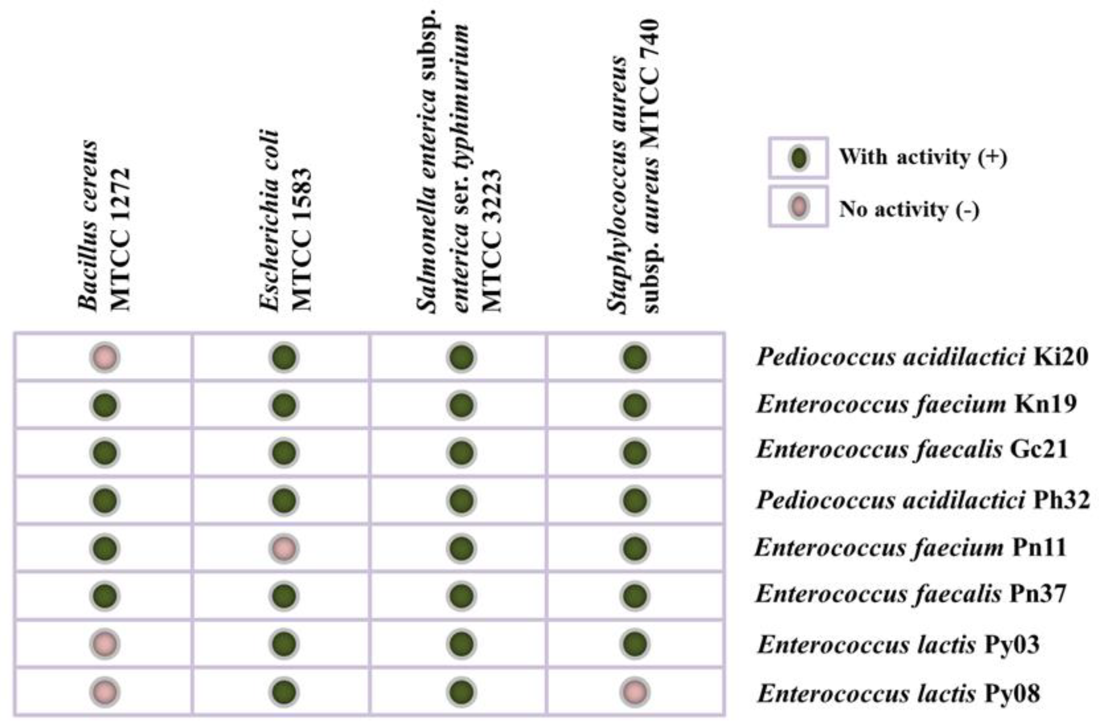
2.5.5. Antagonistic Activity

Antimicrobial activity of isolates against pathogenic bacteria such as Escherichia coli MTCC 1583, Salmonella enterica subsp. enterica ser. typhimurium MTCC 3223, Staphylococcus aureus subsp. aureus MTCC 740, and Bacillus cereus MTCC 1272 was conducted following the method described by Rai and Tamang [37]. Briefly, an overnight freshly prepared tested pathogen was adjusted to a density of 0.08 to 0.1 (±0.05) and spread on the surface of Mueller Hinton agar (M173, HiMedia, Mumbai, India) plates by sterile cotton swab and wells punching, made by a sterile borer. Overnight LAB cultures were inoculated into the wells and incubated at 37 °C for 24–48 h. After incubation, the zone of inhibition was observed.

2.5.6. Genetic Screening for Probiotic Functions

Gene detection of various probiotic functions was screened for isolates using a PCR-based method [38,39,40,41,42,43,44,45]. Details of genes and primers with probiotic function including bile salt tolerance, low pH tolerance, adherence/attachment, and bacteriocin production are listed in Table 1. For gene detection, a PCR reaction was prepared with a total volume of 10-μL containing GoTaq® Green Master Mix (M7122, Promega, WI, USA), DNA template, and primers (forward and reverse). Targeted gene amplification was performed using VeritiTM Thermal cycler (4375305, Applied Biosystems, Thermo Fisher Scientific, Carlsbad, CA, USA) under the following conditions: denaturation of PCR amplification (94 °C for 5 min), followed by 30 cycles denaturation (94 °C for 1 min), annealing process (55 °C for 1 min) and elongation process (72 °C for 1.5 min), respectively. Lastly, PCR amplification was set up for the continuation of the elongation process (72 °C for 10 min) and a stoppage process at 4 °C. The amplified PCR products were checked by agarose gel electrophoresis (0.8%) and visualized using Gel Doc™ EZ Imager (EZ735BR06435, BioRad, Hercules, CA, USA).

2.6. Bioinformatics Analysis

The raw sequence generated by Sanger sequencing was evaluated for the quality assessment using Sequence Scanner (v2.0) software of Applied Biosystems (https://www.thermofisher.com/in/en/home/life-science/sequencing/sanger-sequencing/sanger-dna-sequencing/sanger-sequencing-data-analysis.html) accessed on 2 December 2023. Based on the quality trace score (Q > 20) and length (>600 bp), good-quality sequences were assembled using ChromasPro v2.1.10 (http://technelysium.com.au/wp/chromas/) accessed on 2 December 2022 and chimera sequence filtration checked by Mallard programme [46]. Further, the good-quality assembled sequences were identified by mapping against the NCBI database using BLAST (Basic Local Alignment Search Tool) [47]. Lastly, the identified lactic acid bacterial strains were aligned using ClustalW [48] and constructed the molecular evolutionary analysis by the Neighbor-joining method [49] using MEGA11.0.13 (Molecular Evolutionary Genetics Analysis) [50].
All the experiments on in-vitro probiotic attributes were conducted in triplicates and represented in Mean ± SD (standard deviation).

3. Results

3.1. Preliminary Screening

A total of 352 bacterial isolates from kinema (114 isolates), grep-chhurpi (38 isolates), peha (71 isolates), peron namsing (87 isolates), and peruñyaan (42 isolates) were preliminarily screened for probiotic properties on the basis of low pH and bile salt (0.3%) tolerances. Out of which, only 54 isolates showed tolerances to low acid and bile salt. Further, 54 isolates were again screened for cell surface hydrophobicity. Finally, eight isolates showing >80% hydrophobicity were tentatively selected as probiotic bacteria for in vitro screening of probiotic attributes. Based on 16S rRNA gene sequencing (Figure 1), eight isolates were identified as Pediococcus acidilactici Ki20 and Enterococcus faecium Kn19 (isolated from kinema), Enterococcus faecalis Gc21 (grep-chhurpi), Pediococcus acidilactici Ph32 (peha), Enterococcus faecium Pn11 and Enterococcus faecalis Pn37 (peron namsing), Enterococcus lactis Py03, and Enterococcus lactis Py08 (peruñyaan) (Table 2).

3.2. In-Vitro Probiotic Properties

In vitro probiotic properties of eight LAB, isolated from the Himalayan fermented soybean foods were assessed. The survival rate of tested LAB was found >52% in pH 3.0 and >66% in bile salt, among which Enterococcus faecium Kn19 (kinema) and Pediococcus acidilactici Ph32 (peha) showed the highest survival rate of 73.67 ± 1.05% in low pH and 79.71 ± 0.13% in 3% bile salt, respectively (Table 3). More than 85% of hydrophobicity percentage was observed in all eight LAB, among which Enterococcus faecalis Gc21 (grep-chhurpi) showed the highest hydrophobicity percentage of 90.50 ± 10.14% (Table 3). Auto-aggregation percentage of all tested LAB ranged from 43.8 ± 1.05 to 55.73 ± 0.96 (Table 3). Moreover, all LAB strains showed co-aggregation property against Bacillus cereus MTCC 1272, Escherichia coli MTCC 1583, Salmonella enterica subsp. enterica ser. typhimurium MTCC 3223, and Staphylococcus aureus subsp. aureus MTCC 740 (Table 3). Pediococcus acidilactici Ki20 isolated from kinema showed the highest co-aggregation property against the tested pathogens (Table 3). Similarly, resistance to lysozyme was exhibited by all LAB, among which Pediococcus acidilactici Ph32 exhibited the highest resistance to lysozyme (Table 3). Further, in vitro probiotic attributes were tested for bile salt hydrolase activity (Table 3), in which five isolates were positive to sodium taurodeoxycholate and four isolates to Sodium taurocholate (Table 3). Additionally, the antagonistic activity of LAB, against four pathogens including Bacillus cereus MTCC 1272, Escherichia coli MTCC 1583, Salmonella enterica subsp. enterica ser. typhimurium MTCC 3223, and Staphylococcus aureus subsp. aureus MTCC 740, was performed and found. Only Enterococcus faecium Kn19 (kinema), E. faecalis Gc21 (grep-chhurpi), Pediococcus acidilactici Ph32 (peha), and E. faecalis Pn37 (peron namsing) exhibited antibacterial activity against all the tested pathogens, whereas other LAB strains showed the variable results (Figure 2).

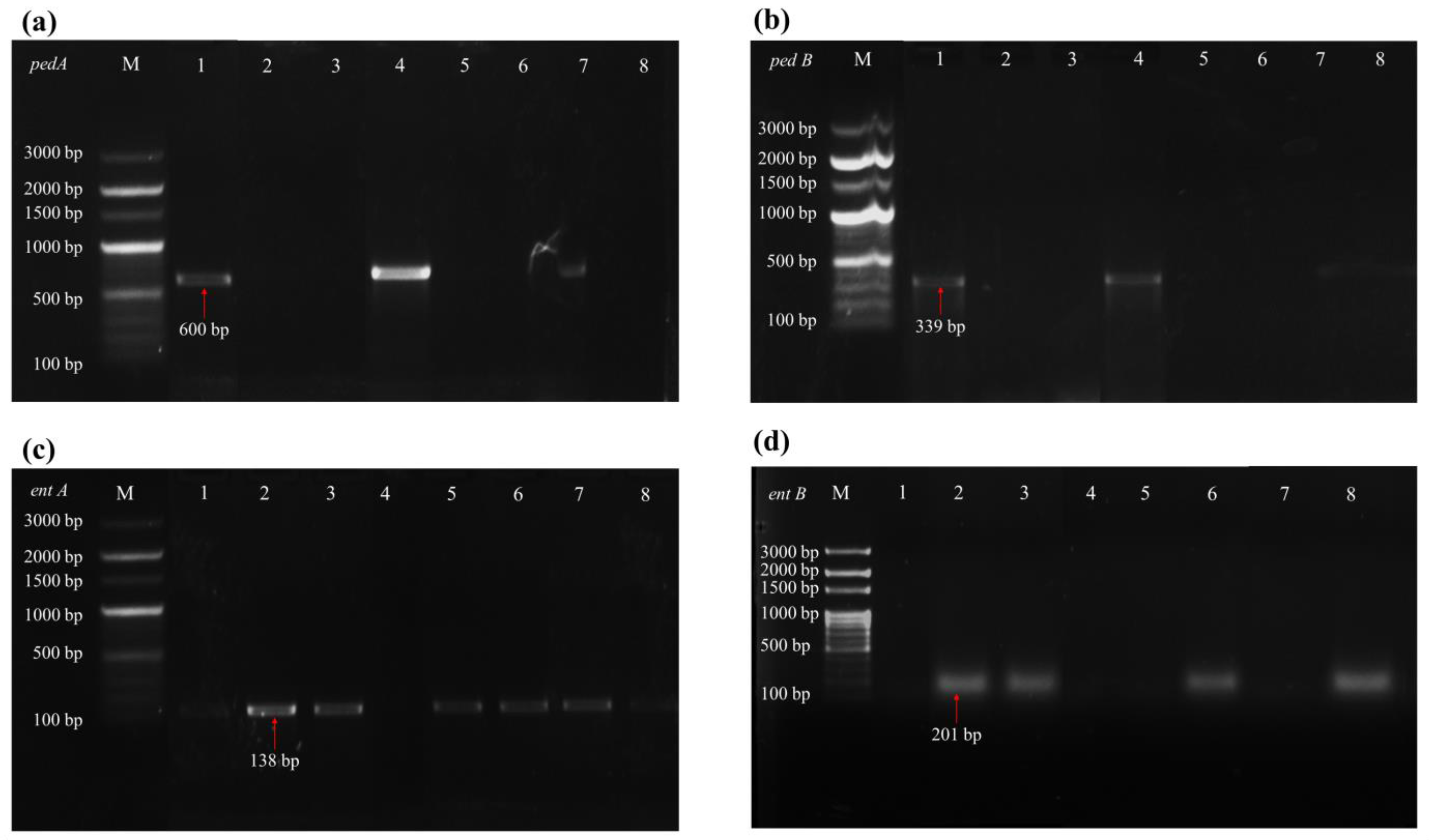
3.3. Gene Detection of Probiotic Functions

Genes responsible for various probiotic traits were screened for all eight LAB strains using a PCR-based method. Genetic screening of probiotic function for survival at low pH showed the presence of groEl in Pediococcus acidilactici Ki20 and Enterococcus faecium Kn19 (kinema), E. faecalis (grep-chhurpi), P. acidilactici Ph32 (peha), E. lactis Py03 and E. lactis Py08 (peruñyaan) (Figure 3a) and clpL genes in P. acidilactici Ki20 and E. faecium Kn19 (kinema), P. acidilactici Ph32 (peha), E. faecium Pn11 and E. faecalis Pn37 (peron namsing) and E. lactis Py08 (peruñyaan) (Figure 3b); however, none of the strains found to harbour odc and tdc genes. Further, genes responsible for bile salt tolerance were screened, and all strains showed the presence of apf (aggregation-promoting factor) gene (Figure 3c). Similarly, bsh gene responsible for bile salt hydrolase (BSH) was also detected in all strains (Figure 3d). All strains harboured the msa gene for cell surface adherence/attachment (Figure 3e), and only four strains viz. E. faecalis Gc21 (grep-chhurpi), E. faecium Pn11 and E. faecalis Pn37 (peron namsing), and E. lactis Py03 (peruñyaan) showed the presence of mub1 gene (Figure 3f). P. acidilactici Ki20 (kinema) and P. acidilactici Ki20 (peha) were detected with genes ped A (Figure 4a) and ped B (Figure 4b), respectively, for pediocin. Gene ent A was detected in E. faecium Kn19 and Gc21 (grep-chhurpi), E. faecium Pn11 and E. faecalis Pn37 (peron namsing), and E. lactis Py03 (peruñyaan) (Figure 4c). Gene ent B was detected in E. faecium Kn19 (kinema), E. faecalis Gc21 (grep-chhurpi), E. faecalis Pn37 (peron namsing), and E. lactis Py08 (peruñyaan) (Figure 4d). None of these genes, odc, tdc, Ir0085, Ir1516, fbp, and cylA, were detected in LAB strains isolated from the Himalayan fermented soybean foods.

4. Discussion

Bacillus species, mostly B. subtilis, have been reported as pre-dominant bacteria from the spontaneous fermentation of soybeans into sticky with umami-flavoured foods in Asia [10,11,13,51,52,53], which dispense several bio-functional and health benefits [7,24,54,55]. Even some of these species, largely Bacillus subtilis, have been developed as the commercial starter for monoculture fermentation of soybean into some popular fermented soybean foods such as natto of Japan [56] and cheonggokjang of Korea [57]. However, the age-old traditional fermentation of soybeans is orchestrated exclusively by spontaneous fermentation, which facilitates the existence and co-existence of the complex microbial community including domains of bacteria (bacilli, lactic acid bacteria, and other non-lactis), eukaryotes (yeasts and fungi), viruses (bacteriophages), and archaea [13,52,58]. The exact role of LAB in soybean fermentation has not been extensively studied; however, their coexistence during natural fermentation has raised concern for their fermentative or non-fermentative roles in soybean fermentation. One of the bio-functional roles of LAB is probiotics, which have been reported in many other fermented foods of plant/animal-based foods. We hypothesized that some LAB in Himalayan naturally fermented soybean foods may have probiotic properties, which may aid the functionality of the soybean foods.
The ability of any bacterial strain to survive in gastrointestinal transit, highly acidic and strong bile [59,60] and the adhesion property to epithelial cells [61,62] are the most important preliminary screening criteria to claim the tentative probiotic properties. Hence, we evaluated 352 bacterial isolates from the Himalayan fermented soybean foods based on their survival in low pH, bile salt, and adhesion property in terms of hydrophobicity index. Finally, eight strains of lactic acid bacteria were selected as probiotic bacteria viz. Pediococcus acidilactici strains Ki20 & Ph32, Enterococcus faecium strains Kn19 & Pn11, E. faecalis Gc21 & Pn37 and E. lactis Py03 & Py08. Species of Enterococcus and Pediococcus, isolated from other fermented soybean foods, also manifested the ability to resist low acid and bile salt, indicating their easy colonization of the human gut [63,64]. Detection of genes groEl, clpL, and apf in identified strains of Enterococcus and Pediococcus justify their responsibility for acid and bile tolerance [65]. Gene (bsh) responsible for bile salt hydrolase (BSH) usually correlated with the ability to lower serum cholesterol levels [66] was also detected in all strains of Pediococcus and Enterococcus. The microbial adhesion of LAB to hydrocarbons with a hydrophobicity index of ≥70% is being classified as highly hydrophobic strains [62]. Moreover, the results in the current study showed the hydrophobicity of ≥80% in all strains of P. acidilactici, E. faecium, E. faecalis, and E. lactis, which indicates their strong adhesion capabilities to the epithelial cells [67]. All strains showed the presence of msa gene, whereas only four strains of Enterococcus showed the presence of mucus adhesion mub1 gene, which is responsible for cell surface adherence/attachment [68]. In addition to adhesion, auto-aggregation and co-aggregation properties of probiotic bacteria that prevent pathogens from surface colonization were also recorded. Auto-aggregation could enable self-joining association of the same species of LAB attached to the mucosal surface [69]. Co-aggregation enables the intercellular attachment/adhesion with other microorganisms bound up with the ability to interact with pathogens [34]. Therefore, the ability of LAB strains for auto-aggregation and co-aggregation is related to the adhesion capacity that has an effect on reducing mucosa colonisation by pathogenic strains [70]. The ability to colonize is an important characteristic for probiotic isolates to exhibit beneficial properties and exclude pathogenic bacteria [71]. In the present study, we also observed the resistivity to lysozyme by the species of Pediococcus and Enterococcus. Tolerance to lysozyme is the first valuable step to evaluate the potential contribution of LAB in extreme environments of gastrointestinal pathways [72]. In addition, the results recorded indicate the ability of LAB strains to inhibit the growth of pathogens, which is one of the most desirable properties of probiotic strains to exhibit antibacterial activity [73]. The antibacterial activity of LAB against pathogens and spoilage microbes could be due to the production of bacteriocin or bacteriocin-like inhibitory substances (BLIS) [74,75]. The PCR-based method also revealed the detection of pediocin genes pedA and pedB, a class of bacteriocin [76] detected in Pediococcus acidilactici. Pediocin produced by Pediococcus acidilactici might be able to create pores in the cell membrane that ultimately leads to cell death [77]. Similarly, the detection of enterocin (entA, entB), which are structural genes for enterocin Xα and Xβ, respectively [78], in Enterococcus species, indicated their antimicrobial spectrum. The detection of enterocin-producing genes in Enterococcus species might attribute to controlling the target bacteria by inducing membrane porosity that causes leakage of target cells or by interfering with the DNA replication and transcription [79].

5. Conclusions

The current study was conducted to evaluate some of the probiotic properties of LAB isolated from Himalayan spontaneously fermented soybean foods. Pediococcus spp. and Enterococcus spp. revealed the potential probiotic characteristics in Himalayan fermented soybean foods. This may be the first report on the role of LAB in Bacillus-dominated Himalayan fermented soybean food. Based on in vitro studies, several probiotic attributes were able to be exhibited by the LAB strains, which were further screened by genetic screening that revealed the presence of some marker genes related to specific probiotic characteristics. However, further studies on whole genome analysis of probiotic strains, their safety assessment, and other functional characteristics may also be of great importance to understanding the role and contribution of probiotic LAB in soybean fermentation. Bacillus species were reported as the main key microbes in fermented soybean foods; however the ability of LAB strains to exhibit some functional characteristics such as organic acid production, and antibacterial properties against pathogens might attract the possible co-culture that suppresses unwanted microorganisms during fermentation. Additionally, probiotic LAB may be developed as starter(s) or co-culture(s) with Bacillus spp. for controlled and optimized fermentation of soybeans.

Author Contributions

Conceptualization, J.P.T.; methodology, P.K.; investigation, P.K.; resources, J.P.T.; data curation, J.P.T.; writing—original draft preparation, P.K.; writing—review and editing, J.P.T.; visualization, P.K.; supervision, J.P.T.; project administration, J.P.T.; funding acquisition, J.P.T. All authors have read and agreed to the published version of the manuscript.

Funding

This research received no external funding.

Institutional Review Board Statement

Not applicable.

Informed Consent Statement

Not applicable.

Data Availability Statement

The 16S rRNA gene sequences of identified lactic acid bacteria were deposited in GenBank NCBI under the accession numbers OP941712, OP941713, OP941714, OP941715, OP941716, OP941717, OP941718 and OP941719.

Acknowledgments

Jyoti P. Tamang is grateful to International Centre for Integrated Mountain Development (ICIMOD)—Mountain Chair for financial support.

Conflicts of Interest

The authors declare no conflict of interest.

References

  1. Durazzo, A.; Carocho, M.; Heleno, S.A.; Pedrosa, M.C.; Ueda, J.M.; Barros, L.; Souto, E.B.; Santini, A.; Lucarini, M. Fermented food/beverage and health: Current perspectives. Rend. Lincei Sci. Fis. Nat. 2022, 33, 729–738. [Google Scholar] [CrossRef]
  2. Nazhand, A.; Souto, E.B.; Lucarini, M.; Souto, S.B.; Durazzo, A.; Santini, A. Ready to use therapeutical beverages: Focus on functional beverages containing probiotics, prebiotics and synbiotics. Beverages 2020, 6, 26. [Google Scholar] [CrossRef]
  3. Tamang, J.P. Dietary culture and antiquity of the Himalayan fermented foods and alcoholic fermented beverages. J. Ethn. Foods 2022, 9, 30. [Google Scholar] [CrossRef]
  4. Singh, T.A.; Devi, K.R.; Ahmed, G.; Jeyaram, K. Microbial and endogenous origin of fibrinolytic activity in traditional fermented foods of Northeast India. Food Res. Int. 2014, 55, 356–362. [Google Scholar] [CrossRef]
  5. Tamang, J.P. Naturally fermented ethnic soybean foods of India. J. Ethn. Foods 2015, 2, 8–17. [Google Scholar] [CrossRef]
  6. Tamang, J.P.; Jeyaram, K.; Rai, A.K.; Mukherjee, P.K. Diversity of beneficial microorganisms and their functionalities in community-specific ethnic fermented foods of the Eastern Himalayas. Food Res. Int. 2021, 148, 110633. [Google Scholar] [CrossRef]
  7. Kharnaior, P.; Das, M.; Tamang, J.P. Therapeutic and anti-thrombotic properties of some naturally fermented soybean foods of the Eastern Himalayas. Fermentation 2023, 9, 91. [Google Scholar] [CrossRef]
  8. Tamang, J.P. Native microorganisms in the fermentation of kinema. Indian J. Microbiol. 2003, 43, 127–130. [Google Scholar]
  9. Chettri, R.; Tamang, J.P. Bacillus species isolated from Tungrymbai and Bekang, naturally fermented soybean foods of India. Int. J. Food Microbiol. 2015, 197, 72–76. [Google Scholar] [CrossRef]
  10. Kharnaior, P.; Tamang, J.P. Bacterial and fungal communities and their predictive functional profiles in kinema, a naturally fermented soybean food of India, Nepal and Bhutan. Food Res. Int. 2021, 140, 110055. [Google Scholar] [CrossRef]
  11. Kharnaior, P.; Tamang, J.P. Metagenomic-Metabolomic Mining of Kinema, a naturally fermented soybean food of the Eastern Himalayas. Front. Microbiol. 2022, 13, 868383. [Google Scholar] [CrossRef] [PubMed]
  12. Pariyar, P.; Yaduvanshi, P.S.; Raghu, P.; Tamang, J.P. Screening of Poly-Glutamic Acid (PGA)-Producing Bacillus Species from Indian Fermented Soybean Foods and Characterization of PGA. Fermentation 2022, 8, 495. [Google Scholar] [CrossRef]
  13. Tamang, J.P.; Kharnaior, P.; Pariyar, P.; Thapa, N.; Lar, N.; Win, K.S.; Mar, A.; Nyo, N. Shotgun sequence-based metataxonomic and predictive functional profiles of Pe poke, a naturally fermented soybean food of Myanmar. PLoS ONE 2021, 16, e0260777. [Google Scholar] [CrossRef]
  14. Chettri, R.; Bhutia, M.; Tamang, J.P. Poly-γ-glutamic acid (PGA)-producing Bacillus species isolated from Kinema, Indian fermented soybean food. Front. Microbiol. 2016, 7, 971. [Google Scholar] [CrossRef] [PubMed]
  15. Rai, A.K.; Sanjukta, S.; Chourasia, R.; Bhat, I.; Bhardwaj, P.K.; Sahoo, D. Production of bioactive hydrolysate using protease, β-glucosidase and α-amylase of Bacillus spp. isolated from kinema. Bioresour. Technol. 2017, 235, 358–365. [Google Scholar] [CrossRef]
  16. Chettri, R.; Tamang, J.P. Organoleptic evaluation of Tungrymbai and Bekang, naturally fermented soybean foods, produced by using selected species of Bacillus. J. Sci. Ind. Res. 2016, 75, 416–419. [Google Scholar]
  17. Sanjukta, S.; Rai, A.K.; Muhammed, A.; Jeyaram, K.; Talukdar, N.C. Enhancement of antioxidant properties of two soybean varieties of Sikkim Himalayan region by proteolytic Bacillus subtilis fermentation. J. Funct. Foods 2015, 14, 650–658. [Google Scholar] [CrossRef]
  18. Sarkar, P.K.; Tamang, J.P.; Cook, P.E.; Owens, J.D. Kinema—A traditional soybean fermented food: Proximate composition and microflora. Food Microbiol. 1994, 11, 47–55. [Google Scholar] [CrossRef]
  19. Kumar, J.; Sharma, N.; Kaushal, G.; Samurailatpam, S.; Sahoo, D.; Rai, A.K.; Singh, S.P. Metagenomic insights into the taxonomic and functional features of kinema, a traditional fermented soybean product of Sikkim Himalaya. Front. Microbiol. 2019, 10, 1744. [Google Scholar] [CrossRef]
  20. Goel, A.; Halami, P.M.; Tamang, J.P. Genome analysis of Lactobacillus plantarum isolated from some Indian fermented foods for bacteriocin production and probiotic marker genes. Front. Microbiol. 2020, 11, 40. [Google Scholar] [CrossRef]
  21. Lim, E.S. Antibacterial activity of lactic acid bacteria against biogenic amine-producing Bacillus spp. isolated from traditional fermented soybean paste. Korean J. Microbiol. 2018, 54, 398–409. [Google Scholar] [CrossRef]
  22. Ma, H.; Wang, L.; Yu, H.; Wang, W.; Wu, G.; Qin, G.; Tan, Z.W.Y.; Pang, H. Protease-producing lactic acid bacteria with antibacterial properties and their potential use in soybean meal fermentation. Chem. Biol. Technol. Agric. 2022, 9, 40. [Google Scholar] [CrossRef]
  23. Sirilun, S.; Sivamaruthi, B.S.; Kesika, P.; Peerajan, S.; Chaiyasut, C. Lactic acid bacteria mediated fermented soybean as a potent nutraceutical candidate. Asian Pac. J. Trop. Biomed. 2017, 7, 930–936. [Google Scholar] [CrossRef]
  24. Jang, C.H.; Oh, J.; Lim, J.S.; Kim, H.J.; Kim, J.S. Fermented soy products: Beneficial potential in neurodegenerative diseases. Foods 2021, 10, 636. [Google Scholar] [CrossRef]
  25. Jeong, J.K.; Chang, H.K.; Park, K.Y. Doenjang prepared with mixed starter cultures attenuates azoxymethane and dextran sulfate sodium-induced colitis-associated colon carcinogenesis in mice. J. Carcinog. 2014, 13, 9. [Google Scholar] [CrossRef]
  26. Fong, F.L.Y.; Lam, K.Y.; San Lau, C.; Ho, K.H.; Kan, Y.H.; Poon, M.Y.; El-Nezami, H.; Sze, E.T.P. Reduction in biogenic amines in douchi fermented by probiotic bacteria. PLoS ONE 2020, 15, e0230916. [Google Scholar] [CrossRef] [PubMed]
  27. Son, S.H.; Jeon, H.L.; Yang, S.J.; Sim, M.H.; Kim, Y.J.; Lee, N.K.; Paik, H.D. Probiotic lactic acid bacteria isolated from traditional Korean fermented foods based on β-glucosidase activity. Food Sci. Biotechnol. 2018, 27, 123–129. [Google Scholar] [CrossRef]
  28. Shangpliang, H.N.J.; Tamang, J.P. Phenotypic and genotypic characterisation of lactic acid bacteria isolated from exotic naturally fermented milk (cow and yak) products of Arunachal Pradesh, India. Int. Dairy J. 2021, 118, 105038. [Google Scholar] [CrossRef]
  29. Nithya, V.; Halami, P.M. Evaluation of probiotic characteristics of Bacillus species isolated from different food sources. Ann. Microbiol. 2013, 63, 129–137. [Google Scholar] [CrossRef]
  30. Bao, Y.; Zhang, Y.; Zhang, Y.; Liu, Y.; Wang, S.; Dong, X.; Wang, Y.; Zhang, H. Screening of potential probiotic properties of Lactobacillus fermentum isolated from traditional dairy products. Food Control 2010, 21, 695–701. [Google Scholar] [CrossRef]
  31. Nath, S.; Sikidar, J.; Roy, M.; Deb, B. In vitro screening of probiotic properties of Lactobacillus plantarum isolated from fermented milk product. Food Qual. Saf. 2020, 4, 213–223. [Google Scholar] [CrossRef]
  32. Lane, D.J. 16S/23S rRNA sequencing. In Nucleic Acid Techniques in Bacterial Systematics; Stackebrandt, E., Goodfellow, M., Eds.; John Wiley and Sons: New York, NY, USA, 1991; pp. 115–175. [Google Scholar]
  33. Mallappa, R.H.; Singh, D.K.; Rokana, N.; Pradhan, D.; Batish, V.K.; Grover, S. Screening and selection of probiotic Lactobacillus strains of Indian gut origin based on assessment of desired probiotic attributes combined with principal component and heatmap analysis. LWT Food Sci. Technol. 2019, 105, 272–281. [Google Scholar] [CrossRef]
  34. Li, M.; Wang, Y.; Cui, H.; Li, Y.; Sun, Y.; Qiu, H.J. Characterization of lactic acid bacteria isolated from the gastrointestinal tract of a wild boar as potential probiotics. Front. Vet. Sci. 2020, 7, 49. [Google Scholar] [CrossRef] [PubMed]
  35. Vera-Pingitore, E.; Jimenez, M.E.; Dallagnol, A.; Belfiore, C.; Fontana, C.; Fontana, P.; von Wright, A.; Vignolo, G.; Plumed-Ferrer, C. Screening and characterization of potential probiotic and starter bacteria for plant fermentations. LWT Food Sci. Technol. 2016, 71, 288–294. [Google Scholar] [CrossRef]
  36. Pradhan, P.; Tamang, J.P. Probiotic properties of lactic acid bacteria isolated from traditionally prepared dry starters of the Eastern Himalayas. World J. Microbiol. Biotechnol. 2021, 37, 1–13. [Google Scholar] [CrossRef] [PubMed]
  37. Rai, R.; Tamang, J.P. In vitro and genetic screening of probiotic properties of lactic acid bacteria isolated from naturally fermented cow-milk and yak-milk products of Sikkim, India. World J. Microbiol. Biotechnol. 2022, 38, 1–20. [Google Scholar] [CrossRef]
  38. Turpin, W.; Humblot, C.; Guyot, J.P. Genetic screening of functional properties of lactic acid bacteria in a fermented pearl millet slurry and in the metagenome of fermented starchy foods. Appl. Environ. Microbiol. 2011, 77, 8722–8734. [Google Scholar] [CrossRef]
  39. Ramiah, K.; Van Reenen, C.A.; Dicks, L.M.T. Expression of the mucus adhesion genes Mub and MapA, adhesion-like factor EF-Tu and bacteriocin gene plaA of Lactobacillus plantarum 423, monitored with real-time PCR. Int. J. Food Microbiol. 2007, 116, 405–409. [Google Scholar] [CrossRef]
  40. Archer, A.C.; Halami, P.M. Probiotic attributes of Lactobacillus fermentum isolated from human feces and dairy products. Appl. Environ. Microbiol. 2015, 99, 8113–8123. [Google Scholar] [CrossRef]
  41. Özdemir, G.B.; Oryaşın, E.; Bıyık, H.H.; Özteber, M.; Bozdoğan, B. Phenotypic and genotypic characterization of bacteriocins in enterococcal isolates of different sources. Indian J. Microbiol. 2011, 51, 182–187. [Google Scholar] [CrossRef]
  42. De Vuyst, L.; Moreno, M.F.; Revets, H. Screening for enterocins and detection of hemolysin and vancomycin resistance in enterococci of different origins. Int. J. Food Microbiol. 2003, 84, 299–318. [Google Scholar] [CrossRef] [PubMed]
  43. Rodrıguez, J.M.; Cintas, L.M.; Casaus, P.; Martınez, M.I.; Suárez, A.; Hernández, P.E. Detection of pediocin PA-1-producing pediococci by rapid molecular biology techniques. Food Microbiol. 1997, 14, 363–371. [Google Scholar] [CrossRef]
  44. El-Arabi, N.I.; Salim, R.G.; Abosereh, N.A.; Abdelhadi, A.A. Molecular characterization of some antilisterial bacteriocin genes from Enterococcus faecium and Pediococcus pentosaceus. Microbiol. Biotechnol. Lett. 2018, 46, 288–299. [Google Scholar] [CrossRef]
  45. Creti, R.; Imperi, M.; Bertuccini, L.; Fabretti, F.; Orefici, G.; Di Rosa, R.; Baldassarri, L. Survey for virulence determinants among Enterococcus faecalis isolated from different sources. J. Med. Microbiol. 2004, 53, 13–20. [Google Scholar] [CrossRef]
  46. Ashelford, K.E.; Chuzhanova, N.A.; Fry, J.C.; Jones, A.J.; Weightman, A.J. New screening software shows that most recent large 16S rRNA gene clone libraries contain chimeras. Appl. Environ. Microbiol. 2006, 72, 5734–5741. [Google Scholar] [CrossRef]
  47. Altschul, S.F.; Gish, W.; Miller, W.; Myers, E.W.; Lipman, D.J. Basic local alignment search tool. J. Mol. Biol. 1990, 215, 403–410. [Google Scholar] [CrossRef]
  48. Thompson, J.D.; Higgins, D.G.; Gibson, T.J. CLUSTAL W: Improving the sensitivity of progressive multiple sequence alignment through sequence weighting, position-specific gap penalties and weight matrix choice. Nucleic Acids Res. 1994, 22, 4673–4680. [Google Scholar] [CrossRef]
  49. Saitou, N.; Nei, M. The neighbor-joining method: A new method for reconstructing phylogenetic trees. Mol. Biol. Evol. 1987, 4, 406–425. [Google Scholar] [CrossRef]
  50. Tamura, K.; Stecher, G.; Kumar, S. MEGA11: Molecular evolutionary genetics analysis version 11. Mol. Biol. Evol. 2021, 38, 3022–3027. [Google Scholar] [CrossRef]
  51. Gopikrishna, T.; Suresh Kumar, H.K.; Perumal, K.; Elangovan, E. Impact of Bacillus in fermented soybean foods on human health. Ann. Microbiol. 2021, 71, 30. [Google Scholar] [CrossRef]
  52. Tamang, J.P.; Das, S.; Kharnaior, P.; Pariyar, P.; Thapa, N.; Jo, S.W.; Yim, E.J.; Shin, D.H. Shotgun metagenomics of Cheonggukjang, a fermented soybean food of Korea: Community structure, predictive functionalities and amino acids profile. Food Res. Int. 2022, 151, 110904. [Google Scholar] [CrossRef] [PubMed]
  53. Yongsawas, R.; In-on, A.; Inta, A.; Kampuansai, J.; Pandith, H.; Suwannarach, N.; Lumyong, S.; Chitov, T.; Disayathanoowat, T. Bacterial communities in Lanna fermented soybeans from three different ethnolinguistic groups in Northern Thailand. Microorganisms 2023, 11, 649. [Google Scholar] [CrossRef]
  54. do Prado, F.G.; Pagnoncelli, M.G.B.; de Melo Pereira, G.V.; Karp, S.G.; Soccol, C.R. Fermented soy products and their potential health benefits: A review. Microorganisms 2022, 10, 1606. [Google Scholar] [CrossRef]
  55. Liu, Y.; Han, Y.; Cao, L.; Wang, X.; Dou, S. Analysis of main components and prospects of natto. Adv. Enzym Res. 2021, 9, 1–9. [Google Scholar] [CrossRef]
  56. Elhalis, H.; Chin, X.H.; Chow, Y. Soybean fermentation: Microbial ecology and starter culture technology. Crit. Rev. Food Sci. Nutr. 2023, 14, 1–23. [Google Scholar] [CrossRef] [PubMed]
  57. Kim, S.H.; Yehuala, G.A.; Bang, W.Y.; Yang, J.; Jung, Y.H.; Park, M.K. Safety evaluation of Bacillus subtilis IDCC1101, newly isolated from cheonggukjang, for industrial applications. Microorganisms 2022, 10, 2494. [Google Scholar] [CrossRef] [PubMed]
  58. Yue, X.; Li, M.; Liu, Y.; Zhang, X.; Zheng, Y. Microbial diversity and function of soybean paste in East Asia: What we know and what we don’t. Curr. Opin. Food Sci. 2021, 37, 145–152. [Google Scholar] [CrossRef]
  59. Urdaneta, V.; Casadesús, J. Interactions between bacteria and bile salts in the gastrointestinal and hepatobiliary tracts. Front. Med. 2017, 4, 163. [Google Scholar] [CrossRef]
  60. Han, S.; Lu, Y.; Xie, J.; Fei, Y.; Zheng, G.; Wang, Z.; Liu, J.; Lv, L.; Ling, Z.; Berglund, B.; et al. Probiotic gastrointestinal transit and colonization after oral administration: A long journey. Front. Cell. Infect. Microbiol. 2021, 11, 609722. [Google Scholar] [CrossRef]
  61. Garcia-Gonzalez, N.; Prete, R.; Battista, N.; Corsetti, A. Adhesion properties of food-associated lactobacillus plantarum strains on human intestinal epithelial cells and modulation of IL-8 release. Front. Microbiol. 2018, 9, 2392. [Google Scholar] [CrossRef]
  62. Petrova, P.; Tsvetanova, F.; Petrov, K. Low cell surface hydrophobicity is one of the key factors for high butanol tolerance of Lactic acid bacteria. Eng. Life Sci. 2019, 19, 133–142. [Google Scholar] [CrossRef] [PubMed]
  63. Krawczyk, B.; Wityk, P.; Gałęcka, M.; Michalik, M. The many faces of Enterococcus spp.-commensal, probiotic and opportunistic pathogen. Microorganisms 2021, 9, 1900. [Google Scholar] [CrossRef] [PubMed]
  64. Qi, Y.; Huang, L.; Zeng, Y.; Li, W.; Zhou, D.; Xie, J.; Xie, J.; Tu, Q.; Deng, D.; Yin, J. Pediococcus pentosaceus: Screening and application as probiotics in food processing. Front. Microbiol. 2021, 12, 762467. [Google Scholar] [CrossRef]
  65. Chen, C.; Yu, L.; Tian, F.; Zhao, J.; Zhai, Q. Identification of novel bile salt-tolerant genes in Lactobacillus using comparative genomics and its application in the rapid screening of tolerant strains. Microorganisms 2022, 10, 2371. [Google Scholar] [CrossRef]
  66. Hernández-Gómez, J.G.; López-Bonilla, A.; Trejo-Tapia, G.; Ávila-Reyes, S.V.; Jiménez-Aparicio, A.R.; Hernández-Sánchez, H. In vitro bile salt hydrolase (BSH) activity screening of different probiotic microorganisms. Foods 2021, 10, 674. [Google Scholar] [CrossRef]
  67. Mladenović, K.G.; Grujović, M.Ž.; Nikodijević, D.D.; Čomić, L.R. The hydrophobicity of enterobacteria and their co-aggregation with Enterococcus faecalis isolated from Serbian cheese. Biosci. Microbiota Food Health 2020, 39, 227–233. [Google Scholar] [CrossRef]
  68. Holst, B.; Glenting, J.; Holmstrøm, K.; Israelsen, H.; Vrang, A.; Antonsson, M.; Ahrné, S.; Madsen, S.M. Molecular switch controlling expression of the mannose-specific adhesin, Msa, in Lactobacillus plantarum. Appl. Environ. Microbiol. 2019, 85, e02954-18. [Google Scholar] [CrossRef]
  69. Sharma, K.; Attri, S.; Goel, G. Selection and evaluation of probiotic and functional characteristics of autochthonous lactic acid bacteria isolated from fermented wheat flour dough babroo. Probiotics Antimicrob. 2019, 11, 774–784. [Google Scholar] [CrossRef] [PubMed]
  70. Zawistowska-Rojek, A.; Kośmider, A.; Stępień, K.; Tyski, S. Adhesion and aggregation properties of Lactobacillaceae strains as protection ways against enteropathogenic bacteria. Arch. Microbiol. 2022, 204, 285. [Google Scholar] [CrossRef]
  71. Alp, D.; Kuleaşan, H. Adhesion mechanisms of lactic acid bacteria: Conventional and novel approaches for testing. World J. Microbiol. Biotechnol. 2019, 35, 1–9. [Google Scholar] [CrossRef]
  72. Samedi, L.; Charles, A.L. Isolation and characterization of potential probiotic Lactobacilli from leaves of food plants for possible additives in pellet feeding. Ann. Agric. Sci. 2019, 64, 55–62. [Google Scholar] [CrossRef]
  73. Silva, D.R.; Sardi, J.D.C.O.; de Souza Pitangui, N.; Roque, S.M.; da Silva, A.C.B.; Rosalen, P.L. Probiotics as an alternative antimicrobial therapy: Current reality and future directions. J. Funct. Foods 2020, 73, 104080. [Google Scholar] [CrossRef]
  74. Abanoz, H.S.; Kunduhoglu, B. Antimicrobial activity of a bacteriocin produced by Enterococcus faecalis KT11 against some pathogens and antibiotic-resistant bacteria. Korean J. Food Sci. Anim. Resour. 2018, 38, 1064. [Google Scholar] [CrossRef]
  75. Sharma, B.R.; Halami, P.M.; Tamang, J.P. Novel pathways in bacteriocin synthesis by lactic acid bacteria with special reference to ethnic fermented foods. Food Sci. Biotechnol. 2021, 31, 1–16. [Google Scholar] [CrossRef] [PubMed]
  76. Kim, W.S.; Lee, J.Y.; Singh, B.; Maharjan, S.; Hong, L.; Lee, S.M.; Cui, L.H.; Lee, K.J.; Kim, G.; Yun, C.H.; et al. A new way of producing pediocin in Pediococcus acidilactici through intracellular stimulation by internalized inulin nanoparticles. Sci. Rep. 2018, 8, 5878. [Google Scholar] [CrossRef] [PubMed]
  77. Wang, J.; Li, L.; Zhao, X.; Zhou, Z. Partial characteristics and antimicrobial mode of pediocin produced by Pediococcus acidilactici PA003. Ann. Microbiol. 2015, 65, 1753–1762. [Google Scholar] [CrossRef]
  78. Hu, C.B.; Malaphan, W.; Zendo, T.; Nakayama, J.; Sonomoto, K. Enterocin X, a novel two-peptide bacteriocin from Enterococcus faecium KU-B5, has an antibacterial spectrum entirely different from those of its component peptides. Appl. Environ. Microbiol. 2010, 76, 4542–4545. [Google Scholar] [CrossRef]
  79. Wu, Y.; Pang, X.; Wu, Y.; Liu, X.; Zhang, X. Enterocins: Classification, synthesis, antibacterial mechanisms and food applications. Molecules 2022, 27, 2258. [Google Scholar] [CrossRef]
Figure 1. Phylogenetic analysis of lactic acid bacteria isolated from spontaneously fermented soybean foods of the Eastern Himalayas by the neighbor-joining method using MEGA11, based on 16S rRNA gene sequences. The bootstrap consensus of 1000 replicates was taken to represent the evolutionary history and Kimura-2 parameter method for evolutionary distances. Pediococcus acidilactici DSM 20284, Enterococcus faecalis ATCC 19433, Enterococcus lactis BT159, and Enterococcus faecium LMG 11423 were used as reference type strains, and Escherichia coli ATCC 11775 as an outgroup.
Fermentation 09 00461 g001
Figure 2. Antibacterial activity of identified lactic acid bacterial strains against some pathogenic bacteria.
Fermentation 09 00461 g002
Figure 3. PCR amplification of marker genes associated with probiotic characteristics. Two marker genes were associated with survival at low pH (a) groEl and (b) clpL. Two marker genes were associated with bile salt (c) apf and (d) bsh. Two marker genes were associated with adhesion/attachment (e) msa and (f) mub1. Details in the image of gel electrophoresis described as: ‘M’ DNA ladder (100 bp); (1) Pediococcus acidilactici Ki20 (Kinema); (2) Enterococcus faecium Kn19 (Kinema); (3) Enterococcus faecalis Gc21 (Grep-chhurpi); (4) Pediococcus acidilactici Ph32 (Peha); (5) Enterococcus faecium Pn11 (Peron namsing); (6) Enterococcus faecalis Pn37 (Peron namsing); (7) Enterococcus lactis Py03 (Peruñyaan); (8) Enterococcus lactis Py08 (Peruñyaan).
Fermentation 09 00461 g003
Figure 4. Marker genes associated with probiotic characteristics detected by PCR amplification. Detection of the gene associated with pediocin (a) pedA and (b) pedB. Marker genes were associated with enterocin (c) entA and (d) entB. Gel electrophoresis image description: ‘M’ DNA ladder (100 bp); (1) Pediococcus acidilactici Ki20 (Kinema); (2) Enterococcus faecium Kn19 (Kinema); (3) Enterococcus faecalis Gc21 (Grep-chhurpi); (4) Pediococcus acidilactici Ph32 (Peha); (5) Enterococcus faecium Pn11 (Peron namsing); (6) Enterococcus faecalis Pn37 (Peron namsing); (7) Enterococcus lactis Py03 (Peruñyaan); (8) Enterococcus lactis Py08 (Peruñyaan).
Fermentation 09 00461 g004
Table 1. Details of marker genes studied, and their respective primers used for detection of probiotic functions.
GenesFunctionsPrimer Sequence (5′ → 3′) (F = Forward; R = Reverse)Annealing Temperature (°C)Size of Amplicon (bp)References
groElSurvival at low pHF-TTCCATGGCKTCAGCRATCA
R-GCTAAYCCWGTTGGCATTCG
58168[38]
clpLSurvival at low pHF-GCTGCCTTYAAAACATCATCTGG
R-AATACAATTTTGAARAACGCAGCTT
50158[38]
odcSurvival at low pHF-TMTWCCAACHGATCGWAATGC
R-CRCCCCAWGCACARTCRAA
52245[38]
tdcSurvival at low pHF-CCACTGCTGCATCTGTTTG
R-CCRTARTCNGGNATAGCRAARTCNGTRTG
50370[38]
Ir0085Bile saltF-RCTTTGACCGRTGGGGCTRT
R-NNNATGGCCGCATGGAAA
57.5150[38]
Ir1516Bile saltF-TRACCACTYTCWCCATTCAACAA
R-CCACTAGCRATGACYAATACKGGT
56.5143[38]
apfBile saltF-YAGCAACACGTTCTTGGTTAGCA
R-GAATCTGGTGGTTCATAYWCAGC
53112[38]
bshBile saltF-ATTGAAGGCGGAACSGGMTA
R-ATWACCGGWCGGAAAGCTG
58155[38]
mub1AdhesionF-GTAGTTACTCAGTGACGATCAATG
R-TAATTGTAAAGGTATAATCGGAGG
50150[39]
msaAdhesionF-GCGATTAGGGGTGTGCAAG
R-GCAGTTGGTGACGTAGGCA
55319[40]
fbpAdhesionF-AGTGCTGAAATYATGGGAAGA
R-AATTGTCCACCTTGTTGCTG
60835[40]
entABacteriocinF-GGT ACC ACT CAT AGT GGA AA
R-CCC TGG AAT TGC TCC ACC TAA
55138[41]
entBBacteriocinF-CAA AAT GTA AAA GAA TTA AGT ACG
R-AGA GTA TAC ATT TGC TAA CCC
56201[42]
pedABacteriocinF-AAAATATCTAACTAATACTTG
R-TAAAAAGATATTTGACCAAAA
44600[43]
pedBBacteriocinF-ATGAATAAGACTAAGTCGGAACATATT
R-CTATTGGCTAGGCCACGTATTG
57339[44]
cylABacteriocinF-ACTCGGGGATTGATAGGC
R-GCTGCTAAAGCTGCGCTT
54688[45]
Table 2. Lactic acid bacteria isolated from naturally fermented soybean food of the Eastern Himalayas with NCBI accession numbers and reference type strains.
ProductsIdentity with Sample CodeType Species (% Similarity)GenBank Accession Number
KinemaPediococcus acidilactici Ki20Pediococcus acidilactici DSM 20284 (99.72%)OP941712
Enterococcus faecium Kn19Enterococcus faecium LMG 11423 (99.65%)OP941713
Grep chhurpiEnterococcus faecalis Gc21Enterococcus faecalis ATCC 19433 (99.85%)OP941714
PehaPediococcus acidilactici Ph32Pediococcus acidilactici DSM 20284 (99.86%)OP941715
Peron namsingEnterococcus faecium Pn11Enterococcus faecium LMG 11423 (99.52%)OP941716
Enterococcus faecalis Pn37Enterococcus faecalis ATCC 19,433 (99.93%)OP941717
PeruñyaanEnterococcus lactis Py03Enterococcus lactis BT159 (99.64%)OP941718
Enterococcus lactis Py08Enterococcus lactis BT159 (99.71%)OP941719
Table 3. Probiotic properties of lactic acid bacterial strains isolated from spontaneously fermented soybean foods of the Eastern Himalayas.
SamplesLactic Acid BacteriaSurvival Rate (%)Cell Surface Hydrophobicity (%)Auto-Aggregation (%)Co-Aggregation (%)Resistance to Lysozyme (%)BSH Activity
Acid (pH 3)Bile (0.3% Oxgall)Bacillus cereus MTCC 1272Escherichia coli MTCC 1583Salmonella enterica subsp. enterica ser. typhimurium MTCC 3223Staphylococcus aureus subsp. aureus MTCC 740Sodium TaurodeoxycholateSodium Taurocholate
KinemaPediococcus acidilactici Ki2067.85 ± 0.2578.03 ± 0.2186.67 ± 3.3055.73 ± 0.9679.51 ± 0.3572.20 ± 0.7275.52 ± 0.3475.14 ± 0.5262.38 ± 0.59++
Enterococcus faecium Kn1973.67 ± 1.0574.08 ± 0.3887.84 ± 3.0650.03 ± 1.1465.02 ± 1.0458.48 ± 1.7758.52 ± 0.8758.97 ± 1.1370.80 ± 0.46++
Grep-chhurpiEnterococcus faecalis Gc2152.02 ± 1.0566.46 ± 0.8890.50 ± 10.1454.15 ± 0.2959.72 ± 0.9356.31 ± 0.4560.99 ± 0.7859.06 ± 0.2575.09 ± 0.77
PehaPediococcus acidilactici Ph3266.11 ± 2.3279.71 ± 0.1388.97 ± 6.5543.80 ± 0.4064.52 ± 0.2558.58 ± 1.1160.97 ± 0.6460.82 ± 0.4177.76 ± 0.25++
Peron namsingEnterococcus faecium Pn1155.92 ± 1.8575.32 ± 0.9589.34 ± 6.6048.13 ± 0.3359.13 ± 0.4059.69 ± 0.3966.41 ± 0.7755.93 ± 0.7966.08 ± 0.30+
Enterococcus faecalis Pn3761.43 ± 0.6575.54 ± 0.5585.67 ± 1.8955.05 ± 0.3663.66 ± 0.2556.90 ± 0.4259.58 ± 0.4258.10 ± 1.0367.78 ± 1.05
PeruñyaanEnterococcus lactis Py0353.24 ± 1.1766.41 ± 0.8589.00 ± 11.3155.46 ± 0.4864.01 ± 0.9251.29 ± 0.7560.23 ± 0.5058.54 ± 0.2464.48 ± 1.75+
Enterococcus lactis Py0864.92 ± 1.6071.88 ± 0.1490.34 ± 8.9654.10 ± 0.3858.14 ± 0.2752.69 ± 0.7053.98 ± 0.2654.41 ± 0.5272.53 ± 1.39+
Note: All experiments were conducted in triplicates represented in Mean ± SD. ‘+’ = Activity; ‘−’ = No activity; BSH = Bile Salt Hydrolase; MTCC = Microbial Type Culture Collection.
Disclaimer/Publisher’s Note: The statements, opinions and data contained in all publications are solely those of the individual author(s) and contributor(s) and not of MDPI and/or the editor(s). MDPI and/or the editor(s) disclaim responsibility for any injury to people or property resulting from any ideas, methods, instructions or products referred to in the content.

