Article

Effects of Lactobacillus curvatus HY7602-Fermented Antlers in Dexamethasone-Induced Muscle Atrophy

1
R&BD Center, hy Co., Ltd., 22, Giheungdanji-ro 24beon-gil, Giheung-gu, Yongin-si 17086, Gyeonggi-do, Korea
2
Department of Oriental Medicine Biotechnology, College of Life Science, Kyung Hee University, Yongin-si 17104, Gyeonggi-do, Korea
*
Authors to whom correspondence should be addressed.
These authors contributed equally to this work.
Fermentation 2022, 8(9), 454; https://doi.org/10.3390/fermentation8090454
Submission received: 23 August 2022 / Revised: 8 September 2022 / Accepted: 9 September 2022 / Published: 12 September 2022
(This article belongs to the Special Issue Bioactivity Change in Fermented Foods)

Abstract

:
This study assessed the improvements yielded by Lactobacillus curvatus HY7602-fermented antlers (FA) in dexamethasone-induced muscle atrophy and the effects of bioactive compounds increased by fermentation. Dexamethasone-treated C2C12 myoblast cells were treated with FA and non-fermented antlers (NFA). FA showed inhibitory effects on muscle protein degradation in the C2C12 cells. Hsb:ICR mice were orally administered saline (control(CON) and dexamethasone only (DEX)), oxymetholone (DEX+OXY), NFA (DEX+NFA), and FA (DEX+FA) via gavage. Before the end of the experiment, dexamethasone was intraperitoneally (IP) injected into the mice, except in the control group, to induce muscle atrophy. Compared with the DEX group, the DEX+FA group exhibited a significant prevention in the reduction of hindlimb strength, calf thickness, calf muscle weight, and the cross-sectional area of muscle fibers (p < 0.05). The FA-induced improvements in muscle atrophy were associated with a decreased gene expression of protein degradation and growth inhibition, and an increased gene expression of protein synthesis and growth factors. Sialic acid, a bioactive compound associated with muscles, was increased by 51.41% after fermentation and suppressed the expression of protein degradation genes in the C2C12 cells. L. curvatus HY7602-fermented antlers with increased sialic acid after fermentation may therefore be useful for preventing and improving muscle atrophy.

1. Introduction

As humans age, they experience a gradual decrease in muscle mass and strength, which leads to functional loss and an increase in intramuscular fat [1,2]. Muscle loss further accumulates and reaches the same level as that in sarcopenia. The underlying molecular mechanisms include decreased division of muscle cells due to aging [3], unbalanced and accumulated apoptosis [4], the accumulation of mitochondrial damage in muscle cells [5], and dissonance of muscle protein degradation [6]. Sarcopenia is highly correlated with frailty, disability, morbidity, and mortality in the elderly [7]. Various mouse models have been developed to study sarcopenia. These include a frequently used model that utilizes the administration of dexamethasone, which is a synthetic glucocorticoid that induces catabolic muscle loss [8,9,10,11,12]. Oxymetholone is an anabolic–androgen hormone that is used as a drug for muscle strengthening or for the treatment of various musculoskeletal systems [13] and is also often used as a positive control in sarcopenia studies. Several studies have shown that oxymetholone prevents calf muscle loss and weakness in dexamethasone-treated mouse models [14] and inhibits muscle atrophy in aging or sarcopenia patients in clinical trials [15,16]. These effects of oxymetholone are associated with an improved level of IGF-1 in the blood and an increased mRNA expression in muscle.
Clinical studies on the prevention of muscle loss have reported that proteins and vitamin D may be beneficial, but the optimal dosages, types of proteins, and duration of treatment remain unclear. Micronutrient sources such as selenium, magnesium, and omega-3 may also be useful in therapies, but their efficacy is unknown [17]. Jones et al. have reported that load exercise helps to maintain muscle mass and strength [18], but there is a need for the prevention and supplementation of muscle loss in the form of simple dietary supplements or foods as the situation and circumstances of each individual are different.
Deer antlers are unique regenerative tissues that grow and fall off every year [19], but not all antlers may be used for medicinal purposes. According to the Korean Herbal Pharmacopoeia, medicinal deer antlers are limited to several types, including Cervus nippon Temminck, C. elaphus Linné, and C. canadensis Erxleben (Family Cervidae). Deer antlers have mainly been used to enhance physical strength, strengthen sexual function and immunity, alleviate symptoms of anemia, and promote growth in children in Korea, China, Japan, and Northeast Asian countries [20,21]. Deer antlers are not only traditional medicinal ingredients but are also listed in the Food Ingredients List in the Food Code of the Ministry of Food and Drug Safety. In Korea, antlers are not only used for medicinal purposes, but are also widely used as a functional food and beverage. Recently, they have been used as health supplements for physical fitness in the United States. The health-related effects of antlers are presumed to be due to various physiologically bioactive substances contained in the antlers. These include ganglioside, which is a type of glycosphingolipid that contains one or more sialic acids; chondroitin, which contains uronic acid or glucosamine; hyaluronic acid; and phospholipids [22,23,24,25]. Sialic acid, which is known to be abundant in antlers, has been reported to cause oxidative stress in muscles when deficient and decreases when muscles are damaged [26,27].
The human intestinal flora plays an important role in the digestion and absorption of nutrients. Microbiota can directly produce enzymes that decompose macronutrients or cause intestinal or systemic changes through the secretion of metabolites, which are beneficial for the human body [28]. The gut flora changes with age. It is unknown whether the changes in the intestinal flora are the causes or results of aging, but it is clear that aging and changes in the intestinal flora are correlated [29,30,31]. Given that such changes in the human microflora have a negative effect on the absorption of macronutrients, processing food or nutrients through fermentation techniques may contribute to improving health. Fermentation is commonly used to increase the content of bioactive compounds in food, and research on the functionality of bioactive compounds increased by fermentation is being actively conducted. Several studies have reported that fermented deer antlers enhance exercise performance [32], the viability of spleen cells, interleukin-12 production [33], and hematopoiesis in mouse bone marrow [34].
Our previous study showed that L. curvatus HY7602-fermented deer antlers improved exercise performance in young mice and recovered muscle strength in middle-aged mice. However, the effects of the dexamethasone-induced muscle atrophy model and the components that increase after fermentation, which affects functionality, have not yet been confirmed. In this study, we investigated the effects of L. curvatus HY7602-fermented antlers (FA) in a dexamethasone-induced muscle atrophy model and assessed the changes in sialic acid contents due to fermentation and the effects of sialic acid on muscle atrophy.

2. Materials and Methods

2.1. Preparation of Fermented Antlers

The deer antlers used in this study were collected from Cervus elaphus Linné and used in accordance with herbal medicine standards (Korean Pharmacopoeia, Ministry of Food and Drug Safety). The middle and lower parts of the hot air-dried antlers were thinly sliced, sealed, and stored at 1–35 °C. The antler slices were then mixed with distilled water (1:30, w/w), extracted at 95 ± 2 °C to 1.0 ± 0.1 °Brix, and filtered. Freeze-dried powder of the L. curvatus strain HY7602 (1%; hy Pyeongtaek Factory Co., Ltd., Pyeongtaek-si, Korea, w/w) was added to the antler extract and fermented at 37 ± 2 °C for 24 ± 1 h. The L. curvatus strain HY7602 was then inactivated through pasteurization, concentrated, and lyophilized by mixing maltodextrin powder (Samyang Corp., Seoul, Korea) as an excipient. The prepared L. curvatus HY7602-fermented antlers were stored at 1–35 °C and used in subsequent experiments.

2.2. C2C12 Cell Culture and Treatments

C2C12 myoblasts were obtained from the American Type Culture Collection (ATCC, Manassas, VA, USA). The cells were grown in a growth medium (Dulbecco’s modified Eagle’s medium [DMEM]) with high glucose (Gibco, Grand Island, NY, USA) containing 10% fetal bovine serum (Gibco, Grand Island, NY, USA) and 1% antibiotic–antimycotic (AA; Gibco). When the cells seeded in 12-well plates reached 90% confluence (day 0), the medium was replaced with differentiation medium (DMEM supplemented with 2% horse serum (Gibco) and 1% AA (Gibco). After 4 days, the cells were cultured in differentiation medium without (CON) or with samples (100 μM of dexamethasone (DEX), dexamethasone+NFA (DEX+NFA), dexamethasone+FA (DEX+FA), and dexamethasone+N-acetylneuraminic acid (DEX+SA)) for another 2 days. RNA was extracted from the cells on day 6. During the cell culture procedure, the cells were incubated in a CO2 incubator (Hera™ VIOS 250i; Thermo Fisher Scientific, Waltham, MA, USA) at 37 °C with 5% CO2.

2.3. Animals, Diet, and Experimental Design

Seven-week-old male Hsd:ICR (CD-1®) mice were purchased from DooYeol Biotech (Seoul, Korea) and acclimatized for 1 week with an AIN-93G diet containing 200 g of casein, 100 g of sucrose, 397.486 g of cornstarch, 132 g of dextrose, 3 g of L-cystine, 50 g of cellulose, 0.014 g of t-butylhydroquinone, 35 g of mineral mix, 10 g of vitamin mix, 2.5 g of choline bitartrate, and 70 g of soybean oil/kg diet. The mice were housed in a controlled environment (temperature, 20–22 °C; humidity, 40–60%; and a 12-h light/dark cycle) and were allowed free access to autoclaved water. After the acclimation period, the mice were divided into five groups with five mice in each: control mice (CON), dexamethasone alone (DEX), dexamethasone+oxymetholone 50 mg/kg (DEX+OXY), dexamethasone+NFA (DEX+NFA), and dexamethasone+FA (DEX+FA). Oxymetholone (OXY, 50 mg/kg body weight), NFA (120 mg/kg body weight), and FA (120 mg/kg body weight) were orally administered via gavage for 24 days, and saline was orally administered via gavage to the CON and DEX groups during the same period (days 1–24). After 14 days of dietary administration, the experimental groups were IP injected with dexamethasone (1 mg/kg body weight) for 10 days to induce muscle atrophy. The CON group was injected with saline (days 15–24). Food intake, body weight, hindlimb strength, and calf thickness were measured weekly. All animal studies were conducted in accordance with hy Co., Ltd. (Yongin-si, Korea) and approved by the Institutional Animal Care and Use Committee of hy Co., Ltd. (IACUC approval number: AEC-2022-0001-Y).

2.4. Measurement of Hindlimb Grip Strength and Calf Thickness

The grip strengths of the hindlimbs were measured on days 0, 7, 14, 16, 18, 21, and 24 using a grip strength testing machine (BIO-GS3; Bioseb, Vitrolles CEDEX, France). Each mouse was placed on the bar to maintain its body level; only its hind paws were attached to the bar and then pulled back gently by holding the tail so that the body was level. This procedure was repeated twice to obtain the peak values.
The calf thickness of the left hindlimb was measured using an electronic digital caliper (CD-10APS; Mitutoyo, Tokyo, Japan) on the same day as the grip strength test.

2.5. Serum and Tissue Collection and Serum Biochemistry

Whole blood and calf muscles (gastrocnemius (GA), plantaris (PLA), and soleus (SOL)) were collected at the end of the study. After the mice had been sacrificed, the calf muscles were weighed immediately, and the GA and SOL muscles were separated. The SOL was then stored in tubes in a freezer at −80 °C.
Blood samples were collected by cardiac punctures into a serum tube. The sera were separated by centrifugation at 2000× g for 15 min at 4 °C. The sera were then stored at −80 °C until further use. The serum levels of alanine aminotransferase (ALT), aspartate aminotransferase (AST), and creatinine (Crea) were measured using an automated analyzer (Hitachi 7020; Hitachi, Japan).

2.6. Histological Analysis

The GA was fixed in 10% formalin, embedded in paraffin, and stained with hematoxylin and eosin (H&E) for a histological analysis of the cross-sectional area. Five mice per group were chosen for muscle fiber size morphometry, and five representative pictures were taken from the muscle sections. The areas and diameters of the fibers were measured using a Motic digital microscope image analysis system (Motic Optical Instruments Co., Ltd., Xiamen, China), and images were captured using the same program at 10× magnification.

2.7. Gene Expression Analysis

2.7.1. Isolation of RNA and cDNA Synthesis

Total RNA from the cells or SOL muscle tissue (20 mg) was isolated using the easy-spin™ Total RNA Extraction Kit (iNtRON Biotechnology, Gyeong-gi, Korea) according to the manufacturer’s instructions. cDNA was then synthesized using 2 μg of the total RNA and Omniscript RT Kit (QIAGEN, Hilden, Germany). The resulting cDNA was used as a template for RT-PCR.

2.7.2. Real-Time RT-PCR

For the gene expression analysis, PCR was performed under the following conditions: 95 °C for 10 min, followed by 40 cycles at 95 °C for 15 s and 60 °C for 1 min. The amplified products were analyzed using the QuantStudio 6 Real-Time PCR program (Thermo Fisher Scientific). The quantification of muscle atrophy F-box (Atrogin-1, Mm00499523_m1), muscle RING-finger Protein-1 (MuRF1, Mm01185221_m1), phosphoinositide 3-kinases (Pi3ks, Mm00440781_m1), protein kinase B (Akt, Mm_01331626_m1), mammalian target of rapamycin (Mtor, Mm_00444968_m1), ribosomal S6 protein kinase (P70s6k, Mm00659517_m1), myostatin (Mstn, Mm01254559_m1), and Glyceraldehyde-3-phosphate dehydrogenase (GAPDH, Mm_99999915_g1) transcripts was performed using gene-specific primers purchased from Applied Biosystems (Middlesex Country, MA, USA).

2.8. Measurement of Sialic Acid Content

The sialic acid content was determined by HPLC using a Sialic Acid Fluorescence Labeling Kit (Takara, Kusatsu, Japan). For the pretreatment, 50 μL of the samples (deer antlers before fermentation, deer antlers after fermentation, and standard) and 50 μL of 50 mM HCl were mixed and heated to 80 °C for 3 h. A mixed solution was then prepared according to the protocol of the kit, and 50 μL of the pretreatment sample and 200 μL of the mixed solution were incubated at 50 °C for 2.5 h. After cooling on ice for 5 min, the solution was filtered by a 0.22-μm filter and used for the analysis. N-acetylneuraminic acid (Sigma-Aldrich, St. Louis, MO, USA) was used as the standard. All processes were performed in a light-shielded environment. The following operating conditions were used: column, CAPCELL PAK C18 (OSAKA SODA, UG120, 4.6 mm I.D*250 mm, 5 μm); flow rate, 1 mL/min; column temperature, 40 °C; injection volume, 10 μL; detector, fluorescence (Ex, 310 nm; Em, 448 nm; Agilent 1260 G1321B); mobile phase, 3% methanol in water (A), 3% methanol in acetonitrile (B); gradient, 0–25 min 5% to 40% B, 25–35 min 40 to 100% B, 35–50 min 100 to 5% B.

2.9. Statistical Analysis

All data were expressed as the mean ± standard deviation (SD). The values obtained from the experimental results were evaluated using a one-way analysis of variance (ANOVA) followed by Tukey’s multiple comparisons test. Statistical analyses were conducted using SPSS software (version 26.0, IBM, Somers, NY, USA), and p-values were considered significant at 0.05.

3. Results

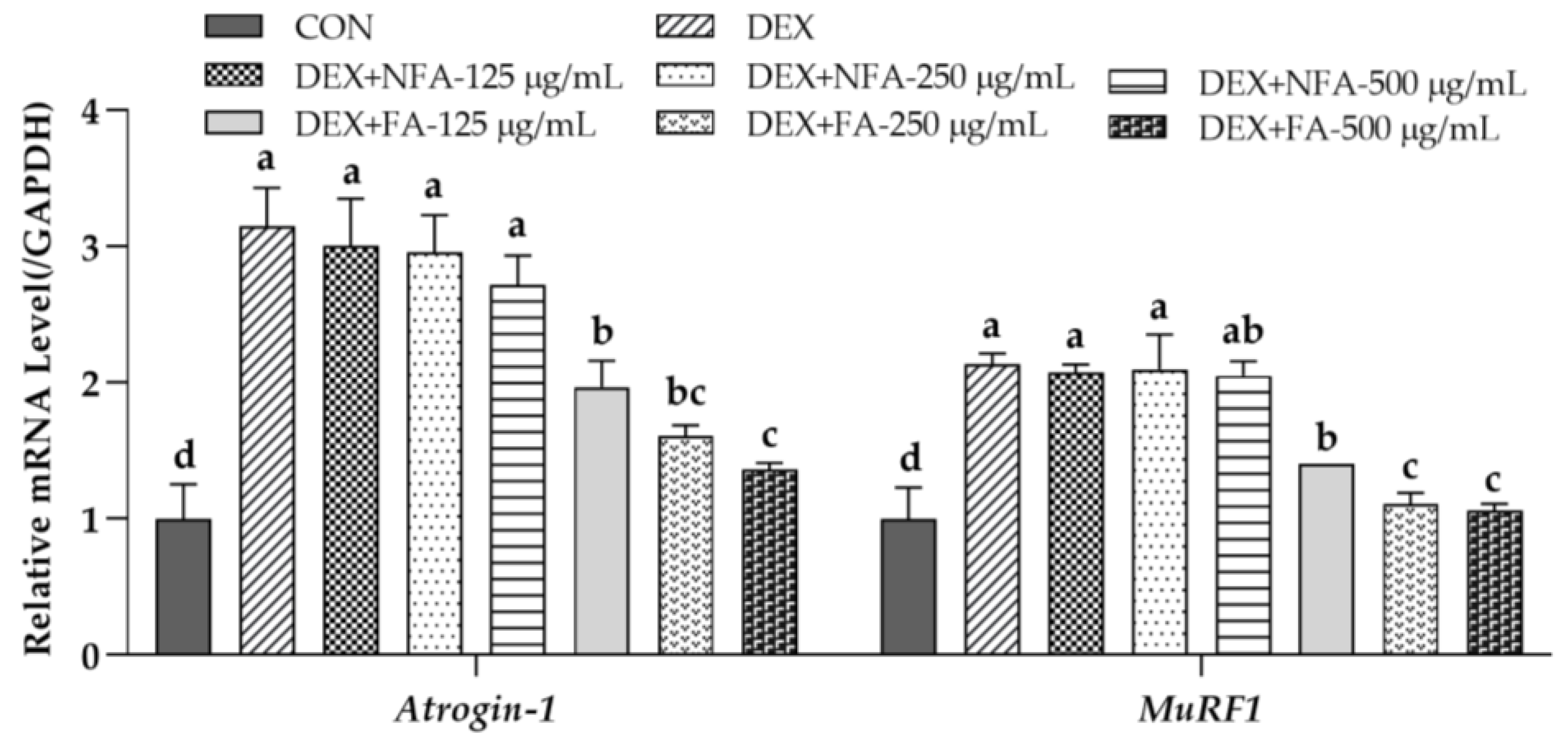
3.1. L. curvatus HY7602-Fermented Antlers Inhibit Muscle Protein Degradation in Dexamethasone-Induced Muscle Atrophy in C2C12 Cells

We used C2C12 cells to investigate the effects of FA on dexamethasone-induced muscle atrophy. The degradation of muscle proteins is mediated through the ubiquitin–proteasome system (UPS), which is promoted by Atrogin-1 and MuRF1 in skeletal muscles [35,36]. DEX group showed the greatest increase in the expression levels of Atrogin-1 and MuRF1 compared to those in the control. In contrast, the FA significantly reduced the expression levels of Atrogin-1 and MuRF1 in the C2C12 cells with dexamethasone-induced muscle atrophy in a dose-dependent manner (Figure 1). The expression of both Atrogin-1 and MuRF1 showed the greatest decrease at DEX+FA-500 μg/mL. However, the DEX+NFA did not reduce the expression levels of Atrogin-1 and MuRF1 at any of the tested concentrations. These results show that FA have a more potent effect than NFA on dexamethasone-induced muscle protein degradation in vitro.

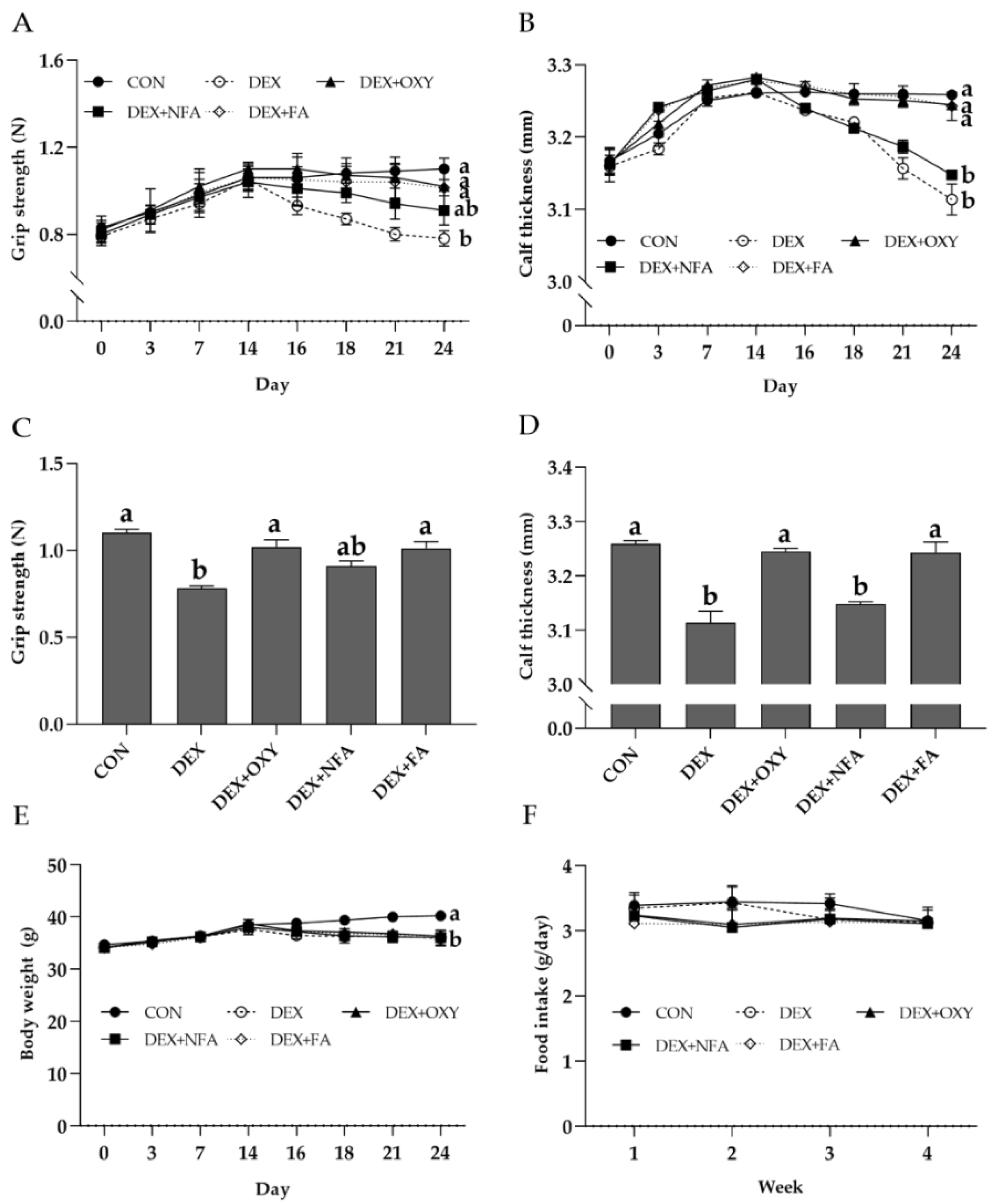
3.2. L. curvatus HY7602-Fermented Antlers Ameliorate Dexamethasone-Induced Reductions in Hindlimb Grip Strength and Calf Thickness in Mice

To evaluate the effects of FA in vivo based on previous in vitro results, we used a young, 7-week-old mouse with dexamethasone-induced muscle atrophy. All experimental groups, except for the CON, were IP injected with dexamethasone (1 mg/kg/day) for 10 days before the end of the study. During the experimental period (24 days), the CON and DEX groups were administered saline; the DEX+OXY group consumed 50 mg/kg/day of OXY; and the DEX+NFA and DEX+FA groups consumed 120 mg/kg/day of NFA or FA, respectively. From day 14 onward, the starting date of the dexamethasone treatment, hindlimb grip strength, and calf thickness decreased in the DEX group. The DEX+NFA group also showed a similar decrease to that in the DEX group, whereas the DEX+OXY and DEX+FA groups showed only a slight decrease in hindlimb grip strength (Figure 2A,B). After 24 days of treatment, the hindlimb grip strength of DEX group was 0.78 ± 0.02 N, which was 29.09% lower than that of the CON group, 1.10 ± 0.02 N (p < 0.05). On the other hand, the grip strength of the DEX+OXY group and the DEX+FA group were 1.02 ± 0.04 N and 1.01 ± 0.04 N, respectively, and no significant decrease was observed compared to the CON group. In the case of the DEX+NFA group, it was 0.91 ± 0.03 N, and there was no significant difference from both CON and DEX groups (Figure 2C). The calf thickness was 3.11 ± 0.05 mm in DEX group, which was reduced by 4.44% compared to 3.26 ± 0.01 mm in the CON group (p < 0.05). The DEX+OXY group and the DEX+FA group were 3.25 ± 0.01 mm and 3.24 ± 0.04 mm, respectively, and no significant decrease was observed compared to the CON group. For the DEX+NFA group, it was measured as 3.15 ± 0.01, which was reduced by 3.40% compared to the CON group (p < 0.05) (Figure 2D).
The body weights of the mice decreased from day 14 in all the dexamethasone-treated groups, except for the CON group, and there was no significant difference between them (Figure 2E). Additionally, food intake did not differ significantly between the groups during the experimental period (Figure 2F).

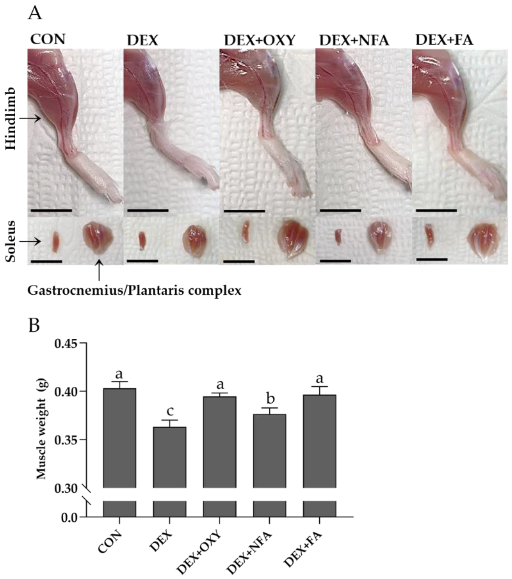
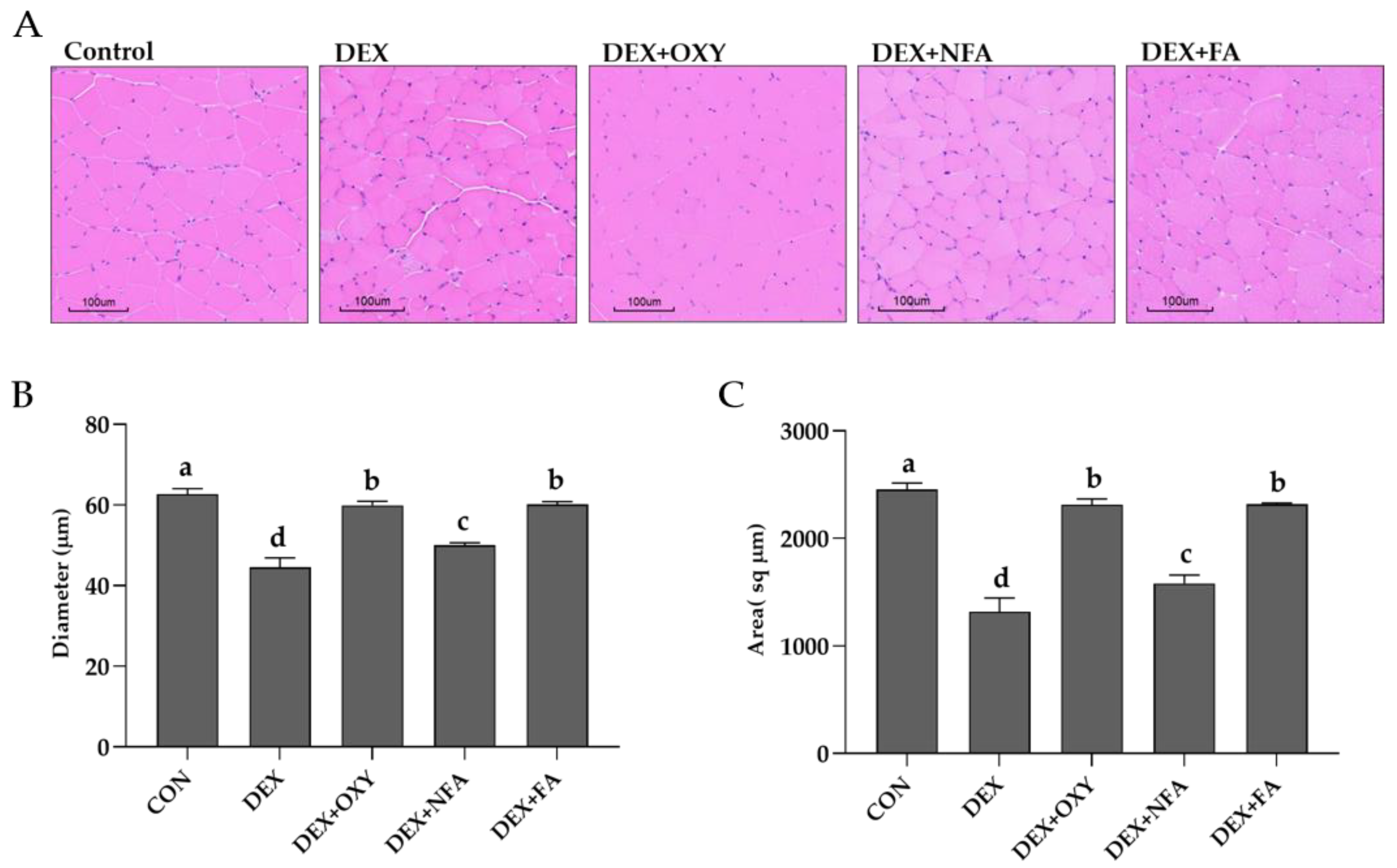
3.3. L. curvatus HY7602-Fermented Antlers Recover Dexamethasone-Induced Muscle Loss in Mice

Dexamethasone, a synthetic glucocorticoid, inhibits protein synthesis and promotes protein degradation, thereby leading to muscle atrophy [37]. In this study, we investigated the effects of dexamethasone and FA on hindlimb muscle mass (Figure 3A). To determine the changes in calf muscle mass (GA/PLA/SOL complex) following dexamethasone and FA treatment, muscle tissue was extracted and weighed after the mice had been sacrificed. As expected, the DEX group showed the greatest reduction in muscle mass of 9.88% at 0.36 ± 0.02 g compared with that in the CON group, 0.40 ± 0.05 g (p < 0.05). In contrast, DEX+OXY and the DEX+FA were 0.39 ± 0.01 g and 0.40 ± 0.02 g, respectively, and there was no significant difference from the CON group. The muscle weight of DEX+NFA was 0.38 ± 0.01 g, which was decreased by 6.59% compared to the CON group (p < 0.05) (Figure 3B).
Additionally, we evaluated the diameters and areas of the muscle fibers using a histological analysis of the calf muscle. The GA muscle tissue was analyzed by staining with H&E. The cross-sectional area of the GA muscle was measured using a digital microscope (Figure 4A). The diameters of the muscle fibers were the smallest in the DEX group at 44.54 ± 5.23 µm, reduced by 28.95% compared to the CON group, 62.69 ± 3.02 µm (p < 0.05). The DEX+OXY and DEX+FA groups were 59.87 ± 2.25 µm and 60.19 ± 1.39 µm, and there was no significant difference from the CON group. The DEX+NFA group was 50.04 ± 1.26 µm, which was decreased by 20.18% compared to the CON group (p < 0.05) (Figure 4B). The area of the DEX group was 1319.22 ± 284.94 sq µm, which was 46.21% lower than that of the CON group, 2452.73 ± 284.94 sq µm (p < 0.05). On the other hand, the DEX+OXY and DEX+FA groups were 2314.34 ± 118.30 sq µm and 2316.20 ± 27.96 sq µm, respectively, and no significant decrease was observed compared to the CON group. For the DEX+NFA group, there was a decrease of 35.51%, compared to the CON group, to 1581.77 ± 173.64 sq µm (p < 0.05) (Figure 4C).

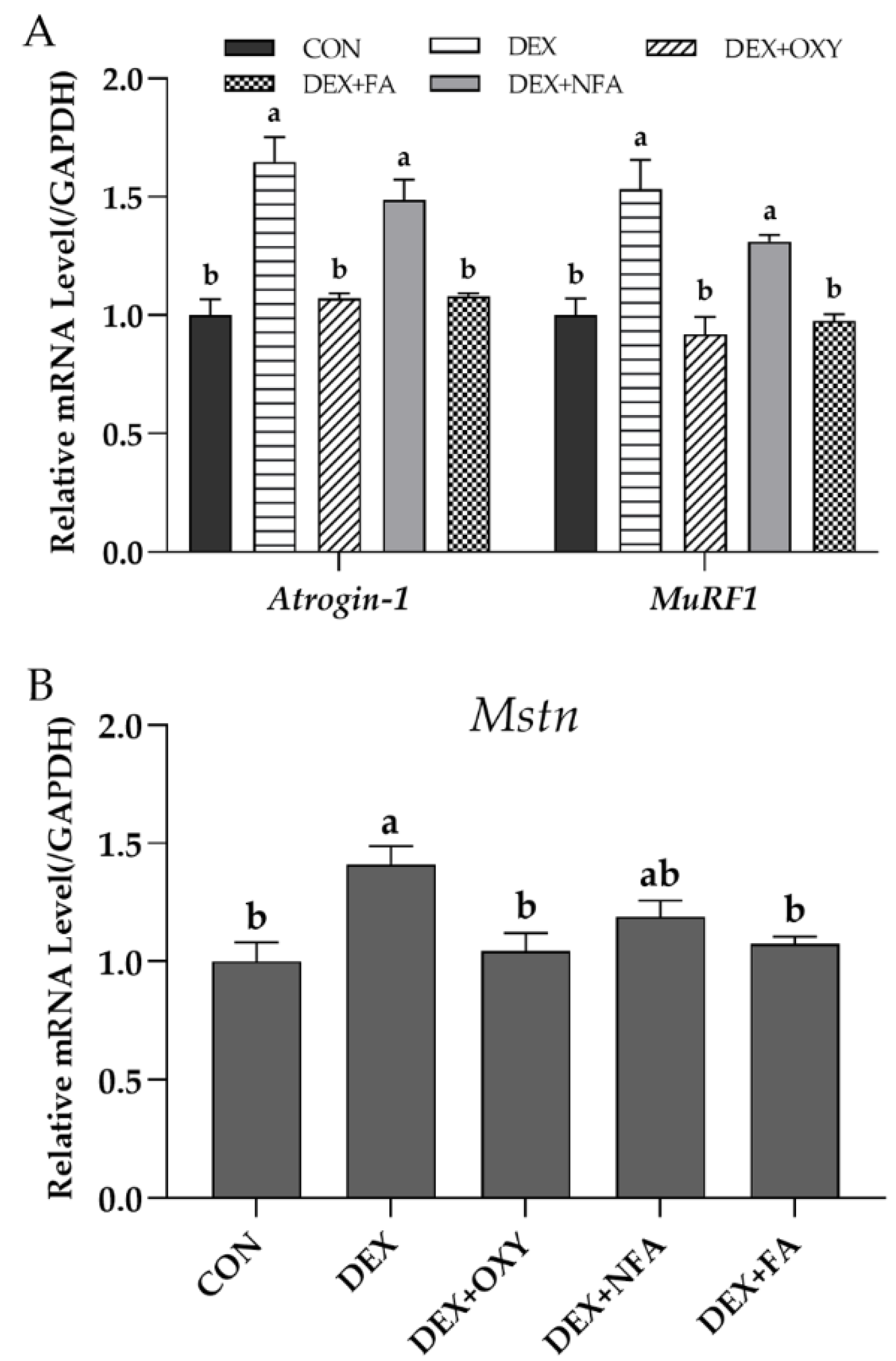
3.4. L. curvatus HY7602-Fermented Antlers Inhibit Muscle Protein Degradation and Prevent Muscle Growth Inhibition in Mice with Dexamethasone-Induced Muscle Atrophy

Atrogin-1 and MuRF1 are expressed in skeletal muscles and promote the UPS, which is a degradation pathway for muscle proteins [35,36]. In a previous in vitro experiment, we confirmed that the expression levels of Atrogin-1 and MuRF1 were increased in dexamethasone-induced muscle atrophy. Likewise, the DEX group presented the highest expression level of both factors, and this result was significantly higher than that in the CON group (Figure 5A). DEX+OXY and the DEX+FA reduced the high expression levels of Atrogin-1 and MuRF1 by dexamethasone, similar to the CON. The DEX+NFA group showed a slight, but not significant, decreasing trend compared to the DEX group (Figure 5A).
Myostatin is mainly expressed in skeletal muscles and acts as a negative regulator of muscle mass [38]. Dexamethasone-induced muscle atrophy is associated with the upregulation of Mstn [37,39]. In our results, the expression of Mstn was the highest in the DEX group. DEX+OXY and the DEX+FA were suppressed to levels similar to those in the CON group. The DEX+NFA group showed a slight decrease compared to the DEX group, but there was no significant difference between the CON and DEX groups (Figure 5B).

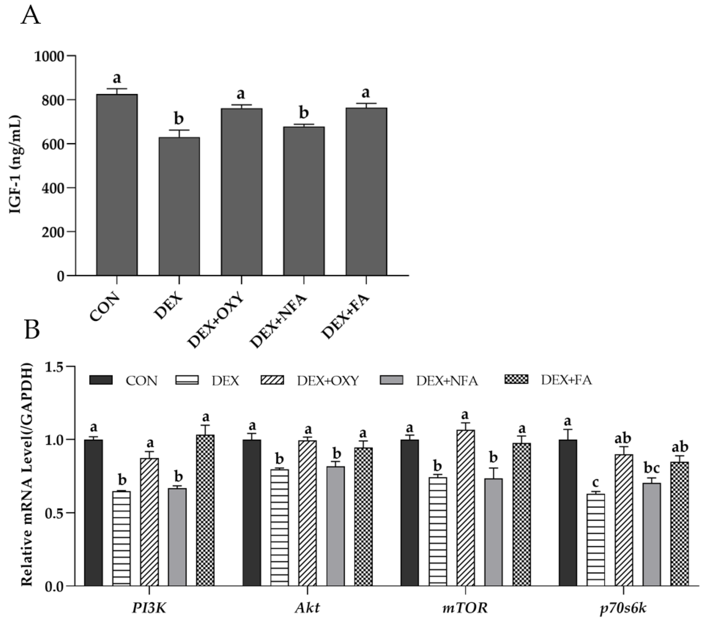
3.5. L. curvatus HY7602-Fermented Antlers Promote Muscle Protein Synthesis in Mice with Dexamethasone-Induced Muscle Atrophy

Insulin-like growth factor-1 (IGF-1) is a major growth factor that promotes growth by increasing protein synthesis in skeletal muscles. Increased protein synthesis due to IGF-1 occurs via the PI3K/Akt/mTOR and PI3K/Akt/GSK3β pathways [40]. Dexamethasone inhibits muscle protein synthesis by decreasing the expression of IGF-1 and inhibiting signaling [41,42]. As shown in Figure 6A, the serum IGF-1 concentration was the lowest in the DEX group. The DEX+OXY and DEX+FA groups showed a suppression of the decrease in the concentration of IGF-1, whereas the DEX+NFA group did not restore the decrease in IGF-1 induced by dexamethasone (Figure 6A). Furthermore, the expression of the genes in the PI3K/Akt/mTOR pathway (PI3K, Akt, mTOR, and p70s6k) in the DEX group was lower than that in the CON group (Figure 6B). The expression of PI3K, Akt, mTOR, and p70s6k was significantly increased in the DEX+OXY and DEX+FA groups, which was similar to the IGF-1 results, but the expression of PI3K, Akt, and mTOR in the DEX+NFA group was not significantly different from that in the DEX group. The expression of p70s6k was increased in the DEX+NFA group and was not significantly affected by DEX, DEX+OXY, and the DEX+FA but was significantly lower than that in the CON.

3.6. Effects of the L. curvatus HY7602-Fermented Antlers on Serum Biochemistry

The serum levels of ALT, AST, and Crea were analyzed to determine the in vivo toxicity of dexamethasone and the FA. ALT and AST levels are the most commonly used hepatotoxicity indicators of liver damage in clinical diagnoses [43,44]. The Crea level is commonly used to evaluate renal function [45]. Dexamethasone treatment in mice increases serum ALT and AST levels [46]. As expected, the DEX group showed a significant increase in the ALT and AST levels compared with those in the CON group, and the Crea level was not significantly different from that in the CON (Table 1). The DEX+OXY group had the highest levels of both ALT and AST and lowest levels of Crea. The results of the DEX+NFA group were comparable to those of the DEX group. The DEX+FA group showed a slightly significant decrease only in the ALT levels compared to the DEX group.

3.7. Increased Sialic Acid Content after Fermentation

Deer antlers are known to contain various bioactive compounds, such as amino acids, saccharides, and polypeptides [47]. We confirmed the content of sialic acid, which is known to be related to muscles [26]. The sialic acid contents of the antlers before and after fermentation were quantified using HPLC and were found to increase by 51.41% after fermentation compared with the contents before fermentation (Table 2).

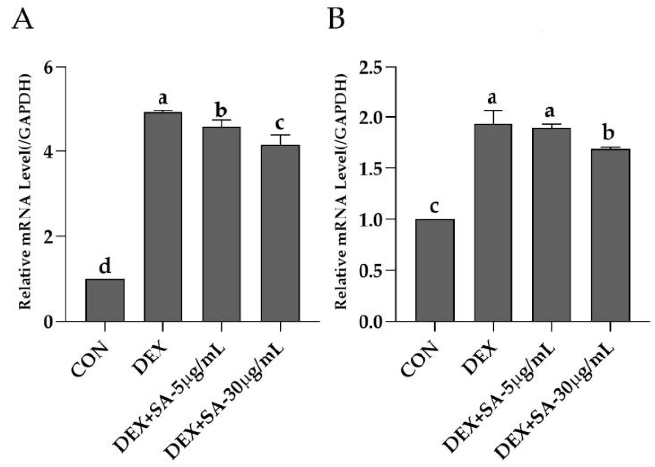
3.8. Effects of Sialic Acid (N-acetylneuraminic Acid) on Muscle Protein Degradation in Dexamethasone-Treated C2C12 Cells

To determine the effects of sialic acid on muscle proteolysis in muscle atrophy, dexamethasone-treated C2C12 cells were used in the same manner as that in a previous in vitro study. Sialic acid decreased the expression of muscle proteolytic genes in a concentration-dependent manner. Compared with that in the DEX group, DEX+SA-5 and DEX+SA-30 μg/mL yielded a significant decrease in Atrogin-1 expression (Figure 7A), and MuRF1 expression was significantly reduced at DEX+SA-30 μg/mL (Figure 7B).

4. Discussion

Lactic acid bacteria are an incredibly important group of microorganisms in the food industry and have been used for centuries as they are generally safe for humans to consume [48]. Strains of the genera Lactobacillus and Bifidobacterium are further known to have beneficial effects on human health, including improved intestinal health and the immune response, and reduced serum cholesterol [49,50]. The most common and traditional way to consume probiotics is through fermented foods. Recently, research on fermented foods has expanded to not only include well-known dairy products, but also non-dairy fermented foods such as vegetables, grains, and meat. Their various health benefits have consequently been revealed [51,52,53].
Deer antlers are used to recover from fatigue, improve liver function, and enhance immunity, and are mainly supplied by New Zealand, Russia, Canada, and China [54]. Various studies have assessed fermented antlers using Bacillus species or L. plantarum to increase the yield, functional compounds, and biological effects of fermentation, but few studies have used L. curvatus [34,54]. L. curvatus is known to have probiotic functions, such as in the prevention of obesity, dyslipidemia, and dexamethasone-induced muscle atrophy, as well as excellent fermentation properties [55].
Sarcopenia occurs with aging and results in the loss of muscle mass and strength [32,56]. Given that the progression of this disease is greatly influenced by diet and lifestyle, current research is focused on the prevention and suppression of sarcopenia with foods [56,57]. Experimental models of sarcopenia include aging, genetically engineered, and chemical or dietary induction models. Among these, dexamethasone is a synthetic glucocorticoid and representative medication in chemically-induced sarcopenia models [58,59]. dexamethasone-induced muscle atrophy is widely used as a model of sarcopenia as it occurs due to a decrease in type II muscle fibers, which is the case with muscle atrophy caused by aging [59].
Several studies have investigated the effects of fermented antler on exercise endurance or muscle differentiation, but the use of fermented antlers in improving dexamethasone-induced muscle atrophy has not yet been studied [60,61]. Therefore, we aimed to establish the efficacy of FA in dexamethasone-induced muscle atrophy and determine the compounds that increased and were effective after fermentation. To investigate the mechanisms underlying the prevention of dexamethasone-induced muscle atrophy in FA, we performed in vitro experiments with dexamethasone-treated C2C12 cells and in vivo experiments with young dexamethasone-injected mice. OXY, which is known to affect muscle strength recovery due to sarcopenia, was used as a positive control [14,15]. We further evaluated sialic acid in dexamethasone-treated C2C12 cells in vitro.
We found that dexamethasone treatment increased the expression of Atrogin-1 and MuRF1 in C2C12 cells. This was consistent with a previous study in which the muscle atrophy mechanisms of dexamethasone were found to be related to the increased expression of Atrogin-1 and MuRF1, which are related to muscle protein degradation [58]. Two major muscle-specific E3 ubiquitin ligases, Atrogin-1 and MuRF1, mediate the UPS, which is a targeted protein degradation system for regulating protein homeostasis [62]. Thus, the increased expression of Atrogin-1 and MuRF1 in muscle tissue indicates protein degradation due to muscle atrophy. In contrast, our results showed that FA reduced the expression of Atrogin-1 and MuRF1 to levels similar to those of the CON. These results indicate a possible role of FA in muscle protein degradation.
Dexamethasone-induced muscle atrophy is mainly caused by decreased protein synthesis and increased protein breakdown and is expressed in various skeletal muscle phenotypes, including decreased muscle mass, thickness, and muscle weakness [9,10]. In an in vivo study, we showed that body weight, calf muscle mass, hindlimb muscle strength, calf thickness, and cross-sectional areas of the GA were significantly lower in the DEX group than in the control group, which is consistent with previous studies. The FA showed similar inhibitory effects to those in the CON and DEX+OXY groups on the reduction of hindlimb muscle strength and calf muscle thickness, calf muscle mass, and cross-sectional areas of the GA caused by dexamethasone, but did not show a significant effect on body weight. These results may be explained by previously-published findings in which dexamethasone-induced weight loss was due not only to muscle atrophy, but also to the decreased volume and density of cortical bones [63]. In contrast, the DEX+NFA showed a slight increase in hindlimb muscle strength, calf muscle mass, and cross-sectional areas of the GA compared to the DEX group but showed a significant difference with the CON group. In our study, the FA prevented and inhibited dexamethasone-induced muscle strength and muscle mass loss to levels similar to those observed in the CON group.
Atgrogin-1 and MuRF1 are representative factors that promote muscle protein degradation and are upregulated in dexamethasone-induced muscle atrophy. We previously found that dexamethasone significantly increased the expression levels of both factors, while FA decreased them. Similarly, the in vivo FA significantly reduced the expression levels of Atrogin-1 and MuRF1 that had been increased by dexamethasone to levels similar to those in the CON and DEX+OXY groups. However, the DEX+NFA group showed no significant difference compared to the DEX group. Another muscle atrophy-associated factor is Mstn, which is a negative regulator of muscle growth that activates and phosphorylates the Smad2/3 transcription factor. Dexamethasone upregulates Mstn, thereby leading to muscle atrophy [37,64]. In our study, Mstn was upregulated by dexamethasone, which was consistent with previous studies. The FA suppressed the upregulation of Mstn to a level similar to that in the CON and DEX+OXY groups. However, the DEX+NFA group was not significantly different with the control and DEX groups. These results suggest that FA prevent muscle protein degradation and negatively regulate growth owing to muscle atrophy.
The IGF-1/PI3K/Akt pathway is a representative muscle protein synthesis pathway in which IGF-1 activates the PI3K/Akt pathway to induce protein synthesis and muscle development [64]. IGF-1 is a systemic growth factor synthesized in the liver and is known to strongly regulate gene expression, especially in skeletal muscles [65]. IGF-1 levels in the blood are reduced by dexamethasone treatment, which inhibits the effects of IGF-1 by impairing PI3K signaling [42]. Reduced IGF-1 levels and signaling disruptions promote muscle atrophy by decreasing protein synthesis. Our findings show that the serum IGF-1 levels in the DEX group were significantly decreased, and the expression of PI3K/Akt pathway-related factors was also significantly decreased in the muscle tissue. The FA significantly restored the decrease in muscle protein synthesis and growth factors induced by dexamethasone. Meanwhile, the DEX+NFA group showed a slight increase in only one factor; however, this was not significant. Our data showed that dexamethasone-induced muscle atrophy reduced muscle protein synthesis factor expression and blood growth factor levels and was improved with the FA.
In the case of healthy, functional foods, sufficient data on disease prevention, health, toxicity, and safety evaluations in the body are important. Serum ALT, AST, and Crea concentrations were determined to assess the toxicity of FA in the body. Serum ALT and AST levels are highly sensitive biomarkers of hepatotoxicity that reflect hepatocellular injury and are the most commonly used biomarkers in non-clinical and clinical studies of liver injury [43,44]. Crea is a waste product produced by muscles, and serum Crea levels are commonly-used biomarkers for impaired renal function, especially in clinical and non-clinical settings [66]. In previous studies, various bioactive compounds have been shown to be effective in preventing dexamethasone-induced muscle atrophy, but few studies have analyzed serum ALT, AST, and Crea levels to evaluate the in vivo toxicity of functional compounds [11,67]. Our data show that blood ALT and AST levels significantly increased in the DEX group. This was consistent with previous studies that showed that short-term treatment with dexamethasone increased blood ALT [46]. In addition, DEX+OXY significantly increased ALT and AST levels compared to DEX, which may be explained by previous studies that suggested that OXY, an anabolic–androgenic steroid, can induce liver damage [68]. In contrast, the FA showed significantly reduced ALT levels and slightly decreased AST levels compared with those in the DEX group. The DEX+NFA showed results similar to those in the DEX group for all the biomarkers. There was a significant difference in the serum Crea levels between CON and DEX+OXY groups. Our results suggest that FAs have no hepatic or renal toxicity and partially protect from liver damage induced by dexamethasone.
In summary, FA restores dexamethasone-induced muscle atrophy. These effects are associated with the regulation of muscle protein degradation, synthesis, growth inhibition factors, and blood growth factor levels. FAs may therefore be functional foods that can be used for muscle atrophy.
Fermentation is a simple food processing technique that increases the bioavailability of nutrients and positively changes the components or flavor of food [69,70]. Research on the increase in bioactive compounds through fermentation is being actively conducted [71,72,73]. Deer antlers are known to have various active ingredients such as ganglioside, chondroitin, glucosamine, hyaluronic acid, phospholipids, collagen, minerals, and saccharides, and the commonly identified ingredients include sialic acid, uronic acid, and S-GAG [21,47,74,75]. Sialic acid deficiencies are known to cause oxidative stress and weaken muscles, and surface sialic acid contents decrease when muscles are damaged [26,27]. Therefore, we investigated the changes in sialic acid contents after fermentation and the effects of sialic acid on muscle atrophy. Our data show that the sialic acid contents of the antlers increased by 51.41% after fermentation. The effects of sialic acid on muscle protein degradation in dexamethasone-induced muscle atrophy were further confirmed. Although previous studies have reported a relationship between muscle atrophy or injury and sialic acid, the effects of sialic acid supplementation on dexamethasone-induced muscle atrophy have not yet been studied. Therefore, we assessed dexamethasone-treated C2C12 cells using the same methods as in the previous in vitro FA efficacy evaluations and confirmed the changes in the expression of the muscle protein degradation factors Atrogin-1 and MuRF1 following treatment with sialic acid. Sialic acid decreased the expression of Atrogin-1 in a concentration-dependent manner and showed significant inhibition compared to the DEX group at 30 μg/mL for MuRF1. Although further studies are needed to confirm the efficacy of sialic acid on phenotypic changes and clear genotypic changes due to muscle atrophy, through the results of this study we can confirm that sialic acid is increased during the fermentation process and may contribute to the prevention of muscle atrophy.
Overall, the FA ameliorated dexamethasone-induced reductions in muscle mass, strength, and fiber thickness. These reductions were mediated in part through the decreased expression of genes related to muscle protein degradation and growth inhibition factors, increased expression of genes related to protein synthesis, and increased levels of growth factor in the blood. This effect on muscle atrophy is related to the fermentation of antlers with increased sialic acid content.

5. Conclusions

This study suggests that FAs are potential functional foods, particularly for relieving muscle atrophy. FAs prevent the reduction in hindlimb strength, calf thickness, muscle mass, and cross-sectional areas of muscle fibers due to muscle atrophy. They further function in part through alterations in muscle protein degradation, synthesis, and muscle growth inhibitory genes or growth factors in the blood. In addition, the sialic acid components of deer antlers increase following fermentation, which is noteworthy as sialic acid is associated with inhibition of muscle protein degradation. These findings therefore highlight potential factors that may be used in the treatment and prevention of muscle atrophy and sarcopenia.

Author Contributions

Conceptualization, H.J., Y.-T.K., W.Y.J., J.-Y.K., K.H., J.-J.S., J.-L.L., D.-C.Y. and S.C.K.; methodology, H.J.; software, H.J.; validation, H.J. and Y.-T.K.; formal analysis, H.J. and Y.-T.K.; investigation, H.J., Y.-T.K. and W.Y.J.; resources, H.J. and Y.-T.K.; data curation, H.J.; writing—original draft preparation, H.J. and Y.-T.K.; writing—review and editing, K.H., D.-C.Y. and S.C.K.; visualization, H.J.; supervision, K.H., D.-C.Y. and S.C.K.; project administration, K.H., D.-C.Y. and S.C.K. All authors have read and agreed to the published version of the manuscript.

Funding

This research received no external funding.

Institutional Review Board Statement

The animal study protocol was approved by the hy Co., Ltd. (Yongin-si, Korea) and approved by the Institutional Animal Care and Use Committee of hy Co., Ltd. (IACUC approval number: AEC-2022-0001-Y).

Informed Consent Statement

Not applicable.

Data Availability Statement

The data presented in this study are available in this article.

Conflicts of Interest

The authors declare no conflict of interest.

References

  1. Beas-Jiménez, J.D.; López-Lluch, G.; Sánchez-Martínez, I.; Muro-Jiménez, A.; Rodríguez-Bies, E.; Navas, P. Sarcopenia, implications of physical exercise in its pathophysiology, prevention and treatment. Rev. Andal. Med. Deporte 2011, 4, 158–166. [Google Scholar]
  2. Moustogiannis, A.; Philippou, A.; Taso, O.; Zevolis, E.; Pappa, M.; Chatzigeorgiou, A.; Koutsilieris, M. The effects of muscle cell aging on myogenesis. Int. J. Mol. Sci. 2021, 22, 3721. [Google Scholar] [CrossRef] [PubMed]
  3. Hikida, R.S. Aging changes in satellite cells and their functions. Curr. Aging Sci. 2011, 4, 279–297. [Google Scholar] [CrossRef] [PubMed]
  4. Brack, A.S.; Muñoz-Cánoves, P. The ins and outs of muscle stem cell aging. Skelet. Muscle. 2016, 6, 1. [Google Scholar] [CrossRef] [PubMed]
  5. Marzetti, E.; Calvani, R.; Cesari, M.; Buford, T.W.; Lorenzi, M.; Behnke, B.J.; Leeuwenburgh, C. Mitochondrial dysfunction and sarcopenia of aging: From signaling pathways to clinical trials. Int. J. Biochem. Cell Biol. 2013, 45, 2288–2301. [Google Scholar] [CrossRef] [PubMed]
  6. Combaret, L.; Dardevet, D.; Béchet, D.; Taillandier, D.; Mosoni, L.; Attaix, D. Skeletal muscle proteolysis in aging. Curr. Opin. Clin. Nutr. Metab. Care 2009, 12, 37–41. [Google Scholar] [CrossRef]
  7. Tsekoura, M.; Kastrinis, A.; Katsoulaki, M.; Billis, E.; Gliatis, J. Sarcopenia and its impact on quality of life. Adv. Exp. Med. Biol. 2017, 987, 213–218. [Google Scholar] [CrossRef]
  8. Kim, J.W.; Ku, S.K.; Han, M.H.; Kim, K.Y.; Kim, S.G.; Kim, G.Y.; Hwang, H.J.; Kim, B.W.; Kim, C.M.; Choi, Y.H. The administration of Fructus Schisandrae attenuates dexamethasone-induced muscle atrophy in mice. Int. J. Mol. Med. 2015, 36, 29–42. [Google Scholar] [CrossRef]
  9. Jesinkey, S.R.; Korrapati, M.C.; Rasbach, K.A.; Beeson, C.C.; Schnellmann, R.G. Atomoxetine prevents dexamethasone-induced skeletal muscle atrophy in mice. J. Pharmacol. Exp. Ther. 2014, 351, 663–673. [Google Scholar] [CrossRef]
  10. Shen, S.; Liao, Q.; Liu, J.; Pan, R.; Lee, S.M.; Lin, L. Myricanol rescues dexamethasone-induced muscle dysfunction via a sirtuin 1-dependent mechanism. J. Cachexia Sarcopenia Muscle 2019, 10, 429–444. [Google Scholar] [CrossRef]
  11. Otsuka, Y.; Egawa, K.; Kanzaki, N.; Izumo, T.; Rogi, T.; Shibata, H. Quercetin glycosides prevent dexamethasone-induced muscle atrophy in mice. Biochem. Biophys. Rep. 2019, 18, 100618. [Google Scholar] [CrossRef] [PubMed]
  12. Gilson, H.; Schakman, O.; Combaret, L.; Lause, P.; Grobet, L.; Attaix, D.; Ketelslegers, J.M.; Thissen, J.P. Myostatin gene deletion prevents glucocorticoid-induced muscle atrophy. Endocrinology 2007, 148, 452–460. [Google Scholar] [CrossRef]
  13. Andrew, M.P.; Oliver, F.; Matthew, J.M.; Anil, V. Review of oxymetholone: A 17α-alkylated anabolic-androgenic steroid. Clin. Ther. 2001, 23, 789–801. [Google Scholar] [CrossRef]
  14. Lim, J.M.; Lee, Y.J.; Cho, H.R.; Park, D.C.; Jung, G.W.; Ku, S.K.; Choi, J.S. Extracellular polysaccharides purified from Aureobasidium pullulans SM-2001 (Polycan) inhibit dexamethasone-induced muscle atrophy in mice. Int. J. Mol. Med. 2018, 41, 1245–1264. [Google Scholar] [CrossRef] [PubMed]
  15. Schroeder, E.T.; Singh, A.; Bhasin, S.; Storer, T.W.; Azen, C.; Davidson, T.; Martinez, C.; Sinha-Hikim, I.; Jaque, S.V.; Terk, M.; et al. Effects of an oral androgen on muscle and metabolism in older, community-dwelling men. Am. J. Physiol. Endocrinol. Metab. 2003, 284, E120–E128. [Google Scholar] [CrossRef]
  16. Supasyndh, O.; Satirapoj, B.; Aramwit, P.; Viroonudomphol, D.; Chaiprasert, A.; Thanachatwej, V.; Vanichakarn, S.; Kopple, J.D. Effect of oral anabolic steroid on muscle strength and muscle growth in hemodialysis patients. Clin. J. Am. Soc. Nephrol. 2013, 8, 271–279. [Google Scholar] [CrossRef]
  17. Ganapathy, A.; Nieves, J.W. Nutrition and sarcopenia-what do we know? Nutrients 2020, 12, 1755. [Google Scholar] [CrossRef]
  18. Jones, T.E.; Stephenson, K.W.; King, J.G.; Knight, K.R.; Marshall, T.L.; Scott, W.B. Sarcopenia—Mechanisms and treatments. J. Geriatr. Phys. Ther. 2009, 32, 83–89. [Google Scholar] [CrossRef]
  19. Li, C.; Zhao, H.; Liu, Z.; McMahon, C. Deer antler—A novel model for studying organ regeneration in mammals. Int. J. Biochem. Cell Biol. 2014, 56, 111–122. [Google Scholar] [CrossRef]
  20. Earnest, C.P.; Quindry, J.; Panton, L.; Broeder, C. Effect of deer antler velvet on aerobic, anaerobic and strength performance. Cent. Eur. J. Sport Sci. Med. 2015, 9, 17–26. [Google Scholar]
  21. Jang, D.W.; Ameer, K.; Oh, J.H.; Park, M.K. Optimization and pretreatment for hot water extraction of Korean deer (Cervus canadensis Erxleben) velvet antlers. J. Microbiol. Biotechnol. 2020, 30, 1116–1123. [Google Scholar] [CrossRef] [PubMed]
  22. Ivankina, N.F.; Isay, S.V.; Busarova, N.G.; Mischenko, T.Y. Prostaglandin-like activity, fatty acid and phospholipid composition of sika deer (Cervus nippon) antlers at different growth stages. Comp. Biochem. Physiol. B 1993, 106, 159–162. [Google Scholar] [CrossRef]
  23. Jhon, G.J.; Park, S.Y.; Han, S.Y.; Lee, S.; Kim, Y.; Chang, Y.S. Studies of the chemical structure of gangliosides in deer antler, Cervus nippon. Chem. Pharm. Bull. 1999, 47, 123–127. [Google Scholar] [CrossRef]
  24. Pothacharoen, P.; Kodchakorn, K.; Kongtawelert, P. Characterization of chondroitin sulfate from deer tip antler and osteogenic properties. Glycoconj. J. 2011, 28, 473–480. [Google Scholar] [CrossRef]
  25. Zhang, L.Q.; Wang, J.; Li, T.; Li, P.Y.; Wang, Y.H.; Yang, M.; Liu, J.P.; Liu, J.H. Determination of the chemical components and phospholipids of velvet antler using UPLC/QTOF-MS coupled with UNIFI software. Exp. Ther. Med. 2019, 17, 3789–3799. [Google Scholar] [CrossRef] [PubMed]
  26. Cho, A.; Christine, M.; Malicdan, V.; Miyakawa, M.; Nonaka, I.; Nishino, I.; Noguchi, S. Sialic acid deficiency is associated with oxidative stress leading to muscle atrophy and weakness in GNE myopathy. Hum. Mol. Genet. 2017, 26, 3081–3093. [Google Scholar] [CrossRef] [PubMed]
  27. Iwata, Y.; Suzuki, O.; Wakabayashi, S. Decreased surface sialic acid content is a sensitive indicator of muscle damage. Muscle Nerve. 2013, 47, 372–378. [Google Scholar] [CrossRef] [PubMed]
  28. Oliphant, K.; Allen-Vercoe, E. Macronutrient metabolism by the human gut microbiome: Major fermentation by-products and their impact on host health. Microbiome 2019, 7, 91. [Google Scholar] [CrossRef]
  29. Ragonnaud, E.; Biragyn, A. Gut microbiota as the key controllers of “healthy” aging of elderly people. Immun. Ageing 2021, 18, 2. [Google Scholar] [CrossRef]
  30. Claesson, M.J.; Jeffery, I.B.; Conde, S.; Power, S.E.; O’Connor, E.M.; Cusack, S.; Harris, H.M.; Coakley, M.; Lakshminarayanan, B.; O’Sullivan, O.; et al. Gut microbiota composition correlates with diet and health in the elderly. Nature 2012, 488, 178–184. [Google Scholar] [CrossRef]
  31. Langille, M.G.I.; Meehan, C.J.; Koenig, J.E.; Dhanani, A.S.; Rose, R.A.; Howlett, S.E.; Beiko, R.G. Microbial shifts in the aging mouse gut. Microbiome 2014, 2, 50. [Google Scholar] [CrossRef] [PubMed]
  32. Jung, S.; Kim, S.; Jeung, W.; Ra, J.; Heo, K.; Shim, J.; Lee, J. Fermented antler improves endurance during exercise performance by increasing mitochondrial biogenesis and muscle strength in mice. Appl. Sci. 2021, 11, 5386. [Google Scholar] [CrossRef]
  33. Yang, H.-Y.; Kim, Y.; Joo, H.-G. Fermented antler extract enhances the viability and interleukin-12 production of spleen cells. Korean J. Vet. Res. 2016, 56, 183–187. [Google Scholar] [CrossRef]
  34. Park, Y.; Choi, H.S.; Lee, H.S.; Suh, H.J. Hematopoietic effect of deer antler extract fermented by Bacillus subtilis on murine marrow cells. Nutr. Res. Pract. 2015, 9, 451–458. [Google Scholar] [CrossRef] [PubMed]
  35. Gumucio, J.P.; Mendias, C.L. Atrogin-1, MuRF-1, and sarcopenia. Endocrine 2013, 43, 12–21. [Google Scholar] [CrossRef] [PubMed]
  36. Clavel, S.; Coldefy, A.S.; Kurkdjian, E.; Salles, J.; Margaritis, I.; Derijard, B. Atrophy-related ubiquitin ligases, atrogin-1 and MuRF1 are up-regulated in aged rat tibialis anterior muscle. Mech. Ageing Dev. 2006, 127, 794–801. [Google Scholar] [CrossRef] [PubMed]
  37. Ma, K.; Mallidis, C.; Bhasin, S.; Mahabadi, V.; Artaza, J.; Gonzalez-Cadavid, N.; Arias, J.; Salehian, B. Glucocorticoid-induced skeletal muscle atrophy is associated with upregulation of myostatin gene expression. Am. J. Physiol. Endocrinol. Metab. 2003, 285, E363–E371. [Google Scholar] [CrossRef] [PubMed]
  38. Elkina, Y.; von Haehling, S.; Anker, S.D.; Springer, J. The role of myostatin in muscle wasting: An overview. J. Cachexia Sarcopenia Muscle 2011, 2, 143–151. [Google Scholar] [CrossRef]
  39. Qin, J.; Du, R.; Yang, Y.Q.; Zhang, H.Q.; Li, Q.; Liu, L.; Guan, H.; Hou, J.; An, X.R. Dexamethasone-induced skeletal muscle atrophy was associated with upregulation of myostatin promoter activity. Res. Vet. Sci. 2013, 94, 84–89. [Google Scholar] [CrossRef]
  40. Yoshida, T.; Delafontaine, P. Mechanisms of IGF-1-Mediated regulation of skeletal muscle hypertrophy and atrophy. Cells 2020, 9, 1970. [Google Scholar] [CrossRef]
  41. Son, Y.H.; Lee, S.J.; Lee, K.B.; Lee, J.H.; Jeong, E.M.; Chung, S.G.; Park, S.C.; Kim, I.G. Dexamethasone downregulates caveolin-1 causing muscle atrophy via inhibited insulin signaling. J. Endocrinol. 2015, 225, 27–37. [Google Scholar] [CrossRef] [PubMed]
  42. Singleton, J.R.; Baker, B.L.; Thorburn, A. Dexamethasone inhibits insulin-like growth factor signaling and potentiates myoblast apoptosis. Endocrinology 2000, 141, 2945–2950. [Google Scholar] [CrossRef] [PubMed]
  43. Ozer, J.; Ratner, M.; Shaw, M.; Bailey, W.; Schomaker, S. The current state of serum biomarkers of hepatotoxicity. Toxicology 2008, 245, 194–205. [Google Scholar] [CrossRef] [PubMed]
  44. McGill, M.R. The past and present of serum aminotransferases and the future of liver injury biomarkers. Excli J. 2016, 15, 817–828. [Google Scholar] [CrossRef] [PubMed]
  45. Keppler, A.; Gretz, N.; Schmidt, R.; Kloetzer, H.M.; Groene, H.J.; Lelongt, B.; Meyer, M.; Sadick, M.; Pill, J. Plasma creatinine determination in mice and rats: An enzymatic method compares favorably with a high-performance liquid chromatography assay. Kidney Int. 2007, 71, 74–78. [Google Scholar] [CrossRef] [PubMed]
  46. Jackson, E.R.; Kilroy, C.; Joslin, D.L.; Schomaker, S.J.; Pruimboom-Brees, I.; Amacher, D.E. The early effects of short-term dexamethasone administration on hepatic and serum alanine aminotransferase in the rat. Drug Chem Toxicol. 2008, 31, 427–445. [Google Scholar] [CrossRef]
  47. Sui, Z.; Zhang, L.; Huo, Y.; Zhang, Y. Bioactive components of velvet antlers and their pharmacological properties. J. Pharm. Biomed. Anal. 2014, 87, 229–240. [Google Scholar] [CrossRef]
  48. Holzapfel, W.H.; Wood, B.J.B. Lactic Acid Bacteria: Biodiversity and Taxonomy, 1st ed.; Wiley-Blackwell: Chichester, UK, 2014. [Google Scholar]
  49. Granato, D.; Branco, G.F.; Cruz, A.G.; Faria, J.A.F.; Shah, N.P. Probiotic dairy products as functional foods. Compr. Rev. Food Sci. Food Saf. 2010, 9, 455–470. [Google Scholar] [CrossRef]
  50. Kechagia, M.; Basoulis, D.; Konstantopoulou, S.; Dimitriadi, D.; Gyftopoulou, K.; Skarmoutsou, N.; Fakiri, E.M. Health benefits of probiotics: A review. ISRN Nutr. 2013, 2013, 481651. [Google Scholar] [CrossRef]
  51. Ranadheera, C.S.; Vidanarachchi, J.; Rocha, R.; Cruz, A.; Ajlouni, S. Probiotic Delivery through Fermentation: Dairy vs. Non-Dairy Beverages. Fermentation 2017, 3, 67. [Google Scholar] [CrossRef]
  52. Hugenholtz, J. Traditional biotechnology for new foods and beverages. Curr. Opin. Biotechnol. 2013, 24, 155–159. [Google Scholar] [CrossRef] [PubMed]
  53. Aspri, M.; Papademas, P.; Tsaltas, D. Review on non-dairy probiotics and their use in non-dairy based products. Fermentation 2020, 6, 30. [Google Scholar] [CrossRef] [Green Version]
  54. Yoo, J.; Lee, J.; Zhang, M.; Mun, D.; Kang, M.; Yun, B.; Kim, Y.A.; Kim, S.; Oh, S. Enhanced γ-aminobutyric acid and sialic acid in fermented deer antler velvet and immune promoting effects. J. Anim. Sci. Technol. 2022, 64, 166–182. [Google Scholar] [CrossRef] [PubMed]
  55. Chen, Y.; Yu, L.; Qiao, N.; Xiao, Y.; Tian, F.; Zhao, J.; Zhang, H.; Chen, W.; Zhai, Q. Latilactobacillus curvatus: A Candidate probiotic with Excellent Fermentation Properties and Health Benefits. Foods 2020, 9, 1366. [Google Scholar] [CrossRef] [PubMed]
  56. Robinson, S.; Cooper, C.; Aihie Sayer, A. Nutrition and sarcopenia: A review of the evidence and implications for preventive strategies. J. Aging Res. 2012, 2012, 510801. [Google Scholar] [CrossRef]
  57. Yanai, H. Nutrition for sarcopenia. J. Clin. Med. Res. 2015, 7, 926–931. [Google Scholar] [CrossRef]
  58. Mankhong, S.; Kim, S.; Moon, S.; Kwak, H.B.; Park, D.H.; Kang, J.H. Experimental models of sarcopenia: Bridging molecular mechanism and therapeutic strategy. Cells 2020, 9, 1385. [Google Scholar] [CrossRef]
  59. Xie, W.Q.; He, M.; Yu, D.J.; Wu, Y.X.; Wang, X.H.; Lv, S.; Xiao, W.F.; Li, Y.S. Mouse models of sarcopenia: Classification and evaluation. J. Cachexia Sarcopenia Muscle 2021, 12, 538–554. [Google Scholar] [CrossRef]
  60. Jang, S.; Park, E.D.; Suh, H.J.; Lee, S.H.; Kim, J.S.; Park, Y. Enhancement of exercise endurance capacity by fermented deer antler in BALB/c mice. Biosci. Biotechnol. Biochem. 2014, 78, 1716–1722. [Google Scholar] [CrossRef]
  61. Jo, K.; Jang, W.Y.; Yun, B.S.; Kim, J.S.; Lee, H.S.; Chang, Y.B.; Suh, H.J. Effect of deer antler extract on muscle differentiation and 5-Aminoimidazole-4-Carboxamide ribonucleoside (AICAR)-induced muscle atrophy in C2C12 cells. Food Sci. Anim. Resour. 2021, 41, 623–635. [Google Scholar] [CrossRef]
  62. Foletta, V.C.; White, L.J.; Larsen, A.E.; Léger, B.; Russell, A.P. The role and regulation of MAFbx/atrogin-1 and MuRF1 in skeletal muscle atrophy. Pflugers Arch. 2011, 461, 325–335. [Google Scholar] [CrossRef] [PubMed]
  63. Filippopoulou, F.; Habeos, G.I.; Rinotas, V.; Sophocleous, A.; Sykiotis, G.P.; Douni, E.; Chartoumpekis, D.V. Dexamethasone administration in mice leads to less body weight gain over time, lower serum glucose, and higher insulin levels independently of NRF2. Antioxidants 2021, 11, 4. [Google Scholar] [CrossRef] [PubMed]
  64. Romanick, M.; Thompson, L.V.; Brown-Borg, H.M. Murine models of atrophy, cachexia, and sarcopenia in skeletal muscle. Biochim. Biophys. Acta 2013, 1832, 1410–1420. [Google Scholar] [CrossRef] [PubMed]
  65. Ascenzi, F.; Barberi, L.; Dobrowolny, G.; Villa Nova Bacurau, A.; Nicoletti, C.; Rizzuto, E.; Rosenthal, N.; Scicchitano, B.M.; Musarò, A. Effects of IGF-1 isoforms on muscle growth and sarcopenia. Aging Cell 2019, 18, e12954. [Google Scholar] [CrossRef]
  66. Perrone, R.D.; Madias, N.E.; Levey, A.S. Serum creatinine as an index of renal function: New insights into old concepts. Clin. Chem. 1992, 38, 1933–1953. [Google Scholar] [CrossRef]
  67. Lee, C.W.; Chang, Y.B.; Park, C.W.; Han, S.H.; Suh, H.J.; Ahn, Y. Protein hydrolysate from Spirulina platensis prevents dexamethasone-induced muscle atrophy via Akt/Foxo3 signaling in C2C12 myotubes. Mar. Drugs 2022, 20, 365. [Google Scholar] [CrossRef]
  68. Robles-Diaz, M.; Gonzalez-Jimenez, A.; Medina-Caliz, I.; Stephens, C.; García-Cortes, M.; García-Muñoz, B.; Ortega-Alonso, A.; Blanco-Reina, E.; Gonzalez-Grande, R.; Jimenez-Perez, M.; et al. Distinct phenotype of hepatotoxicity associated with illicit use of anabolic androgenic steroids. Aliment. Pharm. Ther. 2015, 41, 116–125. [Google Scholar] [CrossRef]
  69. Adebo, O.A.; Gabriela Medina-Meza, I.G. Impact of fermentation on the phenolic compounds and antioxidant activity of whole cereal grains: A mini review. Molecules 2020, 25, 927. [Google Scholar] [CrossRef]
  70. Ren, Q.; Sun, L.; Wu, H.; Wang, Y.; Wang, Z.; Zheng, F.; Lu, X.; Xu, J. The changes of microbial community and flavor compound in the fermentation process of Chinese rice wine using Fagopyrum tataricum grain as feedstock. Sci. Rep. 2019, 9, 3365. [Google Scholar] [CrossRef]
  71. Guilloux-Benatier, M.; Chassagne, D. Comparison of components released by fermented or active dried yeasts after aging on lees in a model wine. J. Agric. Food Chem. 2003, 51, 746–751. [Google Scholar] [CrossRef]
  72. Lee, S.M.; Hwang, Y.R.; Kim, M.S.; Chung, M.S.; Kim, Y.S. Comparison of volatile and nonvolatile compounds in rice fermented by different lactic acid bacteria. Molecules 2019, 24, 1183. [Google Scholar] [CrossRef] [PubMed]
  73. Liu, S.; Laaksonen, O.; Marsol-Vall, A.; Zhu, B.; Yang, B. Comparison of volatile composition between alcoholic bilberry beverages fermented with non-saccharomyces yeasts and dynamic changes in volatile compounds during fermentation. J. Agric. Food Chem. 2020, 68, 3626–3637. [Google Scholar] [CrossRef] [PubMed]
  74. Jeon, B.T.; Cheong, S.H.; Kim, D.H.; Park, J.H.; Park, P.J.; Sung, S.H.; Thomas, D.G.; Kim, K.H.; Moon, S.H. Effect of antler development stage on the chemical composition of velvet antler in Elk (Cervus elaphus canadensis). Asian Australas. J. Anim. Sci. 2011, 24, 1303–1313. [Google Scholar] [CrossRef]
  75. Je, J.-Y.; Park, P.-J.; Lim, D.-H.; Jeon, B.-T.; Kho, K.-H.; Ahn, C.-B. Antioxidant, anti-acetylcholinesterase and composition of biochemical components of Russian deer velvet antler extracts. Korean J. Food Sci. Anim. Resourc. 2011, 31, 349–355. [Google Scholar] [CrossRef] [Green Version]
Figure 1. Effects of L. curvatus HY7602-fermented antlers (FA) on the expression of genes involved in muscle protein degradation (Atrogin-1 and MuRF1) in dexamethasone-induced muscle atrophy in C2C12 cells. Data are expressed as the mean ± SD. Significance was determined by a one-way ANOVA with Tukey’s multiple comparisons test. Different letters indicate statistically significant differences between groups (p < 0.05). Letters shared in common between groups indicate no significant difference.
Fermentation 08 00454 g001
Figure 2. Effects of L. curvatus HY7602-fermented antlers (FA) on hindlimb grip strength and calf thickness in mice with dexamethasone-induced muscle atrophy. Changes in (A) hindlimb grip strength and (B) calf thickness during the 24 days of treatment. (C) Hindlimb grip strength and (D) calf thickness in the experimental groups after 24 days of treatment. (E) Body weight was measured on days 0, 7, 14, 16, 18, 21, and 24, and (F) food intake was measured weekly. All data are expressed as the mean ± SD (n = 5 mice per group). Significance was determined by a one-way ANOVA with Tukey’s multiple comparisons test. Different letters indicate statistically significant differences between groups (p < 0.05). Letters shared in common between groups indicate no significant difference. CON, control mice; DEX, dexamethasone-treated mice; DEX+OXY, dexamethasone + oxymetholone-treated mice; DEX+NFA, dexamethasone + non-fermented antlers-treated mice; DEX+FA, dexamethasone + L. curvatus HY7602-fermented antlers-treated mice.
Fermentation 08 00454 g002
Figure 3. Effects of L. curvatus HY7602-fermented antlers (FA) on dexamethasone-induced muscle loss. (A) Representative images of the hindlimb (top), gastrocnemius/plantaris/soleus complex muscles (bottom) in the mice after 24 days of treatment. The scale bar shows a 10-mm distance. (B) Muscle weight (gastrocnemius/plantaris/soleus complex) was measured. Data are expressed as the mean ± SD (n = 5 mice per group). Significance was determined by a one-way ANOVA with Tukey’s multiple comparisons test. Different letters indicate statistically significant differences between groups (p < 0.05). Letters shared in common between groups indicate no significant difference. CON, control mice; DEX, dexamethasone-treated mice; DEX+OXY, dexamethasone + oxymetholone-treated mice; DEX+NFA, dexamethasone + non-fermented antlers-treated mice; DEX+FA, dexamethasone + L. curvatus HY7602-fermented antlers-treated mice.
Fermentation 08 00454 g003
Figure 4. Effects of L. curvatus HY7602-fermented antlers (FA) on the cross-sectional areas of the muscle fibers in mice with dexamethasone-induced muscle atrophy. (A) Representative images of the gastrocnemius/plantaris complex muscle tissue. (B) The diameters of the calf muscle fibers were measured using digital microscope images, (C) and the areas of the calf muscle fibers were measured using the same method. Data are expressed as the mean ± SD (n = 5 mice per group). Significance was determined by a one-way ANOVA with Tukey’s multiple comparisons test. Different letters indicate statistically significant differences between groups (p < 0.05). Letters shared in common between groups indicate no significant difference. CON, control mice; DEX, dexamethasone-treated mice; DEX+OXY, dexamethasone + oxymetholone-treated mice; DEX+NFA, dexamethasone + non-fermented antlers-treated mice; DEX+FA, dexamethasone + L. curvatus HY7602-fermented antlers-treated mice.
Fermentation 08 00454 g004
Figure 5. Effects of L. curvatus HY7602-fermented antlers (FA) on gene expression in soleus muscle tissues related to muscle protein degradation and growth inhibition. The expression of (A) Atrogin-1 and MuRF1 (related to muscle atrophy) and (B) Mstn (related to muscle growth inhibition) were detected by RT-PCR. Data are expressed as the mean ± SD (n = 5 mice per group). Significance was determined by a one-way ANOVA with Tukey’s multiple comparisons test. Different letters indicate statistically significant differences between groups (p < 0.05). Letters shared in common between groups indicate no significant difference. CON, control mice; DEX, dexamethasone-treated mice; DEX+OXY, dexamethasone + oxymetholone-treated mice; DEX+NFA, dexamethasone + non-fermented antlers-treated mice; DEX+FA, dexamethasone + L. curvatus HY7602-fermented antlers-treated mice.
Fermentation 08 00454 g005
Figure 6. Effects of L. curvatus HY7602-fermented antlers (FA) on muscle protein synthesis in mice with dexamethasone-induced muscle atrophy. (A) The serum level of insulin-like growth factor 1 (IGF-1) in the mice was measured using a commercial colorimetric enzyme-linked immunosorbent assay (ELISA) kit. The expression of (B) PI3K, Akt, mTOR, and p70s6k (related to muscle protein synthesis) was detected by RT-PCR in the soleus muscle tissue. Data are expressed as the mean ± SD (n = 5 mice per group). Significance was determined by a one-way ANOVA with Tukey’s multiple comparisons test. Different letters indicate statistically significant differences between groups (p < 0.05). Letters shared in common between groups indicate no significant difference. CON, control mice; DEX, dexamethasone-treated mice; DEX+OXY, dexamethasone + oxymetholone-treated mice; DEX+NFA, dexamethasone + non-fermented antlers-treated mice; DEX+FA, dexamethasone + L. curvatus HY7602-fermented antlers-treated mice.
Fermentation 08 00454 g006
Figure 7. Inhibitory effect of sialic acid (SA) on gene expression in C2C12 cells related to muscle protein degradation. The expression of (A) Atrogin-1 and (B) MuRF1 was detected by RT-PCR. Data are expressed as the mean ± SD. Significance was determined by a one-way ANOVA with Tukey’s multiple comparisons test. Different letters indicate statistically significant differences between groups (p < 0.05). Letters shared in common between groups indicate no significant difference. CON, control cells; DEX, dexamethasone-treated cells; DEX+SA-5 μg/mL, dexamethasone+sialic acid 5 μg/mL-treated cells; DEX+SA-30 μg/mL, dexamethasone+sialic acid 30 μg/mL-treated cells.
Fermentation 08 00454 g007
Table 1. Serum biochemical analysis in different groups of mice after 24 days of treatment.
ALT (U/L)AST (U/L)Crea (mg/dL)
CON22.0 ± 1.3 d49.9 ± 4.2 c0.40 ± 0.02 a
DEX52.4 ± 3.2 b69.4 ± 6.2 b0.40 ± 0.03 ab
DEX+OXY68.8 ± 4.1 a78.3 ± 0.7 a0.37 ± 0.02 b
DEX+NFA49.6 ± 0.7 b69.8 ± 6.1 b0.43 ± 0.07 ab
DEX+FA39.2 ± 1.0 c64.7 ± 4.3 b0.39 ± 0.02 ab
The data are expressed as the mean ± SD (n = 5 mice per group). Significance was determined using a one-way ANOVA with Tukey’s multiple comparisons test. Different letters indicate statistically significant differences between groups (p < 0.05). Letters shared in common between groups indicate no significant difference. ALT, alanine aminotransferase; AST, aspartate aminotransferase; Crea, creatinine; CON, control mice, DEX, dexamethasone-treated mice; DEX+OXY, dexamethasone + oxymetholone-treated mice; DEX+NFA, dexamethasone + non-fermented antlers-treated mice; DEX+FA, dexamethasone + L. curvatus HY7602-fermented antlers-treated mice.
Table 2. Sialic acid contents of deer antlers before and after fermentation.
Deer AntlerSialic Acid Content (μg/mL)
Before fermentation18.42 ± 0.19
After fermentation27.89 ± 0.06
The data are expressed as the mean ± SD. The samples were analyzed three times.
Publisher’s Note: MDPI stays neutral with regard to jurisdictional claims in published maps and institutional affiliations.

