Article

Cultivation of Microalgae in Unsterile Malting Effluent for Biomass Production and Lipid Productivity Improvement

1
Department of Biology, Lakehead University, Thunder Bay, ON P7B 5E1, Canada
2
School of Marine Sciences, Ningbo University, Ningbo 315211, China
*
Author to whom correspondence should be addressed.
Fermentation 2022, 8(4), 186; https://doi.org/10.3390/fermentation8040186
Submission received: 18 March 2022 / Revised: 8 April 2022 / Accepted: 11 April 2022 / Published: 14 April 2022
(This article belongs to the Special Issue Recent Trends in Biogenic Gas, Waste and Wastewater Fermentation)

Abstract

:
Microalgae have the potential to grow in nutrient-rich environments and have the ability to accumulate nutrients from wastewater. The nutrients in malting wastewater are ideal for microalgae cultivation. However, there is limited published work on the growth characteristics of freshwater microalgae grown in malting effluent. This study examined the potential of diluted malting effluent for the growth of freshwater green algae Chlorella sp. and Chlamydomonas sp. isolated from northern Ontario and subsequent biomass and lipid production. Under the 18:6 h light/dark cultivation cycle, the highest cell number counted (540 × 104 cell·mL−1 on day 20) and total chlorophyll content were found in 50% diluted malting effluents for Chlorella sp., whereas the 70% dilution concentration was the most productive for Chlamydomonas (386 × 104 cell·mL−1 on day 16). The total lipid content was higher in the 50% dilution concentration of malting effluent in both Chlorella sp. (maximum 20.5%–minimum 11.5% of dry weight) and Chlamydomonas sp. (max 39.3%–min 25.9% of dry weight). These results emphasize the suitability of using unsterile diluted malting effluent for microalgae cultivation.

1. Introduction

Renewable and sustainable fuels have garnered a lot of attention as a result of the scarcity of conventional fossil fuels and environmental concerns [1]. The development of new biofuel production methods is projected to reduce the reliance on conventional fossil fuels. Developing renewable and environmentally friendly alternative energy sources is becoming a major concern for environmentalists. Conversion of plant biomass for energy production could be the cheapest and most promising alternative [2]. Currently, third-generation biofuel production from microalgae has drawn the attention of many researchers because microalgae have a higher growth rate, shorter harvesting period, and significantly higher average photosynthetic efficiency than other plants [3,4]. Microalgae perform around 10–50 times higher carbon dioxide fixation through photosynthesis than terrestrial plants [5,6] and can be effectively grown in a nutrient-rich environment to accumulate nutrients from the wastewater, making it a promising candidate in bioremediation [7]. Subsequently, wastewater can be used for algal biomass and biofuel production. Even though biofuel production from microalgae has been extensively explored in recent decades, it has yet to be commercialized due to its relatively high production costs [8].
Large-scale cultivation is required before extracting lipid from algae, however enormous volumes of water and nutrients are required for commercial-scale cultivation [9]. Different strategies, such as including plant growth regulators [10], pretreatment with low-dose cold atmospheric pressure plasma (CAPP) [11], optimization of aeration [12], lipid-free biomass, and waste glycerol [13], have been employed to enhance the economic feasibility in microalgae-based lipid production. To enhance the process feasibility, various wastewaters, such as municipal wastewater [14], paper industry wastewater [15], livestock wastewater [16], etc., have been successfully used for algal growth and lipid production. Although most of these wastewaters contain sufficient nutrients for algal growth, their nutrients are imbalanced (i.e., extremely high/low nitrogen and phosphorus concentrations), which likely restricts the growth of algae [17,18]. In addition, the cultivation of microalgae using wastewater in the industry is still fairly limited since these wastewaters typically require preliminary removal of the impurities, toxicants, and microorganisms before being used for algal growth, which might raise the production cost [18,19]. It has been reported that malting effluents were a better nutrient source for algal growth, but the initial concentration of total carbohydrates of the malting effluent must be adjusted to 2–4 g/L to maintain a stable equilibrium between microalgal growth [20]. Campaña et al. [20] evaluated the fertilizing potential and capability of malting effluents for improving the soil as a way to reuse or recycle this material and found that sludge from malting effluent treatment plants has a lower concentration of toxic metals than sewage sludge and could thereby be applied as soil amendments in moderate to high doses. Malting effluents are nontoxic and also contain abundant organic nutrients, such as nitrogen, organic carbon, and various essential metals, which were the most promising options to be used as a nutrient source for microalgal cultivation [21]. Canada Malting is the largest malt company in Canada, producing approximately 450,000 metric tons of malt per year using barley (Hordeum vulgare L.). The process of malting barley requires immersing the barley into water and subsequently hydrolyzing the starch of barley into maltose, which produces a large amount of malting effluent [22]. There are some potential adverse impacts of malting effluent, such as surface and groundwater pollution and occupational health and safety issues. Untreated malting effluent can add more nutrients to the surface water, leading to eutrophication and dissolved oxygen depletion [23]. Consequently, the government of Canada formulated a Wastewater Systems Effluent Regulations act to reduce the adverse environmental impact [24].
The objectives of this study were: (i) to isolate the suitable microalgal candidate for biomass and lipid accumulation, (ii) to investigate the effectiveness of malting effluent for algal growth, and (iii) to identify the best dilution concentration of malting effluent for microalgae growth and lipid productivity.

2. Materials and Methods

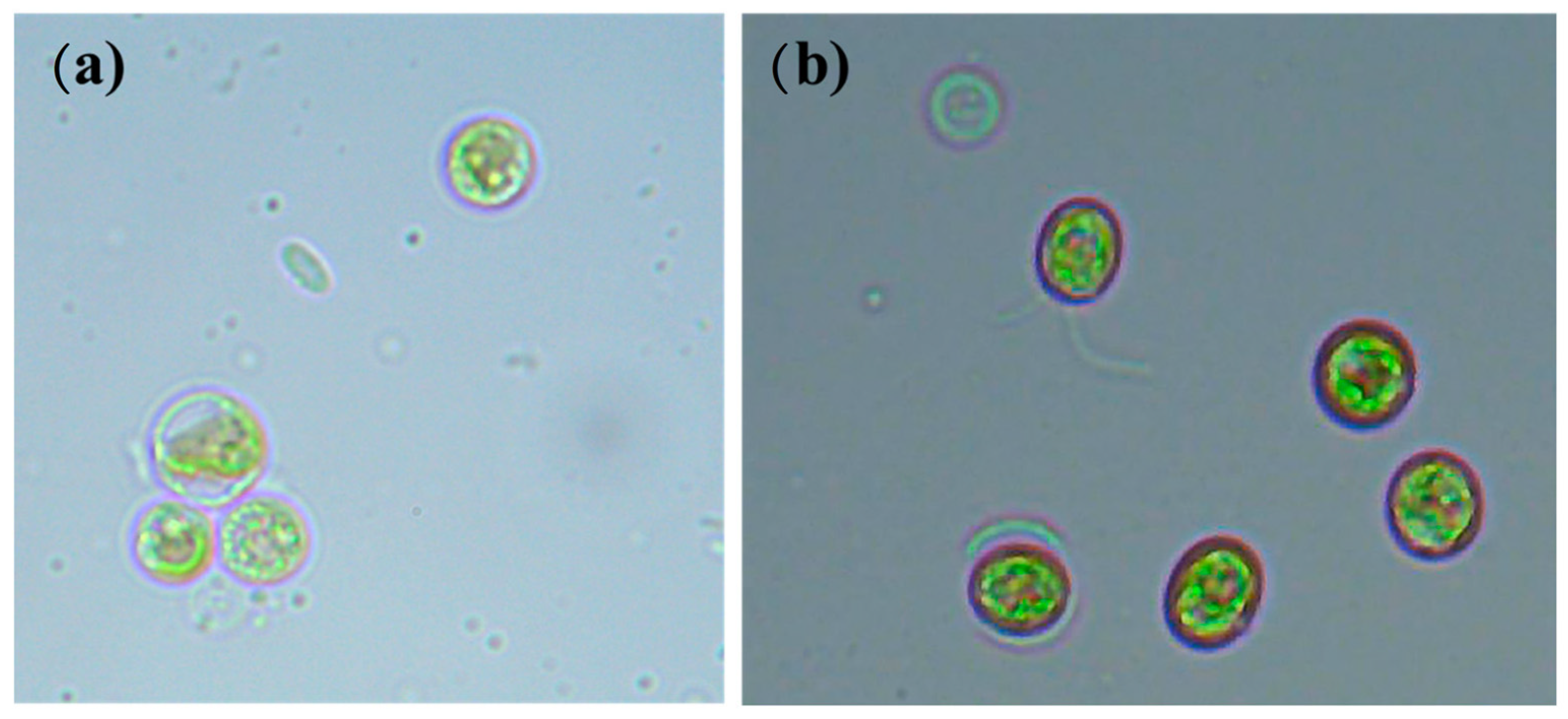
2.1. Sample Collection and Microalgae Isolation

Water samples were collected from Lake Superior and Lake Tamblyn—Lakehead University, Thunder Bay, ON, Canada. Filamentous plants and other coarse materials were removed by filtration using a mesh net. Water samples were spread on the BG-11 [25] agar plate with the help of sterile disposable spreaders and incubated at room temperature at 20 ± 2 °C in continuous illumination for two to three weeks. Different algal colonies from plates were selected and sub-cultured on the BG-11 agar plate by the streak plate technique until a pure isolated colony was obtained. Purely isolated microalgae from the solid culture medium were inoculated in a 1 L glass flask with 800 mL of working volume containing BG-11 medium.

2.2. Microalgae Strain and Cultivation Conditions

Two microalgae strains were isolated and morphologically identified as Chlorella sp. (isolated from Lake Tamblyn—Lakehead University, Thunder Bay, ON, Canada) and Chlamydomonas sp. (isolated from Lake Superior, Thunder Bay, ON, Canada) using the guidebook by Bellinger and Sigee [26] (Figure 1). The inoculum was prepared using a BG-11 medium at the initial pH of 7.1 [25]. Exponential phase of the algal culture was used as inoculants (1 mL of inoculants with optical density (OD) of 1.351 at 680 nm) for experimental trials. All laboratory-scale cultivations were carried out in a 1 L glass flask (Erlenmeyer), with a working volume of 800 mL. The solutions were placed in a constant orbital shaking (125 rpm). The entire experiment was conducted in an environmentally controlled growth room with a constant temperature of 25 °C (±2 °C) and a photoperiod of a 16:8 h light/dark cycle. Light intensity was provided by four cool fluorescence tube-bulbs on the top of the reactors (10 cm distance).

2.3. Characteristics of Malting Effluents

The malting effluent was obtained from Canada Malting (Thunder Bay, ON, Canada). Solid particles from malting effluents were removed by sedimentation and filtration with a clean cheesecloth. The visible solid particles settled down at the bottom and only the supernatant was used for subsequent microalgae growth experiments, stored at 4 °C until use in experiments. The growth characteristics of microalgae were tested in four dilution conditions: 10% malting effluent (10 ME: 10 volumes of effluent were mixed with 90 volumes of distilled water), 30% malting effluent (30 ME: 30 volumes of effluent were mixed with 70 volumes of distilled water), 50% malting effluent (50 ME: 50 volumes of effluent were mixed with 50 volumes of distilled water), and 70% dilution (70 ME: 70 volumes of effluent were mixed with 30 volumes of distilled water), and a control condition (BG-11 medium). The elements and compounds of malting effluents are shown in Table 1.

2.4. Measurement of Microalgae Growth Characteristics

Microalgae growth was monitored by determining the cell count every two days. Total algal cell counts were measured using a microscopic cell count method (0.1 mm Tiefe deep Neubauer Improved Hemocytometer, Hamburg, Germany), with the help of a compound microscope (Olympus, Tokyo, Japan) [27]. The specific growth rate (SGR) refers to the increment cell density per unit area per unit of time during the log phase. The specific growth rate was calculated using the following formula [28]:
SGR   ( mg · L 1 · day 1 ) = ( N 2 N 2 ) ( T 2 T 1 )
where N2 and N1 are the cell density (107 cell·mL−1) at time T2 (at any day) and T1 (at the beginning), respectively.

2.5. Estimation of Total Chlorophyll Content

The total chlorophyll content was measured using methanol as a solvent [28]. Two mL of microalga culture was centrifuged at 10,000 rpm for 10 min. Then, the supernatant was discarded, and the algal pellet was resuspended with 2 mL of methanol and briefly sonicated for algal cell wall breakdown. Then, the mixture was incubated at 60 °C for 5 min and centrifuged again for 10 min at 10,000 rpm. The absorbance of the supernatant was analyzed by reading at 665 and 652 nm with a visible spectrophotometer, and the concentrations of chlorophyll α and chlorophyll β were calculated by using the following formulae [29]:
Chlorophyll   α   ( mg · L 1 ) = 16.82 ×   A 665 9.28 ×   A 652
Chlorophyll   β   ( mg · L 1 ) = 39.92 ×   A 652 16.54 ×   A 665
Total   chlorophyll   ( mg · L 1 ) =   Chlorophyll   α +   Chlorophyll   β

2.6. Lipid Extraction

The total lipids from the wet algal biomass were extracted by chloroform:methanol (1:1, v/v) using the method of Bligh and Dyer [30] and Guo et al. [31], with some modifications. Two mL of the wet algal biomass was collected by centrifuging at 8000× g for 10 min. The cell pellet was first extracted with a mixture of chloroform, methanol, and distilled water (1:2:0.8, v/v/v). The mixture was agitated by vortexing for 5 min and centrifuged at 8000× g for 10 min. The supernatant was transferred to a pre-weighted Eppendorf tube (W1 g), and the cell pellet was re-extracted with the mixture of chloroform and methanol (1:2, v/v). The mixture was centrifuged again, and the supernatant was collected in the same pre-weighed Eppendorf tube. Chloroform and water were then added to the supernatant to form a ratio of 1:1:0.9 (chloroform:methanol:distilled water, v/v/v). The mixture was thoroughly mixed and centrifuged at 8000× g for 5 min. The top layer was then removed, and the bottom layer was evaporated and dried at 80 °C until reaching a constant weight (W2 g). The total lipid content was calculated by subtracting W1 from W2 and was presented as a percentage of the dry weight. The lipid productivity was calculated as described by Cobes et al. [32]:
Percentage   of   total   lipid   content   ( %   dry   weight )   ( L T ) = W L W A × 100
Lipid   productivity   ( L p ) ( mg · L 1 ) =   W A   ( g ) × L T V   ( L ) × T ( d )
where WL (g) is the weight of the total lipids extracted, WA (g) is the weight of the dry microalgae biomass, LT is the percentage of total lipid content of the dry weight, V is the working volume, and T is the cultivation time.

2.7. Statistical Analysis

All the numerical variables were checked for normality before conducting the parametric tests. Polynomial regression analysis was carried out to explore the relationship between algal cell density and number of cultivation days. One-way ANOVA was used to test for differences in the algal dry biomass and lipid contents among the different treatments, followed by Tukey’s HSD post hoc test for pairwise differences. Student’s t-test was carried out to test the mean difference of dry weight, chlorophyll content, and lipid content between the two algal species. All the experiments were performed in triplicates, and the results are shown as mean ± SD (standard deviation). Differences between treatments were evaluated at p < 0.05. All statistical analyses were carried out in R, v. 3.6.1 [33].

3. Results and Discussion

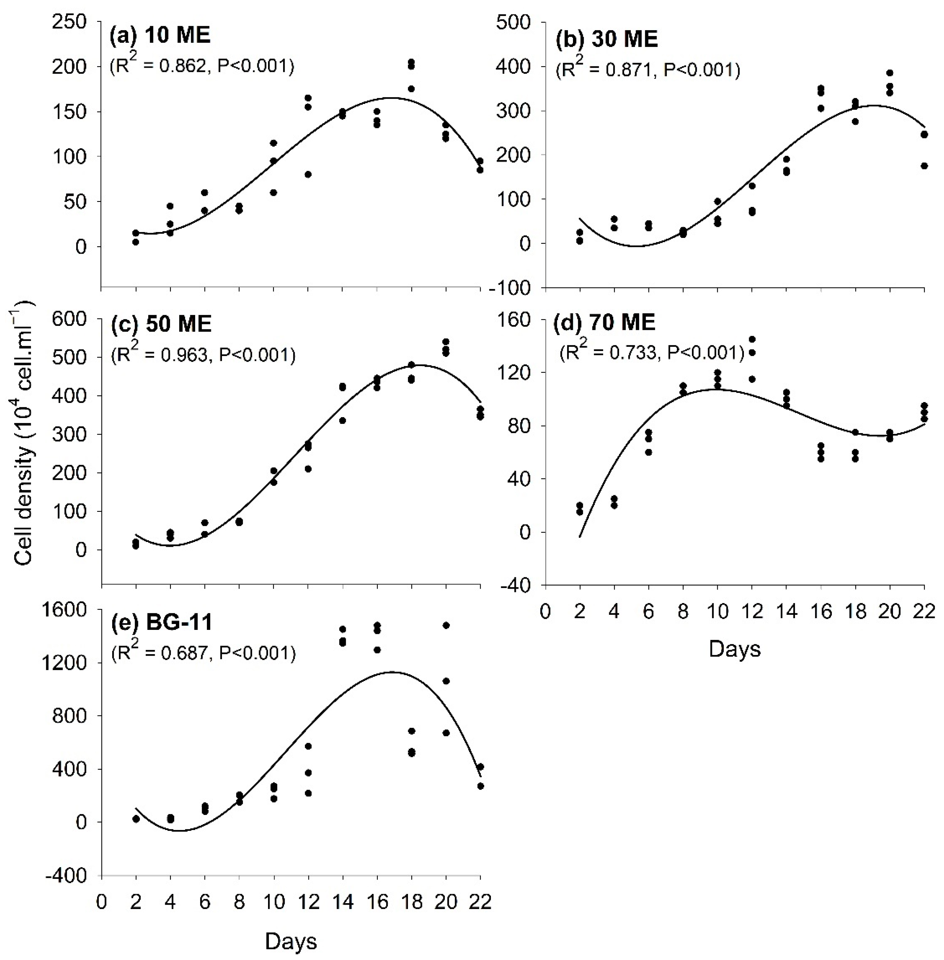
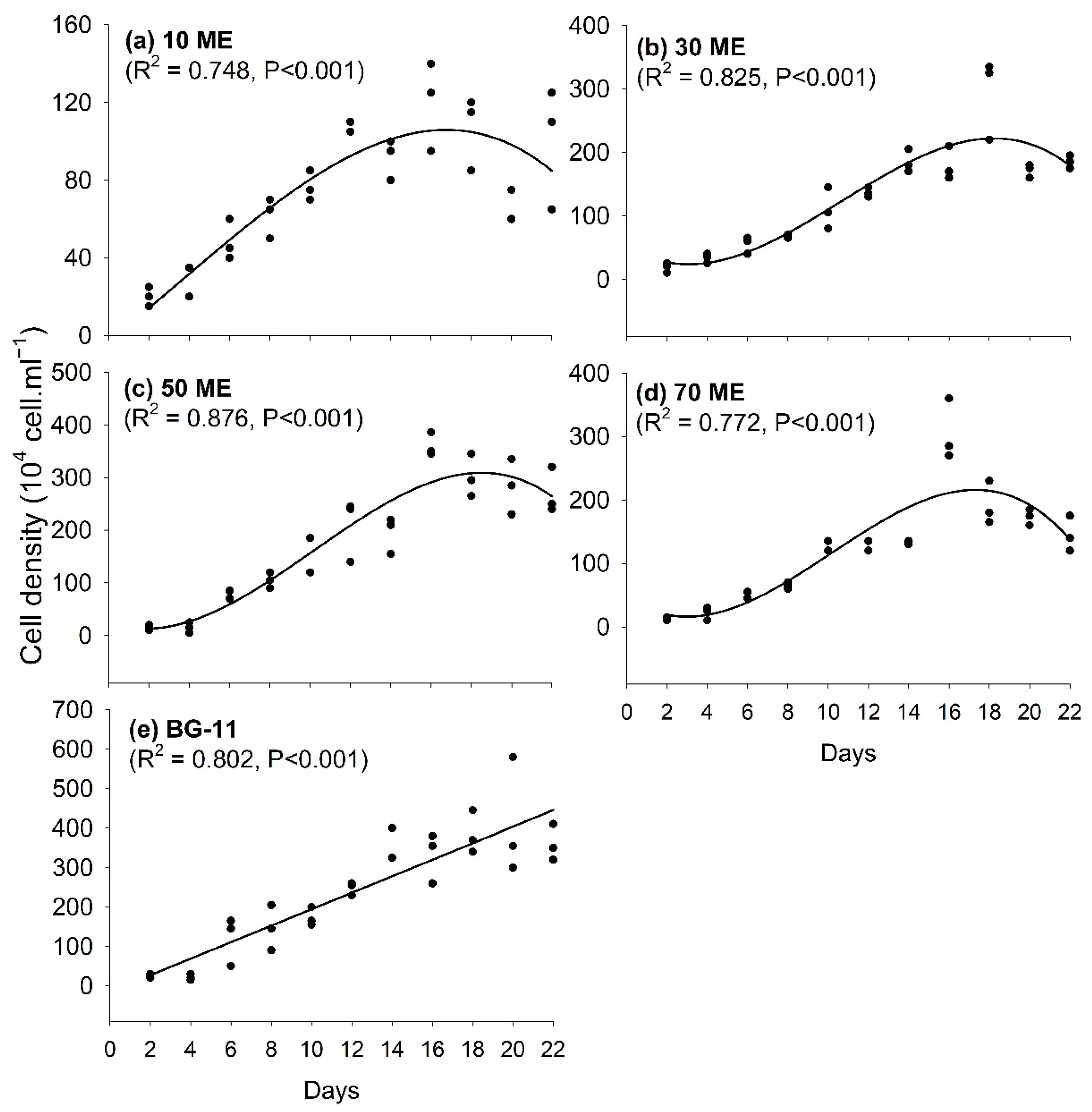
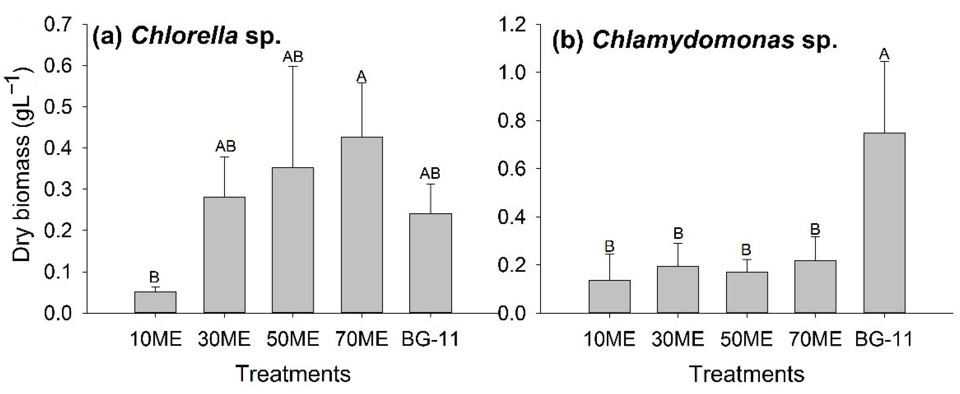
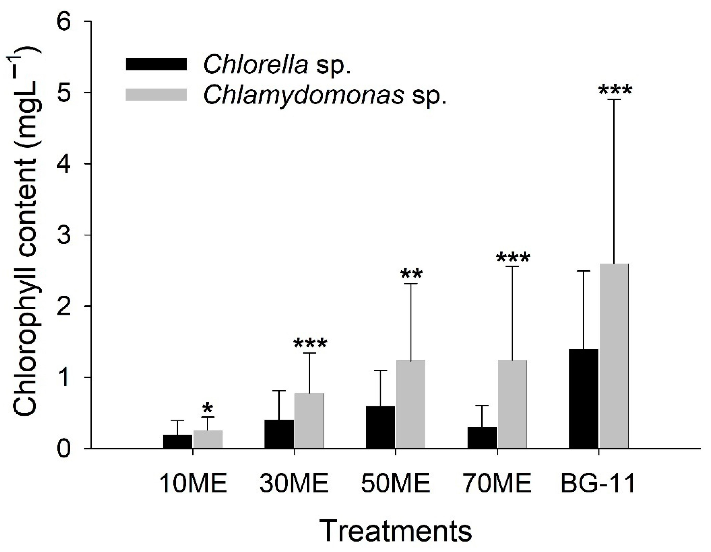
3.1. Effects of Malting Effluent on Algal Growth and Chlorophyll Content

The undiluted malting effluent contained abundant organic matter, such as total organic carbon (1870 mg·L1), nitrogen (NO3-N was 820 mg·L1 and NH3-N was 22.3 mg·L1), phosphorus (PO4-P was 60.5 mg·L1 and TP was 65.7 mg·L1), and amino acids (0.210 mg·L1) (Table 1), which were beneficial to algal growth. The contents of some metal elements, such as potassium (K), iron (Fe), and zinc (Zn), were markedly higher in malting effluent than that in standard BG-11 medium, even though the malting effluent was diluted 10 times. In addition, malting effluent also consisted of some heavy metals and various trance elements, which were not included in the standard medium (Table 1). However, the non-sterile malting effluent may have been contaminated by bacteria, and thus algae cannot grow properly without dilutions. Thus, 10%, 30%, 50%, and 70% diluted malting effluents were used for further experiments. In this study, non-sterile malting effluent was used as a growth medium of two different freshwater microalgae and identified the optimal concentration for microalgal biomass and lipid production. The cell density of both Chlorella sp. and Chlamydomonas sp. was significantly increased when the number of cultivation days increased in experimental groups (different concentrations of malting effluent) and the control group (BG-11) (Figure 2 and Figure 3). In Chlorella sp., algal cell density showed an increasing trend as the number of cultivation days increased for 10 ME medium (R2 = 0.862, p < 0.001), 30 ME (R2 = 0.871, p < 0.001), 50 ME (R2 = 0.963, p < 0.001), and 70 ME (R2 = 0.733, p < 0.001), as well as BG-11 (R2 = 0.687, p < 0.001), respectively (Figure 2). The cell density gradually declined after 14 days of cultivation. The nutrient availability, for example, nitrate, phosphate, and other micronutrients, slowly decreased as cultivation days increased in the culture medium, and these nutrients play a significant role in microalgae growth [34].
A similar trend was found in Chlamydomonas sp., where cell density significantly increased when the number of cultivation days increased for 10 ME medium (R2 = 0.748, p < 0.001), 30 ME (R2 = 0.825, p < 0.001), 50 ME (R2 = 0.786, p < 0.001), and 70 ME (R2 = 0.772, p < 0.001), as well as BG-11 (R2 = 0.876, p < 0.001), respectively (Figure 3). This study revealed that untreated malting effluent supports microalgae growth without the addition of any extra nutrient supplements. This study aligned with the pervious study by Marchão et al. [35], who reported the maximum growth of Scenedesmus obliquus in diluted brewery wastewater.
In addition, the specific growth rates of Chlorella sp. growing in the 10 ME, 30 ME, 50 ME, 70 ME, and BG-11 were 10.25, 20.3, 25.42, 7.25, and 95.17 (104 cell·mL−1), while correspondingly in Chlamydomonas sp. they were 6.81, 15.6, 20.28, 15.52, and 26.29 (104 cell·mL−1), respectively (Table 2). Likewise, the specific growth rates of both species were higher in BG-11 medium compared to species cultivated in malting effluent (Table 2). Similar growth rates were reported for several other freshwater microalgae species [32,36]. Previous studies have indicated that several factors affect cell density and growth rate in microalgae, for example, nitrogen deficiency, light condition, and CO2 availability [32,37,38]. In this study, the experimental setup was adequate for microalgae growth.
The dry weight of Chlamydomonas sp. (mean 0.74 g/L ± SD 0.29) was significantly higher in the standard BG-11 medium (T = 2.86, df = 2, p = 0.05) compared to Chlorella sp. (mean 0.24 g/L ± SD 0.07) on day 22, respectively (Figure 4). A similar dry weight of both species was noticed when they were grown in the different diluted malting effluents (Figure 4). Within Chlorella sp., the highest biomass was observed in 70 ME, followed by 30 ME and 50 ME, and the lowest biomass accumulation was observed in 10 ME after 21 days of growing (Figure 5a). For Chlamydomonas sp., the biomass accumulation was higher in standard BG-11 medium (Figure 5b). This result indicated the nutrients’ differences between culture mediums. In malting effluent, nutrients are largely imbalanced compared to standard BG-11 medium, which leads to lower biomass accumulation. In addition, dark color and turbidity in the malting effluent may impede light penetration, resulting in lower microalgae growth [39]. O’Rourke et al. [40] reported that cultivating Parachlorella kessleri in a 50% concentration of brewery wastewater increased the biomass yield 2-fold (12.3 g·L−1), compared to a 10% brewery wastewater concentration after 14 days of growth.
Our results revealed that the diluted, non-sterile malting effluent could be a nutrient medium for large-scale algal growth. Although limited studies are available on the growth pattern of freshwater microalgae in malting effluent, available studies revealed that malting effluent is an excellent source of nitrogen and other nutrients and stimulates higher algal biomass production [21,40]. Wastewater contains abundant organic matter and amino acids and promotes the algal growth [41]. Furthermore, wastewater also contains some metal elements useful for the algal growth [42] accumulation of microalgae. Some of the elements, for example, potassium, iron, and zinc ions, promote the growth of algae and lipid accumulation through increasing the specific growth rates and provoking the uptake of ammonium in microalgae [41,43,44,45]. Therefore, the diluted malting effluents can supply adequate nutrition for algal growth.

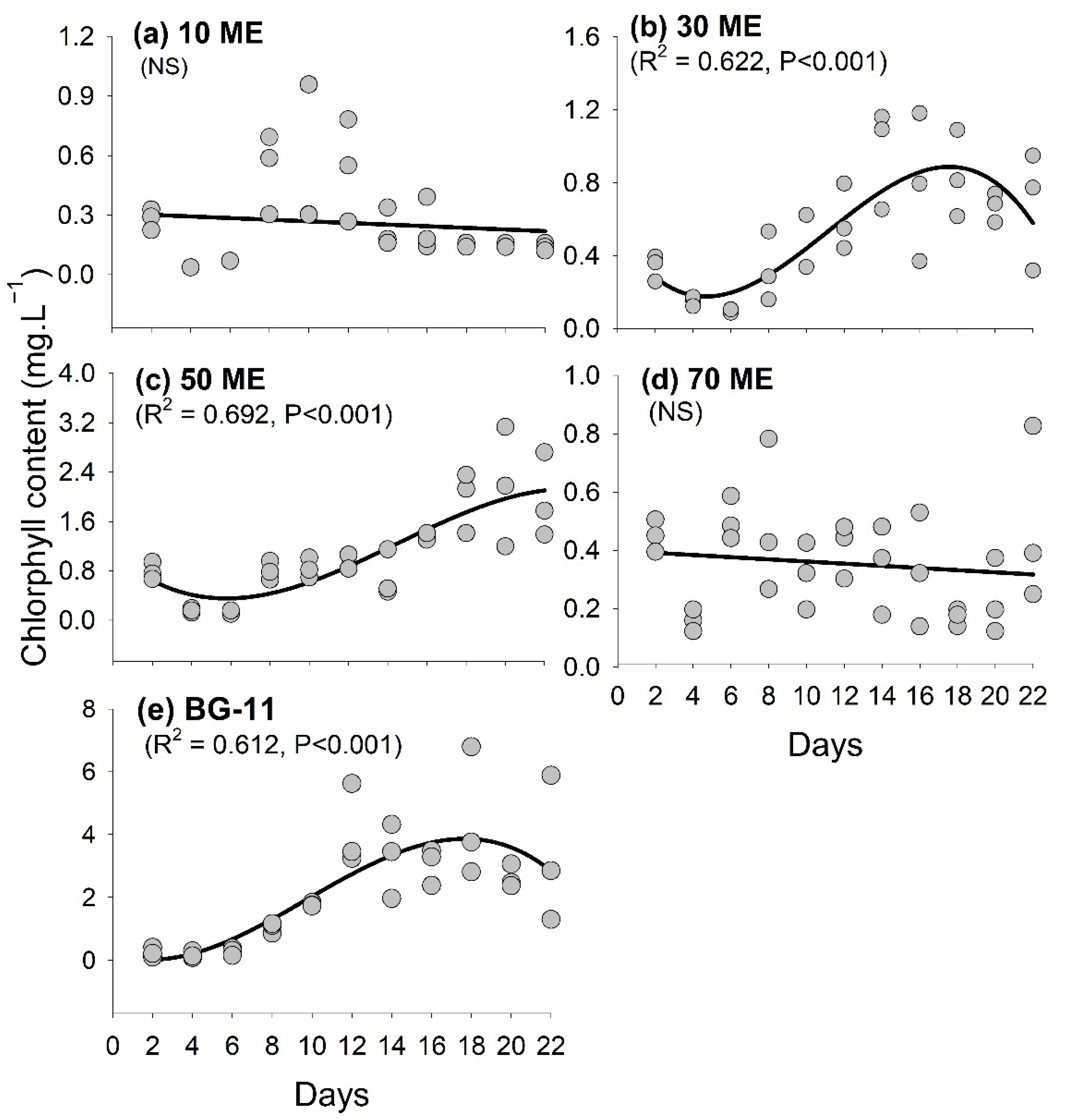
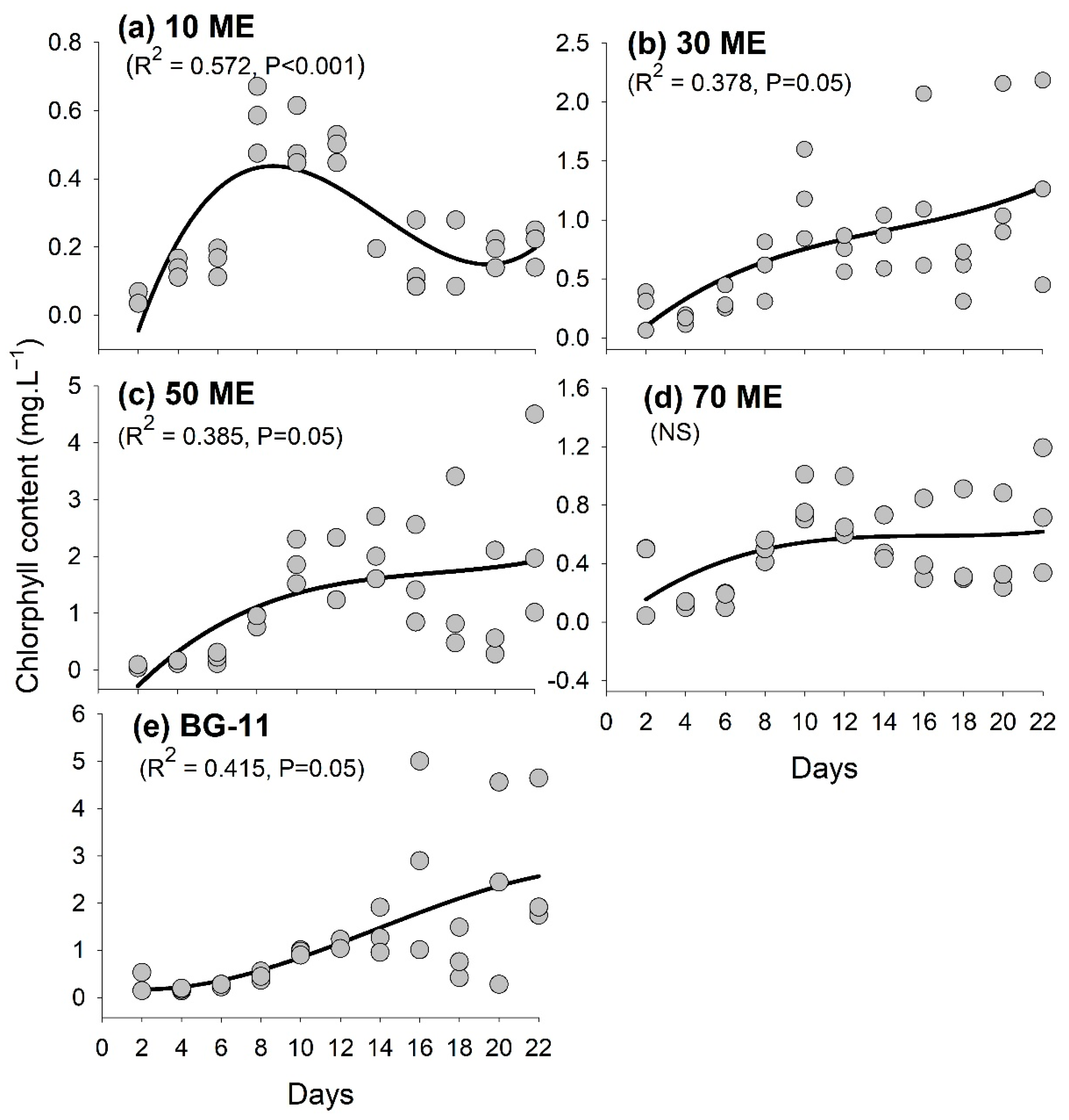
3.2. Chlorophyll Content Variation

In order to explore the effect of malting effluent on microalgae growth, chlorophyll contents of microalgae were determined at various concentrations. Figure 6 and Figure 7 demonstrate that different concentrations of malting effluent had significant effects on the total chlorophyll content. For Chlorella sp., there was a significant increase of chlorophyll content only in 30 ME and 50 ME treatments and the BG-11 medium (Figure 6). Within Chlamydomonas sp., the chlorophyll accumulation was found to be increased in 10 ME, 30 ME, and 50 ME concentrations of ME and the BG-11 medium, along with the increase in the number of cultivation days (Figure 7).
The chlorophyll concentrations were significantly higher in Chlamydomonas sp. compared to Chlorella sp. in all treatments (Figure 8). In our study, the amount of chlorophyll content varied between different concentrations of malting culture medium and species. Chlamydomonas sp. have the potential to survive in higher nutrient conditions and promote growth in malting effluent compared to Chlorella sp. There was a significant positive correlation between the chlorophyll content and algal cell density in BG-11 medium for both Chlorella sp. (r = 0.551, p = 0.05) and Chlamydomonas sp. (r = 0.727, p = 0.001). This further signifies that the increasing chlorophyll content in the culture medium indicates the microalgal growth.

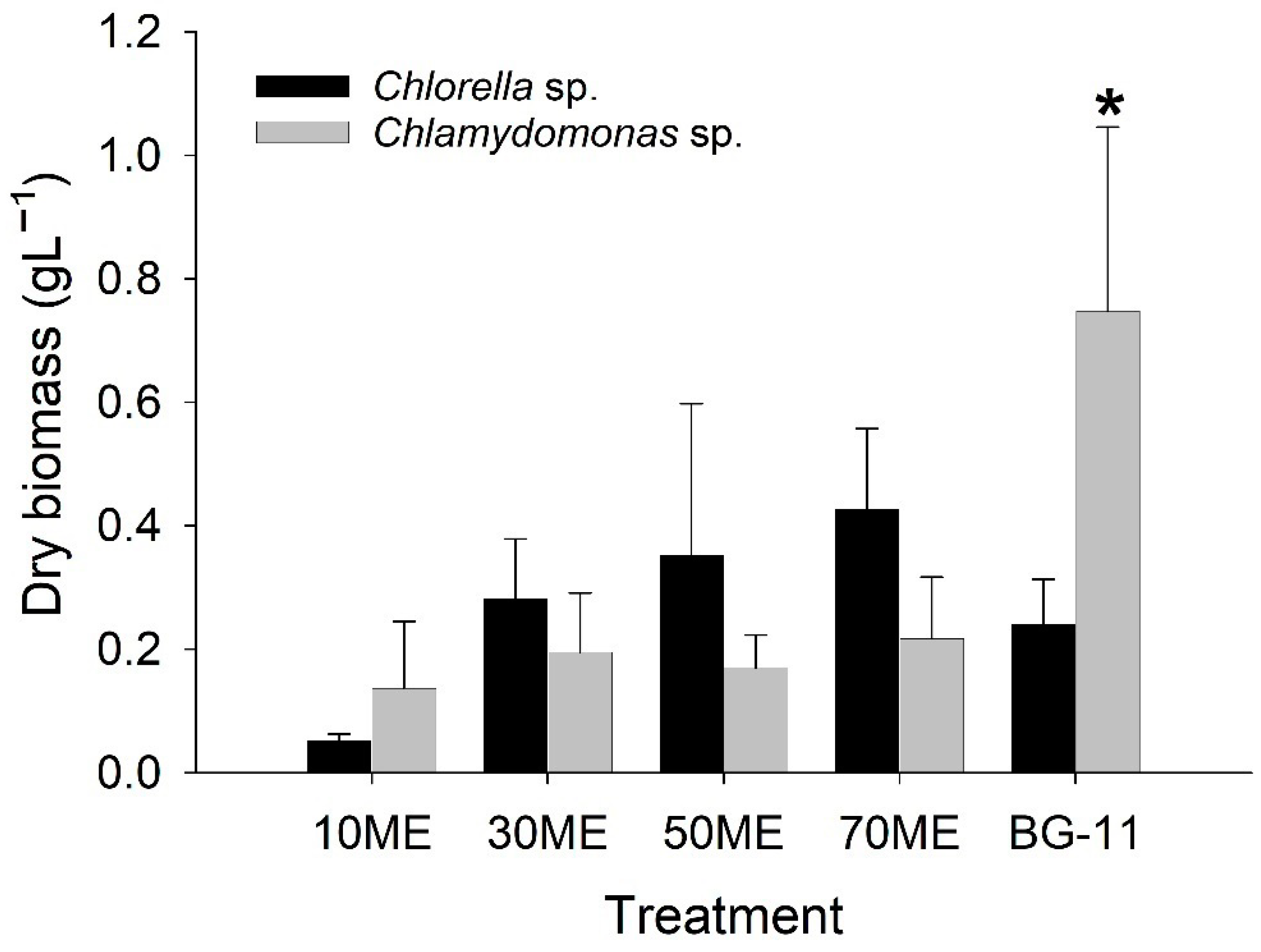
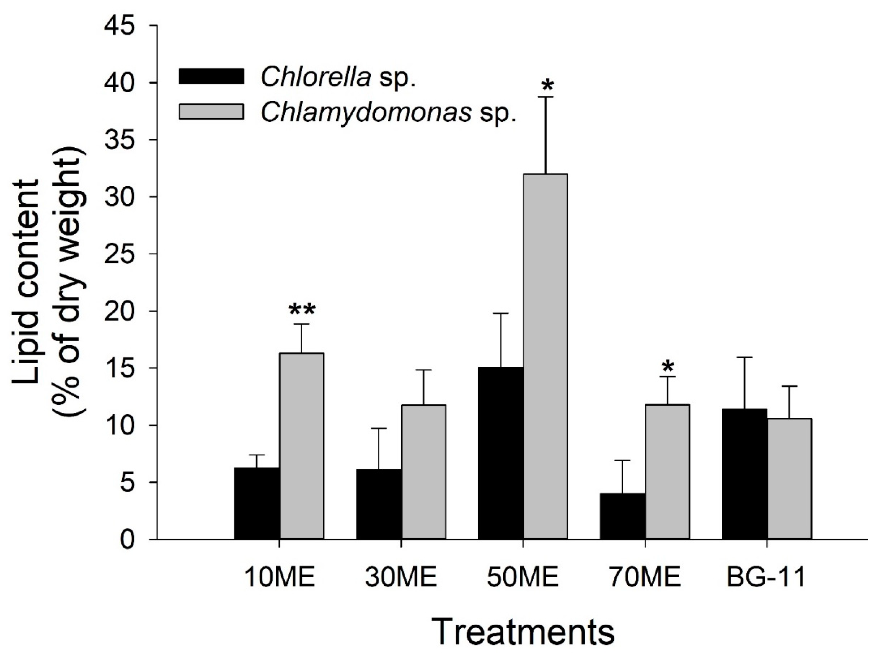
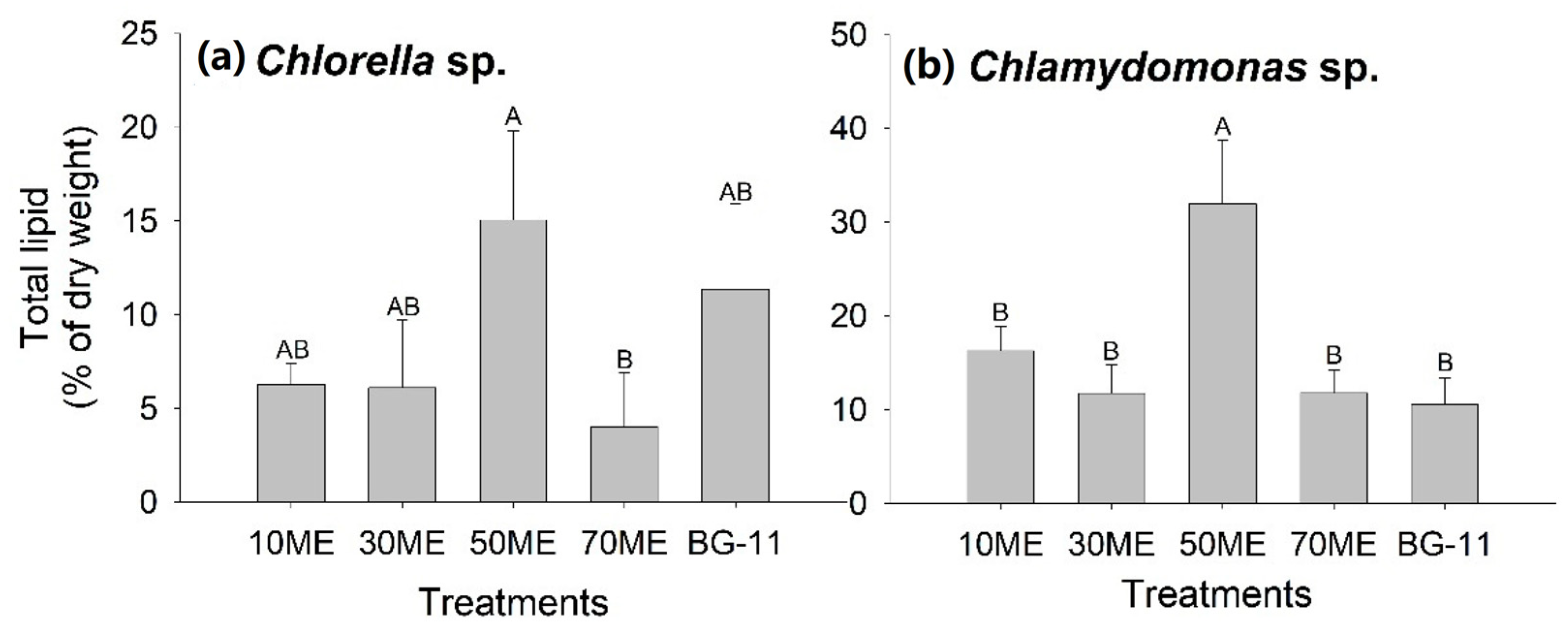
3.3. Effects of Malting Effluent on Lipid Accumulation

To evaluate the effects of the malting effluent on lipid accumulation of microalgae, percentages of total lipid accumulation were determined on day 22. Chlamydomonas sp. had potential for higher lipid production than Chlorella sp. (Figure 9). Similarly, the one-way ANOVA revealed that the highest lipid content was observed in both microalgae species’ ME (Figure 10). The final lipid productivity of Chlorella sp. in 50% of ME ranged from 39 mg·L1 (min) to 145 mg·L1 (max) and 96 mg·L1 (min) to 123 mg·L1 (max) for Chlamydomonas sp., respectively, on day 22. In the 50% concentration of malting effluent, the maximum lipid productivities of Chlorella sp. and Chlamydomonas sp. largely corresponded with higher cell density. The reason for a variation in lipid production between the diluted concentrations of malting effluent was mainly due to the availability of organic carbon, nutrients, and the turbidity of the malting effluent. The amount of organic carbon increased with the higher concentration of malting. Still, at the same time, turbidity also increased, which impeded the light availability and reduced the growth and subsequent lipid production. The increment of total lipids in diluted malting effluents could be partially due to the low concentration of nitrogen. During the nitrogen-sufficient condition, the photosynthetic carbon fixation was higher than nitrogen assimilation. As a result, cells produced nitrogen-containing components such as proteins, nucleotides, and pigments. During nitrogen-deficient conditions, carbon demand increased, and excess carbon was stored in the form of lipids and carbohydrates [46,47]. Studies demonstrated that the nitrogen depletion markedly stimulated the lipid production in many microalgae species: Chlorella vulgaris [48], Desmodesmus sp. [49], Acutodesmus dimorphus [46], Neochloris oleoabundans [50], and Nannochloropsis gaditana [51]. This study is greatly supported by Li et al.’s work [50], which found that nitrogen starvation accelerates the lipid accumulation in microalgae species. It has been well-reported that nitrogen starvation markedly induced the accumulation of triacylglycerol in the green algae by increasing carbon flux towards glycerol-3-phosphate and acyl-CoA for fatty acid synthesis [52].

4. Conclusions

This study isolated the native freshwater microalgae and integrated them for malting wastewater treatment and subsequent biomass and lipid production. Microalgae grown in the diluted malting effluent achieved the highest cell number at 50 ME for Chlorella sp. and 70 ME for Chlamydomonas sp. In addition, the diluted malting effluent markedly stimulated lipid accumulation up to 145 mg·L−1 in Chlorella sp. and 123 mg·L−1 in Chlamydomonas sp. The current study results revealed that the diluted malting effluent could be a promising alternative as a nutrient medium for large-scale algal cultivation and simultaneous biofuel production and bioremediation.

Author Contributions

Conceptualization, J.R.K., H.G. and W.Q.; methodology, J.R.K. and S.S.; validation, W.Q.; formal analysis, J.R.K.; investigation, J.R.K.; data curation, J.R.K., S.S., C.C., X.C. and A.L.M.K.; writing—original draft preparation, J.R.K.; writing—review and editing, J.R.K.; supervision, W.Q.; funding acquisition, W.Q. All authors have read and agreed to the published version of the manuscript.

Funding

This research was funded by the Natural Sciences and Engineering Research Council of Canada, grant number RGPIN-2017-05366.

Institutional Review Board Statement

Not applicable.

Informed Consent Statement

Not applicable.

Data Availability Statement

The data presented in this study are available upon request from the corresponding author.

Conflicts of Interest

The authors declare no conflict of interest.

References

  1. Siwal, S.S.; Zhang, Q.; Devi, N.; Saini, A.K.; Saini, V.; Pareek, B.; Gaidukovs, S.; Thakur, V.K. Recovery processes of sustainable energy using different biomass and wastes. Renew. Sust. Energ. Rev. 2021, 150, 111483. [Google Scholar] [CrossRef]
  2. Macqueen, D.; Korhaliller, S. Bundles of Energy: The Case for Renewable Biomass Energy; IIED: London, UK, 2011. [Google Scholar]
  3. Xia, A.; Jacob, A.; Tabassum, M.R.; Herrmann, C.; Murphy, J.D. Production of hydrogen, ethanol and volatile fatty acids through co-fermentation of macro-and micro-algae. Bioresour. Technol. 2016, 205, 118–125. [Google Scholar] [CrossRef] [PubMed]
  4. Xu, L.; Fan, J.; Wang, Q. Omics Application of Bio-Hydrogen Production Through Green Alga Chlamydomonas reinhardtii. Front. Bioeng. Biotechnol. 2019, 7, 201. [Google Scholar] [CrossRef] [PubMed]
  5. Williams, P.J.l.B.; Laurens, L.M. Microalgae as biodiesel & biomass feedstocks: Review & analysis of the biochemistry, energetics & economics. Energy Environ. Sci. 2010, 3, 554–590. [Google Scholar]
  6. Li, Y.; Horsman, M.; Wu, N.; Lan, C.Q.; Dubois-Calero, N. Biofuels from microalgae. Biotechnol. Prog. 2008, 24, 815–820. [Google Scholar] [CrossRef]
  7. Christenson, L.; Sims, R. Production and harvesting of microalgae for wastewater treatment, biofuels, and bioproducts. Biotechnol. Adv. 2011, 29, 686–702. [Google Scholar] [CrossRef]
  8. Khoo, K.S.; Chew, K.W.; Yew, G.Y.; Leong, W.H.; Chai, Y.H.; Show, P.L.; Chen, W.-H. Recent advances in downstream processing of microalgae lipid recovery for biofuel production. Bioresour. Technol. 2020, 304, 122996. [Google Scholar] [CrossRef]
  9. Kumar, B.R.; Mathimani, T.; Sudhakar, M.P.; Rajendran, K.; Nizami, A.-S.; Brindhadevi, K.; Pugazhendhi, A. A state of the art review on the cultivation of algae for energy and other valuable products: Application, challenges, and opportunities. Renew. Sust. Energ. Rev. 2021, 138, 110649. [Google Scholar] [CrossRef]
  10. Du, H.; Ahmed, F.; Lin, B.; Li, Z.; Huang, Y.; Sun, G.; Ding, H.; Wang, C.; Meng, C.; Gao, Z. The Effects of Plant Growth Regulators on Cell Growth, Protein, Carotenoid, PUFAs and Lipid Production of Chlorella pyrenoidosa ZF Strain. Energies 2017, 10, 1696. [Google Scholar] [CrossRef] [Green Version]
  11. Almarashi, J.Q.M.; El-Zohary, S.E.; Ellabban, M.A.; Abomohra, A.E.-F. Enhancement of lipid production and energy recovery from the green microalga Chlorella vulgaris by inoculum pretreatment with low-dose cold atmospheric pressure plasma (CAPP). Energy Convers. Manag. 2020, 204, 112314. [Google Scholar] [CrossRef]
  12. Han, S.-F.; Jin, W.; Tu, R.; Abomohra, A.E.-F.; Wang, Z.-H. Optimization of aeration for biodiesel production by Scenedesmus obliquus grown in municipal wastewater. Bioprocess Biosyst. Eng. 2016, 39, 1073–1079. [Google Scholar] [CrossRef] [PubMed]
  13. Abomohra, A.E.-F.; Eladel, H.; El-Esawi, M.; Wang, S.; Wang, Q.; He, Z.; Feng, Y.; Shang, H.; Hanelt, D. Effect of lipid-free microalgal biomass and waste glycerol on growth and lipid production of Scenedesmus obliquus: Innovative waste recycling for extraordinary lipid production. Bioresour. Technol. 2018, 249, 992–999. [Google Scholar] [CrossRef] [PubMed]
  14. Chen, Z.; Qiu, S.; Amadu, A.A.; Shen, Y.; Wang, L.; Wu, Z.; Ge, S. Simultaneous improvements on nutrient and Mg recoveries of microalgal bioremediation for municipal wastewater and nickel laterite ore wastewater. Bioresour. Technol. 2020, 297, 122517. [Google Scholar] [CrossRef] [PubMed]
  15. Porto, B.; Gonçalves, A.L.; Esteves, A.F.; Souza, S.M.A.G.U.d.; Souza, A.A.U.d.; Vilar, V.J.P.; Pires, J.C.M. Microalgal Growth in Paper Industry Effluent: Coupling Biomass Production with Nutrients Removal. Appl. Sci. 2020, 10, 3009. [Google Scholar] [CrossRef]
  16. Li, G.; Zhang, J.; Li, H.; Hu, R.; Yao, X.; Liu, Y.; Zhou, Y.; Lyu, T. Towards high-quality biodiesel production from microalgae using original and anaerobically-digested livestock wastewater. Chemosphere 2020, 273, 128578. [Google Scholar] [CrossRef]
  17. Ashokkumar, V.; Chen, W.-H.; Ngamcharussrivichai, C.; Agila, E.; Ani, F.N. Potential of sustainable bioenergy production from Synechocystis sp. cultivated in wastewater at large scale—A low cost biorefinery approach. Energy Convers. Manag. 2019, 186, 188–199. [Google Scholar] [CrossRef]
  18. Javed, F.; Aslam, M.; Rashid, N.; Shamair, Z.; Khan, A.L.; Yasin, M.; Fazal, T.; Hafeez, A.; Rehman, F.; Rehman, M.S.U.; et al. Microalgae-based biofuels, resource recovery and wastewater treatment: A pathway towards sustainable biorefinery. Fuel 2019, 255, 115826. [Google Scholar] [CrossRef]
  19. Hernando, M.D.; Fernández-Alba, A.R.; Tauler, R.; Barceló, D. Toxicity assays applied to wastewater treatment. Talanta 2005, 65, 358–366. [Google Scholar] [CrossRef]
  20. Giraldo, N.D.; Buchelly, R.J.R.; Hincapié, D.E.; Atehortua, L. Transformation of Brewery Subproducts into Valuable Biomass Using Mixotrophic Culture of Chlorella pyrenoidosa and Associated Bacteria. Braz. Arch. Biol. Technol. 2020, 63, e20190229. [Google Scholar] [CrossRef]
  21. Campaña, D.H.; Echevarría, M.E.U.; Airasca, A.O.; Couce, M.L.A. Physicochemical and phytotoxic characterisation of residual sludge from the malting of barley. J. Ind. Pollut. Control. 2014, 2, 115. [Google Scholar]
  22. Vriens, L.; Soest, H.v.; Verachtert, H. Biological treatment of malting and brewing effluents. Crit. Rev. Biotechnol. 1990, 10, 1–46. [Google Scholar] [CrossRef]
  23. EoC. Technical Pollution Prevention Guide for Brewery and Wine Operations in the Lower Fraser Basin; Environment Canada, Environmental Protection and Fraser Pollution Abatement: Vancouver, BC, Canada, 1997; p. 101.
  24. MoJ. Wastewater Systems Effluent Regulations (SOR/2012-139); Minister of Justice, Government of Canada: Ottawa, ON, Canada, 2012; p. 80.
  25. Stanier, R.; Kunisawa, R.; Mandel, M.; Cohen-Bazire, G. Purification and properties of unicellular blue-green algae (order Chroococcales). Bacteriol. Rev. 1971, 35, 171–205. [Google Scholar] [CrossRef] [PubMed]
  26. Bellinger, E.G.; Sigee, D.C. Freshwater Algae: Identification, Enumeration and Use as Bioindicators; John Wiley & Sons: Oxford, UK, 2015. [Google Scholar]
  27. Wahidin, S.; Idris, A.; Shaleh, S.R.M. The influence of light intensity and photoperiod on the growth and lipid content of microalgae Nannochloropsis sp. Bioresour. Technol. 2013, 129, 7–11. [Google Scholar] [CrossRef] [PubMed]
  28. Song, C.; Liu, Z.; Wang, C.; Li, S.; Kitamura, Y. Different interaction performance between microplastics and microalgae: The bio-elimination potential of Chlorella sp. L38 and Phaeodactylum tricornutum MASCC-0025. Sci. Total Environ. 2020, 723, 138146. [Google Scholar] [CrossRef] [PubMed]
  29. Xiong, J.-Q.; Kurade, M.B.; Jeon, B.-H. Ecotoxicological effects of enrofloxacin and its removal by monoculture of microalgal species and their consortium. Environ. Pollut. 2017, 226, 486–493. [Google Scholar] [CrossRef]
  30. Bligh, E.G.; Dyer, W.J. A rapid method of total lipid extraction and purification. Can. J. Biochem. Physiol. 1959, 37, 911–917. [Google Scholar] [CrossRef]
  31. Guo, H.; Chen, H.; Fan, L.; Linklater, A.; Zheng, B.; Jiang, D.; Qin, W. Enzymes produced by biomass-degrading bacteria can efficiently hydrolyze algal cell walls and facilitate lipid extraction. Renew. Energ. 2017, 109, 195–201. [Google Scholar] [CrossRef]
  32. Cobos, M.; Paredes, J.D.; Maddox, J.D.; Vargas-Arana, G.; Flores, L.; Aguilar, C.P.; Marapara, J.L.; Castro, J.C. Isolation and Characterization of Native Microalgae from the Peruvian Amazon with Potential for Biodiesel Production. Energies 2017, 10, 224. [Google Scholar] [CrossRef] [Green Version]
  33. R Development Core Team A Language and Environment for Statistical Computing. R Foundation for Statistical Computing; R Foundation for Statistical Computing: Vienna, Austria, 2019.
  34. Ding, J.; Zhao, F.; Cao, Y.; Xing, L.; Liu, W.; Mei, S.; Li, S. Cultivation of microalgae in dairy farm wastewater without sterilization. Int. J. Phytoremediation 2015, 17, 222–227. [Google Scholar] [CrossRef]
  35. Marchão, L.; da Silva, T.L.; Gouveia, L.; Reis, A. Microalgae-mediated brewery wastewater treatment: Effect of dilution rate on nutrient removal rates, biomass biochemical composition, and cell physiology. J. Appl. Phycol. 2018, 30, 1583–1595. [Google Scholar] [CrossRef] [Green Version]
  36. Vidotti, A.D.S.; Riaño-Pachón, D.M.; Mattiello, L.; Giraldi, L.A.; Winck, F.V.; Franco, T.T. Analysis of autotrophic, mixotrophic and heterotrophic phenotypes in the microalgae Chlorella vulgaris using time-resolved proteomics and transcriptomics approaches. Algal Res. 2020, 51, 102060. [Google Scholar] [CrossRef]
  37. Patel, A.K.; Joun, J.M.; Hong, M.E.; Sim, S.J. Effect of light conditions on mixotrophic cultivation of green microalgae. Bioresour. Technol. 2019, 282, 245–253. [Google Scholar] [CrossRef]
  38. Iwasaki, K.; Evenhuis, C.; Tamburic, B.; Kuzhiumparambil, U.; O’Connor, W.; Ralph, P.; Szabó, M. Improving light and CO2 availability to enhance the growth rate of the diatom, Chaetoceros muelleri. Algal Res. 2021, 55, 102234. [Google Scholar] [CrossRef]
  39. Marcilhac, C.; Sialve, B.; Pourcher, A.-M.; Ziebal, C.; Bernet, N.; Béline, F. Digestate color and light intensity affect nutrient removal and competition phenomena in a microalgal-bacterial ecosystem. Water Res. 2014, 64, 278–287. [Google Scholar] [CrossRef]
  40. O’Rourke, R.; Gaffney, M.; Murphy, R. The effects of Parachlorella kessleri cultivation on brewery wastewater. Water Sci. Technol. 2016, 73, 1401–1408. [Google Scholar] [CrossRef]
  41. George, B.; Pancha, I.; Desai, C.; Chokshi, K.; Paliwal, C.; Ghosh, T.; Mishra, S. Effects of different media composition, light intensity and photoperiod on morphology and physiology of freshwater microalgae Ankistrodesmus falcatus–A potential strain for bio-fuel production. Bioresour. Technol. 2014, 171, 367–374. [Google Scholar] [CrossRef]
  42. Mohan, S.V.; Rohit, M.; Chiranjeevi, P.; Chandra, R.; Navaneeth, B. Heterotrophic microalgae cultivation to synergize biodiesel production with waste remediation: Progress and perspectives. Bioresour. Technol. 2015, 184, 169–178. [Google Scholar] [CrossRef]
  43. Jena, U.; Vaidyanathan, N.; Chinnasamy, S.; Das, K. Evaluation of microalgae cultivation using recovered aqueous co-product from thermochemical liquefaction of algal biomass. Bioresour. Technol. 2011, 102, 3380–3387. [Google Scholar] [CrossRef]
  44. Ren, H.-Y.; Liu, B.-F.; Kong, F.; Zhao, L.; Xing, D.; Ren, N.-Q. Enhanced energy conversion efficiency from high strength synthetic organic wastewater by sequential dark fermentative hydrogen production and algal lipid accumulation. Bioresour. Technol. 2014, 157, 355–359. [Google Scholar] [CrossRef]
  45. Huang, Z.; Li, L.; Huang, G.; Yan, Q.; Shi, B.; Xu, X. Growth-inhibitory and metal-binding proteins in Chlorella vulgaris exposed to cadmium or zinc. Aquat. Toxicol. 2009, 91, 54–61. [Google Scholar] [CrossRef]
  46. Chokshi, K.; Pancha, I.; Ghosh, A.; Mishra, S. Nitrogen starvation-induced cellular crosstalk of ROS-scavenging antioxidants and phytohormone enhanced the biofuel potential of green microalga Acutodesmus dimorphus. Biotechnol. Biofuels 2017, 10, 60. [Google Scholar] [CrossRef] [PubMed] [Green Version]
  47. Chen, B.; Wan, C.; Mehmood, M.A.; Chang, J.-S.; Bai, F.; Zhao, X. Manipulating environmental stresses and stress tolerance of microalgae for enhanced production of lipids and value-added products–A review. Bioresour. Technol. 2017, 244, 1198–1206. [Google Scholar] [CrossRef] [PubMed]
  48. Shen, X.-F.; Chu, F.-F.; Lam, P.K.S.; Zeng, R.J. Biosynthesis of high yield fatty acids from Chlorella vulgaris NIES-227 under nitrogen starvation stress during heterotrophic cultivation. Water Res. 2015, 81, 294–300. [Google Scholar] [CrossRef]
  49. Gojkovic, Z.; Lu, Y.; Ferro, L.; Toffolo, A.; Funk, C. Modeling biomass production during progressive nitrogen starvation by North Swedish green microalgae. Algal Res. 2020, 47, 101835. [Google Scholar] [CrossRef]
  50. Li, Y.; Horsman, M.; Wang, B.; Wu, N.; Lan, C.Q. Effects of nitrogen sources on cell growth and lipid accumulation of green alga Neochloris oleoabundans. Appl. Microbiol. Biotechnol. 2008, 81, 629–636. [Google Scholar] [CrossRef]
  51. Janssen, J.H.; Wijffels, R.H.; Barbosa, M.J. Lipid Production in Nannochloropsis gaditana during Nitrogen Starvation. Biology 2019, 8, 5. [Google Scholar] [CrossRef] [Green Version]
  52. Goncalves, E.C.; Wilkie, A.C.; Kirst, M.; Rathinasabapathi, B. Metabolic regulation of triacylglycerol accumulation in the green algae: Identification of potential targets for engineering to improve oil yield. Plant Biotechnol. J. 2016, 14, 1649–1660. [Google Scholar] [CrossRef]
Figure 1. Microalgae strains (a) Chlorella sp. and (b) Chlamydomonas sp.
Fermentation 08 00186 g001
Figure 2. Variation of cell density of Chlorella sp. in (a) 10 ME, (b) 30 ME, (c) 50 ME, and (d) 70 ME of malting effluent and (e) BG-11 standard mineral media during 22 days of cultivation time. The regression lines are derived from polynomial third-order regression.
Fermentation 08 00186 g002
Figure 3. Variation of cell density of Chlamydomonas sp. in (a) 10 ME, (b) 30 ME, (c) 50 ME, and (d) 70 ME of malting effluent and (e) BG-11 standard mineral media during 22 days of cultivation time. The regression lines are derived from polynomial third-order regression.
Fermentation 08 00186 g003
Figure 4. Dry biomass of microalgae species grown in different dilution concentrations of malting effluent. The bars (black bars: Chlorella sp. and grey bars: Chlamydomonas sp.) represent mean biomass, and errors refer to standard deviation. The level of significance is from two-sample t-test statistics (* p < 0.05).
Fermentation 08 00186 g004
Figure 5. Variation of dry biomass (a) Chlorella sp. and (b) Chlamydomonas sp. cultivated using the diluted malting effluent and standard mineral media (BG-11) during 22 days of cultivation time. The bars represent mean biomass and errors refer to standard deviation. The statistics were derived from a one-way ANOVA with post hoc Tukey test. The different letters above the bar signify the significant difference at p < 0.05.
Fermentation 08 00186 g005
Figure 6. Variation of chlorophyll content of Chlorella sp. cultivated in (a) 10 ME, (b) 30 ME, (c) 50 ME, and (d) 70 ME of malting effluent and (e) BG-11 standard mineral media during 22 days of cultivation time. The regression lines are derived from polynomial third-order regression.
Fermentation 08 00186 g006
Figure 7. Variation of chlorophyll content of Chlamydomonas sp. cultivated in (a) 10 ME, (b) 30 ME, (c) 50 ME, and (d) 70 ME of malting effluent and (e) BG-11 standard mineral media (BG-11) during 22 days of cultivation time. The regression lines are derived from polynomial third-order regression.
Fermentation 08 00186 g007
Figure 8. Variation of chlorophyll content between Chlorella sp. and Chlamydomonas sp. cultivated in different dilution concentrations of malting effluent and standard mineral media (BG-11). The bars (black bars: Chlorella sp. and grey bars: Chlamydomonas sp.) represent mean lipid content and errors refer to standard deviation. The level of significance is from two-sample t-test statistics (* p < 0.05, ** p < 0.01, *** p < 0.001).
Fermentation 08 00186 g008
Figure 9. Percentage of lipid content of microalgae species grown in different dilution concentrations of malting effluent. The bars (black bars: Chlorella sp. and grey bars: Chlamydomonas sp.) represent mean lipid content and errors refer to standard deviation. The level of significance is from two-sample t-test statistics (* p < 0.05, ** p < 0.01).
Fermentation 08 00186 g009
Figure 10. Variation of total lipid (a) Chlorella sp. and (b) Chlamydomonas sp. cultivated using the diluted malting effluent and standard mineral media (BG-11) during 22 days of cultivation time. The bars represent mean lipid content and errors refer to standard deviation. The statistics were derived from a one-way ANOVA with post hoc Tukey test. The different letters above the bar signify the significant difference at p < 0.05.
Fermentation 08 00186 g010
Table 1. Comparison of elements and compounds between undiluted malting effluents and standard mineral media (BG-11) for growth of algae.
Elements and CompoundsConcentrations in Undiluted Malting Effluent (mg·L−1)Concentrations in BG-11 Medium (mg·L−1)Elements and CompoundsConcentration in Undiluted Malting Effluent (mg·L−1)Concentrations in BG-11 Medium (mg·L−1)
Macro ElementsTrace Elements
Na310.2410.3Fe7.3000.688
K101013.7Zn1.2590.051
Ca43.89.8Mn0.6030.503
Mg31.57.4Mo0.0120.1546
Nitrate (as N)820247.1Cu0.0800.020
Ammonia nitrogen
(as N)
22.30.6B0.1870.150
Orthophosphate-Dissolved (as P)60.5NACo0.0050.01
Phosphorus (P)- Total65.75.4V<0.01NA
Sulfate (SO42−)17929.2W<0.1NA
Heavy metalsTi<0.02NA
Cr<0.002NABa0.107NA
Ni0.041NASe0.115NA
Pb0.01NASi12.7NA
As<0.002NAFinal pH4.627.5
Ag0.001NA
Cd<0.002NA
Hg<0.002NA
Other elements
Na2EDTANA1
Citric acid
Amino acids
NA
0.210
10.7
NA
Total Organic Carbon (TOC)1870NA
Biochemical Oxygen Demand (BOD)3220NA
Table 2. Algae specific growth rate, maximum lipid productivity, and percentage of lipid productivity rate under different concentrations of malting effluent and a control medium (BG-11). Values are represented in mean ± SD. The statistics were derived from a one-way ANOVA with post hoc Tukey test. Different letters (a, b, c, ab or bc) in subscript indicate the significant differences (p < 0.05) among different treatments.
SpeciesCultivation MediumSpecific Growth Rate (104 Cell·mL−1)Maximum Lipid Productivity
(mg·L−1·day−1)
Percentage of Lipid Productivity
(mg·L−1·day−1)
Chlorella sp.10 ME10.25 ± 12.69 b7.33 ± 2.54 b6.27 ± 1.13 ab
30 ME20.3 ± 25.93 b35.2 ± 23.28 ab6.11 ± 3.63 ab
50 ME25.42 ± 26.91 b99.73 ± 54.31 a15.05 ± 4.76 a
70 ME7.25 ± 8.31 b32.27 ± 13.44 ab4.01 ± 2.92 b
BG-1195.17 ± 167.52 a55.73 ± 11.07 ab11.38 ± 4.57 ab
Chlamydomonas sp.10 ME6.81 ± 7.5 b45.47 ± 29.3 c16.29 ± 2.59 b
30 ME15.6 ± 18.8 ab49.87 ± 29.3 bc11.75 ± 3.08 b
50 ME20.28 ± 27.16 ab114.4 ± 15.24 ab31.99 ± 6.76 a
70 ME15.52 ± 27.12 ab52.8 ± 15.86 bc11.79 ± 2.46 b
BG-1126.29 ± 29.99 a161.33 ± 29.95 a10.56 ± 2.86 b
Publisher’s Note: MDPI stays neutral with regard to jurisdictional claims in published maps and institutional affiliations.

