Article

Analysis of the Comparative Growth Kinetics of Paenarthrobacter ureafaciens YL1 in the Biodegradation of Sulfonamide Antibiotics Based on Substituent Structures and Substrate Toxicity

1
State Key Laboratory of Urban Water Resource and Environment, School of Environment, Harbin Institute of Technology, Harbin 150090, China
2
College of Energy and Environmental Engineering, Hebei University of Engineering, Handan 056038, China
*
Authors to whom correspondence should be addressed.
Fermentation 2022, 8(12), 742; https://doi.org/10.3390/fermentation8120742
Submission received: 29 October 2022 / Revised: 8 December 2022 / Accepted: 10 December 2022 / Published: 14 December 2022
(This article belongs to the Special Issue Research Progress of Microorganisms in Wastewater Treatment)

Abstract

:
The high consumption and emission of sulfonamide antibiotics (SAs) have a considerable threat to humans and ecosystems, so there is a need to develop safer and more effective methods than conventional strategies for the optimal removal of these compounds. In this study, four SAs with different substituents, sulfadiazine (SDZ), sulfamerazine (SMR), sulfamethoxazole (SMX), and sulfamethazine (SMZ) were removed by a pure culture of Paenarthrobacter ureafaciens YL1. The effect of the initial SAs concentration on the growth rate of strain YL1 was investigated. The results showed that the strain YL1 effectively removed various SAs in the concentration range of 0.05–2.4 mmol·L−1. The Haldane model was used to perform simulations of the experimental data, and the regression coefficient of the model indicated that the model had a good predictive ability. During SAs degradation, the maximum specific growth rate of strain YL1 was ranked as SMX > SDZ > SMR > SMZ with constants of 0.311, 0.304, 0.302, and 0.285 h−1, respectively. In addition, the biodegradation of sulfamethoxazole (SMX) with a five-membered substituent was the fastest, while the six-membered substituent of SMZ was the slowest based on the parameters of the kinetic equation. Also, density functional theory (DFT) calculations such as frontier molecular orbitals (FMOs), and molecular electrostatic potential map analysis were performed. It was evidenced that different substituents in SAs can affect the molecular orbital distribution and their stability, which led to the differences in the growth rate of strain YL1 and the degradation rate of SAs. Furthermore, the toxicity of P. ureafaciens is one of the crucial factors affecting the biodegradation rate: the more toxic the substrate and the degradation product are, the slower the microorganism grows. This study provides a theoretical basis for effective bioremediation using microorganisms in SAs-contaminated environments.

1. Introduction

Sulfonamide antibiotics (SAs) are widely used in animal husbandry, aquaculture, and the treatment of human infectious diseases, which has led to frequent detection of sulfonamides at varying levels in groundwater, sediments, soil, and food [1,2,3]. Sulfadiazine (SDZ) [4], sulfamerazine (SMR) [5], sulfamethoxazole (SMX) [6], and sulfamethazine (SMZ) [7] are four types of SAs that have been widely used. In addition, it has been found that multiple sulfonamide antibiotics are often present together in the same water bodies. For example, different types of sulfonamide antibiotics such as SMX and sulfadiazine co-occur in the Harbin section [8]. Notably, lifestyle changes due to pandemics are altering the use of antibiotics and have the potential to exacerbate environmental risks, including infection of terrestrial organisms, alteration in microbial community composition and activity, and promotion of the spread and proliferation of antibiotic resistance genes [9,10]. Therefore, the harm caused by sulfonamides to the environment and human health is gradually attracting considerable attention.
To overcome the persistent toxicity and the high environmental mobility, various technologies for SAs removal have been reported, such as adsorption, advanced oxidation, photocatalysis, and biological methods [11,12,13,14]. Moreover, these methods are widely used in treating wastewater with sulfonamide antibiotics because these strategies are cost-effective, bio-friendly, and can be performed under mild reaction conditions [15]. Microorganisms with the potential to degrade SAs can be isolated from the environment, such as Actinobacteria, Gammaproteobacteria, Betaproteobacteria, and Alphaproteobacteria. In fact, these microorganisms can be used as functional agents inside various biological treatment devices that can aid in the removal of antibiotics from wastewater and provide better ecological remediation benefits [16,17,18,19]. Previous studies have assessed specifically both single and mixed microbial systems, focusing on the degradation capacity and survival conditions of microorganisms, the degradation pathways of sulfonamides [20], and the functional enzymes and genes of the degradation process [21,22,23,24]. Currently, SAs degraders are usually screened by a substrate, and reports on their degradation ability are often limited to the removal rate of the substrate [19,21,23,25]. There is a lack of studies about the differences in the degradation efficacy of various sulfonamides. Indeed, these research gaps limit the practical application of sulfonamide-degrading microorganisms in complex natural environments.
Different types of sulfonamide antibiotics have similar structural features with amino (NH2-), benzene moiety, sulfonamide bridge (-SO2-NH-), and R substituents (Figure 1), where the R substituent determines the type of SA [26]. In this paper, we investigated the difference in the rate of SAs degradation by strain YL1 and examined the effect of SAs with different substituents. According to previous studies, the degradation of SAs by strain YL1 is initiated by ipso-hydroxylation, followed by fragmentation of the parent compound (Figure 1). The benzene ring part was further transformed while the heterocyclic moiety was a dead-end product [7,26]. Therefore, the performance of SAs biodegradation by the strain YL1 was influenced by the type of R substituent and substrate concentration. This process can be examined with the biodegradation efficacy and rate, and with the inhibition of microbial growth [27]. The aim of this study was to evaluate the biodegradation of four common SAs (SDZ, SMX, SMR, and SMZ). The growth rate and kinetics of strain YL1 were examined using the four common SAs as the sole carbon source to better explain the differences in the biodegradation effects of SAs in mineral salt medium (MSM). Furthermore, the density functional theory (DFT) calculation was used to analyze the charge distribution differences of various SAs to further demonstrate that the substituents affect the reactivity of SAs.

2. Materials and Methods

2.1. Chemicals and Bacterial Strain

SDZ, SMR, SMX, and SMZ were purchased from Tokyo Chemical Industry (Tokyo, Japan). Acetonitrile, high-performance liquid chromatography (HPLC) grade, and formic acid (HPLC grade) were purchased from Dikma Technologies Inc. (Tianjin, China). All other chemicals used in this study were analytical grade and obtained from Kermel Chemical Reagent Ltd. (Tianjin, China).
Paenarthrobacter ureafaciens YL1 was deposited in the China General Microbiological Culture Collection Center under strain number 18365.

2.2. Medium and Culture Conditions

The mineral salt medium (MSM) was composed of 1.5 g·L−1 KH2PO4, 3.5 g·L−1 K2HPO4, 0.5 g·L−1 (NH4)2SO4, 0.5 g·L−1 NaCl, 0.15 g·L−1 MgSO4·7H2O, and 1.0 mL·L−1 trace elements. The trace elements used in this study were ZnSO4·7H2O 0.2 g·L−1, NaHCO3·10H2O 2.0 g·L−1, MnSO4·4H2O 0.3 g·L−1, (NH4)6Mo7O2·4H2O 0.02 g·L−1, CuSO4·5H2O 0.1 g·L−1, CaCl2·2H2O 0.05 g·L−1, CoCl2·6H2O 0.5 g·L−1, and FeSO4·7H2O 0.5 g·L−1 [28]. All media were adjusted to a pH of 7.0 ± 0.3 using NaOH/HCl and sterilized at 120 °C for 20 min before use. The solid medium was prepared by adding 18 g·L−1 agar powder to a liquid medium.
Paenarthrobacter ureafaciens YL1 colonies were grown in the solid medium with SMX for 4 days at 30 °C and the colonies were inoculated into 250 mL Erlenmeyer flasks containing 100 mL of MSM. The initial optical density at 660 nm (OD660) was 0.015. After incubation at 30 °C (shaking rate of 160 r·min−1 for 36 h, strain YL1 reached the mid-exponential phase [14]. The resting cells were washed three times with 0.02 M phosphate buffer solution (PBS, pH = 7), and then resuspended in 0.02 M PBS until OD660 ≈ 1.0. Resting cells were used to control the initial biomass.

2.3. Determination of the Biomass

The cell growth was examined based on the results obtained by spectrophotometry at 660 nm (OD660), and the MSM without bacterial solution was used as a control reference. In addition, the level of dry weight (DW) was determined by the weight-drying method. The fermentation broth of the strain YL1 with the same volume and different growth periods was centrifuged (8000 r·min−1, 10 min). The supernatant was discarded to obtain the centrifuged bacterial cells, and the samples were re-solubilized twice with sterile water and centrifuged under the same conditions [29]. The centrifuged bacteria were dried at 105 °C until a constant weight was reached. The dry weight (DW) of biomass of the strain YL1 was converted from the OD660 using a standard curve:
DW   ( mg · L 1 ) = 473.5   ×   OD 660   15.7  

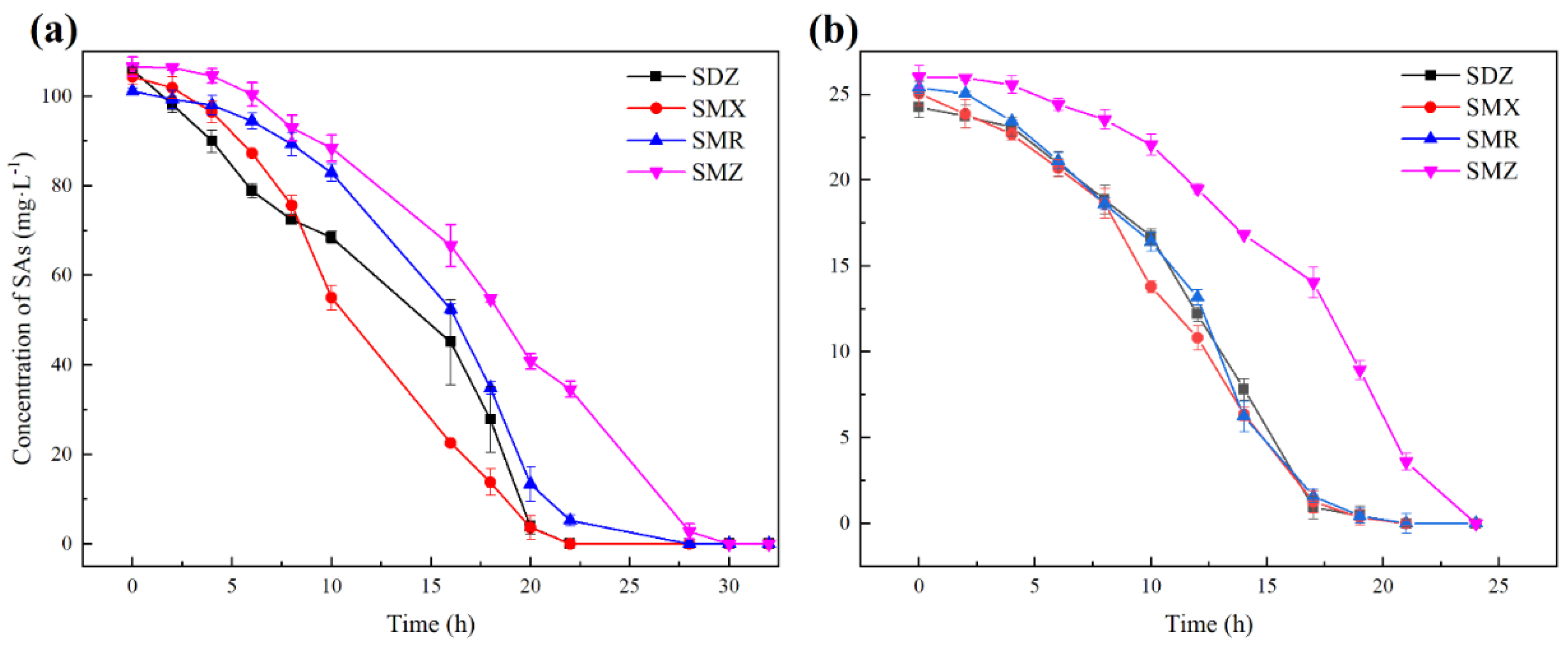
2.4. Determination of the Degradation Rate of SAs

The concentration of SAs was determined based on the method reported in a previous study. Each SA was measured separately [7]. The detection limit is 60 μg/L.
When the four substrates were in the same sample, the mobile phase used a gradient to separate and detect these substrates. A Waters e2695 Acquity Ultra Performance Liquid Chromatograph (UPLC) was used, equipped with a 2489 UV/Visible detector, and a C18 (150 × 4.6 mm, 5 µm) column. The wavelength was set at 272 nm, and the temperature was fixed at 35 °C. The injection volume was 10 µL, and the mobile phase flow rate was 0.3 mL·min−1. The mobile phase was eluted following a gradient scheme. The ratio of the mobile phase was set as follows (Table 1):

2.5. Kinetic Analysis of the Degradation of Sulfonamide Antibiotics

The effect of different initial concentrations of substrate (0.05~2.4 mmol·L−1) on strain YL1 growth was investigated by adding SAs as a sole source of carbon in MSM. The purpose of this experiment was to study the growth kinetics of strain YL1 during the degradation of SAs. The strain YL1 growth rate was limited by the substrate concentration at fixed conditions.
The specific growth rate, μ (1·h−1), was calculated as the slope of a plot of Ln ( X / X 0 ) versus time determined over the exponential growth phase where it is assumed to be linear [5,30]. Moreover, Haldane’s model was used to assess the kinetics of the SAs biodegradation due to the inhibition effect of SAs for the strain YL1. Equation (1) was used to calculate the specific growth rate [31].
μ   = μ m a x S K s + S + S 2 K i
where S is the SMX concentration (mmol/L), μ m a x is the maximum specific cell growth rate (1·h−1), K S is the half-saturation coefficient (mmol·L−1), and K i is the inhibition coefficient (mmol·L−1). The Origin 2016 computer software was used to determine the Haldane model kinetic parameters.
For self-inhibitory compounds such as SAs, the specific affinity could be used to describe specific affinity according to Equation (2), which linked nutrient accumulation rate with transporter density [32].
a A = μ m a x K S
where a A is the specific affinity (L·mmol−1·h−1).
The biomass yield coefficient, dry weight of biomass/weight of the substrate, for SAs can be determined by the following equation [33]:
Y = X X 0 S 0 S
where S 0 is the initial substrate concentration (mmol·L−1) and X 0 is the initial biomass concentration (mg·L−1). Y , is the biomass yield coefficient (mg-DW/mmol).

2.6. Density Functional Theory (DFT) Method

A density functional theory (DFT) method was used to simulate the geometry optimization of SAs. The energy calculations were performed in Gaussian 09 software using the method of B3LYP/6-31+G** combined with the IEFPCM solvent model (water). In addition, the electron density and the energy gap between HOMO and LUMO (ΔE = ELUMOEHOMO) are important stability indices that can help explain the effect of various substituents on the degradation performance of SAs [34,35]. In fact, high HOMO energy indicates high electrophilic reaction activities, while low LUMO energy implies a high nucleophilic reactivity [36]. In addition, a molecule with a narrow HOMO-LUMO gap is more reactive. Therefore, the energy values of different structured SAs were compared to illustrate the different structure reactivity.

3. Results and Discussion

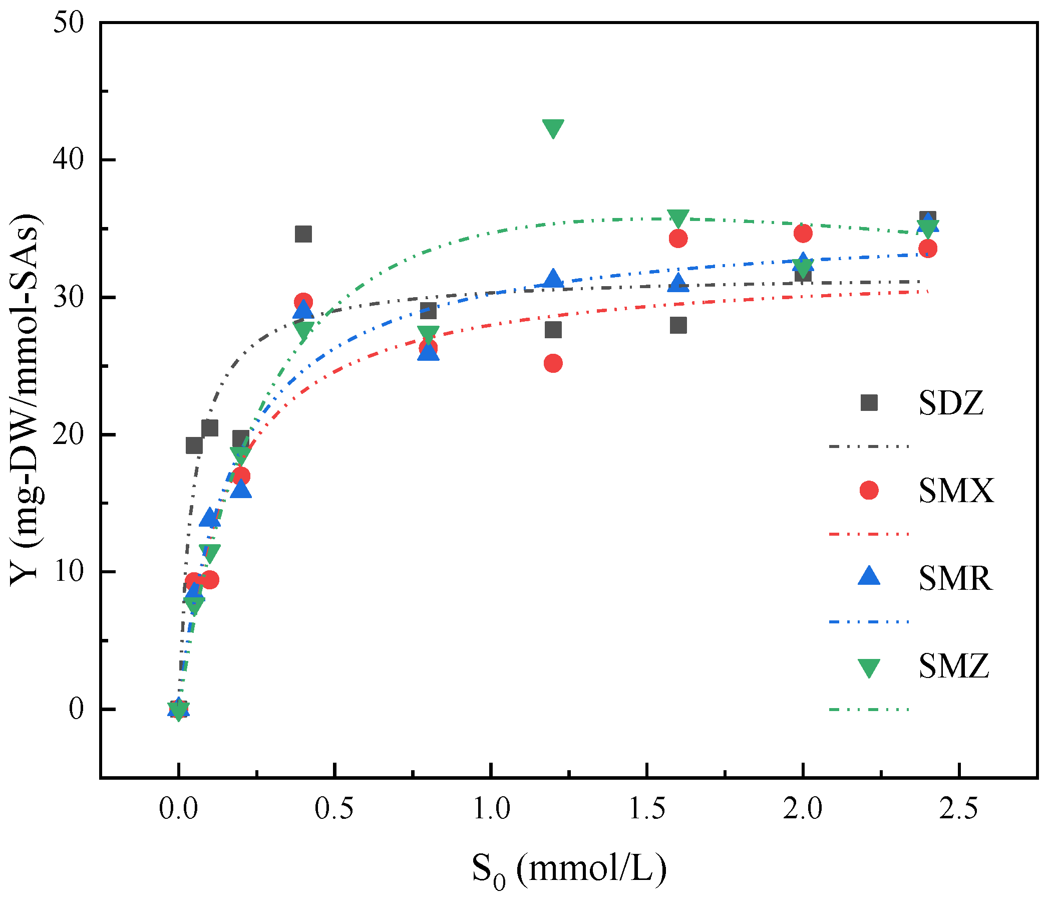
3.1. Substrate Spectrum of Strain YL1

Strain YL1 is capable of utilizing a variety of sulfonamide antibiotics for metabolic activities. SDZ, SMX, SMR, and SMZ were selected at the initial concentration of 100 mg·L−1 to investigate whether strain YL1 could effectively degrade these compounds. Figure 2a shows that strain YL1 degraded the SAs used in this study, which was reflected by rapid substrate elimination. SDZ, SMX, SMR, and SMZ at 100 mg·L−1 were completely degraded to below the detection limit within 22 h, 22 h, 28 h, and 30 h, respectively.
Also, the degradation process by strain YL1 of mixed sulfonamide antibiotics present in the same sample was investigated. SDZ, SMR, SMX, and SMZ were mixed at 25 mg·L−1. Figure 2b shows the variation of substrate concentration with time for the simultaneous degradation of four sulfonamide antibiotics by the strain YL1. In fact, there were some differences in the degradation efficiency of YL1 in degrading multiple sulfonamide antibiotics simultaneously. The removal efficiencies of these four substrates were slightly different due to the structures of the substituents. For example, the removal rate of SMZ by strain YL1 was significantly slower compared to the removal rates of the other substituents. An independent sample T-test was used to examine the variability of strain YL1 in degrading the four sulfonamide antibiotics. Among them, the concentration of SMZ over time was significantly different from SDZ, SMX, and SMR (p-values were 0.009, 0.005, and 0.014, respectively). The result indicated that the SMZ degradation rate by strain YL1 significantly lagged behind the degradation of the other types of sulfonamides.
These results implied that strain YL1 can effectively remove a variety of SAs from the environment. In fact, the ability of microorganisms to degrade more than one SAs simultaneously has been observed in a few other previous studies. According to Flemming et al., specific degraders could rapidly degrade sulfonamides, including sulfadiazine, sulfadimidine, and sulfadimethoxine. This study also indicates that when bacteria are adapted to carbutamide, sulfadoxine, sulfameter, and sulfanilamide, they can rapidly degrade sulfadiazine, sulfadiamidine, and sulfadimethoxypyrimidine [36]. Similarly, Qi et al. isolated a strain named P27, which significantly degraded SDZ, SMR, and SMZ. This degradation was evidenced by rapid substrate elimination, corresponding heterocyclic product formation, and high biomass growth [37]. This previous study reported that SMX had the fastest degradation rate by P27, while the degradation process of sulfadimethoxine required the longest time period. Similar to the present study, the degradation rates of SDZ and SMR were slightly lower than those of SMX [38]. Achromobacter denitrificans PR1, isolated from activated sludge with SMX as the sole carbon and nitrogen source, was able to degrade other sulfonamides [39]. This finding suggested that if a bacterial strain can degrade one type of sulfonamides, it can also be able to reduce the concentration of a wide range of other sulfonamide compounds.

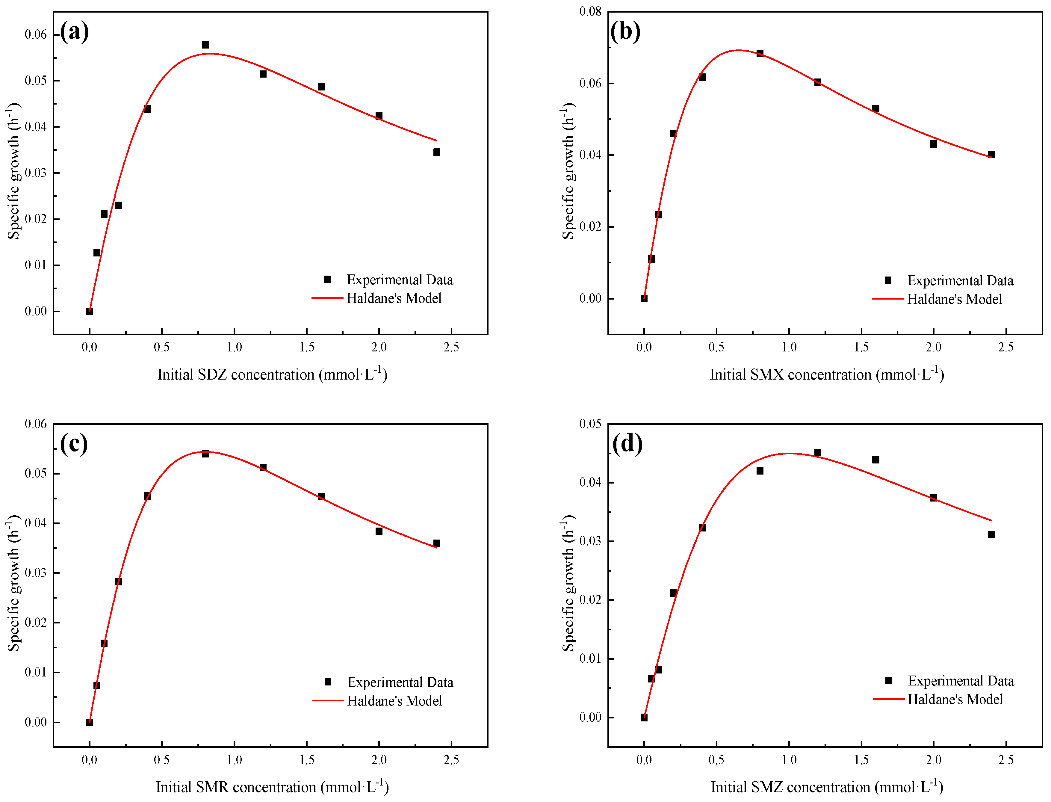
3.2. Growth Kinetics of Strain YL1

There were differences in the use of different SAs compounds by strain YL1 to maintain growth. In fact, Figure 3 shows that high SAs concentrations could inhibit the growth of strain YL1. Therefore, the Haldane growth model was selected to estimate the kinetic parameters. Non-linear regression analysis of bacterial growth data was performed using Origin software (version 2016). Experimental and predicted specific growth rates of strain YL1 using various SAs are shown in Figure 3. The kinetic parameters for strain YL1 growth on SAs were calculated from Equation (3). The kinetic parameter values used in the model are shown in Table 2. For the degradation of SAs by strain YL1, the specific growth rate was positively correlated with the substrate concentration at low concentrations. However, when the concentration exceeded a threshold value, the specific growth rate decreased as the SAs concentrations increased. This implies that above a certain concentration, SAs acts as an inhibitor of bacterial activity. The Haldane model showed a good fit with a high R2 value.
The maximum specific growth rates (μmax) of strain YL1 growing on SDZ, SMX, SMR, and SMZ were 0.304, 0.311, 0.302, and 0.285, respectively. The substrate affinity reflects the ability of cells to receive nutrients, and it is one of the crucial factors affecting the growth rate. Moreover, the Ks value is usually used to represent the substrate affinity under substrate-restricting conditions. However, Ks was not a reliable indicator of substrate affinity [40]. D.K. Button suggested that the specific affinity (aA), as a more reliable indicator, could describe the specific affinity of a restricted substrate due to its linking of as a more reliable indicator because it connects the nutrient accumulation rate to the density of transporter proteins [41]. For this study, the range of aA value was SMX > SDZ > SMR > SMZ, indicating that the fastest growth rate could be obtained when strain YL1 is cultured with SMX as the sole carbon source. In addition, Ki is the inhibition coefficient, which indicates how tolerant bacteria are to toxic substrates toxicity [42]. A high Ki value indicates that the cell is more resistant to substrate toxicity.
The substrate inhibition constant (Ki) for SMZ was higher than the Ki value for other SAs (Table 2), which indicated that SMZ caused less substrate inhibition than SDZ, SMR, and SMX. Therefore, the maximum SMZ biodegradation rate was clearly lower compared to the other SA compounds.
The yield coefficients for different initial SAs concentrations were shown in Figure 4. The yield coefficients of SDZ were significantly higher than the other SAs at low concentrations. As well, when strain YL1 uses SMZ as the single substrate, the biomass biomass yield coefficient is greater than others at high concentrations (>0.4 mmol/L).

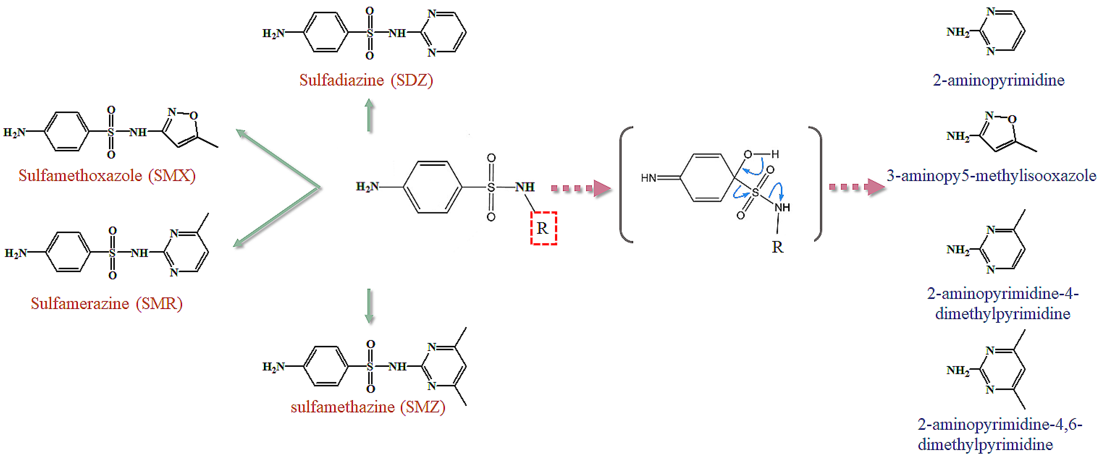
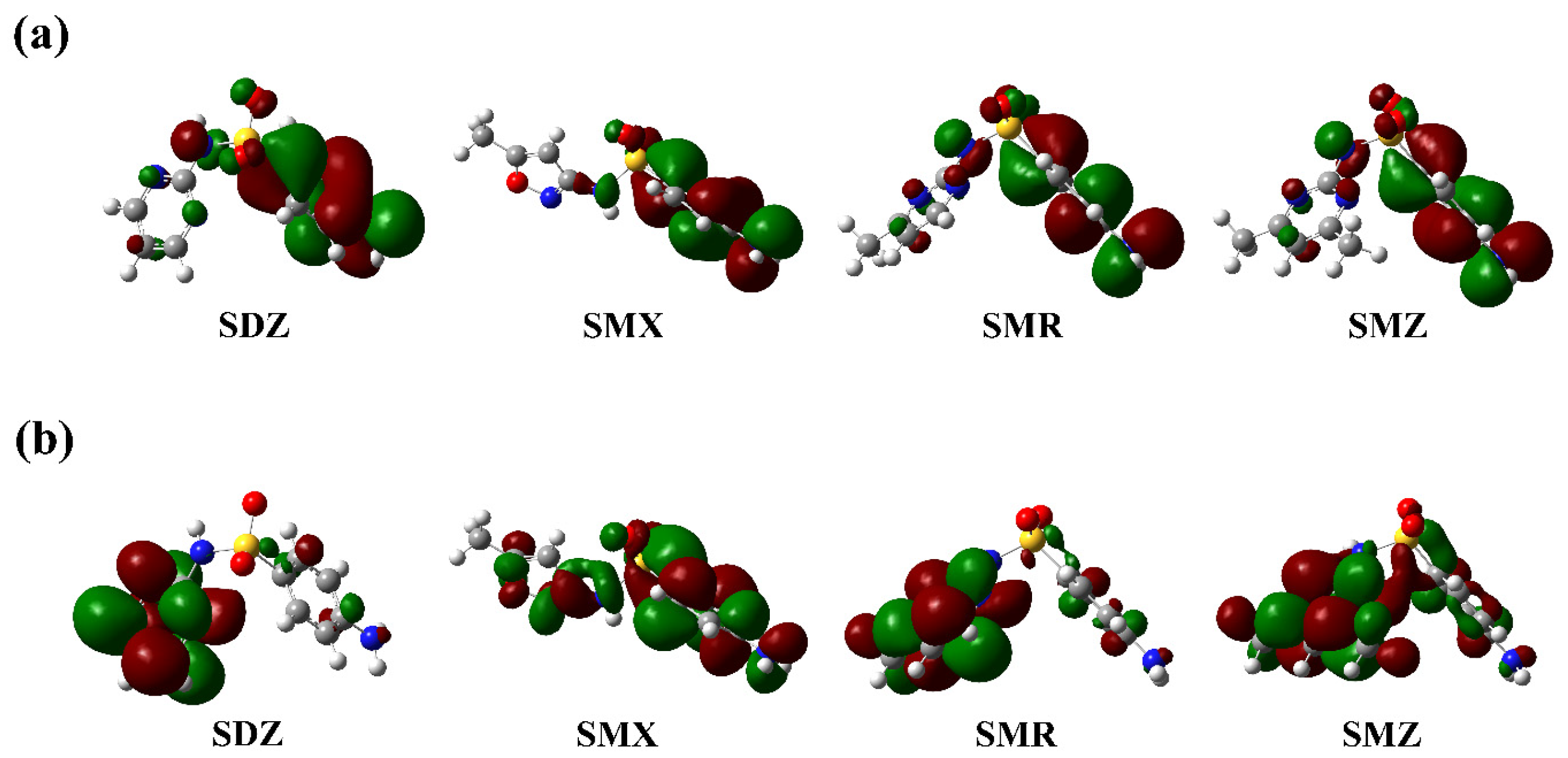
3.3. Effect of the Substitution Groups on the Reactivity of SAs

The explanation for the varied degradation performance of SAs by strain YL1 caused by various substituents was discovered using theoretical simulations [43,44]. The performance of strain YL1 in the degradation of SAs was dependent on the stability of the substrates, and it can be shown with the electron density in HOMO and LUMO and the energy gap between HOMO and LUMO energy ( Δ E = E HOMO ). Figure 5 shows the HOMO and LUMO distributions of SAs with different substituents. The HOMO distributions of the SAs used in this experiment were similar. The HOMO of SDZ, SMX, SMR, and SMZ molecules mainly originated from the aminophenyl structure. Electrons on the LUMO of SDZ, SMR, and SMZ are mainly concentrated in the pyrimidine ring. However, the LUMO of SMX is more uniformly distributed in the aromatic ring and substituent parts. Moreover, the layout of the orbital electron distribution of the four compounds differed due to the substituents, especially the electron-donating substituents, such as the methyloxazole ring. This fact had a significant effect on the orbital electron layout of the molecule and affected the growth of strain YL1.
According to the front molecular orbital (FMO) theory, the energy gap ( Δ E ) between HOMO and LUMO is an essential indicator of the stability of the molecules [45]. A small Δ E value indicates that the molecule is more reactive and less stable. Table 3 lists the Δ E energy values of the four SAs. The smallest Δ E is SDZ, indicating that SDZ is the most unstable and rapidly-degraded substance. In addition to SDZ, the ranking order of Δ E is SMX > SMZ > SMR. Similarly. Yin et al. found that among the selected sulfonamide antibiotics (SMX, SMR, and SMZ), the SMX that had the largest Δ E was SMX [34]. The same degradation rate was also followed in the reduced graphene oxide (rGO) system. Interestingly, although DFT calculations could further demonstrate the effect of substituents on the reactivity of SAs, the experimental results showed that the growth rates of microorganisms when the four SAs were used as substrates were SMX > SDZ > SMR > SMZ. When strain YL1 was grown with SMX as the sole carbon source, the specific growth rate of strain YL1 was significantly higher than that of SMZ, which was similar to the result reported by Qi et al. However, it was different from the results calculated by DFT [40]. Thus, this event needs to be further discussed and analyzed.
In addition, the toxicity of the substances and degradation end-products was one of the reasons for the differences in growth and degradation rates, which can affect the growth of microorganisms during degradation. According to Zou et al., the effects of SAs on microorganisms can be both attributed to acute (15 min exposure) and chronic (24 h exposure) toxicity to P. phosphoreum. Therefore, the acute toxicity of antibiotic mixtures to P. phosphoreum was tested, and SMX was found to be more toxic than SDZ and SMZ. However, the order of chronic toxicity to P. phosphoreum was SDZ ≈ SMZ > SMX [46]. In addition, the substrate toxicity of the degradation products can also have an impact on the growth of the resulting strain YL1. 2-Aminopyrimidine, 3-amino-5-methylisoxazole, 2-aminopyrimidine-4-dimethylpyrimidine, and 2-aminopyrimidine-4,6-dimethylpyrimidine were the end products accumulated during the degradation of SDZ, SMX, SMR, and SMZ by strain YL1, respectively (Figure 1). Notably, the degradation end products of SDZ and SMX were non-toxic [46,47]. However, the degradation end-products of SMR (2-aminopyrimidine-4-dimethylpyrimidine) and SMZ (2-aminopyrimidine-4,6-dimethylpyrimidine) were considered to be as harmful to aquatic organisms [48]. In general, compounds with electron-donating substituents are more easily oxidized than those with electron-withdrawing substituents [49]. The S atom on the SMX substituent can provide more electron density for the sulfonamide bond, which leads to SMX being more active than SDZ, SMR, and SMZ. The findings of the present study were consistent with the experimental and theoretical values of the growth kinetics of strain YL1. In summary, strain YL1 has the largest specific growth rate when cultured with SMX as the sole substrate, followed by SDZ > SMR > SMZ.

4. Conclusions

The degradation rate of SAs with different substituents by strain YL1 was investigated. The capacity of strain YL1 to break down the selected SAs, namely SDZ, SMX, SMR, and SMZ, indicated that the different substituents had no impact on this breakdown ability of strain YL1. However, according to the kinetic study of strain YL1 at different initial concentrations, there were differences in the specific growth rates of the SAs used by the strains with various substituents. Moreover, the order of the four selected substrate utilization rates was SMX > SDZ > SMR > SMZ. Furthermore, the DFT calculations confirmed that the different substituents changed the HOMO and LUMO distribution of the SAs, resulting in the differential reactivity of the molecules and degradation rates of the molecules. Also, the ecotoxicity of the substrate to the microorganisms was one of the important indicators affecting the biodegradation of SAs. Finally, this study provided a theoretical basis and contributed to the knowledge of the microbial treatment of SAs.

Author Contributions

Conceptualization, L.Y. and Y.W.; methodology, software, validation, L.Y., Y.W. and H.G.; investigation, resources, data curation, L.Y., J.X. and F.M.; writing—original draft preparation, L.Y. and Y.W.; writing—review and editing, L.Y., Y.W., F.M., H.G. and J.X.; visualization, L.Y.; supervision, F.M., Y.W. and H.G.; project administration, F.M.; funding acquisition, F.M. All authors have read and agreed to the published version of the manuscript.

Funding

This research was supported by State Key Laboratory of Urban Water Resource and Environment (Harbin Institute of Technology) (No. 2020DX09).

Institutional Review Board Statement

Not applicable.

Informed Consent Statement

Not applicable.

Acknowledgments

We would like to thank our anonymous reviewers for excellent comments to improve our work.

Conflicts of Interest

The authors declare no conflict of interest.

References

  1. Wang, S.; Yuan, R.; Chen, H.; Wang, F.; Zhou, B. Anaerobic biodegradation of four sulfanilamide antibiotics: Kinetics, pathways and microbiological studies. J. Hazard. Mater. 2021, 416, 125840. [Google Scholar] [CrossRef] [PubMed]
  2. K’Oreje, K.O.; Kandie, F.J.; Vergeynst, L.; Abira, M.A.; Van Langenhove, H.; Okoth, M.; Demeestere, K. Occurrence, fate and removal of pharmaceuticals, personal care products and pesticides in wastewater stabilization ponds and receiving rivers in the nzoia basin, kenya. Sci. Total Environ. 2018, 637–638, 336–348. [Google Scholar] [CrossRef] [PubMed]
  3. Ondarza, P.M.; Haddad, S.P.; Avigliano, E.; Miglioranza, K.S.B.; Brooks, B.W. Pharmaceuticals, illicit drugs and their metabolites in fish from argentina: Implications for protected areas influenced by urbanization. Sci. Total Environ. 2019, 649, 1029–1037. [Google Scholar] [CrossRef] [PubMed]
  4. Wang, B.; Ni, B.-J.; Yuan, Z.; Guo, J. Unravelling kinetic and microbial responses of enriched nitrifying sludge under long-term exposure of cephalexin and sulfadiazine. Water Res. 2020, 173, 115592. [Google Scholar] [CrossRef] [PubMed]
  5. Kiki, C.; Rashid, A.; Wang, Y.; Li, Y.; Zeng, Q.; Yu, C.P.; Sun, Q. Dissipation of antibiotics by microalgae: Kinetics, identification of transformation products and pathways. J. Hazard. Mater. 2020, 387, 121985. [Google Scholar] [CrossRef] [PubMed]
  6. Sella, C.F.; Carneiro, R.B.; Sabatini, C.A.; Sakamoto, I.K.; Zaiat, M. Can different inoculum sources influence the biodegradation of sulfamethoxazole antibiotic during anaerobic digestion? Braz. J. Chem. Eng. 2021, 39, 35–46. [Google Scholar] [CrossRef]
  7. Yu, L.; Wang, Y.; Su, X.; Fu, Y.; Ma, F.; Guo, H. Biodiversity, isolation and genome analysis of sulfamethazine-degrading bacteria using high-throughput analysis. Bioprocess Biosyst. Eng. 2020, 2020, 2–3. [Google Scholar] [CrossRef]
  8. Yin, Z. Distribution and ecological risk assessment of typical antibiotics in the surface waters of seven major rivers, China. Environ. Sci. Process Impacts 2021, 23, 1088–1100. [Google Scholar] [CrossRef]
  9. Rizvi, S.G.; Ahammad, S.Z. COVID-19 and antimicrobial resistance: A cross-study. Sci Total Environ. 2022, 807, 150873. [Google Scholar] [CrossRef]
  10. Huang, F.; An, Z.; Moran, M.J.; Liu, F. Recognition of typical antibiotic residues in environmental media related to groundwater in china (2009–2019). J. Hazard. Mater. 2020, 399, 122813. [Google Scholar] [CrossRef]
  11. Zhang, X.; Zhang, Z.; Liu, Y.; Hao Ngo, H.; Guo, W.; Wang, H.; Zhang, Y.; Zhang, D. Impacts of sulfadiazine on the performance and membrane fouling of a hybrid moving bed biofilm reactor-membrane bioreactor system at different c/n ratios. Bioresour. Technol. 2020, 318, 124180. [Google Scholar] [CrossRef] [PubMed]
  12. Song, H.L.; Lu, Y.X.; Yang, X.L.; Xu, H.; Singh, R.P.; Du, K.X.; Yang, Y.L. Degradation of sulfamethoxazole in low-C/N ratio wastewater by a novel membrane bioelectrochemical reactor. Bioresour. Technol. 2020, 305, 123029. [Google Scholar] [CrossRef] [PubMed]
  13. Zheng, J.; Wang, S.; Zhou, A.; Zhao, B.; Dong, J.; Zhao, X.; Li, P.; Yue, X. Efficient elimination of sulfadiazine in an anaerobic denitrifying circumstance: Biodegradation characteristics, biotoxicity removal and microbial community analysis. Chemosphere 2020, 252, 126472. [Google Scholar] [CrossRef] [PubMed]
  14. Yu, L.; Wang, Y.; Shan, X.; Ma, F.; Guo, H. Harnessing paenarthrobacter ureafaciens yl1 and pseudomonas koreensis yl2 interactions to improve degradation of sulfamethoxazole. Microorganisms 2022, 10, 648. [Google Scholar] [CrossRef] [PubMed]
  15. Zeng, L.; Li, W.; Wang, X.; Zhang, Y.; Tai, Y.; Zhang, X.; Dai, Y.; Tao, R.; Yang, Y. Bibliometric analysis of microbial sulfonamide degradation: Development, hotspots and trend directions. Chemosphere 2022, 293, 133598. [Google Scholar] [CrossRef] [PubMed]
  16. Liang, D.H.; Hu, Y.; Liang, D.; Chenga, J.; Chena, Y. Bioaugmentation of moving bed biofilm reactor (mbbr) with achromobacter jl9 for enhanced sulfamethoxazole (smx) degradation in aquaculture wastewater. Ecotoxicol. Environ. Saf. 2021, 207, 111258. [Google Scholar] [CrossRef]
  17. Wang, S.; Wang, J. Biodegradation and metabolic pathway of sulfamethoxazole by a novel strain Acinetobacter sp. Appl. Microbiol. Biotechnol. 2018, 102, 425–432. [Google Scholar] [CrossRef]
  18. Hu, S.; Hu, H.; Li, W.; Hong, X.; Cai, D.; Lin, J.; Li, M.; Zhao, Y. Investigating the biodegradation of sulfadiazine in soil using enterobacter cloacae t2 immobilized on bagasse. RSC Adv. 2020, 10, 1142–1151. [Google Scholar] [CrossRef] [Green Version]
  19. Cao, L.; Zhang, J.; Zhao, R.; Deng, Y.; Liu, J.; Fu, W.; Lei, Y.; Zhang, T.; Li, X.; Li, B. Genomic characterization, kinetics, and pathways of sulfamethazine biodegradation by Paenarthrobacter sp. A01. Environ. Int. 2019, 131, 104961. [Google Scholar] [CrossRef]
  20. Bouju, H.; Ricken, B.; Beffa, T.; Corvini, P.F.; Kolvenbach, B.A. Isolation of bacterial strains capable of sulfamethoxazole mineralization from an acclimated membrane bioreactor. Appl. Environ. Microbiol. 2012, 78, 277–279. [Google Scholar] [CrossRef]
  21. Jiang, B.; Li, A.; Cui, D.; Cai, R.; Ma, F.; Wang, Y. Biodegradation and metabolic pathway of sulfamethoxazole by pseudomonas psychrophila ha-4, a newly isolated cold-adapted sulfamethoxazole-degrading bacterium. Appl. Microbiol. Biotechnol. 2014, 98, 4671–4681. [Google Scholar] [CrossRef] [PubMed]
  22. Liang, D.H.; Hu, Y. Application of a heavy metal-resistant Achromobacter sp. For the simultaneous immobilization of cadmium and degradation of sulfamethoxazole from wastewater. J. Hazard. Mater. 2021, 402, 124032. [Google Scholar] [CrossRef] [PubMed]
  23. Deng, Y.; Mao, Y.; Li, B.; Yang, C.; Zhang, T. Aerobic degradation of sulfadiazine by arthrobacter spp.: Kinetics, pathways and genomic characterization. Environ. Sci. Technol. 2016, 50, 9566–9575. [Google Scholar] [CrossRef] [PubMed]
  24. Hegedus, B.; Kos, P.B.; Balint, B.; Maroti, G.; Gan, H.M.; Perei, K.; Rakhely, G. Complete genome sequence of novosphingobium resinovorum sa1, a versatile xenobiotic-degrading bacterium capable of utilizing sulfanilic acid. J. Biotechnol. 2017, 241, 76–80. [Google Scholar] [CrossRef] [PubMed]
  25. Hu, J.; Li, X.; Liu, F.; Fu, W.; Lin, L.; Li, B. Comparison of chemical and biological degradation of sulfonamides: Solving the mystery of sulfonamide transformation. J. Hazard. Mater. 2022, 424, 127661. [Google Scholar] [CrossRef]
  26. Ricken, B.; Kolvenbach, B.A.; Bergesch, C.; Benndorf, D.; Kroll, K.; Strnad, H.; Vlcek, C.; Adaixo, R.; Hammes, F.; Shahgaldian, P.; et al. Fmnh2-dependent monooxygenases initiate catabolism of sulfonamides in Microbacterium sp. Strain br1 subsisting on sulfonamide antibiotics. Sci. Rep. 2017, 7, 15783. [Google Scholar] [CrossRef] [Green Version]
  27. Sam, S.P.; Tan, H.T.; Sudesh, K.; Adnan, R.; Ting, A.S.Y.; Ng, S.L. Phenol and p-nitrophenol biodegradations by acclimated activated sludge: Influence of operational conditions on biodegradation kinetics and responding microbial communities. J. Environ. Chem. Eng. 2021, 9, 105420. [Google Scholar] [CrossRef]
  28. Ma, F.; Sun, Y.; Li, A.; Zhang, X.; Yang, J. Activation of accumulated nitrite reduction by immobilized pseudomonas stutzeri t13 during aerobic denitrification. Bioresour. Technol. 2015, 187, 30–36. [Google Scholar] [CrossRef]
  29. Min, J.; Wang, J.; Chen, W.; Hu, X. Biodegradation of 2-chloro-4-nitrophenol via a hydroxyquinol pathway by a gram-negative bacterium, Cupriavidus sp. Strain cnp-8. AMB Express 2018, 8, 43. [Google Scholar] [CrossRef] [Green Version]
  30. Ghosh, I.; Jasmine, J.; Mukherji, S. Biodegradation of pyrene by a pseudomonas aeruginosa strain rs1 isolated from refinery sludge. Bioresour. Technol. 2014, 166, 548–558. [Google Scholar] [CrossRef]
  31. Min, J.; Xu, L.; Fang, S.; Chen, W.; Hu, X. Microbial degradation kinetics and molecular mechanism of 2,6-dichloro-4-nitrophenol by a cupriavidus strain. Environ. Pollut. 2020, 258, 113703. [Google Scholar] [CrossRef] [PubMed]
  32. Button, D.K. Nutrient-limited microbial growth kinetics: Overview and recent advances. Antonie Van Leeuwenhoek 1993, 63, 225–235. [Google Scholar] [CrossRef] [PubMed]
  33. Bakhshi, Z.; Najafpour, G.; Kariminezhad, E.; Pishgar, R.; Mousavi, N.; Taghizade, T. Growth kinetic models for phenol biodegradation in a batch culture of pseudomonas putida. Environ. Technol. 2011, 33, 1835–1841. [Google Scholar] [CrossRef] [PubMed]
  34. Chen, Q.; Zheng, J.; Xu, J.; Dang, Z.; Zhang, L. Insights into sulfamethazine adsorption interfacial interaction mechanism on mesoporous cellulose biochar: Coupling dft/fot simulations with experiments. Chem. Eng. J. 2019, 356, 341–349. [Google Scholar] [CrossRef]
  35. Wang, H.; Guo, W.; Yin, R.; Du, J.; Wu, Q.; Luo, H.; Liu, B.; Sseguya, F.; Ren, N. Biochar-induced fe(iii) reduction for persulfate activation in sulfamethoxazole degradation: Insight into the electron transfer, radical oxidation and degradation pathways. Chem. Eng. J. 2019, 362, 561–569. [Google Scholar] [CrossRef]
  36. Luo, S.; Wei, Z.; Spinney, R.; Zhang, Z.; Dionysiou, D.D.; Gao, L.; Chai, L.; Wang, D.; Xiao, R. Uv direct photolysis of sulfamethoxazole and ibuprofen: An experimental and modelling study. J. Hazard. Mater. 2018, 343, 132–139. [Google Scholar] [CrossRef]
  37. Ingerslev, F.; Halling-Sorensen, B. Biodegradability properties of sulfonamides in activated sludge. Environ. Toxicol. Chem. 2000, 19, 2467–2473. [Google Scholar] [CrossRef]
  38. Qi, M.; Ma, X.; Liang, B.; Zhang, L.; Kong, D.; Li, Z.; Wang, A. Complete genome sequences of the antibiotic sulfamethoxazole-mineralizing bacteria Paenarthrobacter sp. P27 and Norcardiodes sp. N27. Environ. Res. 2022, 204, 112013. [Google Scholar] [CrossRef]
  39. Reis, P.J.M.; Reis, A.C.; Ricken, B.; Kolvenbach, B.A.; Manaia, C.M.; Corvini, P.F.X.; Nunes, O.C. Biodegradation of sulfamethoxazole and other sulfonamides by Achromobacter denitrificans pr1. J. Hazard. Mater. 2014, 280, 741–749. [Google Scholar] [CrossRef] [Green Version]
  40. Nedwell, D.B. Effect of low temperature on microbial growth: Lowered affinity for substrates limits growth at low temperature. FEMS Microbiol. Ecol. 1999, 30, 101–111. [Google Scholar] [CrossRef]
  41. Cui, T.; Wang, P.; Li, J.; Su, Y.; Liu, N.; Hong, M. Effects of temperature, ph, and salinity on the growth kinetics of Pseudomonas sp. Nb-1, a newly isolated cold-tolerant, alkali-resistant, and high-efficiency nitrobenzene-degrading bacterium. Environ. Technol. 2022, 1–13. [Google Scholar] [CrossRef] [PubMed]
  42. Pramparo, L.; Suarez-Ojeda, M.E.; Perez, J.; Carrera, J. Kinetics of aerobic biodegradation of dihydroxybenzenes by a p-nitrophenol-degrading activated sludge. Bioresour. Technol. 2012, 110, 57–62. [Google Scholar] [CrossRef]
  43. Yin, R.; Guo, W.; Ren, N.; Zeng, L.; Zhu, M. New insight into the substituents affecting the peroxydisulfate nonradical oxidation of sulfonamides in water. Water Res. 2020, 171, 115374. [Google Scholar] [CrossRef] [PubMed]
  44. Gurkan, Y.Y.; Turkten, N.; Hatipoglu, A.; Cinar, Z. Photocatalytic degradation of cefazolin over n-doped tio2 under uv and sunlight irradiation: Prediction of the reaction paths via conceptual dft. Chem. Eng. J. 2012, 184, 113–124. [Google Scholar] [CrossRef]
  45. Feng, L.; Liu, J.; Abu-Hamdeh, N.H.; Bezzina, S.; Eshaghi Malekshah, R. Molecular dynamics and quantum simulation of different cationic dyes removal from contaminated water using uio-66 (zr)-(cooh)2 metal–organic framework. J. Mol. Liq. 2022, 349, 118085. [Google Scholar] [CrossRef]
  46. Zhu, F.; Pan, J.; Zou, Q.; Wu, M.; Wang, H.; Xu, G. Electron beam irradiation of typical sulfonamide antibiotics in the aquatic environment: Kinetics, removal mechanisms, degradation products and toxicity assessment. Chemosphere 2021, 274, 129713. [Google Scholar] [CrossRef]
  47. Nguyen, P.Y.; Carvalho, G.; Reis, A.C.; Nunes, O.C.; Reis, M.A.M.; Oehmen, A. Impact of biogenic substrates on sulfamethoxazole biodegradation kinetics by achromobacter denitrificans strain pr1. Biodegradation 2017, 28, 205–217. [Google Scholar] [CrossRef]
  48. Shao, S.; Wu, J.; Meng, F.; Liu, J. Natural attenuation of sulfometuron-methyl in seawater: Kinetics, intermediates, toxicity change and ecological risk assessment. J. Environ. Manag. 2022, 313, 114980. [Google Scholar] [CrossRef]
  49. Hsu, M.-H.; Kuo, T.-H.; Chen, Y.-E.; Huang, C.-H.; Hsu, C.-C.; Lin, A.Y.-C. Substructure reactivity affecting the manganese dioxide oxidation of cephalosporins. Environ. Sci. Technol. 2018, 52, 9188–9195. [Google Scholar] [CrossRef]
Figure 1. Chemical structures of the sulfonamides used in this study (red) and the end products accumulated after degradation by strain YL1 (blue). ([·] denotes that the compound was not detected).
Fermentation 08 00742 g001
Figure 2. Changes in SAs concentration over time. (a) strain YL1 degraded 100 mg·L−1 SDZ, SMX, SMR, and SMZ, respectively; (b) Strain YL1 degrades a mixture of SDZ, SMX, SMR, and SMZ at an initial concentration of 25 mg·L−1.
Fermentation 08 00742 g002
Figure 3. Experimental and predicted specific growth rates of strain YL1 against SDZ (a), SMX (b), SMR (c), and SMZ (d) according to the Haldane model.
Fermentation 08 00742 g003
Figure 4. Biomass Yield coefficient for bacterial growth on initial SAs concentrations.
Fermentation 08 00742 g004
Figure 5. The HOMO (a) and LUMO (b) electron density contour of SDZ, SMX, SMR, and SMZ. The substituted heterocyclic rings and aniline are on the left and right sides of the SA molecules, respectively.
Fermentation 08 00742 g005
Table 1. Gradient elution procedure.
t·min−1Methanol (%)0.1% Formic Acid (%)
01090
105050
111090
121090
Table 2. Kinetic parameters for strain YL1 growth on SAs used in the Haldane model.
Haldane Modelμmax (1·h−1)KS (mmol·L−1)Ki (mmol·L−1)R2 a A   ( L · mmol 1 · h 1 )
SDZ μ = 0 . 304 × S 1 . 858 + S + S 2 0 . 372 0.304 ± 0.2871.858 ± 2.0420.372 ± 0.4190.9680.164
SMX μ = 0 . 311 × S 1 . 139 + S + S 2 0.373 0.311 ± 0.0831.139 ± 0.3670.373 ± 0.1210.9950.273
SMR μ = 0 . 302 × S 1 . 817 + S + S 2 0.351 0.302 ± 0.0551.817 ± 0.3830.351 ± 0.0760.9990.166
SMZ μ = 0 . 285 × S 2 . 684 + S + S 2 0 . 376 0.285 ± 0.2262.684 ± 2.410.376 ± 0.3510.9890.106
Table 3. HOMO and LUMO distributions of SAs calculated at the B3LYP/6-31+G** level of theory.
E HOMO   ( eV ) E LUMO   ( eV ) E   ( LUMO - HOMO ,   eV )
SDZ−6.246−1.6614.585
SMX−6.337−1.2885.049
SMR−6.238−1.5514.687
SMZ−6.235−1.444.795
Publisher’s Note: MDPI stays neutral with regard to jurisdictional claims in published maps and institutional affiliations.

