Article

Uncovering the Effects of Ammonium Sulfate on Neomycin B Biosynthesis in Streptomyces fradiae SF-2

1
Engineering Laboratory for Industrial Microbiology Molecular Beeding of Anhui Province, College of Biologic & Food Engineering, Anhui Polytechnic University, Wuhu 241000, China
2
Key Laboratory of Industrial Biotechnology, Ministry of Education, School of Biotechnology, Jiangnan University, Wuxi 214122, China
*
Author to whom correspondence should be addressed.
These authors contributed equally to this work.
Fermentation 2022, 8(12), 678; https://doi.org/10.3390/fermentation8120678
Submission received: 11 October 2022 / Revised: 14 November 2022 / Accepted: 23 November 2022 / Published: 26 November 2022
(This article belongs to the Special Issue Pharmaceutical Fermentation: Antibiotic Production and Processing)

Abstract

:
The aminoglycoside antibiotic neomycin has broad antibacterial properties and is widely used in medicine and agriculture. With the discovery of neomycin’s potential applications in treating tumors and SARS-CoV-2, it is necessary to accelerate the biosynthesis of neomycin. In the present study, we investigated the effects of various inorganic salts on neomycin B (the main active neomycin) biosynthesis in Streptomyces fradiae SF-2. We found that 60 mM (NH4)2SO4 could promote neomycin B biosynthesis and cell growth most effectively. Further comparative transcriptomic analyses revealed that 60 mM (NH4)2SO4 inhibited the EMP and TCA cycles and enhanced the expression of neo genes involved in the neomycin B biosynthesis pathway. Finally, a neomycin B potency of 17,399 U/mL in shaking flasks was achieved by overexpressing neoE and adding 60 mM (NH4)2SO4, corresponding to a 51.2% increase compared with the control S. fradiae SF-2. In the present study, the mechanism by which (NH4)2SO4 affects neomycin biosynthesis was revealed through transcriptomics, providing a reference for the further metabolic engineering of S. fradiae SF-2 for neomycin B production.

1. Introduction

Streptomyces fradiae is a species of Actinomycetota Phylum, a genus of filamentous bacteria of the family Streptomycetaceae. The strain is a Gram-positive bacterium with a genomic G+C content of up to 70–74% [1,2,3]. The precursor compounds and energy generated by primary metabolism can be used to synthesize a variety of secondary metabolites with complex structures, diverse functions, and various biological activities [4]. Neomycin, a secondary metabolite first isolated from S. fradiae (GenBank: GCA_008704425.1) in the 1940s by Waksman, is a classical aminoglycoside antibiotic made from carbohydrates through the pentose phosphate pathway [5], which is bactericidal against both Gram-positive and Gram-negative bacteria, including Staphylococcus aureus, Escherichia coli, Haemophilus influenzae, Proteus spp., and Serratia spp. The antibiotic includes three classes, A, B, and C, each with its typical chemical structure and functions, including biological activities. Neomycin B, the main active neomycin, exhibits higher antimicrobial activity than other classical antibiotics (streptomycin, bacitracin, etc.). Neomycin B can bind to the 16S rRNA site of the 30S ribosome and interfere with the synthesis of bacterial proteins [5,6,7]. In recent years, with the development of medical diagnostic and clinical intervention technology, the potential function of neomycin B has become apparent in the medical field. To prevent respiratory and intestinal bacterial infections in livestock, neomycin can be added to the feed [8]. Additionally, neomycin can promote tumor cell apoptosis by activating the inhibitory factor p53 in tumor cells [9,10]. Furthermore, a recent study has shown that neomycin may inhibit SARS-CoV-2 as the protease inhibitor [11].
The biosynthesis of neomycin by S. fradiae is strongly affected by the composition of the medium. Recent studies have reported optimization of the fermentation medium to promote Streptomyces growth and neomycin biosynthesis [12,13]. In addition, some studies have shown that ammonium ions also affect growth and metabolism [14]. It was found that the nitrogen source affects growth and nitrate depletion causes biphasic growth patterns in batch cultures of strain OB3b [15]. To balance carbon and nitrogen metabolism, bacteria have evolved complex mechanisms to sense the nutrient supply and adapt their metabolism accordingly [16]. It has been demonstrated that nitrogen compounds, such as sodium nitrate, aspartic acid, and glutamic acid, could promote the growth of S. fradiae 3535 and neomycin production [17]. The results of these studies indicated that nitrogen metabolism and its metabolic intermediates might affect the central metabolism of methanotrophs, thereby controlling their growth. The biosynthesis of neomycin B in S. fradiae is mainly regulated by the neo gene clusters, which contain two operons, one comprised of 12 genes from neoE to neoD (responsible for neomycin B biosynthesis) and the other containing neoGHaphA and other regulatory genes [1]. At present, metabolic engineering and other technical methods are being studied to improve neomycin production by enhancing its biosynthesis. For example, the overexpression of two regulatory genes of neomycin B biosynthesis (afsA-g and neoR) increased the neomycin B titer to 722.9 ± 20.1 mg/L and 564.7 ± 32.5 mg/L, respectively [18]. Moreover, an important conserved enzyme in 2-DOS synthesis is NeoE, a zinc-containing dehydrogenase that utilizes NAD(P)+ as a coenzyme [19]. This enzyme belongs to the medium-chain dehydrogenase (MDR) family. MDR proteins contain a highly conserved structure of GxGxxG that could bind NAD(P)+. Studies have shown that NeoE could catalyze the dehydrogenation of 2-deoxyscyllo inosamine (2-DOIA) to 3-amino-2,3-deoxyscyllo inosone (amino-DOI), and a neoE deletion mutant strain lost the ability to synthesize neomycin B [18]. Therefore, NeoE may be a key enzyme in the biosynthesis of neomycin B.
Comparative transcriptomics can be used to mine genes that are obviously positively regulated or negatively regulated by comparing and analyzing transcripts of samples in different states at different times [20,21]. Therefore, comparative transcriptomic sequencing is widely used to explore the mechanism of action on cellular metabolism under specific conditions [22]. For example, comparative transcriptomic analysis revealed that the transcription factor RosR could regulate L-glutamate metabolism and the L-glutamate biosynthesis network by interacting with promoter regions of many related genes [23]. Hence, we investigated the effects of different inorganic salts on neomycin B biosynthesis in S. fradiae. It was determined that 60 mM (NH4)2SO4 most effectively promoted the biosynthesis of neomycin B and growth in S. fradiae. Next, we conducted a comparative transcriptomic analysis, which revealed that (NH4)2SO4 affects neomycin B potency. Based on comparative transcriptomic analysis, the expression of related genes (neo gene cluster) involved in neomycin B was enhanced. In addition, the neomycin B potency of 17,399 U/mL at shake-flask was achieved by overexpressing the neoE gene and supplying 60 mM (NH4)2SO4, which was 51.2% higher than that of the control S. fradiae SF-2. The present study reveals (NH4)2SO4’s effect on neomycin B biosynthesis through transcriptomics, providing a good reference for the further metabolic engineering of S. fradiae SF-2 for neomycin B production.

2. Materials and Methods

2.1. Strains, Plasmids, and Growth Conditions

The strains and plasmids used in this work are listed in Table 1. The primers used in this study are listed in Table 2. With the primer pair NeoE F/NeoE R, the target fragment NeoE was amplified using the genome of S. fradiae SF-2 as a template. S. fradiae SF-2 was obtained through ARTP mutagenesis in the laboratory, and the whole genome sequencing was analyzed (data unpublished). To construct the PPR-NeoE recombinant plasmid, the vector pSET152 (PPR) was digested with NotI and EcoRV and then connected to the target DNA fragments by T4 DNA ligase (TaKaRa). Other recombinant plasmids were constructed in the same way with the corresponding primers. All the recombinant plasmids constructed were transformed into E. coli DH5α and confirmed by colony PCR and sequencing.
S. fradiae SF-2, E. coli DH5α, and E. coli ET12567 were deposited in the laboratory. E. coli DH5α was used as a host cell for cloning. E. coli ET12567 was used for conjugative transfer with actinomycetes. S. fradiae SF-2 was grown with AS-1 solid medium at 35 °C [25]. E. coli DH5α and E. coli ET12567 were grown in LB medium at 37 °C. S. fradiae SF-2 single colonies were first cultivated in seed medium at 35 °C and 260 rpm to the log phase (40–50 h) and then seed spores were inoculated (1% v/v) into fermentation medium at 35 °C, 260 rpm, and 75% relative humidity for 7 days. AS-1 medium contained 1 g/L yeast powder, 0.2 g/L L-alanine, 0.2 g/L L-arginine, 0.5 g/L L-aspartate, 2.5 g/L NaCl, 10 g/L Na2SO4, 5 g/L soluble starch, and 20 g/L agar powder, pH 7.3–7.8. Seed medium contained 1 g/L (NH4)2SO4, 20 g/L yeast powder, 10 g/L groundnut meal, 10 g/L soluble starch, 30 g/L glucose, 10 g/L corn steep liquor, 5 g/L trypsin, 1 g/L Na2HPO4, 10 g/L CaCO3, and 2 g/L bean oil, pH 7.3–7.8. The fermentation medium contained 70 g/L soluble starch, 28 g/L groundnut meal, 6 g/L yeast powder, 6 g/L (NH4)2SO4, 20 g/L glucose, 2.5 g/L corn steep liquor, 9 g/L trypsin, 5 g/L medium-temperature bean cake powder, 4.5 g/L NaCl, 0.3 g/L high-temperature amylase, 0.4 g/L Na2HPO4, 4 g/L CaCO3, and 3 g/L bean oil, pH 6.8–7.3.

2.2. Comparative Transcriptomic Sequencing

S. fradiae SF-2 preserved at −80 °C was cultured on AS-1 solid medium for 5–7 days at 35 °C. Then, single colonies were inoculated in seed medium at 35 °C and 260 rpm to the log phase. Next, the cultured cells were transferred to fermentation medium with or without 60 mM (NH4)2SO4 and cultured for 48 h at 35 °C and 260 rpm. Finally, the fermented media were centrifuged for 10 min at 4 °C and 8000 rpm. The cultured cells were collected, snap-frozen in liquid nitrogen, and sent to Shanghai Megji Biomedical Technology Co., Ltd. for comparative transcriptomic sequencing. For annotation, S. fradiae DSM 40063 (GenBank: AJ629247.1) was used as the reference.

2.3. RT-qPCR Analysis

RT-qPCR reactions were conducted with ChamQ Universal SYBR qPCR Master Mix*Q711-02 (Vazyme Biotech Co., Ltd., Nanjing, China) to confirm the validity of the RNA-seq data. The StepOnePlus 96 real-time PCR system (Applied Biosystems Inc., Waltham, MA, USA) was used to amplify and quantify the PCR products. The program was as follows: 30 s at 95 °C, followed by 40 cycles of 10 s at 95 °C and 30 s at 60 °C. Relative transcript levels were calculated by the 2−ΔΔCt method. RT-qPCR was tested with three reactions in parallel. The primers used for RT-qPCR analysis are listed in Table 2.

2.4. Optimizing the Conjugation between E. coli and S. fradiae SF-2

The spore suspension of S. fradiae SF-2 was incubated for 10 min at 50 °C and then for 3 h at 37 °C at 200 rpm. Next, the donor cells were mixed with a cultured spore suspension of S. fradiae SF-2 (donor cells: receptor cells = 10:1). Afterwards, the mixture was centrifuged at 4000 rpm for 5 min at 4 °C, spread on AS-1 solid medium with 75 mM MgCl2, and incubated for 14 h at 30 °C. Next, 50 μg/mL apramycin and 500 μg/mL nalidixic acid were used to cover the AS-1 plate, followed by incubation at for 4–5 days at 30 °C.

2.5. Detection of Neomycin and Residual Sugar

A spectrophotometer (UV-1800) was used to determine the optical density at 600 nm (OD600). Neomycin B potency was determined as described previously [1]. The method for detection of neomycin B (HPLC) was as follows: chromatographic Agilent C18 column, flow rate: 1 mL/min, flow phase: acetonitrile/water (95:5, v/v); temperature: 25 °C; injection volume: 10 µL; absorption: 265 nm. Reducing sugar levels were determined as previously described [23]. All assays were performed in triplicate.

3. Results and Discussion

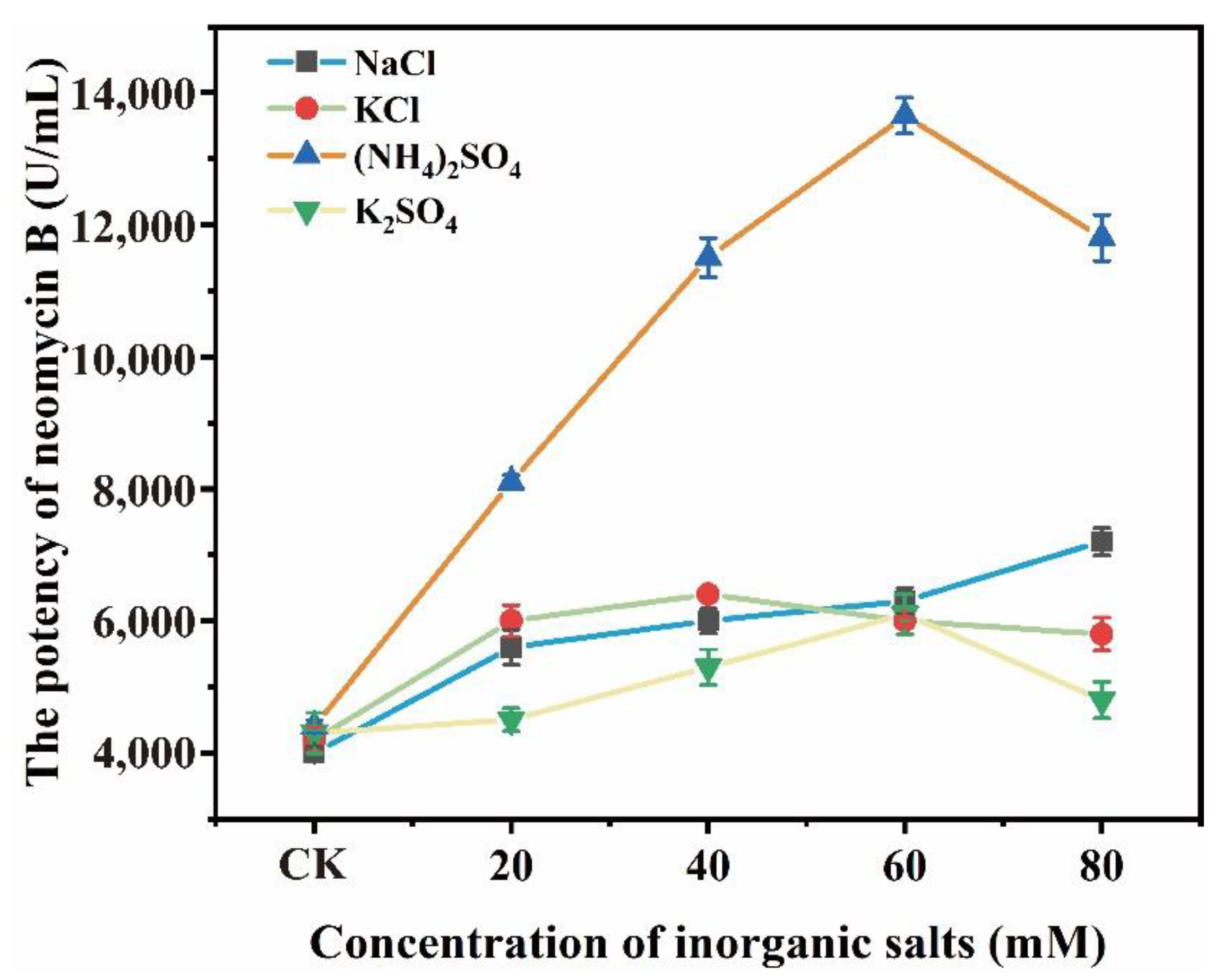
3.1. (NH4)2SO4 Promoted the Biosynthesis of Neomycin B

To investigate the effects of various inorganic salts on neomycin B biosynthesis, different concentrations (CK: without (NH4)2SO4 addition, 20, 40, 60, and 80 mM) of NaCl, KCl, (NH4)2SO4, and K2SO4 were added to the fermentation medium. The addition of inorganic salts affected the potency of neomycin B in shake flasks differently (Figure 1). In the presence of 60 mM (NH4)2SO4, 80 mM NaCl, 40 mM KCl, and 60 mM K2SO4, the highest potency of neomycin B was achieved, corresponding to 13,650.0, 7429.7, 6574.1, and 6317.2 U/mL, respectively, which was 3.3, 0.82, 0.61, and 0.54 times higher than the control without inorganic salts. According to the results, it was found that the accumulation of neomycin B was promoted by adding 60 mM (NH4)2SO4 more efficiently than in the other test groups, which might be attributed to three aspects. On the other hand, to control the fed-batch cultures, the culture phase has to be divided into three sections with different C/N ratios: initial, exponential, and neomycin production. First, (NH4)2SO4 could increase the cell’s osmotic pressure to efficiently utilize carbon and nitrogen sources, thereby enhancing neomycin biosynthesis. Second, the addition of (NH4)2SO4 can moderately reduce the C/N ratios in the fermentation medium, which is quite beneficial for the growth and metabolism of the strain and further neomycin B production. Third, (NH4)2SO4 may also act as an amino donor to increase the transaminase activity involved in neomycin B biosynthesis.

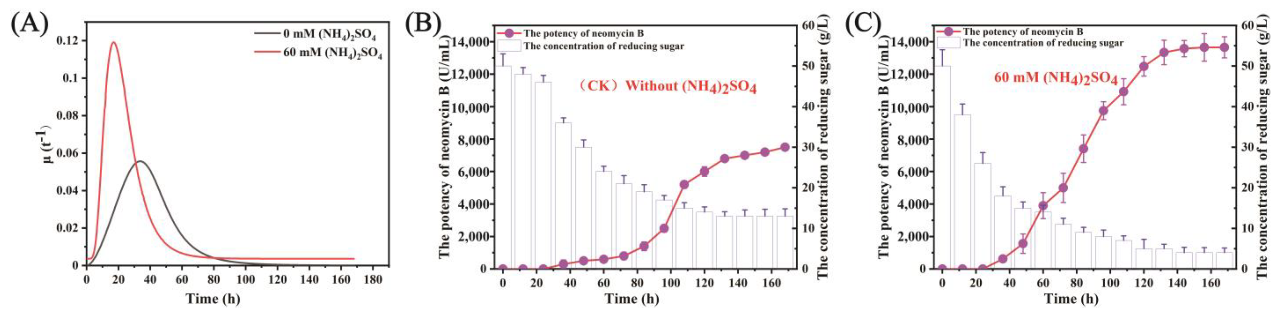
3.2. (NH4)2SO4 Enhanced Cell Growth and Utilization of Reducing Sugar

In antibiotic production, nitrogen is an essential nutrient used by bacteria for cell growth and secondary metabolite synthesis. The bacteria can directly absorb and utilize appropriate amounts of inorganic nitrogen or organic nitrogen in the form of protein degradation products. The inorganic nitrogen (NH4)2SO4 can facilitate the conversion of α-ketoglutarate to L-glutamate by the TCA cycle; L-glutamate is transformed into L-glutamine by transamination, thereby promoting cell growth [16,26,27,28]. To further investigate the effects of (NH4)2SO4 on cell growth and neomycin B biosynthesis, the specific growth rate, the efficiency of neomycin biosynthesis, and the utilization of reducing sugar of S. fradiae SF-2 were determined in the presence or absence of 60 mM (NH4)2SO4.
It was found that 60 mM (NH4)2SO4 promoted S. fradiae SF-2 growth, causing the cells to enter the logarithmic growth phase more quickly and reach a maximum specific growth rate of 0.122 h−1 at about 24 h (Figure 2A). Moreover, S. fradiae SF-2 without (NH4)2SO4 supplementation reached a maximum specific growth rate of 0.056 h−1 at 36 h and entered the stable phase at 72 h. These results indicate that the addition of appropriate amounts of (NH4)2SO4 could accelerate the growth of S. fradiae SF-2, which may be attributed to the fact that it reduced the C/N ratio in the fermentation medium. Secondly, in the presence of 60 mM (NH4)2SO4, S. fradiae SF-2 began synthesizing neomycin B at 48 h, which significantly improved the efficiency of neomycin B biosynthesis compared to the control without (NH4)2SO4. This may be related to the ability of (NH4)2SO4 to alter the physical and chemical properties of the cell wall to promote the absorption of carbon and nitrogen sources by S. fradiae SF-2 used for the synthesis of secondary metabolites. This was consistent with previous studies that reported that the addition of ammonium boosts product biosynthesis, such as gentamicin in Micromonospora purpurea, glycopeptide A40926 in Actinomadura sp. ATCC 39727, and avilamycin in Streptomyces viridochromogenes [29,30,31]. Moreover, neomycin B biosynthesis requires several aminotransferases, and thus adding (NH4)2SO4 may increase aminotransferase activity. However, excessive nitrogen sources can inhibit secondary metabolite biosynthesis. It was reported that when the NH4+ concentration exceeds 20 mM, valine dehydrogenase and glucose-6-phosphate dehydrogenase activity was inhibited, thereby decreasing erythromycin production [32]. Therefore, 60 mM (NH4)2SO4 was found to be a suitable concentration for neomycin biosynthesis in this study. Third, at 60 mM (NH4)2SO4, the final residual sugar content at the end of fermentation was 4.2 g/L, which was much lower than the condition without (NH4)2SO4 (Figure 2B,C), suggesting that (NH4)2SO4 could promote the utilization of reducing sugar in S. fradiae SF-2. In conclusion, (NH4)2SO4 could improve neomycin B potency in S. fradiae by promoting the utilization of carbon sources.

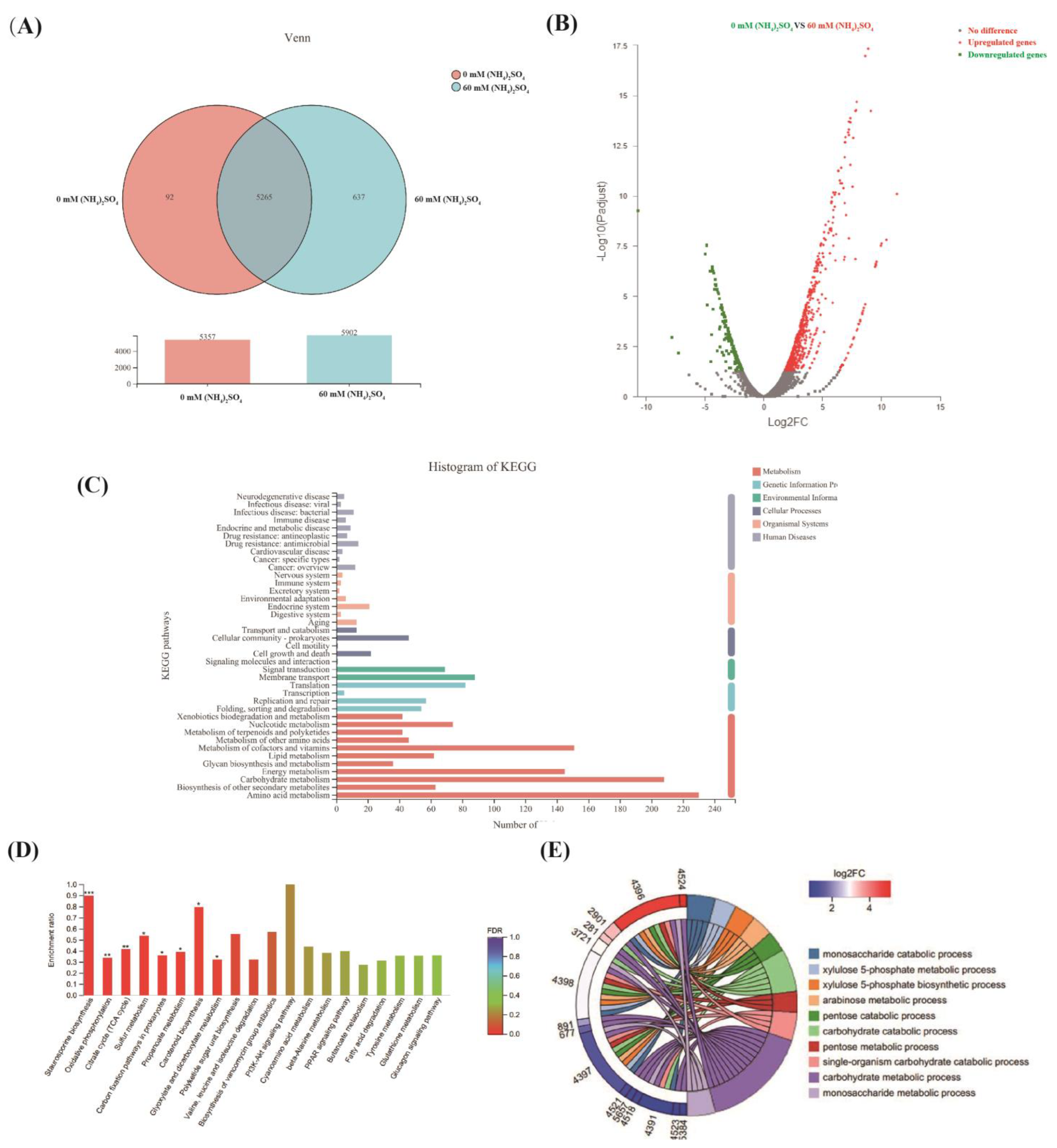
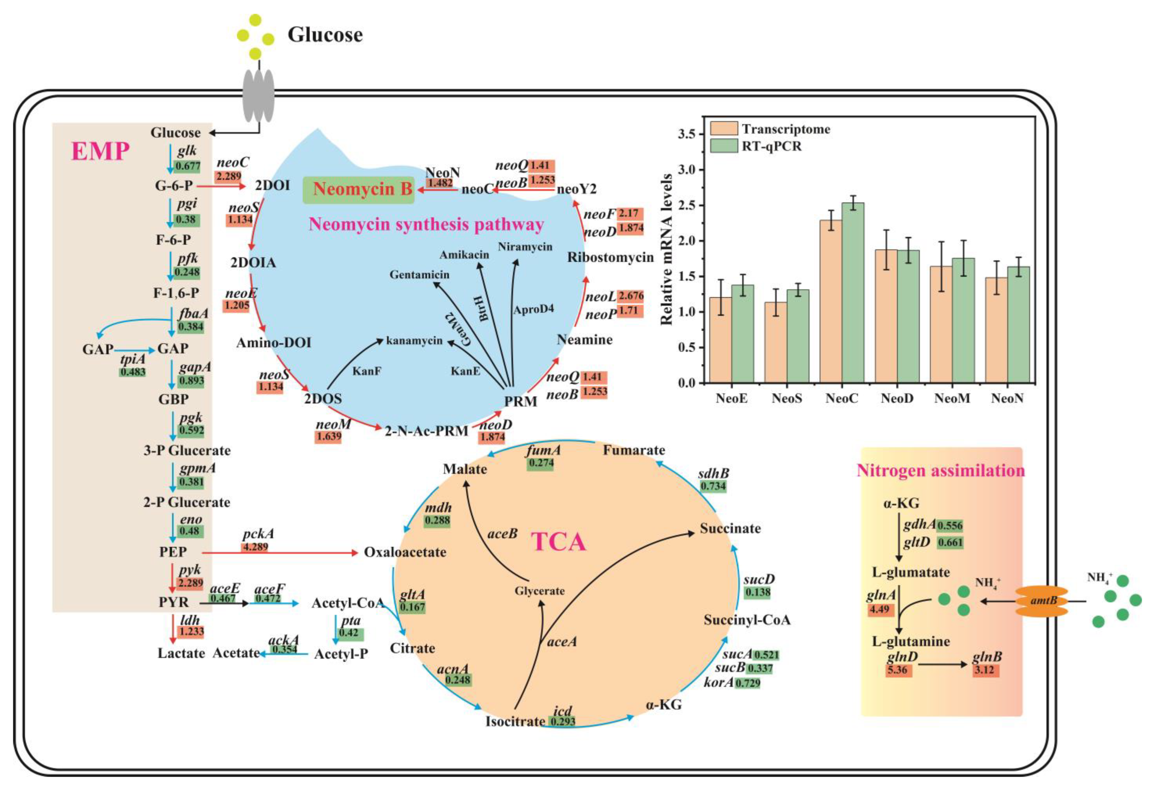
3.3. Comparative Transcriptomics Revealed the Mechanisms Underlying the Effect of (NH4)2SO4 on Neomycin B Biosynthesis

To further investigate the mechanisms underlying the effect of (NH4)2SO4 on neomycin B biosynthesis in S. fradiae, the cells were fermented for 48 h with and without 60 mM (NH4)2SO4. The results showed that a total of 5902 genes were expressed in the medium with 60 mM (NH4)2SO4 compared with the condition with no (NH4)2SO4 addition, of which 637 were specifically expressed (Figure 3A), based on comparative transcriptomic analysis. Compared with the culture condition without (NH4)2SO4, the expression levels of a total of 880 genes changed significantly, among which 651 genes were significantly upregulated and 229 genes were significantly downregulated (Figure 3B and Supplementary Materials). The results indicated that 60 mM (NH4)2SO4 significantly influenced the gene expression of S. fradiae SF-2.
KEGG pathway enrichment analysis of the differentially expressed genes revealed that the genes related to metabolism were mainly involved in amino acid metabolism, carbohydrate metabolism, glycan biosynthesis and metabolism, cofactor and vitamin metabolism, nucleic acid metabolism, and energy metabolism. The genes differentially expressed related to the genetic information processing system are mainly involved in translation, replication, and repair. The differentially expressed genes related to environmental information processing are primarily involved in membrane transport and signal transduction (Figure 3C). The analysis also revealed that there was a significant difference in the expression of genes involved in the TCA cycle, oxidative phosphorylation, amino acid metabolism, propionate metabolism, carboxylic acid metabolism, polyketose metabolism, and vancomycin and staurosporine biosynthesis (Figure 3D). Functional enrichment analysis of the differentially expressed genes showed that they were largely involved in the biosynthesis and metabolism of xylulose-5-phosphate, monosaccharide decomposition, arabinose metabolism, pentose catabolism, and carbohydrate catabolism. The transcription levels of these genes were significantly changed, which may have significant effects on the biosynthesis of neomycin (Figure 3E). Finally, we found that the transcript levels of genes involved in the EMP and TCA cycles were significantly downregulated compared with the control without (NH4)2SO4, indicating that the carbon metabolism flow was pushed toward neomycin biosynthesis (Figure 4). Moreover, the transcript levels of neo genes that regulate neomycin biosynthesis were upregulated compared to the control without (NH4)2SO4. Furthermore, the upregulation of nitrogen assimilation-related genes also indicated that the nitrogen transport and utilization capacity were significantly improved by (NH4)2SO4. Additionally, the genes related to pentose catabolism and carbohydrate catabolism were significantly upregulated, which provided precursors for the biosynthesis of neomycin B. To further verify the reliability of the comparative transcriptomic data, RT-qPCR analysis was performed on the neo gene cluster genes. The RT-qPCR results were consistent with the results of the comparative transcriptomic analysis (Figure 4), which indicated that the comparative transcriptomic analysis was reliable.

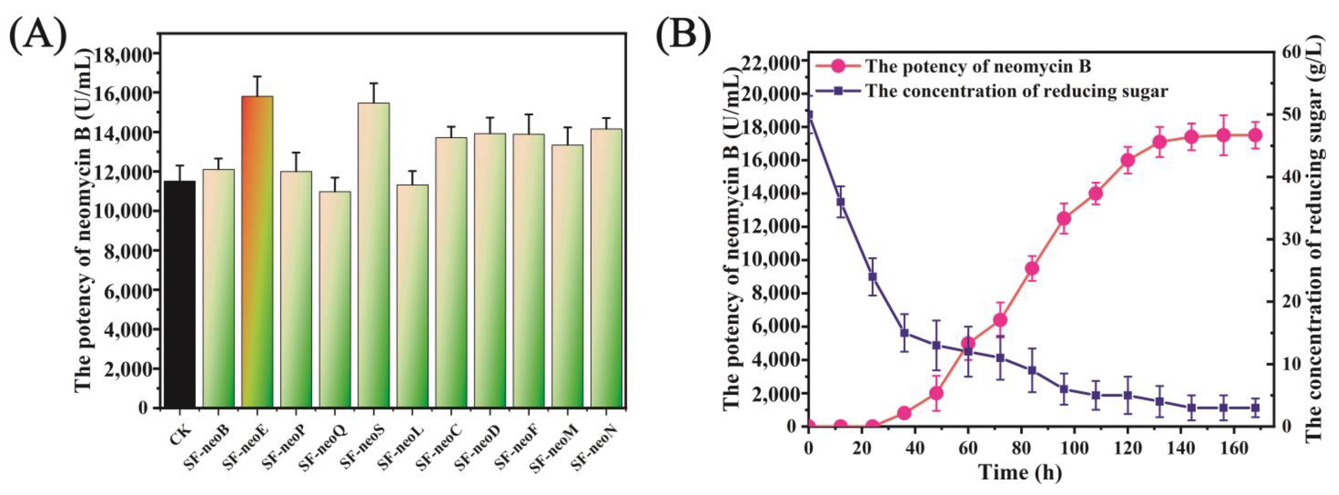
3.4. neoE Overexpression Improved the Biosynthesis of Neomycin B

Using comparative transcriptomic analysis, it was found that (NH4)2SO4 enhanced the expression of neo genes involved in neomycin B biosynthesis and reduced the expression of genes involved in the EMP and TCA cycles, thereby promoting neomycin B production. To further improve neomycin B potency, each gene in the neo gene clusters was overexpressed. Overexpression of neoE, neoS, neoC, neoD, neoF, neoM, and neoN significantly promoted the accumulation of neomycin B. Overexpression of neoE resulted in 15,810.8 U/mL of neomycin B after fermentation for 168 h, which was 37.5% higher than that achieved with the S. fradiae SF-2 control (Figure 5A). Finally, the engineered S. fradiae SF-neoE strain was fermented for 168 h with 60 mM (NH4)2SO4 supplementation. A potency of 17,399 U/ mL was achieved, corresponding to a 51.2% increase compared to the wild-type strain S. fradiae SF-2 (Figure 5B).

4. Conclusions

This study proved that 60 mM (NH4)2SO4 could inhibit the EMP and TCA cycles, promote the utilization of reducing sugars, and enhance the expression of neo genes involved in the neomycin B biosynthesis pathway, thereby improving the neomycin B potency. Upon neoE overexpression and the addition of 60 mM (NH4)2SO4, the engineered S. fradiae SF-neoE strain presented a 51.2% increase (17,399 U/mL) compared with the control strain S. fradiae SF-2. In summary, uncovering the mechanisms underlying the effects of (NH4)2SO4 on neomycin B biosynthesis in S. fradiae is beneficial for enhancing neomycin B production and applications.

Supplementary Materials

The following supporting information can be downloaded at: https://www.mdpi.com/article/10.3390/fermentation8120678/s1, Table S1: Raw data of comparative transcriptomic.

Author Contributions

Conceptualization, X.L., F.Y. and Z.X.; writing—original draft preparation, X.L. and F.Y.; methodology, K.L., M.Z., Y.C., F.W., S.W. and R.H.; supervision, Z.X. All authors have read and agreed to the published version of the manuscript.

Funding

This study was supported by the National Nature Science Foundation of China (31471615, 31871781, and 31772081).

Institutional Review Board Statement

Not applicable.

Data Availability Statement

The data that support the findings of this study are available from the corresponding author upon reasonable request.

Conflicts of Interest

The authors declare no conflict of interest.

Abbreviations

EMP: glycolytic pathway; TCA: tricarboxylic acid cycle. RH: relative humidity.

References

  1. Zheng, J.; Li, Y.; Guan, H.; Li, J.; Li, D.; Zhang, J.; Tan, H. Component optimization of neomycin biosynthesis via the reconstitution of a combinatorial mini-gene-cluster in Streptomyces fradiae. ACS Synth. Biol. 2020, 9, 2493–2501. [Google Scholar] [CrossRef]
  2. Wright, F.; Bibb, M.J. Codon usage in the G+C-rich Streptomyces genome. Gene 1992, 113, 55–65. [Google Scholar] [CrossRef] [PubMed]
  3. Bekker, O.B.; Klimina, K.M.; Vatlin, A.A.; Zakharevich, N.V.; Kasianov, A.S.; Danilenko, V.N. Draft genome sequence of Streptomyces fradiae ATCC 19609, a strain highly sensitive to antibiotics. Genome Announc. 2014, 2, e01247-14. [Google Scholar] [CrossRef] [PubMed] [Green Version]
  4. Xu, Z.; Ji, L.; Tang, W.; Guo, L.; Gao, C.; Chen, X.; Liu, J.; Hu, G.; Liu, L. Metabolic engineering of Streptomyces to enhance the synthesis of valuable natural products. Eng. Microbiol. 2022, 2, 100022. [Google Scholar] [CrossRef]
  5. Champney, W.S. Antibiotics targeting bacterial ribosomal subunit biogenesis. J. Antimicrob. Chemother. 2020, 75, 787–806. [Google Scholar] [CrossRef]
  6. Foster, C.; Champney, W.S. Characterization of a 30S ribosomal subunit assembly intermediate found in Escherichia coli cells growing with neomycin or paromomycin. Arch. Microbiol. 2008, 189, 441–449. [Google Scholar] [CrossRef] [PubMed]
  7. Mehta, R.; Champney, W.S. Neomycin and paromomycin inhibit 30S ribosomal subunit assembly in Staphylococcus aureus. Curr. Microbiol. 2003, 47, 237–243. [Google Scholar] [CrossRef] [PubMed]
  8. Xiao, C.; Liu, J.; Yang, A.; Zhao, H.; He, Y.; Li, X.; Yuan, Z. Colorimetric determination of neomycin using melamine modified gold nanoparticles. Microchim. Acta 2015, 182, 1501–1507. [Google Scholar] [CrossRef]
  9. Swiatkowska, A.; Dutkiewicz, M.; Machtel, P.; Janecki, D.M.; Kabacinska, M.; żydowicz-Machtel, P.; Ciesiołka, J. Regulation of the p53 expression profile by hnRNP K under stress conditions. RNA Biol. 2020, 17, 1402–1415. [Google Scholar] [CrossRef]
  10. Jaiswal, S.K.; Oh, J.J.; Depamphilis, M.L. Cell cycle arrest and apoptosis are not dependent on p53 prior to p53-dependent embryonic stem cell differentiation. Stem Cells 2020, 9, 1091–1106. [Google Scholar] [CrossRef] [PubMed]
  11. Ahmed, M.Z.; Zia, Q.; Haque, A.; Alqahtani, A.S.; Almarfadi, O.M.; Banawas, S.; Alqahtani, M.S.; Ameta, K.L.; Haque, S. Aminoglycosides as potential inhibitors of SARS-CoV-2 main protease: An in silico drug repurposing study on FDA-approved antiviral and anti-infection agents. J. Infect. Public Health 2021, 14, 611–619. [Google Scholar] [CrossRef] [PubMed]
  12. Vastrad, B.M.; Neelagund, S.E. Optimization of medium composition for the production of neomycin by Streptomyces fradiae NCIM 2418 in solid state fermentation. Biotechnol. Res. Int. 2014, 2014, 674286. [Google Scholar] [CrossRef] [PubMed] [Green Version]
  13. Lee, S.H.; Rho, Y.T. Improvement of tylosin fermentation by mutation and medium optimization. Lett. Appl. Microbiol. 1998, 28, 142–144. [Google Scholar] [CrossRef]
  14. Yang, X.; Yang, Y.; Huang, J.; Man, D.; Guo, M. Comparisons of urea or ammonium on growth and fermentative metabolism of Saccharomyces cerevisiae in ethanol fermentation. World J. Microbiol. Biotechnol. 2021, 37, 1–7. [Google Scholar] [CrossRef] [PubMed]
  15. Park, S.; Shah, N.N.; Taylor, R.T.; Droege, M.W. Batch cultivation of Methylosinus trichosporium OB3b: II. Production of particulate methane monooxygenase. Biotechnol. Bioeng. 1992, 40, 151–157. [Google Scholar] [CrossRef] [PubMed]
  16. Commichau, F.M.; Forchhammer, K.; Stulke, J. Regulatory links between carbon and nitrogen metabolism. Curr. Opin. Microbiol. 2006, 9, 167–172. [Google Scholar] [CrossRef] [PubMed]
  17. Majumdar, M.K.; Majumdar, S.K. Utilization of carbon and nitrogen-containing compounds for neomycin production by Streptomyces fradiae. Appl. Microbiol. 1967, 15, 744–749. [Google Scholar] [CrossRef] [PubMed]
  18. Meng, X.; Wang, W.; Xie, Z.; Li, P.; Li, Y.; Guo, Z.; Lu, Y.; Yang, J.; Guan, K.; Lu, Z.; et al. Neomycin biosynthesis is regulated positively by AfsA-g and NeoR in Streptomyces fradiae CGMCC 4.7387. Sci. China Life Sci. 2017, 60, 980–991. [Google Scholar] [CrossRef]
  19. Kudo, F.; Eguchi, T. Aminoglycoside antibiotics: New insights into the biosynthetic machinery of old drugs. Chem. Rec. 2016, 16, 4–18. [Google Scholar] [CrossRef] [PubMed]
  20. Wu, J.; Chen, D.; Wu, J.; Chu, X.; Yang, Y.; Fang, L.; Zhang, W. Comparative transcriptome analysis demonstrates the positive effect of the cyclic AMP receptor protein Crp on daptomycin biosynthesis in Streptomyces roseosporus. Front. Bioeng. Biotechnol. 2021, 9, 401. [Google Scholar] [CrossRef] [PubMed]
  21. Pinilla, L.; Toro, L.F.; Laing, E.; Alzate, J.F.; Ríos-Estepa, R. Comparative transcriptome analysis of Streptomyces clavuligerus in response to favorable and restrictive nutritional conditions. Antibiotics 2019, 8, 96. [Google Scholar] [CrossRef] [PubMed] [Green Version]
  22. Liu, T.; Huang, Z.; Gui, X.; Xiang, W.; Jin, Y.; Chen, J.; Zhao, J. Multi-omics comparative analysis of Streptomyces mutants obtained by iterative atmosphere and room-temperature plasma mutagenesis. Front. Microbiol. 2021. [Google Scholar] [CrossRef] [PubMed]
  23. Li, X.; Bao, T.; Osire, T.; Qiao, Z.; Liu, J.; Zhang, X.; Xu, M.; Yang, T.; Rao, Z. MarR-type transcription factor RosR regulates glutamate metabolism network and promotes accumulation of L-glutamate in Corynebacterium glutamicum g01. Bioresour. Technol. 2021, 342, 125945. [Google Scholar] [CrossRef] [PubMed]
  24. Yu, F.; Zhang, M.; Sun, J.; Wang, F.; Li, X.; Liu, Y.; Wang, Z.; Zhao, X.; Li, J.; Chen, J.; et al. Improved neomycin sulfate potency in streptomyces fradiae using atmospheric and room temperature plasma (ARTP) mutagenesis and fermentation medium optimization. Microorganisms 2022, 10, 94. [Google Scholar] [CrossRef] [PubMed]
  25. Huang, F.; Li, Y.; Yu, J.; Spencer, J.B. Biosynthesis of aminoglycoside antibiotics: Cloning, expression and characterisation of an aminotransferase involved in the pathway to 2-deoxystreptamine. Chem. Commun. 2002, 23, 2860–2861. [Google Scholar] [CrossRef] [PubMed]
  26. Dam, B.; Dam, S.; Kim, Y.; Liesack, W. Ammonium induces differential expression of methane and nitrogen metabolism-related genes in Methylocystis sp. Strain SC2. Environ. Microbiol. 2014, 16, 3115–3127. [Google Scholar] [CrossRef] [PubMed]
  27. Cueto-Rojas, H.F.; Maleki, S.R.; Ten, P.A.; van Helmond, W.; Pieterse, M.M.; Heijnen, J.J.; Wahl, S.A. In vivo analysis of NH4+ transport and central nitrogen metabolism in Saccharomyces cerevisiae during aerobic nitrogen-limited growth. Appl. Environ. Microbiol. 2016, 82, 6831–6845. [Google Scholar] [CrossRef] [PubMed] [Green Version]
  28. Stitt, M. Nitrate regulation of metabolism and growth. Curr. Opin. Plant Biol. 1999, 2, 178–186. [Google Scholar] [CrossRef] [PubMed]
  29. Technikova-Dobrova, Z.; Damiano, F.; Tredici, S.M.; Vigliotta, G.; Di Summa, R.; Palese, L.; Abbrescia, A.; Labonia, N.; Gnoni, G.V.; Alifano, P. Design of mineral medium for growth of Actinomadura sp. ATCC 39727, producer of the glycopeptide A40926: Effects of calcium ions and nitrogen sources. Appl. Microbiol. Biot. 2004, 6, 671–677. [Google Scholar] [CrossRef]
  30. Gonzalez, R.; Islas, L.; Obregon, A.M.; Escalante, L.; Sanchez, S. Gentamicin formation in Micromonospora purpurea: Stimulatory effect of ammonium. J. Antibiot. 1995, 48, 479–483. [Google Scholar] [CrossRef]
  31. Zhu, C.H.; Lu, F.P.; He, Y.N.; Han, Z.L.; Du, L.X. Regulation of avilamycin biosynthesis in Streptomyces viridochromogenes: Effects of glucose, ammonium ion, and inorganic phosphate. Appl. Microbiol. Biotechnol. 2007, 73, 1031–1038. [Google Scholar] [CrossRef] [PubMed]
  32. Hong, M.; Mou, H.; Liu, X.; Huang, M.; Chu, J. 13C-assisted metabolomics analysis reveals the positive correlation between specific erythromycin production rate and intracellular propionyl-CoA pool size in Saccharopolyspora erythraea. Bioprocess Biosyst. Eng. 2017, 40, 1337–1348. [Google Scholar] [CrossRef] [PubMed]
Figure 1. The effects of different inorganic salts on the biosynthesis of neomycin B.
Fermentation 08 00678 g001
Figure 2. The effects of ammonium sulfate on the growth, reducing sugar utilization, and neomycin B biosynthesis in S. fradiae SF-2: (A) The effects of the addition of 60 mM (NH4)2SO4 on the specific growth rate. (B,C) The impact of the addition of 60 mM (NH4)2SO4 on reducing sugar utilization and neomycin B potency.
Fermentation 08 00678 g002
Figure 3. Comparative transcriptomic analysis of the effects of 60 mM (NH4)2SO4 on neomycin B biosynthesis: (A) Venn diagram analysis of differentially expressed genes. (B) Volcano plot analysis of differentially expressed genes. The x-axis represents the log2-transformed expression fold-change values. The y-axis represents the log10-transformed adjusted p-values. Red dots indicate upregulated genes and blue dots indicate downregulated genes. (C) KEGG enrichment analysis of the metabolic pathways in which differentially expressed genes are involved. The y-axis is the name of the KEGG metabolic pathway, and the abscissa is the number of genes annotated to the pathway. KEGG metabolic pathways can be divided into seven major categories: metabolism, genetic information processing, environmental information processing, cellular processes, organismal systems, human diseases, and drug development. (D) KEGG enrichment analysis of differentially expressed genes. The X-axis is the name of the pathway. The Y-axis represents the enrichment rate, which is the ratio of the Sample number of genes annotated to the pathway and the background number of all genes annotated to the pathway. The higher the value of the rich factor, the greater the degree of enrichment. The color represents the enrichment significance (p-value), and the darker the color, the more significantly the pathway is enriched, where p-value < 0.001 is labeled as ***, p-value < 0.01 as **, p-value < 0.05 as *, and the color gradient on the right indicates the p-value size. (E) Functional enrichment analysis of differentially expressed genes. On the left is the gene, which is arranged in the order of log2FC from largest to smallest. Larger log2FC values indicate larger differential expression ploidy for upregulated genes. Smaller log2FC values indicate larger differential expression ploidy for downregulated genes. A log2FC closer to 0 indicates smaller differential expression ploidy for genes.
Fermentation 08 00678 g003
Figure 4. The expression levels of genes involved in neomycin B metabolism in S. fradiae SF-2 with 60 mM (NH4)2SO4 relative to 0 mM (NH4)2SO4. The red arrows show genes whose expression levels are upregulated, and the green arrows show those whose expression levels are downregulated.
Fermentation 08 00678 g004
Figure 5. (A) The effects of overexpression of single genes of the neo gene clusters on neomycin B biosynthesis in S. fradiae SF-2. (B) The effect of 60 mM (NH4)2SO4 on reducing sugar utilization and neomycin B biosynthesis in the SF-neoE strain.
Fermentation 08 00678 g005
Table 1. Strains and plasmids used in this study.
Strains and PlasmidsDescriptionSource
Strains
E. coli DH5αGeneral cloning hostThe lab
E. coli ET12567Demethylated strain containing pUZ8002 plasmid for conjugative transfer with actinomycetesThe lab
S. fradiae SF-2Streptomyces fradiae, neomycin B-producing strains generated by ARTP [24]
ET12567/PPR-NeoBE. coli ET12567 derivative harboring PPR-NeoBThis study
ET12567/PPR-NeoEE. coli ET12567 derivative harboring PPR-NeoEThis study
ET12567/PPR-NeoPE. coli ET12567 derivative harboring PPR-NeoPThis study
ET12567/PPR-NeoQE. coli ET12567 derivative harboring PPR-NeoQThis study
ET12567/PPR-NeoSE. coli ET12567 derivative harboring PPR-NeoSThis study
ET12567/PPR-NeoLE. coli ET12567 derivative harboring PPR-NeoLThis study
ET12567/PPR-NeoCE. coli ET12567 derivative harboring PPR-NeoCThis study
ET12567/PPR-NeoDE. coli ET12567 derivative harboring PPR-NeoDThis study
ET12567/PPR-NeoFE. coli ET12567 derivative harboring PPR-NeoFThis study
ET12567/PPR-NeoME. coli ET12567 derivative harboring PPR-NeoMThis study
ET12567/PPR-NeoNE. coli ET12567 derivative harboring PPR-NeoNThis study
SF-NeoBS. fradiae SF-2 derivative the expression of NeoBThis study
SF-NeoES. fradiae SF-2 derivative the expression of NeoEThis study
SF-NeoPS. fradiae SF-2 derivative the expression of NeoPThis study
SF-NeoQS. fradiae SF-2 derivative the expression of NeoQThis study
SF-NeoSS. Fradiae SF-2 derivative the expression of NeoSThis study
SF-NeoLS. fradiae SF-2 derivative the expression of NeoLThis study
SF-NeoCS. fradiae SF-2 derivative the expression of NeoCThis study
SF-NeoDS. fradiae SF-2 derivative the expression of NeoDThis study
SF-NeoFS. fradiae SF-2 derivative the expression of NeoFThis study
SF-NeoMS. fradiae SF-2 derivative the expression of NeoMThis study
SF-NeoNS. fradiae SF-2 derivative the expression of NeoNThis study
Plasmids
PPR (pSET152-PermE*)E. coli-S. fradiae shuttle vector for the expression of target proteinThe lab
PPR-NeoBDerived from PPR, for the expression of NeoBThis study
PPR-NeoEDerived from PPR, for the expression of NeoEThis study
PPR-NeoPDerived from PPR, for the expression of NeoPThis study
PPR-NeoQDerived from PPR, for the expression of NeoQThis study
PPR-NeoSDerived from PPR, for the expression of NeoSThis study
PPR-NeoLDerived from PPR, for the expression of NeoLThis study
PPR-NeoCDerived from PPR, for the expression of NeoCThis study
PPR-NeoDDerived from PPR, for the expression of NeoDThis study
PPR-NeoFDerived from PPR, for the expression of NeoFThis study
PPR-NeoMDerived from PPR, for the expression of NeoMThis study
PPR-NeoNDerived from PPR, for the expression of NeoNThis study
Table 2. Primers used in this study.
Primer NameSequencesDigest Sites
NeoB FATAGCGGCCGCGATGACGAAAAACTCTTCCCTGCNotI
NeoB RCGAGATATCTCAGTCGTCCAGCAGCCGEcoRV
NeoE FATAGCGGCCGCGATGAAGGCTCTGGTGTTCGAGGNotI
NeoE RCGAGATATCTCAGGCCCGGAGGTTGAAGTAEcoRV
NeoP FATAGCGGCCGCGATGACGGCCGCCCAGCNotI
NeoP RCGAGATATCTCATGCCGTCCTGGCCAGEcoRV
NeoQ FATAGCGGCCGCGATGAAGCGCCTTCGAGGCACNotI
NeoQ RCGAGATATCTCAGACGTGCGCGGTGTGCEcoRV
NeoS FATAGCGGCCGCGATGGTCTCCCCGTTGGCANotI
NeoS RCGAGATATCTCAAGTGGCCAGGTCGGCEcoRV
NeoL FATAGCGGCCGCGGTGGTGACGACCGGCGTGGCNotI
NeoL RCGAGATATCTCAGGCCAGTGCGGCGACEcoRV
NeoC FATAGCGGCCGCGATGCAGACCACCCGCATNotI
NeoC RCGAGATATCTTACGGCACGGGTCCGGCEcoRV
NeoD FATAGCGGCCGCGGTGGGTGAGCCGACGTGGNotI
NeoD RCGAGATATCTCACCGGGCACCCGCCGEcoRV
NeoF FATAGCGGCCGCGGTGGCTGAGGCGCCTGCNotI
NeoF RCGAGATATCTCACCCACCGTGCTCCTCCEcoRV
NeoM FATAGCGGCCGCGGTGCTGCGGCTCACCCNotI
NeoM RCGAGATATCTCACGGCGCCCACCCGEcoRV
NeoN FATAGCGGCCGCGATGACCACCGACNotI
NeoN RCGAGATATCTCATACGAGCGEcoRV
RT-NeoE FTGACGGCCACCTTCTCGC
RT-NeoE RACCTGCTCCTGCGGCACCT
RT-NeoS FTAGGTGTAGTACGTACGGG
RT-NeoS RATGGGCAGCAACCGCTGCCT
RT-NeoC FGTTCTTGACGAGCGCGGT
RT-NeoC RGGACACGCCATCGAGCACG
RT-NeoD FCTCGGTCTCGTCGTCGTA
RT-NeoD RACGCGACGCTCCTGACGGTGT
RT-NeoM FTGGACGTGCACCAGGT
RT-NeoM RAGCAGCTCGTCATGACCGT
RT-NeoN FTGGTAGTTGTAGCCGTTGGT
RT-NeoN RACTGCTCCACTTCATGCCGC
Publisher’s Note: MDPI stays neutral with regard to jurisdictional claims in published maps and institutional affiliations.

