Article

Potentially Probiotic Fermented Glutinous Rice (Oryza sativa L.) with Lactiplantibacillus plantarum Improved Immune System Response in a Small Sample of BALB/cByJ Mice

1
Faculty of Food Science and Technology, Universiti Putra Malaysia, Serdang 43400, Selangor, Malaysia
2
School of Pharmacy, The University of Nottingham Malaysia Campus, Semenyih 43500, Selangor, Malaysia
3
Department of Food Science and Nutrition, University of Minnesota, Minneapolis, MN 55455, USA
4
Institute of Bioscience, University Putra Malaysia, Serdang 43400, Selangor, Malaysia
5
Faculty of Veterinary Medicine, Universiti Putra Malaysia, Serdang 43400, Selangor, Malaysia
6
Faculty of Medical and Health Sciences, Universiti Putra Malaysia, Serdang 43400, Selangor, Malaysia
7
Science and Food Technology Research Centre, MARDI Headquarters, Serdang 43400, Selangor, Malaysia
8
Halal Products Research Institute, Universiti Putra Malaysia, Serdang 43400, Selangor, Malaysia
*
Author to whom correspondence should be addressed.
Fermentation 2022, 8(11), 612; https://doi.org/10.3390/fermentation8110612
Submission received: 12 August 2022 / Revised: 1 November 2022 / Accepted: 3 November 2022 / Published: 8 November 2022
(This article belongs to the Special Issue Recent Trends in Lactobacillus and Fermented Food)

Abstract

:
L. plantarum strains displayed different abilities to exhibit high survivability to acid (pH 3.0), bile salts (3%), enzyme (pepsin), and temperature (40 °C) and good antibiotic susceptibility. The isolates were further supplemented in traditional tapai pulut to study the immunomodulation properties of tapai pulut based on the splenic T- and B-cell populations. The mice groups were divided into group one (unfermented glutinous rice), group two (tapai pulut group), and group three (probiotic tapai pulut group). Group one showed consistent body weight gain, with the highest observed after four weeks. Group three exhibited the most significant reduction in the percentage of CD19+ B-cells. The CD3+ T-cells population of Group three increased significantly compared with the control mice, followed by Group two. The results suggest that traditional tapai pulut supplemented with L. plantarum has a high potential for supporting the immune system’s immunomodulatory effect.

Graphical Abstract

1. Introduction

Fermented food consumption has existed for thousands of years due to their aromas and taste preferences. The development of food science has led to the recognition of the health benefits of fermented foods. Several microorganisms exist in fermented foods, including bacteria, fungi, and yeast. Lactic acid bacteria (LAB) is the most common bacteria used in food fermentation, and is estimated to be applied as a starter culture in 80% of fermented foods as probiotics [1]. According to the expert consensus, probiotics are “live microorganisms that, when administered in adequate amounts, confer a health benefit on the host”. The expert panels accepted the following bacterial species of Bifidobacterium (adolescentis, animalis, bifidum, breve, and longum) and Lactobacillus (acidophilus, casei. fermentum, gasseri, johnsonii, paracasei, plantarum, rhamnosus, and salivarius), when delivered in food at a level of 1 × 10⁹ colony forming units (CFU) per serving, as probiotics [1]. These genera produce several antimicrobial compounds, such as organic acids, bacteriocins, and bioactive peptides [2]. Thus, the fermentation process can enhance the bioavailability of the nutrients in fermented foods via the release of featured nutrients from the cells [3].
Two mechanisms were suggested for the improved immune system function: the IL-12 cytokines enhancement that stimulate natural killer cells, and the modulation of gut microbiota to favor friendly bacteria over harmful bacteria [4]. Lee et al. (2018) studied the effect of Korean fermented soybean products (Doenjang and Cheonggukjang) on the immune cells [5]. They reported the enhancement of natural killer cell cytotoxicity in mice after four weeks of consuming Doenjang and Cheonggukjang. The T helper type-1 cell activity was enhanced, the cytokines ratio of IFN-gamma versus IL-4 was up-regulated, and the mice were more resistant to Listeria monocytogenes. In another study, the phagocytosis cytotoxicity and anti-inflammatory activity were enhanced towards lipopolysaccharides’ stimulation in vitro and in vivo [6]. Furthermore, the consumption of milk rich in Bifidobacteria demonstrated significantly increased antibody IgA production detected in the feces and milk [7]. However, there is no research on the use of probiotics in traditional fermented food in Malaysia, such as Tapai Pulut, as most studies focused on the physiochemical properties of traditional fermented food.
Fermented glutinous rice (tapai pulut) is a fermented food prepared from simple raw materials and consumed as fresh or added to a dessert such as ice cream or cendol. The starter cultures used for their production are called “ragi tapai” (Tapai yeast), which are dry starters that contain several species of yeast and fungi [8]. Ragi consists of Candida spp., Saccharomyces spp., Rhizopus spp., and Aspergillus spp. The presence of these species was proven in a study using ragi from Pahang and Sarawak using API kits 32 C and 20 C AUX for yeast profiling [9]. However, the genotypic identification was not profiled. In other studies of ragi, the metabarcoding analysis was employed to identify the populations in ragi genotypically, in which it was found that the bacterial population was higher than the fungal population [10]. The selection of LABs strains to be incorporated into Tapai pulut leads to isolating strains, as they originated from different sources [9].
Furthermore, the varieties of ragi microbes lead to unclear symbiosis relationships in which microbes are majorly responsible throughout the fermentation process. The LABs were previously characterized in ragi used in Brem (black and white glutinous rice), in which the LABs found were homofermentative rods and heterofermentative rods [11], respectively, and they multiplied rapidly during the liquefaction process [12]. Hence, the availability of well-characterized probiotics is excellent in numbers. However, limited studies have been conducted to screen and identify novel strains to improve probiotic production to meet global market demand. Moreover, no evidence was established to determine the impact of consuming fermented glutenous rice on improving immune system function. Therefore, this study aims to identify LAB strains from animal-based and plant-based strains with probiotic properties, and hypothesizes that consuming fermented glutenous rice containing probiotic strains will modulate the immune system’s function in a pre-clinical trial.

2. Materials and Methods

2.1. Isolation of Lactic acid Bacteria (LAB)

Fifty-three LABs were isolated from a variety of animal-based and plant-based food samples by plating on MRS agar media (Oxoid Ltd., Hampshire, UK) and incubated anaerobically in an anaerobic jar placed in an incubator (Memmert, Schwabach, Germany) at 37 °C for 48 h. The strains’ origins are tabulated in Supplementary Files (Table S1). The identification of single colonies was carried out through gram staining, catalase production, and microscopic examination. The cultured strains, characterized as LABs, were kept as a stock culture in 20% glycerol at −80 °C. In addition, LAB strains from the stock media were activated for the use of upcoming assays and tests by subculturing anaerobically in an MRS broth media containing 0.05% L-Cysteine at 37 °C for 18 to 24 h. The tests were conducted in triplicate, and the average values were recorded.

2.2. Acid Tolerance, Bile Tolerance, Enzyme Resistance, and High-Temperature Resistance

The test was carried out according to the previous study, with minor modifications, by employing MRS broth as the growth media [13]. For the acid tolerance test, 200 μL of hydrochloric acid (99.8%, w/v) at pH 3.0 was evaluated and placed in the 96-well microtiter plates. For the bile tolerance test, 200 μL of bile salts prepared at 1, 2, and 3% (w/v) were placed in the 96-well microtiter plates. For the enzyme resistance test, the enzyme pepsin (Sigma Aldrich, St. Louis, MO, USA) was prepared as two-enzymes units, respectively. Precisely, 200 μL of the enzyme mixture was placed in the 96-well microtiter plates. For the high-temperature resistance test, 200 μL of MRS broth was placed in the 96-well microtiter plates. Subsequently, 20 μL of the starter culture was added to the wells in triplicate for each starter culture in all tests. The plates were incubated at 37 °C in all of the parameters, except for the high-temperature resistance test (40 °C) for 6 h. The OD600 nm readings were measured at 0 h and every interval of 1 h using GENESYS 10S UV-Vis spectrophotometer (Thermo Fisher scientific, Waltham, MA, USA).

2.3. Antibiotic Susceptibility

The standardized method for the disc diffusion method was applied, as previously described [14]. The following antibiotics used were: ampicillin (AMP, 10 μg/mL), clindamycin (DA, 2 μg/mL), chloramphenicol (C, 2 μg/mL), erythromycin (E, 5 μg/mL), imipenem (IPM, 10 μg/mL), tetracycline (TE, 30 μg/mL), vancomycin (VA, 30 μg/mL), amoxycillin/clavulanic acid (AMC, 30 μg/mL), and gentamicin (GN, 10 μg/mL).

2.4. Phenotypic and Genotypic Identifications

Of the fifty-three strains, ten strains were selected based on the researched probiotic properties. Their phenotypics were identified using an API 50CHL kit (BioMerieux, Marcy L’Etoile, France), according to the manufacturer’s instructions. For genotypic identification, the DNAs of 10 selected isolates were extracted using a Nucleospin Extraction Kit (Nucleospin Microbial DNA handbook, July 2018, Revision 3, Macherey-Nagel, UK) with some amendments according to technical trials. The preparation was carried out at 4 °C in an ice bath. The DNA was run on gel electrophoresis (Bio-Rad Laboratories, Hercules, CA, USA) at 110 V for 60 min, and the DNAs of the 10 isolates were identified under UV rays (Major Science, UV Transilluminator, Taiwan). The isolates were identified genotypically using the 16S rRNA gene sequencing technique. Two universal LAB primers were employed for the amplification of the 16S rRNA gene, namely forward primer 27F (AGAGTTTGATCMTGGCTCAG) and reverse primer 1492 R (TACGGYTACCTTGTTACGACTT) [12,13]. The PCR (SureCycler 8800A, Agilent, CA, USA) protocols were fixed at 94°C for 4 min for initial denaturation, 30 cycles of denaturation at 94 °C for 30 s, annealing at 5 °C for 30 s and 68 °C for 1 min, and a final extension at 68 °C for 7 min. The DNA sequences of data sets were assembled using the Bioedit sequence alignment editor software (version 7.0.9.0. Ibis Biosciences, Carlsbad, CA, USA). The similarity sequences percentages were identified using the basic local alignment search tool (BLAST) (http://blast.ncbi.nlm.nih.gov/Blast.cgi, accessed on 11 November 2020) and the accession numbers were attained from GeneBank (NCBI, Rockville, MD, USA).

2.5. Growth Condition for the Probiotic Bacteria

The potential probiotic bacteria Lactiplantibacillus plantarum [14] was isolated from the red bean, inoculated in MRS broth, and incubated in an incubator (Memmert, Schwabach, Germany) at 37 °C for 48 h. After 48 h, the culture was washed using sterile 0.5% (w/v) sodium chloride, and the cell count was adjusted to 109 CFU mL−1.

2.6. Preparation of Tapai Pulut

The tapai pulut was prepared following the traditional method [15]. Briefly, 1000 g of glutinous rice was washed, soaked overnight, cooked for 20 min, and cooled down to 40 °C. The commercial powder starter culture (ragi tapai) purchased from MARDI was added to the cooked glutinous rice, and the inoculum size was at 0.2% w/w. The starter culture contained a diversity of microorganisms, including Amylomyces rouxii, Saccharomycopsis fibuligera, Chlamydomucor sp., Rhizopus oryzae, Rhizopus oryzae, and Hansenula sp. The inoculated glutinous rice was divided into 50 g portions, placed in plastic containers, and incubated at 28 °C for 48 h. The tapai pulut with probiotics was prepared using the same method. Thus, washed probiotic cells were added to the rice alongside the ragi tapai, the inoculum size was 2% v/w, and the final cell count was 107 CFU g−1.

2.7. Chemical and Microbial Analyses

The unfermented glutenous rice was analyzed directly after the cooking process, while the fermented samples were collected after the 48 h fermentation and subjected to the same analysis. The analysis included the pH value, total acidity, and cell counts. The samples (1 g) were diluted in 6 mL of sterilized water, and the pH values were measured at room temperature of 28 °C using a digital pH meter (JENWAY 3505, Cole-Palmer, Staffordshire, UK). The total soluble solid (TSS) contents were measured using a refractometer (Schmidt Haensch Co., Berlin, Germany) and presented as Brix values. The moisture contents were measured following the AOAC (2005) method [16]. The total acid contents (% lactic acid) were measured following the AOAC (2005) method [16]. The cell counts were carried out by mixing the samples (25 g) with 225 mL sterilized peptone water (0.1%) using a stomacher bag and placing them in the stomacher for 2 min at room temperature (28 ± 2 °C). The cell counts were based on the presence of microorganisms on the plate using a haemacytometer (Blaubrand Neubauer, Essex, CT, USA). The lactic acid bacteria cell counts were determined on MRS Agar at 37 °C for 48 h in an anaerobic jar with anaerobic bags (Oxoid) [17].

2.8. Animals and Diets

The animal study’s ethics were endorsed and approved by the secretariat board of the Institutional Animal Care and Use Committee, Universiti Putra Malaysia (UPM/IACUC/AUP-R004/2019). The animals were obtained from the Animal Resource Unit, Faculty of Veterinary, Universiti Putra Malaysia. The experiment was carried out with 12 male mice BALB/cByJ aged 4–6 weeks and 25 g of body weight on average, as described [5]. The diets were as the following: Group 1 was fed rodent chow and unfermented glutenous rice (NG), Group 2 was fed with rodent chow and traditional tapai pulut (TPG), and Group 3 was fed with rodent chow and probiotic tapai pulut (PTPG). The animals were fed the selected diets for four weeks. The body weights were measured on days 0, 7, 14, 21, and 28, while the feed intake was recorded daily. The average weekly weight gain and feed efficiency (ratio of weight gain to feed intake (G/F)) were measured from the collected data. After 4 weeks, the animals were euthanized and the organs, including the spleen, thymus, and lymph nodes, were harvested and kept in buffered formalin (10%) to determine the histopathological changes in immune organs and the populations of T-cells and B-cells.

2.9. Biochemical Analysis for Blood Samples

The blood samples were collected on week 4 using Vacutainer tubes containing K2EDTA (BD Biosciences, Franklin Lakes, NJ, USA) and were analyzed for peripheral blood counts using the ADVIA 2120 automatic analyzer (Siemens, Munich, Germany). The blood samples were centrifuged to obtain serum and plasma and were stored at −70 °C until analysis. The liver and kidney function tests were assayed using the standard markers, namely alanine aminotransferase (ALT) and aspartate aminotransferase (AST). Other biomarkers were measured, including glucose (Gluc), triglycerides (Tgl), low-density lipoprotein (LDL), and high-density lipoprotein (HDL).

2.10. Flow Cytometry Analysis

Fluorescence-activated cell sorting (FACS) flow cytometry was used to determine the cytokine levels in the T- and B-cells in all three mice groups. Splenocytes from the BALB/cByJ mice were harvested in a mixture of RPMI-1640 medium through a grinding method between two frosted microscope slides. The suspended cells were centrifuged and collected, whereas supernatants were discarded. Ammonium-chloride-potassium lysing (ACK) buffer was used to treat the pellets before flowing through a filter known as a nylon cell strainer at 40 μm. The splenocytes were counted and resuspended at 4 × 10⁶ cells in 100 μL of staining buffer. The splenocytes were stained with PE-labeled anti-mouse CD19 and APC-labeled anti-mouse CD3 for each group of mice washed with staining buffer and centrifuged. The splenocytes were later fixed in 100 μL of 2.0% paraformaldehyde, resuspended in PBS, and stored at 4 °C. Splenocytes were analyzed by BD Accuri C6 Flow Cytometer (Becton Dickinson, San Jose, CA, USA), and 50,000 events were collected. Data analyses of the T-cell and B-cell populations were performed using the BD Accuri software (BD Biosciences, Ann Arbor, MI, USA). Based on the percentage of total CD3+ and CD19+, dot plots were generated for each group of treatment types. The results were expressed as a percentage of cells (%) ± S.E.M. T-test using GraphPad QuickCalcs online tool was performed to determine the p-values (* p ≤ 0.005, ** p ≤ 0.01, and *** p ≤ 0.0001).

2.11. Statistical Analysis

The results were collected in triplicate and subjected to a one-way analysis of variance (ANOVA) and Tukey’s test for evaluating the significant differences at α = 0.05 using Minitab (Version 18, Minitab LLC, State College, PA, USA).

3. Results

3.1. Probiotic Properties

The survivability rates of the 53 isolated strains after exposure to pH 3.0 and 3% bile salt, temperature 40 °C and enzyme pepsin for 6 h are tabulated in Supplementary Files (Table S2). The survivability rates of the selected ten strains are tabulated in Table 1. ATCC:14931 was randomly selected during the screening process to ensure that there was no bias in selection and to avoid a repetitive number of subcultures. The strain also acted as a control to confirm that no contamination occurred during the subculturing process. All strains could survive under low pH conditions, with different survival rates. The strain RB5 had the highest (98.24%) survival rate compared with the other strains, in which TA8 exhibited the lowest (66.14%). Other strains showed survivabilities ranging from 67.64% to 88.74%. The ten strains could withstand the exposure to 3% bile salts for 6 h.

3.2. Antibiotic Susceptibility

The antibiotic susceptibilities of the ten isolated strains results against the tested antibiotics is depicted are Table 2. There were no contaminations found, and the results indicate that all of the strains were resistant to DA (clindamycin), E (Erythromycin), IPM (Imipenem), VA (Vancomycin), and GN (Gentamicin). In contrast, all of the strains were sensitive to C (Chloramphenicol), and all strains except for CL8 were sensitive to TE (tetracycline).

3.3. Phenotypic and Genotypic Identification

Fifty-three LAB strains were described as rod-shaped, gram-positive, and catalase-negative. Ten isolates were selected to determine their probiotic characteristics according to probiotic characterization using optical density (OD) values (data shown in Table 1). The presence of ten DNA isolates is depicted in Figure 1, while the identifications and characterizations of the ten isolates are shown in Table 3.
After the total bacterial count (the data are not shown), six isolates were selected. For identification at genotypic level, six out of ten isolates were chosen based on the highest total bacterial count. They were identified using carbohydrates fermentation profiles and 16S rRNA gene sequences (97–100% similarity) from NCBI, as shown in Table 4.

3.4. Feeding Samples and Growth Performances

The feeding samples, including unfermented glutinous rice, fermented glutinous rice, and probiotic glutinous rice, showed significant differences in their pH values, acidity contents, and TSS contents (Table 5). The LAB cells were only found in the probiotic sample and not in the other two samples. This study shows significant differences in the growth performance of the animals from different groups.
The mice group subjected to unfermented glutenous rice (NG)’s body weights were slightly increased after four weeks. In comparison, the TPG and PTPG groups showed significant body weight increases after four weeks of the experiment (Table 6). The TPG group had significantly high overall body weight gains compared with the other groups. Mice fed with fermented and probiotic glutinous rice demonstrated significant weekly weight gains. Thus, no significant differences were observed in the feed intake of the group fed with unfermented glutinous rice. The body weight gain was significantly low for the NG group during the first three weeks, but the body weight significantly increased on week four. No difference was observed in the amount of feed consumed between the experimental groups (fermented and probiotic glutinous rice) and the control group (unfermented glutinous rice). The feed efficiency results show a significant difference for NG (018), TPG (0.30), and PTPG (0.22).

3.5. Haematology Analysis

The results show that the red blood cell count did not differ significantly between the groups in this study (Table 7). However, the group fed with probiotic glutinous rice showed a significant (p < 0.05) increase in the white blood cells count, including WBC (4.60 × 10⁹/L), Neutro (0.74 × 10⁹/L), and Lymp (3.54 × 10⁹/L). In addition, the group fed with fermented glutinous rice showed very high levels of AST (509.00 U/L), ALT (184.00 U/L), and LDL (0.50 mmol/L). Furthermore, the experimental groups demonstrated significantly high white blood cells and HDLs compared with the control group.

3.6. T-Cell and B-Cell Analysis

To determine the effect of fermented glutinous rice and probiotic glutinous rice on the immune cells’ response, T-cell and B-cell populations were examined and compared against the unfermented glutinous rice control group. Harvested spleens from the control and two experimental groups were stained with anti-mouse CD19 (PE-labeled) antibody and anti-mouse CD3 (APC-labeled) antibody for detecting B-cell and T-cell populations after four weeks of feeding. Figure 2 shows a significant decrease in B-cells and an increase in T-cell populations for both the fermented (TPG2) and probiotic groups (PTPG2) when compared with the unfermented control group (NG2). Notably, PTPG2 showed the most significant decrease in the B-cell population at 29.8% (p = 0.0046), whereas TPG2 showed a similar but smaller decrease at 38.8% (p = 0.0356) in comparison with the control group (47.2%). The probiotic glutinous rice group has a significantly higher effect in inhibiting B-cell proliferation than fermented glutinous rice (p = 0.0195). PTPG2 also exhibited the most significant increase in T-cell populations at 44.0% (p < 0.001), rather than the increase at 28.1 % in TPG2 (p = 0.0024) when compared with the unfermented control group (17.5%). PTPG2 also demonstrated a significantly greater effect in stimulating T-cell proliferation compared with fermented glutinous rice (p < 0.001). Probiotic glutinous rice is more effective at decreasing B-cell and T-cell proliferation than the fermented glutinous rice group.

4. Discussion

4.1. Probiotic Properties

Probiotic characterization from plant sources has gained attention as it integrates human health benefits. The current study comprised 53 strains that were isolated to be co-cultured in tapai pulut (Oryza Sativa L.) and intensively studied to evaluate their potential probiotics as an added value. The present study is consistent with previous studies on traditional fermented food known as tempoyak [18,19], in which LAB was involved in both traditional fermented foods. L. plantarum is a dominant species of Lactobacilli in vegetables and fruits such as cucumber and cassava [20]. However, LAB isolated from different strains has different capabilities of becoming lactic starters/probiotics, as they depend on the temperature, time, pH, and medium for growth, including carbon and nitrogen sources [21].
The tolerance to the harsh environment of the gastrointestinal tract is one of the main factors limiting probiotics’ use. Acid and bile salt tolerances are vital for probiotics to survive in the gut [22]. The ability of potential probiotic strains to withstand the low-pH environment (at pH 2.0) of the stomach is the first limitation before they can reach and enter to colonize the host’s small intestine. The survival of the strains at pH 3.0 is considered an optimum acid tolerance in the stomach. Likewise, food digestion and ingestion provide a buffering effect that elevates the pH to 3.0 [23]. Hence, this study examined the strains’ abilities to tolerate pH 3.0. Good resistance to low pH with some minor differences was depicted among the ten strains. These results from RB5 strains are aligned with those obtained from previous studies, which proves that resistance of Lactobacillus strains of human or animal origin or fermented food, after exposure to pH, ranges from 1.0 to 3.0 because of strain-specific attitudes, and the surviving and resistance percentages of strains were higher at pH 3.0 [24].
High bile salts (%) cause disorganization of the cell wall, oxidative stress, DNA damage, protein denaturation, and intracellular acidification [25]. Therefore, it is necessary to examine the ability of the strains to withstand the bile salts through the probiotic screening process. The range of bile salt in the gut is between 3.5 to 4% after an hour of food ingestion, and it reduces gradually to around 3% [26]. Instead, it has been proven that the amount of bile salt in percentage in the human small intestine is 3% (w/v). Hence, bile salts at 3% were used in this study to investigate the bile salts’ survivabilities of the strains. The results show that the ten strains showed good resistance when exposed to 3% bile salts. These findings match those observed in earlier studies [27], where all nine isolated strains (CL3, PC2, TA8, PC4, AFSF2, FS3, RB5, CL8, and HML3) exhibited good tolerance to 3% bile salt. This study also confirmed that the L. acidophilus group and L. fermentum show tolerance to high bile salt.
At 40 °C, the survivability of the strains is more than 65%, which aligns with another study [28] that studied three incubation temperatures of 37, 40, and 44 °C and three cold storage temperatures (2, 5, and 8 °C) on the probiotic’s survivability levels during the production of yogurt. It was shown that 20 days of fermentation at 37 °C and at a temperature of 2 °C of storage led to the highest survival rate of L. acidophilus, whereas Bifidobacteria survived optimally at 8 °C. It was noted that the survival levels of probiotics during fermentation at 40 °C was higher than 44 °C. This is because of the production of acetic acid in a large amount released by Bifidobacteria species and the antagonistic effect of yogurt starter bacteria in the end product stage [29]. The same study depicted that throughout the fermentation process at 37 °C and 44 °C, 6 h was required to detect and attain probiotic survival levels at the lowest and highest, respectively.
The presence of enzymes could be effective in the maintenance of probiotic cells [30]. It has been reported that using pepsin and glucose in fermented samples positively supports potential probiotic survival through the reduction of oxidation [31]. The mechanism of action occurs when the enzyme catalyzes the conversion of glucose to gluconic acid and hydrogen peroxide by engulfing the oxygen that threatens the survival of some probiotics in the fermented medium. The observed optimum concentrations of glucose oxidase and glucose are 62.32 and 4.35 mg/kg, respectively [30]. In a separate study [32] to compare the plastic packages with lower oxygen permeability and glucose oxidase for preserving the potential probiotics in the fermented product, yogurt, a reduction of oxygen level was observed in the probiotic counts (higher than 7 log CFU) without changes in the sensorial and organoleptic properties of the yogurt. Enzyme tolerance is a variable to determine the ability of the selected probiotics to function during the presence of enzymes used in modulating the human gut.
Antibiotic resistance phenotype is a criterion for assessing probiotic safety issues such as the presence of pathogens and health risk issues. The probiotics should not have a resistance to antibiotics because resistant strains that harbor acquired and transferable antibiotic resistance genes can transfer these genes to pathogenic microorganisms [33]. In this study, all of the isolated strains are resistant to DA (clindamycin), E (Erythromycin), IPM (Imipenem), VA (Vancomycin), and GN (Gentamicin). However, all the strains are sensitive to C (Chloramphenicol), and all strains except for CL8 are sensitive to TE (tetracycline). Consistent with our results, in a study [32], it was found that most Lactobacillus species are intrinsically resistant to vancomycin, which indicates their safety [34]. Furthermore, Jafari-Nasab [35] reported that Lactobacilli are susceptible to erythromycin, while Lactobacillus strains are susceptible to gentamicin and erythromycin [36]. The susceptibility to such antibiotics is due to nucleic acid synthesis inhibitors, which have a low inhibitory effect on most Lactobacillus species.

4.2. Phenotypic and Genotypic Identification

The sugar fermentation in LAB strains was determined by employing the API 50 CHL test strips (bioMérieux, Craponne, France). The strips work by utilizing 50 single-carbon sources to determine and identify LAB species, according to the manual provided. The strains of LAB were phenotypically characterized by their cell morphologies, gas productions from glucose, arginine hydrolysis, and lactate configurations. Out of 50 sugars, a few highlighted sugars were tabulated. The positive coloration in the strip was indicated in yellow color, whereas the negative remains blue. However, in some of the results obtained, the coloration was distinctively different from what they were supposed to be. Hence, technical support directly from BioMeriux was sought. The strain code CL8 with the tetrad-forming cocci was presumptively classified as belonging to the genus Pediococcus spp. In contrast, the rod-forming shape is known as L. plantarum. Table 5 shows some sugar fermentation, in which all nine isolates were absent of arabinose, whereas the CL8 isolates showed as positive. The commercial identification systems API 50 CHL and Biolog allow only for identification at the species level, which does not allow a clear identification at the strain level.
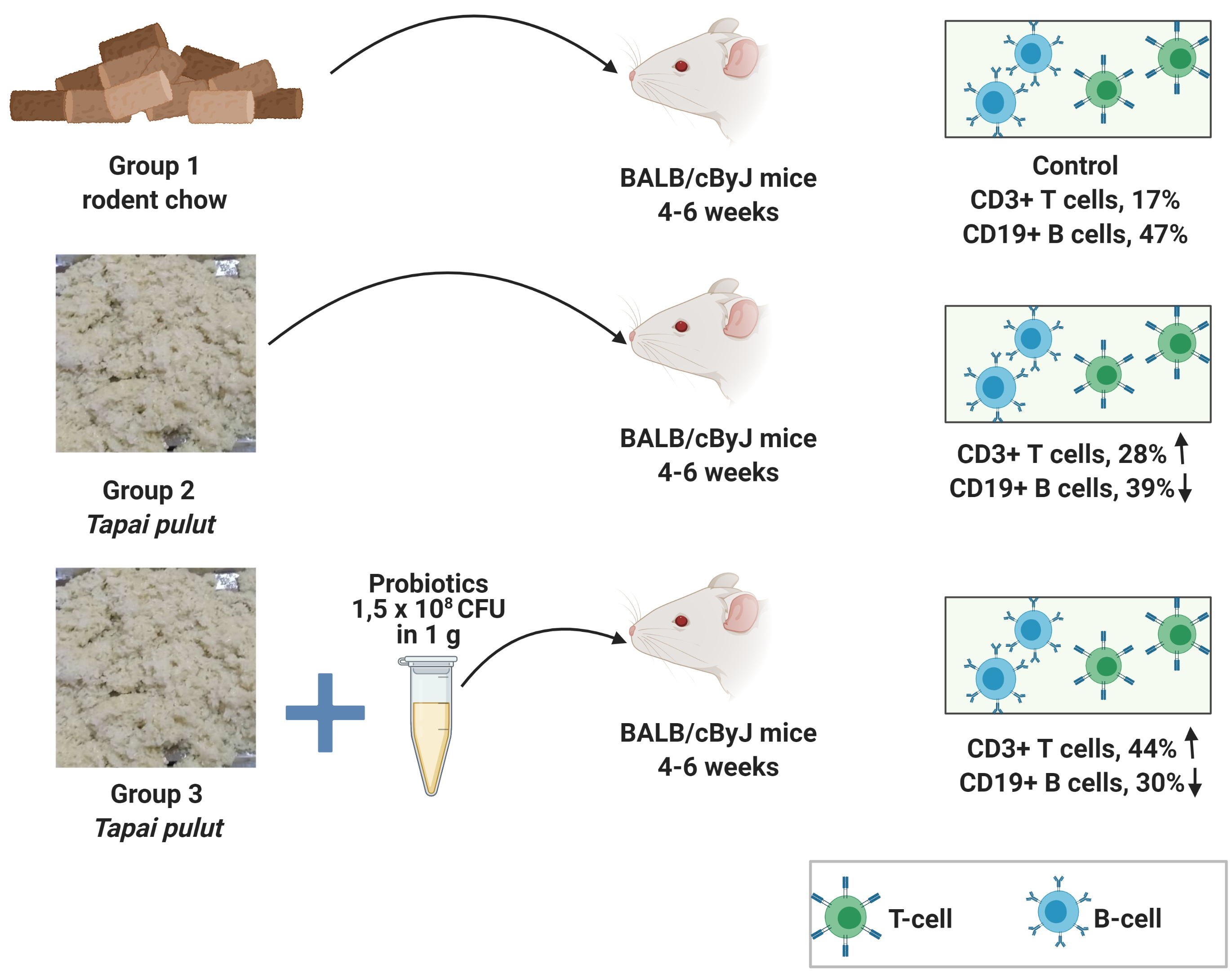
The patterns of the six strains (TA8, PC4, AFSF2, RB5, CL8, and HML3) (after the results from the total bacterial count) were obtained using PCR (SureCycler 8800A, Agilent, Mississauga, ON, Canada) with two universal primers, 27F (AGAGTTTGATCMTGGCTCAG) and 1492 R (TACGGYTACCTTGTTACGACTT) showed high similarity. Therefore, 16S rRNA gene sequencing of six strains was performed and compared with the 16S rDNA gene sequence of Leuc. lactis DSM 20202 revealed the identities of 97.8% to 100 %. In addition, the DNA of each strain was identified. The results shown were agreed in the study by Adesulu-Dahunsi [37], where the microbials of fermented Nigerian food revealed that in LABs comprising lactobacilli, pediococci were the predominant micro-organisms present in viable numbers above 107 CFU g−1. A study that compiled the gene sequence data of Lactobacillus and Pediococcus spp. was used to determine the gene evolution and its phylogeny [38] to identify the strains at the species level. Lactobacilli spp. are differentiated into obligate homofermentative, facultative heterofermentative, and obligate heterofermentative. The differences in this classification of these Lactobacilli spp. are based on the carbohydrate metabolism, which is the pentose phosphate pathway [39]. Thus, a combination of phenotypic properties and molecular techniques 16S rRNA gene sequencing allows for identification at the strain level [40,41]. The phylogenetic tree of the Lactobacilli spp. maybe a cornerstone for future studies on the fermented food industry

4.3. Feeding Samples and Growth Performance

The added probiotic effect was significant on the glutinous rice compared with the traditionally fermented sample using the powder yeast starter culture, as observed for the high acidity content and low pH value. On the other hand, the TSS content was significantly increased for the potential probiotic glutinous rice and fermented glutinous rice. Simultaneously, the highest was observed for the glutinous rice fermented with the traditional starter culture. In a previous study, the fermentation process with different starter cultures increased the TSS content and acidity content of glutinous rice [42]. The strains present only in the potential probiotic glutinous rice sample indicated the absence of the LAB in the powder starter culture known as (ragi tapai). Probiotics play an essential role in restoring the gut microbiota, and their bioactive compounds (probioactives) are suggested to contribute to the health functionality of fermented foods [43]. Therefore, the selected strains (RB5) were added jointly with the starter cultures that produce traditional fermented foods. The fermentation process that includes the added potential probiotics is recommended to increase the cell density of the friendly microorganisms and their bioactive compounds in the final product [44]. In this study, the correlation between the acid and TSS contents was an indicator of the chemical changes due to the probiotic addition alongside the starter culture.
The overall body weight gain in the TPG group was 4.63 g, followed by the PTPG group (3.69 g) and NG group (2.90 g), which correlated with the TSS content in their diets. The traditional fermented glutinous rice had the highest TSS content (53.7 Brix). However, TSS content alone cannot significantly increase body weight. In a recent study, fermented rice products were reported to increase body weight in mice due to the modification of the intestinal microbiota [45]. It was reported that diets containing probiotics increased body weight gain in tested animals (New Zealand male rats) [46]. In another study, a diet containing probiotics belonging to different genera, including Lactobacillus, Clostridium sensu stricto, Prevotella, and Alloprevotella, was reported to increase the body weight in obese mice compared with the control group. The increased body weight was associated with gut microbiota improvement and the reduction of gut pathogens [47]. Thus, a probiotic diet was reported to regulate body weight gain in obese mice and reduce the risk of obesity [48]. There is a high potential that the traditional starter culture contains several probiotics belonging to species other than LAB. Therefore, the microorganisms in the starter cultures may be profiled to determine the important probiotics’ potential presence.

4.4. Haematology Analysis

In this study, we demonstrated that the levels of immune cells in the blood, including WBCs, neutros, and lymphs, were significantly (p < 0.05) increased in mice treated with the probiotic glutinous rice. Probiotic foods and beverages are well known for elevating the immune system cell populations [49,50,51]. Probiotics colonizing in the epithelial cells will boost immune system function by excluding or reducing pathogenic bacteria colonization and reducing the immune cells’ stress [52]. On the other hand, the antimicrobial compounds (organic acids, hydrogen peroxide, bacteriocins, and bioactive peptides) produced by the probiotic strains will enhance intestinal barriers and improve immunomodulation [53]. Probiotic soy milk was observed to increase the white blood cells, including lymphocytes, in rats and improve immune system function [54]. Furthermore, the probiotic and prebiotic combination was reported to significantly increase the neutrophil levels in mice, indicating potential benefits for immune system function [55]. In this study, the increased WBC levels, including neutrophils, demonstrated the probiotic glutinous rice’s immunomodulation effects on the innate immune system [56].

4.5. T-Cell and B-Cell Analysis

The results show a significantly increased glucose level in the TPTG group, followed by PTG and NG (Table 2). The results were correlated with the TSS content in the un-fermented (37.6 Brix), traditionally fermented (53.7 Brix), and probiotic glutinous rice samples (42.8 Brix). On the other hand, this study demonstrated increased triglycerides and LDL contents in the PTPG group. The increase in glucose and lipid levels may be due to the high TSS content. According to Frayn & Kingman [57], high sugar content diets can alter the plasma lipid profile and cause elevation of the triglycerides and low-density lipoprotein due to the metabolism of sugars stored as fats. Diets with high carbohydrate content were reported as high-risk foods for consumers due to the negative impact on sugar and lipid levels in the blood [58]. In this study, the serum AST and ALT enzymes were significantly increased for the TPG and PTPG groups. However, the PTG group showed a significantly higher level than the PTPG. The diet supplemented with RB5 strains results showed that excessive consumption of traditional fermented glutinous rice might have a risk of causing slight liver damage. This can be justified because of the high carbohydrate content in the traditional fermented glutinous rice, as indicated by the high TSS.
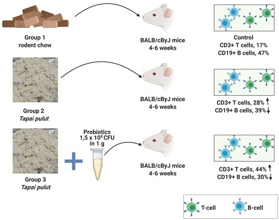
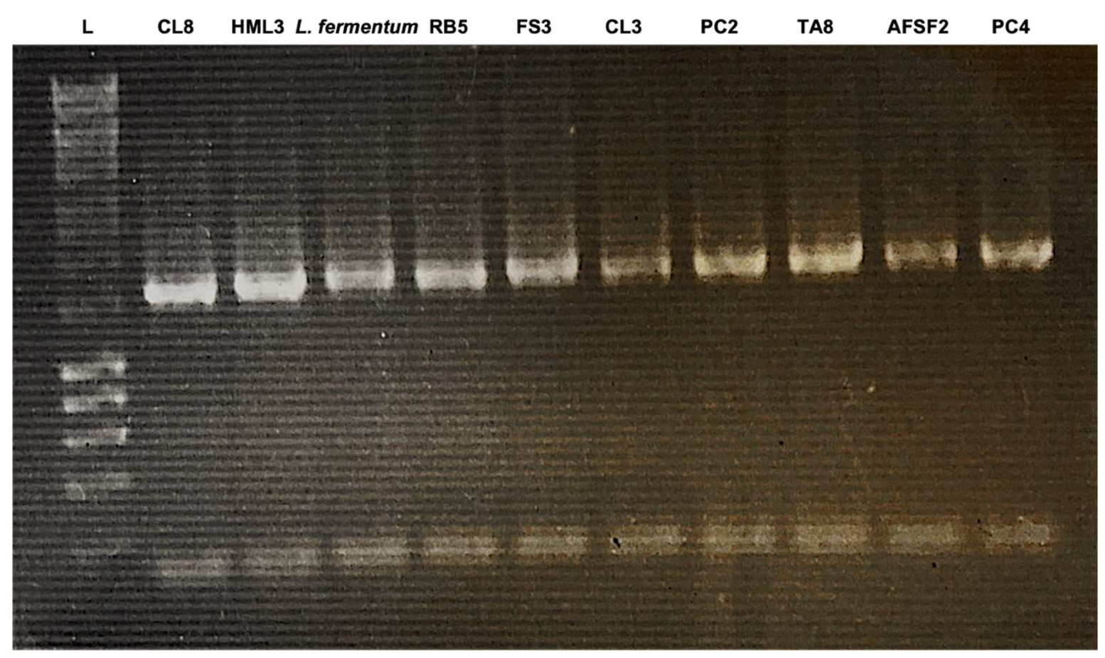
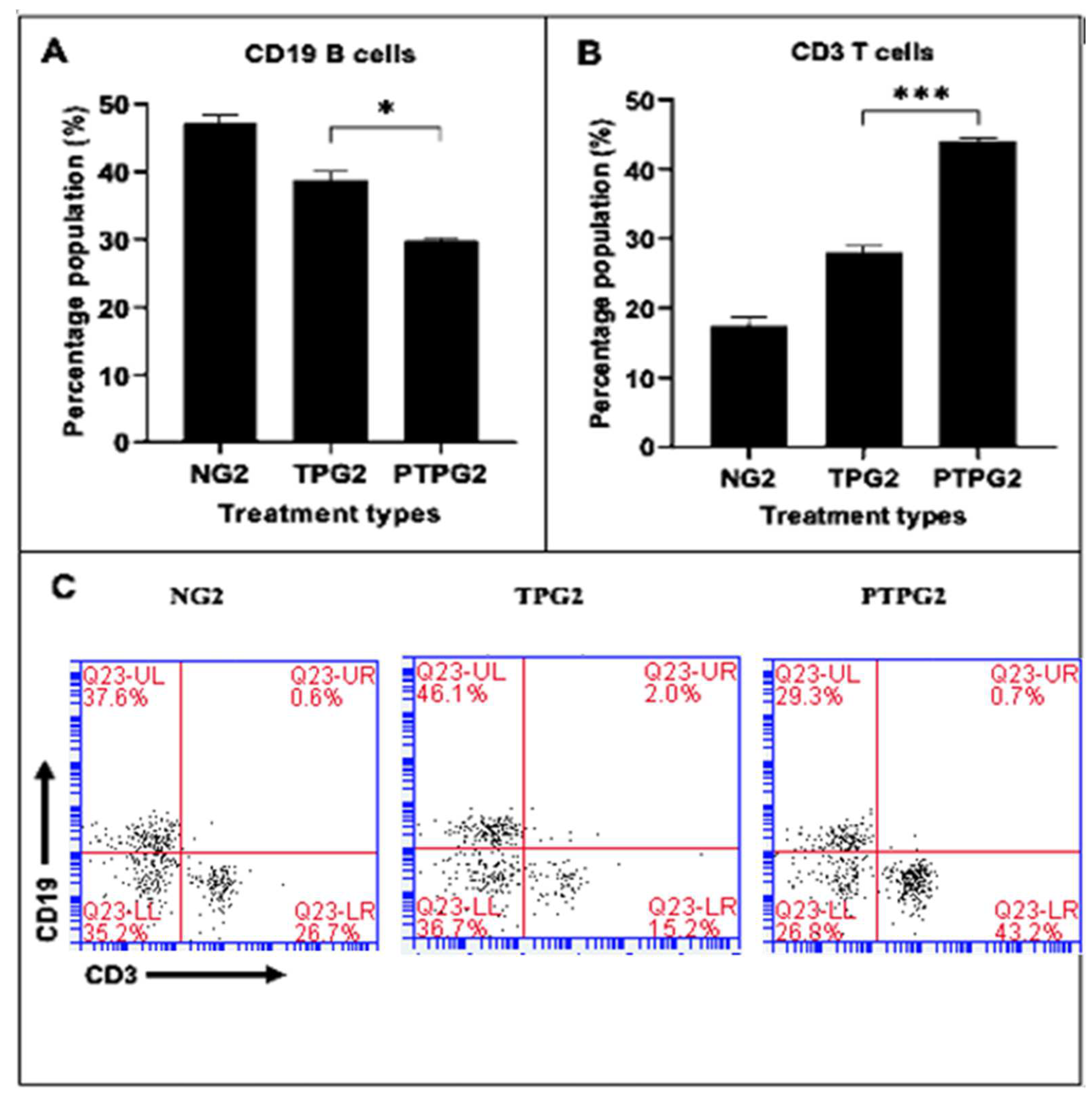
The effects of probiotic, fermented, and non-fermented glutinous rice (tapai pulut) diets on the BALB/cByJ mice’s immune cells, specifically B-cell and T-cells derived from splenocytes, were investigated. The PTPG mice showed the most significant decrease in B-cells and an increase in T-cells after four weeks of treatment, followed by the TPG mice group, which showed a similar trend but a weaker response. Similar observations were seen when the supplementation of Lp. plantarum and Lacticaseibacillus casei in mice showed similar trends in the B-cell and T-cell distribution in the Peyer’s patches and mesenteric lymph nodes. The study found that the CD19+ B-cell population was reduced, while the CD3+ T-cell population was increased [59].
In contrast, the increase in regulatory T-cells (Treg) was specifically highlighted [60]. Recent research has shown that B-cells play an important role in shaping the microbiome by producing an extensive array of secretory IgA antibodies in response to commensals. The decrease or absence of B-cells or IgA could lead to the upregulation of epithelium-inherent immune defense mechanisms conferred by the intestinal epithelia, which eventually causes the microbiome’s changes [61]. On the other hand, the Lactobacillus strains have been reported to have anti-inflammatory properties by promoting T-cell responses, particularly Treg cells, in an IL-10-dependent manner [62]. A previous study in a swine model fed with a high dose of Lactobacillus rhamnosus observed an increased and higher CD3+ T-cells percentage among Peyer’s patch and small intestinal lamina propria lymphocytes [63]. The probiotic diet exhibited an ameliorative effect against potential enteric pathogens.
Furthermore, the administration of probiotic bacteria leads to the increase of CD4 T-cells and CD8 T-cells in the small intestinal lamina propria, which shifts the Th1/Th2 ratio towards a Th2 bias response [64]. Furthermore, a high treatment dose of L.plantarum has been linked to the immunomodulating activity by the significant increase in IL-17 and IL-19 cytokines from the Th17-type immune response. The regulation of Th17 further highlights probiotics’ potential for improving intestinal mucosa immunity [65]. Hence, this study’s results hypothesize the immunomodulatory effect of Lp. plantarum as a probiotic in fermented glutinous rice by reducing the CD19+ B = cell population and stimulating the CD3+ T-cell population in mice splenocytes. Further research can be conducted using T-cell- and B-cell-specific assays to identify the specific cytokines and antibodies released to confirm the findings.

5. Conclusions

The current study demonstrates that RB5 strains (identified as Lactiplantibacillus plantarum) such as the probiotic tapai pulut have a high potential to improve traditional fermented foods’ health benefits. RB5 strains can withstand harsh environmental conditions such as a low pH, bile salts, high temperatures, the presence of an enzyme, and exhibit antibiotic susceptibility. The fermented glutinous rice (tapai pulut) contained higher soluble solids and pH values than the probiotic tapai pulut. However, the probiotics belonging to the lactic acid bacteria group were not detected in the tapai pulut and the unfermented glutinous rice. In addition, tapai pulut significantly increased the mice group’s body weight due to the high sugar content. However, the probiotic tapai pulut increased the immune cell population, including white blood cells, neutrophils, and lymphocytes.
Moreover, the probiotic tapai pulut group demonstrated a high CD3+ T cells population. The results reveal the potential immunomodulation properties of the tapai pulut in which Lactiplantibacillus plantarum serves as a co-culture. Hence, the mechanism of action in the immunomodulatory pathway has been clearly depicted from the selection and identification of potential probiotics.

Supplementary Materials

The following supporting information can be downloaded at: https://www.mdpi.com/article/10.3390/fermentation8110612/s1. Table S1: The origins of the 53 isolates with its species name; Table S2: The survivability rate of 53 strains after 6 h of exposure at different parameters.

Author Contributions

Conceptualization, M.H. (Muhaini Hussin), A.A. and C.X.-Q.L.; Formal analysis, M.H. (Muhaini Hussin), C.X.-Q.L. and B.J.M.; Funding acquisition, B.J.M. and A.S.M.H.; Investigation, M.H. (Muhaini Hussin), A.A. and C.X.-Q.L.; Methodology, M.H. (Muhaini Hussin), A.A. and C.X.-Q.L.; Project administration, A.S.M.H.; Resources, C.-M.F., M.Z.S. and A.S.M.H.; Software, B.J.M.; Supervision, C.-M.F., M.Z.S., N.H.A., M.H. (Masriana Hassan), H.A. and A.S.M.H.; Validation, M.H. (Muhaini Hussin), A.A. and C.X.-Q.L.; Visualization, M.H. (Muhaini Hussin) and A.A.; Writing—original draft, M.H. (Muhaini Hussin) and C.X.-Q.L.; Writing—review & editing, M.H. (Muhaini Hussin) and A.Z.M. All authors have read and agreed to the published version of the manuscript.

Funding

This study was funded by the Ministry of Science, Technology, and Innovation (MOSTI) under the International Collaboration Fund (ICF), grant number (IF0419A1077).

Institutional Review Board Statement

The compulsory ethical approval was obtained from the Institutional Animal Care and Use Committee on 10 April 2019, and the reference number was UPM/IACUC/AUP-R004/2019.

Informed Consent Statement

Not applicable.

Data Availability Statement

Not applicable.

Acknowledgments

The authors are grateful to the Comparative Medicine and Technology Unit’s (COMeT) staff, Universiti Putra Malaysia. We thank Sharina Omar and Sabri of the Faculty of Veterinary Science, Universiti Malaysia, and the technical staff of the Division of Biomedical Science, The University of Nottingham, Malaysia. We appreciate the help from Muna Mahmood Taleb Abadl in helping the team to prepare for the animal study.

Conflicts of Interest

The authors declare no conflict of interest.

References

  1. Hill, C.; Guarner, F.; Reid, G.; Gibson, G.R.; Merenstein, D.J.; Pot, B.; Morelli, L.; Canani, R.B.; Flint, H.J.; Salminen, S.; et al. Expert consensus document: The International Scientific Association for Probiotics and Prebiotics consensus statement on the scope and appropriate use of the term probiotic. Nat. Rev. Gastroenterol. Hepatol. 2014, 11, 506–514. [Google Scholar] [CrossRef] [Green Version]
  2. Gerez, C.L.; Torres, M.J.; De Valdez, G.F.; Rollán., G. Control of spoilage fungi by lactic acid bacteria. Biol. Control 2013, 64, 231–237. [Google Scholar] [CrossRef]
  3. Samtiya, M.; Aluko, R.E.; Puniya, A.K.; Dhewa, T. Enhancing micronutrients bioavailability through fermentation of plant-based foods: A concise review. Fermentation 2021, 7, 63. [Google Scholar] [CrossRef]
  4. Azad, M.; Kalam, A.; Sarker, M.; Wan, D. Immunomodulatory effects of probiotics on cytokine profiles. BioMed Res. Int. 2018, 2018, 8063647. [Google Scholar] [CrossRef] [Green Version]
  5. Lee, Y.J.; Lee, A.; Yoo, H.J.; Kim, M.; Noh, G.M.; Lee, J.H. Supplementation with the probiotic strain Weissella cibaria JW15 enhances natural killer cell activity in nondiabetic subjects. J. Funct. Foods 2018, 48, 153–158. [Google Scholar] [CrossRef]
  6. Markov, A.V.; Sen’kova, A.V.; Babich, V.O.; Odarenko, K.V.; Talyshev, V.A.; Salomatina, O.V.; Salakhutdinov, N.F.; Zenkova, M.A.; Logashenko, E.B. Dual effect of soloxolone methyl on LPS-induced inflammation in vitro and in vivo. Int. J. Mol. Sci. 2020, 21, 7876. [Google Scholar] [CrossRef]
  7. Fukushima, Y.; Kawata, Y.; Mizumachi, K.; Kurisaki, J.I.; Mitsuoka, T. Effect of bifidobacteria feeding on fecal flora and production of immunoglobulins in lactating mouse. Int. J. Food Microbiol. 1999, 46, 193–197. [Google Scholar] [CrossRef]
  8. Merican, Z.; Quee-Lan, Y. Tapai processing in Malaysia: A technology in transition. In Industrialization of Indigenous Fermented Foods, Revised and Expanded, 2nd ed.; Steinkraus, K., Ed.; Marcel Dekker, Inc.: New York, NY, USA, 2004; pp. 247–269. [Google Scholar]
  9. Kofli, N.T.; Dayaon, S.H.M. Identification of Microorganism from Ragi for Bioethanol Production by API Kit. J. Appl. Sci. 2010, 10, 2751–2753. [Google Scholar] [CrossRef] [Green Version]
  10. Hermanto, F.E.; Warsito, W.; Rifa’I, M.; Widodo, N.; Jatmiko, Y.D. Metabarcoding dataset on the elicitation of Soybean and Mungbean using Ragi Tape as elicitors for enhancing secondary metabolites production. Data Brief 2022, 42, 108209. [Google Scholar] [CrossRef]
  11. Hamid, T.H.T.A.; Amsya, N.F. Lactic acid bacterium with antimicrobial properties from selected malay traditional fermented foods. Int. J. Life Sci. Biotechnol. 2020, 4, 13–24. [Google Scholar] [CrossRef]
  12. Sujaya, I.; Amachi, S.; Yokota, A.; Asano, K.; Tomita, F. Identification and characterization of lactic acid bacteria in ragi tape. World J. Microbiol. Biotechnol. 2001, 17, 349–357. [Google Scholar] [CrossRef]
  13. Martinez, R.C.; Aynaou, A.E.; Albrecht, S.; Schols, H.A.; De Martinis, E.C.; Zoetendal, E.G., Venema; Zoetendal, E.G.; Venema, K.; Smidt, H. In vitro evaluation of gastrointestinal survival of Lactobacillus amylovorus DSM 16698 alone and combined with galactooligosaccharides, milk and/or Bifidobacterium animalis subsp. lactis Bb-12. Int. J. Food Microbio. 2011, 149, 152–158. [Google Scholar] [CrossRef]
  14. Liasi, S.A.; Azmi, T.I.; Hassan, M.D.; Shuhaimi, M.; Rosfarizan, M.; Ariff, A.B. Antimicrobial activity and antibiotic sensitivity of three isolates of lactic acid bacteria from fermented fish product, Budu. Malays. J. Microb. 2009, 5, 33–37. [Google Scholar]
  15. D’Aimmo, M.R.; Modesto, M.; Biavati, B. Antibiotic resistance of lactic acid bacteria and Bifidobacterium spp. isolated from dairy and pharmaceutical products. Int. J. Food. Microbiol. 2007, 115, 35–42. [Google Scholar] [CrossRef]
  16. Lane, D.J. 16S/23S rRNA Sequencing. In Nucleic Acid Techniques in Bacterial Systematic; Stackebrandt, E., Goodfellow, M., Eds.; John Wiley and Sons: New York, NY, USA, 1991; pp. 115–175. [Google Scholar]
  17. Tamura, K.; Stecher, G.; Peterson, D.; Filipski, A.; Kumar, S. MEGA6: Molecular evolutionary genetics analysis version 6.0. Mol. Biol. Evol. 2013, 30, 2725–2729. [Google Scholar] [CrossRef] [Green Version]
  18. Association of Official Analytical Chemists (AOAC). Official Methods of Analysis of the Association Analytical Chemists, 18th ed.; AOAC: Gaithersburg, MD, USA, 2005. [Google Scholar]
  19. Zhao, J.; Fleet, G. Yeasts are essential for cocoa bean fermentation. Int. J. Food Microbiol. 2014, 174, 72–87. [Google Scholar] [CrossRef]
  20. Chuah, L.O.; Shamila-Syuhada, A.K.; Liong, M.T.; Rosma, A.; Thong, K.L.; Rusul, G. Physio-chemical, microbiological properties of tempoyak and molecular characterisation of lactic acid bacteria isolated from tempoyak. Food Microbiol. 2016, 58, 95–104. [Google Scholar] [CrossRef]
  21. Adnan, A.F.M.; Tan, I.K. Isolation of lactic acid bacteria from Malaysian foods and assessment of the isolates for industrial potential. Bioresour. Technol. 2007, 98, 1380–1385. [Google Scholar] [CrossRef]
  22. Swain, M.R.; Anandharaj, M.; Ray, R.C.; Rani, R.P. Fermented fruits and vegetables of Asia: A potential source of probiotics. Biotechnol. Res. Int. 2014, 2014, 250424. [Google Scholar] [CrossRef]
  23. Ruas-Madiedo, P.; Moreno, J.A.; Salazar, N.; Delgado, S.; Mayo, B.; Margolles, A.; de Los Reyes-Gavilán, C.G. Screening of exopolysaccharide-producing Lactobacillus and Bifidobacterium strains isolated from the human intestinal microbiota. Appl. Environ. Microbiol. 2007, 73, 4385–4388. [Google Scholar] [CrossRef] [Green Version]
  24. Ren, D.; Li, C.; Qin, Y.; Yin, R.; Du, S.; Ye, F.; Liu, C.; Liu, H.; Wang, M.; Li, Y.; et al. In vitro evaluation of the probiotic and functional potential of Lactobacillus strains isolated from fermented food and human intestine. Anaerobe 2014, 30, 1–10. [Google Scholar] [CrossRef]
  25. Liu, W.J.; Chen, Y.F.; Kwok, L.Y.; Li, M.H.; Sun, T.; Sun, C.L.; Wang, X.N.; Dan, T.; Zhang, H.P.; Sun, T.S. Preliminary selection for potential probiotic Bifidobacterium isolated from subjects of different Chinese ethnic groups and evaluation of their fermentation and storage characteristics in bovine milk. J. Dairy Sci. 2013, 96, 6807–6817. [Google Scholar] [CrossRef] [Green Version]
  26. Anandharaj, M.; Sivasankari, B.; Santhanakaruppu, R.; Manimaran, M.; Rani, R.P.; Sivakumar, S. Determining the probiotic potential of cholesterol-reducing Lactobacillus and Weissella strains isolated from gherkins (fermented cucumber) and south Indian fermented koozh. Res. Microbiol. 2015, 166, 428–439. [Google Scholar] [CrossRef]
  27. García-Ruiz, A.; de Llano, D.G.; Esteban-Fernández, A.; Requena, T.; Bartolomé, B.; Moreno-Arribas, M.V. Assessment of probiotic properties in lactic acid bacteria isolated from wine. Food Microbiol. 2014, 44, 220–225. [Google Scholar] [CrossRef]
  28. Noriega, L.; Gueimonde, M.; Sánchez, B.; Margolles, A.; de los Reyes-Gavilán, C.G. Effect of the adaptation to high bile salts concentrations on glycosidic activity, survival at low pH and cross-resistance to bile salts in Bifidobacterium. Int. J. Food Microbiol. 2004, 94, 79–86. [Google Scholar] [CrossRef]
  29. Shokryazdan, P.; Faseleh Jahromi, M.; Liang, J.B.; Ramasamy, K.; Sieo, C.C.; Ho, Y.W. Effects of a Lactobacillus salivarius mixture on performance, intestinal health and serum lipids of broiler chickens. PLoS ONE 2017, 12, e0175959. [Google Scholar] [CrossRef] [Green Version]
  30. Meybodi, N.M.; Mortazavian, A.M.; Arab, M.; Nematollahi, A. Probiotic viability in yoghurt: A review of influential factors. Int. Dairy J. 2020, 109, 104793. [Google Scholar] [CrossRef]
  31. Mortazavian, A.M.; Ehsani, M.R.; Mousavi, S.M.; Reinheimer, J.A.; Emamdjomeh, Z.; Sohrabvandi, S.; Rezaei, K. Preliminary investigation of the combined effect of heat treatment and incubation temperature on the viability of the probiotic micro-organisms in freshly made yogurt. Int. J. Dairy Technol. 2006, 59, 8–11. [Google Scholar] [CrossRef]
  32. Corona-Hernandez, R.I.; Álvarez-Parrilla, E.; Lizardi-Mendoza, J.; Islas-Rubio, A.R.; de la Rosa, L.A.; Wall-Medrano, A. Structural stability and viability of microencapsulated probiotic bacteria: A review. Compr. Rev. Food Sci. Food Saf. 2013, 12, 614–628. [Google Scholar] [CrossRef]
  33. Cruz, A.G.D.; Castro, W.F.; Faria, J.; Bolini, H.M.A.; Celeghini, R.M.D.S.; Raices, R.S.L.; Oliveira, C.A.F.; Freitas, M.Q.; Júnior, C.C.; Mársico, E.T. Stability of probiotic yogurt added with glucose oxidase in plastic materials with different permeability oxygen rates during the refrigerated storage. Food Res. Int. 2013, 51, 723–728. [Google Scholar] [CrossRef]
  34. Zoumpopoulou, G.; Tzouvanou, A.; Mavrogonatou, E.; Alexandraki, V.; Georgalaki, M.; Anastasiou, R.; Papadelli, M.; Manolopoulou, E.; Kazou, M.; Kletsas, D.; et al. Probiotic features of lactic acid bacteria isolated from a diverse pool of traditional Greek dairy products regarding specific strain-host interactions. Probiotics Antimicrob. Proteins 2018, 10, 313–322. [Google Scholar] [CrossRef]
  35. Rahayu, H.M.; Qurbaniah, M. Selection of Tempoyak Lactic Acid Bacteria as Candidate Strain for Yoghurt Starter Culture. Biosaintifika: J. Biol. Educ. 2019, 11, 39–46. [Google Scholar] [CrossRef] [Green Version]
  36. Cao, M.; Feng, Y.; Zhang, Y.; Kang, W.; Lian, K.; Ai, L. Studies on the metabolism and degradation of vancomycin in simulated in vitro and aquatic environment by UHPLC-Triple-TOF-MS/MS. Sci. Rep. 2018, 8, 15471. [Google Scholar] [CrossRef] [Green Version]
  37. Jafari-Nasab, T.; Khaleghi, M.; Farsinejad, A.; Khorrami, S. Probiotic potential and anticancer properties of Pediococcus sp. isolated from traditional dairy products. Biotechnol. Rep. 2021, 29, e00593. [Google Scholar] [CrossRef]
  38. Gueimonde, M.; Sánchez, B.G.; de los Reyes-Gavilán, C.; Margolles, A. Antibiotic resistance in probiotic bacteria. Front. Microbiol. 2013, 4, 202. [Google Scholar] [CrossRef] [Green Version]
  39. Adesulu-Dahunsi, A.T.; Sanni, A.I.; Jeyaram, K. Rapid differentiation among Lactobacillus, Pediococcus and Weissella species from some Nigerian indigenous fermented foods. LWT 2017, 77, 39–44. [Google Scholar] [CrossRef] [Green Version]
  40. Zheng, J.; Ruan, L.; Sun, M.; Gänzle, M. A genomic view of lactobacilli and pediococci demonstrates that phylogeny matches ecology and physiology. Appl. Environ. Microbiol. 2015, 81, 7233–7243. [Google Scholar] [CrossRef] [Green Version]
  41. Xu, X.; Zhou, S.; McClements, D.J.; Huang, L.; Meng, L.; Xia, X.; Dong, M. Multistarter fermentation of glutinous rice with Fu brick tea: Effects on microbial, chemical, and volatile compositions. Food Chem. 2020, 309, 125790. [Google Scholar] [CrossRef]
  42. Champagne, C.P.; da Cruz, A.G.; Daga, M. Strategies to improve the functionality of probiotics in supplements and foods. Curr. Opin. Food Sci. 2018, 22, 160–166. [Google Scholar] [CrossRef]
  43. Shiby, V.K.; Mishra, H.N. Fermented milks and milk products as functional foods—A review. Crit. Rev. Food Sci. Nutr. 2013, 53, 482–496. [Google Scholar] [CrossRef]
  44. Huang, Z.R.; Chen, M.; Guo, W.L.; Li, T.T.; Liu, B.; Bai, W.D.; Ai, L.Z.; Rao, P.F.; Ni, L.; Lv, X.C. Monascus purpureus-fermented common buckwheat protects against dyslipidemia and non-alcoholic fatty liver disease through the regulation of liver metabolome and intestinal microbiome. Food Res. Int. 2020, 136, 109511. [Google Scholar] [CrossRef] [PubMed]
  45. Ayyat, M.S.; Al-Sagheer, A.A.; El-Latif, A.; Khaled, M.; Khalil, B.A. Organic selenium, probiotics, and prebiotics effects on growth, blood biochemistry, and carcass traits of growing rabbits during summer and winter seasons. Biol. Trace Elem. Res. 2018, 186, 162–173. [Google Scholar] [CrossRef] [PubMed]
  46. Kong, C.; Gao, R.; Yan, X.; Huang, L.; Qin, H. Probiotics improve gut microbiota dysbiosis in obese mice fed a high-fat or high-sucrose diet. Nutrition 2019, 60, 175–184. [Google Scholar] [CrossRef] [PubMed]
  47. de Carvalho Marchesin, J.; Celiberto, L.S.; Orlando, A.B.; de Medeiros, A.I.; Pinto, R.A.; Zuanon, J.A.S.; Spolidorio, L.C.; dos Santos, A.; Taranto, M.P.; Cavallini, D.C.U. A soy-based probiotic drink modulates the microbiota and reduces body weight gain in diet-induced obese mice. J. Funct. Foods 2018, 48, 302–313. [Google Scholar] [CrossRef] [Green Version]
  48. Lehtoranta, L.; Latvala, S.; Lehtinen, M.J. Role of probiotics in stimulating the immune system in viral respiratory tract infections: A narrative review. Nutrients 2020, 12, 3163. [Google Scholar] [CrossRef] [PubMed]
  49. Ganguly, S.; Sabikhi, L.; Singh, A.K. Effect of whey-pearl millet-barley based probiotic beverage on Shigella-induced pathogenicity in murine model. J. Funct. Foods 2019, 54, 498–505. [Google Scholar] [CrossRef]
  50. Tang, C.; Zhaoxin, L. Health promoting activities of probiotics. J. Food Biochem. 2019, 43, e12944. [Google Scholar] [CrossRef]
  51. Sanders, M.E.; Merenstein, D.J.; Reid, G.; Gibson, G.R.; Rastall, R.A. Probiotics and prebiotics in intestinal health and disease: From biology to the clinic. Nat. Rev. Gastroenterol. Hepatol. 2019, 16, 605–616. [Google Scholar] [CrossRef]
  52. Wan, M.L.Y.; Forsythe, S.J.; El-Nezami, H. Probiotics interaction with foodborne pathogens: A potential alternative to antibiotics and future challenges. Crit. Rev. Food Sci. Nutr. 2019, 59, 3320–3333. [Google Scholar] [CrossRef]
  53. Niamah, A.K.; Sahi, A.A.; Al-Sharifi, A.S. Effect of feeding soy milk fermented by probiotic bacteria on some blood criteria and weight of experimental animals. Probiotics Antimicrob. Proteins 2017, 9, 284–291. [Google Scholar] [CrossRef]
  54. Sharma, R.; Kumari, M.; Kumari, A.; Sharma, A.; Gulati, A.; Gupta, M.; Padwad, Y. Diet supplemented with phytochemical epigallocatechin gallate and probiotic Lactobacillus fermentum confers second generation synbiotic effects by modulating cellular immune responses and antioxidant capacity in aging mice. Eur. J. Nutr. 2019, 58, 2943–2957. [Google Scholar] [CrossRef] [PubMed]
  55. Mortaz, E.; Alipoor, S.D.; Adcock, I.M.; Mumby, S.; Koenderman, L. Update on neutrophil function in severe inflammation. Front. Immunol. 2018, 9, 2171. [Google Scholar] [CrossRef] [PubMed] [Green Version]
  56. Frayn, K.N.; Kingman, S.M. Dietary sugars and lipid metabolism in humans. Am. J. Clin. Nutr. 1995, 62, 250S–261S. [Google Scholar] [CrossRef] [PubMed]
  57. Park, S.; Ahn, J.; Kim, N.S.; Lee, B.K. High carbohydrate diets are positively associated with the risk of metabolic syndrome irrespective to fatty acid composition in women: The KNHANES 2007–2014. Int. J. Food Sci. Nutr. 2017, 68, 479–487. [Google Scholar] [CrossRef]
  58. Aminlari, L.; Shekarforoush, S.S.; Hosseinzadeh, S.; Nazifi, S.; Sajedianfard, J.; Eskandari, M.H. Effect of probiotics Bacillus coagulans and Lactobacillus plantarum on lipid profile and feces bacteria of rats fed cholesterol-enriched diet. Probiotics Antimicrob. Proteins 2019, 11, 1163–1171. [Google Scholar] [CrossRef]
  59. van Beek, A.A.; Sovran, B.; Hugenholtz, F.; Meijer, B.; Hoogerland, J.A.; Mihailova, V.; van der Ploeg, C.; Belzer, C.; Boekschoten, M.V.; Hoeijmakers, J.H.; et al. Supplementation with Lactobacillus plantarum WCFS1 prevents decline of mucus barrier in colon of accelerated aging Ercc1−/Δ7 mice. Front. Immunol. 2016, 7, 408. [Google Scholar] [CrossRef] [Green Version]
  60. Zheng, D.; Liwinski, T.; Elinav, E. Interaction between microbiota and immunity in health and disease. Cell Res. 2020, 30, 492–506. [Google Scholar] [CrossRef]
  61. Ding, Y.H.; Qian, L.Y.; Pang, J.; Lin, J.Y.; Xu, Q.; Wang, L.H.; Huang, D.S.; Zou, H. The regulation of immune cells by Lactobacilli: A potential therapeutic target for anti-atherosclerosis therapy. Oncotarget 2017, 8, 59915. [Google Scholar] [CrossRef] [Green Version]
  62. Zhu, Y.H.; Li, X.Q.; Zhang, W.; Zhou, D.; Liu, H.Y.; Wang, J.F. Dose-dependent effects of Lactobacillus rhamnosus on serum interleukin-17 production and intestinal T-cell responses in pigs challenged with Escherichia coli. Appl. Environ. Microbiol. 2014, 80, 1787–1798. [Google Scholar] [CrossRef] [Green Version]
  63. Smelt, M.J.; de Haan, B.J.; Bron, P.A.; van Swam, I.; Meijerink, M.; Wells, J.M.; Faas, M.M.; de Vos, P. Probiotics can generate FoxP3 T-cell responses in the small intestine and simultaneously inducing CD4 and CD8 T cell activation in the large intestine. PLoS ONE 2013, 8, e68952. [Google Scholar] [CrossRef] [Green Version]
  64. Xie, J.; Nie, S.; Yu, Q.; Yin, J.; Xiong, T.; Gong, D.; Xie, M. Lactobacillus plantarum NCU116 attenuates cyclophosphamide-induced immunosuppression and regulates Th17/Treg cell immune responses in mice. J. Agric. Food Chem. 2016, 64, 1291–1297. [Google Scholar] [CrossRef] [PubMed]
  65. Santiago-López, L.; Hernández-Mendoza, A.; Garcia, H.S.; Mata-Haro, V.; Vallejo-Cordoba, B.; González-Córdova, A.F. The effects of consuming probiotic-fermented milk on the immune system: A review of scientific evidence. Int. J. Dairy Technol. 2015, 68, 153–165. [Google Scholar] [CrossRef]
Figure 1. The amplified DNA from PCR reactions of 0.8% agarose gel ran at 110 V for 60 min using Hyperladder 1 kB as DNA ladder (L), and each sample was loaded at 10 uL, respectively. The first strains from well number 2 (start from left is CL8, HML3, Lactobacillus fermentum RB5, FS3, CL3, PC2, TA8, AFSF2, PC4).
Fermentation 08 00612 g001
Figure 2. T-cells and B-cells populations following the different diet treatment types were measured by fluorescence-activated cell sorting (FACS) flow cytometry. The percentage of (A) B-cells and (B) T-cells populations were calculated as % of total CD19+ B cells and CD3+ T cells, respectively. (C) Representative flow cytometric dot plots showing T-cells (CD3+) and B-cells (CD19+) as calculated from the percentages of total live cells (50,000 events). Unfermented glutinous rice (NG) served as the control group. Both traditional Tapai Pulut (TPG) and probiotic Tapai Pulut (PTPG) showed a significant decrease in B-cells and an increase in T-cells populations. PTPG2 exhibited the most significant decrease in the percentage of CD19+ B-cells and an increase in the CD3+ T-cells populations compared with TPG2 against the NG2 control. Results are expressed as percentage of cells population (%) ± S.E.M. Asterisk over bars indicates the degree of significance as compared with the traditional Tapai Pulut (TPG) group; * p ≤ 0.005, and *** p ≤ 0.0001.
Fermentation 08 00612 g002
Table 1. The survivability rates of 10 strains after 6 h of exposure at different parameters against Lactobacillus fermentum ATCC:14931.
StrainsControlAcid
(pH 3.0)
Temperature (40 °C)Bile Salts (3%)Enzyme (Pepsin)Overall SR (%) 1
CL39.35 ± 0.018.35 ± 0.016.57 ± 0.027.12 ± 0.016.24 ± 0.0567.64 a
PC28.34 ± 0.107.34 ± 0.106.38 ± 0.107.78 ± 0.036.03 ± 0.0277.21 a
TA88.44 ± 0.017.44 ± 0.016.52 ± 0.057.00 ± 0.016.03 ± 0.0166.14 a,b
PC47.97 ± 0.107.07 ± 0.106.38 ± 0.017.13 ± 0.026.39 ± 0.0489.62 b
AFSF28.78 ± 0.017.48 ± 0.016.59 ± 0.068.13 ± 0.106.13 ± 0.0177.21 c
L. fermentum ATCC:149318.99 ± 0.037.39 ± 0.036.37 ± 0.057.13 ± 0.036.06 ± 0.0184.99 a,b
FS39.20 ± 0.027.20 ± 0.026.26 ± 0.018.13 ± 0.076.21 ± 0.0276.38 a
RB58.35 ± 0.158.35 ± 0.156.78 ± 0.007.04 ± 0.076.91 ± 0.0198.94 a
CL88.49 ± 0.028.49 ± 0.026.52 ± 0.027.00 ± 0.026.23 ± 0.0688.74 b
HML37.07 ± 0.088.07 ± 0.086.24 ± 0.047.41± 0.096.37 ± 0.0665.96 a,b,c
1, a,b,c within the survivability rate (SR) column represents significant differences (p ≤ 0.05) according to Tukey’s test.
Table 2. Antibiotic susceptibilities of seven isolated strains of Lactobacillus and two reference strains.
StrainBAMPDACEIPMTEVAAMCGN
CL3RRRSRRSRRS
PC2RSRSRRSRSS
TA8RSRSRRRRSR
PC4RRRSRRRRRR
AFSF2RSRSRRSRRR
L. fermentum ATCC:14931RRRSRRSRSR
FS3RRRSRRSRRR
RB5RRRSRRSRSR
CL8RRRSRRRRRR
HML3RRRSRRSRSR
B, Blank; AMP, Ampicillin; DA Clindamycin; C, Chloramphenicol; E, Erythromycin; IPM, Imipenem; TE, Tetracycline; VA, Vancomycin; AMC, Amoxycillin/Clavulanic Acid; GN, Gentamicin; S, Sensitive; R, Resistant.
Table 3. API 50CHL kit identification of the ten LABs with their characterizations and fermentable sugars.
Sugars Fermented
Strains NameCell ShapeGas from GlucoseNH3 from ArginineLactate IsomerGrowth at 45 °CArabinoseCellobioseEsculinGalactoseLactoseMaltose
CL3 *rodDL+++
PC2 *rodDL+++
TA8 *rodDL+++
PC4 *rodDL+++
AFSF2 *rodDL+++
L. fermentum ATCC:14931 *rodDL+++
FS3 *rodDL+++
RB5 *rodDL+++
CL8 *coccoid+DL++4/14/1++
HML3 *rodDL+++
* +, all strains fermented positive; −, all strains negative.
Table 4. Identification of the six isolates using API 50 CHL kits and 16S rRNA gene.
StrainPhenotypic IdentificationGenotypic IdentificationGene Accession No.
TA8Lactobacillus plantarum 1Lactiplantibacillus plantarum strain 10334MW090367.1
PC4Lactobacillus plantarum 1Lactiplantibacillus plantarum strain 11112 MW092969.1
AFSF2Lactobacillus plantarum 1Lactiplantibacillus plantarum strain 11905 sequenceMW116637.1
RB5Lactobacillus plantarum 1Lactiplantibacillus plantarum strain NHD2 16S ribosomal RNA gene, partial sequenceMW345831.1
CL8Pediococcus spp.Pediococcus acidilactici strain FCP-1 MN367973.1
HML3Lactobacillus plantarum 1Lactiplantibacillus plantarum strain XYY2MW301118.1
Table 5. Chemical variables analyses and cell counts for the unfermented and fermented glutinous rice samples.
SamplepH ValueAcidity (% w/v)Moisture (%)°BrixCell Count (log10 CFU g−1)
NG5.96 ± 0.03 a0.17 ± 0.04 c38.69 ± 0.51 a37.6 ± 1.43 cND
PTG4.54 ± 0.12 b0.26 ± 0.06 b33.29 ± 0.41 b53.7 ± 0.98 aND
PTPG3.77 ± 0.06 c0.531 ± 0.04 a34.15 ± 0.48 b42.8 ± 1.62 b8.11 ± 0.45
NG (normal group), TPG (tapai pulut group), PTPG (probiotic tapai pulut group), ND (not detected). a,b,c represent significant differences within the column (p < 0.05).
Table 6. Effects on the body weight, food intake, and feed efficiency.
GroupsAverage Weekly Weight Gain (g)Average Feed IntakeFeed Efficiency
NGWeek 1: 0.52 ± 0.82 cWeek 1: 17.89 ± 1.35Week 1: 0.02
Week 2: 0.52 ± 0.74 cWeek 2: 14.25 ± 0.91Week 2: 0.03
Week 3: 0.54 ± 0.76 bWeek 3: 13.59 ± 0.87Week 3: 0.03
Week 4: 1.32 ± 0.21 aWeek 4: 16.67 ± 0.54Week 4: 0.07
Overall: 2.90 ± 0.39 cOverall: 15.62 ± 2.03Overall: 0.18
TPGWeek 1: 1.93 ± 0.49 aWeek 1: 15.96 ± 2.10Week 1: 0.12
Week 2: 0.66 ± 0.69 bWeek 2: 15.98 ± 0.86Week 2: 0.04
Week 3: 0.87 ± 0.70 aWeek 3: 11.15 ± 0.54Week 3: 0.07
Week 4: 1.17 ± 0.23 bWeek 4: 16.84 ± 0.63Week 4: 0.06
Overall: 4.63 ± 0.55 aOverall: 14.98 ± 2.58Overall: 0.30
PTPGWeek 1: 1.10 ± 0.92 bWeek 1: 17.21 ± 1.90Week 1: 0.06
Week 2: 0.90 ± 0.55 aWeek 2: 18.46 ± 1.03Week 2: 0.04
Week 3: 0.78 ± 0.20 aWeek 3: 14.70 ± 1.42Week 3: 0.05
Week 4: 0.91 ± 0.18 bWeek 4: 15.09 ± 0.15Week 4: 0.06
Overall: 3.69 ± 0.13 bOverall: 16.36 ± 1.77Overall: 0.22
NG (normal group), TPG (tapai pulut group), PTPG (probiotic tapai pulut group). a,b,c represent significant differences within the column (p < 0.05).
Table 7. The biochemical examination results of the blood obtained from the different mice groups.
TestNGTPGPTPG
WBC × 109/L3.70 ± 0.35 c4.30 ± 1.62 b4.60 ± 1.20 a
RBC × 1012/L9.69 ± 2.80 a9.35 ± 2.43 b9.66 ± 0.79 a,b
Neutrophil × 109/L0.59 ± 0.07 c0.69 ± 0.37 b0.74 ± 0.23 a
Lymp × 109/L2.81 ± 0.38 c3.27 ± 0.19 b3.54 ± 0.26 a
Mono × 109/L0.22 ± 0.04 b0.26 ± 0.06 a0.23 ± 0.01 a,b
Eosin × 109/L0.04 ± 0.00 a0.04 ± 0.00 a0.05 ± 0.01 a
AST (U/L)102.00 ± 17.00 c509.00 ± 24.00 a398.00 ± 22.00 b
ALT (U/L)36.00 ± 8.00 c184.00 ± 19.00 a54.00 ± 17.00 b
Gluc (mmol/L)11.90 ± 4.00 c18.30 ± 5.00 a14.10 ± 1.00 b
Tgl mmol/L1.51 ± 0.20 b2.66 ± 0.05 a1.56 ± 0.01 b
LDL mmol/L0.35 ± 0.33 b0.50 ± 0.21 a0.33 ± 0.01 b
HDL mmol/L2.67 ± 0.45 b3.04 ± 0.83 a3.03 ± 0.27 a
White blood cells (WBCs), red blood cells (RBC), neutrophils (Neutro), lymphocyte (Lymp), monocytes (Mono), eosinophil (Eosin), aspartate aminotransferase (AST), alanine aminotransferase (ALT), glucose (Gluc), triglycerides (Tgl), low-density lipoprotein (LDL), high-density lipoprotein (HDL), NG (normal group), TPG (tapai pulut group), PTPG (probiotic tapai pulut group). a,b,c represent significant differences within the row (p < 0.05).
Publisher’s Note: MDPI stays neutral with regard to jurisdictional claims in published maps and institutional affiliations.

