Article

Streptomyces sp. ADR1, Strain Producing β- and γ-Rubromycin Antibiotics, Isolated from Algerian Sahara Desert

by
Ali Zineddine Boumehira
1,2,3,
Bronywn Kirby
4,
Marla Trindade
4,
Hocine Hacène
5,
Enoch Y. Park
6 and
Hesham A. El Enshasy
7,8,*
1
Ecole Nationale Supérieure Agronomique—ENSA, Algiers 16004, Algeria
2
Laboratory of Valorization and Bioengineering of Natural Resources—LVBRN, Faculty of Science, University of Algiers, Algiers 16000, Algeria
3
Centre de Recherche Scientifique et Technique en Analyses Physico Chimiques—CRAPC, Bou-Ismail 42004, Algeria
4
Institute for Microbial Biotechnology and Metagenomics—IMBM, University of the Western Cape, Modderdam Road, Bellville 7535, South Africa
5
Laboratory of Cellular and Molecular Biology, Microbiology Team, Faculty of Biological Sciences, University of Sciences and Technology of Houari Boumediene, Bab Ezzouar 16111, Algeria
6
Research Institute of Green Science and Technology, Shizouka University, 836 Ohya, Suruga-ku, Shizuoka 422-8529, Japan
7
Institute of Bioproduct Development, Universiti Teknologi Malaysia—UTM, Skudai 81310, Johor, Malaysia
8
City of Scientific Research and Technology Applications, New Burg Al Arab, Alexandria 21934, Egypt
*
Author to whom correspondence should be addressed.
Fermentation 2022, 8(10), 473; https://doi.org/10.3390/fermentation8100473
Submission received: 8 August 2022 / Revised: 13 September 2022 / Accepted: 14 September 2022 / Published: 21 September 2022
(This article belongs to the Special Issue Production of Pharmaceuticals and Nutraceuticals by Fermentation)

Abstract

:
A Gram-positive strain, ADR1, was isolated from soil collected from the Algerian Sahara Desert. The ethyl acetate extract of the fermentation broth showed cytotoxic activity against the PANC-1 cell line (37.1 ± 1.3% viability when applied at a concentration of 100 µg/mL). Fractionation and NMR analysis of two peaks absorbing at 490 nm revealed that they represented β- and γ-rubromycin, anticancer antibiotic compounds. The ADR1 strain contained LL-diaminopimelic acid in the whole-cell hydrolysate, and the partial 16S ribosomal RNA gene sequence (1392 bp, Accession No. KF947515) showed 99% sequence similarity to Streptomyces species. Therefore, the name Streptomyces sp. ADR1 was proposed and deposited in the Wellness Industries Culture Collection (WICC) of the Institute of Bioproduct Development, UTM, Malaysia, under the number (WICC- B86). In a 16 L stirred-tank bioreactor, the stain was adapted to submerged culture conditions and produced rubromycins at a relatively high concentration, with maximums of 24.58 mg/L and 356 mg/L for β- and γ-rubromycins, respectively.

1. Introduction

Cancer is one of the main causes of death in the world; almost 10.0 million cancer deaths occurred in 2020 [1,2]. Although progress has been made in the two last decades in terms of disease prevention and care for patients, the number of sufferers from this disease remains high, and certain tumors have the ability to develop drug resistance [3]. Moreover, a great inequality is observed in the management of this disease between high-, middle-, and low-income countries [4]. Therefore, there is an urgent need to develop new anticancer drugs. However, the number of new anticancer molecules being introduced into the market is insufficient because the development of new drugs encounters several obstacles, such as the cumbersome approval process by regulation authorities and the high cost of investment [5].
Natural products have historically been considered an important source of anticancer drug compounds. However, the development of drugs from natural products does not represent a priority for pharmaceutical companies, which prefer investing in research projects focusing on structural modification or on the screening of molecules derived from chemical synthesis. Therefore, academic research centers play an important role in anticancer drug discovery and the development of new anticancer treatments from natural products [6]. The Streptomyces genus has provided a large number of therapeutic molecules with a wide spectrum of biological activity. Many antibacterial and anticancer drugs used today were isolated from strains belonging to this genus [7,8]. In the course of our screening program to isolate microbial strains producing cytotoxic molecules, the strain Streptomyces sp. ADR1, isolated from soil collected in the Algerian Sahara Desert, was selected for the production of cytotoxic compounds. The purification and physicochemical analysis revealed that the ADR1 strain produces two antibiotics: β- and γ-rubromycin.

2. Materials and Methods

2.1. Isolation of Antibiotic-Producing Strain

Soil samples were collected from a sebkha near Timadinine, Algeria (26.41 N 0.06 E) on 30 September 2012. The soil sample was heated at 70 °C for one hour, then 1 g of soil was suspended in 10 mL of sterile distilled water and was subsequently treated with ultrasound for 80 s (Fisher Bioblock Scientific 88156, 320W, Illkirch, France). The sample was briefly vortexed, and serial dilutions were made with distilled water (10−1, 10−2, 10−3 and 10−4), and 0.1 mL was inoculated from each dilution onto inorganic salt starch agar ISP4 plates (composition in (g/L): starch, 10.0; (NH4)2SO4, 2.0; CaCO3, 2.0; NaCl, 1.0; K2HPO4, 1.0; MgSO4·7H2O, 1.0; FeSO4·7H2O, 0.001; sMnCl2·4H2O, 0.001; ZnSO4·7H2O, 0.001) [9]. The plates were incubated for 21 days at 28 °C. The resulting colonies were transferred and maintained on ISP4 medium.

2.2. Taxonomic Study of the Isolated Strain

The morphology, physiology, and chemical properties of the isolated strain were investigated according to the recommendations of [9,10,11]. To observe cultural and morphological properties, this strain was cultured for 2 weeks at 28 °C on the following media: yeast extract malt agar (ISP2), oatmeal agar (ISP3), inorganic salt starch agar (ISP4), glycerol asparagine agar (ISP5), peptone yeast extract iron agar (ISP6), tyrosine agar (ISP7) [9], Glycerol Arginine Agar (GAA) [12], Sabouraud 4% Glucose Agar (SGA) (84088, Sigma-Aldrich Co., Poole, UK), and Nutrient Agar (NA) (1177, Laboratorios Conda SA, Pronadisa, Madrid, Spain). The color of aerial and vegetative mycelia and soluble pigments were described by the colors of the RAL code (Deutsches Institut für Gütesicherung und Kennzeichnung, Sankt Augustin, Germany).
For observation with scanning electron microscopy, the strain was cultivated on ISP9 agar [9] supplemented with 1% D-xylose for 1 week at 28 °C before observation by scanning electron microscope (SEM, JSM-6380-LA, JEOL, Tokyo, Japan). The description of the spores was carried out according to the method of Locci [13].
The temperature range, pH range, and sodium chloride tolerance for growth were determined using ISP2 medium. ISP4 medium was used to detect starch hydrolysis, and API 20E strips (BioMérieux SA, Marcy l’Etoile, France) were used for H2S production, gelatin liquefaction, and citrate utilization. All carbon sources for carbon-utilization tests were filter-sterilized and tested at the concentrations recommended by Shirling and Gottlieb [9]. For chemotaxonomic analysis, the diaminopimelic acid (DAP) isomer in whole-organism hydrolysates was determined by the TLC (Thin-Layer Chromatography) method [14].
For molecular systematics study, cell biomass was obtained after cultivation on a rotary shaker in ISP2 broth for 5 days. Genomic DNA extraction was conducted using a modified version of the method of Wang et al. [15]. The modifications made were identical to those cited by Kirby et al. [16]. For 16S rRNA gene amplification, a standard PCR (Polymerase Chain Reaction) that contained 0.2 mM each of dATP, dCTP, dGTP, and dTTP, 2 mM of each primer (the universal bacterial 16S rRNA primers F1 and R5 [17]), 2 mM MgCl2, and 0.25 U of polymerase enzyme Fermentas DreamTaq™ (Thermo Fisher Scientific Inc., Waltham, MA, USA) was carried out in 25 μL reaction volumes on aT100™ Thermal Cycler (BIO-RAD Laboratories, Inc., Hercules, CA, USA). The PCR program used was as follows: an initial denaturation at 94 °C for 5 min, followed by 30 cycles of denaturation (94 °C for 30 s), annealing for 30 s (55 °C for 30 s), extension (72 °C for 90 s), and a final extension at 72 °C for 10 min. The amplified DNA was cleaned with ExoSAP-IT® (Affymetrix, Inc., Sunnyvale, CA, USA). Sequencing was done using the Applied Biosystems 3100 sequencer (Thermo Fisher Scientific Inc., Walthem, CA, USA). Output .abi sequence files were edited using DNA Baser v3.5.4 (BioSoft, 2013). Local alignments were obtained by performing a standard nucleotide–nucleotide BLAST search (BLASTN) [18] using the GenBank database.
For phylogenetic analysis, sequences were aligned using MUSCLE [19], and the pairwise distances of sequences were computed [20]. The phylogenetic tree was constructed based on the neighbor-joining method [21], sampled with 1000 bootstrap replications [22], and all positions containing gaps and missing data were eliminated from the dataset (complete deletion option). The maximum-likelihood [23] and maximum-parsimony [24] trees were also designed. The evolutionary analyses were conducted in MEGA Version 6.0 [20], and the values of sequence similarities with the closest strains were determined using the EzTaxon-e server (http://eztaxon-e.ezbiocloud.net (accessed on 20 November 2014)) [25].

2.3. Fermentation in 16 L Stirred-Tank Bioreactor and Ethyl Acetate (EtOAc) Extraction

For the fermentation process, the slant, seed, and fermentation medium was ISP2. Five 250 mL flasks containing 20 mL of ISP2 were inoculated with a stock culture of the isolated strain maintained on agar slant. After incubation at 28 °C for 2 days on a rotary shaker (Classic Series C24, New Brunswick Scientific Co., Edison, NJ, USA) at 200 rpm, 5 mL of the culture was transferred to each of twenty 250 mL flasks containing 50 mL of the seed medium, then incubated at 28 °C for 2 days on a rotary shaker at 200 rpm. The 1 L obtained from seed culture was used as inoculum for a fermentation process carried out using a 16 L stirred-tank bioreactor (BioEngineering AG, Wald, Switzerland) with a working volume of 10 L. The fermentation was in uncontrolled pH mode, and the pH was adjusted to 7.2 before inoculation. During the experiments, the temperature, aeration rate, and the agitation speed were controlled at 28 °C, 1 volume per volume per min (vvm), and 400 rpm, respectively. The dissolved oxygen was adjusted to 100% saturation before inoculation and kept uncontrolled during the cultivation process. Fermentation proceeded for 4 days, after which the culture broth was centrifuged and filtered to obtain a cell-free supernatant, followed by liquid extraction with an equal volume (v/v) of ethyl acetate (EtOAc). The EtOAc layer was concentrated using a 20 L rotary evaporator (Laborota 20 Control, Heidolph Instruments, Schwabach, Germany). From this extraction step, 1.452 g of EtOAc extract was collected.

2.4. Cytotoxic Activity of the Microbial EtOAc Extract

Human pancreatic carcinoma cell line PANC1 (ATCC No., CRL 1469), obtained from the American Type Culture Collection (Rockville, MD, USA), was used in this study to determine the cytotoxicity of the EtOAc extract. PANC-1 cells in concentration of 5 × 10³ were incubated with EtOAc extract in 96-well plates for 72 h. Cell viability was determined using an MTT assay [26].

2.5. Physicochemical Analysis of the Microbial Extract and Identification of Antibiotics

LC-MS (Liquid Chromatography–Mass Spectrometry) analysis of the EtOAc extract was conducted using a Q Exactive™ Orbitrap Mass Spectrometer (Thermo Scientific Inc., Waltham, MA, USA); Column: Capcell Pak MGIII (2 × 50 mm, Shiseido Co., Ltd., Tokyo, Japan); Solvent system: 5 to 95% CH3CN with 0.1% HCOOH (linear gradient, 0 to 10 min), 95% CH3CN with 0.1% HCOOH (10–15 min); Flow rate: 0.3 mL/min. For the isolation of the bioactive compounds, a portion of the EtOAc extract (667 mg) was fractionated by silica gel column chromatography and eluted with mixtures of CHCl3/MeOH. The fractions were analyzed by reversed-phase HPLC: Ultimate 3000 (Dionex, Germering, Germany); Column: Capcell Pak MGIII (2 × 50 mm); Solvent system: 5 to 95% CH3CN with 0.1% HCOOH (liner gradient, 0 to 10 min), 95% CH3CN with 0.1% HCOOH (10–15 min); Flow rate: 0.3 mL/min; Detector: UV 220 nm. Fraction 1 contained compound 2, and fraction 2 was separated to soluble and insoluble portions against 3 mL of CH3CN. To obtain compound 1, the soluble fraction was purified by preparative HPLC: Delta 600 (Waters Corp., Milford, MA, USA); Column: Capcell Pak UG (30 × 250 mm, Shiseido Co., Ltd., Tokyo, Japan); Solvent system: 60% CH3CN with 0.1% acetic acid; Flow rate: 20 mL/min. The structures of the two compounds were determined by NMR (Nuclear Magnetic Resonance) spectroscopy; spectra were acquired on a JNM-A400 (JEOL Ltd., Tokyo, Japan).

3. Results

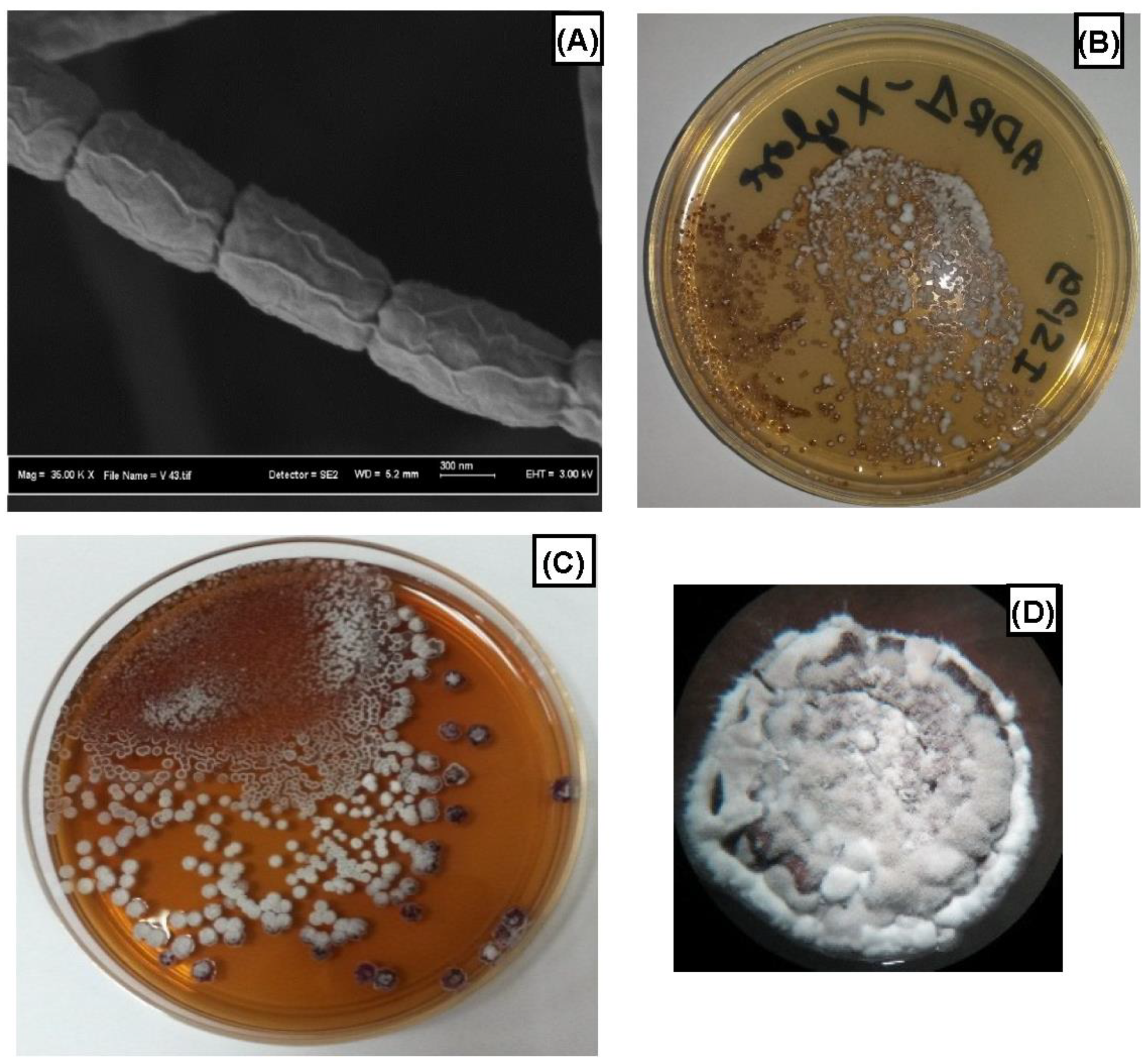
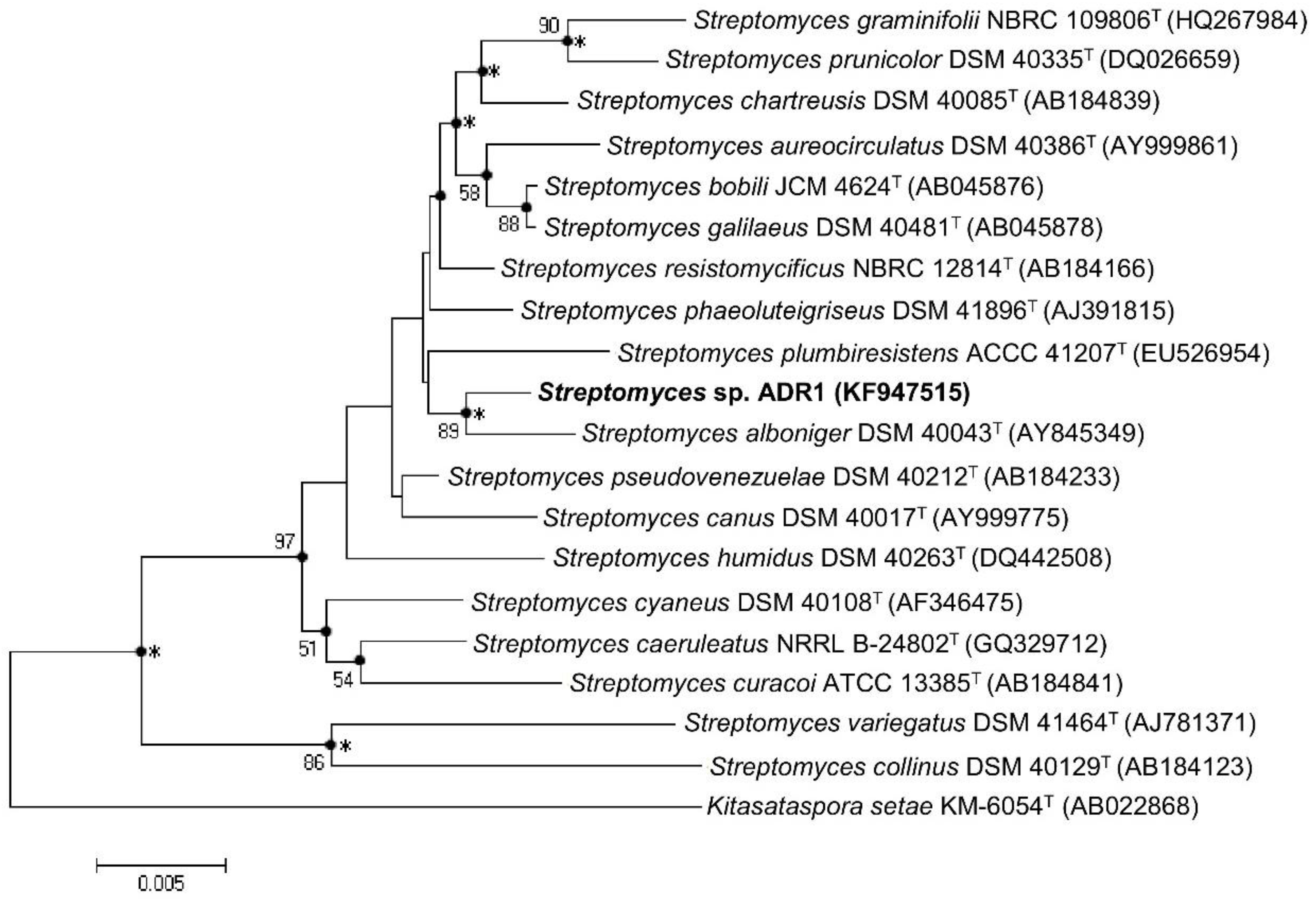
3.1. Isolation and Identification of ADR1 Strain

Among the actinomycete strains isolated, one strain that showed a significant cytotoxic activity was designated as strain ADR1. The ADR1 strain is Gram-positive, producing doliform spores, with irregular rugose surface ornamentation (Figure 1A,B), and (0.617 − 1.188) × (0.524 − 0.604) μm in size. The spore chains are in rectiflexible sections, with mature spore chains generally having 30 to 60 or more spores per chain.
On studying the cultural characteristics of the ADR1 strain, it was found that ISP2 and ISP4 media were the best for growth (Table 1). In the ISP2 medium, the substrate mycelia were purple-red, the aerial mycelia were white, and the cells produced a brown-red soluble pigment in the medium (Figure 1C). The aerial mycelia also formed in the ISP4 medium, but without soluble pigment production. The growth was moderate in the other media (ISP3, ISP5, ISP6, ISP7, GAA, SGA, and NA), without aerial mycelium formation and soluble pigment production. For physiological characteristics, the temperature range for growth in the ISP2 medium was 15–40 °C, with optimal growth between 25–30 °C. The pH range of growth was between 4 and 11, with optimal growth in the range between 7 and 8. The NaCl range for growth was 0–4%, with the best growth in NaCl-free medium (Table 2).
In terms of biochemical characteristics, the ADR1 strain did not produce melanin in the ISP6 medium. It was catalase- and beta-galactosidase-positive, did not produce H2S, liquefied gelatin, and hydrolyzed starch in the ISP4 medium. D-glucose, D-xylose, L-rhamnose, L-arabinose, D-fructose, D-mannitol, glycerol, D-sucrose, D-melibiose, chitine, pectin A, and amygdaline can be used as sole carbon sources in the ISP9 medium. There was no growth or only traces of growth when the carbon source was Iso-inositol, D-sorbitol, or D-galacturonic acid. LL-DAP was the type of diaminopimelic acid isomer found in the whole-cell hydrolysates of the ADR1 strain.
The partial 16S ribosomal RNA gene sequence (1392 bp, Accession No. KF947515) showed high sequence similarity with species of the genus Streptomyces, such as Streptomyces pseudovenezuelae (NBRC 12904(T), 1383/1391 bp, Pairwise Similarity 99.42%) and Streptomyces alboniger (NBRC 12738(T), 1381/1391bp, Pairwise Similarity 99.28%). Therefore, the ADR1 strain belongs to the genus Streptomyces; the name Streptomyces sp. ADR1 was proposed and deposited in the Wellness Industries Culture Collection (WICC) of the Institute of Bioproduct Development, UTM, Malaysia, under the number (WICC-B86). Figure 2 shows a neighbor-joining phylogenetic tree of the strain based on 16S rRNA gene sequences, showing relationships between the Streptomyces sp. ADR1 strain and related strain types of the genus Streptomyces.

3.2. Cytotoxic Activity of the Microbial EtOAc Extract

The cytotoxic activities of the EtOAc extract obtained from the small-scale fermentation of Streptomyces sp. ADR1 on the ISP2 medium were investigated using the MTT assay against human pancreatic cancer PANC-1 cells. Results showed a 37.1 ± 1.3% survival rate at the concentration of 100 µg/mL of EtOAc extract.

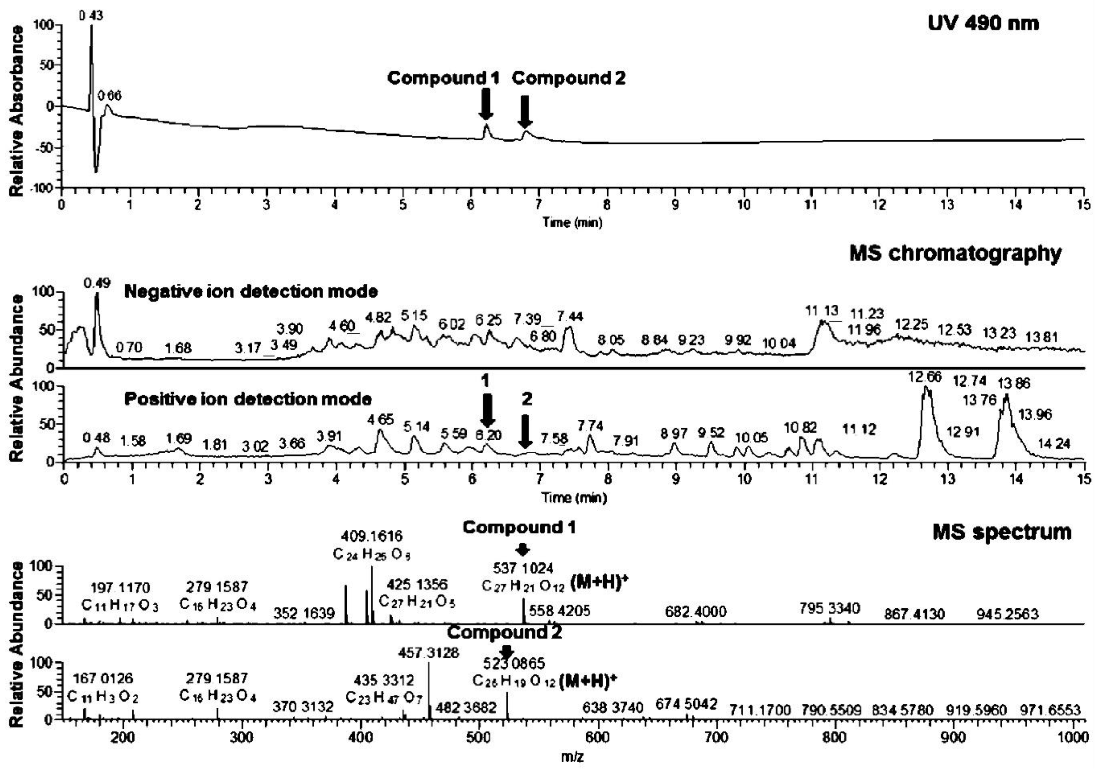
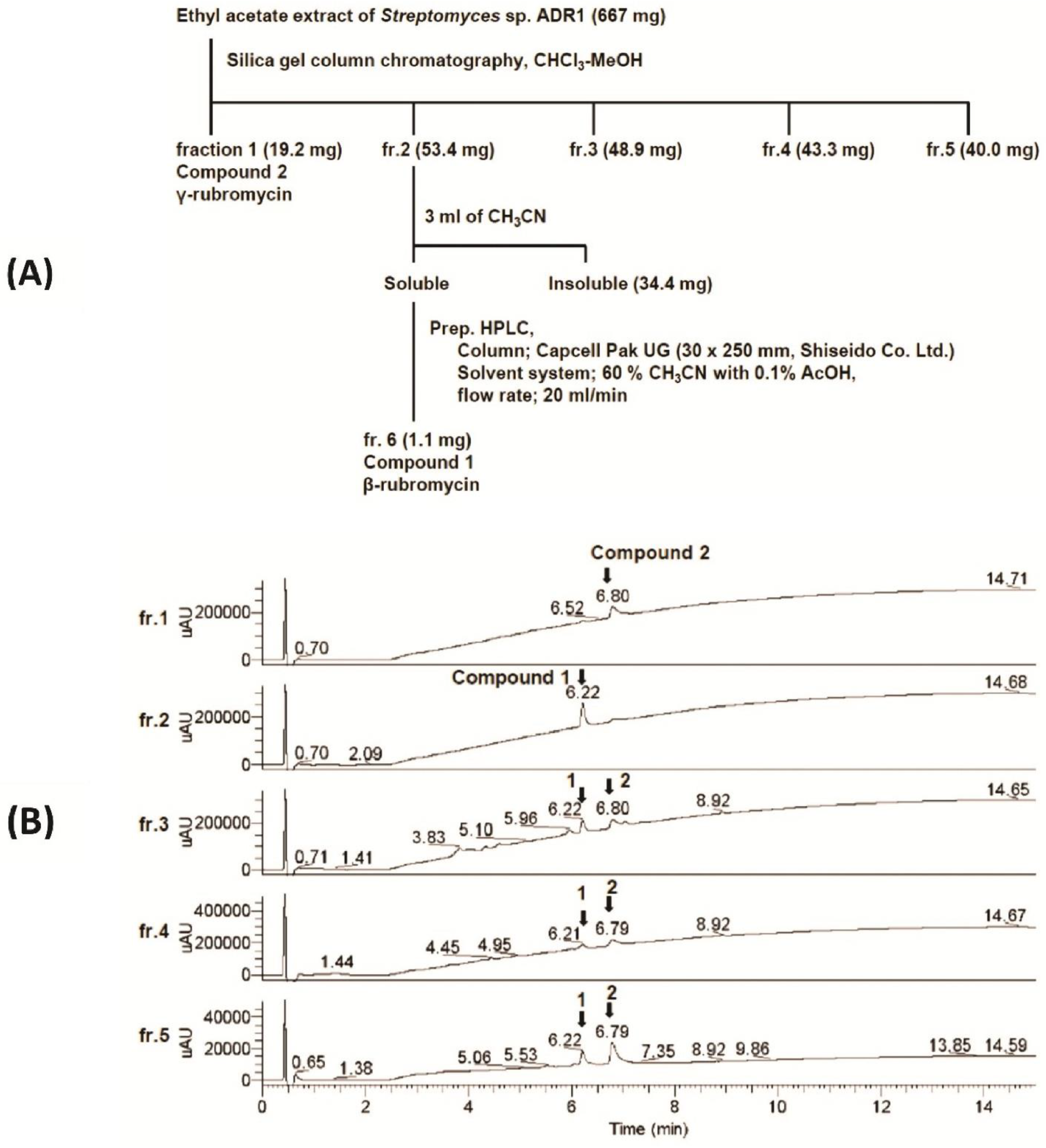
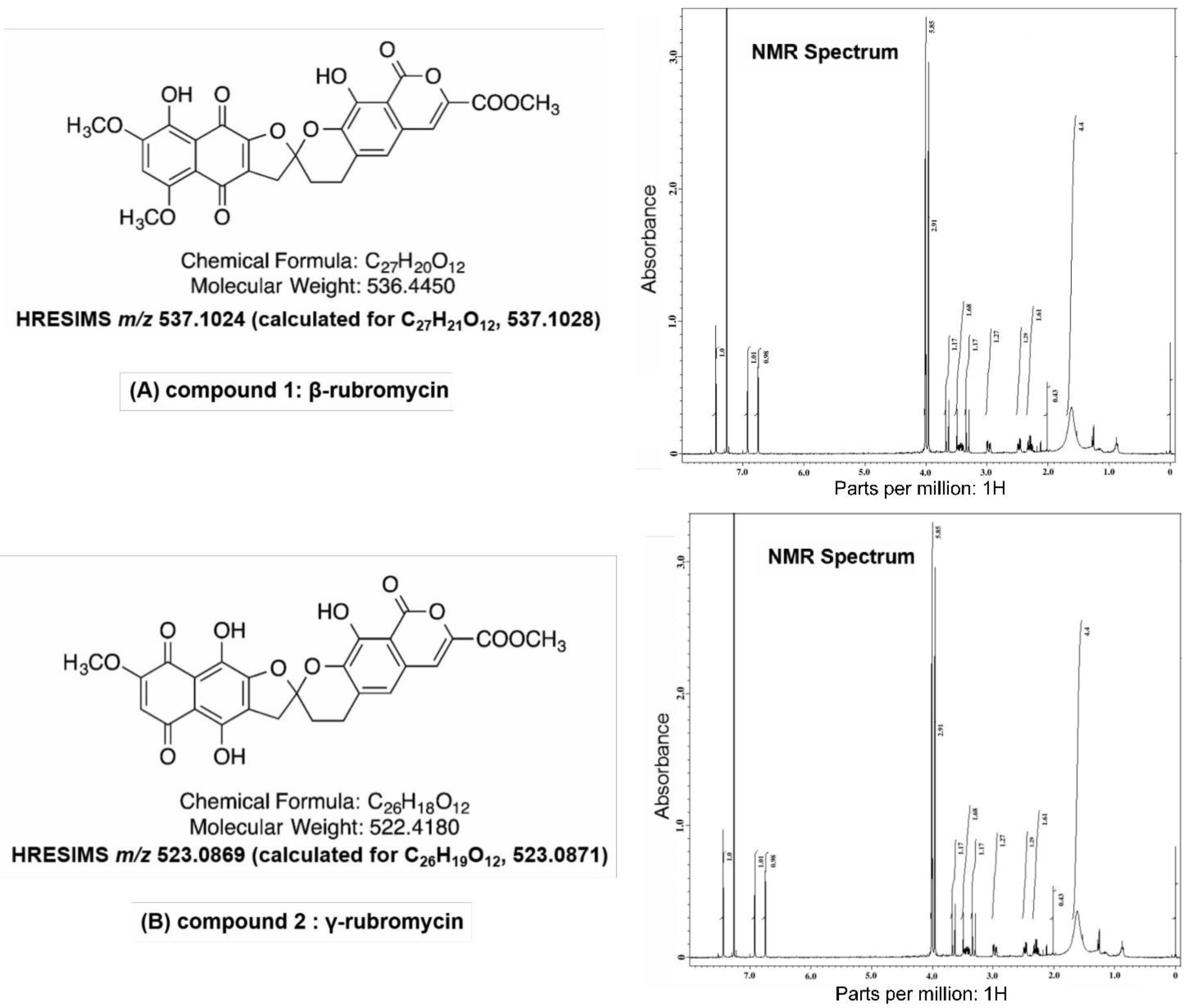
3.3. Physicochemical Analysis of the Microbial Extract and Isolation of β- and γ-Rubromycin

The EtOAc extract from the ADR1 fermentation broth was red in color, and at the UV wavelength of 490 nm, two compounds were identified. The MS spectrum data show that the molecular formula of compound 1 was C27H20O12, and that of compound 2 was C26H18O12 (Figure 3). After fractionation of the EtOAc extract by silica gel column chromatography (Figure 4A) and purification by HPLC (Figure 4B), the NMR spectra of the pure compounds obtained showed that compound 1 was β-rubromycin, and compound 2 was γ-rubromycin (Figure 5).

4. Discussion

Soil is a well-known source of bioactive microorganisms [27,28], and recently, it has been described that arid soils show the richest biosynthetic diversity compared to brackish sediments and pine forest soils [29]. The β- and γ-rubromycin-producing strain Streptomyces sp. ADR1 was isolated from soil collected in the Algerian Sahara Desert. It showed differences in some phonotypical and biochemical characteristics compared to phylogenetically related species Streptomyces pseudovenezuelae NBRC 12904T and Streptomyces alboniger NBRC 12738T (Table 3), and these two strains are not identified as rubromycin-producing strains. The first description of rubromycins was made by Brockmann et al. in 1953, and they were purified from a culture of Streptomyces collinus isolated from soil collected in Baden, Germany [30,31]. On the basis of the 16S rRNA gene sequence analysis, we showed that Streptomyces sp. ADR1 has low similarity with Streptomyces collinus (DSM 40,129 (T), 50/1390 bp, Pairwise Similarity 96.4%) (Figure 2), and the two strains are different in some phonotypical characteristics, such as morphology and carbon source utilization (Table 3). Therefore, we consider that Streptomyces sp. ADR1 is a new β- and γ-rubromycin-producing strain.
The bioactive compounds were initially extracted from the fermentation broth using EtOAc followed by purification using HPLC, and the full identification of the compounds was carried out using NMR. The NMR spectra clearly demonstrated the presence of two bioactive compounds: (A) of molecular weight of 536.4450 and molecular structure (C27H20O12), and (B) of molecular weight of 522.4180 and molecular structure (C26H18O12), which are typical to β-rubromycin and γ-rubromycin, respectively [31]. From a biosynthetic pathway perspective, quinone antibiotics such as rubromycins are derived from a type-II polyketide synthase (PKS) pathway [32]. β- and γ-rubromycins exhibit antibacterial activity against S. aureus and B. subtilis [33] and cytotoxic activity against stomach, colon, breast, and liver cancer cell lines at low concentrations [32]. These antibiotics also inhibit several enzymes, such as the retroviral reverse transcriptase, DNA polymerases, and terminal deoxynucleotidyl transferase [33]. However, the most remarkable property of β- and γ-rubromycins is their ability to inhibit human telomerase with IC50 values from 2.64–12.2 µM [33]. Studies have shown that the spiroketal moiety of rubromycin plays a key role in its telomerase inhibitory action [31,34]. Currently, it is clearly recognized that a strong relationship exists between telomerase and cancer. This enzyme is not expressed in most normal human cells; therefore, it is a good target for the development of new anticancer drugs [31,35,36].

5. Conclusions

The results of this study clearly showed the high potential of desert soil as an important source of isolation of actinomycetes that have the capacity to produce high-value antibiotic compounds. This work presents a complete platform for bioactive molecule discovery, from strain isolation and complete identification using physiological and molecular biology methods to purification and chemical structure determination of the bioactive compounds using reversed-phase HPLC, LC-MS, and NMR techniques. The two isolated compounds were identified as β- and γ-rubromycins. These two compounds belong to a special class of antibiotics that has the capacity to act as telomerase-inhibitor molecules and thus exhibit antitumor activity. Further studies are now being conducted in our laboratories for medium and bioprocess optimization for the production of these two important antibiotics at semi-industrial scale.

Author Contributions

Conceptualization, A.Z.B., H.A.E.E., H.H. and E.Y.P.; methodology, A.Z.B.; validation, H.A.E.E., H.H. and E.Y.P.; strain isolation, A.Z.B.; strain identification, A.Z.B.; molecular biology identification, A.Z.B., B.K. and M.T.; fermentation, A.Z.B. and H.A.E.E.; extraction, A.Z.B. and H.A.E.E.; antibiotic chemical characterization, A.Z.B. and E.Y.P.; writing—original draft preparation, A.Z.B.; writing—review and editing, A.Z.B., H.A.E.E., B.K., M.T., H.H. and E.Y.P.; supervision, H.A.E.E. and H.H.; project administration, A.Z.B. and H.A.E.E.; funding acquisition, A.Z.B., H.A.E.E., B.K., M.T., H.H. and E.Y.P. All authors have read and agreed to the published version of the manuscript.

Funding

This work was supported by the project No. 284/SA/2012, Centre de Recherche Scientifique et Technique en Analyses Physico-Chimiques—C.R.A.P.C. Algeria, as well as the Algerian scholarship program “PNE” and the Institute of Bioproduct Development, Universiti Teknologi Malaysia (UTM), Johor, Malaysia with industrial fund: R.J130000.7609.4C336 and R.J13000.7609.4C240.

Institutional Review Board Statement

Not applicable.

Informed Consent Statement

Not applicable.

Data Availability Statement

Not applicable.

Acknowledgments

This work is dedicated to the memory of Roslinda Abd Malek, Institute of Bioproduct Development, Universiti Teknologi Malaysia (UTM). We thank Isao Momose of the Institute of Microbial Chemistry (Numazu Branch 18–24 Miyamoto, Numazu-shi Shizuoka 410-0301, Japan) for analysis of the physicochemical properties of the microbial extract. We also thank Heide Goodman and Gerda du Plessis (IMBM, University Western Cape) for the precious help in the extraction of DNA.

Conflicts of Interest

The authors declare no conflict of interest.

References

  1. Sung, H.; Ferlay, J.; Siegel, R.L.; Laversanne, M.; Soerjomataram, I.; Jemal, A.; Bray, F. Global cancer statistics 2020: GLOBOCAN estimates of incidence and mortality worldwide for 36 cancers in 185 countries. CA Cancer J. Clin. 2021, 71, 209–249. [Google Scholar] [CrossRef] [PubMed]
  2. Boumehira, A.Z.; Akchiche, Y.F.; Chorfi, W.; Talhi, O.; Djidjik, R.; Dailin, J.D.; El Enshasy, H.A. Metabolomics approaches for early cancer diagnosis: A review. Asian J. Agric. Biol. 2022, 4, 1–22. [Google Scholar]
  3. Magee, P.; Shi, L.; Garofalo, M. Role of microRNAs in chemoresistance. Ann. Transl. Med. 2015, 3, 332. [Google Scholar] [PubMed]
  4. de Souza, J.A.; Hunt, B.; Asirwa, F.C.; Adebamowo, C.; Lopes, G. Global Health Equity: Cancer Care Outcome Disparities in High-, Middle-, and Low-Income Countries. J. Clin. Oncol. 2016, 34, 6–13. [Google Scholar] [CrossRef] [PubMed]
  5. Lu, D.; Chen, E.; Lu, T. Anticancer drug development, a matter of money or a matter of idea. Metabolomics 2015, 5, e134. [Google Scholar]
  6. Amirkia, V.; Heinrich, M. Natural products and drug discovery: A survey of stakeholders in industry and academia. Front. Pharmacol. 2015, 6, 237. [Google Scholar] [CrossRef]
  7. Bérdy, J. Bioactive Microbial Metabolites. J. Antibiot. 2005, 58, 1–26. [Google Scholar] [CrossRef]
  8. Donald, L.; Pipite, A.; Subramani, R.; Owen, J.; Keyzers, R.A.; Taufa, T. Streptomyces: Still the Biggest Producer of New Natural Secondary Metabolites, a Current Perspective. Microbiol. Res. 2022, 13, 31. [Google Scholar] [CrossRef]
  9. Shirling, E.B.; Gottlieb, D. Methods for characterization of Streptomyces species. Int. J. Syst. Evol. Microbiol. 1966, 16, 313–340. [Google Scholar] [CrossRef]
  10. Lechevalier, H.A. The actinomycetes III. A practical guide to generic identification of actinomycetes. In Bergey’s Manual Systematic Bacteriology; Williams, S.T., Ed.; Springer: Berlin, Germany, 1989; Volume 4, pp. 2344–2347. [Google Scholar]
  11. Wink, J.M. Compendium of Actinobacteria; University of Braunschweig: Braunschweig, Germany, 2012. [Google Scholar]
  12. El-Nakeeb, M.A.; Lechevalier, H.A. Selective isolation of aerobic Actinomycetes. J. Appl. Microbiol. 1963, 11, 75–77. [Google Scholar] [CrossRef]
  13. Locci, R. Actinomycete Spores. In Encyclopedia of Life Sciences; John Wiley & Sons, Ltd.: Hoboken, NJ, USA, 2001. [Google Scholar]
  14. Schon, R.; Groth, I. Practical thin layer chromatography techniques for diaminopimelic acid and whole cell sugar analyses in the classification of environmental actinomycetes. J. Basic Microbiol. 2006, 46, 243–249. [Google Scholar] [CrossRef] [PubMed]
  15. Wang, Y.; Zhang, Z.S.; Ruan, J.S. A proposal to transfer Microbispora bispora (Lechevalier 1965) to a new genus, Thermobispora gen. nov., as Thermobispora bispora comb. nov. Int. J. Syst. Evol. Microbiol. 1996, 46, 933–938. [Google Scholar]
  16. Kirby, B.M.; Everest, G.J.; Meyers, P.R. Phylogenetic analysis of the genus Kribbella based on the gyrB gene: Proposal of a gyrB-sequence threshold for species delineation in the genus Kribbella. Antoni van Leeuwenhoek 2010, 97, 131–142. [Google Scholar] [CrossRef] [PubMed]
  17. Cook, A.E.; Meyers, P.R. Rapid identification of filamentous actinomycetes to the genus level using genus-specific 16S rRNA gene restriction fragment patterns. Int. J. Syst. Evol. Microbiol. 2003, 53, 1907–1915. [Google Scholar] [CrossRef]
  18. Altschul, S.F.; Madden, T.L.; Schaffer, A.A.; Zhang, J.H.; Zhang, Z.; Miller, W.; Lipman, D.J. Gapped BLAST and PSI-BLAST: A new generation of protein database search programs. Nucleic Acids Res. 1997, 25, 3389–3402. [Google Scholar] [CrossRef]
  19. Edgar, R.C. MUSCLE: Multiple sequence alignment with high accuracy and high throughput. Nucleic Acids Res. 2004, 32, 1792–1797. [Google Scholar] [CrossRef]
  20. Tamura, K.; Stecher, G.; Peterson, D.; Filipski, A.; Kumar, S. MEGA6: Molecular Evolutionary Genetics Analysis Version 6.0. Mol. Biol. Evol. 2013, 30, 2725–2729. [Google Scholar] [CrossRef]
  21. Saitou, N.; Nei, M. The neighbor-joining method: A new method for reconstructing phylogenetic trees. Mol. Biol. Evol. 1987, 4, 406–425. [Google Scholar]
  22. Felsenstein, J. Confidence Limits on Phylogenies: An Approach Using the Bootstrap. Evolution 1985, 39, 783–791. [Google Scholar] [CrossRef]
  23. Felsenstein, J. Evolutionary trees from DNA sequences: A maximum likelihood approach. J. Mol. Evol. 1981, 17, 368–376. [Google Scholar] [CrossRef]
  24. Fitch, W.M. Toward Defining the Course of Evolution: Minimum Change for a Specific Tree Topology. Syst. Biol. 1971, 20, 406–416. [Google Scholar] [CrossRef]
  25. Chun, J.; Lee, J.-H.; Jung, Y.; Kim, M.; Kim, S.; Kim, B.K.; Lim, Y.-W. EzTaxon: A web-based tool for the identification of prokaryotes based on 16S ribosomal RNA gene sequences. Int. J. Syst. Evol. Microbiol. 2007, 57, 2259–2261. [Google Scholar] [CrossRef] [PubMed]
  26. Mosmann, T. Rapid colorimetric assay for cellular growth and survival: Application to proliferation and cytotoxicity assays. J. Immunol. Methods 1983, 65, 55–63. [Google Scholar] [CrossRef]
  27. Waksman, S.A.; Woodruff, H.B. The Soil as a Source of Microorganisms Antagonistic to Disease-Producing Bacteria. J. Bacteriol. 1940, 40, 581–600. [Google Scholar] [CrossRef] [Green Version]
  28. Boumehira, A.Z.; El-Enshasy, H.A.; Hacène, H.; Elsayed, E.A.; Aziz, R.; Park, E.Y. Recent progress on the development of antibiotics from the genus Micromonospora. Biotechnol. Bioprocess Eng. 2016, 21, 199–223. [Google Scholar] [CrossRef]
  29. Charlop-Powers, Z.; Owen, J.G.; Reddy, B.V.B.; Ternei, M.A.; Brady, S.F. Chemical-biogeographic survey of secondary metabolism in soil. Proc. Natl. Acad. Sci. USA 2014, 111, 3757–3762. [Google Scholar] [CrossRef]
  30. Brockmann, H.; Renneberg, K.-H. Rubromycin, ein rotes Antibiotikum aus Actinomyceten. Naturwissenschaften 1953, 40, 59–60. [Google Scholar] [CrossRef]
  31. Boumehira, A.Z.; Hacène, H.; El-Enshasy, H.A. Rubromycins: A Class of Telomerase Inhibitor Antibiotics Produced by Streptomyces spp. In New and Future Developments in Microbial Biotechnology and Bioengineering; Elsevier: Amsterdam, The Netherlands, 2019; pp. 141–150. [Google Scholar]
  32. Puder, C.; Loya, S.; Hizi, A.; Zeeck, A. Structural and biosynthetic investigations of the rubromycins. Eur. J. Org. Chem. 2000, 2000, 729–735. [Google Scholar] [CrossRef]
  33. Atkinson, D.J.; Brimble, M.A. Isolation, biological activity, biosynthesis and synthetic studies towards the rubromycin family of natural products. Nat. Prod. Rep. 2015, 32, 811–840. [Google Scholar] [CrossRef] [PubMed]
  34. Ueno, T.; Takahashi, H.; Oda, M.; Mizunuma, M.; Yokoyama, A.; Goto, Y.; Mizushina, Y.; Sakaguchi, K.; Hayashi, H. Inhibition of human telomerase by rubromycins: Implication of spiroketal system of the compounds as an active moiety. Biochemistry 2000, 39, 5995–6002. [Google Scholar] [CrossRef]
  35. Buseman, C.M.; Wright, W.E.; Shay, J.W. Is telomerase a viable target in cancer? Mutat. Res.-Fundam. Mol. Mech. Mutagenesis 2012, 730, 90–97. [Google Scholar] [CrossRef] [PubMed]
  36. Ivancich, M.; Schrank, Z.; Wojdyla, L.; Leviskas, B.; Kuckovic, A.; Sanjali, A.; Puri, N. Treating Cancer by Targeting Telomeres and Telomerase. Antioxidants 2017, 6, 15. [Google Scholar] [CrossRef] [PubMed]
Figure 1. (A) Scanning electron micrograph of Streptomyces sp. ADR1 strain. (B) Streptomyces sp. ADR1 grown on ISP9—D-xylose agar—for 7 days at 28 °C. (C) Streptomyces sp. ADR1 strain grown on ISP2 medium for 14 days at 28 °C. (D) A single colony of Streptomyces sp. ADR1 strain grown on ISP2 medium under stereomicroscope (×45) (Zoom 2000™, Leica Microsystems, Germany).
Fermentation 08 00473 g001
Figure 2. Neighbor-joining tree based on 16S rRNA gene sequences, showing relationships between Streptomyces sp. ADR1 strain and related strain types of the genus Streptomyces. Only bootstrap values above 50% (percentages of 1000 replications) are indicated. Branches marked with a solid circle were also recovered in the maximum-likelihood tree; asterisks show branches recovered in the maximum-parsimony tree. Bar, 0.01 substitutions per nucleotide position. * show branches recovered in the maximum-parsimony tree.
Fermentation 08 00473 g002
Figure 3. LC-MS analysis of the EtOAc extract from the culture broth of Streptomyces sp. ADR1.
Fermentation 08 00473 g003
Figure 4. (A) Work-up procedure of extraction and purification of β- and γ-rubromycin from Streptomyces sp. ADR1; (B) fraction purification by reversed-phase HPLC.
Fermentation 08 00473 g004
Figure 5. NMR spectra and chemical structures of (A) compound 1, β-rubromycin, and (B) compound 2, γ-rubromycin.
Fermentation 08 00473 g005
Table 1. Cultural characteristics of Streptomyces sp. ADR1 strain.
MediumGrowthSubstrateSoluble PigmentAerial
Yeast extract malt agar (ISP2)GoodPurple-redBrown-redWhite
Oatmeal agar (ISP3)ModerateBrown-beigeNoneNone
Inorganic salt starch agar (ISP4)GoodPurple-redNoneWhite
Glycerol asparagine agar (ISP5)ModerateBrown-beigeNoneNone
Peptone yeast extract iron agar (ISP6)ModerateBrown-beigeNoneNone
Tyrosine agar (ISP7)ModerateBrown-beigeNoneNone
Glycerol Arginine Agar (GAA)ModerateBrown-beigeNoneNone
Sabouraud 4% Glucose Agar (SGA)ModerateBrown-beigeNoneNone
Nutrient Agar (NA)ModerateBrown-beigeNoneNone
Table 2. Characteristics of the Streptomyces sp. ADR1 strain.
Microscopic Aspect
Gram stainingPositive
Spore morphologyDoliform
Spore surface ornamentationIrregular rugose
Spore dimensions (μm)(0.617 − 1.188) × (0.524 − 0.604)
Spore chain morphology Rectiflexible
Spores per chain30–60
Physiological Characteristics
Temperature range for growth (°C)15–40
Optimal temperature for growth (°C)25–30
pH range of growth4–11
Optimal pH for growth7–8
NaCl range for growth (%)0–4
Optimal NaCl for growth (%)0
Biochemical Characteristics
Melanin production (ISP6)
Catalase +
β-galactosidase+
Citrate utilization
H2S production
Gelatin liquefaction+
Hydrolysis of starch in ISP4+
Utilization as sole carbon source
D-glucose+
D-xylose+
L-rhamnose+
L-arabinose +
D-fructose+
D-mannitol+
Iso-inositol
D-sorbitol
Glycerol+
D-galacturonic acid
D-sucrose+
D-melibiose+
Chitine+
Pectin A+
Amygdaline+
Table 3. Morphological, cultural features and physiologically different characteristics of strain Streptomyces sp. ADR1 and related phylogenetic and rubromycin-producing species strains *.
MediumStreptomyces sp. ADR1Streptomyces pseudovenezuelae NBRC 12904TStreptomyces alboniger
NBRC 12738T
Streptomyces collinus
DSM 40129T
Morphology on ISP2
GrowthGoodGood GoodGood
Colony colorPurple-redSafran yellowBlack-greyDahlia yellow
Aerial myceliumWhiteSparse/creamBlack-greyCream
Soluble pigmentBrown-redNoneUmbra greyNone
Utilization as sole carbon source
D-glucose+++
D-xylose++
L-rhamnose+++
L-arabinose +++
D-fructose++
Iso-inositol+++
D-sucrose++
Production of rubromycins++
16S rRNA gene pairwise similarity with Streptomyces sp. ADR199.42%99.28%96.4%
(*): Morphological and Physiological tests of species as described by [9,11].
Publisher’s Note: MDPI stays neutral with regard to jurisdictional claims in published maps and institutional affiliations.

