Article

Identification and Characterization of a Novel Cold-Adapted GH15 Family Trehalase from the Psychrotolerant Microbacterium phyllosphaerae LW106

1
School of Food Science and Technology, Shihezi University, Shihezi 832002, China
2
Institute of Microbiology, Xinjiang Academy of Agricultural Sciences, Urumqi 830052, China
*
Authors to whom correspondence should be addressed.
These authors contributed equally to this work.
Fermentation 2022, 8(10), 471; https://doi.org/10.3390/fermentation8100471
Submission received: 16 August 2022 / Revised: 10 September 2022 / Accepted: 15 September 2022 / Published: 21 September 2022
(This article belongs to the Special Issue Extremophiles—Source for Novel Biomolecules with Applied Potential)

Abstract

:
Psychrophiles inhabiting various cold environments are regarded as having evolved diverse physiological and molecular strategies, such as the accumulation of trehalose to alleviate cold stress. To investigate the possible contributions of trehalose metabolism-related enzymes to cold-adaption in psychrotrophic bacteria and enrich the resource bank of trehalose hydrolysis enzymes, a novel cold-adapted GH15 GA-like trehalase (MpTre15A) from psychrotolerant Microbacterium phyllosphaerae LW106 isolated from glacier sediments was cloned and characterized. The recombinant MpTre15A from M. phyllosphaerae LW106 was expressed and purified in Escherichia coli BL21(DE3). The purified MpTre15A functioned as a hexamer and displayed maximal activity at pH 5.0 and 50 °C. Substrate specificity assay proved MpTre15A only showed hydrolytic activity toward α,α-trehalose. Site-directed mutation verified the key catalytic sites of Glu392 and Glu557 in MpTre15A. The kcat and kcat/Km values of MpTre15A at 4 °C (104.50 s−1 and 1.6 s−1 mM−1, respectively) were comparable to those observed for thermophilic GH15 trehalases at 50 °C, revealing its typical cold-adaptability. MpTre15A showed a trehalose conversion rate of 100% and 99.4% after 10 min and 15 min of incubation at 50 °C and 37 °C, respectively. In conclusion, this novel cold-adapted α,α-trehalase MpTre15A showed potential application for developing therapeutic enzymes, enzyme-based biosensors, and enzyme additives in the fermentation industry.

1. Introduction

Trehalose is a kind of stable disaccharide that consists of two glucose units linked primarily by an α, α-(1,1) bond, which is ubiquitous in soils and a prominent metabolite in bacteria, yeast, fungi, insects, invertebrates, and plants [1,2]. It plays multiple roles in various microorganisms, including acting as a source of glucose and/or energy [3], protecting proteins and membranes against various stress conditions such as dehydration, heat, cold, and oxygen radicals [1,4], regulating glucose metabolism [5], and serving as an essential component of various cell wall glycolipids in some mycobacteria species [6]. Trehalose synthesis is induced upon exposure of Escherichia coli to cold and is essential for viability at low temperatures [7]. The yeast Saccharomyces cerevisiae also accumulates trehalose during heat shock to protect its proteome against thermal denaturation and aggregation [8]. This disaccharide is expected to be a valuable ingredient in pharmaceuticals, food, and cosmetic industries [1,4,9].
Compared to enzymes related to trehalose synthesis [10], relatively less attention has been paid to trehalose degrading enzymes due to their limited application prospect in biotechnology. The major enzyme involved in trehalose hydrolysis is trehalase (α, α-1,1-glucosyl hydrolase). Trehalases (EC 3.2.1.28) are widely distributed in eukaryotes, archaea, and bacteria, and play important roles in various physiological processes. Currently, trehalases are categorized mainly as family members of glycoside hydrolase 65 (GH65), GH37 as well as GH15 in the Carbohydrate-Active enZymes database (CAZy, http://www.cazy.org/, accessed on 14 August 2022). Among these carbohydrate-active enzymes, GH37 trehalases have been the most extensively characterized group in respect of the activity regulation and catalytic mechanism. Two types of GH37 trehalases, periplasmic TreA and cytoplasmic TreF were reported in E. coli [11,12]. Trehalose is first transferred into the periplasm or cytoplasm and then degraded directly into two glucose molecules [13]. Bacterial degradation of trehalose can also be achieved via the phosphotransferase system (PTS) in the cytoplasm. In this trehalose degradation pathway, the transport and cleavage of trehalose occur simultaneously, resulting in the production of glucose and glucose-6-phosphate [14]. Unlike the diverse distribution of GH37 trehalases, GH65 trehalases have been isolated only from fungi and yeasts and exhibit activity under acidic conditions, whereas GH15 trehalases have been reported only in archaea and bacteria. Although the resolved crystal structures are only available in GH37 trehalases, GH15 and GH65 trehalases are assumed to share common catalytic domains with (α/α) 6-barrel structures [13]. Because of the absence of a signature motif, GH37 enzymes were classified into clan GH-G, while GH65 and GH15 trehalases were assigned to clan GH-L according to the CAZy database. Due to the relatively late discovery of the first two GH15 trehalases [6], only a few GH15 trehalases from archaea including Thermoplasma volcanium, T. acidophilum, and Sulfolobus acidocaldarius have been biochemically characterized [15,16,17]. There have also been rare reports on the characterization of GH15 bacterial trehalase except for a recently isolated trehalase from the strictly aerobic bacterium Microvirga sp. MC18 [18].
Although trehalose has been approved as an additive in the preparation of dried food, intestinal trehalase deficiency, a metabolic disorder in which the human body is not able to convert trehalose into glucose, exists in some regional populations [19]. Moreover, hydrolysis of extracellular trehalose by trehalase also finds applications during ethanol fermentation since yeast generates trehalose but cannot utilize trehalose as a carbon source [20]. Thus, from the perspective of the application, trehalase has shown potential to be developed as a therapeutic enzyme and enzyme additive in the fermentation industry.
More than three-quarters of the earth’s surface is occupied by cold ecosystems, including the ocean depths, and polar and alpine regions [21]. The bacteria inhabiting these cold environments were regarded as having evolved diverse physiological and molecular strategies to alleviate the cold stress, such as the production of cold-shock proteins, cold-adapted enzymes, and accumulation of various compatible solutes including trehalose [22]. However, detailed investigation of the trehalose metabolism-related enzyme and their contribution to cold-adaption in psychrophiles has been rare. Moreover, cold-adapted enzymes produced by psychrophiles also gained much attention due to their unique characteristics, such as high activity at low temperatures and thermolabile compared to their mesophilic and thermophilic counterparts, which are favorable for the enzyme inactivation with moderate heating and help reduce the energy consumption required for the catalyzed reaction [23].
A psychrotolerant bacterial strain, Microbacterium phyllosphaerae LW106, was recently isolated from sediments at Glacier No.1, northwest China [24], and a putative GH15 GA-like trehalase MpTre15A was discovered from M. phyllosphaerae LW106. To explore the enzymatic properties of this putative GH15 trehalase of psychrotolerant origin, we attempted to express the MpTre15A gene in E. coli. This putative GH15 trehalase could be efficiently expressed in its active form in E. coli and was able to specifically hydrolyze trehalose at an optimum temperature of 50 °C and an optimum pH of 5.0. MpTre15A was found to be thermolabile at temperatures above 45 °C but retained high catalytic activity at 4 °C, suggesting it was a typical cold-adapted GH15 trehalase that has been discovered for the first time. The exploration of the enzymatic properties of this cold-adapted GH15 trehalase enriched the resource bank of trehalose hydrolysis enzymes and provided insights into the role that trehalose hydrolysis may play in bacteria cold adaption.

2. Materials and Methods

2.1. Chemical Reagents and Strains

Carbohydrates such as trehalose, sucrose, lactose, maltose, cellobiose, glucose, other media reagents, and regular chemicals, were purchased from Sangon Biotech Ltd. (Shanghai, China). p-nitrophenyl-α-glucopyranose was ordered from Carbosynth Ltd. (Beijing, China). The Prime STAR HS DNA Polymerase and restriction enzymes employed for DNA manipulation were ordered from TaKaRa Biotechnology Ltd. (Dalian, China). The genetic engineering manipulation was performed as described previously [25]. The psychrotolerant strain M. phyllosphaerae LW106 was isolated from subglacial sediments at the headwaters of Urumqi River, Glacier No. 1, Tianshan Mountains in northwest China (43°07′ N, 86°48′ E, 3800 m altitude). The E. coli BL21(DE3) strain was used for enzyme recombinant expression.

2.2. Bacteria Cultivation

Tryptic soy broth (1/4 TSB) was used to propagate the cells of M. phyllosphaerae LW106 strain. To investigate the effects of various sugars on the growth of M. phyllosphaerae LW106 strain, the bacteria cells were grown in a 1/4 TSB liquid medium containing glucose, lactose, sucrose, maltose, and trehalose, at a final concentration of 3.0 g/L, respectively, with shaking (200 rpm) for aeration at 16 °C. Optical density at 600 nm (OD600) was measured using a Multiskan FC microplate reader (Thermo). All growth experiments were conducted in triplicate and the average values were shown. Luria-Bertani (LB) medium (pH 7.2) was used to cultivate the E. coli cells by shaking at 200 rpm and 37 °C for 12 h before protein expression. Ampicillin (50 μg/mL) was added to the media as needed.

2.3. Sequence, Phylogenetic, and Modeling Analysis

A similarity search of MpTre15A was performed using the BLASTp algorithm at the National Center for Biotechnology Information (NCBI) database (https://blast.ncbi.nlm.nih.gov/Blast.cgi, accessed on 14 August 2022). The conserved domains and the GH family classification were predicted using the cdd online tool (http://www.ncbi.nlm.nih.gov/Structure/cdd/cdd.shtml, accessed on 14 August 2022). Computer-assisted protein sequence analysis was performed using Clustal W version 2.0, and the secondary structure elements and key catalysis residues based on alignments were depicted using the online tool ESPript version 3 [26]. The reference sequences selected in the phylogenetic analysis were retrieved from the NCBI database and aligned using Clustal W. The phylogenetic tree for MpTre15A was constructed using the neighbor-joining method with the bootstrap method phylogeny test and 1000 replications using the MEGA7 program.

2.4. Cloning, Mutant Plasmid Construction, Expression, and Purification of MpTre15A

To amplify the MpTre15A coding sequence, genomic DNA of M. phyllosphaerae LW106 was used as a template for PCR reaction. The amplification procedure included one cycle of 94 °C for 5 min, 30 cycles of 94 °C for 30 s, 56 °C for 30 s, 72 °C for 2 min, and incubation at 72 °C for 10 min. The primer sequences for the MpTre15A gene were as follows: the forward primer was 5′-ATGCCGGCTCCGATTGAAGATTAT-3′ and the reverse primer was 5′-ACGACGATGTGCTGCACGACCACC-3′. The amplified MpTre15A DNA fragments were purified, ligated to the pMD18-T vector using a Mighty TA-cloning Reagent Set for PrimeSTAR (TaKaRa), and delivered for sequencing.
The nucleotide sequence of the MpTre15A gene (GenBank: OM456201) was codon-optimized for E. coli, synthesized, and directly subcloned into the expression vector pET21a with NdeI and BamHI restriction sites by Zoonbio Biotechnology Co., Ltd. (Nanjing, China). The MpTre15A gene was fused with the coding gene of a hexahistidine tag (His-tag) to achieve a C-terminal tail for subsequent affinity chromatography purification. After being confirmed by sequencing, the recombinant plasmid carrying MpTre15A gene, named pET21-MpTre15A, was transformed into the cells of E. coli BL21 (DE3).
E. coli transformants carrying the expression vectors were grown overnight at 37 °C in an LB medium containing 50 μg/mL ampicillin, then inoculated in a fresh LB medium containing 50 μg/mL ampicillin. The E. coli cells were grown at 37 °C until the optical density at 600 nm (OD600) reached 0.6 and then the target protein was induced for expression for approximately 18 h with 0.5 mM isopropyl-β-D-thiogalactopyranoside (IPTG). The E. coli cells were harvested by centrifugation at 4 °C, 8000 rpm for 10 min, and washed 3 times with 50 mM sodium phosphate buffer (pH 7.0). The cells were finally resuspended in lysis buffer (50 mM sodium phosphate, 300 mM sodium chloride, 30 mM imidazole, pH 7.0), and were lysed by sonication at 4 °C (2 s on with 4 s interval for 10 min at 200 W) with a Sonicator JY92-IIN (Scientz). The supernatant was collected after centrifugation at 4 °C, 12,000 rpm for 20 min.
For purification of the recombinant trehalase, lysate obtained from sonication supernatant was added to a Ni-NTA resin column (Sangon, China). The non-specific binding proteins were washed out with 10 column volumes of wash buffer (50 mM sodium phosphate, 300 mM sodium chloride, and 50 mM imidazole, pH 7.0). The target protein was then eluted with 3 column volumes of elution buffer (50 mM sodium phosphate, 300 mM sodium chloride, and 250 mM imidazole, pH 7.0). To remove the residual salts and imidazole, the eluted protein solution was added to a Millipore spin column (10 kDa cutoff) and buffer-exchanged with the appropriate buffer solution. Aliquots of 10 μL of lysate and purified protein were applied on the SDS-PAGE and Native-PAGE gels, respectively, to analyze the purity and oligomeric state of the target protein.
The pET21-MpTre15A plasmid was used as a template for site-directed mutagenesis analysis by the NEBase Changer method (Q5 Site-Directed Mutagenesis Kit). The following oligonucleotide primers were used as mutagenic primers (mismatched bases are underlined): E392Q F: 5′-CACCGCGAATctgCCAAATACCAT-3′, E392Q R: 5′- AACCGCAGTGGTTTACCC-3′, E557Q F, 5′- CATCATATTCctgGCTCAGCAGGCC-3′, E557Q R, 5′- TGGCAAATGCACGCCAGG-3′. The nucleotide sequences of the MpTre15A mutant genes were confirmed by DNA sequencing as described above. The expression, purification, and activity determination for these two mutants were performed the same as the wild-type MpTre15A described above.

2.5. Trehalase Activity and Protein Concentration Determination

The activity of the recombinant trehalase was measured by determining the content of glucose released from trehalose. The reaction mixture consisted of 4.5 mM trehalose and 0.025 mg/mL purified enzyme in 100 μL sodium citrate buffer (100 mM, pH 5.0). The reaction was incubated at 50 °C for 10 min and stopped by boiling for 10 min. Then 100 μL 3,5-Dinitrosalicylic acid (DNS) solution was added to determine the produced glucose. One unit of trehalase activity was defined as the amount of enzyme required for releasing 1 μmol of glucose per minute under a specific assay condition indicated. Protein concentration was assayed by employing a Bradford protein assay kit (Sangon Biotech) using bovine serum albumin as the standard.

2.6. Substrate Specificity Assay

Enzyme activities toward soluble starch, different disaccharides including trehalose, maltose, cellobiose, and p-nitrophenyl-α-glucoside, were measured at a substrate concentration of 4.5 mM in 100 mM sodium citrate buffer at pH 5.0 and 50 °C. The reaction was kept at 50 °C for 10 min and stopped by boiling for 10 min. The amount of the released glucose was determined using the DNS method [18].

2.7. Optimum pH, Optimum Temperature, and Stability

The optimum reaction pH for the recombinant MpTre15A was evaluated by analyzing with 4.5 mM trehalose dissolved in various buffers with pH values ranging from 3.0 to 9.0 at 50 °C. The pH stability of the purified enzyme was assayed after incubation in buffers with pH values ranging from 3.0 to 9.0 for 12 h at 4 °C. Buffers for pH 3.0–6.6, pH 6.6–7.8, and pH 8.2–9.0 were 100 mM sodium citrate buffer, 100 mM sodium phosphate buffer, and 100 mM sodium borate buffer, respectively. Aliquots were withdrawn at different time points and the residual activity was measured at 50 °C, pH 5.0 as described above.
The influence of temperature on enzyme activity over trehalose was investigated by incubating the purified enzyme at temperatures ranging from 10 °C to 65 °C in 100 mM sodium citrate buffer (pH 5.0) for 10 min. To determine the enzyme thermostability, reaction aliquots were withdrawn for activity assay after incubating the purified enzymes in 100 mM sodium citrate buffer (pH 5.0) at various temperatures for up to 12 h. Residual activity was measured at 50 °C, pH 5.0 for 10 min as described above.

2.8. Effects of Metal Ions and Other Chemicals

Hydrolytic activities against 4.5 mM trehalose were assayed at their optimal temperature of 50 °C and pH 5.0 in the absence or presence of various cations added as chloride form as indicated. The influences of various chemicals including ethylenediaminetetraacetic acid, urea, sodium dodecyl sulfate, 1, 4-dithiothreitol, and β-mercaptoethanol on the enzyme activities were investigated under the same condition described above.

2.9. Kinetic Constants

For kinetic parameter measurement, the enzyme activity was detected using the substrate trehalose with concentrations ranging from 4 mM to 50 mM. Activity assay of the MpTre15A was performed as described above at optimum pH and specific temperature indicated. The kinetic parameters (Km, Vmax) were calculated by non-linear fitting the experimental data to the Michaelis Menten equation using the Michaelis Menten function in the software Origin. The apparent kcat values were estimated using the theoretic molecular mass of MpTre15A (62.5 kDa).

2.10. Analysis of Trehalose Hydrolysis Products

Aliquots of purified MpTre15A (2 mg/mL) were mixed with trehalose at a final concentration of 5 mg/mL and 50 mg/mL, respectively, and incubated for up to 120 min. Then, the reaction mixture was boiled for 5 min to inactivate the enzyme. The hydrolyzed sugar solutions were visually analyzed by thin-layer chromatography (TLC) on a silica gel plate (10 cm × 20 cm; Merck, Germany) with a solvent system consisting of n-butyl alcohol/methyl alcohol/deionized water (8:4:3, v/v/v), and the spots were visualized by spraying the silica gel plate with methyl alcohol and concentrated sulfuric acid (1:1, v/v), followed by heating at 90 °C for 15 min [18]. Trehalose consumption and the produced glucose were determined by high-performance liquid chromatography (HPLC, Shimazu, Japan) using a column of Hi-Plex Na Carbohydrate Column (300 mm × 7.7 mm, Agilent) at 80 °C with a refractive index detector (RID). The mobile phase was triple distilled water (Watsons, China) at a flow rate of 0.2 mL min−1.

3. Results

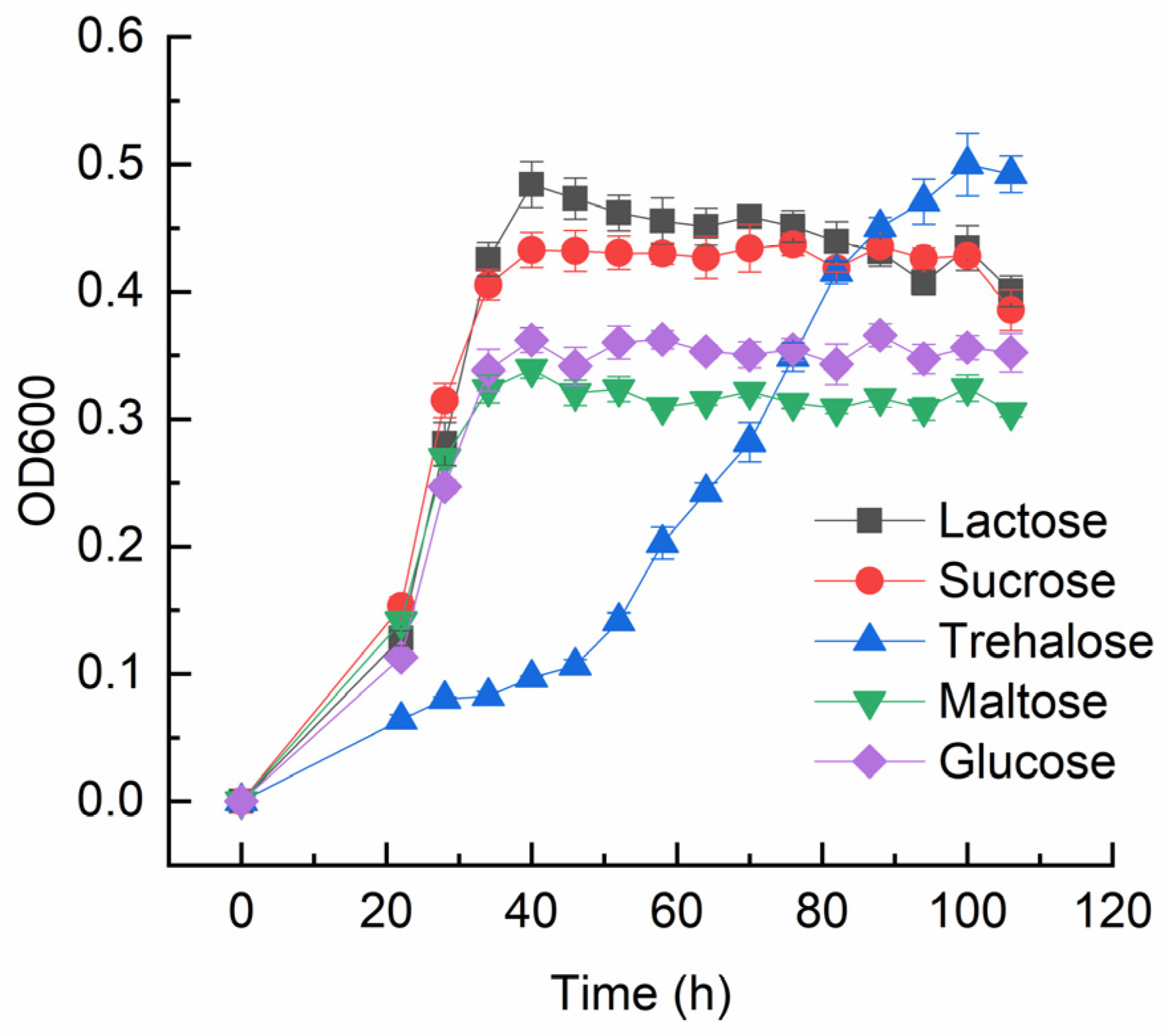
3.1. The Ability of M. phyllosphaerae LW106 to Utilize Trehalose as the Sole Source of Carbon

To validate the trehalose utilization ability of M. phyllosphaerae LW106 under ambient temperature, the time course growth of this psychrotolerant strain cultured in a 1/4 TSB liquid medium supplemented with various sources of carbon was investigated. M. phyllosphaerae LW106 was able to grow normally on a modified 1/4 TSB medium containing glucose, lactose, sucrose, maltose, and trehalose as a sole carbon source, suggesting this strain owns multiple disaccharide hydrolytic enzymes. However, the growth rate on a medium with trehalose as a sole carbon source was significantly slower than that of the other carbon sources (Figure 1).

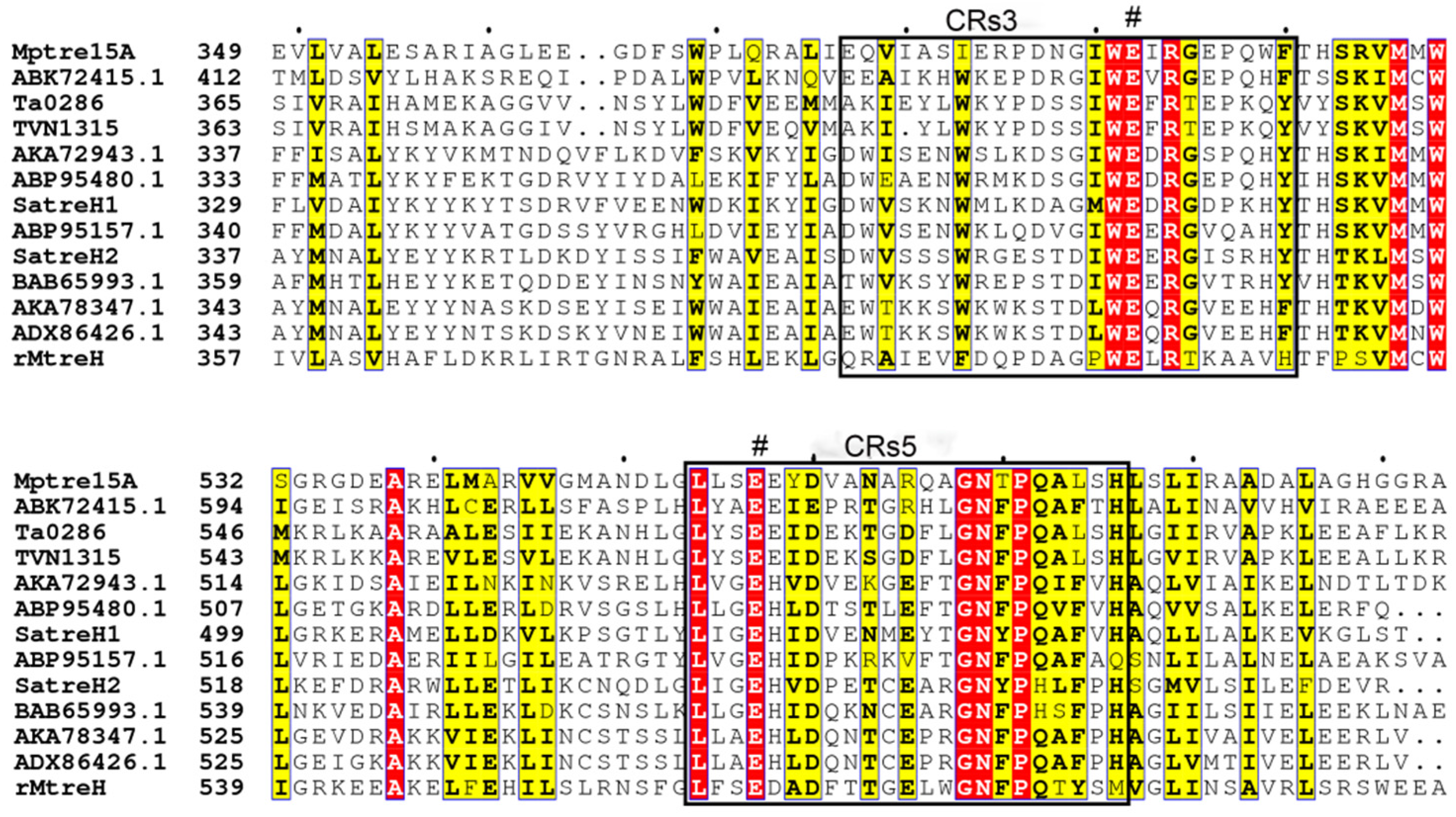
3.2. Bioinformatics Analysis of MpTre15A

The discovered putative GH15 family trehalase MpTre15A consisted of 597 amino acid residues without a signal peptide, with a theoretical molecular mass of 66.2 kDa. The isoelectric point (pI) was predicted to be 5.40. The deduced amino acid sequences of MpTre15A showed 93.97% identity with a predicted glycoside hydrolase from Microbacterium sp. Leaf161, followed by predicted glycoside hydrolases from Microbacterium sp. Leaf320 (93.79%) and the type strain of Microbacterium phyllosphaerae (93.45%). MpTre15A shared a low amino acid sequence identity with the functionally verified bacterial trehalases from Mycolicibacterium smegmatis (36.63%) and Mycobacterium tuberculosis (36.30%), and archaeal trehalases from Thermoplasma volcanium (33.90%) and Thermoplasma acidophilum (34.43%).
The phylogenetic relationships among GH37 and GH65 trehalases and relevant functional verified GH15 members were built. As shown in Figure 2, three major branches were formed. GH37 enzymes including trehalases from bacteria, yeasts, insects, algae, plants, and animals formed one branch, GH65 enzymes, which include acidic trehalases and periplasmic trehalose from fungi, were clustered in the same branch, and GH15 enzymes, which include MpTre15A, trehalase from Mycobacterium smegmatis, and archaeal trehalases from Sulfolobus acidocaldarius, Thermoplasma volcanium, and Thermoplasma acidophilum together with several glucoamylases (GAs) of different origin, were clustered in one branch.
Multiple sequence alignment of MpTre15A with selected relevant trehalases of different origins showed that MpTre15A possessed the five conserved regions in GH15 family glycoside hydrolases. Furthermore, two conserved residues (Glu392 and Glu557) in regions 3 and 5, respectively, which may correspond to the conserved catalytic residues in GH15 family trehalases, were found (Figure 3).

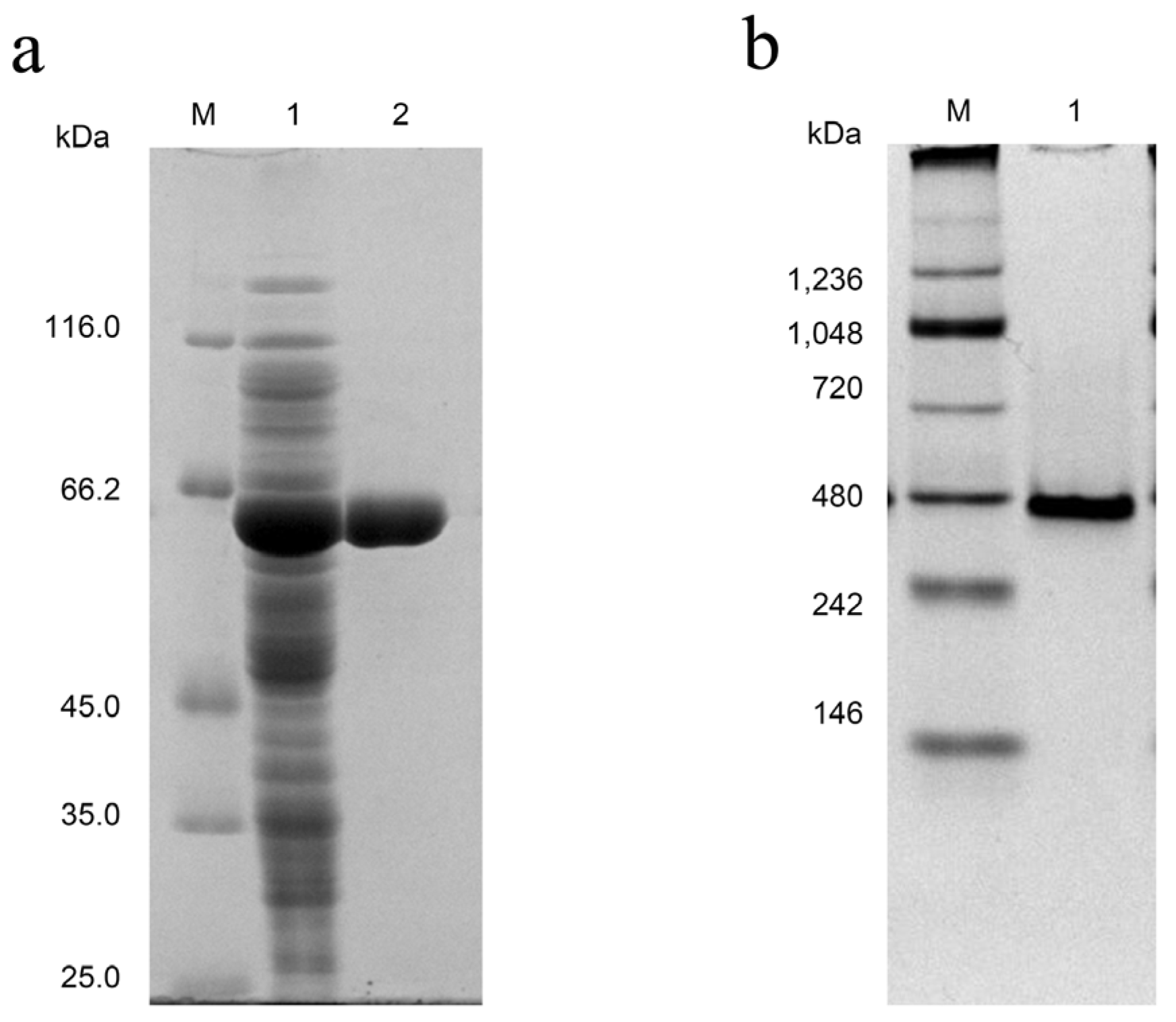
3.3. Expression, Purification, and Activity Assay of GA-Like Trehalase MpTre15A

The recombinant MpTre15A could be expressed in soluble form and at a high level, typically with a yield of 26.8 mg/L. The expressed gene products were purified via affinity chromatography to generate single bands in SDS-PAGE analysis (Figure 4a). The apparent molecular weight of His6 tagged MpTre15A correspond to their theoretical one (66.2 kDa). The Native-PAGE analysis (Figure 4b) of the purified MpTre15A provided a rough estimate of the apparent molecular mass of approximately 397.2 kDa, suggesting that the holoenzyme of MpTre15A should function as a hexamer.
The purified MpTre15A was able to hydrolyze trehalose yielding glucose but showed no activity toward the other tested disaccharides including maltose, cellobiose, sucrose, soluble starch, and p-nitrophenyl-α-glucoside (Table 1). Based on the low amino acid sequence identity of MpTre15A shared with the other functionally verified trehalases, and the highly specific hydrolytic activity toward the substrate trehalose shown by MpTre15A, we identified MpTre15A as a novel member of GH15 family trehalases.

3.4. Characterization of the Catalytic Residues in MpTre15A

To verify the catalytic involvement of the amino acid residue Glu392 and Glu557 in the hydrolytic activity of trehalose MpTre15A, two mutants E392Q and E557Q were constructed and introduced into E. coli for recombinant expression, respectively. The mutants were purified similarly to the wild-type MpTre15A (Figure S1). As shown in Table 2, both the purified E392Q and E557Q mutants lost all their trehalose hydrolytic activity when tested under the same condition as the wild-type MpTre15A. Taken together, these results proved that the two amino acid residues, Glu392 and Glu557, located in the conserved region 3 and 5 of MpTre15A, respectively, functioned as its two key catalytic residues, which are similar to other reported GH15 family GA-like bacterial trehalases [13].

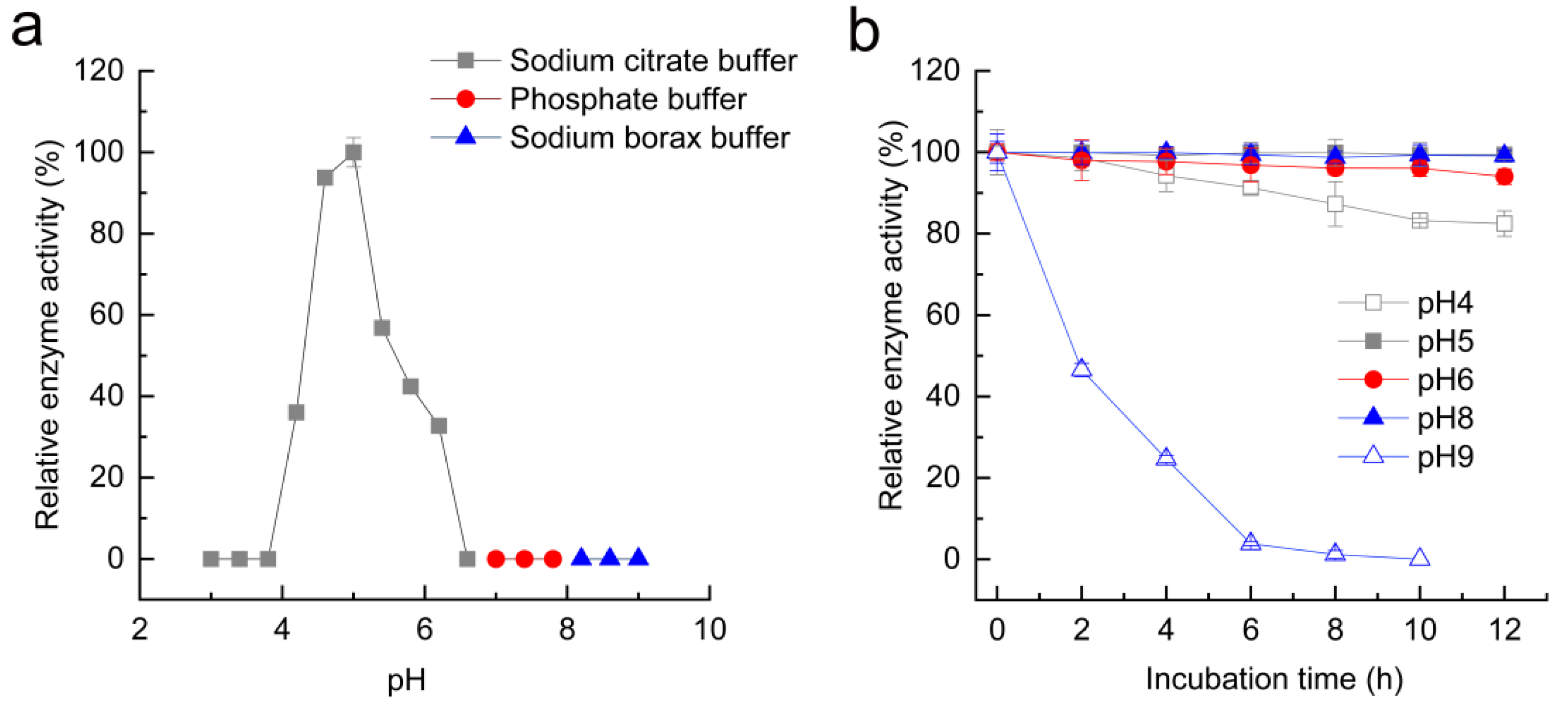
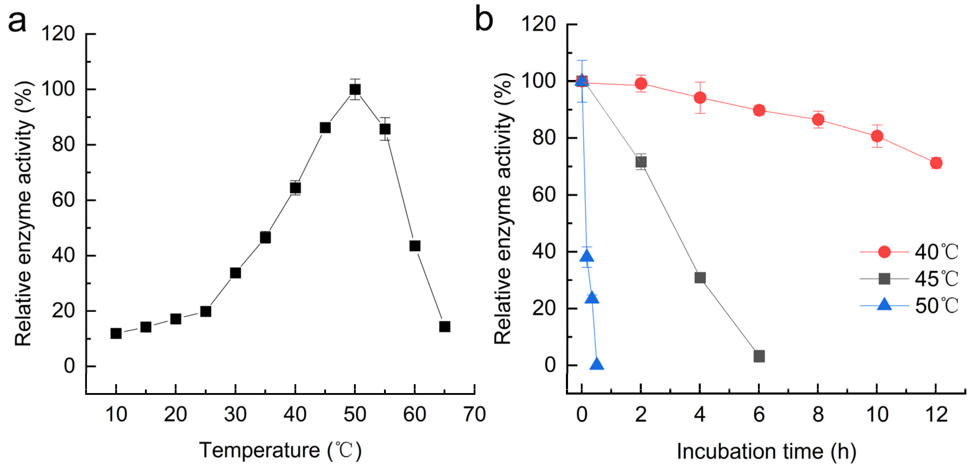
3.5. Effects of pH and Temperature on MpTre15A

The influences of pH on MpTre15A activity are illustrated in Figure 5a. The trehalose-hydrolyzing activity of MpTre15A was maximal at pH 5.0. MpTre15A was active within a narrow pH range from 4.4 to 6.2. MpTre15A exhibited more than half of its maximal activity at pH between 4.8 and 5.4 but lost its activity greatly at pH below 4.4 and above 6.2. The pH stability of MpTre15A was also investigated (Figure 5b). MpTre15A retained over 80% of its maximum activity after 12 h treatment at 4 °C and a pH of 4.0, 5.0, 6.0, and 8.0. However, the residual activity of MpTre15A decreased to less than 30% after 4 h treatment at pH 9.0.
The effects of temperature on the enzymatic activity of the recombinant MpTre15A are shown in Figure 6a. Under the conditions used, the trehalose-hydrolyzing activity of MpTre15A was maximal at approximately 50 °C. The relative activity of MpTre15A dropped to about 10% at 10 °C. As the results demonstrated in Figure 6b, MpTre15A retained more than 90% of its initial activity after 24 h of incubation at 25 °C and showed good stability even after 14 days of incubation at 4 °C (data not shown). After 12 h of incubation at 40 °C, the residual activity of MpTre15A was about 90%. The residual activities of MpTre15A decreased to less than 40% of their initial activity after incubation at 45 °C for 4 h and 50 °C for 20 min. The enzyme activity disappeared after 6 h and 30 min of incubation at 45 °C and 50 °C, respectively.

3.6. Effects of Different Chemicals on the Activity of MpTre15A

The effects of metal ions and other substances on the activity of recombinant MpTre15A were tested at pH 5.0 and 50 °C with the chemicals’ final concentrations of 1 mM and 10 mM. As shown in Table 3, the addition of the monovalent cations Na+ and K+, and the divalent cations Co2+ and Fe2+ did not affect the activity of MpTre15A significantly. It was reported that inorganic phosphate and Mg2+ were required for the activity of Mycobacterium trehalase [6]. In contrast, the addition of 1 mM or 10 mM ethylenediaminetetraacetic acid (EDTA) did not affect the activity of MpTre15A significantly, suggesting MpTre15A was able to hydrolyze trehalose without a requirement for any specific metal ions. Nevertheless, Mn2+ showed a stimulating effect on MpTre15A at a final concentration of 1 mM. When added in 10 mM, both Mn2+ and Mg2+ had a moderate stimulating effect on the activity of MpTre15A. In contrast, the divalent cations Ni2+ and Ca2+, and the trivalent cations Fe3+ and Al3+ showed a moderate inhibitory effect on trehalase activity at the concentration of 10 mM. Similar inhibitory effect of Fe3+ and stimulating effect of Mn2+ on the trehalose hydrolytic activity of rMtreH from Microvirga sp. MC18 was observed as well, indicating trivalent cations like Fe3+ may play a negative role in GH15 family trehalases, whereas some divalent cations such as Mn2+ and Mg2+ may be favorable for the activity of GH15 trehalases. The detergent sodium dodecyl sulfate (SDS) strongly inhibited the activity of MpTre15A at a final concentration of 1 mM, and the addition of 1 mM or 10 mM cetyltrimethylammonium bromide (CTAB) moderately inhibited the activity. Surprisingly, urea, 1,4-dithiothreitol (DTT), and β-mercaptoethanol at a final concentration of both 1 mM and 10 mM significantly stimulated the activity of MpTre15A.

3.7. Steady-State Kinetics Using Trehalose as the Substrate at Different Temperature

Due to the psychrotolerant bacterial origin of MpTre15A, we determined the kinetic parameters of MpTre15A toward trehalose at 50, 37, 25, and 4 °C, respectively (Table 4). In 100 mM sodium citrate buffer (pH 5.0), the trehalose hydrolysis reaction catalyzed by MpTre15A at different temperatures obeyed the Michaelis-Menten kinetics. The kcat and Km values at 50 °C were 345.4 s−1 and 38.0 mM, respectively. As the reaction temperatures decreased to 37, 25, and 4 °C, the Km values toward trehalose increased to 48.38, 53.66, and 65.43 mM, respectively, indicating a decreased affinity of MpTre15A to trehalose as the reaction temperature dropped. As expected, the kcat value of MpTre15A decreased with the dropped reaction temperature from 50 °C to 4 °C. The kcat/Km of MpTre15A at 50 °C was the highest with a value of 9.15 s−1 mM−1 and that of MpTre15A at 4 °C was the lowest with a value of 1.60 s−1 mM−1. Notably, the kcat and kcat/Km values of MpTre15A at 4 °C (104.50 s−1 and 1.6 s−1mM−1, respectively) were comparable to those observed for thermophilic GH15 trehalase TVN1315, Ta0286, and SaTreH1 from T. volcanium, T. acidophilum, and S. acidocaldarius at 50 °C (Table 4).

3.8. Time-Course Conversion of Trehalose into Glucose by MpTre15A

The time-course conversion efficiency of trehalose into glucose catalyzed by MpTre15A at different temperatures (4, 25, 37, and 50 °C) was further investigated by TLC and HPLC. TLC analysis demonstrated that the trehalose hydrolysis and synchronous formation of glucose catalyzed by purified MpTre15A occurred at different temperatures with initial trehalose concentrations of 5 mg/mL and 50 mg/mL (Figure S2). The trehalose consumption and glucose production were further determined by HPLC (Figure 7). When 5 mg/mL trehalose was used as the substrate, the residual trehalose at 50 °C after incubation for 10 min and that at 37 °C after incubation for 15 min was only 3.0% of the initial trehalose (Figure 7a). The substrate trehalose was completely degraded after being incubated at 50 °C and/or 37 °C for less than 60 min with an initial substrate concentration of 5 mg/mL. The conversion rate of trehalose into glucose decreased as the reaction temperature decreased. The residual trehalose after 120 min of incubation at 25 °C and 4 °C was about 2.4% and 9.3%, respectively. The formation of glucose at different temperatures was in agreement with the trehalose consumption (Figure 7b), indicating a direct conversion of trehalose to glucose without any other side products. The produced glucose after 10 min incubation at 50 °C and 15 min incubation at 37 °C were 5.07 g/L and 4.97 g/L, suggesting the conversion rate of trehalose at 50 °C and 37 °C reached 100% and 99.4%, respectively. Consistent with reduced trehalose hydrolysis rate at a lower temperature, the glucose production at 25 °C and 4 °C after 120 min was 5.02 g/L and 4.15 g/L.
When 50 mg/mL trehalose was used as the substrate, trehalose was not completely degraded after incubated at 50 °C and 37 °C for up to 120 min, and the residual trehalose at 50 °C and 37 °C was 1.2% and 13.7% of the initial trehalose, respectively (Figure 7c). The trehalose hydrolysis rate at 25 °C and 4 °C was only 60.6% and 44.9% after 120 min of incubation, which is much lower than that at 50 °C and 37 °C. Correspondingly, the glucose production at 25 °C and 4 °C after 120 min was only 27.58 g/L and 17.80 g/L, respectively, both of which were significantly lower than those (48.78 g/L and 41.29 g/L, respectively) at 50 °C and 37 °C (Figure 7d). Approximately 39.9% of the total trehalose could still be detected after 6 h of incubation at 4 °C. In addition, compared with the trehalose conversion rate with an initial substrate concentration of 5 mg/mL at 4 °C and 25 °C, the trehalose conversion with an initial substrate concentration of 50 mg/mL at the same reaction temperature was significantly lower, which may be caused by inhibition effects of a high concentration of substrate or produced glucose.

4. Discussion

Trehalose is generally regarded as an osmoprotectant as part of bacterial stress responses during periods of freezing [27]. However, the trehalose metabolism in psychrophiles has not been addressed in detail yet. The bacterium M. phyllosphaerae LW106 could grow at 4–25 °C and showed an optimal growth at 16–20 °C. According to Morita’s definition, M. phyllosphaerae LW106 should be categorized as a psychrotolerant bacterium [28]. This strain was found to be able to grow on a medium containing glucose, lactose, sucrose, maltose, and trehalose as a sole carbon source but displayed a slower growth rate on a medium with trehalose as a sole carbon source at 16 °C (Figure 1). Whole genomic sequencing results revealed both a GH15 family GA-like trehalose and a GH13_16 family trehalose synthase in M. phyllosphaerae LW106 (data not shown). Thus, we speculate that MpTre15A catalyzed trehalose degradation may not be the priority way for providing glucose as a cellular carbon source of energy in M. phyllosphaerae LW106. MpTre15A should be more involved in maintaining the homeostasis of cellular trehalose, which functioned as a compatible solute, when the growth temperature changes, since this psychrotolerant bacterium has to undergo temperature fluctuations caused by the glacier basal freezing and the internal melting cycles as well as the season and climate changes [29].
There have been only a few identified bacterial trehalases, especially from the GH15 family [6,18]. MpTre15A shared only a low sequence identity with its closest homolog that has been partially characterized, which is a trehalase from Mycobacterium tuberculosis (35.03%). In addition, different from the recombinant, archaeal trehalases functions as a monomer, dimer, or trimer [16,17,30], and Mycobacterium trehalase functions as a multimeric structure with a molecular mass of 1500 kDa [6], the native MpTre15A seemed to function as a hexamer with a molecular mass of approximately 397.2 kDa (Figure 4). Therefore, the description of MpTre15A is valuable due to its sequence novelty and psychrotrophic origin.
In addition to trehalase, enzymes assigned to the GH15 family also include GAs, glucodextranases (GDases), dextran dextrinase, and isomaltose glucohydrolase. Similar to other reported GH15 trehalases, the cold-adapted GH15 trehalase MpTre15A, discovered in the current study, showed more similar amino acid sequences to GAs than to GH37 and GH65 trehalases (Figure 2). An [S/G/A]E[H/E] sequence around one of the catalytic residue Glu in the conserved region 5 (CRs5) was regarded as an essential motif for the catalytic reaction of GH15 family trehalases [13]. For instance, a GEH sequence existed in the two archaeal trehalases, SaTreH1 and SaTreH2 from Sulfolobus acidocaldarius, whereas a SEE sequence was observed in the other two archaeal trehalases, TVN1315 and Ta0286, from the thermophilic Thermoplasma volcanium and T. acidophilum [17]. Unlike the previously reported bacterial Mycobacterium trehalase [6], which possessed an AEE sequence, a SEE sequence was discovered in MpTre15A in the current study (Figure 3). Similar to the other GH15 enzymes, such as GAs and GDases, GH15 trehalases including those of archaeal origin possess five CRs in their primary structures [6,17,17]. Consistently, mutation of the two glutamic acid residues, E392Q and E557Q, in CRs 3 and 5 of MpTre15A, led to a total loss of trehalose hydrolytic activity, indicating the two glutamic acid residues’ crucial involvement in its catalytic activity.
Another recently reported bacterial GH15 trehalase rMtreH from Microvirga sp. MC18 displayed maximum activity at 40 °C and retained more than 60% residual activity after 1 h incubation at 50 °C [18]. In contrast, the optimum temperature of MpTre15A was found to be 50 °C, and the residual activity of MpTre15A disappeared after being treated at 50 °C for only 30 min (Figure 6b). Interestingly, the enzyme activity of MpTre15A at 50 °C exhibited in the trehalose conversion assay was not lost up to 120 min when incubated with an initial trehalose concentration of 50 mg/mL (Figure 7d). Taking into account that enzyme stability is enhanced in the presence of sugars [31,32], these results suggested that a high concentration of trehalose was favorable for the stability of MpTre15A. Substrate specificity analysis of MpTre15A from M. phyllosphaerae sp. LW106 confirmed this novel cold-adapted trehalase could specifically hydrolyze trehalose but had no activity toward maltose, cellobiose, sucrose, and soluble starch (Table 1). Unlike the bacterial GH15 family trehalases from Mycobacterium smegmatis [6] and Microvirga sp. MC18, both of which showed an optimal pH of around 7.0, MpTre15A from the psychrotolerant M. phyllosphaerae LW106 hydrolyzed trehalose with an optimal pH of 5.0 and was stable at a pH range of 4.0 to 8.0. Nevertheless, similar results were observed in the archaeal GH15 family trehalases from thermophilic Thermoplasma volcanium and T. acidophilum, and acidophilic Sulfolobus acidocaldarius, which functioned within a narrow range of acidic pH values but were stable over a wide pH range [16,17].
The above enzymatic properties of MpTre15A suggested this enzyme was thermolabile, and the kinetic parameters of MpTre15A further revealed its typical cold-adaptability. The kinetic behavior of MpTre15A at different temperatures showed decreased affinity toward trehalose with the decrease in reaction temperature (Table 4). This decrease in substrate affinity of MpTre15A at low temperatures should be a result of its inherent flexible structure, which compensates for the low kinetic energy of cold-adapted enzymes in cold environments [21,33]. Nevertheless, both the kcat and kcat/Km values of MpTre15A at 4 °C (104.50 s−1 and 1.6 s−1 mM−1) were comparable to or even higher than those observed for the GH15 family trehalases from thermophiles at 50 °C (Table 4). It has been generally regarded that cold-adapted enzyme features with high localized structural flexibility, where the rapid change of conformation at low temperatures allows for substrates to access the active center [21]. Higher values are usually found for psychrophilic enzymes in terms of kcat, Km as well as kcat/Km when comparing the catalytic activities of psychrophilic, mesophilic, and thermophilic enzymes at the same temperature below the melting point [34]. Thus, the countered detrimental effect of low temperature on the catalytic turnover and catalytic efficiency displayed by MpTre15A proved that this novel GA-like bacterial trehalase was a typical cold-adapted trehalase of the GH15 family, which may decrease the activation energies and increase the protein flexibility (entropic compensation) like the other psychrophilic enzymes did [35,36]. In addition, since GH15 trehalases are structurally similar to GA but display different substrate specificities, MpTre15A will be a good model for further dissecting the differences in substrate binding sites and elucidating the substrate recognition mechanism of cold-adapted GH15 trehalases.
To date, the trehalase with the highest turnover number (730 s−1) is a GH37 family trehalase from the midgut of Spodoptera frugiperda larvae [37]. According to the kinetic parameters of the current study, MpTre15A showed the highest kcat value (347.45 s−1) at its optimum temperature among the characterized GH15 family trehalases (Table 4). Compared with the other reported trehalases of bacterial origin such as E. coli (a turnover number of 199 s−1) and Zunongwangia sp. (a turnover number of 263.25 s−1) [38,39], the kcat value of MpTre15A made it a good candidate for efficient conversion of trehalose into glucose as therapeutic enzyme and food additive from an application perspective [18,40]. In addition, MpTre15A could be recombinantly expressed in the soluble form at a high yield (typically 26.8 mg/L), which is advantageous for its scale-up expression and purification as well. The optimal pH of 5.0 and good stability at a pH range of 4.0 to 8.0 also made it convenient to achieve the maximum trehalose hydrolysis at a pH range usually employed for ethanol fermentation of the yeast S. cerevisiae and regulate the enzyme activity by simply adjusting the medium pH. Moreover, MpTre15A showed an efficient trehalose conversion rate at a temperature range between 50 °C and 25 °C (Figure 7). Considering that yeast generates trehalose during the process of ethanol fermentation but cannot utilize trehalose as a carbon source [40], recombinant trehalase MpTre15A with a high expression level and efficient conversion rate of trehalose into glucose at a temperature range between 37 °C and 25 °C could be of great interest for application in improving the process of ethanol fermentation. In addition, regression analysis of the trehalose consumption and glucose production catalyzed by MpTre15A at ambient temperature with an initial trehalose concentration below 5 mg/mL showed a good linear correlation with an R2 of 0.9885 and 0.9995, respectively (data not shown). These results suggested this cold-adapted trehalose also had good potential in the development of an enzyme-based biosensor for trehalose content quantification, which is of great value for life science, biomedicine, and other research fields [41].

5. Conclusions

We described detailed identification and characterization of a putative GH15 α,α-trehalase MpTre15A from M. phyllosphaerae LW106, which is the first report of a cold-adapted GH15 family α,α-trehalase to our knowledge. Investigation of the enzymatic properties of this cold-adapted α,α-trehalase proved its involvement in trehalose hydrolysis in the psychrotolerant M. phyllosphaerae LW106. The purified MpTre15A functioned as a hexamer and displayed maximal activity at pH 5.0 and 50 °C without a requirement for any metal ions. MpTre15A was thermolabile at temperatures above 45 °C but retained a high kcat and kcat/Km values of 104.50 s1 and 1.6 s1 mM1 at 4 °C, respectively, which proved its typical cold-adaptability. These properties added to the benefit of using this cold-adapted trehalose in industrial ethanol fermentation processes with energy savings. The capability of MpTre15A to efficiently catalyze the conversion of trehalose at a substrate concentration of up to 5 mg/mL with high rates at a temperature range from 4 °C to 50 °C, also constitutes advantages for developing potential therapeutic enzymes and enzyme-based biosensors for trehalose content quantification.

Supplementary Materials

The following supporting information can be downloaded at: https://www.mdpi.com/article/10.3390/fermentation8100471/s1. Figure S1: SDS-PAGE analysis of the MpTre15A mutants. Lane M, molecular size marker (Unstained Protein Molecular Weight Marker: 116, 66.2, 45, 35, 25 kDa); lane 1, crude enzyme of E392Q mutant; lane 2, purified enzyme of E392Q mutant after Ni-IDA column; lane 3, crude enzyme of E557Q mutant; lane 4, purified enzyme of E557Q mutant. Figure S2: Thin layer chromatography (TLC) analysis of the hydrolytic products of trehalose. Panel a and b, TLC analysis of trehalose hydrolysates at different temperatures with 5 mg/mL trehalose. Panel a: Lane G indicates glucose and trehalose mixed standard; Lane 1 to Lane 5 indicate the hydrolytic product of trehalose at 50 ℃ for 1, 3, 5, 10, and 60 min, respectively; Lane 6 to Lane 11 indicate the hydrolytic product of trehalose at 37 ℃ for 5, 15, 30, 45, 60, and 120 min, respectively. Panel b: Lane G indicates glucose and trehalose mixed standard; Lane 1 to Lane 6 indicate the hydrolytic product of trehalose at 25 ℃ for 10, 30, 45, 60, 90, and 120 min, respectively; Lane 7 to Lane 12 indicates the hydrolytic product of trehalose at 4 ℃ for 20, 40, 60, 120, 180, and 240 min, respectively. Panel c and d, TLC analysis of trehalose hydrolysates at different temperatures with 50 mg/mL trehalose. Panel c: Lane G indicate glucose and trehalose mixed standard; Lane 1 to Lane 5 indicate the hydrolytic product of trehalose at 50 ℃ for 15, 30, 45, 60, and 120 min, respectively; Lane 6 to Lane 10 indicate the hydrolytic product of trehalose at 37 °C for 15, 30, 45, 60, and 120 min, respectively; Lane 11 to Lane 16 indicate the hydrolytic product of trehalose at 25 °C for 30, 60, 90, 120, 180, and 240 min, respectively; Lane 17 indicates glucose and trehalose mixed standard. Panel d: Lane G indicates glucose and trehalose mixed standard; Lane 1 to Lane 7 indicate the hydrolytic product of trehalose at 4 °C for 30, 60, 90, 120, 180, 240, and 360 min, respectively.

Author Contributions

Conceptualization, B.G. and Y.H.; methodology, J.Z. (Jun Zeng); software, B.G.; validation, X.Y. and B.G.; formal analysis, J.Z. (Junhua Zhang); investigation, J.Z. (Junhua Zhang), B.G. and Y.H.; resources, Y.N.; data curation, X.Y. and Y.H.; writing—original draft preparation, J.Z. (Junhua Zhang); writing—review and editing, X.Y., B.G. and Y.H.; visualization, J.Z. (Junhua Zhang); supervision, B.G. and Y.H.; project administration, X.L.; funding acquisition, B.G. and Y.H. All authors have read and agreed to the published version of the manuscript.

Funding

This research was funded by the National Natural Science Foundation of China, grant number No. 32060522, No. 31560432, and the Project of Youth Innovative Talent Cultivation Plan of Shihezi University, grant number No. CXRC201702.

Institutional Review Board Statement

Not applicable.

Informed Consent Statement

Not applicable.

Data Availability Statement

The data presented in this study are available on reasonable request from the corresponding authors.

Acknowledgments

The authors would like to thank the National Natural Science Foundation of China and the Project of Youth Innovative Talent Cultivation Plan of Shihezi University for financial support.

Conflicts of Interest

The authors declare no conflict of interest.

References

  1. Elbein, A.D.; Pan, Y.T.; Pastuszak, I.; Carroll, D. New insights on trehalose: A multifunctional molecule. Glycobiology 2003, 13, 17R–27R. [Google Scholar] [CrossRef] [PubMed]
  2. Garcia, C.A.; Narrett, J.A.; Gardner, J.G. Trehalose degradation by Cellvibrio japonicus exhibits no functional redundancy and is solely dependent on the Tre37A enzyme. Appl. Environ. Microbiol. 2020, 86, 22. [Google Scholar] [CrossRef] [PubMed]
  3. Sussman, A.S.; Lingappa, B.T. Role of trehalose in ascospores of Neurospora tetrasperma. Science 1959, 130, 1343. [Google Scholar] [CrossRef]
  4. Argüelles, J.C. Physiological roles of trehalose in bacteria and yeasts: A comparative analysis. Arch. Microbiol. 2000, 174, 217–224. [Google Scholar] [PubMed]
  5. Thevelein, J.M.; Hohmann, S. Trehalose synthase: Guard to the gate of glycolysis in yeast? Trends Biochem. Sci. 1995, 20, 3–10. [Google Scholar] [CrossRef]
  6. Carroll, J.D.; Pastuszak, I.; Edavana, V.K.; Pan, Y.T.; Elbein, A.D. A novel trehalase from Mycobacterium smegmatis—Purification, properties, requirements. FEBS J. 2007, 274, 1701–1714. [Google Scholar] [CrossRef] [PubMed]
  7. Kandror, O.; DeLeon, A.; Goldberg, A.L. Trehalose synthesis is induced upon exposure of Escherichia coli to cold and is essential for viability at low temperatures. Proc. Natl. Acad. Sci. USA 2002, 99, 9727–9732. [Google Scholar] [CrossRef] [PubMed]
  8. Singer, M.A.; Lindquist, S. Multiple effects of trehalose on protein folding in vitro and in vivo. Mol. Cell 1998, 1, 639–648. [Google Scholar] [CrossRef]
  9. Higashiyama, T. Novel functions and applications of trehalose. Pure Appl. Chem. 2002, 74, 1263–1269. [Google Scholar] [CrossRef]
  10. Iturriaga, G.; Suárez, R.; Nova-Franco, B. Trehalose metabolism: From osmoprotection to signaling. Int. J. Mol. Sci. 2009, 10, 3793–3810. [Google Scholar] [CrossRef] [Green Version]
  11. Boos, W.; Ehmann, U.; Bremer, E.; Middendorf, A.; Postma, P. Trehalase of Escherichia coli. Mapping and cloning of its structural gene and identification of the enzyme as a periplasmic protein induced under high osmolarity growth conditions. J. Biol. Chem. 1987, 262, 13212–13218. [Google Scholar] [CrossRef]
  12. Horlacher, R.; Uhland, K.; Klein, W.; Ehrmann, M.; Boos, W. Characterization of a cytoplasmic trehalase of Escherichia coli. J. Bacteriol. 1996, 178, 6250–6257. [Google Scholar] [CrossRef] [PubMed]
  13. Sakaguchi, M. Diverse and common features of trehalases and their contributions to microbial trehalose metabolism. Appl. Microbiol. Biotechnol. 2020, 104, 1837–1847. [Google Scholar] [CrossRef] [PubMed]
  14. Rimmele, M.; Boos, W. Trehalose-6-phosphate hydrolase of Escherichia coli. J. Bacteriol. 1994, 176, 5654–5664. [Google Scholar] [CrossRef]
  15. Moon, J.H.; Lee, W.; Park, J.; Choi, K.-H.; Cha, J. Characterization of a trehalose-degrading enzyme from the hyperthermophilic archaeon Sulfolobus acidocaldarius. J. Biosci. Bioeng. 2016, 122, 47–51. [Google Scholar] [CrossRef]
  16. Sakaguchi, M.; Shimodaira, S.; Ishida, S.; Amemiya, M.; Honda, S.; Sugahara, Y.; Oyama, F.; Kawakita, M. Identification of GH15 family thermophilic archaeal trehalases that function within a narrow acidic-pH range. Appl. Environ. Microbiol. 2015, 81, 4920–4931. [Google Scholar] [CrossRef]
  17. Yuasa, M.; Okamura, T.; Kimura, M.; Honda, S.; Shin, Y.; Kawakita, M.; Oyama, F.; Sakaguchi, M. Two trehalose-hydrolyzing enzymes from Crenarchaeon Sulfolobus acidocaldarius exhibit distinct activities and affinities toward trehalose. Appl. Microbiol. Biotechnol. 2018, 102, 4445–4455. [Google Scholar] [CrossRef]
  18. Dong, C.; Fan, Q.; Li, X.; Huang, Y.; Han, J.; Fang, X.; Huan, M.; Ye, X.; Li, Z.; Cui, Z. Expression and characterization of a novel trehalase from Microvirga sp. strain MC18. Protein Expr. Purif. 2021, 182, 105846. [Google Scholar] [CrossRef]
  19. Murray, I.A.; Coupland, K.; Smith, J.A.; Ansell, I.D.; Long, R.G. Intestinal trehalase activity in a UK population: Establishing a normal range and the effect of disease. Br. J. Nutr. 2000, 83, 241–245. [Google Scholar] [CrossRef]
  20. Kocot, B.; Vroemen, C.; Torres Pazmino, D.E.; Cannon, D.M., Jr.; Ward, D.E.; Foerster, H.; Shetty, J.K.; Clarkson, K.A.; Albers, K.; Van Brussel, M. Trehalase in fermentations. US Patent WO2015065978; filed 28 October 2014, and issued 7 May 2015,
  21. Feller, G.; Gerday, C. Psychrophilic enzymes: Hot topics in cold adaptation. Nat. Rev. Microbiol. 2003, 1, 200–208. [Google Scholar] [CrossRef]
  22. Mangiagalli, M.; Lotti, M. Cold-active β-galactosidases: Insight into cold adaptation mechanisms and biotechnological exploitation. Mar. Drugs. 2021, 19, 43. [Google Scholar] [CrossRef] [PubMed]
  23. Bruno, S.; Coppola, D.; di Prisco, G.; Giordano, D.; Verde, C. Enzymes from marine polar regions and their biotechnological applications. Mar. Drugs. 2019, 17, 544. [Google Scholar] [CrossRef] [PubMed]
  24. Zhang, Y.; Wang, L.; Li, M.; Gu, Y.; Guan, B.; Ni, Y. Isolation and identification of a Microbacterium sp. LW106 strain producing cold-active β-galactosidase, and study on its enzyme production conditions and enzymatic properties. Microbiol. China 2019, 46, 609–617. [Google Scholar]
  25. Wang, L.; Mou, Y.; Guan, B.; Hu, Y.; Zhang, Y.; Zeng, J.; Ni, Y. Genome sequence of the psychrophilic Cryobacterium sp. LW097 and characterization of its four novel cold-adapted β-galactosidases. Int. J. Biol. Macromol. 2020, 163, 2068–2083. [Google Scholar] [CrossRef] [PubMed]
  26. Gouet, P.; Robert, X. Deciphering key features in protein structures with the new ENDscript server. Nucleic Acids Res. 2014, 42, W320–W324. [Google Scholar]
  27. Strom, A.R.; Kaasen, I. Trehalose metabolism in Escherichia coli: Stress protection and stress regulation of gene expression. Mol. Microbiol. 1993, 8, 205–210. [Google Scholar] [CrossRef]
  28. Morita, R.Y. Psychrophilic bacteria. Bacteriol. Rev. 1975, 39, 144–167. [Google Scholar] [CrossRef]
  29. Alley, R.B.; Cuffey, K.M.; Larson, G.J. How glaciers entrain and transport basal sediment: Physical constraints. Quat. Sci. Rev. 1997, 16, 1017–1038. [Google Scholar] [CrossRef]
  30. Lee, J.; Lee, A.; Moon, K.; Choi, K.-H.; Cha, J. Saci_1816: A Trehalase that Catalyzes Trehalose Degradation in the Thermoacidophilic Crenarchaeon Sulfolobus acidocaldarius. J. Mol. Biol. 2018, 28, 909–916. [Google Scholar] [CrossRef]
  31. Prasad, S.; Roy, I. Effect of disaccharides on the stabilization of bovine trypsin against detergent and autolysis. Biotechnol. Prog. 2009, 26, 627–635. [Google Scholar] [CrossRef]
  32. Rodriguez-Colinas, B.; Poveda, A.; Jimenez-Barbero, J.; Ballesteros, A.O.; Plou, F.J. Galacto-oligosaccharide synthesis from lactose solution or skim milk using the β-galactosidase from Bacillus circulans. J. Agric. Food Chem. 2012, 60, 6391–6398. [Google Scholar] [CrossRef] [PubMed]
  33. Cavicchioli, R.; Charlton, T.; Ertan, H.; Omar, S.M.; Siddiqui, K.S.; Williams, T.J. Biotechnological uses of enzymes from psychrophiles. Microb. Biotechnol. 2011, 4, 449–460. [Google Scholar] [CrossRef] [PubMed] [Green Version]
  34. Åqvist, J.; Isaksen, G.V.; Brandsdal, B.O. Computation of enzyme cold adaptation. Nat. Rev. Chem. 2017, 1, 0051. [Google Scholar] [CrossRef]
  35. Lonhienne, T.; Zoidakis, J.; Vorgias, C.E.; Feller, G.; Gerday, C.; Bouriotis, V. Modular structure, local flexibility and cold-activity of a novel chitobiase from a psychrophilic antarctic bacterium. J. Mol. Biol. 2001, 310, 291–297. [Google Scholar] [CrossRef] [PubMed]
  36. Santiago, M.; Ramírez-Sarmiento, C.A.; Zamora, R.A.; Parra, L.P. Discovery, molecular mechanisms, and industrial applications of cold-active enzymes. Front. Microbiol. 2016, 7, 1408. [Google Scholar] [CrossRef] [PubMed]
  37. Silva, M.C.P.; Terra, W.R.; Ferreira, C. The catalytic and other residues essential for the activity of the midgut trehalase from Spodoptera frugiperda. Insect Biochem. Mol. Biol. 2010, 40, 733–741. [Google Scholar] [CrossRef] [PubMed]
  38. Cheng, Q.; Gao, H.; Hu, N. A trehalase from Zunongwangia sp.: Characterization and improving catalytic efficiency by directed evolution. BMC Biotechnol. 2016, 16, 9. [Google Scholar] [CrossRef]
  39. Gibson, R.; Gloster, T.; Roberts, S.; Warren, R.; Storch de Gracia, I.; García, A.; Chiara, J.; Davies, G. Molecular basis for trehalase inhibition revealed by the structure of trehalase in complex with potent inhibitors. Angew. Chem. Int. Ed Engl. 2007, 46, 4115–4119. [Google Scholar] [CrossRef]
  40. Dong, L.; Yu, D.; Lin, X.; Wang, B.; Pan, L. Improving expression of thermostable trehalase from Myceliophthora sepedonium in Aspergillus niger mediated by the CRISPR/Cas9 tool and its purification, characterization. Protein Expr. Purif. 2020, 165, 105482. [Google Scholar] [CrossRef]
  41. Fang, X.; Mei, Z.; Chen, J.; Xiong, S.; Ning, J.; Jiang, C.; Gao, Y.; Yi, X.; Hui, G. A Trehalose quantitative sensor based on Ni foam material modified with graphene oxidated and non-linear analysis model. Food Anal. Methods 2021, 14, 1977–1985. [Google Scholar] [CrossRef]
Figure 1. Growth analysis of M. phyllosphaerae LW106 strains. M. phyllosphaerae LW106 cells were grown in a 1/4 TSB medium containing 3 g/L of lactose, sucrose, trehalose, maltose, and glucose, respectively. Experiments were performed in triplicate, and the average values are shown.
Fermentation 08 00471 g001
Figure 2. Phylogenetic relationship of related enzyme sequences. The phylogenetic tree was constructed by MEGA6 (52) using the neighbor-joining method. Bootstrap values based on 1000 replicates are shown at nodes, accession number, enzyme name, and organism name are shown. MpTre15A is indicated by a closed circle.
Fermentation 08 00471 g002
Figure 3. Multiple sequence alignments of M. phyllosphaerae trehalase partial conserved regions with relevant GH15 family trehalases. The sequences aligned are the following: ABK72415.1, Mycobacterium smegmatis MC2 155; LC004405.1, Thermoplasma volcanium TVN1315; LC004406, Thermoplasma acidophilum Ta0286; LC330867, Sulfolobus acidocaldarius SaTreH2; LC330868, Sulfolobus acidocaldarius SaTreH1; ABP95480.1, Metallosphaera sedula DSM 5348; ABP95157.1, Metallosphaera sedula DSM 5348; BAB65993.1, Sulfolobus tokodaii str. 7; AKA78347.1, Saccharolobus solfataricus; ADX86426.1, Sulfolobus islandicus REY15A; AKA72943.1, Saccharolobus solfataricus; UHN91757.1, Microvirga sp. MC18 rMtreH. The number sign (#) denotes putative catalytic residues. Fully conserved amino acid residues and related amino acid residues are red and yellow, respectively. Boxes show two highly conserved regions, conserved regions 3 (CRs3) and conserved regions 5 (CRs5).
Fermentation 08 00471 g003
Figure 4. SDS-PAGE (a) and Native-PAGE (b) analysis of the purified proteins: (a) Lane M, molecular size marker (Unstained Protein Molecular Weight Marker: 116, 66.2, 45, 35, 25 kDa); lane 1, crude enzyme; lane 2, purified MpTre15A after Ni-IDA column; (b) Lane M, molecular size marker (Unstained Protein Molecular Weight Marker: 1234, 1048, 720, 480, 242, 146 kDa); lane 1, purified MpTre15A.
Fermentation 08 00471 g004
Figure 5. Influences of pH on MpTre15A activity (a), and pH stability (b). Relative activity under different pH buffer conditions (pH 3.0–6.4, citrate buffer, gray square; pH 7.0–7.8, phosphate buffer, red circle; pH 8.2–9.0, borate buffer, blue triangle) and residual activity after treatment in different pH buffers (pH 4.0 to 9.0) for 12 h were measured. Experiments were performed in triplicate, and the average values are shown.
Fermentation 08 00471 g005
Figure 6. Effects of temperature on MpTre15A activity (a), and thermo-stability (b): (a) the activities of MpTre15A were measured at various temperatures (10 to 65 °C) in 100 mM citrate buffer (pH 5.0); (b) MpTre15A were incubated at various temperatures (40, 45, and 50 °C) for 60 min, and the remaining activities of MpTre15A were measured at 50 °C in 100 mM citrate buffer (pH 5.0). The initial activities without heat treatment were set as 100% with a specific activity of 62.5 U/mg. Experiments were performed in triplicate, and the average values are shown.
Fermentation 08 00471 g006
Figure 7. HPLC analysis of the time-course trehalose conversion by MpTre15A at the different reaction temperatures. The enzyme was incubated with 5 mg/mL (a,b) and 50 mg/mL trehalose (c,d), respectively, for up to 120 min to detect the consumption of trehalose (a,c) and the production of glucose (b,d).
Fermentation 08 00471 g007
Table 1. Substrate specificity analysis of MpTre15A.
SubstrateRelative Activity (%)
trehalose100
maltose0
cellobiose0
sucrose0
soluble starch0
p-nitrophenyl-α-D-glucopyranoside0
MpTre15A was added to the sugar solution with a final protein concentration of 0.01 mg/mL and determined at pH 5.0 with 100 mM sodium citrate buffer.
Table 2. Activity analysis of MpTre15A mutants.
EnzymeRelative Activity (%)
MpTre15A100
E392Q0
E557Q0
The specific activity of MpTre15A toward trehalose was 69.4 U/mg protein tested in 100 mM sodium citrate buffer (pH 5.0).
Table 3. Effects of various chemicals on the relative activity of MpTre15A.
ReagentsRelative Activity (%)
1 mM10 mM
No addition a100.0 ± 1.2100.0 ± 1.2
NaCl100.4 ± 3.6103.2 ± 4.9
KCl100.5 ± 1.6105.1 ± 2.4
MgCl2101.3 ± 2.6126.4 ± 6.3
MnCl2114.8 ± 1.7143.6 ± 6.4
NiCl287.6 ± 4.157.2 ± 5.8
CaCl284.0 ± 3.980.6 ± 4.3
CoCl292.6 ± 6.790.6 ± 3.5
FeCl293.2 ± 0.790.7 ± 3.8
FeCl393.9 ± 5.860.1 ± 3.6
AlCl396.0 ± 6.165.6 ± 1.2
Cetyltrimethylammonium bromide (CTAB)77.3 ± 5.166.9 ± 5.5
Ethylenediaminetetraacetic acid (EDTA)97.4 ± 4.690.1 ± 4.9
Urea138.3 ± 1.8156.8 ± 4.8
Sodium dodecyl sulfate (SDS)00
1,4-Dithiothreitol (DTT)117.7 ± 5.4265.3 ± 4.9
β-Mercaptoethanol149.5 ± 4.9251.6 ± 2.6
a The activity of control (no additions) was set as 100% with a specific activity of 77.6 U/mg protein.
Table 4. Kinetic parameters of various GH15 family trehalases toward substrate trehalose.
Organism (Enzyme Name)kcat (s−1)Km (mM)kcat/Km (s−1mM−1)Temperature (°C)pHReferences
M. phyllosphaerae (Mptre15A)347.4537.969.15505.0This study
M. phyllosphaerae (Mptre15A)204.7048.384.23375.0This study
M. phyllosphaerae (Mptre15A)156.5753.662.92255.0This study
M. phyllosphaerae (Mptre15A)104.5065.431.6045.0This study
Mycolicibacterium smegmatisND a20.0ND a377.1Carroll et al. 2007
Thermoplasma volcanium (TVN1315)63.048.71.29503.7Sakaguchi et al. 2015
T. acidophilum (Ta0286)66.740.21.66503.7Sakaguchi et al. 2015
Sulfolobus acidocaldarius (SaTreH1)77.041.81.84504.0Yuasa et al. 2018
S. acidocaldarius (SaTreH1)102.554.51.88604.0Yuasa et al. 2018
S. acidocaldarius (SaTreH2)1.602.390.67503.7Yuasa et al. 2018
S. acidocaldarius (SaTreH2)3.433.470.99703.7Yuasa et al. 2018
Microvirga sp. MC18ND a23.45ND a407.0Dong et al. 2021
a Not determined.
Publisher’s Note: MDPI stays neutral with regard to jurisdictional claims in published maps and institutional affiliations.

