Article

Amperometric Biosensor Based on Laccase Enzyme, Gold Nanoparticles, and Glutaraldehyde for the Determination of Dopamine in Biological and Environmental Samples

by
Anderson M. Santos
,
Ademar Wong
,
Orlando Fatibello-Filho
* and
Fernando C. Moraes
Department of Chemistry, Federal University of São Carlos, São Carlos 13560-970, SP, Brazil
*
Author to whom correspondence should be addressed.
Submission received: 12 July 2022 / Revised: 25 July 2022 / Accepted: 27 July 2022 / Published: 1 August 2022
(This article belongs to the Special Issue Carbon Nanohybrids for Biomedical Applications)

Abstract

:
The present work reports the development and application of an amperometric biosensor based on carbon paste electrode modified with laccase enzyme, glutaraldehyde, and gold nanoparticles (Lac-Glu-AuNPs/CPE) for the determination of the neurotransmitter dopamine (DA). The materials were characterized morphologically and chemically using scanning electron microscopy (SEM), transmission electron microscopy (TEM), and cyclic voltammetry. Optimization studies were performed in order to determine the optimal amount of enzyme and pH level that can yield the best conditions of analysis. The application of the biosensor in optimal conditions using the amperometric technique yielded a linear concentration range of 8.0 × 10⁻⁷–6.2 × 10⁻⁵ mol L−1 with a limit of detection of 6.0 × 10⁻⁸ mol L−1. The proposed biosensor was successfully applied for the determination of DA in biological and environmental samples. In addition, the application of the biosensor for the conduct of electrochemical measurements showed that the sensing device has good repeatability and stability, and it does not suffer from matrix interference effects. The proposed biosensor exhibited an analytical signal of 85% after 10 days of consecutive use.

1. Introduction

The dramatic rise in global population and the massive advances in medicine and drugs manufacturing have resulted in widespread use and often unbridled consumption of pharmaceutical products worldwide. Numerous pharmaceutical products are manufactured and consumed daily, and these products are discarded in the aquatic environment through domestic sewage and industrial effluents which are not properly treated. The rampant disposal of these pharmaceutical products in water bodies leads to environmental pollution, which poses serious risks to both human and animal health, as well as to the aquatic biota. These pharmaceutical products, composed of chemical or natural compounds, which find their way into the aquatic environment through human and industrial activities are referred to as emerging pollutants. The persistence and/or toxicity of these pollutants in water bodies can alter the metabolism of living beings [1,2].
Dopamine (DA) is a monoaminergic neurotransmitter, from the catecholamine and phenylethylamine family, which plays different important roles in the body. This neurotransmitter is particularly important for the proper functioning of the cardiovascular, renal, and hormonal systems [3,4]. The monitoring of DA in human body is essentially important as it helps in the diagnosis of health problems or neuropsychiatric disorders such as the Parkinson’s disease and Schizophrenia.
Thus, the detection and quantification of this compound is of extreme importance for the preservation of human health and for environmental monitoring since the effects of the bioaccumulation of these neurotransmitters in aquatic animals, such as fish and other aquatic organisms. A wide range of techniques have been employed for the detection of dopamine in different matrices; the techniques applied have included spectrophotometry, chromatography, electrochemiluminescence, and electrochemical methods [5,6,7,8,9,10]. Among the aforementioned techniques employed for dopamine determination, electrochemical methods have proven to be essentially outstanding due to the following advantages that they possess over other techniques: (i) The methods exhibit relatively higher sensitivity with excellent repeatability and good stability; (ii) they require fewer or no sample preparation steps; and (iii) they are relatively less expensive to implement [11]. A wide range of materials can be used for the development of electrochemical sensors and biosensors; these include metal nanoparticles (Au, Pt, Ag), carbon materials (graphite and nanodiamond), enzymes (laccase, peroxidase), and so forth.
Laccases or multicopper oxidase enzymes are produced by different types of bacteria, insects, and fungi [12,13]. These enzymes catalyze the oxidation of a variety of phenolic compounds, which is followed by the reduction of molecular oxygen to water [14]. The development of biosensors occurs through the direct transfer of electrons between the electrode surface and the copper atoms present in the laccase [15]. Portaccio et al. [16] developed a biosensor based on laccase-thionine-carbon black modified screen-printed electrode for the determination of Bisphenol A in tomato juice samples; the authors obtained satisfactory results with recovery percentages of approximately 100%.
Carbonaceous materials have several important properties that make them extremely attractive for application in the fabrication of sensors. Among the suitable properties of carbonaceous materials include the following: high electrical conductivity and surface area, fast electronic transfer, and wide potential range; in addition to that, carbonaceous materials can be modified with other materials to form films or folders [17,18,19]. Although a wide range of carbon-based electrodes can be used for the construction of sensors, carbon paste electrodes (CPEs) have been found to be remarkably outstanding due to their practicality; this is because after using a CPE-based sensor for analysis, the sensor surface can be renewed by simply polishing the surface, and this helps to save preparation time. The use of metal nanoparticles as electrode modifying agents in the development of biosensors has been found to help improve the electronic transfer between the analyte and the electrode [20]. Gold nanoparticles (AuNPs) are highly efficient metal nanoparticles which are commonly employed for the construction of electrochemical biosensors due to their excellent biocompatibility [21]. In an important review on sensing devices published in the literature, Pingaron et al. [22] presented recent advances in the construction of electrochemical biosensors using gold nanoparticles. Among the types of biosensors reported in the review included enzyme biosensors, immunosensors, and DNA sensors [22].
In the present work, we report the development and application of an electrochemical biosensor, based on carbon paste electrode (CPE) and AuNPs modified with laccase enzyme and glutaraldehyde, for the detection of dopamine in environmental and biological fluid samples.

2. Materials and Methods

2.1. Reagents and Solutions

The following reagents and solutions employed in the experiments were acquired from Sigma Aldrich, St. Louis, MO, USA: laccase (≥50 U mg−1 of solid) from trametes versicolor, dopamine (DA), graphite powder (<20 µm), glutaraldehyde (25% v/v in water), citrate sodium, mineral oil, HAuCl4.3H2O, KCl, NaCl, KH2PO4, CaCl2, NH4Cl, and urea. All the aforementioned materials and the other reagents used in the experiments were of analytical grade (purity ≥ 98%). All the solutions used in the experiments were prepared with ultrapure water (resistivity not less than 18 MΩ cm) obtained from a Milli-Q Direct-0.3 purification system (Millipore, Burlington, MA, USA).
About 0.1 mmol L−1 DA stock solution was prepared by dissolving 3.06 mg of DA in 2.0 mL of ultrapure water.

2.2. Apparatus

The electrochemical measurements were performed using a potentiostat/galvanostat Autolab model PGSTAT-30 (Utrecht, The Netherlands) controlled by the GPES 4.9 software (Eco Chemie) fitted to a 10 mL electrochemical cell which consisted of three electrodes: Ag/AgCl/KCl (3.0 mol L−1) reference electrode, a platinum wire used as counter electrode, and carbon paste electrode (CPE) (2.5 mm diameter) used as the working electrode.
The pH measurements were performed using Orion Expandable Ion Analyzer (model EA-940, USA), with the combined application of a glass electrode and Ag/AgCl/KCl (3.0 mol L−1) external reference electrode.
Morphological characterization of the materials were evaluated using images acquired with a gun scanning electron microscopy (SEM) (Oxford, model X-ACT microscope), and transmission electron microscopy (TEM) images were obtained with a JEOL transmission electron microscope.

2.3. Preparation of the Gold Nanoparticles

Gold nanoparticles (AuNPs) were synthesized based on the technique described by Ojea-Jimenez [23]. An amount of approximately 2.0 × 10⁻⁴ mol HAuCl4 was added in a 200 mL of deionized water at a temperature of 85 °C. After that, 0.12 mol of sodium citrate was added to the mixture, which was subjected to stirring for 4 min. Subsequently, the solution was placed in an ice bath until it reached room temperature (25 °C). The color change of the solution from yellow to red was visible during cooling. This abrupt color change indicated the formation of AuNPs.

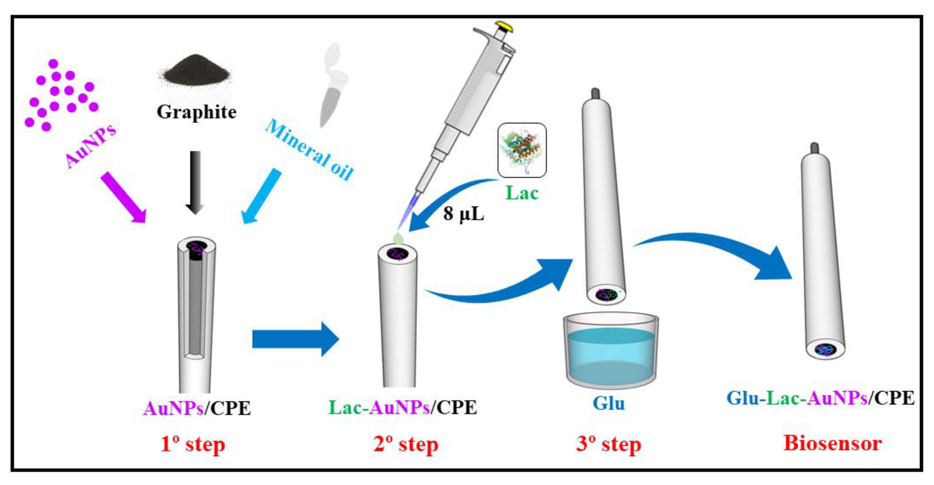
2.4. Preparation of the Biosensor Using Laccase Enzyme, AuNPs, and Glutaraldehyde

The biosensor was prepared by mixing AuNPs, graphite (Gr), and mineral oil in different proportions, where the optimal proportions obtained were as follows: 1.0 mL of AuNPs solution, 100 mg of Gr powder, and 3 drops (45 µL) of mineral oil. The preparation procedure involved mixing 100 mg of Gr powder with 1.0 mL of AuNPs solution, and placing the mixture to dry in an oven at 80 °C. After drying, 3 drops of mineral oil were added to the mixture, and this yielded the AuNPs-modified carbon paste material. The modified carbon paste was placed in a Teflon® tube (Ø = 3 mm) and compacted by pressing it against a smooth surface, obtaining the modified carbon paste electrode; after that, the paste on the electrode surface was then polished by rubbing it on a clean paper (A4 sheet). Subsequently, laccase solution (10 mg mL−1) was prepared and 8.0 µL (4 U by electrode) of this solution was added on the surface of the modified carbon paste electrode. After 30 min, the electrode was dipped in a 0.2% v/v glutaraldehyde (Glu) solution three times, and was left to dry at 25 °C for 30 min. Finally, the Lac-Glu-AuNPs/CPE was washed to remove any excess of reagent before using it in the electrochemical system [24]. A schematic view of the biosensor preparation is shown in Figure 1.

2.5. Preparation of the Synthetic Urine, Bovine Serum and River Water Samples

River water samples were obtained from a river located in the city of São Carlos (São Paulo State, Brazil). The river water samples were collected from different points of the river with the aid of a flask coupled to a rod. The water collected (1.0 L) was filtered to remove any solid particles and was transferred into amber polyethylene vials and stored in a refrigerator at approximately 2 °C. The synthetic urine sample was prepared in a volumetric flask of 25 mL capacity by mixing the following reagents: 2.0 × 10⁻² mol L−1 KCl; 4.9 × 10⁻² mol L−1 NaCl; 1.5 × 10⁻² mol L−1 KH2PO4; 1.0× 10⁻² mol L−1 CaCl2; 1.8 × 10⁻² mol L−1 NH4Cl; and 1.8 × 10⁻² mol L−1 urea. The flask volume was filled up with ultrapure water [25]. The serum samples were prepared using commercial bovine calf serum obtained from Sigma-Aldrich.
The different types of samples prepared were divided into 10 mL flasks, and aliquots of 0.01 mol L1 DA standard solutions were judiciously added into each flask. Finally, an amount of 100 µL of the mixture was placed into the electrochemical cell in the presence of the supporting electrolyte (pH 7.0 phosphate buffer solution). The electrochemical analysis was then performed using the Lac-Glu-AuNPs/CPE sensor.

3. Results and Discussion

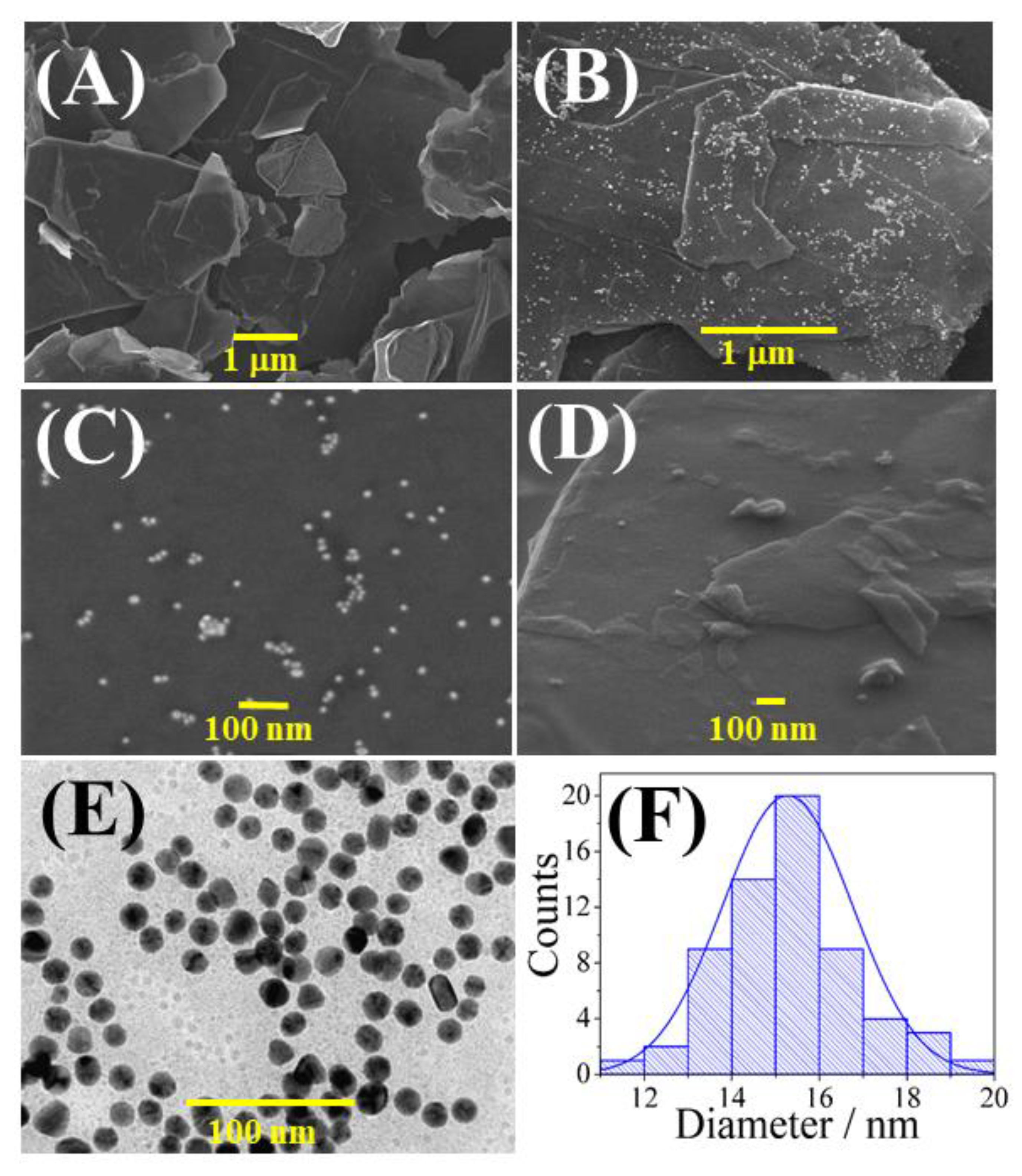
3.1. Morphological Characterization of the Materials

The materials were characterized by field emission gun-scanning electron microscopy (FEG-SEM) and transmission electron microscopy (TEM). The FEG-SEM and TEM images of (A) Gr, (B) AuNPs-Gr (C–F) AuNPs, and (D) Lac-Glu-AuNPs/CPE are shown in Figure 2. As can be seen in Figure 2A, the Gr has a block-like structure, which is typically characteristic of this material. Figure 2B shows the mixture composed of Gr and AuNPs, where several AuNPs (grains) can be found highly dispersed over the Gr (plates). Figure 2C shows the image of the AuNPs. Figure 2D shows the morphology of the product obtained by mixing all the materials used in the construction of the proposed sensor (Lac-Glu-AuNPs/CPE). Finally, Figure 2E shows the average size of the AuNPs obtained by TEM image and in Figure 2F, the corresponding histogram of AuNPs diameters with an average diameter of 15 nm.

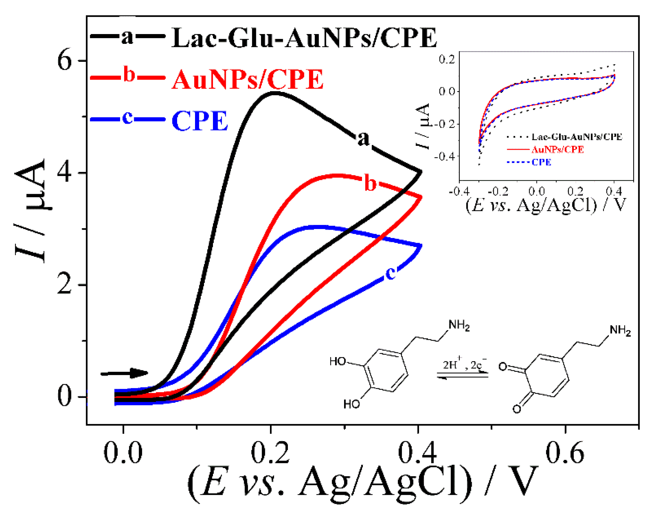
3.2. Using the Lac-Glu-AuNPs/CPE Biosensor for the Analysis of the Electrochemical Behavior of Dopamine

The application of the cyclic voltammetry technique enabled us to effectively and conveniently evaluate the electrochemical response of DA on the surface of the Lac-Glu-AuNPs/CPE. Figure 3 shows the cyclic voltammograms obtained from the application of the Lac-Glu-AuNPs/CPE (a), AuNPs/CPE (b), and CPE (c) in the presence of DA and without the addition of DA (inserted), in the potential range of −0.30 to 0.40 V. The electrochemical response of DA was confirmed by the appearance of an anodic peak current at the potential of 0.2 V, where DA is oxidized to o-quinone on biosensor surface, and the current resulting from the process toward enzyme-catalyzed DA oxidation was proportional to the DA concentration. As can be clearly observed, the modification of the sensor with AuNPs and laccase led to a noticeable improvement in the electrochemical signal. The anodic peak current obtained for the Lac-Glu-AuNPs/CPE was 1.4 and 2.0 times higher than that of AuNPs/CPE and CPE, respectively. These results point to the catalytic effect of the laccase enzyme and AuNPs on DA oxidation on the electrode surface.

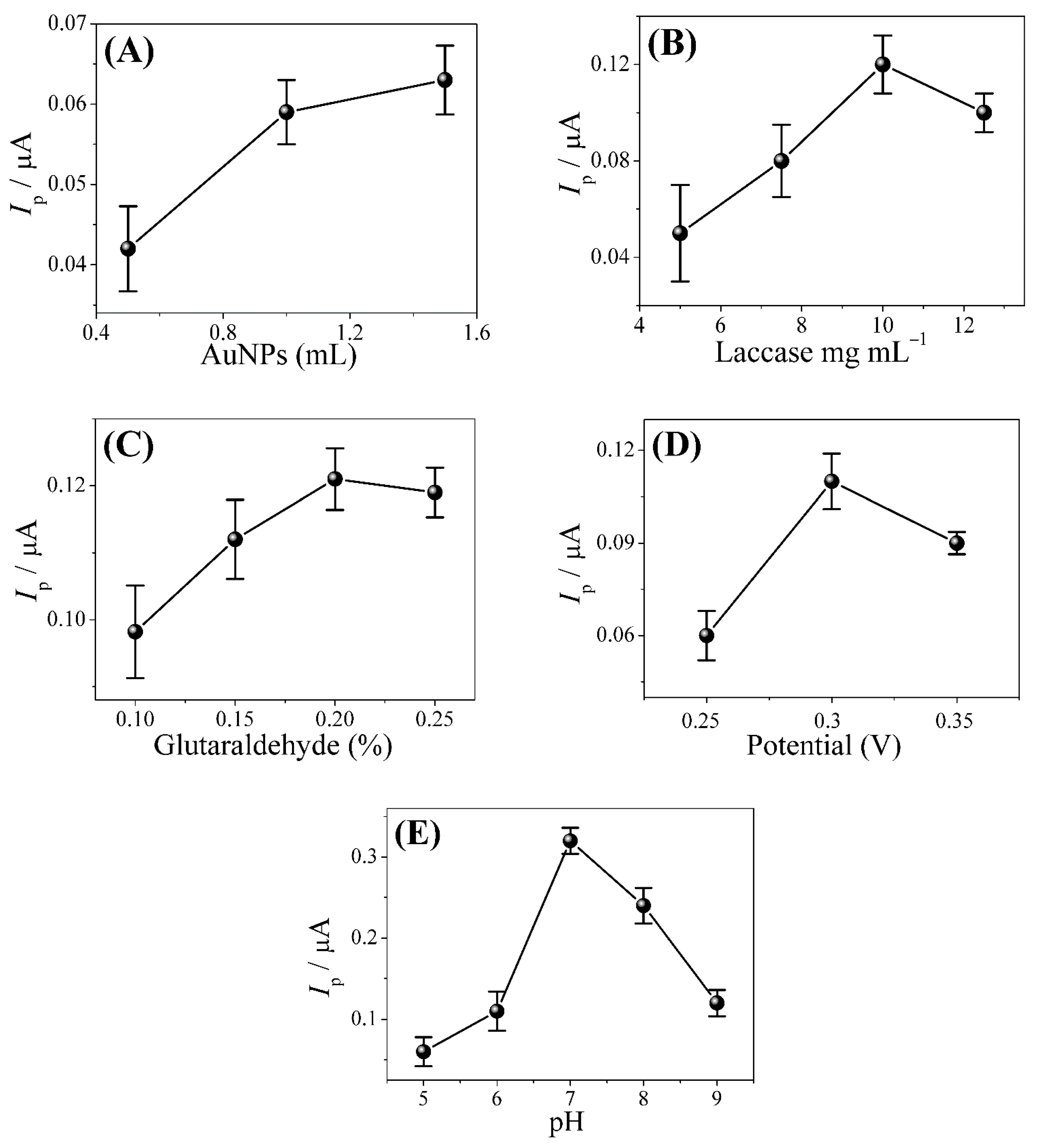
3.3. Optimization of the Lac-Glu-AuNPs/CPE Sensor

First, an analysis was conducted in order to evaluate the optimal concentration of AuNPs solution which should be incorporated into the carbon paste electrode so as to obtain optimum results. This analysis was carried out based on the application of 0.50, 1.0, and 1.5 mL of AuNPs (Figure 4A) solution in 100 mg of Gr, followed by the addition of mineral oil in order to obtain the modified carbon paste electrode. The results obtained from this test showed that 1.0 mL of AuNPs solution was the optimal concentration that yielded the best results. Subsequently, further optimization studies were conducted in order to evaluate the optimal amount of enzyme and the optimal percentage of glutaraldehyde needed to be incorporated into the biosensor so as to obtain the best results. To perform this analysis, we tested the following enzyme concentrations: 5.0, 7.5, 10, and 12.5 mg mL−1 (Figure 4B) and the following glutaraldehyde percentages: 0.10, 0.15, 0.20, and 0.25% v/v (Figure 4C). The optimal enzyme concentration and glutaraldehyde percentage obtained from the optimization studies conducted were 10 mg mL−1 and 0.20% v/v, respectively. These optimal values were then employed for the conduct of all further experiments.
For the analysis of the sensor sensitivity in electrochemical detection, the response of the proposed biosensor to DA was evaluated relative to applied potential based on the application of the amperometric technique using phosphate buffer solution (pH 7.0) as supporting electrolyte. As can be seen in Figure 4D, the highest peak current magnitude was obtained at the potential of 0.3 V; thus, this working potential was applied in subsequent amperometric measurements.
We then analyzed the effect of pH by varying the pH level from 5.0 to 9.0 using 0.1 mol L−1 phosphate buffer solution; this analysis was performed taking into account the electrochemical signals obtained from the amperometric measurements. The results obtained showed that the buffer solution at pH 7.0 presented the best conditions (Figure 4E); as such, this pH value was chosen for the conduct of further studies.

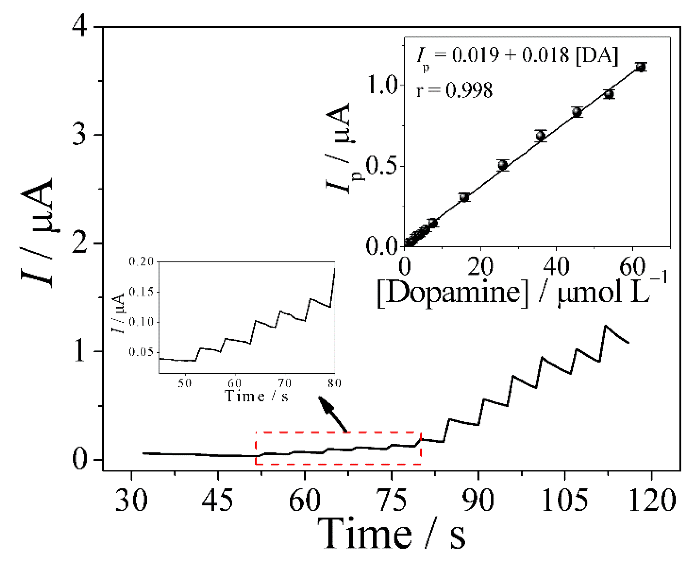
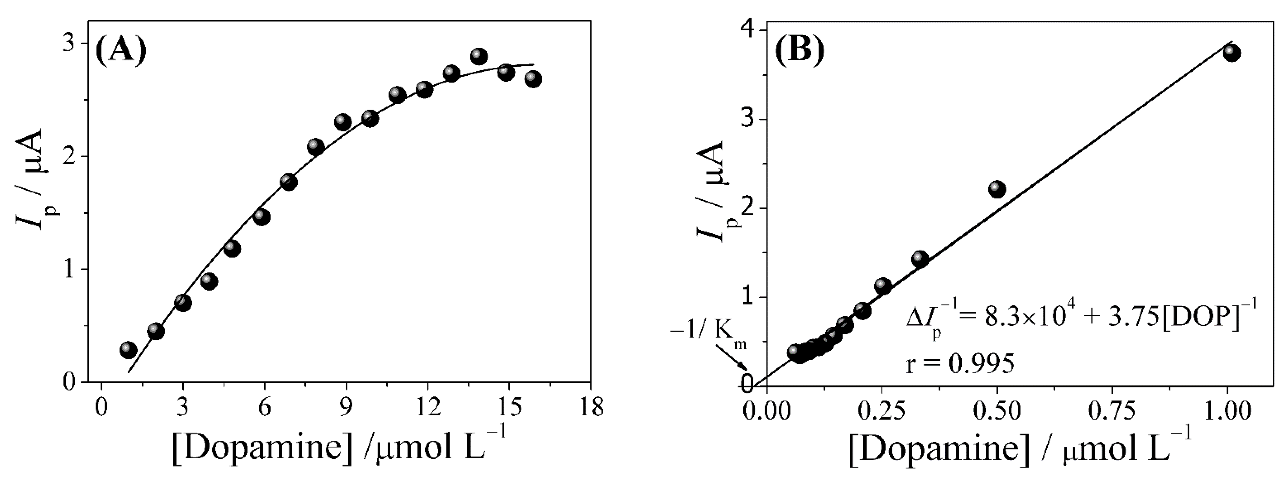
3.4. Amperometric Determination of Dopamine

Through the application of the amperometric technique, a thorough analysis was performed in order to evaluate the electrochemical efficiency of the Lac-Glu-AuNPs/CPE biosensor in the electrocatalytic oxidation of DA; this analysis was conducted under optimized conditions using 0.1 mol L−1 phosphate buffer solution (pH = 7.0) in the presence of different concentrations of DA. As can be seen in Figure 5, a well-defined response was obtained from the successive addition of DA in the supporting electrolyte. Moreover, no electrode deactivation was observed during the amperometric analysis. The application of the amperometric technique at the potential of 0.2 V yielded a linear concentration range of 8.0 × 10⁻⁷ to 6.2 × 10⁻⁵ mol L−1 for DA, with limit of detection (LOD) of 6.0 × 10⁻⁸ mol L−1 and a signal-to-noise ratio of 3 (S/N = 3). The relationship between the current and the concentration of DA is shown in Equation (1) below:
Ip (μA) = 0.019 + 0.018[DA] (µmol L−1) (r = 0.998)
Table 1 shows a comparative analysis of the analytical performance/efficiency of the Lac-Glu-AuNPs/CPE sensor and other sensors reported in the literature applied for the determination of DA [26,27,28,29,30,31,32,33,34,35]. Looking at the data in Table 1, one will observe that the proposed Lac-Glu-AuNPs/CPE biosensor presented linear concentration range and limit of detection (LOD) quite similar to the values obtained for other sensors reported in the literature. Compared to the other sensors reported in the literature, the main advantages of the Lac-Glu-AuNPs/CPE biosensor lie in the following: (i) The biosensor is easy to prepare; (ii) the sensor can be used to perform consecutive analyses by simply polishing its surface; (iii) the biosensor presents excellent repeatability, good stability, and high selectivity, as will be shown in the sequence.
In order to demonstrate the enzymatic behavior of the biosensor, DA concentration was added successively, as shown in Figure 6. This analysis gave rise to a saturation behavior, which is a typical behavior reflected in a Michaelis–Menten plot; thus, based on the application of the Lineweaver–Burk plot, the apparent Michaelis–Menten constant ( K m M M a p p ) was calculated using Equation (2) below [36,37]:
1 V = K m + [ S ] V max × [ S ] = K m V max × 1 [ S ] + 1 V max
where V is the reaction velocity, Km is the Michaelis–Menten constant, Vmax is the maximum reaction velocity, and [S] is the substrate concentration. The linear regression equation of the ΔIp−1 vs. [DA] plot was ΔIp−1 (μA) = 8.3 × 10⁴ + 3.8 [DA]−1. The value obtained for Michaelis–Menten constant was 4.5 × 10⁻⁵ mol L−1; this value indicated a high affinity of the immobilized enzyme for the substrate. Moreover, the K m M M a p p value obtained was found to be comparable to those obtained in other studies reported in the literature [36,37].
The proposed electrochemical mechanism of action involving dopamine and laccase enzyme is based on the catalysis of dopamine oxidation reaction in the o-quinone, which is followed by the reduction of the oxygen molecule [17] (Equation (3)). To demonstrate the occurrence of this electrochemical reaction, electrochemical experiments were performed in the absence of oxygen. The results obtained showed that the presence of oxygen in the medium is essential for the reaction to occur.
Dopamine + O 2     laccase   enzyme     o -quinone + H 2 O   ( reaction )

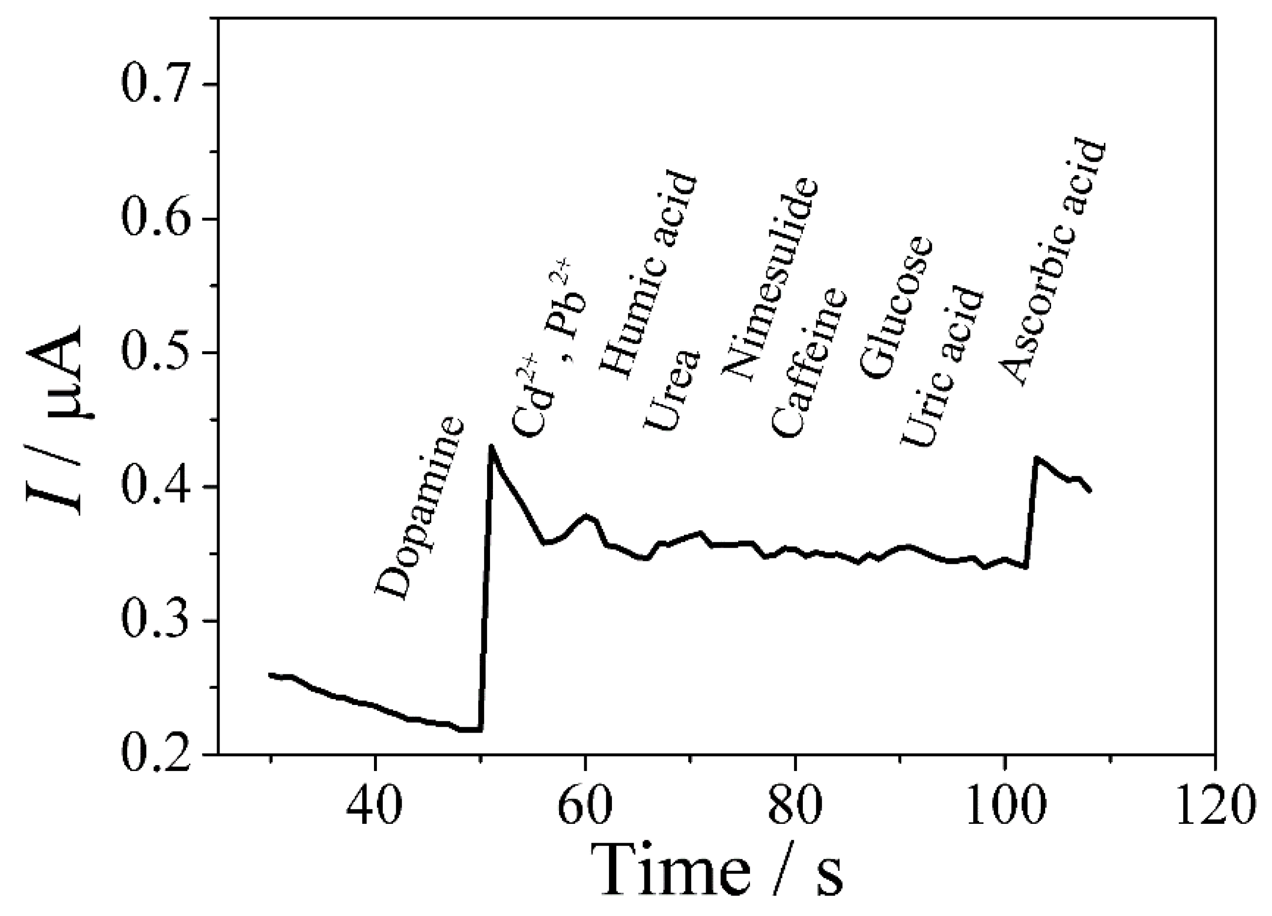
3.5. Study of Repeatability and Interference Effects

The analysis of repeatability and selectivity of the Lac-Glu-AuNPs/CPE biosensor was performed using 0.1 mol L−1 phosphate buffer (pH 7.0) in the presence of DA concentrations of 1.0 × 10⁻⁵ mol L−1 and at Eapl of 0.2 V. To evaluate the repeatability of the proposed sensor, ten consecutive amperometric measurements were performed. The relative standard deviation (RSD) obtained from this analysis was 2.5%; this result shows that the analytical method proposed in this study exhibits excellent precision and repeatability when applied toward the detection of DA. Furthermore, it is worth mentioning that after polishing the Lac-Glu-AuNPs/CPE sensor, a similar RSD was obtained; this outcome points to the considerable homogeneity of the modified carbon paste material.
The selectivity of the proposed biosensor was evaluated through the addition of different concomitants (Cd2+, Pb2+, nimesulide, urea, humic acid, glucose, caffeine, uric acid and ascorbic acid) as possible interferents in the DA detection process. Figure 7 shows the amperometric response obtained for DA, Cd2+, Pb2+, humic acid, urea, nimesulide, caffeine, glucose, uric acid, and ascorbic acid applied in the ratio of 1:10 (analyte: concomitant). As can be clearly seen, among the analyzed compounds, only DA and ascorbic acid showed an electrochemical response. Therefore, ascorbic acid is a potential interferent in the proposed method for DA. The electrochemical experiments were conducted in phosphate buffer solution at pH 7.0.

3.6. Biosensor Stability after Storage

The stability of the biosensor was evaluated after the storage of the device in a refrigerator at 5 °C for 10 days; this analysis was conducted using phosphate buffer solution at pH 7.0 (as supporting electrolyte). The results obtained from this analysis (see Figure 8) showed that, after the 10-day period, the electrochemical response of the biosensor dropped to 85% compared to its initial response. It should be emphasized that after polishing the electrode, its surface is renewed, and this allows one to perform various consecutive electrochemical analyses with the electrode.

3.7. Application of the Proposed Amperometric Biosensor in Synthetic Urine, Bovine Serum and Water Samples

To check the effectiveness of the proposed amperometric method, the Lac-Glu-AuNPs/CPE biosensor was applied for the determination of DA in synthetic urine, serum, and river water samples. These samples were fortified with two known concentration levels (0.5 and 5.0 × 10⁻⁶ mol L−1) of DA (Table 2). The results obtained from the experiments performed in triplicate (n = 3) showed that the application of the sensor yielded recovery percentages between 95 and 105%, in all cases. These results show that the proposed biosensor is reliable and is an efficient analytical alternative tool for the determination of DA. Essentially, the biosensor can be used for the successful determination of DA in different matrices without any interference effects caused by the presence of other compounds.

4. Conclusions

The present study reported the development and application of a highly efficient biosensor, which was constructed based on the immobilization of laccase in carbon paste electrode modified with gold nanoparticles, for the amperometric determination of DA. The Lac-Glu-AuNPs/CPE biosensor is relatively easy to prepare and possesses quick analytical response; apart from that, the sensor has high reproducibility, excellent selectivity, and good stability. Another key advantage of the proposed biosensor is that it can be used to perform numerous analyses over a long period of time by simply polishing the electrode surface with clean paper and modifying it with laccase and glutaraldehyde. All these properties make the sensing device an effective alternative tool for DA determination in biological and environmental fluid samples.

Author Contributions

Conceptualization: A.M.S., A.W., F.C.M. and O.F.-F. Methodology: A.M.S. and A.W. Validation: A.M.S. and A.W. Formal analysis: A.M.S. and A.W. Investigation: A.M.S. and A.W. Resources: F.C.M. and O.F.-F. Data curation/analysis: A.M.S. and A.W. Writing—original draft preparation: A.M.S. and A.W. Writing—review and editing: F.C.M. and O.F.-F. Visualization: A.M.S. and A.W. Supervision: F.C.M. and O.F.-F. Project administration: F.C.M. and O.F.-F. Funding acquisition: F.C.M. and O.F.-F. All authors have read and agreed to the published version of the manuscript.

Funding

This study was financed in part by the Coordenação de Aperfeiçoamento de Pessoal de Nível Superior-Brazil (CAPES)-Finance Code 001, process number 88887.469180/2019-00. The authors also gratefully acknowledge the financial support granted by FAPESP (grant numbers 2017/10118-0, 2020/01050-5 and 2022/05454-9).

Acknowledgments

The authors are grateful to the CNPq, CAPES and FAPESP for the financial assistance granted in support of this work (see the grants and grant numbers cited previously).

Conflicts of Interest

The authors have no conflict of interest to declare.

References

  1. Fontenele, E.G.; Martins, M.R.; Quidute, A.R.; Montenegro, R.M., Jr. Environmental contaminants and endocrine disruptors. Arq. Bras. Endocrinol. E Metabol. 2010, 54, 6–16. [Google Scholar] [CrossRef] [PubMed] [Green Version]
  2. Waring, R.H.; Harris, R.M. Endocrine disrupters: A human risk? Mol. Cell. Endocrinol. 2005, 244, 2–9. [Google Scholar] [CrossRef] [PubMed]
  3. Eisenhofer, G.; Kopin, I.J.; Goldstein, D.S. Catecholamine metabolism: A contemporary view with implications for physiology and medicine. Pharmacol. Rev. 2004, 56, 331–349. [Google Scholar] [CrossRef] [PubMed]
  4. Ko, J.H.; Strafella, A.P. Dopaminergic neurotransmission in the human brain: New lessons from perturbation and imaging. Neurosci. A Rev. J. Bringing Neurobiol. Neurol. Psychiatry 2012, 18, 149–168. [Google Scholar] [CrossRef] [PubMed] [Green Version]
  5. Reddy, N.R.; Sreedevi, G.; Prabhavathi, K.; Chakravarthy, I.E. Spectrophotometric determination of dopamine in pharmaceutical formulations. J. Anal. Chem. 2005, 60, 252–253. [Google Scholar] [CrossRef]
  6. Chatterjee, D.; Gerlai, R. High precision liquid chromatography analysis of dopaminergic and serotoninergic responses to acute alcohol exposure in zebrafish. Behav. Brain Res. 2009, 200, 208–213. [Google Scholar] [CrossRef]
  7. Zuo, F.; Jin, L.; Fu, X.; Zhang, H.; Yuan, R.; Chen, S. An electrochemiluminescent sensor for dopamine detection based on a dual-molecule recognition strategy and polyaniline quenching. Sens. Actuators B Chem. 2017, 244, 282–289. [Google Scholar] [CrossRef]
  8. Kartsova, L.A.; Sidorova, A.A.; Kazakov, V.A.; Bessonova, E.A.; Yashin, A.Y. Determination of Catecholamines by Capillary Electrophoresis and Reversed-Phase High-Performance Liquid Chromatography. J. Anal. Chem. 2004, 59, 737–741. [Google Scholar] [CrossRef]
  9. Fotopoulou, M.A.; Ioannou, P.C. Post-column terbium complexation and sensitized fluorescence detection for the determination of norepinephrine, epinephrine and dopamine using high-performance liquid chromatography. Anal. Chim. Acta 2002, 462, 179–185. [Google Scholar] [CrossRef]
  10. Carrera, V.; Sabater, E.; Vilanova, E.; Sogorb, M.A. A simple and rapid HPLC–MS method for the simultaneous determination of epinephrine, norepinephrine, dopamine and 5-hydroxytryptamine: Application to the secretion of bovine chromaffin cell cultures. J. Chromatogr. B 2007, 847, 88–94. [Google Scholar] [CrossRef]
  11. Cho, I.-H.; Kim, D.H.; Park, S. Electrochemical biosensors: Perspective on functional nanomaterials for on-site analysis. Biomater. Res. 2020, 24, 6. [Google Scholar] [CrossRef] [PubMed] [Green Version]
  12. Baldrian, P. Fungal laccases—Occurrence and properties. FEMS Microbiol. Rev. 2006, 30, 215–242. [Google Scholar] [CrossRef] [PubMed] [Green Version]
  13. Gianfreda, L.; Xu, F.; Bollag, J.-M. Laccases: A Useful Group of Oxidoreductive Enzymes. Bioremediation J. 1999, 3, 1–26. [Google Scholar] [CrossRef]
  14. Yaropolov, A.I.; Skorobogat’ko, O.V.; Vartanov, S.S.; Varfolomeyev, S.D. Laccase. Appl. Biochem. Biotechnol. 1994, 49, 257–280. [Google Scholar] [CrossRef]
  15. Ramírez, P.; Mano, N.; Andreu, R.; Ruzgas, T.; Heller, A.; Gorton, L.; Shleev, S. Direct electron transfer from graphite and functionalized gold electrodes to T1 and T2/T3 copper centers of bilirubin oxidase. Biochim. Biophys. Acta 2008, 1777, 1364–1369. [Google Scholar] [CrossRef] [PubMed]
  16. Portaccio, M.; Di Tuoro, D.; Arduini, F.; Moscone, D.; Cammarota, M.; Mita, D.G.; Lepore, M. Laccase biosensor based on screen-printed electrode modified with thionine–carbon black nanocomposite, for Bisphenol A detection. Electrochim. Acta 2013, 109, 340–347. [Google Scholar] [CrossRef]
  17. Gouveia-Caridade, C.; Pauliukaite, R.; Brett, C.M.A. Development of electrochemical oxidase biosensors based on carbon nanotube-modified carbon film electrodes for glucose and ethanol. Electrochim. Acta 2008, 53, 6732–6739. [Google Scholar] [CrossRef] [Green Version]
  18. Švancara, I.; Vytřas, K.; Barek, J.; Zima, J. Carbon Paste Electrodes in Modern Electroanalysis. Crit. Rev. Anal. Chem. 2001, 31, 311–345. [Google Scholar] [CrossRef]
  19. Silva, T.A.; Moraes, F.C.; Janegitz, B.C.; Fatibello-Filho, O. Electrochemical Biosensors Based on Nanostructured Carbon Black: A Review. J. Nanomater. 2017, 2017, 4571614. [Google Scholar] [CrossRef] [Green Version]
  20. Islam, T.; Hasan, M.M. Metal Nanoparticles for Electrochemical Sensing: Progress and Challenges in the Clinical Transition of Point-of-Care Testing. Molecules 2020, 25, 5787. [Google Scholar] [CrossRef]
  21. Zeng, S.; Yong, K.-T.; Roy, I.; Dinh, X.-Q.; Yu, X.; Luan, F. A Review on Functionalized Gold Nanoparticles for Biosensing Applications. Plasmonics 2011, 6, 491. [Google Scholar] [CrossRef]
  22. Pingarrón, J.M.; Yáñez-Sedeño, P.; González-Cortés, A. Gold nanoparticle-based electrochemical biosensors. Electrochim. Acta 2008, 53, 5848–5866. [Google Scholar] [CrossRef]
  23. Ojea-Jiménez, I.; Romero, F.M.; Bastús, N.G.; Puntes, V. Small Gold Nanoparticles Synthesized with Sodium Citrate and Heavy Water: Insights into the Reaction Mechanism. J. Phys. Chem. C 2010, 114, 1800–1804. [Google Scholar] [CrossRef]
  24. Baluta, S.; Lesiak, A.; Cabaj, J. Graphene Quantum Dots-based Electrochemical Biosensor for Catecholamine Neurotransmitters Detection. Electroanalysis 2018, 30, 1781–1790. [Google Scholar] [CrossRef]
  25. Laube, N.; Mohr, B.; Hesse, A. Laser-probe-based investigation of the evolution of particle size distributions of calcium oxalate particles formed in artificial urines. J. Cryst. Growth 2001, 233, 367–374. [Google Scholar] [CrossRef]
  26. Anuar, N.S.; Basirun, W.J.; Shalauddin, M.; Akhter, S. A dopamine electrochemical sensor based on a platinum–silver graphene nanocomposite modified electrode. RSC Adv. 2020, 10, 17336–17344. [Google Scholar] [CrossRef] [PubMed]
  27. Sun, X.; Zhang, L.; Zhang, X.; Liu, X.; Jian, J.; Kong, D.; Zeng, D.; Yuan, H.; Feng, S. Electrochemical dopamine sensor based on superionic conducting potassium ferrite. Biosens. Bioelectron. 2020, 153, 112045. [Google Scholar] [CrossRef] [PubMed]
  28. Atta, N.F.; Galal, A.; El-Said, D.M. Novel Design of a Layered Electrochemical Dopamine Sensor in Real Samples Based on Gold Nanoparticles/β-Cyclodextrin/Nafion-Modified Gold Electrode. ACS Omega 2019, 4, 17947–17955. [Google Scholar] [CrossRef] [PubMed] [Green Version]
  29. Pramoda, K.; Moses, K.; Maitra, U.; Rao, C.N.R. Superior Performance of a MoS2-RGO Composite and a Borocarbonitride in the Electrochemical Detection of Dopamine, Uric Acid and Adenine. Electroanalysis 2015, 27, 1892–1898. [Google Scholar] [CrossRef]
  30. Palanisamy, S.; Ku, S.; Chen, S.-M. Dopamine sensor based on a glassy carbon electrode modified with a reduced graphene oxide and palladium nanoparticles composite. Microchim. Acta 2013, 180, 1037–1042. [Google Scholar] [CrossRef]
  31. Palanisamy, S.; Velusamy, V.; Ramaraj, S.; Chen, S.-W.; Yang, T.C.K.; Balu, S.; Banks, C.E. Facile synthesis of cellulose microfibers supported palladium nanospindles on graphene oxide for selective detection of dopamine in pharmaceutical and biological samples. Mater. Sci. Eng. C 2019, 98, 256–265. [Google Scholar] [CrossRef] [PubMed] [Green Version]
  32. Song, N.-N.; Wang, Y.-Z.; Yang, X.-Y.; Zong, H.-L.; Chen, Y.-X.; Ma, Z.; Chen, C.-X. A novel electrochemical biosensor for the determination of dopamine and ascorbic acid based on graphene oxide /poly(aniline-co-thionine) nanocomposite. J. Electroanal. Chem. 2020, 873, 114352. [Google Scholar] [CrossRef]
  33. Min, K.; Yoo, Y.J. Amperometric detection of dopamine based on tyrosinase–SWNTs–Ppy composite electrode. Talanta 2009, 80, 1007–1011. [Google Scholar] [CrossRef] [PubMed]
  34. Li, Y.; Zhang, L.; Li, M.; Pan, Z.; Li, D. A disposable biosensor based on immobilization of laccase with silica spheres on the MWCNTs-doped screen-printed electrode. Chem. Cent. J. 2012, 6, 103. [Google Scholar] [CrossRef] [PubMed] [Green Version]
  35. Wang, K.; Liu, P.; Ye, Y.; Li, J.; Zhao, W.; Huang, X. Fabrication of a novel laccase biosensor based on silica nanoparticles modified with phytic acid for sensitive detection of dopamine. Sens. Actuators B Chem. 2014, 197, 292–299. [Google Scholar] [CrossRef]
  36. Singhal, A.; Choudhary, G.; Thakur, I.S. Characterization of laccase activity produced by Cryptococcus albidus. Prep. Biochem. Biotechnol. 2012, 42, 113–124. [Google Scholar] [CrossRef] [PubMed]
  37. Fernández-Sánchez, C.; Tzanov, T.; Gübitz, G.M.; Cavaco-Paulo, A. Voltammetric monitoring of laccase-catalysed mediated reactions. Bioelectrochemistry 2002, 58, 149–156. [Google Scholar] [CrossRef] [Green Version]
Figure 1. Illustrative scheme showing the procedures involved in the preparation of the biosensor.
Carbon 08 00040 g001
Figure 2. FEG-SEM images of graphite (A), AuNPs-Gr (B), AuNPs (C), and the Lac-Glu-AuNPs/CPE biosensor (D). TEM image of (E) AuNPs and (F) corresponding histogram of AuNPs diameters.
Carbon 08 00040 g002
Figure 3. Cyclic voltammograms obtained from the application of (a) Lac-Glu-AuNPs/CPE, (b) AuNPs/CPE, and (c) CPE in 0.1 mol L−1 phosphate buffer solution (pH 7.0) employed as supporting electrolyte in the absence (inserted) and presence of 1.0 × 10⁻⁴ mol L−1 DA; scan rate (v) = 50 mV s−1. Inserted: cyclic voltammogram in the absence of AD.
Carbon 08 00040 g003
Figure 4. Effect of the amount of (A) AuNPs, (B) enzyme and (C) glutaraldehyde on the biosensor response for 1.0 × 10⁻⁵ mol L−1 DA in 0.1 mol L−1 phosphate buffer solution (pH = 7.0) at a potential of 0.3 V. (D) Effect of the applied potential of 0.25, 0.30 and 0.35 V on the biosensor response for 1.0 × 10⁻⁵ mol L−1 DA in 0.1 mol L−1 phosphate buffer solution (pH = 7.0) and 10 mg mL−1 laccase concentration. (E) pH effect on the biosensor response for a 2.0 × 10⁻⁵ mol L−1 DA in 0.1 mol L−1 phosphate buffer at an applied potential (Eapp) of 0.3 V and 10 mg mL−1 laccase concentration (4 U by electrode).
Carbon 08 00040 g004
Figure 5. Amperograms obtained from the application of the Lac-Glu-AuNPs/CPE biosensor using 0.1 mol L−1 phosphate buffer solution (pH 7.0) as supporting electrolyte in the presence of different concentrations of DA (8.0 × 10⁻⁷–6.2 × 10⁻⁵ mol L−1), with Eapl of 0.3 V. Inset: corresponding analytical curves.
Carbon 08 00040 g005
Figure 6. Michaelis–Menten plot obtained from the successive addition of DA (A) and Lineweaver–Burk plot (B).
Carbon 08 00040 g006
Figure 7. Lac-Glu-AuNPs/CPE biosensor selectivity for a 2.0 × 10⁻⁵ mol L−1 DA concentration and 2.0 × 10⁻⁴ mol L−1 of the following concomitants: Cd2+, Pb2+, humic acid, urea, nimesulide, caffeine, glucose, uric acid and ascorbic acid.
Carbon 08 00040 g007
Figure 8. Corresponding histogram of the Lac-Glu-AuNPs/CPE sensor stability analysis for a period of 10 days (n = 3).
Carbon 08 00040 g008
Table 1. A comparative analysis of the results obtained from the application of the proposed Lac-Glu-AuNPs/CPE biosensor and other sensors reported in the literature used for the electrochemical determination of DA.
AnalyteElectrodeMethodLinear Range (mol L−1)LOD (mol L−1)Ref.
DopaminePt–Ag/Gr/GCEDPV1.0 × 10−7–6.0 × 10−51.2 × 10−8[26]
K2Fe4O7/GCEDPV1.0 × 10−6–1.4 × 10−42.2 × 10−7[27]
NF-CD-AuNPs/AuDPV5.0 × 10−8–2.0 × 10−46.0 × 10−10[28]
MoS2-RGO/CPEDPV1.5 × 10−6–1.0 × 10−49.4 × 10−7[29]
RGO-PdNPs/GCELSV1.0 × 10−6–1.5 × 10−42.3 × 10−7[30]
GO-CMF/PdSPs/GCEDPV3.0 × 10−7–1.96 × 10−42.3 × 10−8[31]
GO/P(ANI-co-THI)/GCEDPV2.0 × 10−6–5.0 × 10−42.0 × 10−6[32]
Tyrosinase-SWNTs-Ppy/GCEAmperometry5.0 × 10−6–5.0 × 10−55.0 × 10−6[33]
Lac/Si/MWCNTs/SPEDPV1.3 × 10−7–8.55 × 10−54.2 × 10−7[34]
Lac/SiO2-PA/GCEAmperometry9.9 × 10−7–1.03 × 10−42.6 × 10−7[35]
Lac-Glu-AuNPs/CPEAmperometry8.0 × 10−7–6.2 × 10−56.0 × 10−8This work
Pt-Ag = platinum–silver; Gr = graphene; GCE = glassy carbon electrode; K2Fe4O7 = potassium ferrite; NF = Nafion; CD = β-cyclodextrin; MOS2 = molybdenum sulfide; PbNPs = lead nanoparticles; RGO = reduced graphene oxide; GO = graphene oxide; CMF = cellulose microfiber; PdSPs = spindle-like palladium nanostructures; P(ANI-co-THI) = poly(aniline-co-thionine) nanocomposite; PA = phytic acid; SWNTs = single wall carbon nanotubes; Ppy = polypyrrole.
Table 2. Using the Lac-Glu-AuNPs/CPE biosensor for dopamine determination in river water, synthetic urine and serum samples.
MatricesAdded
(mol L−1)
Proposed Method (mol L−1)Recovery **
(Sensor, %)
Found *
Urine5.0 × 10−7(4.9 ± 0.2) × 10−798
5.0 × 10⁻⁶(5.1 ± 0.1) × 10−7102
Serum5.0 × 10−7(4.6 ± 0.3) × 10−792
5.0 × 10⁻⁶(4.8 ± 0.2) × 10−695
River water5.0 × 10−7(4.7 ± 0.2) × 10−794
5.0 × 10⁻⁶(5.0 ± 0.2) × 10−6100
* Average of 3 concentrations. ** Recovery percentage = [Found/Added] × 100.
Publisher’s Note: MDPI stays neutral with regard to jurisdictional claims in published maps and institutional affiliations.

