
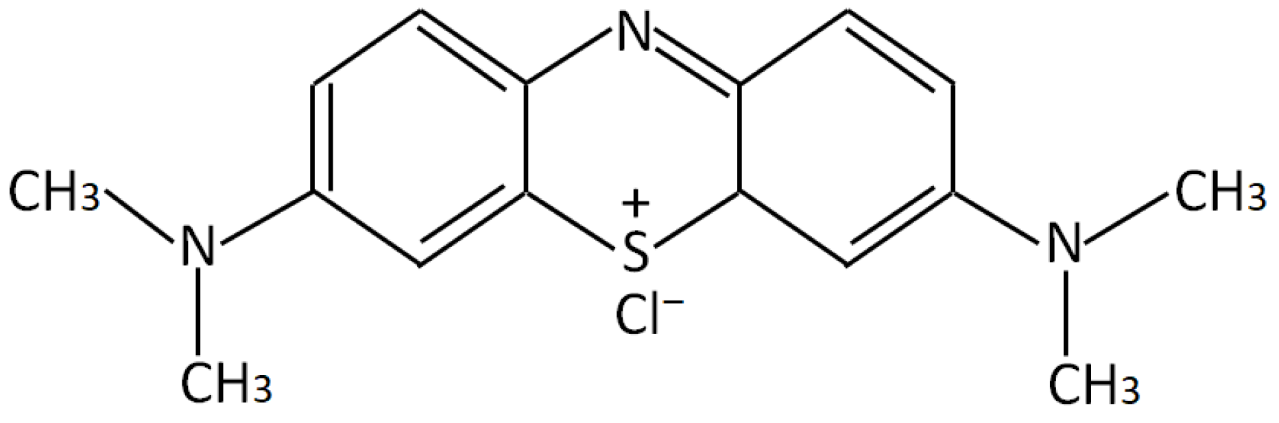
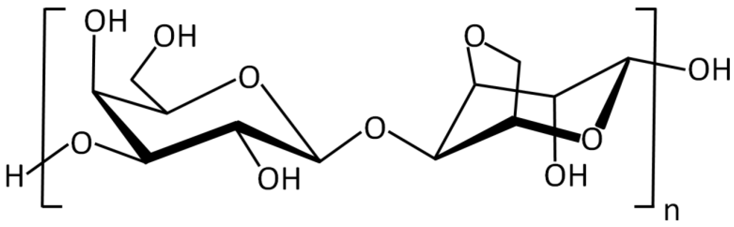
Synthesis of New Hydrogels Involving Acrylic Acid and Acrylamide Grafted Agar-Agar and Their Application in the Removal of Cationic Dyes from Wastewater
 ,
,  ,
,
Abstract
:1. Introduction
2. Results and Discussion
2.1. Characterization
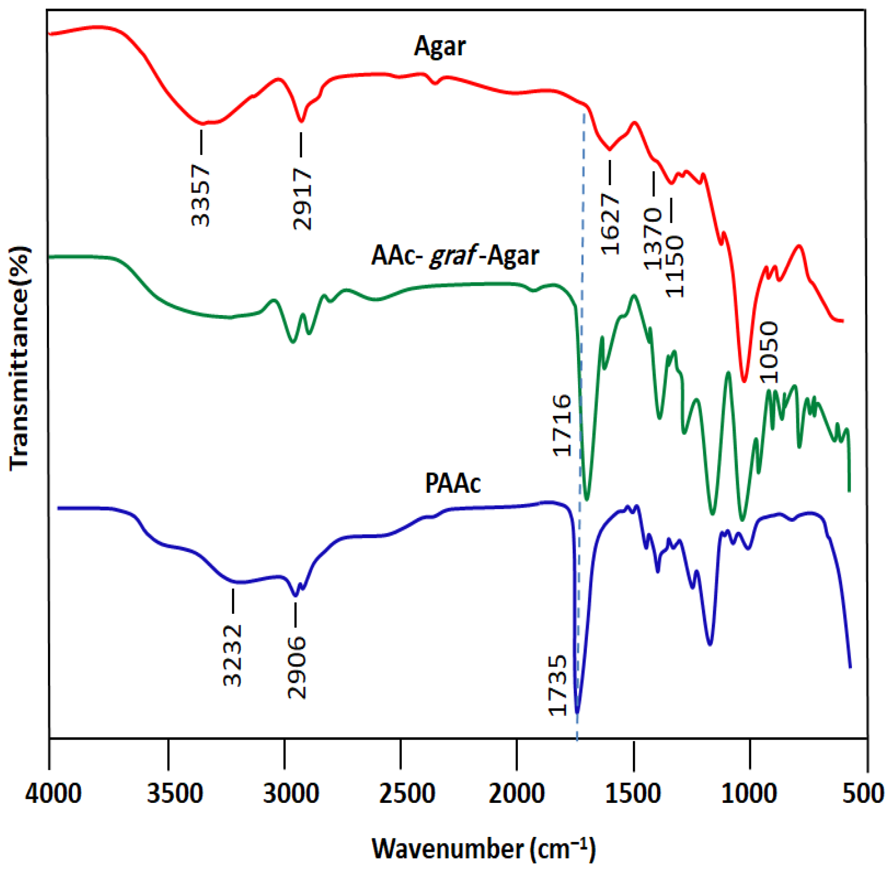
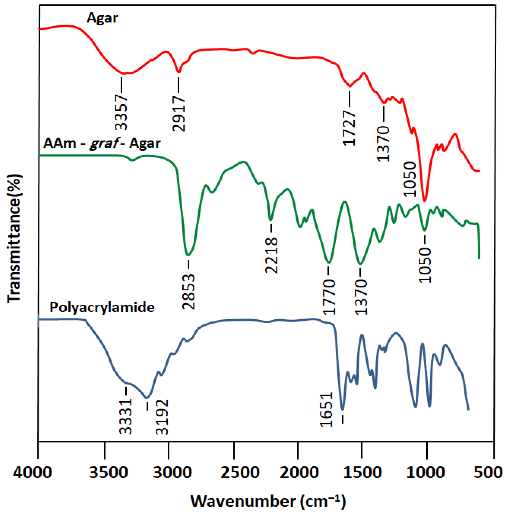
2.1.1. FTIR Analysis
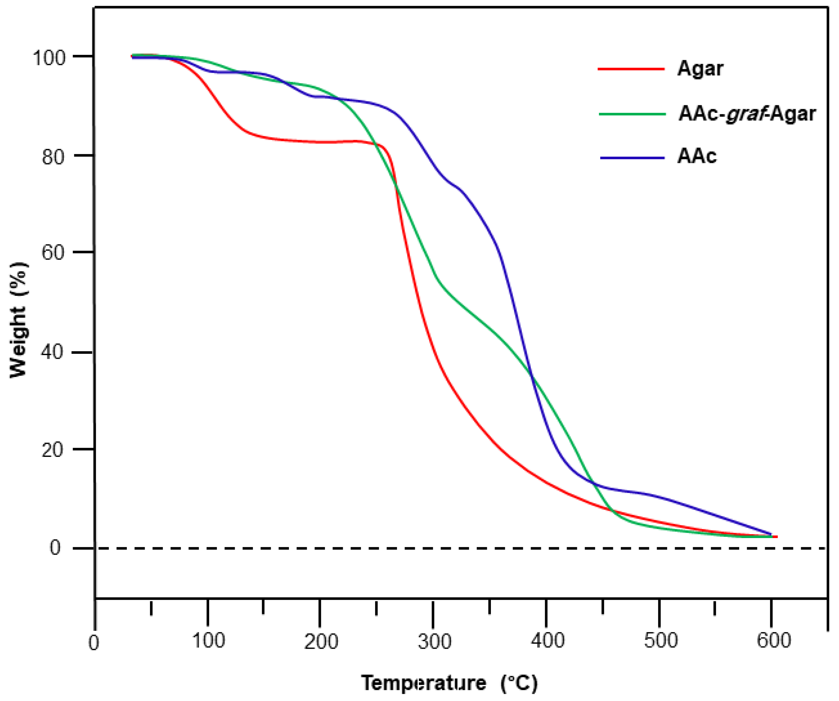
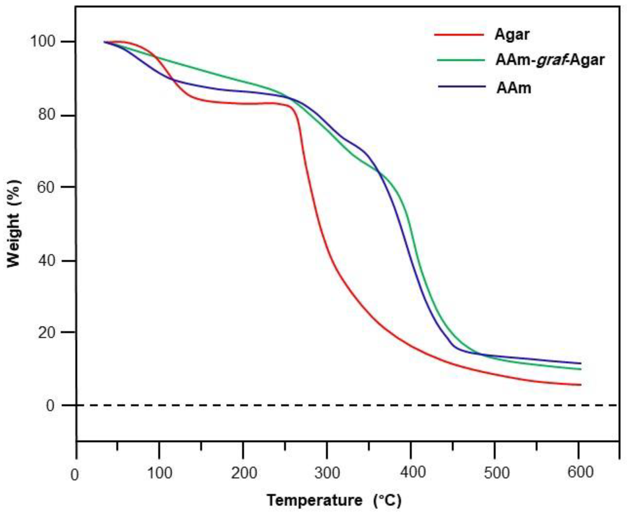
2.1.2. TG-Analysis
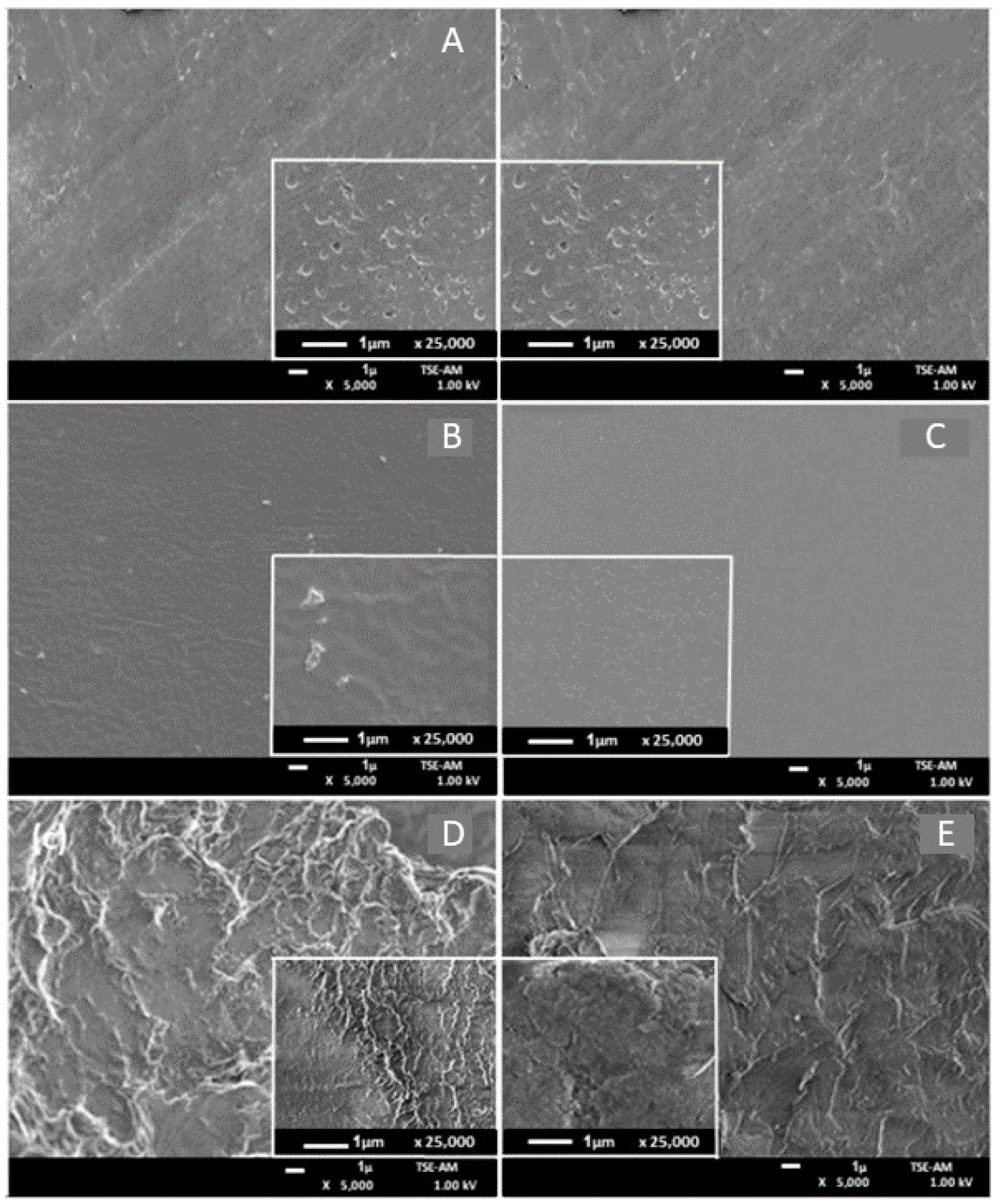
2.1.3. SEM Analysis
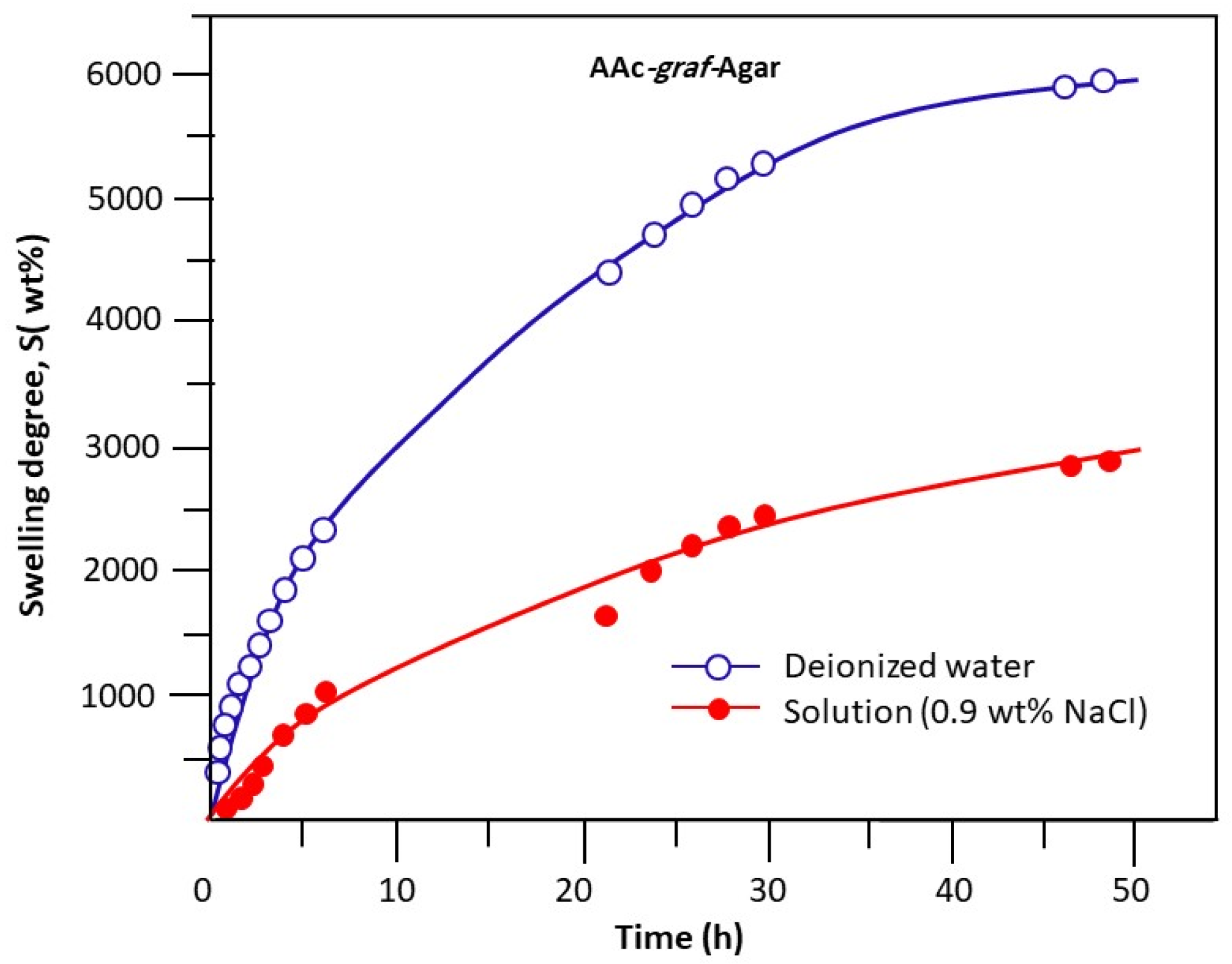
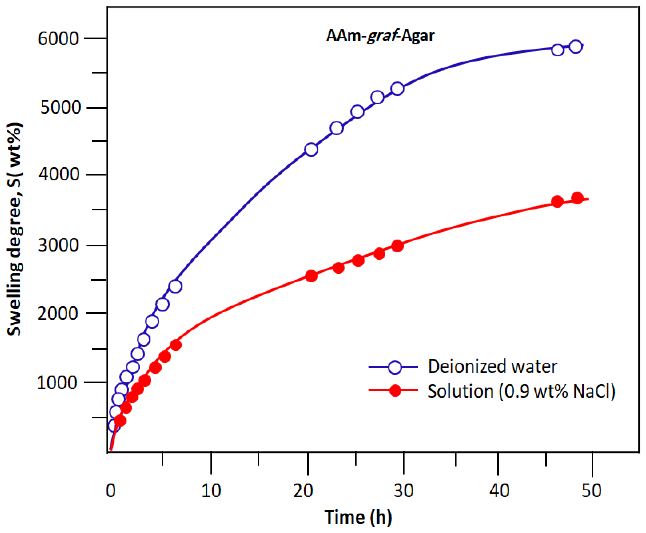
2.2. Swelling Behaviour of Hydrogls
2.3. Adsorption Parameters
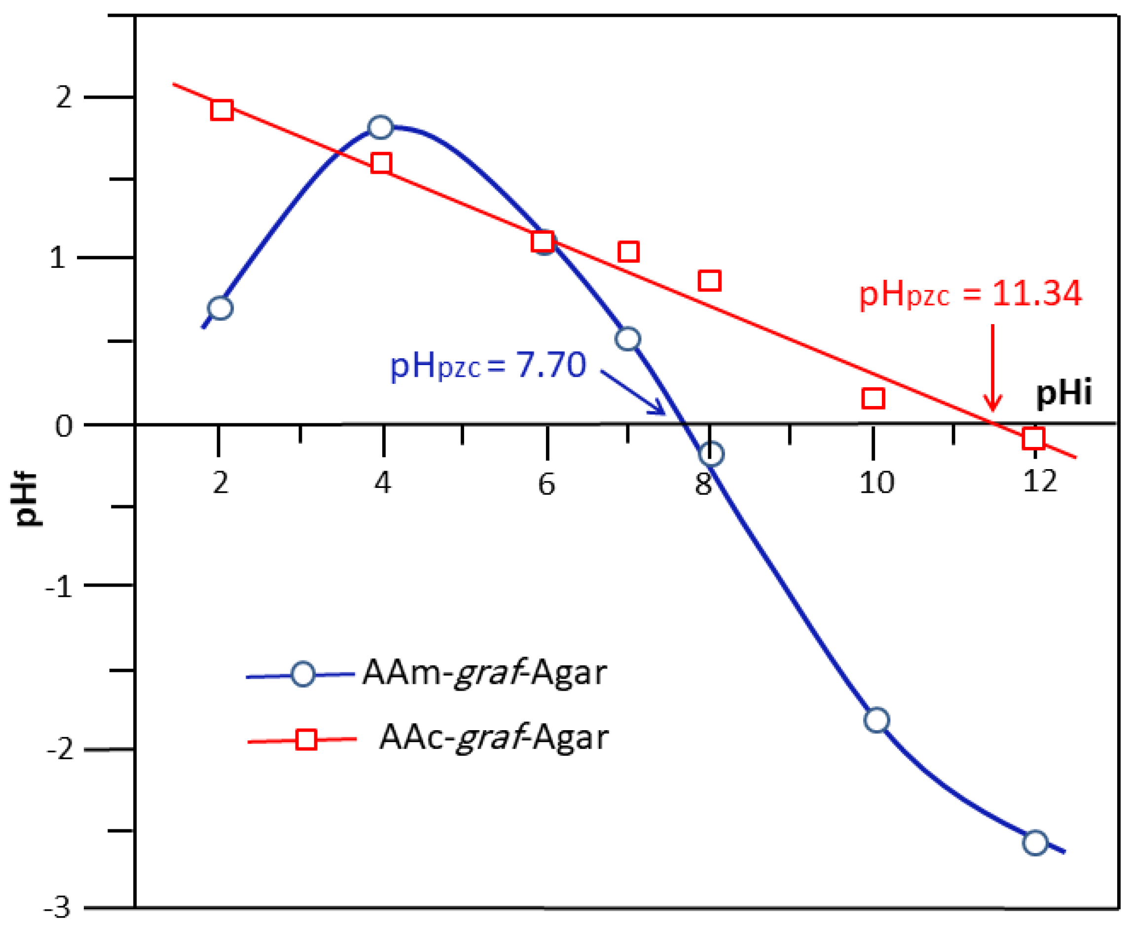
2.3.1. Point of Zero Charge (pHpzc)
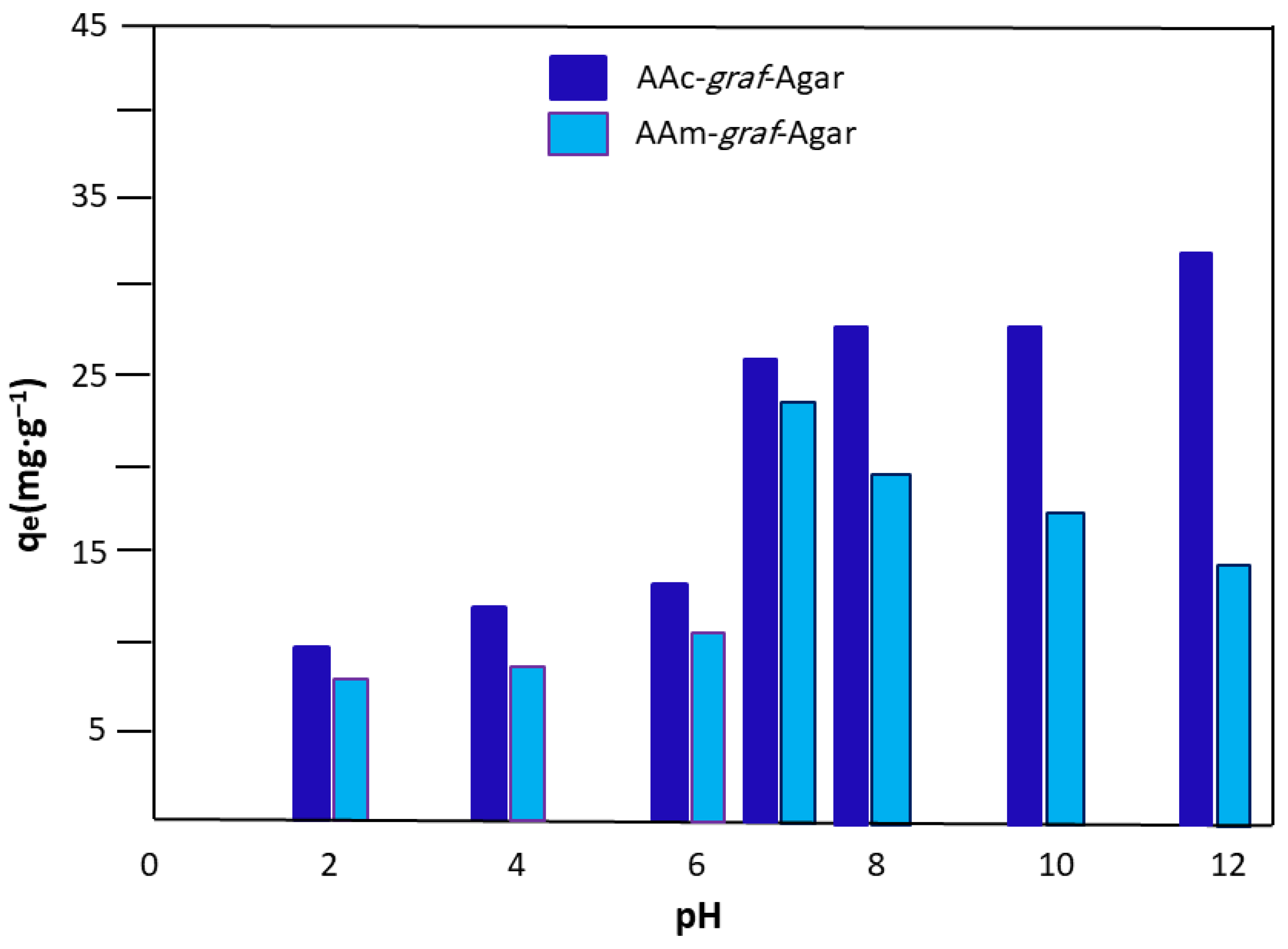
2.3.2. Effect of Initial pH Medium
2.3.3. Effect of Initial Concentration of Dye
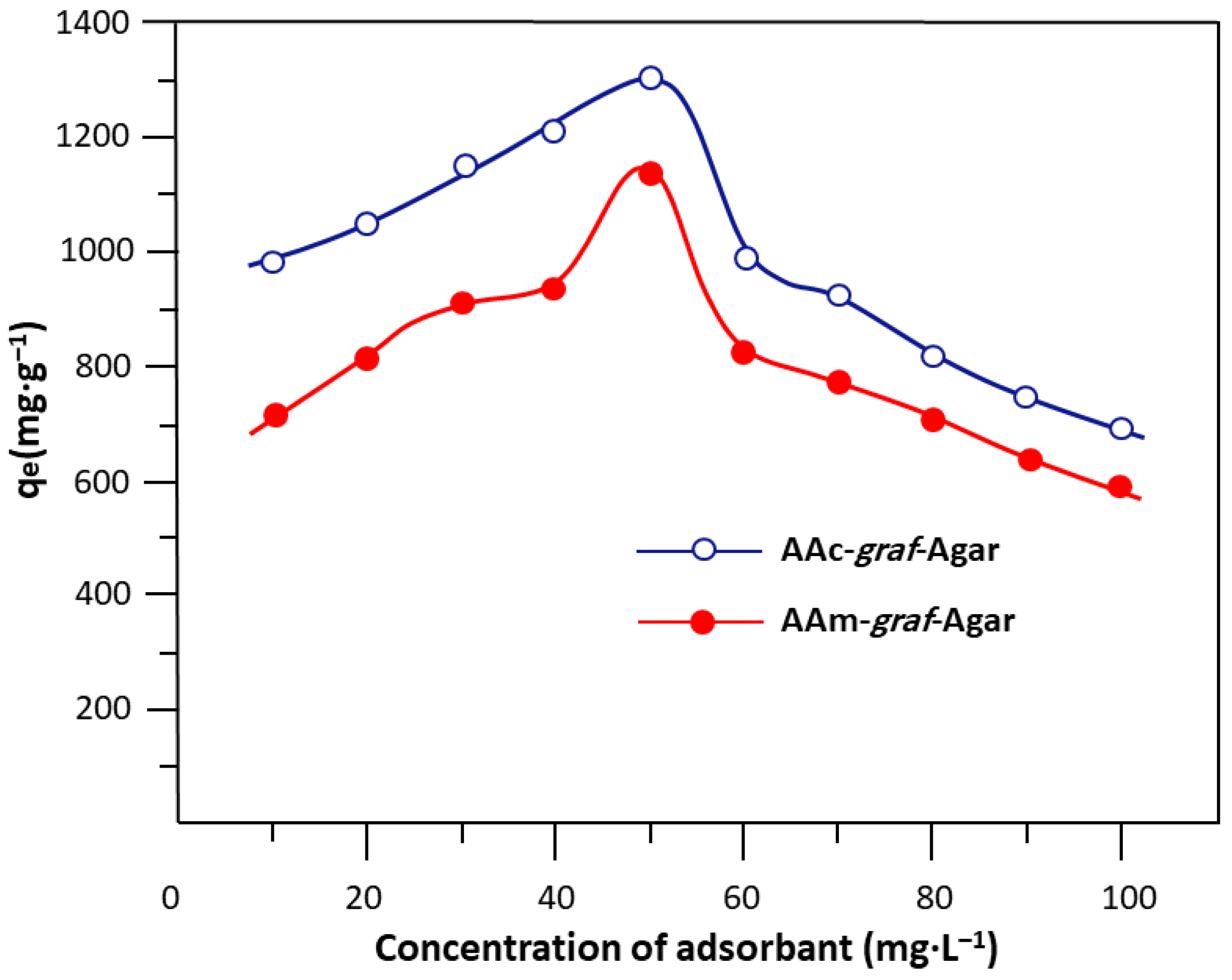
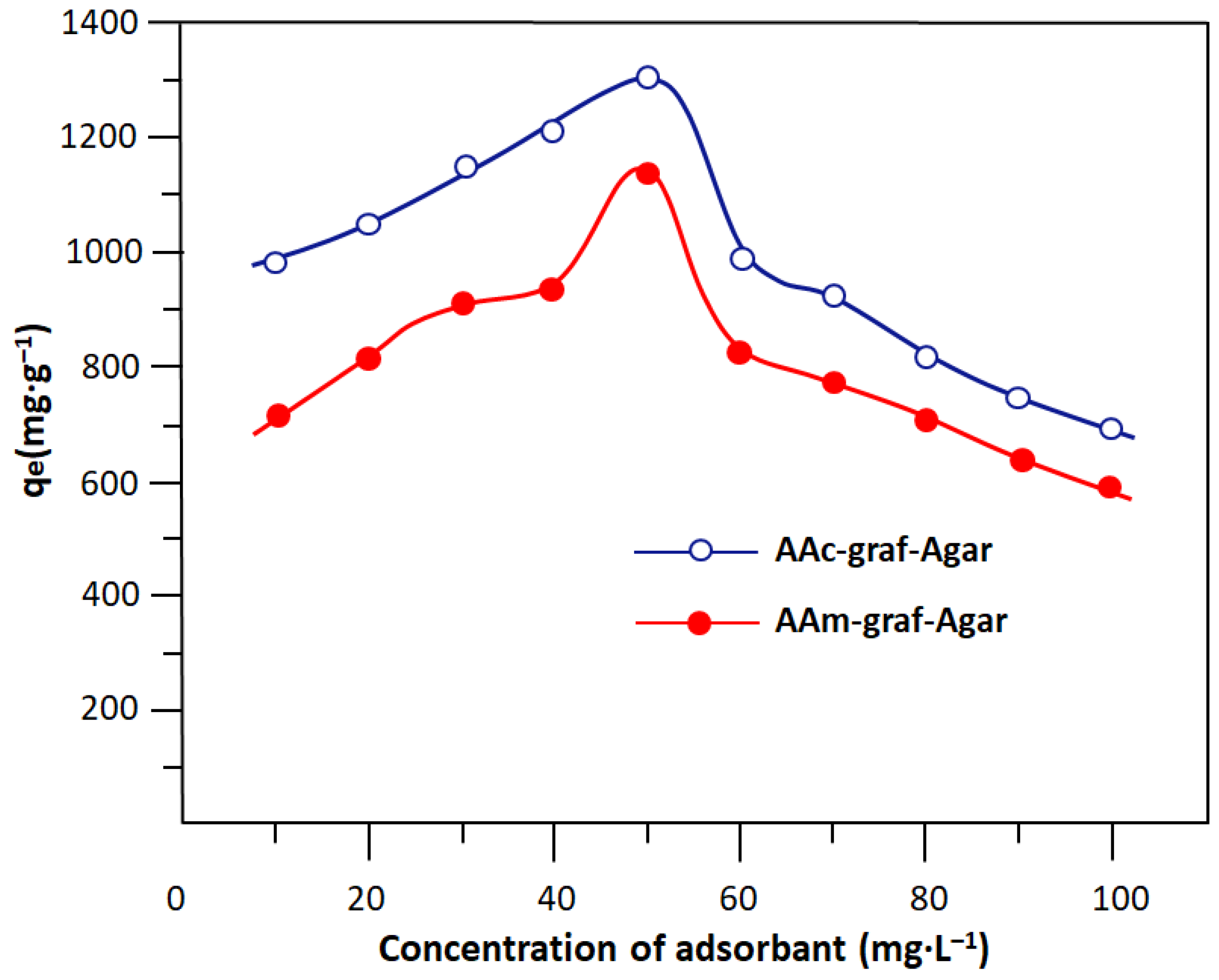
2.3.4. Effect of Adsorbent Dose
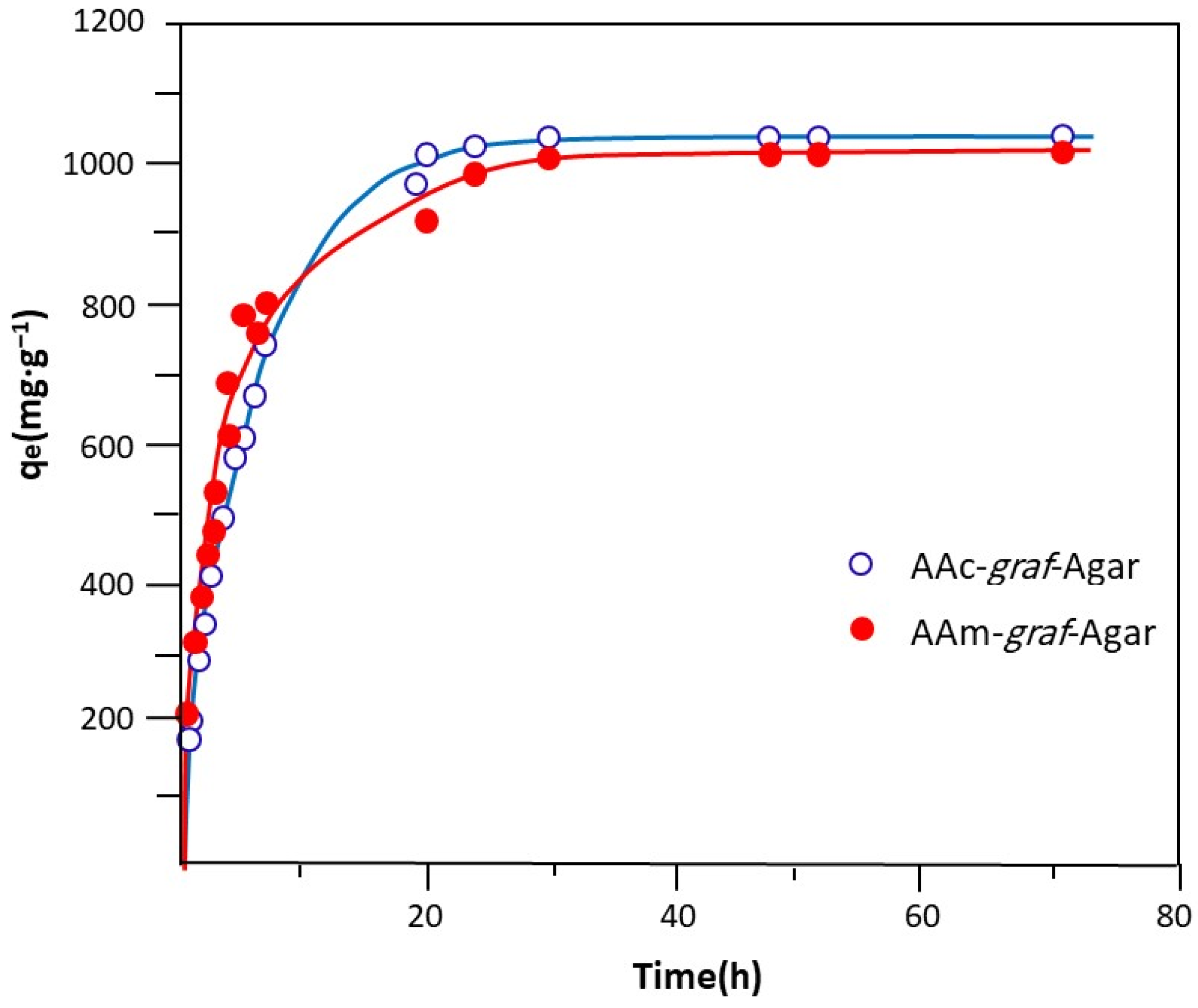
2.3.5. Effect of Contact Time
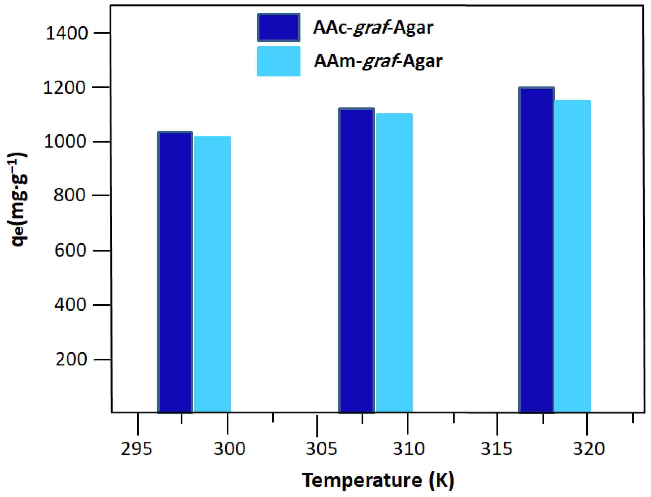
2.3.6. Effect of Temperature
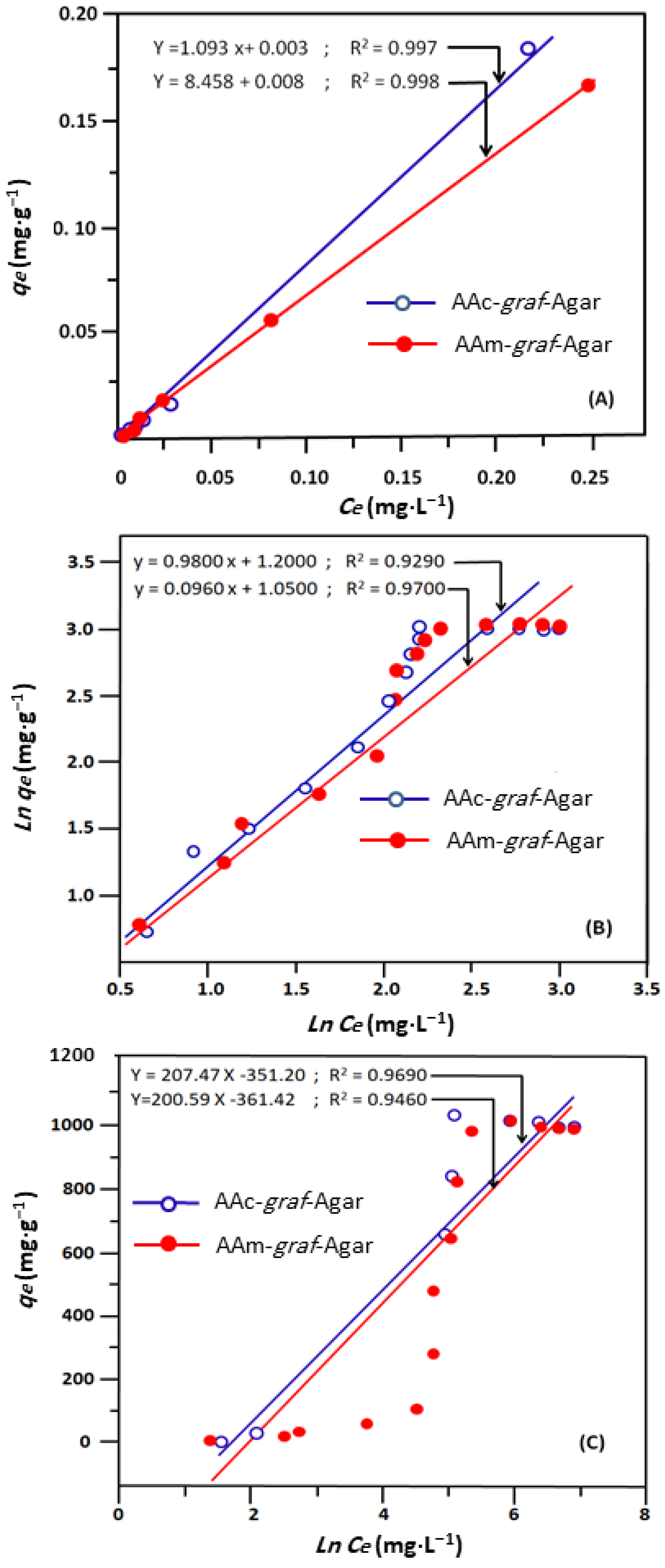
2.4. Adsorption Isotherm Models
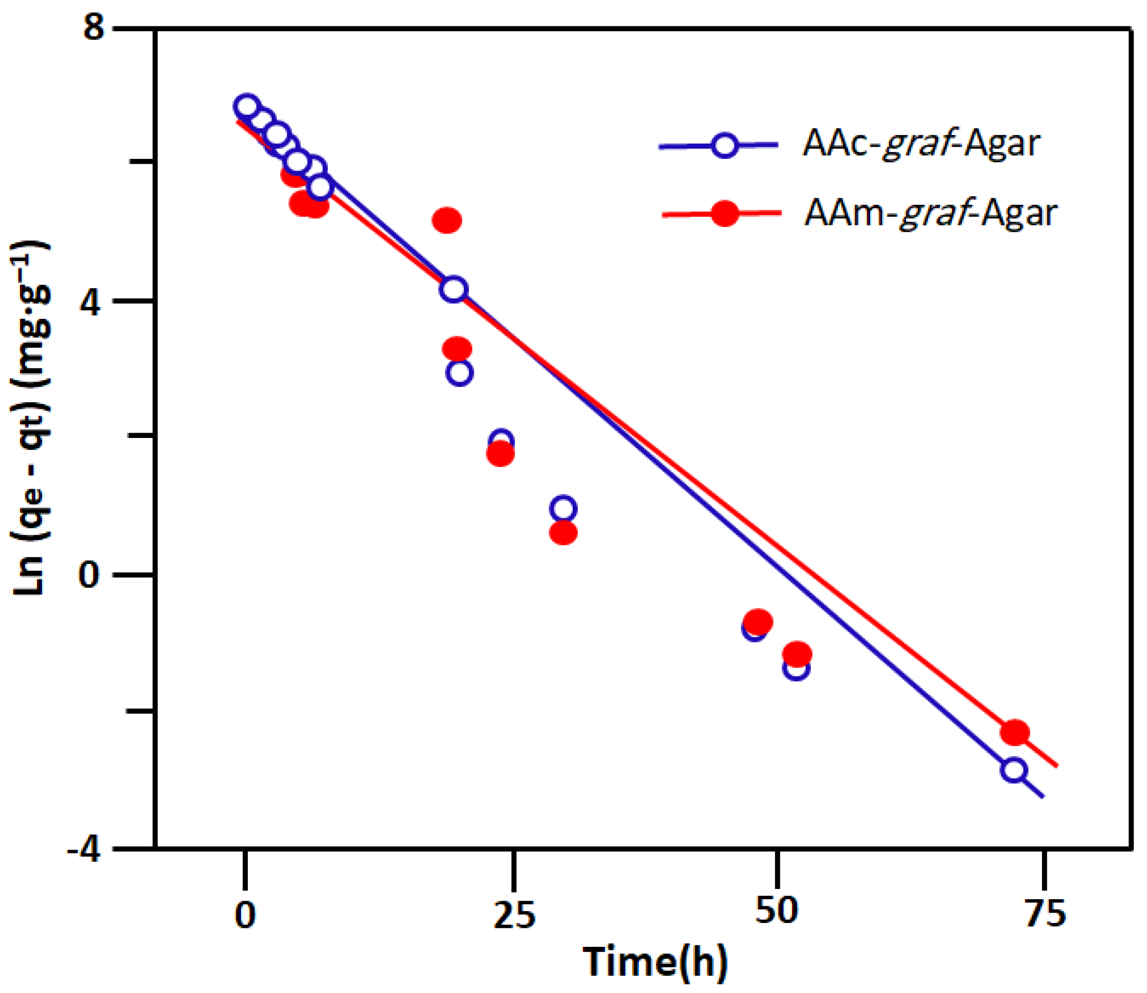
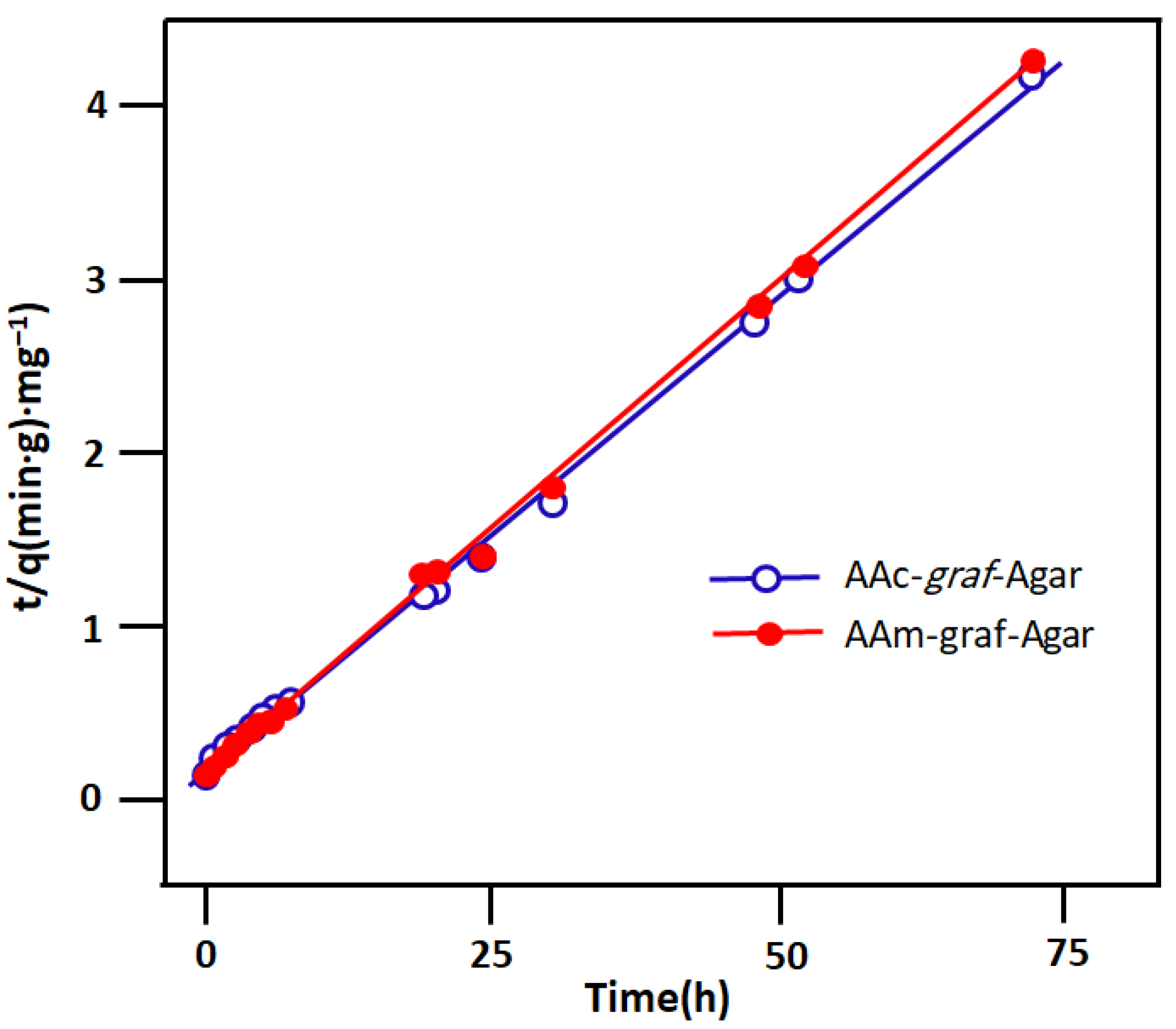
2.5. Adsorption Kinetics
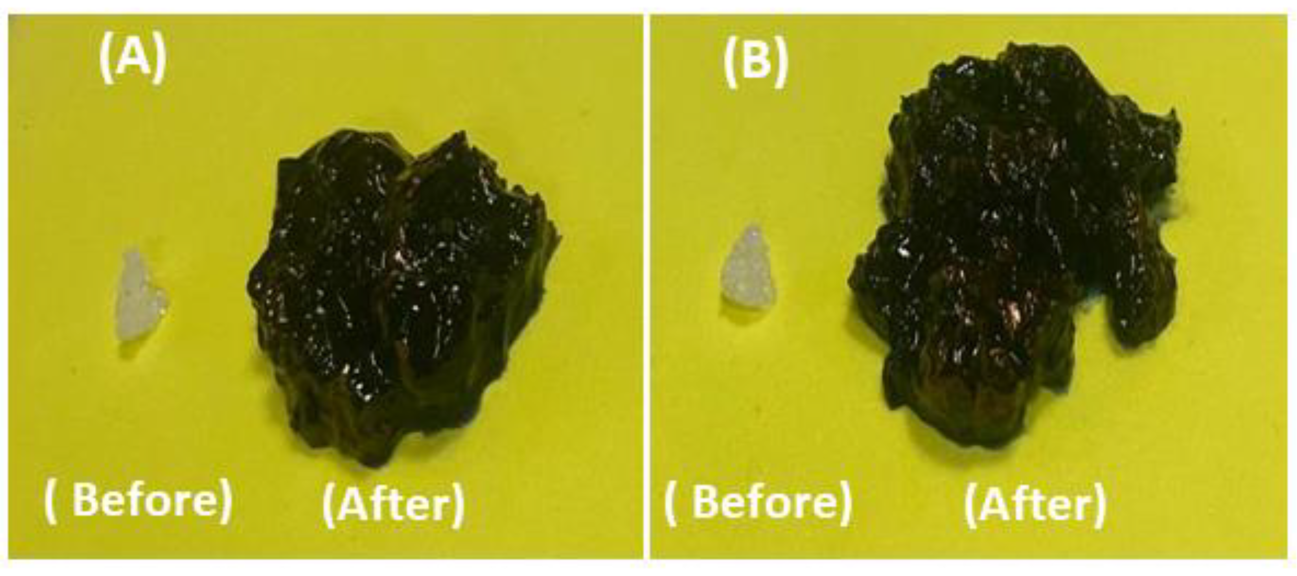
2.6. Recovery, Regeneration, and Reusability
3. Conclusions
4. Materials and Methods
4.1. Materials
4.2. Preparation of PAAm-graf-Ag and PAAc-graf-Ag Hydrogels
4.3. Characterisation
4.4. Swelling Behaviour
4.5. Adsorption Experiment
4.6. Determination of the Point of the Zero Charge (pHpzc)
Author Contributions
Funding
Institutional Review Board Statement
Informed Consent Statement
Data Availability Statement
Conflicts of Interest
References
- Beyranvand, N.S.; Samiey, B.; Tehrani, A.D.; Soleimani, K. Graphene oxide–cellulose nanowhisker hydrogel nanocomposite as a novel adsorbent for methylene blue. J. Chem. Eng. Data 2019, 64, 5558–5570. [Google Scholar] [CrossRef]
- Nguyen, T.T.; Hoang, B.N.; Van Tran, T.; Nguyen, T.D.; Vo, D.-V.N. Agar/maltodextrin/poly (vinyl alcohol) walled montmorillonite composites for removal of methylene blue from aqueous solutions. Surf. Interfaces 2021, 26, 101410. [Google Scholar] [CrossRef]
- Duman, O.; Polat, T.G.; Diker, C.Ö.; Tunç, S. Agar/κ-carrageenan composite hydrogel adsorbent for the removal of Methylene Blue from water. Int. J. Biol. Macromol. 2020, 160, 823–835. [Google Scholar] [CrossRef] [PubMed]
- Pitak-Arnnop, P.; Subbalekha, K.; Sirintawat, N.; Auychai, P.; Klaisiri, A.; Neff, A. Intraoperative injection of combined fibrin sealant and methylene blue dye for surgery of branchial cleft cysts: A case report. J. Stomatol. Oral Maxillofac. Surg. 2019, 120, 378–382. [Google Scholar] [CrossRef] [PubMed]
- Nath, H.; Saikia, A.; Goutam, P.J.; Saikia, B.K.; Saikia, N. Removal of methylene blue from water using okra (Abelmoschus esculentus L.) mucilage modified biochar. Bioresour. Technol. Rep. 2021, 14, 100689. [Google Scholar] [CrossRef]
- Hanafy, H. Adsorption of methylene blue and bright blue dyes on bayleaf capertree pods powder: Understanding the adsorption mechanism by a theoretical study. J. Mol. Liq. 2021, 332, 115680. [Google Scholar] [CrossRef]
- Yoon, H.S.; Cho, C.H.; Yun, M.S.; Jang, S.J.; You, H.J.; Kim, J.-H.; Han, D.; Cha, K.H.; Moon, S.H.; Lee, K. Akkermansia muciniphila secretes a glucagon-like peptide-1-inducing protein that improves glucose homeostasis and ameliorates metabolic disease in mice. Nat. Microbiol. 2021, 6, 563–573. [Google Scholar] [CrossRef]
- Yi, J.-Z.; Zhang, L.-M. Removal of methylene blue dye from aqueous solution by adsorption onto sodium humate/polyacrylamide/clay hybrid hydrogels. Bioresour. Technol. 2008, 99, 2182–2186. [Google Scholar] [CrossRef]
- Tsade Kara, H.; Anshebo, S.T.; Sabir, F.K.; Adam Workineh, G. Removal of methylene blue dye from wastewater using periodiated modified nanocellulose. Int. J. Chem. Eng. 2021, 2021, 9965452. [Google Scholar] [CrossRef]
- Shakoor, S.; Nasar, A. Removal of methylene blue dye from artificially contaminated water using citrus limetta peel waste as a very low cost adsorbent. J. Taiwan Inst. Chem. Eng. 2016, 66, 154–163. [Google Scholar] [CrossRef]
- Munir, M.; Nazar, M.F.; Zafar, M.N.; Zubair, M.; Ashfaq, M.; Hosseini-Bandegharaei, A.; Khan, S.U.-D.; Ahmad, A. Effective adsorptive removal of methylene blue from water by didodecyldimethylammonium bromide-modified Brown clay. ACS Omega 2020, 5, 16711–16721. [Google Scholar] [CrossRef]
- Cheng, J.; Zhan, C.; Wu, J.; Cui, Z.; Si, J.; Wang, Q.; Peng, X.; Turng, L.-S. Highly efficient removal of methylene blue dye from an aqueous solution using cellulose acetate nanofibrous membranes modified by polydopamine. ACS Omega 2020, 5, 5389–5400. [Google Scholar] [CrossRef] [Green Version]
- Mosoarca, G.; Popa, S.; Vancea, C.; Dan, M.; Boran, S. Removal of methylene blue from aqueous solutions using a new natural lignocellulosic adsorbent—Raspberry (Rubus idaeus) leaves powder. Polymers 2022, 14, 1966. [Google Scholar] [CrossRef]
- Al-husseiny, R.A.; Ebrahim, S.E. Effective Removal of Methylene Blue from Wastewater Using Magnetite/Geopolymer Composite: Synthesis, Characterization and Column Adsorption Study. Inorg. Chem. Commun. 2022, 139, 109318. [Google Scholar] [CrossRef]
- Wang, H.; Yang, L.; Qin, Y.; Chen, Z.; Wang, T.; Sun, W.; Wang, C. Highly effective removal of methylene blue from wastewater by modified hydroxyl groups materials: Adsorption performance and mechanisms. Colloids Surf. A Physicochem. Eng. Asp. 2023, 656, 130290. [Google Scholar] [CrossRef]
- Ahmed, T.; Noor, W.; Faruk, O.; Bhoumick, M.; Uddin, M. Removal of methylene blue (MB) from waste water by adsorption on jackfruit leaf powder (JLP) in continuously stirred tank reactor. J. Phys. Conf. Ser. 2018, 1086, 012012. [Google Scholar] [CrossRef] [Green Version]
- Rahman, M.A.; Amin, S.R.; Alam, A.S. Removal of methylene blue from waste water using activated carbon prepared from rice husk. Dhaka Univ. J. Sci. 2012, 60, 185–189. [Google Scholar] [CrossRef]
- Gul, S.; Gul, S.; Gul, H.; Khitab, F.; Khattak, R.; Khan, M.S.; Ullah, R.; Ullah, R.; Wasil, Z.; Krauklis, A.E. Dried Leaves Powder of Adiantum capillus-veneris as an Efficient Biosorbent for Hazardous Crystal Violet Dye from Water Resources. Separations 2023, 10, 165. [Google Scholar] [CrossRef]
- Gul, S.; Gul, A.; Gul, H.; Khattak, R.; Ismail, M.; Khan, S.U.; Khan, M.S.; Aouissi, H.A.; Krauklis, A. Removal of Brilliant Green Dye from Water Using Ficus benghalensis Tree Leaves as an Efficient Biosorbent. Materials 2023, 16, 521. [Google Scholar] [CrossRef]
- Gul, S.; Kanwal, M.; Qazi, R.A.; Gul, H.; Khattak, R.; Khan, M.S.; Khitab, F.; Krauklis, A.E. Efficient removal of methyl red dye by using bark of hopbush. Water 2022, 14, 2831. [Google Scholar] [CrossRef]
- Ozola-Davidane, R.; Burlakovs, J.; Tamm, T.; Zeltkalne, S.; Krauklis, A.E.; Klavins, M. Bentonite-ionic liquid composites for Congo red removal from aqueous solutions. J. Mol. Liq. 2021, 337, 116373. [Google Scholar] [CrossRef]
- Xu, L.; Qiu, L.; Sheng, Y.; Sun, Y.; Deng, L.; Li, X.; Bradley, M.; Zhang, R. Biodegradable pH-responsive hydrogels for controlled dual-drug release. J. Mater. Chem. B 2018, 6, 510–517. [Google Scholar] [CrossRef] [PubMed] [Green Version]
- Narula, A.; Rao, C.P. Hydrogel of the supramolecular complex of graphene oxide and sulfonatocalix [4] arene as reusable material for the degradation of organic dyes: Demonstration of adsorption and degradation by spectroscopy and microscopy. ACS Omega 2019, 4, 5731–5740. [Google Scholar] [CrossRef]
- Bhattacharya, A.; Rawlins, J.W.; Ray, P. Polymer Grafting and Crosslinking; John Wiley & Sons: Hoboken, NJ, USA, 2008. [Google Scholar]
- Tiwari, A.; Prabaharan, M. An amphiphilic nanocarrier based on guar gum-graft-poly (ε-caprolactone) for potential drug-delivery applications. J. Biomater. Sci. Polym. Ed. 2010, 21, 937–949. [Google Scholar] [CrossRef] [PubMed]
- Rani, G.U.; Mishra, S.; Sen, G.; Jha, U. Polyacrylamide grafted Agar: Synthesis and applications of conventional and microwave assisted technique. Carbohydr. Polym. 2012, 90, 784–791. [Google Scholar] [CrossRef] [PubMed]
- Ouyang, Q.-Q.; Hu, Z.; Li, S.-D.; Quan, W.-Y.; Wen, L.-L.; Yang, Z.-M.; Li, P.-W. Thermal degradation of agar: Mechanism and toxicity of products. Food Chem. 2018, 264, 277–283. [Google Scholar] [CrossRef]
- Rhein-Knudsen, N.; Ale, M.T.; Ajalloueian, F.; Yu, L.; Meyer, A.S. Rheological properties of agar and carrageenan from Ghanaian red seaweeds. Food Hydrocoll. 2017, 63, 50–58. [Google Scholar] [CrossRef]
- Safarzadeh, H.; Peighambardoust, S.J.; Mousavi, S.H.; Mohammadi, R.; Peighambardoust, S.H. Adsorption of methyl violet dye from wastewater using poly (methacrylic acid-co-acrylamide)/bentonite nanocomposite hydrogels. J. Polym. Res. 2022, 29, 113. [Google Scholar] [CrossRef]
- Yetimoğlu, E.K.; Kahraman, M.; Ercan, Ö.; Akdemir, Z.; Apohan, N.K. N-vinylpyrrolidone/acrylic acid/2-acrylamido-2-methylpropane sulfonic acid based hydrogels: Synthesis, characterization and their application in the removal of heavy metals. React. Funct. Polym. 2007, 67, 451–460. [Google Scholar] [CrossRef]
- Pakdel, P.M.; Peighambardoust, S.J. A review on acrylic based hydrogels and their applications in wastewater treatment. J. Environ. Manag. 2018, 217, 123–143. [Google Scholar] [CrossRef]
- Lai, J.C.; Rahman, W.A.W.A.; Toh, W.Y. Characterisation of sago pith waste and its composites. Ind. Crops Prod. 2013, 45, 319–326. [Google Scholar] [CrossRef]
- Wu, Y.; Geng, F.; Chang, P.R.; Yu, J.; Ma, X. Effect of agar on the microstructure and performance of potato starch film. Carbohydr. Polym. 2009, 76, 299–304. [Google Scholar] [CrossRef]
- Brown, T.L. Infrared Spectra of Inorganic and Coordination Compounds. By K. Nakamoto. Inorg. Chem. 1964, 3, 306–307. [Google Scholar] [CrossRef]
- Nakanishi, K.; Solomon, P.H. Infrared Absorption Spectroscopy; Holden-Day: San Francisco, CA, USA, 1977. [Google Scholar]
- Huang, H.; Hu, Y.; Zhang, J.; Sato, H.; Zhang, H.; Noda, I.; Ozaki, Y. Miscibility and hydrogen-bonding interactions in biodegradable polymer blends of poly (3-hydroxybutyrate) and a partially hydrolyzed poly (vinyl alcohol). J. Phys. Chem. B 2005, 109, 19175–19183. [Google Scholar] [CrossRef]
- Lejardi, A.; Meaurio, E.; Fernández, J.; Sarasua, J.-R. Miscibility of poly (vinyl alcohol)-graft-hydroxy ester/poly (vinylpyrrolidone) blends. Macromolecules 2011, 44, 7351–7363. [Google Scholar] [CrossRef]
- Moharram, M.; Khafagi, M. Thermal behavior of poly (acrylic acid)–poly (vinyl pyrrolidone) and poly (acrylic acid)–metal–poly (vinyl pyrrolidone) complexes. J. Appl. Polym. Sci. 2006, 102, 4049–4057. [Google Scholar] [CrossRef]
- Pino-Ramos, V.H.; Duarte-Peña, L.; Bucio, E. Highly Crosslinked Agar/Acrylic Acid Hydrogels with Antimicrobial Properties. Gels 2021, 7, 183. [Google Scholar] [CrossRef]
- Peppas, N.A.; Colombo, P. Analysis of drug release behavior from swellable polymer carriers using the dimensionality index. J. Control. Release 1997, 45, 35–40. [Google Scholar] [CrossRef]
- Nesrinne, S.; Djamel, A. Synthesis, characterization and rheological behavior of pH sensitive poly (acrylamide-co-acrylic acid) hydrogels. Arab. J. Chem. 2017, 10, 539–547. [Google Scholar] [CrossRef] [Green Version]
- Huang, Y.; Yu, H.; Xiao, C. pH-sensitive cationic guar gum/poly (acrylic acid) polyelectrolyte hydrogels: Swelling and in vitro drug release. Carbohydr. Polym. 2007, 69, 774–783. [Google Scholar] [CrossRef]
- Kim, S.J.; Park, S.J.; Kim, S.I. Properties of smart hydrogels composed of polyacrylic acid/poly (vinyl sulfonic acid) responsive to external stimuli. Smart Mater. Struct. 2004, 13, 317. [Google Scholar] [CrossRef]
- Chang, C.; Duan, B.; Cai, J.; Zhang, L. Superabsorbent hydrogels based on cellulose for smart swelling and controllable delivery. Eur. Polym. J. 2010, 46, 92–100. [Google Scholar] [CrossRef]
- Harrache, Z.; Abbas, M.; Aksil, T.; Trari, M. Thermodynamic and kinetics studies on adsorption of Indigo Carmine from aqueous solution by activated carbon. Microchem. J. 2019, 144, 180–189. [Google Scholar] [CrossRef]
- Delgado-Mellado, N.; Ayuso, M.; Villar-Chavero, M.M.; García, J.; Rodríguez, F. Ecotoxicity evaluation towards Vibrio fischeri of imidazolium-and pyridinium-based ionic liquids for their use in separation processes. SN Appl. Sci. 2019, 1, 896. [Google Scholar] [CrossRef] [Green Version]
- Forutan, R.; Ehsandoost, E.; Hadipour, S.; Mobaraki, Z.; Saleki, M.; Mohebbi, G. Kinetic and equilibrium studies on the adsorption of lead by the chitin of pink shrimp (Solenocera melantho). Entomol. Appl. Sci. Lett. 2016, 3, 20–26. [Google Scholar]
- Rahimi, K.; Mirzaei, R.; Akbari, A.; Mirghaffari, N. Preparation of nanoparticle-modified polymeric adsorbent using wastage fuzzes of mechanized carpet and its application in dye removal from aqueous solution. J. Clean. Prod. 2018, 178, 373–383. [Google Scholar] [CrossRef]
- Hajizadeh, H.; Peighambardoust, S.J.; Peighambardoust, S.H.; Peressini, D. Physical, mechanical, and antibacterial characteristics of bio-nanocomposite films loaded with Ag-modified SiO2 and TiO2 nanoparticles. J. Food Sci. 2020, 85, 1193–1202. [Google Scholar] [CrossRef]
- Agnihotri, S.; Singhal, R. Effect of sodium alginate content in acrylic acid/sodium humate/sodium alginate superabsorbent hydrogel on removal capacity of MB and CV dye by adsorption. J. Polym. Environ. 2019, 27, 372–385. [Google Scholar] [CrossRef]
- Atangana, E. Adsorption of Zn (II) and Pb (II) ions from aqueous solution using chitosan cross-linked formaldehyde adsorbent to protect the environment. J. Polym. Environ. 2019, 27, 2281–2291. [Google Scholar] [CrossRef]
- García, F.E.; Plaza-Cazón, J.; Montesinos, V.N.; Donati, E.R.; Litter, M.I. Combined strategy for removal of Reactive Black 5 by biomass sorption on Macrocystis pyrifera and zerovalent iron nanoparticles. J. Environ. Manag. 2018, 207, 70–79. [Google Scholar] [CrossRef]
- Wang, M.; Gu, Q.; Luo, Y.; Bukhvalov, D.; Ma, X.; Zhu, L.; Li, G.; Luo, Z. Understanding mechanism of adsorption in the decolorization of aqueous methyl violet (6B) solution by okra polysaccharides: Experiment and theory. ACS Omega 2019, 4, 17880–17889. [Google Scholar] [CrossRef] [Green Version]
- Ma, D.; Zhu, B.; Cao, B.; Wang, J.; Zhang, J. Fabrication of the novel hydrogel based on waste corn stalk for removal of methylene blue dye from aqueous solution. Appl. Surf. Sci. 2017, 422, 944–952. [Google Scholar] [CrossRef]
- Ahmadi, A.; Foroutan, R.; Esmaeili, H.; Tamjidi, S. The role of bentonite clay and bentonite clay@ MnFe2O4 composite and their physico-chemical properties on the removal of Cr (III) and Cr (VI) from aqueous media. Environ. Sci. Pollut. Res. 2020, 27, 14044–14057. [Google Scholar] [CrossRef]
- Pakdel, P.M.; Peighambardoust, S.J. Review on recent progress in chitosan-based hydrogels for wastewater treatment application. Carbohydr. Polym. 2018, 201, 264–279. [Google Scholar] [CrossRef]
- Abdolhosseinzadeh, M.; Peighambardoust, S.J.; Erfan-Niya, H.; Mohammadzadeh Pakdel, P. Swelling and auramine-O adsorption of carboxymethyl cellulose grafted poly (methyl methacrylate)/Cloisite 30B nanocomposite hydrogels. Iran. Polym. J. 2018, 27, 807–818. [Google Scholar] [CrossRef]
- Dai, Y.; Niu, J.; Yin, L.; Xu, J.; Xi, Y. Sorption of polycyclic aromatic hydrocarbons on electrospun nanofibrous membranes: Sorption kinetics and mechanism. J. Hazard. Mater. 2011, 192, 1409–1417. [Google Scholar] [CrossRef]
- Zhu, Y.; Yi, B.; Yuan, Q.; Wu, Y.; Wang, M.; Yan, S. Removal of methylene blue from aqueous solution by cattle manure-derived low temperature biochar. RSC Adv. 2018, 8, 19917–19929. [Google Scholar] [CrossRef] [Green Version]
- Pathania, D.; Sharma, S.; Singh, P. Removal of methylene blue by adsorption onto activated carbon developed from Ficus carica bast. Arab. J. Chem. 2017, 10, S1445–S1451. [Google Scholar] [CrossRef] [Green Version]
- Oliveira, F.R.; Patel, A.K.; Jaisi, D.P.; Adhikari, S.; Lu, H.; Khanal, S.K. Environmental application of biochar: Current status and perspectives. Bioresour. Technol. 2017, 246, 110–122. [Google Scholar] [CrossRef]
- Taşdelen, B.; Çifçi, D.İ.; Meriç, S. Preparation of N-isopropylacrylamide/itaconic acid/Pumice highly swollen composite hydrogels to explore their removal capacity of methylene blue. Colloids Surf. A Physicochem. Eng. Asp. 2017, 519, 245–253. [Google Scholar] [CrossRef]
- Paulino, A.T.; Guilherme, M.R.; Reis, A.V.; Campese, G.M.; Muniz, E.C.; Nozaki, J. Removal of methylene blue dye from an aqueous media using superabsorbent hydrogel supported on modified polysaccharide. J. Colloid Interface Sci. 2006, 301, 55–62. [Google Scholar] [CrossRef] [PubMed]
- Pourjavadi, A.; Bassampour, Z.; Ghasemzadeh, H.; Nazari, M.; Zolghadr, L.; Hosseini, S.H. Porous Carrageenan-g-polyacrylamide/bentonite superabsorbent composites: Swelling and dye adsorption behavior. J. Polym. Res. 2016, 23, 60. [Google Scholar] [CrossRef]
- Coşkun, R.; Delibaş, A. Removal of methylene blue from aqueous solutions by poly (2-acrylamido-2-methylpropane sulfonic acid-co-itaconic acid) hydrogels. Polym. Bull. 2012, 68, 1889–1903. [Google Scholar] [CrossRef]
- Sousa, A.M.; Gonçalves, M.P. Strategies to improve the mechanical strength and water resistance of agar films for food packaging applications. Carbohydr. Polym. 2015, 132, 196–204. [Google Scholar] [CrossRef] [PubMed]
- Koehler, J.; Brandl, F.P.; Goepferich, A.M. Hydrogel wound dressings for bioactive treatment of acute and chronic wounds. Eur. Polym. J. 2018, 100, 1–11. [Google Scholar] [CrossRef]
- Stoica, A.E.; Chircov, C.; Grumezescu, A.M. Hydrogel dressings for the treatment of burn wounds: An up-to-date overview. Materials 2020, 13, 2853. [Google Scholar] [CrossRef]
- Madaghiele, M.; Demitri, C.; Sannino, A.; Ambrosio, L. Polymeric hydrogels for burn wound care: Advanced skin wound dressings and regenerative templates. Burn. Trauma 2014, 2, 2321–3868.143616. [Google Scholar] [CrossRef] [Green Version]
- Zhang, X.; Wang, Y.; Luo, X.; Lu, A.; Li, Y.; Li, B.; Liu, S. O/W Pickering emulsion templated organo-hydrogels with enhanced mechanical strength and energy storage capacity. ACS Appl. Bio Mater. 2018, 2, 480–487. [Google Scholar] [CrossRef]
- Rani, P.; Sen, G.; Mishra, S.; Jha, U. Microwave assisted synthesis of polyacrylamide grafted gum ghatti and its application as flocculant. Carbohydr. Polym. 2012, 89, 275–281. [Google Scholar] [CrossRef]
- Singh, B.; Chauhan, G.; Bhatt, S.; Kumar, K. Metal ion sorption and swelling studies of psyllium and acrylic acid based hydrogels. Carbohydr. Polym. 2006, 64, 50–56. [Google Scholar] [CrossRef]
- Irani, M.; Keshtkar, A.R.; Mousavian, M.A. Removal of Cd (II) and Ni (II) from aqueous solution by PVA/TEOS/TMPTMS hybrid membrane. Chem. Eng. J. 2011, 175, 251–259. [Google Scholar] [CrossRef]
- Hameed, B.; Tan, I.; Ahmad, A. Adsorption isotherm, kinetic modeling and mechanism of 2, 4, 6-trichlorophenol on coconut husk-based activated carbon. Chem. Eng. J. 2008, 144, 235–244. [Google Scholar] [CrossRef]


| Hydrogel | Adsorption Isotherm Parameters | |||||||||
|---|---|---|---|---|---|---|---|---|---|---|
| Langmuir | Freundlich | Temkin | ||||||||
| qmax | KL | RL | R2 | n | Kf | R2 | Bt | Kt | R2 | |
| AAc-graf-Agar | 304.87 | 0.003 | 0.84 | 0.997 | 1.025 | 2.26 | 0.929 | 207.47 | 0.184 | 0.969 |
| AAm-graf-Agar | 118.22 | 0.001 | 0.94 | 0.998 | 1.032 | 1.93 | 0.970 | 200.59 | 0.165 | 0.901 |
| Hydrogel | Initial MB Dye (mg·L−1) | Adsorption Dose (mg·L−1) | Stirring Rate (rpm) | Contact Period (h) | Temperature (°C) |
|---|---|---|---|---|---|
| AAc-graf-Agar | 1400 | 50 | 100 | 48 | 25 |
| AAm-graf-Agar | 1200 | 50 | 100 | 48 | 25 |
| Hydrogel | Pseudo-First-Order Model | |||||
|---|---|---|---|---|---|---|
| Ln(qe) | K1t | qe (mg·g−1) | K1 | R2 | 1/qe (g·mg−1) | |
| AAc-graf-Agar | 6.7362 ± 0.0238 | 0.00222 ± 1.89359 × 10⁻⁵ | 842.353 | 5.138 × 10⁻⁷ | 0.999 | 9.67 × 10⁻⁴ ± 1.0213 × 10⁻⁵ |
| AAm-graf-Agar | 6.5217 ± 0.05913 | 0.00205 ± 4.66628 × 10⁻⁵ | 679.746 | 4.745 × 10⁻⁷ | 0.994 | 9.89 × 10⁻⁴ ± 7.8794 × 10⁻⁶ |
| Pseudo-second-order model | ||||||
| Hydrogel | qe (mg·g−1) | K2 | R2 | 1/k2qe2 (g2·mg−2) | ||
| AAc-graf-Agar | 1030 | 4.94 × 10⁻⁶ | 0.997 | 0.18922 ± 001621 | ||
| AAm-graf-Agar | 1010 | 6.55 × 10⁻⁶ | 0.998 | 0.14939 ± 0.01251 | ||
| Adsorbent | qmax (mg·g−1) | Adsorbate | Ref. |
|---|---|---|---|
| Agar/κ-carrageenan hydrogel | 205.9 | MB | [3] |
| N-isopropylacrylamide/itaconicacid/pumice composite hydrogel | 22.18 | MB | [62] |
| Polyacrylamide/polyacrylate/gum Arabic | 48.00 | MB | [63] |
| κ-Carrageenan-graf-polyacrylamide/bentonite composite hydrogel | 156.3 | MB | [64] |
| PTCS/OMMT hydrogel composite | 49.01 | MB | [54] |
| Agar/maltodextrin/polyvinyl alcohol-montmorillonite (20%) | 71.51 | MB | [2] |
| Poly(AMPS-co-IA) | 24.1 | MB | [65] |
| AAc-graf-Agar | 304.84 | MB | This work |
| AAm-graf-Agar | 118.22 | MB | This work |
| Hydrogel | Agar (g) | AAc (mL) | AAm (g) | APS (g) | G (%) |
|---|---|---|---|---|---|
| AAc-graf-Agar | 1.0 | - | 10.0 | 0.007 | 896 |
| AAm-graf-Agar | 1.0 | 10.0 | - | 0.007 | 1020 |
Disclaimer/Publisher’s Note: The statements, opinions and data contained in all publications are solely those of the individual author(s) and contributor(s) and not of MDPI and/or the editor(s). MDPI and/or the editor(s) disclaim responsibility for any injury to people or property resulting from any ideas, methods, instructions or products referred to in the content. |
© 2023 by the authors. Licensee MDPI, Basel, Switzerland. This article is an open access article distributed under the terms and conditions of the Creative Commons Attribution (CC BY) license (https://creativecommons.org/licenses/by/4.0/).
Share and Cite
Betraoui, A.; Seddiki, N.; Souag, R.; Guerfi, N.; Semlali, A.; Aouak, T.; Aliouche, D. Synthesis of New Hydrogels Involving Acrylic Acid and Acrylamide Grafted Agar-Agar and Their Application in the Removal of Cationic Dyes from Wastewater. Gels 2023, 9, 499. https://doi.org/10.3390/gels9060499
Betraoui A, Seddiki N, Souag R, Guerfi N, Semlali A, Aouak T, Aliouche D. Synthesis of New Hydrogels Involving Acrylic Acid and Acrylamide Grafted Agar-Agar and Their Application in the Removal of Cationic Dyes from Wastewater. Gels. 2023; 9(6):499. https://doi.org/10.3390/gels9060499
Chicago/Turabian StyleBetraoui, Amina, Nesrinne Seddiki, Rafika Souag, Nabila Guerfi, Abdelhabib Semlali, Taieb Aouak, and Djamel Aliouche. 2023. "Synthesis of New Hydrogels Involving Acrylic Acid and Acrylamide Grafted Agar-Agar and Their Application in the Removal of Cationic Dyes from Wastewater" Gels 9, no. 6: 499. https://doi.org/10.3390/gels9060499