

Collagen Hydrolysate Effects on Photodynamic Efficiency of Gallium (III) Phthalocyanine on Pigmented Melanoma Cells


Abstract
:1. Introduction
2. Results and Discussion
3. Conclusions
4. Materials and Methods
4.1. Chemicals and Synthesis
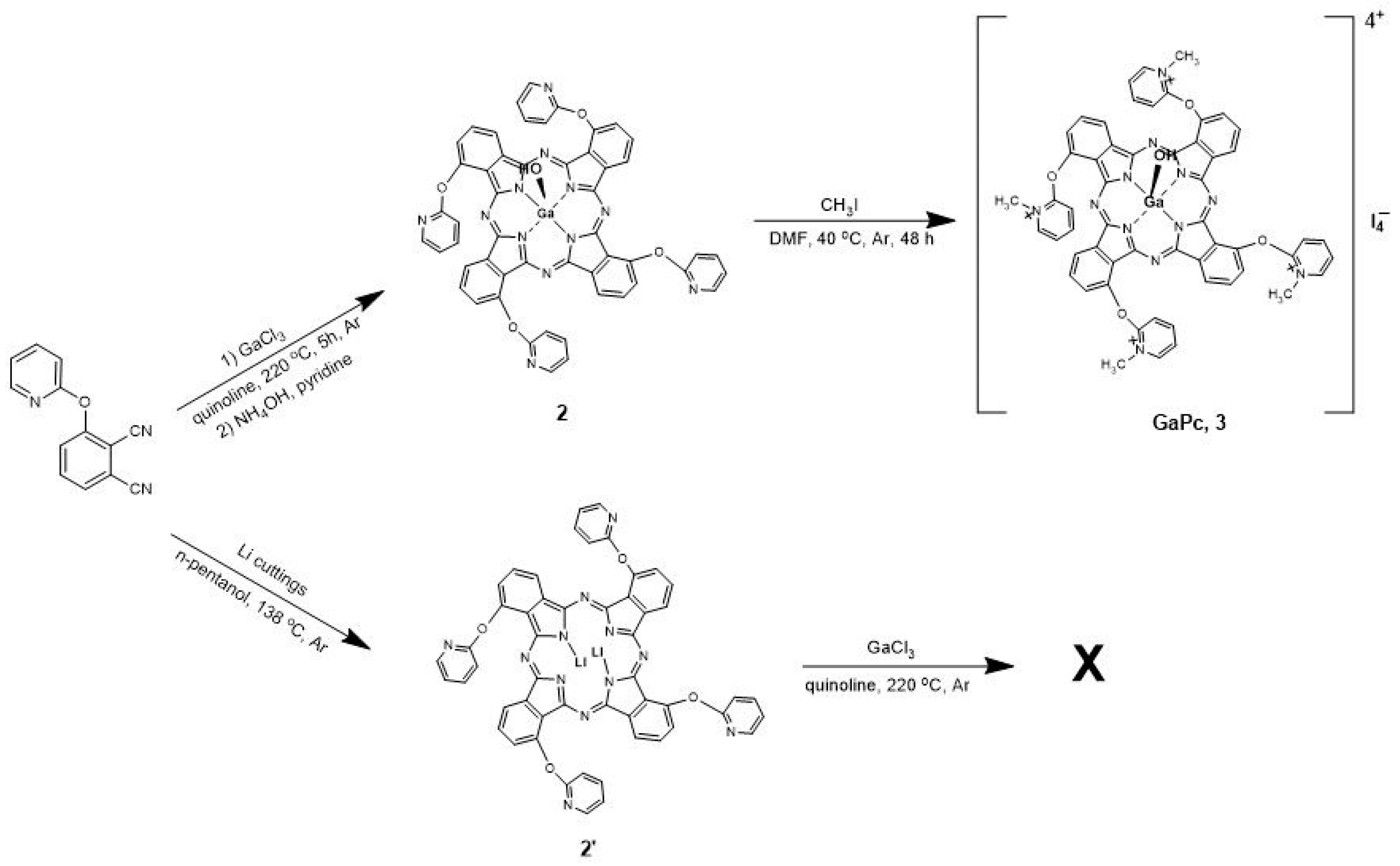
4.1.1. 1(4),8(11),15(18),22(25)-Tetrakis-[(2-pyridiloxy) phthalocyaninato] hydroxygallium(III) (2)
4.1.2. 1(4),8(11),15(18),22(25)-Tetrakis-[(N-methyl-2-pyridiloxy) phthalocyaninato] hydroxygallium(III) Iodide (GaPc, 3)
4.2. Photo-Physicochemical Study
4.3. Photobiology Study
4.3.1. Cell Lines and Cultivation
4.3.2. Light Source and Parameters
4.3.3. Photo- and Cytotoxicity Study
4.4. Statistics
Author Contributions
Funding
Institutional Review Board Statement
Informed Consent Statement
Data Availability Statement
Acknowledgments
Conflicts of Interest
References
- Huis in ‘t Veld, R.V.; Heuts, J.; Ma, S.; Cruz, L.J.; Ossendorp, F.A.; Jager, M.J. Current Challenges and Opportunities of Photodynamic Therapy against Cancer. Pharmaceutics 2023, 15, 330. [Google Scholar] [CrossRef] [PubMed]
- Sun, J.; Zhao, H.; Fu, L.; Cui, J.; Yang, Y. Global Trends and Research Progress of Photodynamic Therapy in Skin Cancer: A Bibliometric Analysis and Literature Review. Clin. Cosmet. Investig. Dermatol. 2023, 16, 479–498. [Google Scholar] [CrossRef] [PubMed]
- Sharma, D.; Singh, S.; Kumar, P.; Jain, G.K.; Aggarwal, G.; Almalki, W.H.; Kesharwani, P. 2—Mechanisms of Photodynamic Therapy. In Nanomaterials for Photodynamic Therapy; Kesharwani, P., Ed.; Woodhead Publishing Series in Biomaterials; Woodhead Publishing: Sawston, UK, 2023; pp. 41–54. [Google Scholar] [CrossRef]
- Maharjan, P.S.; Bhattarai, H.K. Singlet Oxygen, Photodynamic Therapy, and Mechanisms of Cancer Cell Death. J. Oncol. 2022, 2020, 7211485. [Google Scholar] [CrossRef] [PubMed]
- Bacellar, I.; Tsubone, T.; Pavani, C.; Baptista, M. Photodynamic Efficiency: From Molecular Photochemistry to Cell Death. Int. J. Mol. Sci. 2015, 16, 20523–20559. [Google Scholar] [CrossRef]
- Ogura, S.-I.; Tabata, K.; Fukushima, K.; Kamachi, T.; Okura, I. Development of phthalocyanines for photodynamic therapy. J. Porphyr. Phthalocyanines 2006, 10, 1116–1124. [Google Scholar] [CrossRef]
- Nyokong, T. Desired properties of new phthalocyanines for photodynamic therapy. Pure Appl. Chem. 2011, 83, 1643–1799. [Google Scholar] [CrossRef]
- Dias, L.M.; de Keijzer, M.J.; Ernst, D.; Sharifi, F.; de Klerk, D.J.; Kleijn, T.G.; Desclos, E.; Kochan, J.A.; de Haan, L.R.; Franchi, L.P.; et al. Metallated phthalocyanines and their hydrophilic derivatives for multi-targeted oncological photodynamic therapy. J. Photochem. Photobiol. B Biol. 2022, 234, 112500. [Google Scholar] [CrossRef]
- Li, X.; Zheng, B.-D.; Peng, X.-H.; Li, S.-Z.; Ying, J.-W.; Zhao, Y.; Huang, J.-D.; Yoon, J. Phthalocyanines as medicinal photosensitizers: Developments in the last five years. Coord. Chem. Rev. 2019, 379, 147–160. [Google Scholar] [CrossRef]
- Chen, D.; Song, M.; Huang, J.; Chen, N.; Xue, J.; Huang, M. Photocyanine: A novel and effective phthalocyanine-based photosensitizer for cancer treatment. J. Innov. Opt. Health Sci. 2020, 13, 2030009. [Google Scholar] [CrossRef]
- Arun, A.; Malrautu, P.; Laha, A.; Luo, H.; Ramakrishna, S. Collagen Nanoparticles in Drug Delivery Systems and Tissue Engineering. Appl. Sci. 2021, 11, 11369. [Google Scholar] [CrossRef]
- Yewale, C.; Baradia, D.; Vhora, I.; Misra, A. Proteins: Emerging carrier for delivery of cancer therapeutics. Expert Opin. Drug Deliv. 2013, 10, 1429–1448. [Google Scholar] [CrossRef]
- Kasoju, N.; Ali, S.S.; Dubey, V.K.; Bora, U. Exploiting the Potential of Collagen as a Natural Biomaterial in Drug Delivery. J. Proteins Proteom. 2013, 1, 9–14. [Google Scholar]
- Kaltbeitzel, J.; Wich, P.R. Protein-based Nanoparticles: From Drug Delivery to Imaging, Nanocatalysis and Protein Therapy. Angew. Chem. Int. Ed. 2023, 2023, e202216097. [Google Scholar] [CrossRef]
- Korbelik, M.; Sun, J.; Cecic, I. Photodynamic Therapy-Induced Cell Surface Expression and Release of Heat Shock Proteins: Relevance for Tumor Response. Cancer Res. 2005, 65, 1018–1026. [Google Scholar] [CrossRef]
- Sánchez-Cid Bueno, P.; Jiménez Rosado, M.; Romero García, A.; Pérez-Puyana, V.M. Novel Trends in Hydrogel Development for Biomedical Applications: A Review. Polymers 2022, 14, 3023. [Google Scholar] [CrossRef]
- Verrico, A.K.; Moore, J.V. Expression of the collagen-related head shock protein HSP47 in fibroblasts treated with hyperthermia or photodynamic therapy. Br. J. Cancer 1997, 76, 719–724. [Google Scholar] [CrossRef]
- Deyl, Z.; Praus, P.; Lcova, H.J.; Goldman, J.N. Fluorescence of collagen—Properties of tyrosine residues and another fluorescent element in calf skin collagen. FEBS Lett. 1969, 5, 187–191. [Google Scholar] [CrossRef]
- Chan, B.P.; Chan, O.C.M.; So, K.-F. Effects of photochemical crosslinking on the microstructure of collagen and a feasibility study on controlled protein release. Acta Biomater. 2008, 4, 1627–1636. [Google Scholar] [CrossRef]
- Yova, D.M.; Hovhannisyan, V.A.; Theodossiou, T. Photochemical effects and hypericin photosensitized processes in collagen. J. Biomed. Opt. 2001, 6, 52–57. [Google Scholar] [CrossRef]
- Katz, A.; Alfano, R.R. Optical Biopsy—Detecting Cancer with Light. In Biomedical Optical Spectroscopy and Diagnostics; Sevick-Muraca, E., Benaron, D., Eds.; Trends in Optics and Photonics Series; Paper FT1; Optica Publishing Group: Washington, DC, USA, 1996. [Google Scholar]
- Marcu, L.; Grundfest, W.S.; Maarek, J.-M.I. Photobleaching of arterial fluorescent compounds: Characterization of elastin, collagen and cholesterol time-resolved spectra during prolonged ultraviolet irradiation. Photochem. Photobiol. 1999, 69, 713–721. [Google Scholar] [CrossRef]
- Foote, C.S. Definition of type I and type l photosensitised oxidation. Photochem. Photobiol. 1991, 54, 659–671. [Google Scholar] [CrossRef] [PubMed]
- Mantareva, V.; Kussovski, V.; Angelov, I.; Wöhrle, D.; Dimitrov, R.; Popova, E.; Dimitrov, S. Non-aggregated Ga (III)-phthalocyanines in the photodynamic inactivatio planktonic and biofilm cultures of pathogenic microorganisms. Photochem. Photobiol. Sci. 2011, 10, 92–102. [Google Scholar] [CrossRef] [PubMed]
- Mantareva, V.; Iliev, I.; Sulikovska, I.; Durmuş, M.; Angelov, I. Cobalamin (Vitamin B12) in Anticancer Photodynamic Therapy with Zn(II) Phthalocyanines. Int. J. Mol. Sci. 2023, 24, 4400. [Google Scholar] [CrossRef] [PubMed]
- Mantareva, V.; Kril, A.; Dimitrov, R.; Wöhrle, D.; Angelov, I. Selective photodynamic therapy induced by pre-irradiation of galactopyranosyl Zn(II) phthalocyanines with UV and red lights. J. Porphyr. Phthalocyanines 2013, 17, 529–539. [Google Scholar] [CrossRef]
- Baldea, I.; Ion, R.M.; Olteanu, D.E.; Nenu, I.; Tudor, D.; Filip, A.G. Photodynamic therapy of melanoma using new, synthetic porphyrins and phthalocyanines as photosensitisers—A comparative study. Clujul Med. 2015, 88, 175–180. [Google Scholar] [CrossRef]
- Castro, K.A.D.F.; Prandini, J.A.; Biazzotto, J.C.; Tomé, J.P.C.; da Silva, R.S.; Lourenço, L.M.O. The Surprisingly Positive Effect of Zinc-Phthalocyanines with High Photodynamic Therapy Efficacy of Melanoma Cancer. Front. Chem. 2022, 10, 825716. [Google Scholar] [CrossRef]
- Nkune, N.W.; Matlou, G.G.; Abrahamse, H. Photodynamic Therapy Efficacy of Novel Zinc Phthalocyanine Tetra Sodium 2-Mercaptoacetate Combined with Cannabidiol on Metastatic Melanoma. Pharmaceutics 2022, 14, 2418. [Google Scholar] [CrossRef]
- Lopes, J.; Rodrigues, C.M.P.; Gaspar, M.M.; Reis, C.P. Melanoma Management: From Epidemiology to Treatment and Latest Advances. Cancers 2022, 14, 4652. [Google Scholar] [CrossRef]
- Huang, Y.Y.; Vecchio, D.; Avci, P.; Yin, R.; Garcia-Diaz, M.; Hamblin, M.R. Melanoma resistance to photodynamic therapy: New insights. Biol. Chem. 2013, 394, 239–250. [Google Scholar] [CrossRef]
- Sharma, K.V.; Davids, L.M. Depigmentation in melanomas increases the efficacy of hypericin-mediated photodynamic-induced cell death. Photodiagnosis Photodynamic Ther. 2012, 9, 156–163. [Google Scholar] [CrossRef]
- Sharma, K.V.; Bowers, N.; Davids, L.M. Photodynamic therapy-induced killing is enhanced in depigmented metastatic melanoma cells. Cell Biol. Int. 2011, 35, 939–944. [Google Scholar] [CrossRef]
- Ma, L.W.; Nielsen, K.P.; Iani, V.; Moan, J. A new method for photodynamic therapy of melanotic melanoma – effects of depigmentation with violet light photodynamic therapy. J. Environ. Pathol. Toxicol. Oncol. 2007, 26, 165–172. [Google Scholar] [CrossRef]
- Naidoo, C.; Kruger, C.A.; Abrahamse, H. Photodynamic Therapy for Metastatic Melanoma Treatment: A Review. Technol. Cancer Res. Treat. 2018, 17, 1533033818791795. [Google Scholar] [CrossRef]
- Lopes, J.; Rodrigues, C.M.P.; Gaspar, M.M.; Reis, C.P. How to Treat Melanoma? The Current Status of Innovative Nanotechnological Strategies and the Role of Minimally Invasive Approaches like PTT and PDT. Pharmaceutics 2022, 14, 1817. [Google Scholar] [CrossRef]
- ESAC. Statement on the scientific validity of the 3T3 NRU PT test (an in vitro test for phototoxic potential). In Proceedings of the 9th Meeting of ECVAM Scientific Advisory Committee, Hamburg, Germany, 1–2 October 1997. [Google Scholar]
- OECD. Guidelines for the Testing of Chemicals, Section 4 Health Effects, Test Guideline No. 432 In Vitro 3T3 NRU Phototoxicity Test. 18 June 2019. Available online: https://www.oecd-ilibrary.org/environment/test-no-432-in-vitro-3t3-nru-phototoxicity-test_9789264071162-en (accessed on 5 June 2023).
- McCain, J.; Colón, K.L.; Barrett, P.C.; Monro, S.M.A.; Sainuddin, T.; Roque, J., III; Pinto, M.; Yin, H.; Cameron, C.G.; McFarland, S.A. Photophysical Properties and Photobiological Activities of Ruthenium(II) Complexes Bearing π-Expansive Cyclometallation Ligands with Thienyl Groups. Inorg. Chem. 2019, 58, 10778–10790. [Google Scholar] [CrossRef]
- Indrayanto, G.; Putra, G.S.; Suhud, F. Validation of in-vitro bioassay methods: Application in herbal drug research. Profiles Drug Subst. Excip. Relat. Methodol. 2021, 46, 273–307. [Google Scholar]
- Peña-Morán, O.A.; Villarreal, M.L.; Álvarez-Berber, L.; Meneses-Acosta, A.; Rodríguez-López, V. Cytotoxicity Post-Treat. Recovery, and selectivity analysis of naturally occurring podophyllotoxins from Bursera fagaroides var. fagaroides on breast Cancer cell lines. Molecules 2016, 21, 1013. [Google Scholar] [CrossRef]
- López-Lázaro, M. Editorial: How many times should we screen a chemical library to discover an anticancer drug? Drug Discov. Today 2015, 20, 167–169. [Google Scholar] [CrossRef]
- Weerapreeyakul, N.; Nonpunya, A.; Barusrux, S.; Thitimetharoch, T.; Sripanidkulchai, B. Evaluation of the anticancer potential of six herbs against a hepatoma cell line. Chin. Med. 2012, 7, 15. [Google Scholar] [CrossRef]
- Rashidi, M.; Seghatoleslam, A.; Namavari, M.; Amiri, A.; Fahmidehkar, M.A.; Ramezani, A.; Eftekhar, E.; Hosseini, A.; Erfani, N.; Fakher, S. Selective Cytotoxicity and Apoptosis-Induction of Cyrtopodion scabrum Extract Against Digestive Cancer Cell Lines. Int. J. Cancer Manag. 2017, 10, e8633. [Google Scholar] [CrossRef]


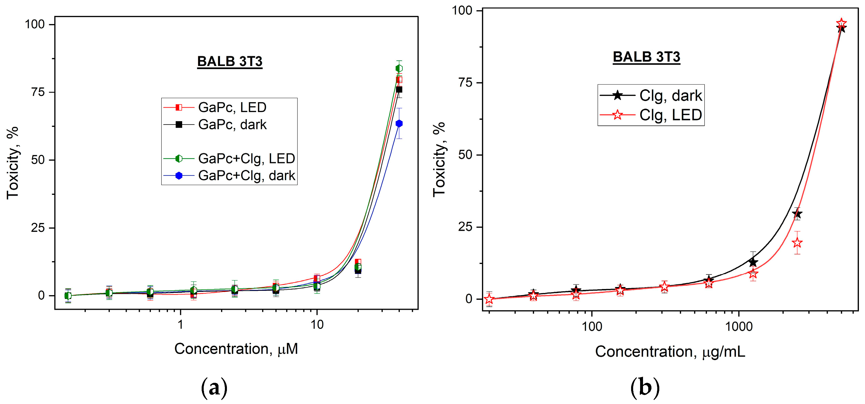
| Compounds | Cytotoxicity | Phototoxicity | PIF * |
|---|---|---|---|
| Clg ** | 3148.63 ± 80.48 | 3299.40 ± 67.79 | 0.95 |
| GaPc | 33.90 ± 1.78 | 29.51 ± 0.44 | 1.15 |
| GaPc–Clg | 30.55 ± 0.63 | 29.11 ± 0.38 | 1.05 |
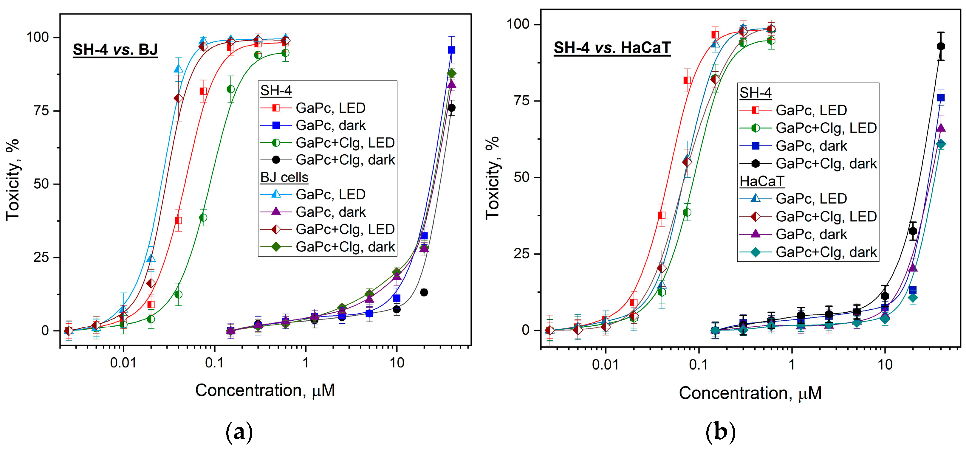
| Cell lines | Compounds | Cytotoxicity | Phototoxicity | PI * | SI 1 ** | SI 2 *** |
|---|---|---|---|---|---|---|
| tumor SH-4 | Clg | 3507.38 ± 111.38 | 3227.80 ± 63.26 | 1 | 1.11 | 1.02 |
| GaPc | 30.06 ± 0.4150 | 0.0367 ± 0.0016 | 819 | 1.49 | 0.54 | |
| GaPc–Clg | 27.74 ± 0.8150 | 0.0770 ± 0.0035 | 360 | 0.71 | 0.28 | |
| HaCaT | Clg | 3716.47 ± 36.82 | 3583.67 ± 69.74 | 1 | ||
| GaPc | 32.67 ± 1.37 | 0.0548 ± 0.0059 | 596 | |||
| GaPc–Clg | 31.61 ± 0.47 | 0.0545 ± 0.0045 | 580 |
Disclaimer/Publisher’s Note: The statements, opinions and data contained in all publications are solely those of the individual author(s) and contributor(s) and not of MDPI and/or the editor(s). MDPI and/or the editor(s) disclaim responsibility for any injury to people or property resulting from any ideas, methods, instructions or products referred to in the content. |
© 2023 by the authors. Licensee MDPI, Basel, Switzerland. This article is an open access article distributed under the terms and conditions of the Creative Commons Attribution (CC BY) license (https://creativecommons.org/licenses/by/4.0/).
Share and Cite
Mantareva, V.; Iliev, I.; Sulikovska, I.; Durmuş, M.; Genova, T. Collagen Hydrolysate Effects on Photodynamic Efficiency of Gallium (III) Phthalocyanine on Pigmented Melanoma Cells. Gels 2023, 9, 475. https://doi.org/10.3390/gels9060475
Mantareva V, Iliev I, Sulikovska I, Durmuş M, Genova T. Collagen Hydrolysate Effects on Photodynamic Efficiency of Gallium (III) Phthalocyanine on Pigmented Melanoma Cells. Gels. 2023; 9(6):475. https://doi.org/10.3390/gels9060475
Chicago/Turabian StyleMantareva, Vanya, Ivan Iliev, Inna Sulikovska, Mahmut Durmuş, and Tsanislava Genova. 2023. "Collagen Hydrolysate Effects on Photodynamic Efficiency of Gallium (III) Phthalocyanine on Pigmented Melanoma Cells" Gels 9, no. 6: 475. https://doi.org/10.3390/gels9060475