
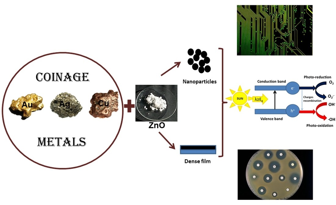
Coinage Metals Doped ZnO Obtained by Sol-Gel Method—A Brief Review

Abstract
:
1. Introduction
2. Synthesis of Coinage Metals Doped ZnO
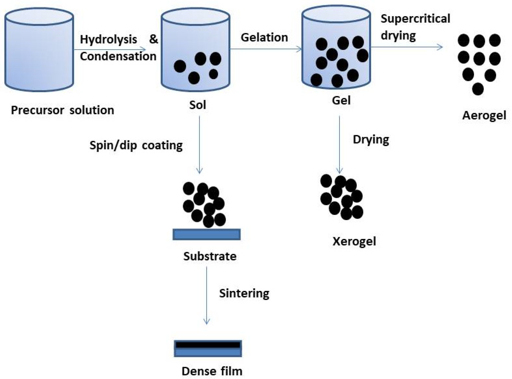
2.1. Sol-Gel Sample Preparation
2.1.1. Precursors
2.1.2. Additives
2.1.3. Solvents
2.2. Nanostructures
2.2.1. Xerogels
2.2.2. Aerogels Dried in Supercritical Conditions of Ethanol
2.2.3. Thin Films (Spin-Coating and Dip-Coating Methods)
2.2.4. Substrate
2.2.5. Heat Treatment
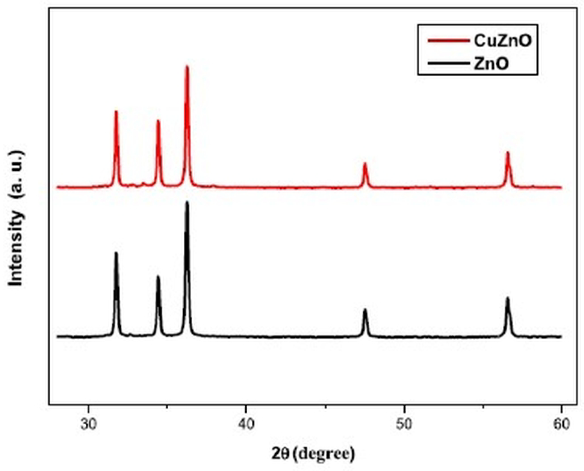
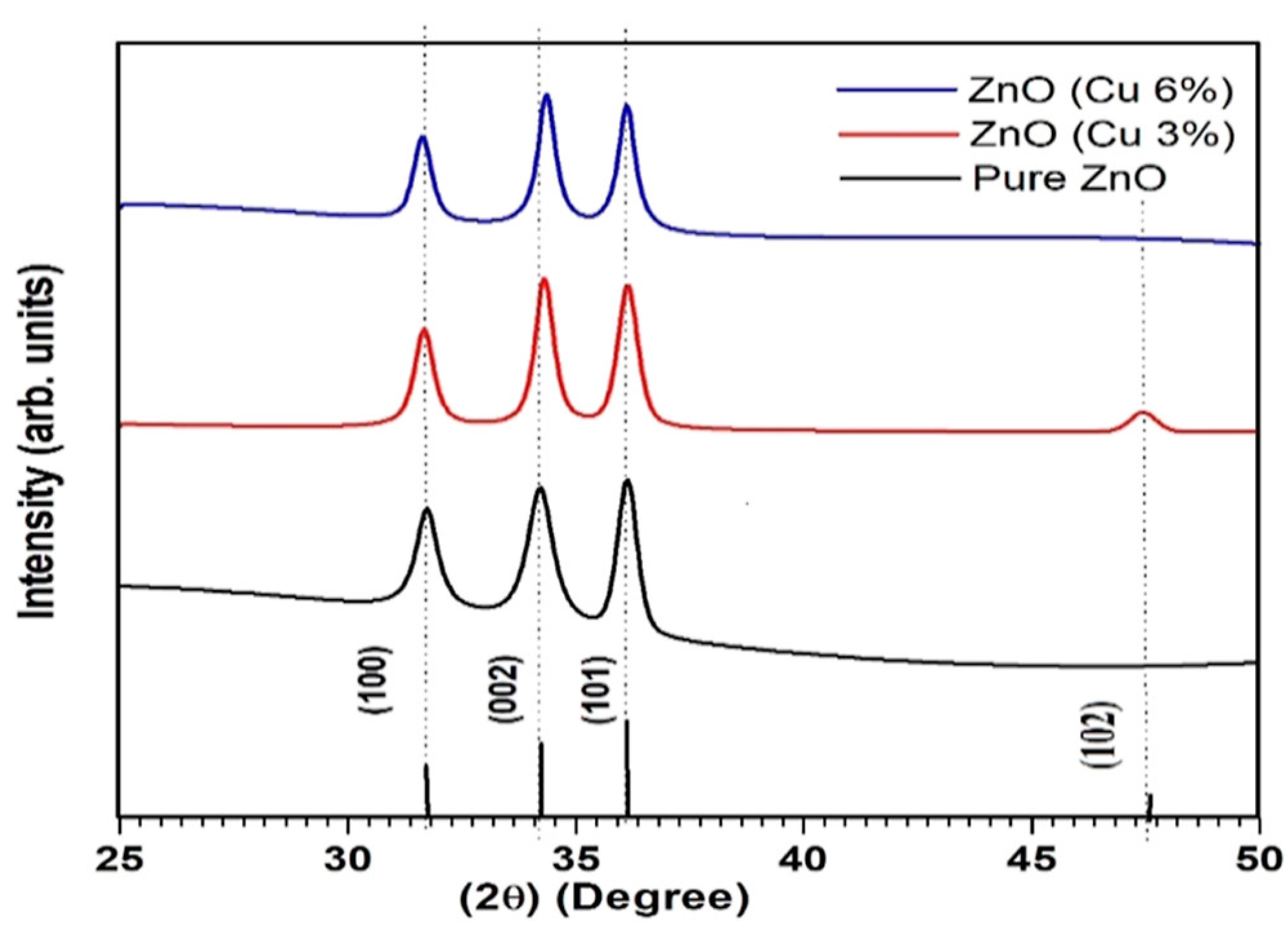
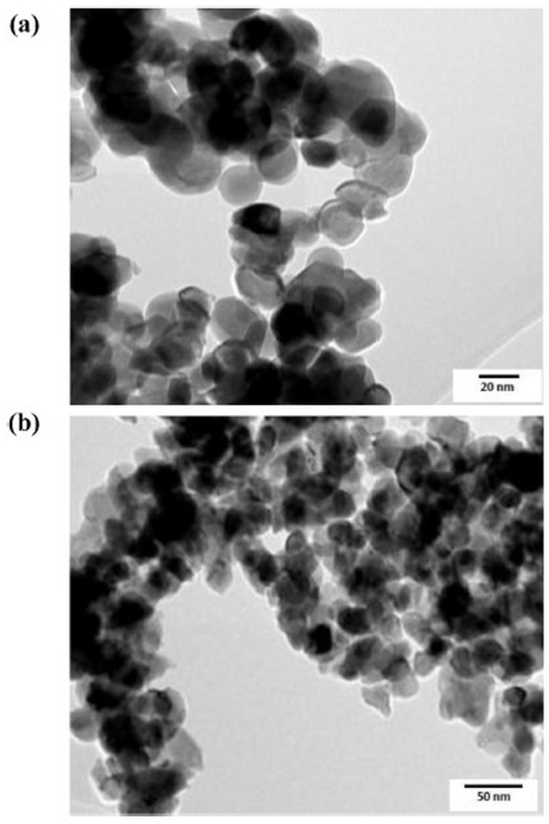
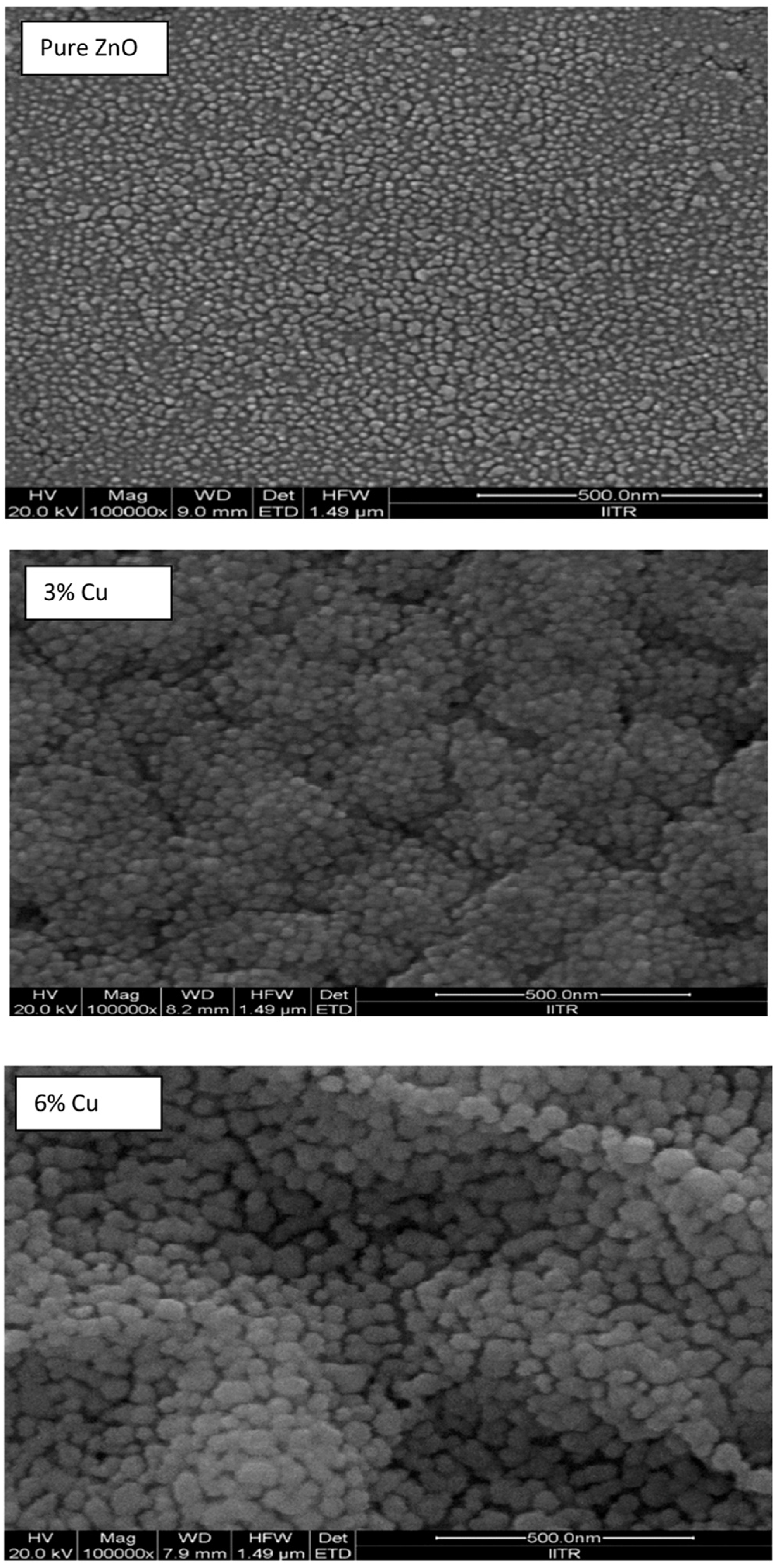
3. Structure and Morphology
4. Applications
4.1. Microelectronics
4.1.1. Optical Properties
4.1.2. Electrical Properties
4.1.3. Sensors
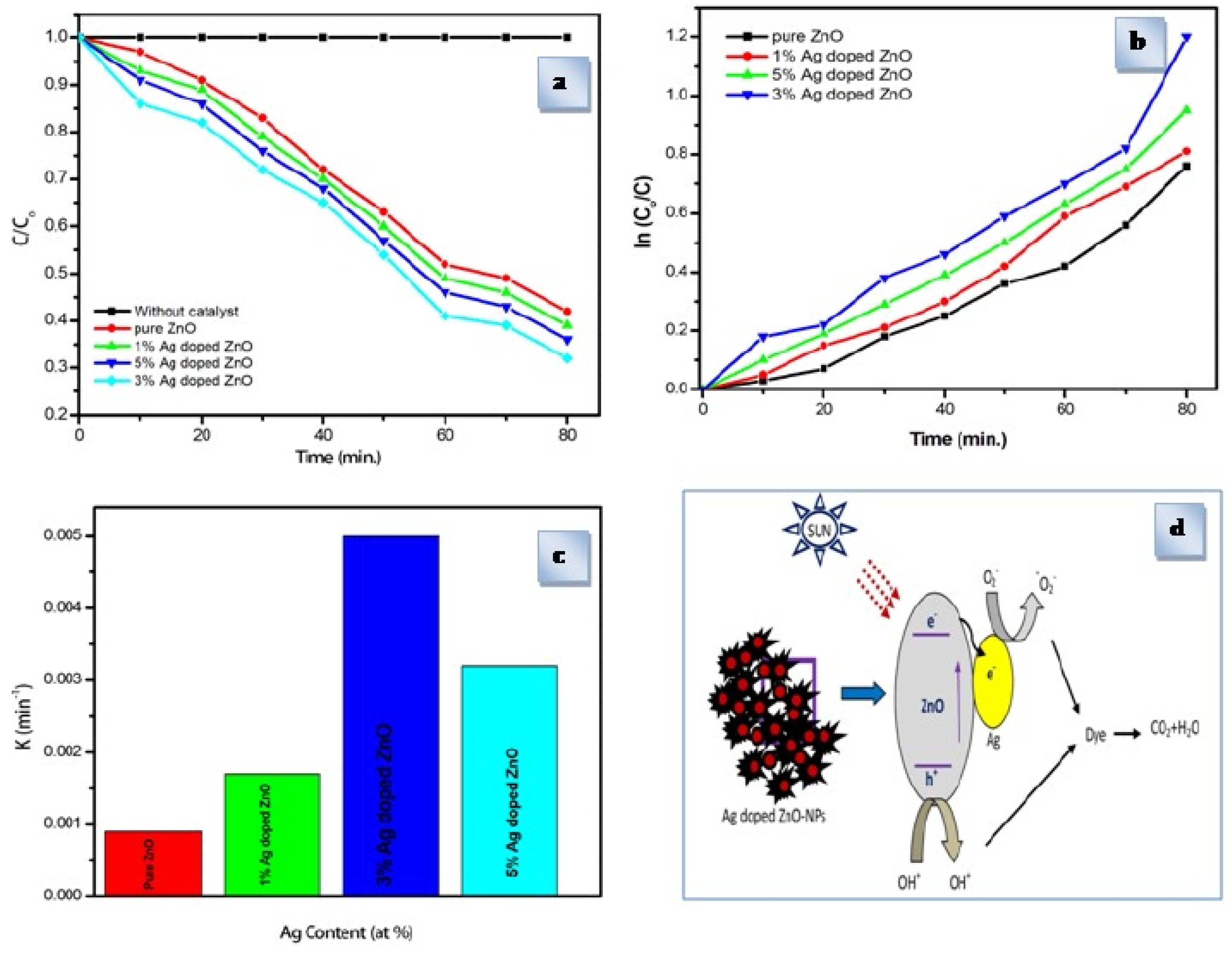
4.2. Photocatalyst
4.3. Biomaterials
5. Conclusions
Author Contributions
Funding
Institutional Review Board Statement
Informed Consent Statement
Data Availability Statement
Acknowledgments
Conflicts of Interest
References
- Jiang, J.; Pi, J.; Cai, J. The advancing of zinc oxide nanoparticles for biomedical applications. Bioinor. Chem. Appl. 2018, 2018, 1062562. [Google Scholar] [CrossRef] [PubMed]
- Chaudhary, S.; Umar, A.; Bhasin, K.K.; Baskoutas, S. Chemical sensing applications of ZnO nanomaterials. Materials 2018, 11, 287. [Google Scholar] [CrossRef] [PubMed]
- Sarmah, K.; Pratihar, S. Synthesis, characterization and photocatalytic application of iron oxalate capped Fe, Fe-Cu, FeCo, and Fe-Mn oxide nanomaterial. ACS Sustain. Chem. Eng. 2017, 5, 310–324. [Google Scholar] [CrossRef]
- Das, P.; Sarmah, K.; Hussain, N.; Pratihar, S.; Das, S.; Bhattacharyya, P.; Patil, S.A.; Kim, H.S.; Iqbal, M.; Khazie, A.; et al. Novel synthesis of an iron oxalate capped iron oxide nanomaterial; a unique soil conditioner and slow release eco-friendly source of iron sustenance in plants. RSC Adv. 2016, 6, 103012–103025. [Google Scholar] [CrossRef]
- Alshamsi, H.A.H.; Hussein, B.S. Hydrothermal preparation of silver doping zinc oxide nanoparticles: Synthesis, characterization and photocatalytic activities. Orient. J. Chem. 2018, 34, 1898–1907. [Google Scholar] [CrossRef]
- Kwoka, M.; Lyson-Sypien, B.; Kulis, A.; Maslyk, M.; Borysiewicz, M.A.; Kaminska, E.; Szuber, J. Surface properties of nanostructured, porous ZnO thin films prepared by direct current reactive magnetron sputtering. Materials 2018, 11, 131. [Google Scholar] [CrossRef]
- Ren, X.; Zi, W.; Wei, Q.; Liu, S. Fabrication gallium/graphene core–shell Nanoparticles by pulsed laser deposition and their applications in surface enhanced Raman scattering. Mater. Lett. 2015, 143, 194–196. [Google Scholar] [CrossRef]
- Chhikara, D.; Senthil, K.M.; Srivatsa, K.M.K. On the synthesis of Zn/ZnO core–shell solid microspheres on quartz substrate by thermal evaporation technique. Superlatt. Microstruc. 2015, 82, 368–377. [Google Scholar] [CrossRef]
- Varnamkhasti, M.G.; Fallah, H.R.; Zadsar, M. Effect of heat treatment on characteristics of nanocrystalline ZnO films by electron beam evaporation. Vacuum 2012, 86, 871–875. [Google Scholar] [CrossRef]
- Maleki-Ghaleh, H.; Shahzadeh, M.; Hoseinizadeh, S.A.; Arabi, A.; Aghaie, E.; Siadati, M.H. Evaluation of the photoelectro-catalytic behavior of nano-structured ZnO films fabricated by electrodeposition process. Mater. Lett. 2016, 169, 140–143. [Google Scholar] [CrossRef]
- Zhu, L.; Li, Y.; Zeng, W. Hydrothermal synthesis of hierarchical flower-like ZnO nanostructure and its enhanced ethanol gas-sensing properties. Appl. Surf. Sci. 2018, 427, 281–287. [Google Scholar] [CrossRef]
- Wojnarowicz, J.; Chudoba, T.; Lojkowski, W. A Review of Microwave Synthesis of Zinc Oxide Nanomaterials: Reactants, Process Parameters and Morphologies. Nanomaterials 2020, 10, 1086. [Google Scholar] [CrossRef] [PubMed]
- Noman, M.T.; Amor, N.; Petru, M. Synthesis and applications of ZnO nanostructures (ZONSs): A review. Crit. Rev. Solid State Mater. Sci. 2022, 47, 99–141. [Google Scholar] [CrossRef]
- Shaba, E.Y.; Jacob, J.O.; Tijani, J.O.; Suleiman, M.A.T. A critical review of synthesis parameters affecting the properties of zinc oxide nanoparticle and its application in wastewater treatment. Appl. Water Sci. 2021, 11, 48. [Google Scholar] [CrossRef]
- Brahma, S.; Shivashankar, S.A. Microwave irradiation assisted rapid growth of ZnO nanorods over metal coated/electrically conducting substrate. Mater. Lett 2020, 264, 127370. [Google Scholar] [CrossRef]
- Dwivedi, S.; Wahab, R.; Khan, F.; Mishra, Y.K.; Musarrat, J.; Al-Khedhairy, A.A. Reactive oxygen species mediated bacterial biofilm inhibition via zinc oxide nanoparticles and their statistical determination. PLoS ONE 2014, 9, e111289. [Google Scholar] [CrossRef]
- Sahu, K.; Kuriakose, S.; Singh, J.; Satpati, B.; Mohapatra, S. Facile synthesis of ZnO nanoplates and nanoparticle aggregates for highly efficient photocatalytic degradation of organic dyes. J. Phys. Chem. Sol. 2018, 121, 186–195. [Google Scholar] [CrossRef]
- Ghorbani, H.R.; Mehr, F.P.; Pazoki, H.; Rahmani, B.M. Synthesis of ZnO nanoparticles by precipitation method. Orient. J. Chem. 2015, 31, 1219–1221. [Google Scholar] [CrossRef]
- Gopal, V.R.V.; Kamila, S. Effect of temperature on the morphology of ZnO nanoparticles: A comparative study. Appl. Nanosci. 2017, 7, 75–82. [Google Scholar] [CrossRef]
- Mahdavi, R.; Talesh, S.S.A. The effect of ultrasonic irradiation on the structure, morphology and photocatalytic performance of ZnO nanoparticles by sol-gel method. Ultrason. Sonochem. 2017, 39, 504–510. [Google Scholar] [CrossRef]
- Kwabena, D.E.; Wee, B.S.; Fun, C.S.; Kok, K.Y.; Bin Assim, Z.; Aquisman, A.E. Comparative evaluation of antibacterial efficacy of biological synthesis of ZnO nanoparticles using fresh leaf extract and fresh stembark of Carica papaya. Nano Biomed. Eng. 2019, 11, 264–271. [Google Scholar] [CrossRef]
- Ghanbari, M.; Bazarganipour, M.; Salavati-Niasari, M. Photodegradation and removal of organic dyes using cui nanostructures, green synthesis and characterization. Separa. Purific. Technol. 2017, 173, 27–36. [Google Scholar] [CrossRef]
- Dimitriev, Y.; Ivanova, Y.; Iordanova, R. History of sol-gel science and technology (review). J. Univ. Chem. Technol. Metallu. 2008, 43, 181–192. [Google Scholar]
- Parihar, V.; Raja, M.; Paulose, R. A brief review of structural, electrical and electrochemical properties of zinc oxide nanoparticles. Rev. Adv. Mater. Sci. 2018, 53, 119–130. [Google Scholar] [CrossRef]
- Catauro, M.; Tranquillo, E.; Poggetto, G.D.; Pasquali, M.; Dell’Era, A.; Ciprioti, S. Influence of the heat treatment on the particles size and on the crystalline phase of TiO2 synthesized by sol-gel method. Materials 2018, 11, 2364. [Google Scholar] [CrossRef]
- Singh, G.; Singh, S.P. Synthesis of zinc oxide by sol-gel method and to study its structural properties. AIP Conf. Proc. 2020, 2220, 020184. [Google Scholar] [CrossRef]
- Chen, X.; Wu, Z.; Liu, D.; Gao, Z. Preparation of ZnO photocatalyst for the efficient and rapid photocatalytic degradation of azo dyes. Nanoscale Res. Lett. 2017, 12, 143. [Google Scholar] [CrossRef]
- Tee, S.Y.; Enyi, Y. Recent advancements in coinage metal nanostructures and bio-applications. Mater. Adv. 2021, 2, 1507–1529. [Google Scholar] [CrossRef]
- Saidani, T.; Zaabat, M.; Aida, M.S.; Boudine, B. Effect of copper doping on the photocatalytic activity of ZnO thin films prepared by sol–gel method. Superlatt. Microstruct. 2015, 88, 315–322. [Google Scholar] [CrossRef]
- Osali, S.; Esfahani, H.; Dabir, F.; Tajaslan, P. Structural and electro-optical properties of electrospun Cu-Doped ZnO thin films. Solid State Sci. 2019, 98, 106038. [Google Scholar] [CrossRef]
- Sreedhar, A.; Kwon, J.H.; Yi, J.; Kim, J.S.; Gwag, J.S. Enhanced photoluminescence properties of Cu-doped ZnO thin films deposited by simultaneous RF and DC magnetron sputtering. Mater. Sci. Semicond. Proc. 2016, 49, 8–14. [Google Scholar] [CrossRef]
- Roguai, S.; Djelloul, A. A structural and optical properties of Cu-doped ZnO films prepared by spray pyrolysis. Appl. Phys. A 2020, 126, 122. [Google Scholar] [CrossRef]
- Lee, J.H.; Oh, K.; Jung, K.; Wilson, K.; Lee, M.J. Tuning the morphology and properties of nanostructured Cu-ZnO thin films using a two-step sputtering technique. Metals 2020, 10, 437. [Google Scholar] [CrossRef]
- Das, B.K.; Das, T.; Parashar, K.; Parashar, S.; Kumar, R.; Choudhary, H.K.; Khopkar, V.B.; Anupama, A.; Sahoo, B. Investigation of structural, morphological and NTCR behaviour of Cu-doped ZnO nanoceramics synthesized by high energy ball milling. Mater. Chem. Phys. 2019, 221, 419–429. [Google Scholar] [CrossRef]
- Shewale, P.; Yu, Y. UV photodetection properties of pulsed laser deposited Cu–doped ZnO thin film. Ceram. Int. 2017, 43, 4175–4182. [Google Scholar] [CrossRef]
- Das, S.; Srivastava, V.C. An overview of the synthesis of CuO-ZnO nanocomposite for environmental and other applications. J. Nanotechnol. Rev. 2018, 7, 267–282. [Google Scholar] [CrossRef]
- Vlăduţ, C.M.; Mihaiu, S.; Szilágyi, I.M.; Kovács, T.N.; Atkinson, I.; Mocioiu, O.C.; Petrescu, S.; Zaharescu, M. Thermal investigations of the Sn-Zn-O gels obtained by sol-gel method. J. Therm. Anal. Calorim. 2019, 136, 461–470. [Google Scholar] [CrossRef]
- Gómez-Pozos, H.; Arredondo, E.J.L.; Álvarez, A.M.; Biswal, R.; Kudriavtsev, Y.; Pérez, J.V.; Casallas-Moreno, Y.L.; Olvera Amador, M.D.l.L. Cu-doped ZnO thin films deposited by a sol-gel process using two copper precursors—Gas-sensing performance in a propane atmosphere. Materials 2016, 9, 87. [Google Scholar] [CrossRef]
- Xu, L.; Xian, F.; Zheng, G.; Lai, M. Realization of strong violet and blue emissions from ZnO thin films by incorporation of Cu ions. Mater. Res. Bull. 2018, 99, 144–151. [Google Scholar] [CrossRef]
- Zahirullah, S.S.; Joseph, P.J.; Inbaraj, P.F.H. Structural and optical properties of Cu-doped ZnO nanorods by silar method. Mater. Technol. 2017, 32, 755–763. [Google Scholar] [CrossRef]
- Chang, Y.C.; Hsu, C.C.; Wu, S.H.; Chuang, K.W.; Chen, Y.F. Fabrication of Cu-doped ZnO nanoneedles on different substrate via wet chemical approach–Structural characterization and photocatalytic performance. J. Appl. Surf. Sci. 2018, 447, 213–221. [Google Scholar] [CrossRef]
- Slimi, O.; Djouadi, D.; Hammiche, L.; Chelouche, A.; Touam, T. Structural and optical properties of Cu doped ZnO aerogels synthesized in supercritical ethanol. J. Porous Mater. 2017, 25, 595–601. [Google Scholar] [CrossRef]
- Liau, L.C.-K.; Huang, J.-S. Energy-level variations of Cu-doped ZnO fabricated through sol-gel processing. J. Alloys Compd. 2017, 702, 153–160. [Google Scholar] [CrossRef]
- Nimbalkar, A.R.; Patil, M.G. Synthesis of highly selective and sensitive Cu-doped ZnO thin film sensor for detection of H2S gas. Mater. Sci. Semicond. 2017, 71, 332–341. [Google Scholar] [CrossRef]
- Omri, K.; Bettaibi, A.; Khirouni, K.; El Mir, L. The optoelectronic properties and role of Cu concentration on the structural and electrical properties of Cu doped ZnO nanoparticles. Phys. B Condens. 2018, 537, 167–175. [Google Scholar] [CrossRef]
- Asikuzun, E.; Ozturk, O.; Arda, L.; Terzioglu, C. Preparation, growth and characterization of nonvacuum Cu-doped ZnO thin films. J. Mol. Struct. 2018, 1165, 1–7. [Google Scholar] [CrossRef]
- Xu, M.; Chen, Y.; Hu, W.Y.; Liu, Y.T.; Zhang, Q.P.; Yuan, H.; Wang, X.Y.; Zhang, J.X.; Luo, K.Y.; Li, J.; et al. Designed synthesis of microstructure and defect-controlled Cu-doped ZnO-Ag nanoparticles: Exploring high high-efficiency sunlight-driven photocatalysts. J. Phys. D Appl. Phys. 2019, 53, 025106. [Google Scholar] [CrossRef]
- Mahmoud, A.; Echabaane, M.; Omri, K.; El Mir, L.; Chaabane, R.B. Development of an impedimetric non enzymatic sensor based on ZnO and Cu doped ZnO nanoparticles for the detection of glucose. J. Alloys Compd. 2019, 786, 960–968. [Google Scholar] [CrossRef]
- Joshi, B.C.; Chaudhri, A.K. Sol−Gel-Derived Cu-Doped ZnO Thin Films for Optoelectronic Applications. ACS Omega 2022, 7, 21877–21881. [Google Scholar] [CrossRef]
- Yousefi, H.F.; Hashemi, B.; Mirzaei, A.; Roshan, H.; Sheikhi, M.H. Effect of Ag on the ZnO nanoparticles properties as an ethanol vapor sensor. Mater. Sci. Semicond. Process. 2020, 117, 105172. [Google Scholar] [CrossRef]
- Vallejo, W.; Cantillo, A.; Díaz-Uribe, C. Methylene Blue Photodegradation under Visible Irradiation on Ag-Doped ZnO Thin Films. Int. J. Photoenergy 2020, 2020, 1627498. [Google Scholar] [CrossRef]
- Azfar, A.K.; Kasim, M.F.; Lokman, I.M.; Rafaie, H.A.; Mastuli, M.S. Comparative study on photocatalytic activity of transition metals (Ag and Ni)-doped ZnO nanomaterials synthesized via sol–gel method. R. Soc. Open Sci. 2020, 7, 191590. [Google Scholar] [CrossRef] [PubMed]
- Chitradevi, T.; Lenus, A.J.; Jaya, N.V. Structure, morphology and luminescence properties of sol-gel method synthesized pure and Ag-doped ZnO nanoparticles. Mater. Res. Express 2020, 7, 015011. [Google Scholar] [CrossRef]
- Jothibas, M.; Muthuvel, A.; Senthilkannan, K.; Mohana, V. Structural, Optical and Photocatatic Activity of Ag Doped ZnO Nanoparticles Obtained by Sol-Gel Method. AIP Conf. Proc. 2019, 2162, 020151. [Google Scholar] [CrossRef]
- AL-Jawad, S.M.H.; Sabeeh, S.H.; Taha, A.A.; Jassim, H.A. Studying structural, morphological and optical properties of nanocrystalline ZnO:Ag films prepared by sol–gel method for antimicrobial activity. J. Sol Gel Sci. Technol. 2018, 87, 362–371. [Google Scholar] [CrossRef]
- Xu, L.; Miao, J.; Chena, Y.; Su, J.; Yang, M.; Zhang, L.; Zhao, L.; Ding, S. Characterization of Ag-doped ZnO thin film for its potential applications in optoelectronic devices. Optik 2018, 170, 484–491. [Google Scholar] [CrossRef]
- Sagadevan, S.; Pal, K.; Chowdhury, Z.Z.; Hoque, M.E. Structural, dielectric and optical investigation of chemically synthesized Ag-doped ZnO nanoparticles composites. J. Sol Gel Sci. Technol. 2017, 83, 394–404. [Google Scholar] [CrossRef]
- Sathya, B.; Anburaj, D.B.; Porkalai, V.; Nedunchezhian, G. Raman scattering and photoluminescence properties of Ag doped ZnO nano particles synthesized by sol–gel method. J. Mater. Sci. Mater. Electron. 2017, 28, 6022–6032. [Google Scholar] [CrossRef]
- Saleem, M.; Irshad, K.; Rehman, S.U.; Javed, M.S.; Hasan, M.A.; Ali, H.M.; Ali, A.; Malik, M.Z.; Islam, S. Characteristics and photovoltaic applications of Au-Doped ZnO–Sm nanoparticle films. J. Nanomater. 2021, 11, 702. [Google Scholar] [CrossRef]
- Mihaiu, S.; Mocioiu, O.C.; Predoana, L.; Atkinson, I.; Preda, S.; Vladut, C.; Tenea, E.; Stoica, M.; Cusu-Pandele, J.; Anastasescu, C.; et al. Prestine and Au-Modified ZnO vs. TiO2 nano-powders prepared by sol-gel: Synthesis, structural properties and photocatalytic degradation of rhodamine B. Rev. Roum. Chim. 2018, 63, 407–417. [Google Scholar]
- Deshwal, M.; Aror, A. Enhanced acetone detection using Au doped ZnO thin film sensor. J. Mater. Sci. Mater. Electron. 2018, 29, 15315–15320. [Google Scholar] [CrossRef]
- Ouarez, L.; Chelouche, A.; Touam, T.; Mahiou, R.; Djouadi, D.; Potdevin, A. Au-doped ZnO sol-gel thin films: An experimental investigation on physical and photoluminescence properties. J. Lumin. 2018, 203, 222–229. [Google Scholar] [CrossRef]
- Rahman, F.A.; Foo, K.L. Synthesis and characterization of gold doped zinc oxide nanostructure for biosensor application. AIP Conf. Proc. 2017, 1892, 1. [Google Scholar] [CrossRef]
- Rakhsha, A.H.; Abdizadeh, H.; Pourshaban, E.; Golobostanfard, M.R.; Mastelaro, V.R.; Montazerian, M. Ag and Cu-doped ZnO nanowires–A pH-Controlled synthesis via chemical bath deposition. J. Mater. 2019, 5, 100212. [Google Scholar] [CrossRef]
- El Sayed, A.; Said, G.; Taha, S.; Ibrahim, A.; Yakuphanoglu, F. Influence of copper incorporation on the structural and optical propertiesof ZnO nanostructured thin films. J. Superlattices Microstruct. 2013, 62, 47–58. [Google Scholar] [CrossRef]
- Laurent, S.; Forge, D.; Port, M.; Roch, A.; Robic, C.; Vander Elst, L.; Muller, R.N. Magnetic iron oxide nanoparticles: Synthesis, stabilization, vectorization, physicochemical characterizations, and biological applications. Chem. Rev. 2010, 110, 2574. [Google Scholar] [CrossRef]
- Tiwari, J.N.; Tiwari, R.N.; Kim, K.S. Zero-dimensional, onedimensional, two-dimensional and three-dimensional nanostructured materials for advanced electrochemical energy devices. Prog. Mater. Sci. 2012, 57, 724–803. [Google Scholar] [CrossRef]
- Khan, I.; Saeed, K.; Khan, I. Nanoparticles: Properties, applications and toxicities. Arab. J. Chem. 2019, 12, 908–931. [Google Scholar] [CrossRef]
- Czarnobaj, K. Preparation and characterization of silica xerogels as carriers for drugs. Drug Deliv. 2008, 15, 485–492. [Google Scholar] [CrossRef]
- Nayak, A.K.; Das, B. Introduction to polymeric gels. In Woodhead Publishing Series in Biomaterials, Polymeric Gels; Kunal, P., Banerjee, I., Eds.; Woodhead Publishing: Sawston, UK, 2018; pp. 3–27. ISBN 9780081021798. [Google Scholar] [CrossRef]
- García-Gonzalez, C.A.; Alnaief, M.; Smirnova, I. Polysaccharide-based aerogels promising biodegradable carriers for drug delivery systems. Carbohydr. Polym. 2011, 86, 1425–1438. [Google Scholar] [CrossRef]
- Du, A.; Zhou, B.; Zhang, Z.H.; Shen, J. A special material or a new state of matter: A review and reconsideration of the aerogel. Materials 2013, 6, 941–968. [Google Scholar] [CrossRef]
- Djouadi, D.; Meddouri, M.; Chelouche, A. Structural and optical characterizations of ZnO aerogel nanopowder synthesized from zinc acetate ethanolic solution. Opt. Mater. 2014, 37, 567–571. [Google Scholar] [CrossRef]
- Meddouri, M.; Hammiche, L.; Djouadi, D.; Chelouche, A.; Touam, T.; Boudine, B. Synthesis of ZnO aerogels nanopowders in supercritical methanol: Effect of sol concentration on structural, morphological and optical properties. J. Sol-Gel Sci. Technol. 2016, 80, 642–650. [Google Scholar] [CrossRef]
- Roig, Y.; Marre, S.; Cardinal, T.; Aymonier, C. Synthesis of Exciton Luminescent ZnO Nanocrystals Using Continuous Supercritical Microfluidics. Angew. Chem. Int. Ed. 2011, 50, 12071. [Google Scholar] [CrossRef] [PubMed]
- Aydemir, S. Effects of withdrawal speed on the microstructural and optical properties of sol–gel grown ZnO–Al thin films. J. Vac. 2015, 120, 51–58. [Google Scholar] [CrossRef]
- Sbeta, M.; Atilgan, A.; Atli, A.; Yildiz, A. Influence of the spin acceleration time on the properties of ZnO–Ga thin films deposited by sol–gel method. J. Sol-Gel Sci. Technol. 2018, 86, 513–520. [Google Scholar] [CrossRef]
- Znaidi, L. Sol-gel-deposited ZnO thin films—A review. Mater. Sci. Eng. B 2010, 174, 18–30. [Google Scholar] [CrossRef]
- Hashim, N.H.; Subramani, S.; Devarajan, M.; Ibrahim, A.R. Structural and surface characterization of undoped ZnO and Cu-doped ZnO using sol–gel spin coating method. J. Mater. Sci. Mater. Electron. 2016, 27, 3520–3530. [Google Scholar] [CrossRef]
- Di Paolo Emilio, M. Review of Microelectronics. In Microelectronics; Springer: Cham, Switzerland, 2016. [Google Scholar]
- Mocioiu, O.C.; Vladut, C.M.; Atkinson, I.; Bratan, V.; Mocioiu, A.-M. The influence of gel preparation and thermal treatment on the optical properties of SiO2-ZnO powders obtained by sol–gel method. GELS 2022, 8, 498. [Google Scholar] [CrossRef] [PubMed]
- Vladut, C.M.; Mocioiu, O.C.; Preda, S.; Pandele-Cusu, J.; Bratan, V.; Trusca, R.; Zaharescu, M. Effect of thermal treatment on the structure and morphology of vanadium doped ZnO nanostructures obtained by microwave assisted sol–gel method. GELS 2022, 8, 811. [Google Scholar] [CrossRef]
- Srinet, G.; Kumar, R.; Sajal, V. Effects of Ni doping on structural, optical and dielectric properties of ZnO. Ceram. Int. 2013, 39, 7557–7561. [Google Scholar] [CrossRef]
- Umar, A.; Lee, J.-H.; Kumar, R.; Al-Dossary, O.; Ibrahim, A.A.; Baskoutas, S. Development of highly sensitive and selective ethanol sensor based on lance shaped CuO nanostructures. Mater. Des. 2016, 105, 16–24. [Google Scholar] [CrossRef]
- Tang, H.; Li, Y.; Zheng, C.; Ye, J.; Hou, X.; Lv, Y. An ethanol sensor based on cataluminescence on ZnO nanoparticles. Talanta 2007, 72, 1593–1597. [Google Scholar] [CrossRef]
- Luo, S.; Shen, Y.; Wu, Z.; Cao, M.; Gu, F.; Wang, L. Enhanced ethanol sensing performance of mesoporous Sn-doped ZnO. Mater. Sci. Semicond. Process. 2016, 41, 535–543. [Google Scholar] [CrossRef]
- Li, X.; Yu, J.; Jaroniec, M. Hierarchical photocatalysts. Chem. Soc. Rev. 2016, 45, 2603–2636. [Google Scholar] [CrossRef]
- Wang, S.; Yang, X.; Zhang, X.; Ding, X.; Yang, Z.; Dai, K.; Chen, H. A plate-on-plate sandwiched Z-scheme heterojunction photocatalyst: BiOBr-Bi2MoO6 with enhanced photocatalytic performance. Appl. Surf. Sci. 2017, 391, 194–201. [Google Scholar] [CrossRef]
- Zhang, Y.; Zhu, G.; Hojamberdiev, M.; Gao, J.; Hao, J.; Zhou, J.; Liu, P. Synergistic effect of oxygen vacancy and nitrogen doping on enhancing the photocatalytic activity of Bi2O2CO3 nanosheets with exposed {001} facets for the degradation of organic pollutants. Appl. Surf. Sci. 2016, 371, 231–241. [Google Scholar] [CrossRef]
- Lee, K.M.; Lai, C.W.; Ngai, K.S.; Juan, J.C. Recent developments of zinc oxide based photocatalyst in water treatment technology: A review. Water Res. 2016, 88, 428–448. [Google Scholar] [CrossRef]
- Cun, T.; Dong, C.; Huang, Q. Ionothermal precipitation of highly dispersive ZnO nanoparticles with improved photocatalytic performance. Appl. Surf. Sci. 2016, 384, 73–82. [Google Scholar] [CrossRef]
- Abbas, K.N.; Bidin, N. Morphological driven photocatalytic activity of ZnO nanostructures. Appl. Surf. Sci. 2017, 394, 498–508. [Google Scholar] [CrossRef]
- Khademalrasool, M.; Farbod, M.; Zad, A.I. Preparation of ZnO nanoparticles/Ag nanowires nanocomposites as plasmonic photocatalysts and investigation of the effect of concentration and diameter size of Ag nanowires on their photocatalytic performance. J. Alloys Compd. 2016, 664, 707–714. [Google Scholar] [CrossRef]
- Gancheva, M.; Markova-Velichkova, M.; Atanasova, G.; Kovacheva, D.; Uzunov, I.; Cukeva, R. Design and photocatalytic activity of nanosized zinc oxides. Appl. Surf. Sci. 2016, 368, 258–266. [Google Scholar] [CrossRef]
- Hariharan, C. Photocatalytic degradation of organic contaminants in water by ZnO nanoparticles Revisited. Appl. Catal. A Gen. 2006, 304, 55–61. [Google Scholar] [CrossRef]
- Akyol, A.; Yatmaz, H.C.; Bayramoglu, M. Photocatalytic decolorization of remazol red RR in aqueous ZnO suspensions. Appl. Catal. B Environ. 2004, 54, 19–24. [Google Scholar] [CrossRef]
- Colon, G.; Hidalgo, M.C.; Navío, J.A.; Melian, E.P.; Díaz, O.G.; Rodríguez, J.M.D. Highly photoactive ZnO by amine capping-assisted hydrothermal treatment. Appl. Catal. B Environ. 2008, 83, 30–38. [Google Scholar] [CrossRef]
- Lizama, C.; Freer, J.; Baeza, J.; Mansilla, H.D. Optimized photodegradation of reactive blue 19 on TiO2 and ZnO suspensions. Catal. Today 2002, 76, 235–246. [Google Scholar] [CrossRef]
- Mijin, D.; Savic, M.; Snezana, P.; Smiljanic, A.; Glavaski, O.; Jovanovic, M.; Petrovic, S. A study of the photocatalytic degradation of metamitron in ZnO water suspensions. Desalination 2009, 249, 286–292. [Google Scholar] [CrossRef]
- Chandiran, A.K.; Abdi-Jalebi, M.; Nazeeruddin, M.K.; Gratzel, M. Analysis of electron transfer properties of ZnO and TiO2 photoanodes for dye-sensitized solar cells. ACS Nano 2014, 8, 2261–2268. [Google Scholar] [CrossRef]
- Anta, J.A.; Guillen, E.; Tena-Zaera, R. ZnO-based dye-sensitized solar cells. J. Phys. Chem. C 2012, 116, 11413–11425. [Google Scholar] [CrossRef]
- Wang, Z.L. Nanostructures of zinc oxide. Mater. Today 2004, 7, 26–33. [Google Scholar] [CrossRef]
- Kandavelu, V.; Kastien, H.K.; Thampi, R. Photocatalytic degradation of isothiazolin-3-ones in water and emulsion paints containing nanocrystalline TiO2 and ZnO catalysts. Appl. Catal. B Environ. 2004, 48, 101–111. [Google Scholar] [CrossRef]
- Ansari, S.A.; Khan, M.M.; Ansari, M.O.; Lee, J.; Cho, M.H. Biogenic synthesis, photocatalytic, and photoelectrochemical performance of AgeZnO nanocomposite. J. Phys. Chem. C 2013, 117, 27023–27030. [Google Scholar] [CrossRef]
- Güy, N.; Ozacar, M. The influence of noble metals on photocatalytic activity of ZnO for Congo red degradation. Int. J. Hydrog. Energy 2016, 41, 20100–20112. [Google Scholar] [CrossRef]
- Podasca, V.E.; Buruiana, T.; Buruiana, E.C. UV-cured polymeric films containing ZnO and silver nanoparticles with UV-vis light-assisted photocatalytic activity. Appl. Surf. Sci. 2016, 377, 262–273. [Google Scholar] [CrossRef]
- Sohrabnezhad, S.; Seifi, A. The green synthesis of Ag/ZnO in montmorillonite with enhanced photocatalytic activity. Appl. Surf. Sci. 2016, 386, 33–40. [Google Scholar] [CrossRef]
- Hernandez-Alonso, M.D.; Fresno, F.; Suarez, S.; Coronado, J.M. Development of alternative photocatalysts to TiO2: Challenges and opportunities. Energy Environ. Sci. 2009, 2, 1231–1257. [Google Scholar] [CrossRef]
- Shi, R.; Yang, P.; Song, X.; Wang, J.; Che, Q.; Zhang, A. ZnO flower: Self-assembly growth from nanosheets with exposed {1–100} facet, white emission, and enhanced photocatalysis. Appl. Surf. Sci. 2016, 366, 506–513. [Google Scholar] [CrossRef]
- Deng, Q.; Tang, H.; Liu, G.; Song, X.; Xu, G.; Li, Q.; Ng, D.H.L.; Wang, G. The fabrication and photocatalytic performances of flower-like Ag nanoparticles/ZnO nanosheets-assembled microspheres. Appl. Surf. Sci. 2015, 331, 50–57. [Google Scholar] [CrossRef]
- Wang, Y.; Fang, H.-B.; Zheng, Y.-Z.; Ye, R.; Tao, X.; Chen, J.-F. Controllable assembly of well-defined monodisperse Au nanoparticles on hierarchical ZnO microspheres for enhanced visible-light-driven photocatalytic and antibacterial activity. Nanoscale 2015, 7, 19118–19128. [Google Scholar] [CrossRef]


| Dopant | Conc. Dopant (%) Nominal Value | Precursors | Reaction Parameters | Obtained Material | Properties | Ref. |
|---|---|---|---|---|---|---|
| Cu | 1–5 at% | Zinc acetate dihydrate, methanol, copper acetate monohydrate | Drying in supercritical conditions of ethanol | Aerogels | Optical | [42] |
| Cu | 1–5 mol% | Zinc acetate dihydrate, naoh, copper acetate, deionized water | 60 °C/1 h, 60 °C/2 h | Thin films (spin coating) | Optical and electrical | [43] |
| Cu | 1–3 at% | Zinc acetate dihydrate, ethanol, m-cresol, copper acetate monohydrate | 70 °C/1.5 h, 200 °C/5 min, 600 °C/1 h | Thin films (spin coating) | Optical, electrical, H2S gas sensing | [44] |
| Cu | 2–4 at% | Zinc acetate dihydrate, methanol, copper acetate | Drying in supercritical conditions of ethanol | Nanoparticles | Optical, electrical | [45] |
| Cu | 0–5 at% | Zinc acetate dihydrate, methanol, acetylacetone, copper acetate tetrahydrate | 60 °C/8 h, 600 °C/30 min | Thin films (dip coating) | Optical | [46] |
| Cu, Ag | 0–5 mol% Cu, 3 mol%Ag | Zinc acetate dihydrate, ethanol, ethanolamine, silver nitrate, copper acetate monohydrate | 60 °C/2 h, 48 h aging, 80 °C/10 h, 200 °C/0.5 h, 450 °C/3 h | Nanoparticles | Optical and photocatalytic | [47] |
| Cu | 3 at% | Zinc acetate dihydrate, ethanol, copper acetate | Drying in supercritical conditions of ethanol | Nanoparticles | Electrochemical, glucose sensor | [48] |
| Cu | 3 at%, 6 at% | Zinc acetate dihydrate, 2-methoxy ethanol, ethanolamine, copper acetate anhydrous | 2 h stirring, 24 h room temperature aging, annealing at 450 °C/2 h | Thin films (spin coating) | Optical, electrical | [49] |
| Ag | 1.5–5.5 wt% | Zinc acetate dehydrate, silver nitrate, Water, ethylene glycol | 70 °C/3 h, 120 °C, 500 °C/2 h | Nanoparticles and films | Ethanol gas sensing | [50] |
| Ag | 1 wt% 3 wt% 5 wt% | Zinc acetate dehydrate, ammonium hydroxide, Silver nitrate | 85 °C 100 °C 500 °C, 2 h | Thin Films | Photocatalysis | [51] |
| Ag | 1–10 mol% | Zinc acetate dehydrate, silver acetate, Ethanol, ammonium hydroxide | 2 h, 80 °C/24 h, 400 °C/3 h | Nanoparticles | Photocatalysis | [52] |
| Ag | 2 mol% | Zinc acetate dehydrate, silver nitrate, Sodium hydroxide, citric acid | 60 °C/1 h, 100 °C, 550 °C/2 h | Nanoparticles | Luminescence | [53] |
| Ag | 1–5 mol% | Zinc chloride, Silver nitrate, Ammonia solution | 30 °C/2 h, 100 °C, 400 °C/2 h | Nanoparticles | Photocatalysis | [54] |
| Ag | 2–8 mol% | Zinc acetate dehydrate, silver nitrate, methanol, isopropanol | 60 °C/1 h, 150 °C, 500 °C/2 h | Thin Films | Antibacterial properties | [55] |
| Ag | 3 mol% | Zinc acetate dehydrate, silver nitrate, Ethanol, mea | 400 °C/ 10–50 min | Thin Films | Electrical | [56] |
| Ag | - | Zinc acetate dehydrate, silver nitrate, distilled water, ammonia solution, tea | 300 °C/2 h | Nanoparticles | Dielectric and optical | [57] |
| Ag | - | Zinc acetate dehydrate, silver nitrate, distilled water, ammonia solution | 60 °C/2 h, 300–450 °C/2 h | Nanoparticles | Photoluminescence | [58] |
| Au | 0–1.5 wt% | Zinc acetate dehydrate, gold nitrate, samarium nitrate, deionized water, ammonia solution | 80 °C/1 h, 150 °C/12 h, 450 °C/3 h. | Thin film | Photovoltaic, optical | [59] |
| Au | 0.14 wt% | Zinc acetate dehydrate Zn(CH3COO)2x2H2O, HAuCl4x3H2O, triethanolamine, ethanol | 500 °C/3 h | Powder | Optical properties, photocatalytic degradation | [60] |
| Au | 3% | Zn(CH3COO)2x2H2O HAuCl4x4H2O, ethanol, monoethanolamine (MEA) | 650 °C for 6 h | Thin film | Optical properties gas sensing | [61] |
| Au | 10–30 at% | Zn(CH3COO)2x2H2O gold(III) chloride hydrate HAuCl4xH2O, ethanol, monoethanolamine | 50 °C/1 h 200 °C/10 min, 500 °C/1 h. | Thin film | Optical properties, photoluminescence, photocatalytic and sensing properties | [62] |
| Au | - | Zn(CH3COO)2x2H2O, 2-methoxyethanol, MEA, Gold wires | 400 °C/2 h | Thin film | Optical and electrical properties | [63] |
Disclaimer/Publisher’s Note: The statements, opinions and data contained in all publications are solely those of the individual author(s) and contributor(s) and not of MDPI and/or the editor(s). MDPI and/or the editor(s) disclaim responsibility for any injury to people or property resulting from any ideas, methods, instructions or products referred to in the content. |
© 2023 by the authors. Licensee MDPI, Basel, Switzerland. This article is an open access article distributed under the terms and conditions of the Creative Commons Attribution (CC BY) license (https://creativecommons.org/licenses/by/4.0/).
Share and Cite
Vlăduț, C.M.; Mocioiu, O.-C.; Soare, E.M. Coinage Metals Doped ZnO Obtained by Sol-Gel Method—A Brief Review. Gels 2023, 9, 424. https://doi.org/10.3390/gels9050424
Vlăduț CM, Mocioiu O-C, Soare EM. Coinage Metals Doped ZnO Obtained by Sol-Gel Method—A Brief Review. Gels. 2023; 9(5):424. https://doi.org/10.3390/gels9050424
Chicago/Turabian StyleVlăduț, Cristina Maria, Oana-Cătălina Mocioiu, and Elena Mirabela Soare. 2023. "Coinage Metals Doped ZnO Obtained by Sol-Gel Method—A Brief Review" Gels 9, no. 5: 424. https://doi.org/10.3390/gels9050424