

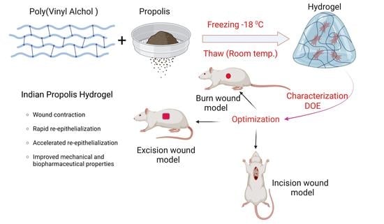
Formulation Development and Evaluation of Indian Propolis Hydrogel for Wound Healing
 , , ,
, , ,
Abstract
:
1. Introduction
2. Results and Discussion
2.1. Extraction and Standardization of Propolis
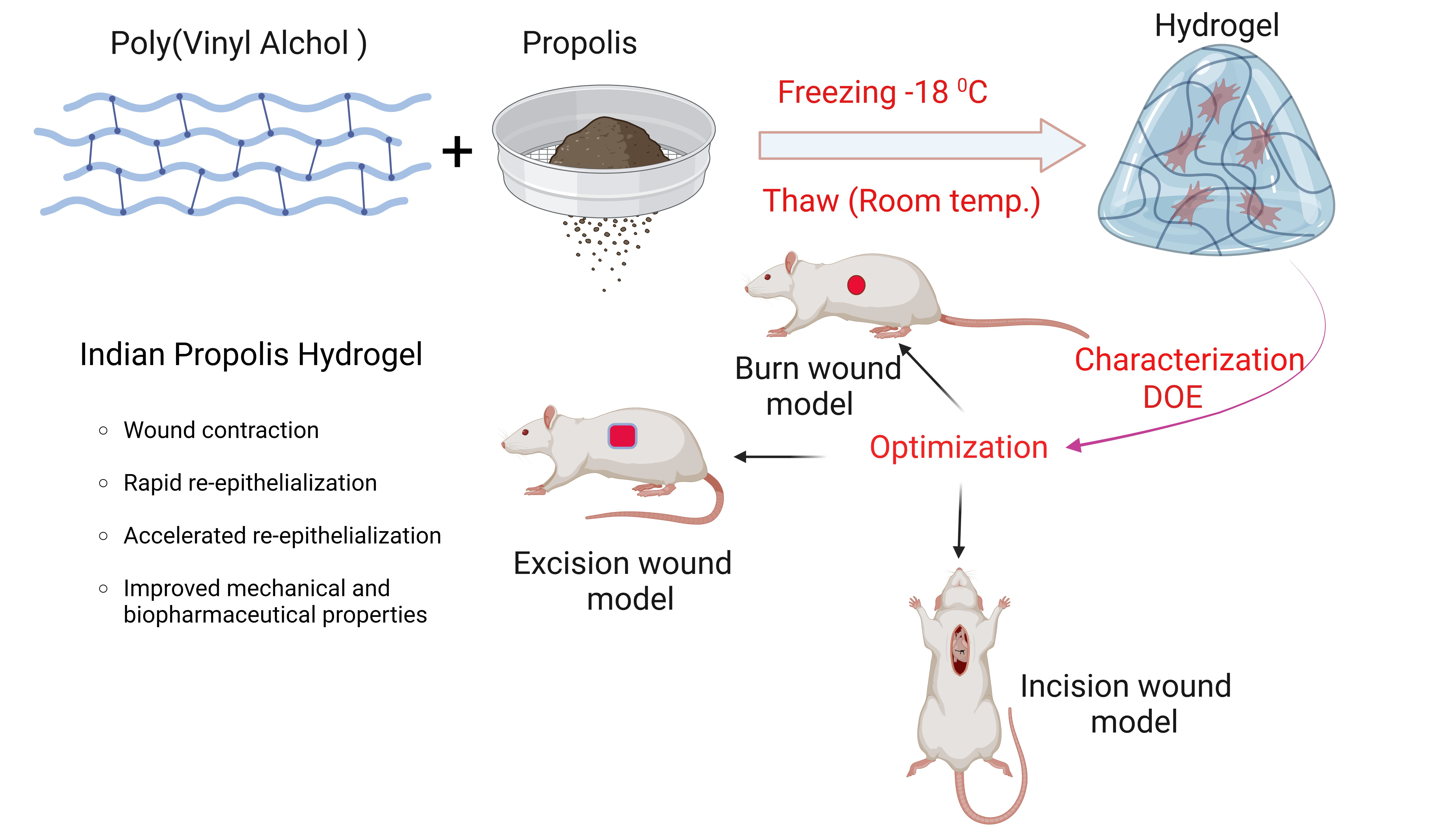
2.2. Formulation Optimization
Effect on Drug Release and Viscosity
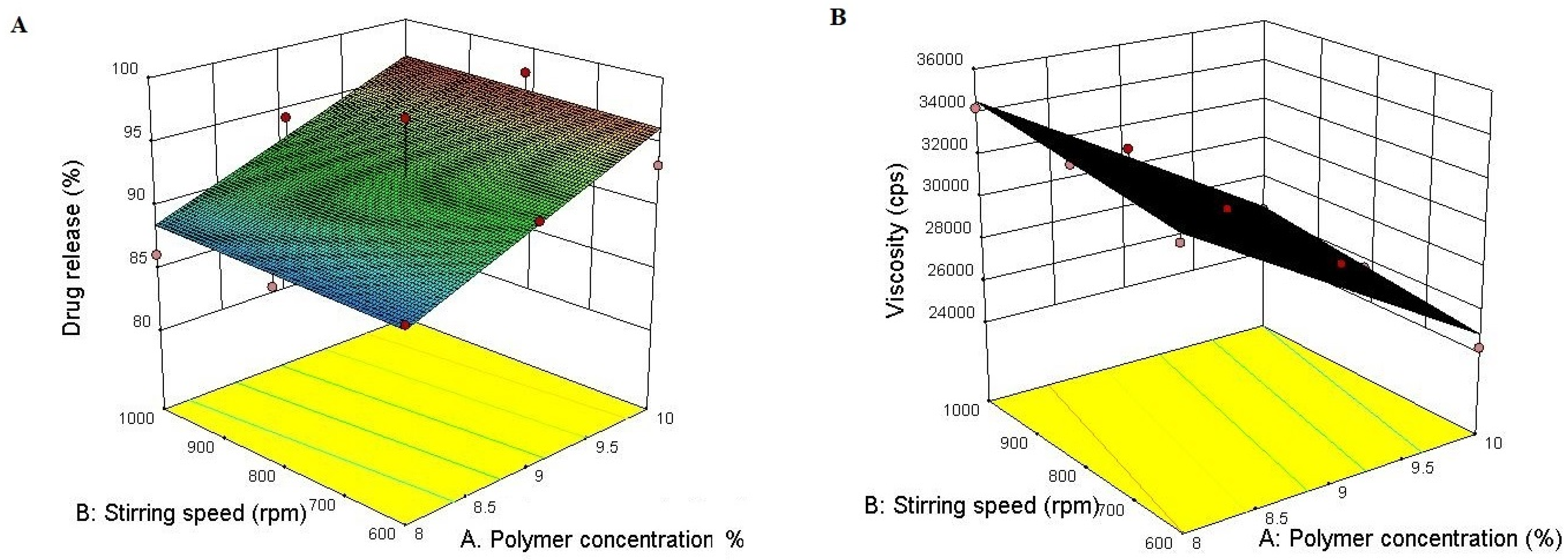
2.3. Visual Appearance
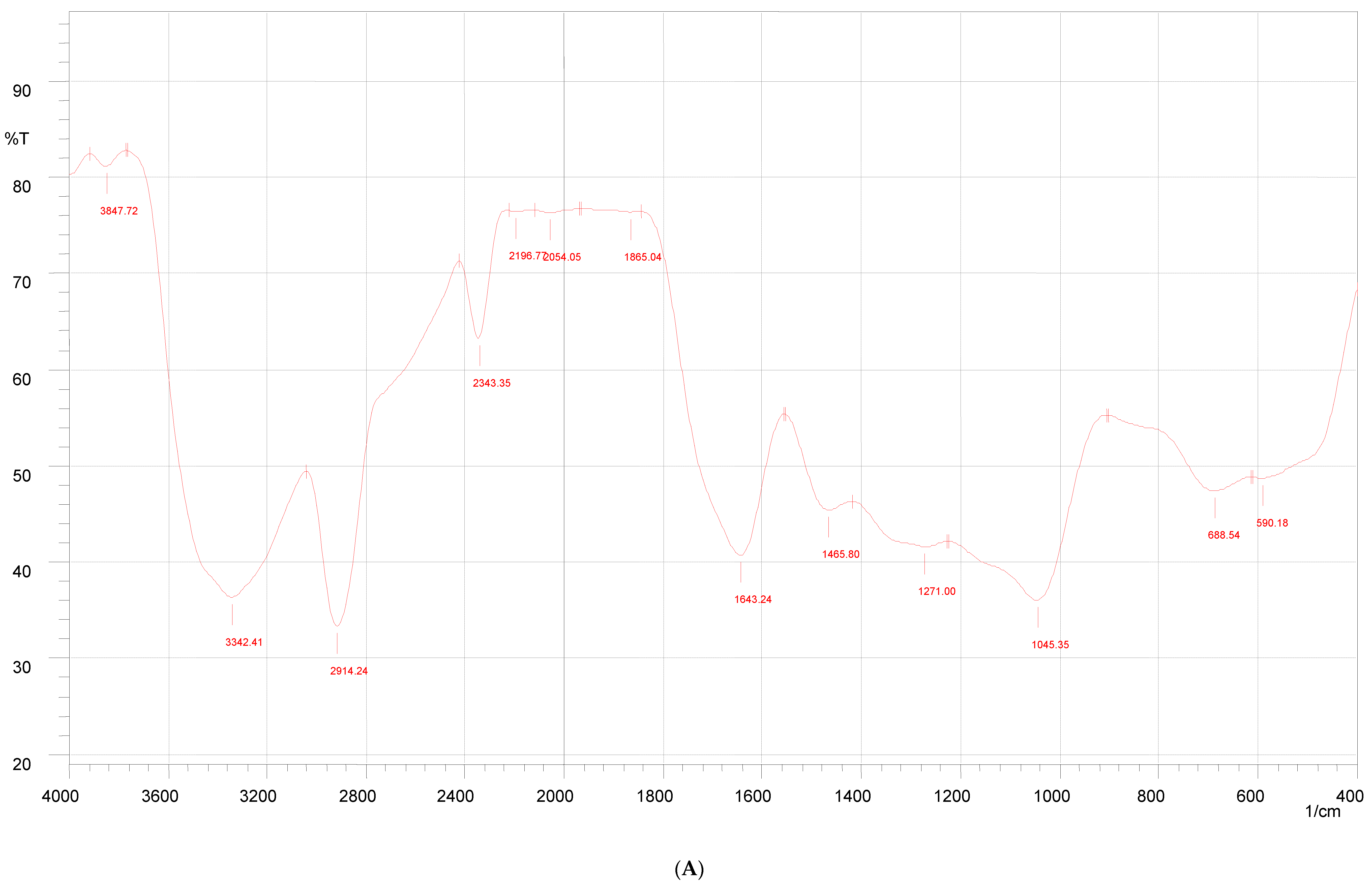
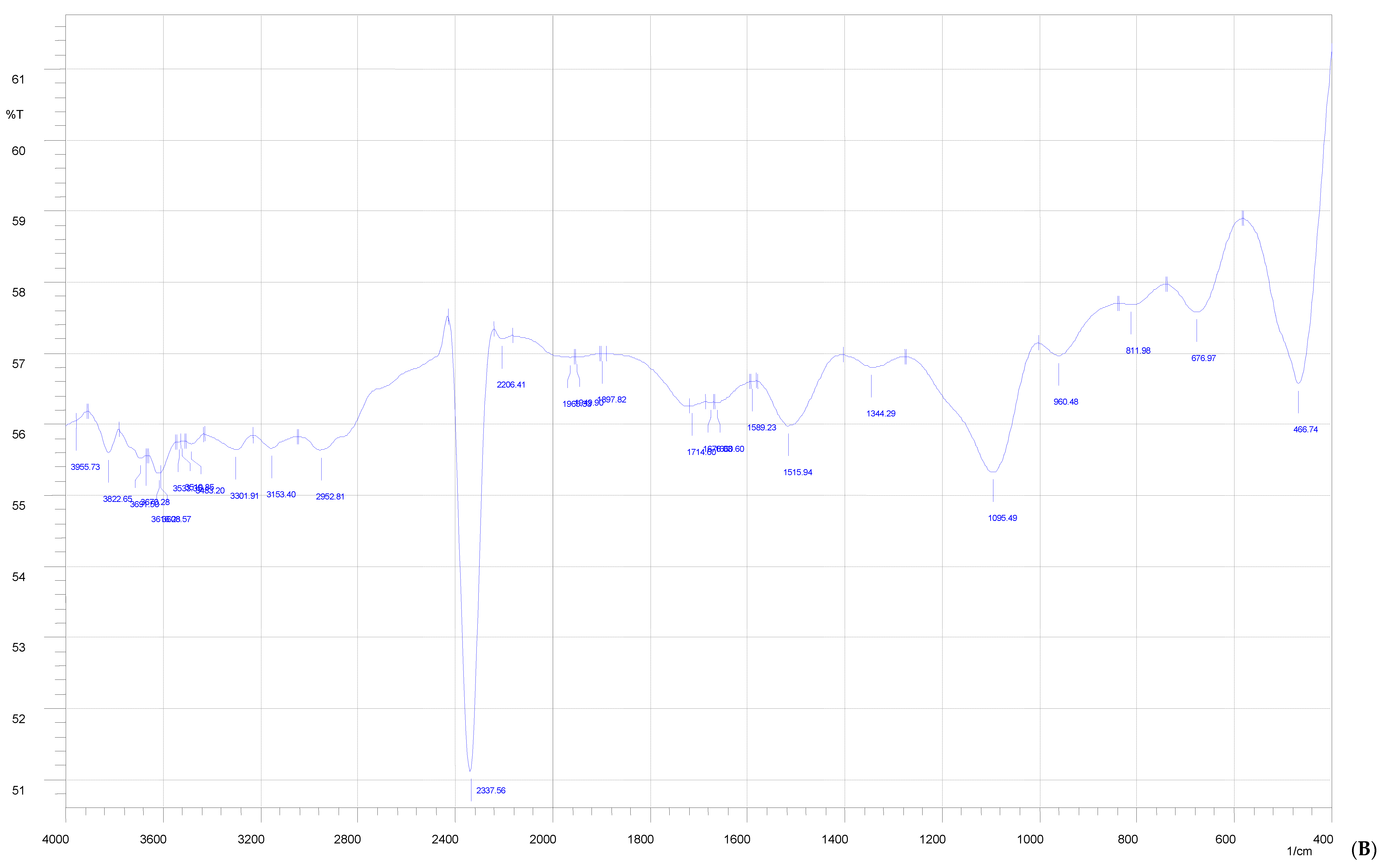
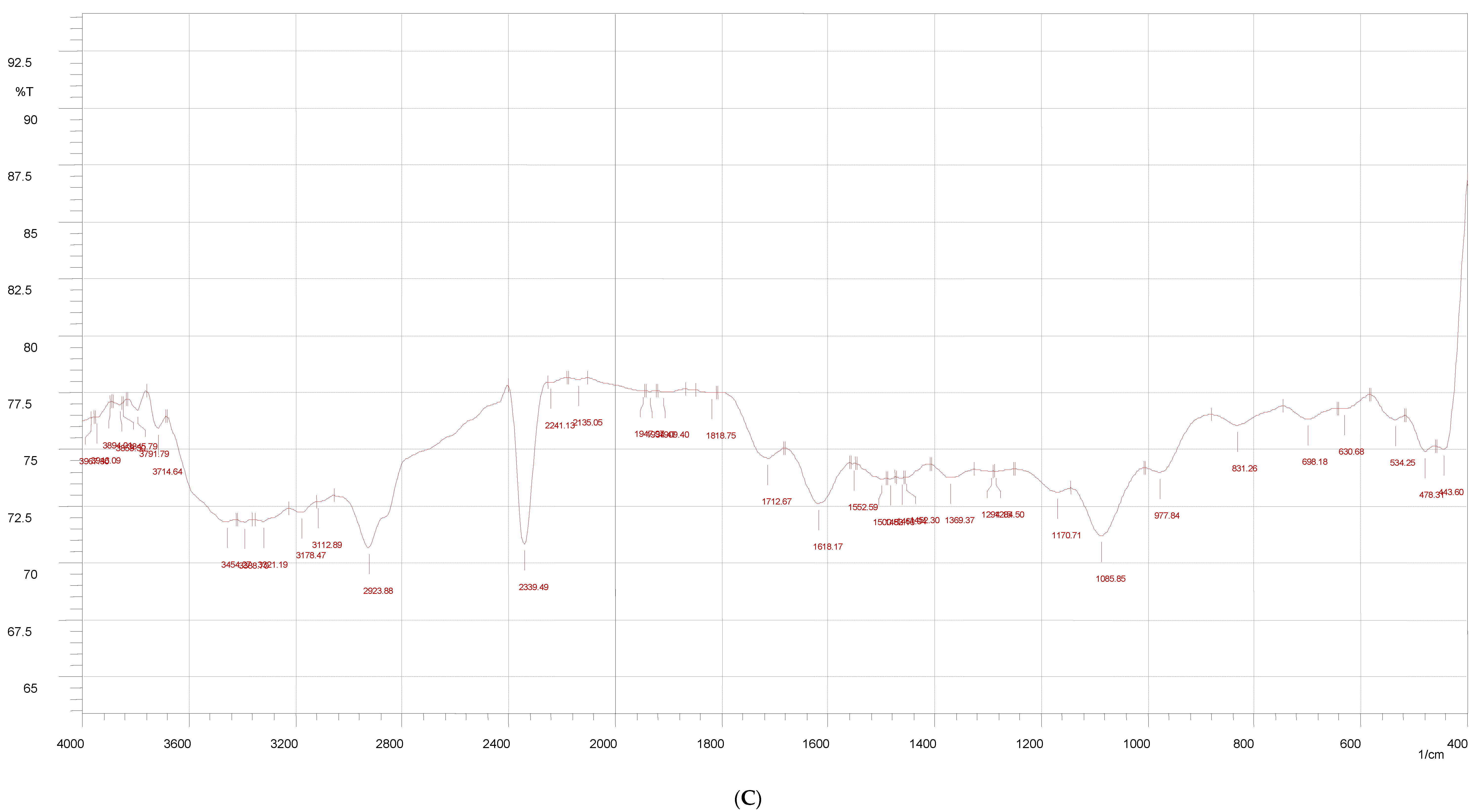
2.4. FT-IR Study
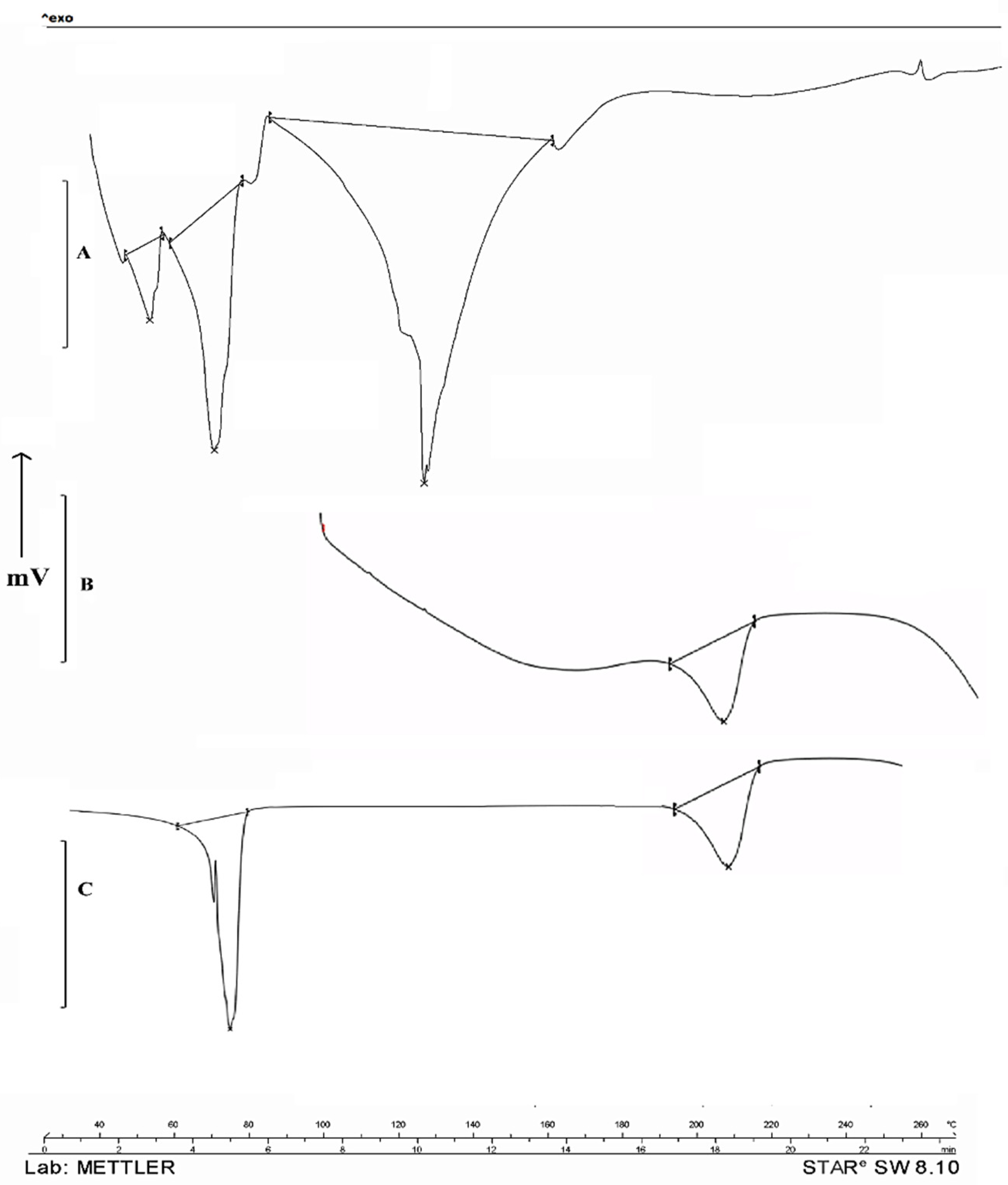
2.5. DSC
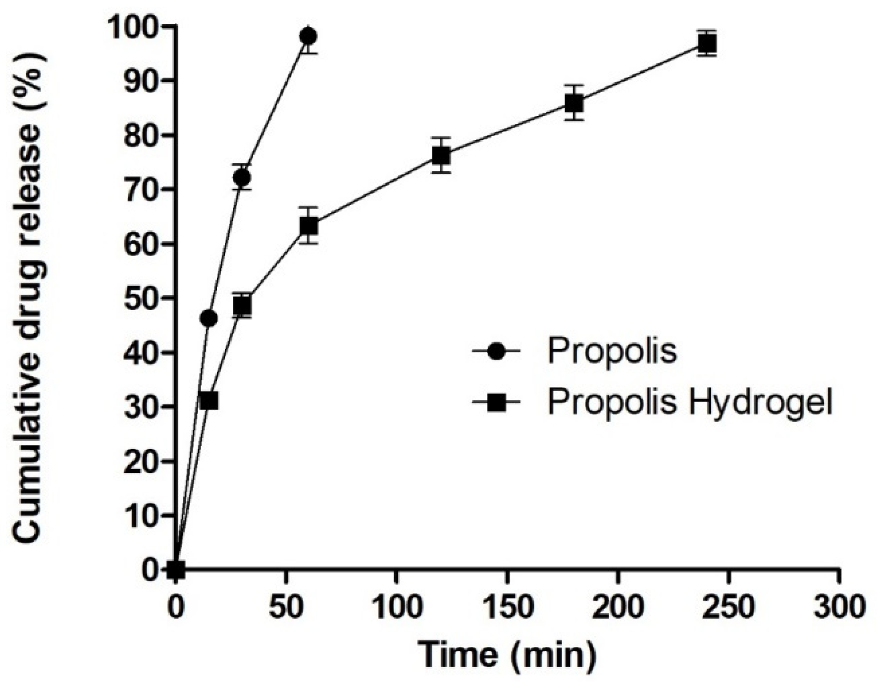
2.6. In Vitro Drug Release Study
2.7. Acute Dermal Toxicity Study
2.8. Wound Healing Studies of Hydrogel Formulation
2.8.1. Burn Wound Model
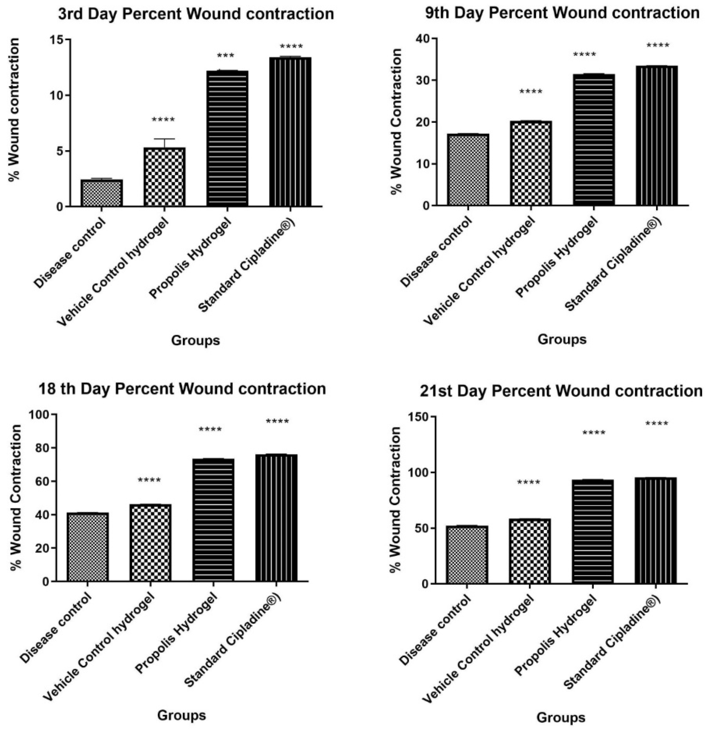
2.8.2. Excision Wound Healing
Measurement of Wound Contraction
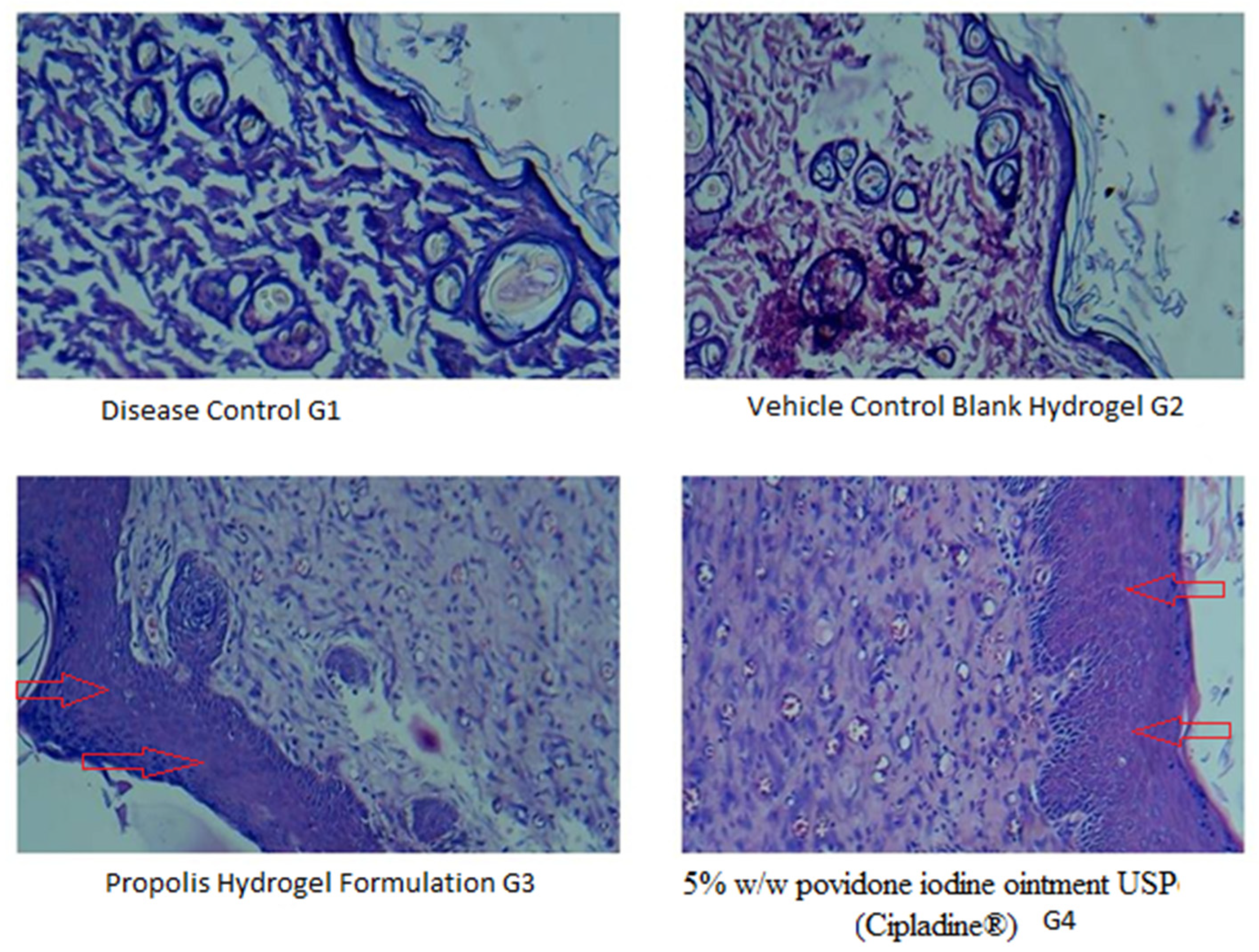
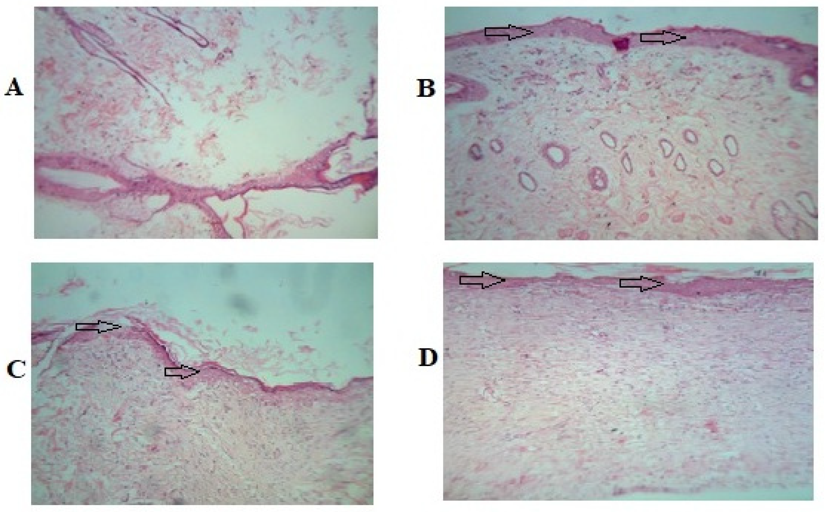
Histopathological Studies
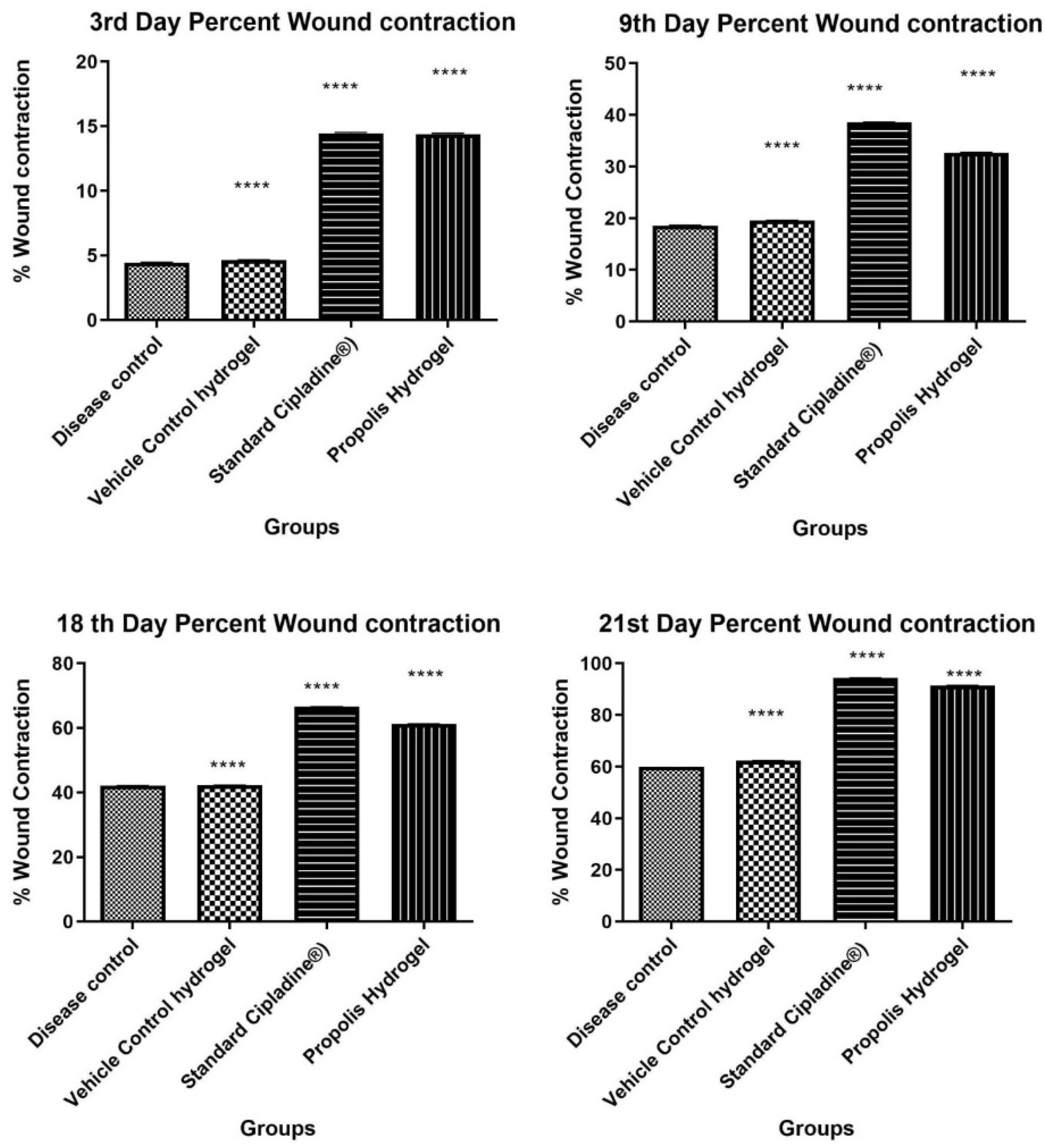
2.8.3. Incision Wound Healing
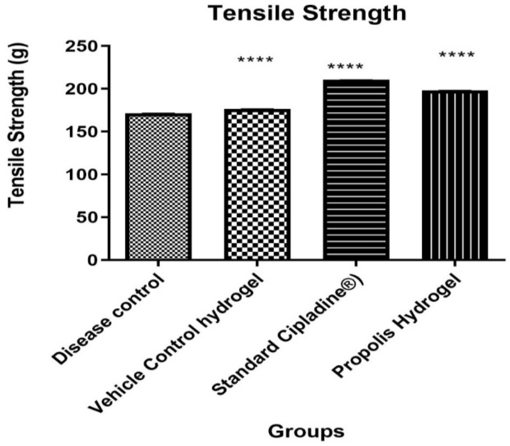
Measurement of Tensile Strength
2.9. Stability Studies
3. Conclusions
4. Material and Methods
4.1. Chemicals
4.2. Extraction and Standardization of Propolis Extract
4.3. FTIR Study
4.4. DSC
4.5. Formulation Development of Hydrogel and Optimization by DOE
4.6. Characterization of Hydrogel
4.6.1. Physical Appearance, pH, and Viscosity
4.6.2. In Vitro Drug Release Study
4.6.3. Acute Dermal Toxicity
4.6.4. Wound Healing Activity
Burn Wound Model
Excision and Incision Wound Model
4.6.5. Statistical Analysis
4.6.6. Stability Testing of Hydrogel Formulation
Author Contributions
Funding
Institutional Review Board Statement
Informed Consent Statement
Data Availability Statement
Acknowledgments
Conflicts of Interest
References
- Kapare, H.S.; Sathiyanarayanan, L. Nutritional and Therapeutic potential of Propolis: A Review. Res. J. Pharm. Tech. 2020, 13, 3545–3549. [Google Scholar] [CrossRef]
- Kapare, H.S.; Sathiyanarayanan, L.; Mahadik, K.R.; Arulmozhi, S. Indian Propolis Loaded Folic Acid Conjugated PLGA Nanoparticles: Formulation Development, Characterization, In Vitro and In Vivo Anticancer Study. J. Pharm. Drug Deliv. Res. 2017, 6, 1. [Google Scholar] [CrossRef]
- Wagh, V.D.; Borkar, R.D. Indian propolis: A potential natural antimicrobial and antifungal agent. Int. J. Pharm. Pharm. Sci. 2012, 4, 12–17. [Google Scholar]
- Krol, W.; Czuba, Z.; Scheller, S.; Gabrys, J.; Grabiec, S.; Shani, J. Anti-oxidant property of ethanolic extract of propolis (EEP) as evaluated by inhibiting the chemiluminescence oxidation of luminol. Biochem. Int. 1990, 21, 593–597. [Google Scholar]
- Li, H.; Kapur, A.; Yang, J.X.; Srivastava, S.; McLeod, D.G.; Paredes-Guzman, J.F. Antiproliferation of human prostate cancer cells by ethanolic extracts of Brazilian propolis and its biological origin. Int. J. Oncol. 2007, 31, 601–606. [Google Scholar] [PubMed]
- Kumar, M.R.; Bose, V.S.C.; Sathyabama, S.; Priyadarshini, V.B. Antimicrobial and DPPH free radical-scavenging activities of the ethanol extract of propolis collected from India. J. Ecobiotechnol. 2011, 3, 8–13. [Google Scholar]
- Cai, R.; Gimenez-Camino, N.; Xiao, M.; Bi, S.; Kyle, A. Technological advances in three-dimensional skin tissue engineering. Rev. Adv. Mater. Sci. 2023, 62, 20220289. [Google Scholar] [CrossRef]
- Kapare, H.S.; Metkar, S.R.; Wakalkar, S.V. Natural products in wound healing: Nano-technology based approaches. Indian Drugs 2020, 57, 7–14. [Google Scholar] [CrossRef]
- Yazarlu, O.; Iranshahi, M.; Kashani, H.R.K.; Reshadat, S.; Habtemariam, S.; Iranshahy, M.; Hasanpour, M. Perspective on the application of medicinal plants and natural products in wound healing: A mechanistic review. Pharm. Res. 2021, 174, 105841. [Google Scholar] [CrossRef]
- Sen, C.K. Human Wounds and Its Burden: An Updated Compendium of Estimates. Adv. Wound Care 2019, 8, 39–48. [Google Scholar] [CrossRef]
- Stanicka, K.; Dobrucka, R.; Woźniak, M.; Sip, A.; Majka, J.; Kozak, W.; Ratajczak, I. The Effect of Chitosan Type on Biological and Physicochemical Properties of Films with Propolis Extract. Polymers 2021, 13, 3888. [Google Scholar] [CrossRef]
- da Rosa, C.; Bueno, I.L.; Quaresma, A.C.M.; Longato, G.B. Healing Potential of Propolis in Skin Wounds Evidenced by Clinical Studies. Pharmaceuticals 2022, 15, 1143. [Google Scholar] [CrossRef]
- Oryan, A.; Alemzadeh, E.; Moshiri, A. Potential role of propolis in wound healing: Biological properties and therapeutic activities. Biomed. Pharm. 2018, 98, 469–483. [Google Scholar] [CrossRef]
- Stojko, M.; Wolny, D.; Włodarczyk, J. Nonwoven Releasing Propolis as a Potential New Wound Healing Method-A Review. Molecules 2021, 26, 5701. [Google Scholar] [CrossRef]
- Yang, J.; Pi, A.; Yan, L.; Li, J.; Nan, S.; Zhang, J.; Hao, Y. Research Progress on Therapeutic Effect and Mechanism of Propolis on Wound Healing. Evid. Based Complement Altern. Med. 2022, 2022, 5798941. [Google Scholar] [CrossRef] [PubMed]
- Martinotti, S.; Ranzato, E. Propolis: A new frontier for wound healing? Burns Trauma 2015, 3, 9. [Google Scholar] [CrossRef] [PubMed]
- Wojciech, S.; Aleksandra, G.; Aleksandra, K.; Filip, C.; Sławomir, B.; Hanna Maria, B.; Katarzyna, W.; Ewa, K.; Maciej, J. Design of vitamin-loaded emulsions in agar hydrogel matrix dispersed with plant surfactants. Food Biosci. 2023, 53, 102559. [Google Scholar]
- Pardeshi, S.; Damiri, F.; Zehravi, M.; Joshi, R.; Kapare, H.; Prajapati, M.K.; Munot, N.; Berrada, M.; Giram, P.S.; Rojekar, S.; et al. Functional Thermoresponsive Hydrogel Molecule to Material Design for Biomedical Applications. Polymers 2022, 14, 3126. [Google Scholar] [CrossRef] [PubMed]
- Peppas, N.A.; Merrill, E.W. Differential scanning calorimetry of crystallized PVA hydrogels. J. Appl. Polym. Sci. 1976, 20, 1457–1465. [Google Scholar] [CrossRef]
- Hickey, A.S.; Peppas, N.A. Mesh size and diffusive characteristics of semicrystalline poly (vinyl alcohol) membranes prepared by freezing/thawing techniques. J. Membr. Sci. 1995, 107, 229–237. [Google Scholar] [CrossRef]
- Khan, A.W.; Kotta, S.; Ansari, S.H.; Sharma, R.K.; Kumar, A.; Ali, J. Formulation development, optimization and evaluation of aloe vera gel for wound healing. Pharm. Mag. 2013, 9, S6–S10. [Google Scholar]
- Wang, M.; Bai, J.; Shao, K.; Tang, W.; Zhao, X.; Lin, D.; Huang, S.; Chen, C.; Ding, Z.; Ye, J. Poly(vinyl alcohol) Hydrogels: The Old and New Functional Materials. Int. J. Polym. Sci. 2021, 16, 2225426. [Google Scholar] [CrossRef]
- Afkhamizadeh, M.; Aboutorabi, R.; Ravari, H.; Fathi Najafi, M.; Ataei Azimi, S.; Javadian Langaroodi, A.; Yaghoubi, M.A.; Sahebkar, A. Topical propolis improves wound healing in patients with diabetic foot ulcer: A randomized controlled trial. Nat. Prod. Res. 2018, 32, 2096–2099. [Google Scholar] [CrossRef] [PubMed]
- Kapare, H.S.; Lohidasan, S.; Arulmozhi, S.; Mahadik, K.R. Standardization, chemical profiling, in vitro cytotoxic effects, in vivo anti-carcinogenic potential and biosafety profile of Indian propolis. J. Ayurveda Integr. Med. 2019, 10, 81–87. [Google Scholar] [CrossRef] [PubMed]
- Kapare, H.S.; Sathiyanarayanan, L.; Arulmozhi, S.; Mahadik, K.R. Caffeic Acid Phenethyl Ester Loaded Poly (ε –caprolactone) Nanoparticles for Improved Anticancer Efficacy: Formulation Development, Characterization and in Vitro Cytotoxicity Study. Nanomed. Res. J. 2020, 5, 324–331. [Google Scholar] [CrossRef]
- Kapare, H.S.; Lohidasan, S.; Sinnathambi, A.; Mahadik, K. Formulation Development of Folic Acid Conjugated PLGA Nanoparticles for Improved Cytotoxicity of Caffeic Acid Phenethyl Ester. Pharm. Nanotechnol. 2021, 9, 111–119. [Google Scholar] [CrossRef]
- Oliveira, R.N.; McGuinness, G.B.; Rouze, R.; Quilty, B.; Cahill, P.; Soares, G.D.; Thiré, R.M. PVA hydrogels loaded with a Brazilian propolis for burn wound healing applications. J. Appl. Polym. Sci. 2015, 132, 42129. [Google Scholar] [CrossRef]
- Peppas, N.A.; Merrill, E.W. PVA hydrogels: Reinforcement of radiation-crosslinked networks by crystallization. J. Polym. Sci. Polym. Chem. Ed. 1976, 14, 441–457. [Google Scholar] [CrossRef]
- Wang, F.; Gao, Y.; Li, H.; Zhou, L.; Shi, H.; Feng, S.; Chen, J.; Mei, Z. Effect of natural-based biological hydrogels combined with growth factors on skin wound healing. Nanotechnol. Rev. 2022, 11, 2493–2512. [Google Scholar] [CrossRef]
- Stauffer, S.R.; Peppas, N.A. Poly (vinyl alcohol) hydrogels prepared by freezing thawing cyclic processing. Polymer 1992, 33, 3932–3936. [Google Scholar] [CrossRef]
- Hua, S. Comparison of in vitro dialysis release methods of loperamide-encapsulated liposomal gel for topical drug delivery. Int. J. Nanomed. 2014, 9, 735–744. [Google Scholar] [CrossRef] [PubMed]
- Organisation for Economic Co-operation and Development (OECD). Test No. 402: Acute Dermal Toxicity, OECD Guidelines for the testing of Chemicals, Section 4; OECD: Paris, France, 2017. [Google Scholar] [CrossRef]
- Nagar, H.K.; Srivastava, A.K.; Srivastava, R.; Kurmi, M.L.; Chandel, H.S.; Ranawat, M.S. Pharmacological investigation of the wound healing activity of Cestrum nocturnum (L.) ointment in Wistar albino rats. J. Pharm. 2016, 2016, 9249040. [Google Scholar] [CrossRef] [PubMed]
- Hemalatha, S.; Subramanian, N.; Ravich, V.; Chinnaswamy, K. Wound healing activity of Indigoferaenneaphylla Linn. Indian J. Pharm. Sci. 2001, 63, 331. [Google Scholar]
- Mukherjee, P.K.; Verpoorte, R.; Suresh, B. Evaluation of in-vivo wound healing activity of Hypericumpatulum (Family: Hypericaceae) leaf extract on different wound model in rats. J. Ethnopharmacol. 2000, 70, 315–321. [Google Scholar] [CrossRef]
- Qindeel, M.; Ahmed, N.; Sabir, F.; Khan, S.; Ur-Rehman, A. Development of novel pH-sensitive nanoparticles loaded hydrogel for transdermal drug delivery. Drug Dev. Ind. Pharm. 2019, 45, 629–641. [Google Scholar] [CrossRef]


| Batch Codes | Polymer Concentration X1 | Stirring Speed X2 | Polymer Concentration (%) X1 | Stirring Speed (rpm) X2 |
|---|---|---|---|---|
| Code | Code | Actual | Actual | |
| F1 | +1 | +1 | 10 | 800 |
| F2 | +1 | 0 | 10 | 700 |
| F3 | +1 | −1 | 10 | 600 |
| F4 | 0 | +1 | 09 | 800 |
| F5 | 0 | 0 | 09 | 700 |
| F6 | 0 | −1 | 09 | 600 |
| F7 | −1 | +1 | 08 | 800 |
| F8 | −1 | 0 | 08 | 700 |
| F9 | −1 | −1 | 08 | 600 |
| Batch No | Polymer Concentration X1 | Stirring Speed X2 | Response 1 | Response 2 |
|---|---|---|---|---|
| % Cumulative Drug Release (4 h) | Viscosity cps | |||
| Unit | % | rpm | % | cps |
| F1 | +1 | +1 | 88.16 | 32,500 |
| F2 | +1 | 0 | 87.89 | 33,600 |
| F3 | +1 | −1 | 86.16 | 34,200 |
| F4 | 0 | +1 | 92.11 | 29,900 |
| F5 | 0 | 0 | 96.89 | 30,100 |
| F6 | 0 | −1 | 94.26 | 30,900 |
| F7 | −1 | +1 | 93.25 | 24,200 |
| F8 | −1 | 0 | 97.90 | 25,600 |
| F9 | −1 | −1 | 95.36 | 26,300 |
| Sr.N | Colour | Homogeneity | Consistency | Grittiness | Phase Separation |
|---|---|---|---|---|---|
| F2 | Brownish | Homogenous | Consistent | Non-greasy | No phase separation |
| Group Number | Name of Group | Day 3 | Day 9 | Day 18 | Day 21 |
|---|---|---|---|---|---|
| 1. | Disease control | 2.45 ± 0.11 | 17.21 ± 0.08 | 41.26 ± 0.11 | 52.37 ± 0.14 |
| 2. | Vehicle control hydrogel | 3.32 ± 0.15 | 20.29 ± 0.10 | 46.32 ± 0.12 | 58.38 ± 0.19 |
| 3. | Propolis hydrogel | 12.39 ± 0.16 a ****, b ****, c **** | 31.51 ± 0.12 a ****, b ****, c **** | 73.50 ± 0.27 a ****, b ****, c **** | 93.58 ± 0.15 a ****, b ****, c **** |
| 4. | 5% w/w povidone iodine ointment USP (Cipladine®) | 13.44 ± 0.13 | 33.46 ± 0.06 | 76.33 ± 0.26 | 95.39 ± 0.16 |
| Groups | % Wound Contraction | |||
|---|---|---|---|---|
| 3rd Day | 9th Day | 18th Day | 21st Day | |
| Group 1: Disease control | 4.41 ± 0.13 | 18.61 ± 0.94 | 42.09 ± 0.10 | 59.95 ± 0.07 |
| Group 2: Vehicle control | 4.64 ± 0.07 | 19.57 ± 0.07 | 42.29 ± 0.67 | 62.30 ± 0.40 |
| Group 3: 5% w/w povidone iodine ointment USP (Cipladine®) | 14.45 ± 0.16 | 38.63 ± 0.07 | 66.57 ± 0.12 | 94.38 ± 0.21 |
| Group 4: Propolis hydrogel | 14.39 ± 0.13 a ****, b ****, c **** | 32.69 ± 0.13 a ****, b ****, c **** | 61.26 ± 0.03 a ****, b ****, c **** | 91.45 ± 0.29 a ****, b ****, c **** |
| Groups | Tensile Strength in g |
|---|---|
| Group1: Disease control | 171.40 ± 0.17 |
| Group 2: Vehicle control | 176.49 ± 0.21 |
| Group 3: 5% w/w povidone iodine ointment USP (Cipladine®) | 210.46 ± 0.19 |
| Group 4: Propolis hydrogel | 198.18 ± 0.22 a ****, b ****, c **** |
| Propolis Hydrogel Formulation | |||||
|---|---|---|---|---|---|
| Evaluation Parameters | 0 Days | 30 Days | 60 Days | 90 Days | 180 Days |
| Physical Appearance | Dark brownish, homogeneous, non-greasy | ||||
| Phase separation | Nil | Nil | Nil | Nil | Nil |
| pH | 5.46 ± 0.246 | 5.44 ± 0.142 | 5.44 ± 0.554 | 5.41 ± 0.311 | 5.41 ± 0.110 |
| Drug content | 98.96% | 98.60% | 98.52% | 98.51% | 98.12% |
| Viscosity | 31,500 cps | 31,450 cps | 31,400 cps | 31,440 cps | 31,400 cps |
Disclaimer/Publisher’s Note: The statements, opinions and data contained in all publications are solely those of the individual author(s) and contributor(s) and not of MDPI and/or the editor(s). MDPI and/or the editor(s) disclaim responsibility for any injury to people or property resulting from any ideas, methods, instructions or products referred to in the content. |
© 2023 by the authors. Licensee MDPI, Basel, Switzerland. This article is an open access article distributed under the terms and conditions of the Creative Commons Attribution (CC BY) license (https://creativecommons.org/licenses/by/4.0/).
Share and Cite
Kapare, H.S.; Giram, P.S.; Raut, S.S.; Gaikwad, H.K.; Paiva-Santos, A.C. Formulation Development and Evaluation of Indian Propolis Hydrogel for Wound Healing. Gels 2023, 9, 375. https://doi.org/10.3390/gels9050375
Kapare HS, Giram PS, Raut SS, Gaikwad HK, Paiva-Santos AC. Formulation Development and Evaluation of Indian Propolis Hydrogel for Wound Healing. Gels. 2023; 9(5):375. https://doi.org/10.3390/gels9050375
Chicago/Turabian StyleKapare, Harshad S., Prabhanjan S. Giram, Sadhana S. Raut, Hemant K. Gaikwad, and Ana Cláudia Paiva-Santos. 2023. "Formulation Development and Evaluation of Indian Propolis Hydrogel for Wound Healing" Gels 9, no. 5: 375. https://doi.org/10.3390/gels9050375