
Qbd-Based Approach to Optimize Niosomal Gel of Levosulpiride for Transdermal Drug Delivery
 , , , , , , , and
Abstract
:1. Introduction
2. Results and Discussion
2.1. Results of Preliminary Trials for Selection of Excipients
2.2. Optimization of Variables Using Box-Behnken Design
2.3. Data Analysis for Dependent Variables of Design Batches
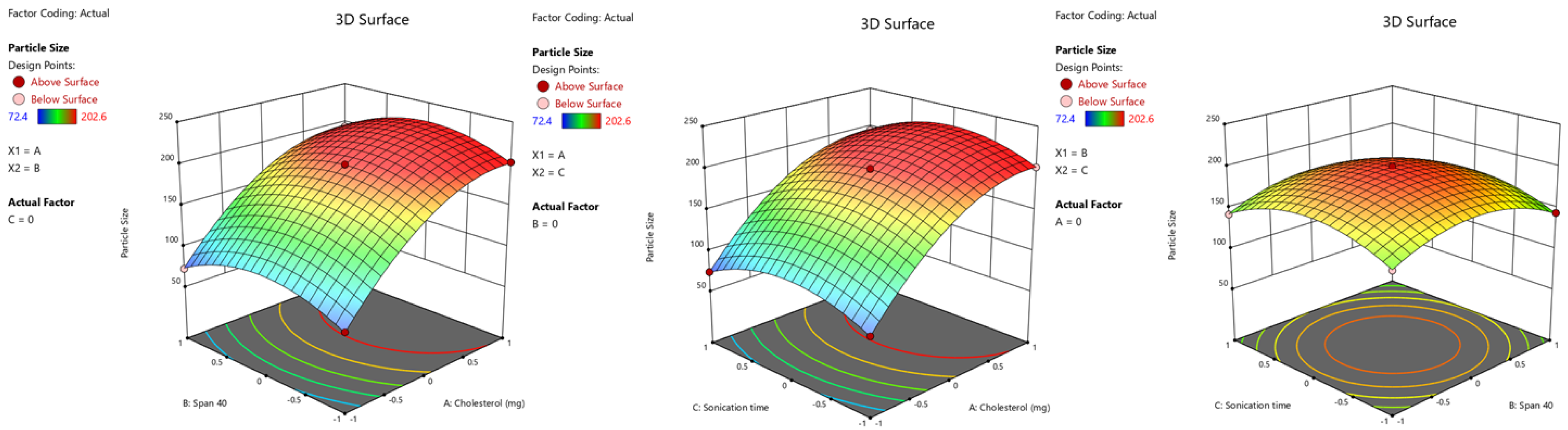
2.3.1. Particle Size
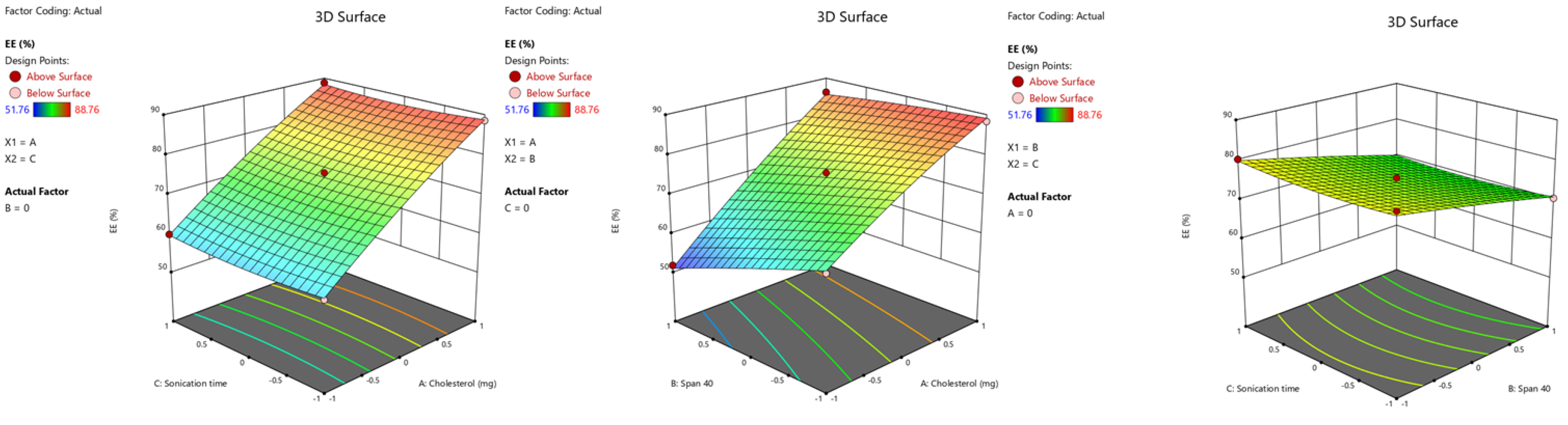
2.3.2. %. EE
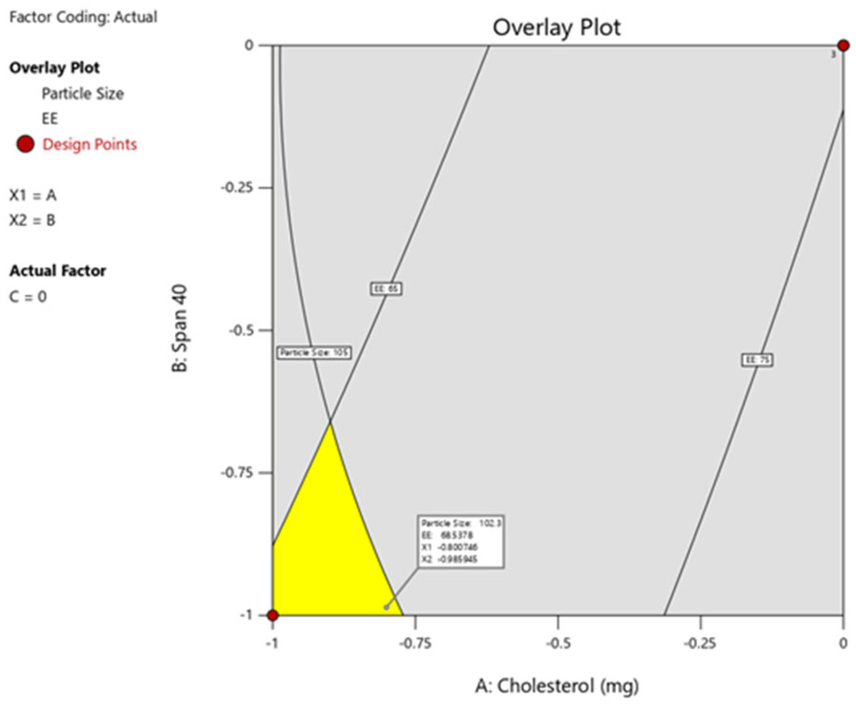
2.4. Optimization and Validation of Response Surface Analysis
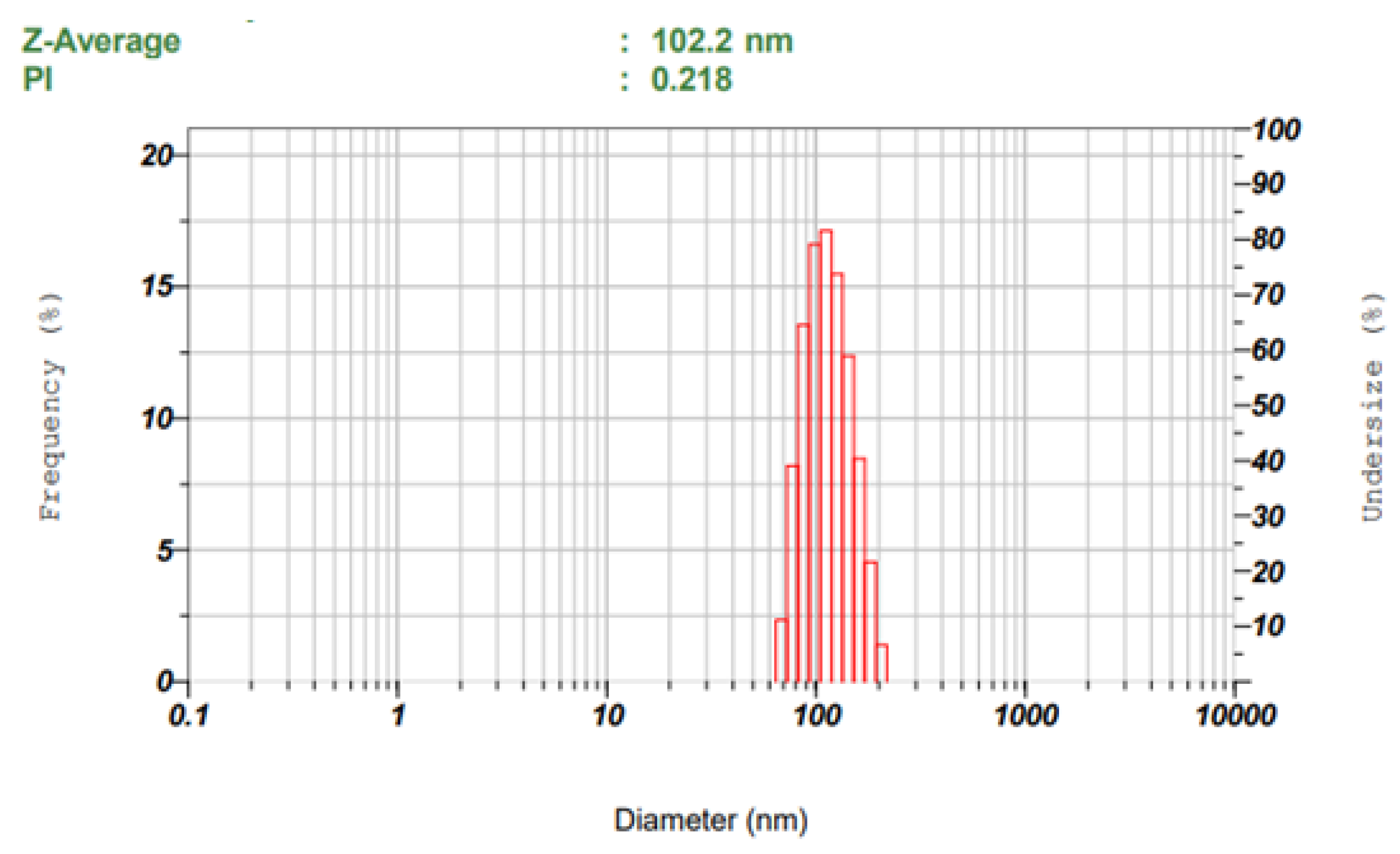
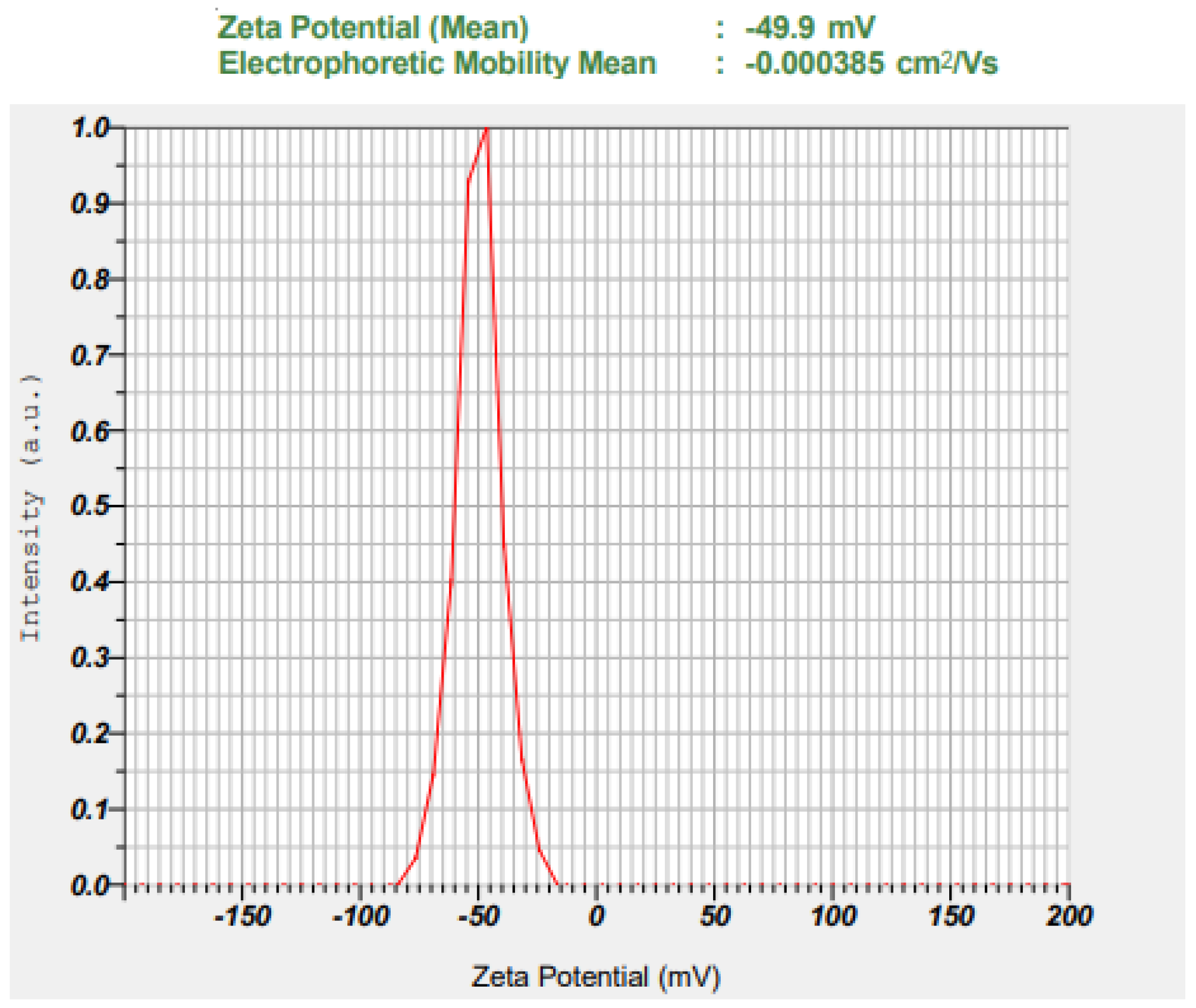
Particle Size and Zeta Potential of Optimized Batch
2.5. Check Point Analysis
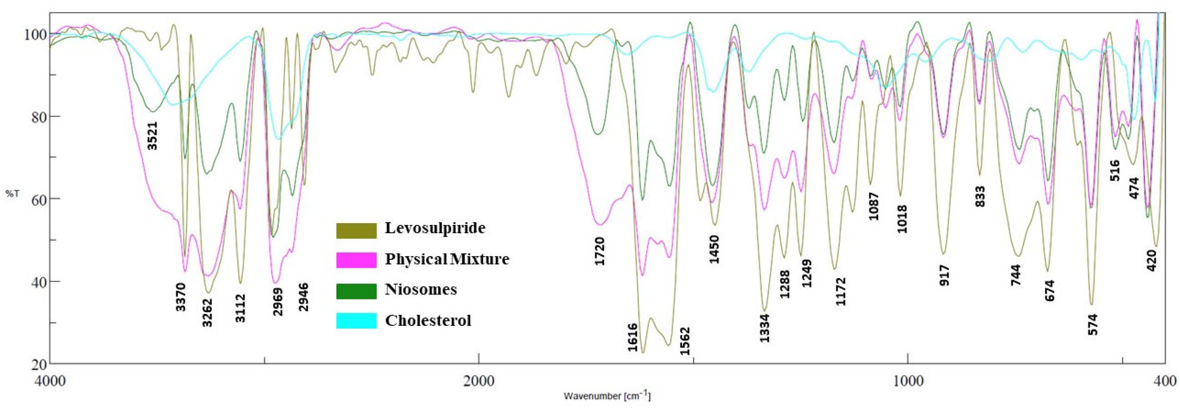
2.6. FTIR
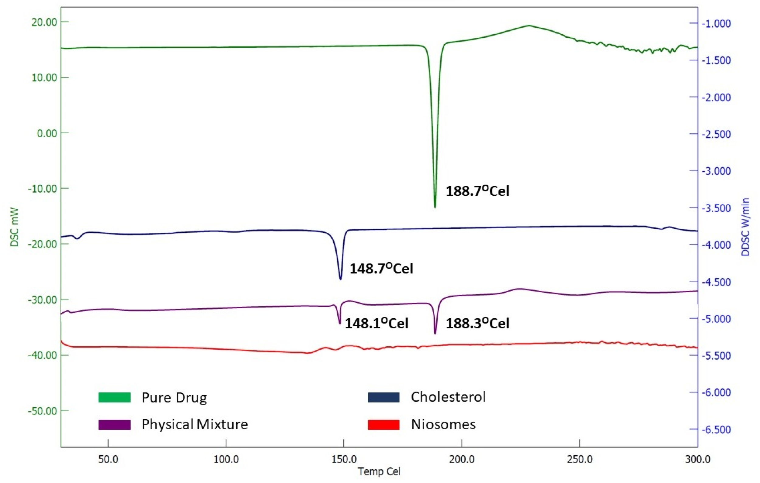
2.7. DSC
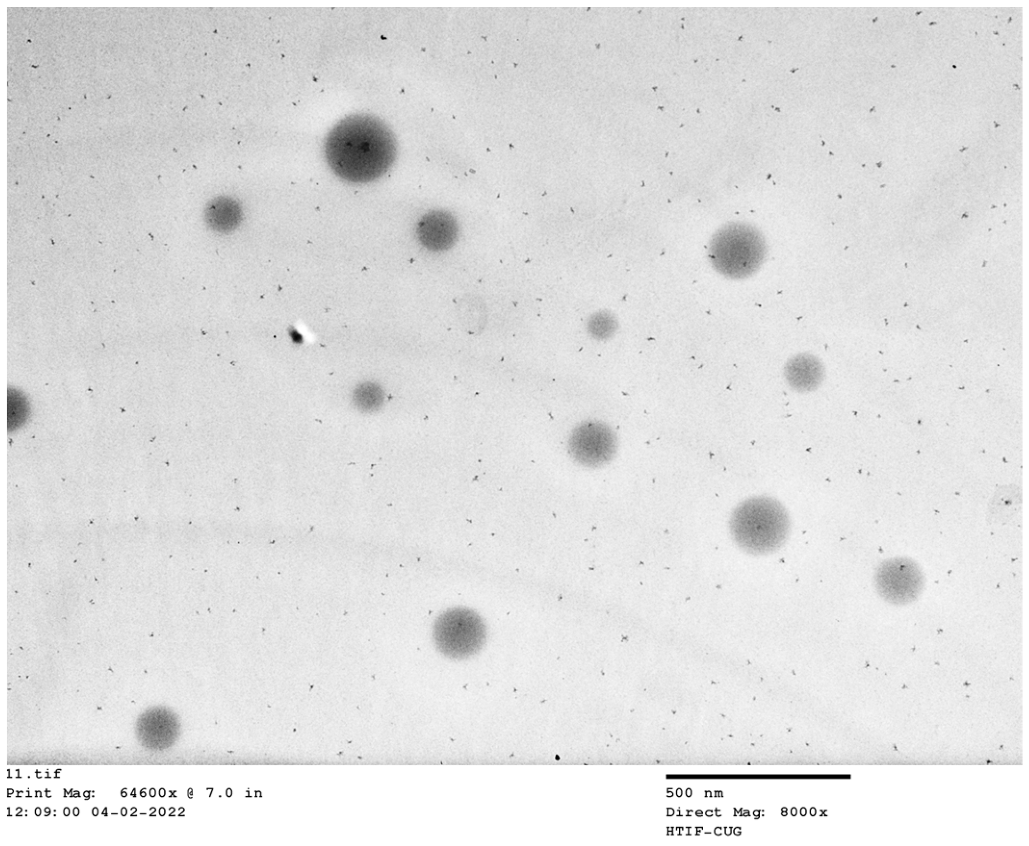
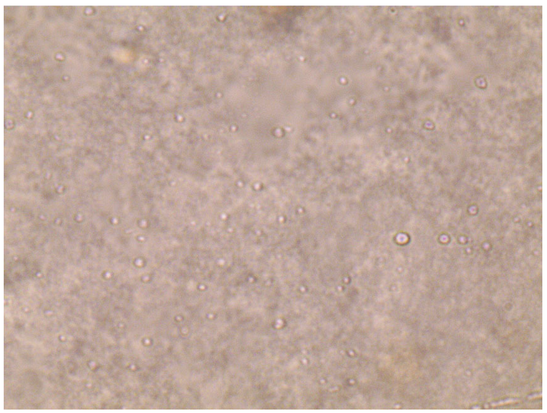
2.8. TEM
2.9. Evaluation of Levosulpiride-Niosomal Gel
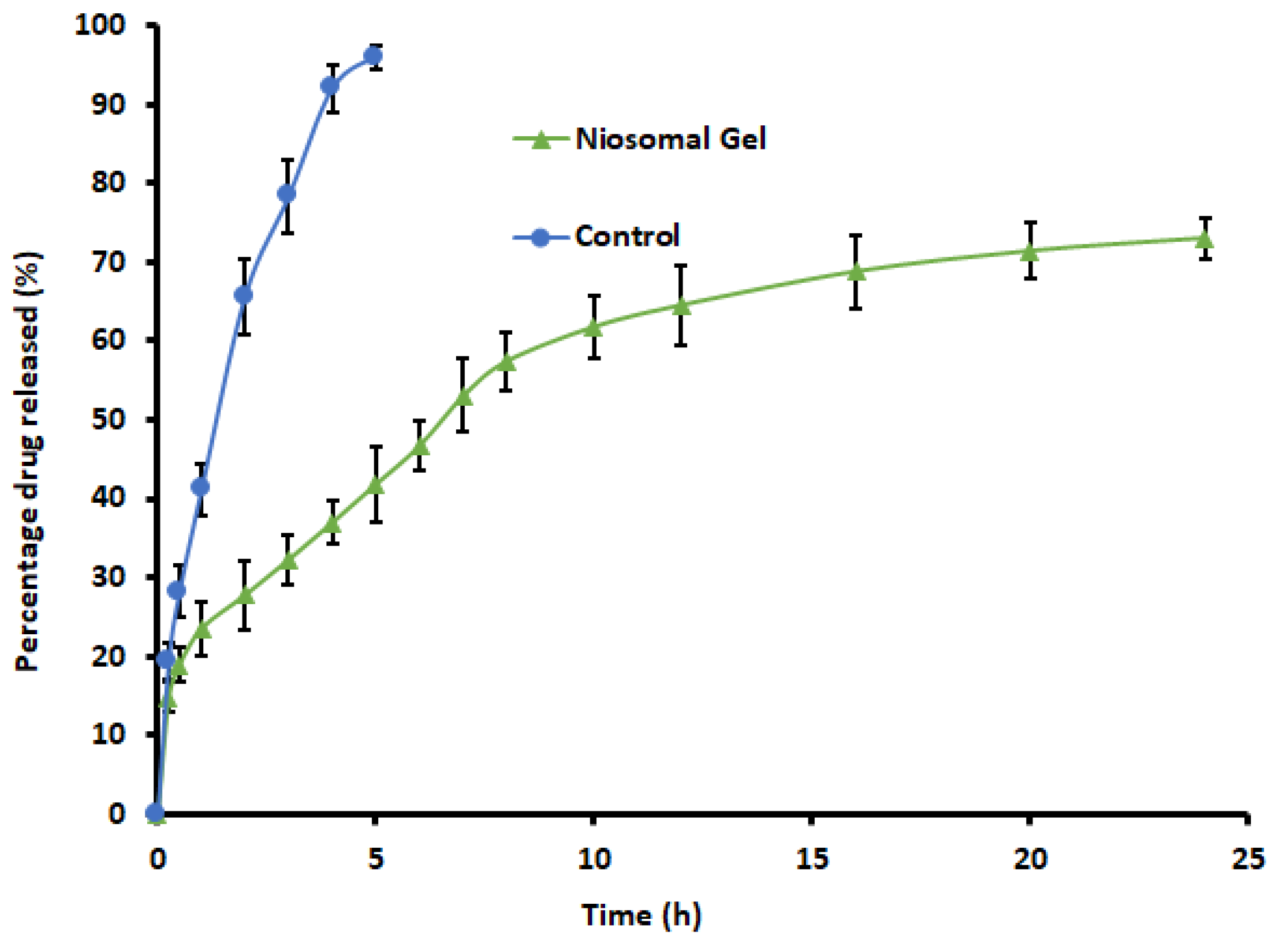
2.10. In Vitro Release Study
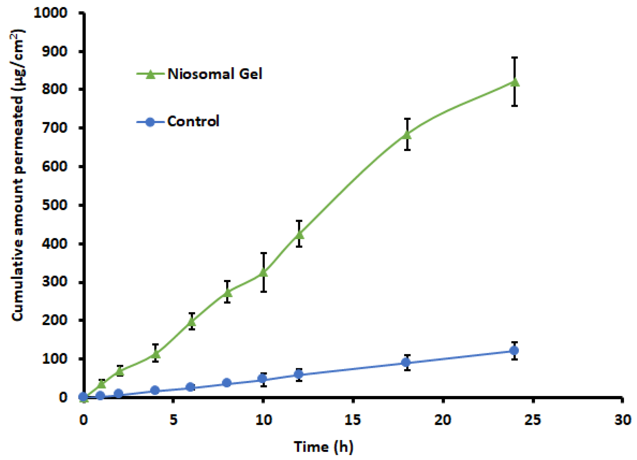
2.11. Ex Vivo Permeation Study
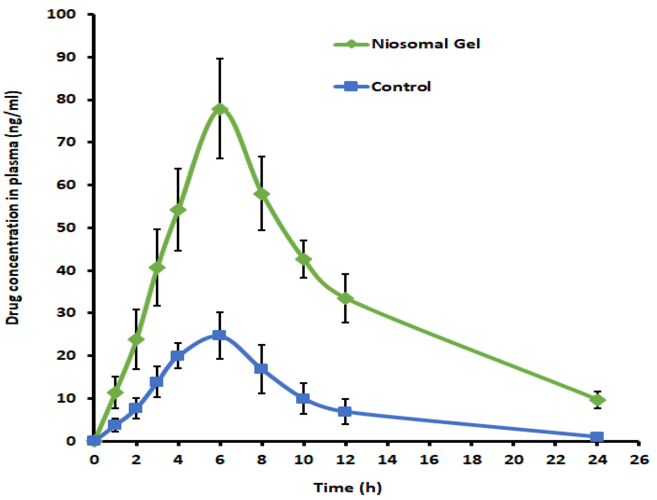
2.12. In Vivo Pharmacokinetic Studies
2.13. Stability Studies
3. Conclusions
4. Materials and Methods
4.1. Materials
4.2. Preliminary Trials for Selection of Excipients
4.3. Formulation of Levosulpiride-Loaded Niosomes
4.4. Estimation of Levosulpiride
4.5. Optimization Study of Variables
4.6. Characterization of Levosulpiride-Loaded Niosomes
4.6.1. Particle Size and Zeta Potential
4.6.2. Entrapment Efficiency
4.7. Formulation of Check Point Batch
4.8. Fourier Transform Infrared (FTIR)
4.9. Differential Scanning Calorimetry (DSC)
4.10. Transmission Electron Microscopy (TEM)
4.11. Preparations of Levosulpiride Niosomal Gel
4.12. Evaluation of Levosulpiride-Niosomal Gel
4.12.1. Viscosity
4.12.2. pH
4.12.3. Drug Content
4.12.4. Spreadability
4.12.5. Morphology
4.12.6. Mechanical Properties
4.13. In Vitro Release Study
4.14. Ex Vivo Permeation Study
4.15. In Vivo Pharmacokinetic Studies
4.16. Stability Studies
4.17. Statistical Analysis
Supplementary Materials
Author Contributions
Funding
Institutional Review Board Statement
Informed Consent Statement
Data Availability Statement
Acknowledgments
Conflicts of Interest
References
- Ng, K.W. Penetration Enhancement of Topical Formulations. Pharmaceutics 2018, 10, 51. [Google Scholar] [CrossRef] [PubMed] [Green Version]
- Yu, Y.Q.; Yang, X.; Wu, X.F.; Fan, Y.B. Enhancing Permeation of Drug Molecules Across the Skin via Delivery in Nanocarriers: Novel Strategies for Effective Transdermal Applications. Front. Bioeng. Biotechnol. 2021, 9, 646554. [Google Scholar] [CrossRef]
- Opatha, S.A.T.; Titapiwatanakun, V.; Chutoprapat, R. Transfersomes: A Promising Nanoencapsulation Technique for Transdermal Drug Delivery. Pharmaceutics 2020, 12, 855. [Google Scholar] [CrossRef] [PubMed]
- Ramadon, D.; McCrudden, M.T.C.; Courtenay, A.J.; Donnelly, R.F. Enhancement strategies for transdermal drug delivery systems: Current trends and applications. Drug Deliv. Transl. Res. 2022, 12, 758–791. [Google Scholar] [CrossRef] [PubMed]
- Fresta, M.; Mancuso, A.; Cristiano, M.C.; Urbanek, K.; Cilurzo, F.; Cosco, D.; Iannone, M.; Paolino, D. Targeting of the Pilosebaceous Follicle by Liquid Crystal Nanocarriers: In Vitro and In Vivo Effects of the Entrapped Minoxidil. Pharmaceutics 2020, 12, 1127. [Google Scholar] [CrossRef] [PubMed]
- Jacob, S.; Nair, A.B.; Patel, V.; Shah, J. 3D Printing Technologies: Recent Development and Emerging Applications in Various Drug Delivery Systems. AAPS PharmSciTech 2020, 21, 220. [Google Scholar] [CrossRef]
- Kurmi, B.D.; Tekchandani, P.; Paliwal, R.; Paliwal, S.R. Transdermal Drug Delivery: Opportunities and Challenges for Controlled Delivery of Therapeutic Agents Using Nanocarriers. Curr. Drug Metab. 2017, 18, 481–495. [Google Scholar] [CrossRef]
- Güngör, S.; Kahraman, E. Nanocarriers Mediated Cutaneous Drug Delivery. Eur. J. Pharm. Sci. Off. J. Eur. Fed. Pharm. Sci. 2021, 158, 105638. [Google Scholar] [CrossRef]
- Prausnitz, M.R.; Langer, R. Transdermal drug delivery. Nat. Biotechnol. 2008, 26, 1261–1268. [Google Scholar] [CrossRef]
- Khatoon, M.; Shah, K.U.; Din, F.U.; Shah, S.U.; Rehman, A.U.; Dilawar, N.; Khan, A.N. Proniosomes derived niosomes: Recent advancements in drug delivery and targeting. Drug Deliv. 2017, 24, 56–69. [Google Scholar] [CrossRef] [Green Version]
- Shinu, P.; Nair, A.B.; Kumari, B.; Jacob, S.; Kumar, M.; Tiwari, A.; Tiwari, V.; Venugopala, K.N.; Attimarad, M.; Nagaraja, S. Recent Advances and Appropriate use of Niosomes for the Treatment of Skin Cancer. Indian J. Pharm. Educ. Res. 2022, 56, 924–937. [Google Scholar] [CrossRef]
- Witika, B.A.; Bassey, K.E.; Demana, P.H.; Siwe-Noundou, X.; Poka, M.S. Current Advances in Specialised Niosomal Drug Delivery: Manufacture, Characterization and Drug Delivery Applications. Int. J. Mol. Sci. 2022, 23, 9668. [Google Scholar] [CrossRef]
- Shah, H.; Nair, A.B.; Shah, J.; Jacob, S.; Bharadia, P.; Haroun, M. Proniosomal vesicles as an effective strategy to optimize naproxen transdermal delivery. J. Drug Deliv. Sci. Technol. 2021, 63, 102479. [Google Scholar] [CrossRef]
- Bhardwaj, P.; Tripathi, P.; Gupta, R.; Pandey, S. Niosomes: A review on niosomal research in the last decade. J. Drug Deliv. Sci. Technol. 2020, 56, 101581. [Google Scholar] [CrossRef]
- Tariq, L.; Arafah, A.; Ali, S.; Beigh, S.; Dar, M.A.; Dar, T.U.H.; Dar, A.I.; Alsaffar, R.M.; Masoodi, M.H.; Rehman, M.U. Nanogel-Based Transdermal Drug Delivery System: A Therapeutic Strategy with Under Discussed Potential. Curr. Top. Med. Chem. 2022, 23, 44–61. [Google Scholar] [CrossRef]
- Rehman, K.; Zulfakar, M.H. Recent advances in gel technologies for topical and transdermal drug delivery. Drug Dev. Ind. Pharm. 2014, 40, 433–440. [Google Scholar] [CrossRef] [PubMed]
- Almoshari, Y. Novel Hydrogels for Topical Applications: An Updated Comprehensive Review Based on Source. Gels 2022, 8, 174. [Google Scholar] [CrossRef] [PubMed]
- Anand, K.; Ray, S.; Rahman, M.; Shaharyar, A.; Bhowmik, R.; Bera, R.; Karmakar, S. Nano-emulgel: Emerging as a Smarter Topical Lipidic Emulsion-based Nanocarrier for Skin Healthcare Applications. Recent Pat. Anti-Infect. Drug Discov. 2019, 14, 16–35. [Google Scholar] [CrossRef]
- Choudhury, H.; Gorain, B.; Pandey, M.; Chatterjee, L.A.; Sengupta, P.; Das, A.; Molugulu, N.; Kesharwani, P. Recent Update on Nanoemulgel as Topical Drug Delivery System. J. Pharm. Sci. 2017, 106, 1736–1751. [Google Scholar] [CrossRef]
- Laomeephol, C.; Ferreira, H.; Kanokpanont, S.; Luckanagul, J.A.; Neves, N.M.; Damrongsakkul, S. Osteogenic differentiation of encapsulated cells in dexamethasone-loaded phospholipid-induced silk fibroin hydrogels. Biomater. Transl. 2022, 3, 213–220. [Google Scholar] [CrossRef]
- Zhang, H.; Wu, S.; Chen, W.; Hu, Y.; Geng, Z.; Su, J. Bone/cartilage targeted hydrogel: Strategies and applications. Bioact. Mater. 2023, 23, 156–169. [Google Scholar] [CrossRef] [PubMed]
- Xue, X.; Zhang, H.; Liu, H.; Wang, S.; Li, J.; Zhou, Q.; Chen, X.; Ren, X.; Jing, Y.; Deng, Y. Rational Design of Multifunctional CuS Nanoparticle-PEG Composite Soft Hydrogel-Coated 3D Hard Polycaprolactone Scaffolds for Efficient Bone Regeneration. Adv. Funct. Mater. 2022, 32, 2202470. [Google Scholar] [CrossRef]
- Shah, H.; Nair, A.B.; Shah, J.; Bharadia, P.; Al-Dhubiab, B.E. Proniosomal gel for transdermal delivery of lornoxicam: Optimization using factorial design and in vivo evaluation in rats. DARU J. Pharm. Sci. 2019, 27, 59–70. [Google Scholar] [CrossRef]
- Poorani, G.; Uppuluri, S.; Uppuluri, K.B. Formulation, characterization, in vitro and in vivo evaluation of castor oil based self-nano emulsifying levosulpiride delivery systems. J. Microencapsul. 2016, 33, 535–543. [Google Scholar] [CrossRef] [PubMed]
- Gong, C.; Agbokponto, J.E.; Yang, W.; Simpemba, E.; Zheng, X.; Zhang, Q.; Ding, L. Pharmacokinetics of levosulpiride after single and multiple intramuscular administrations in healthy Chinese volunteers. Acta Pharm. Sin. B 2014, 4, 402–407. [Google Scholar] [CrossRef] [Green Version]
- Lozano, R.; Concha, M.P.; Montealegre, A.; de Leon, L.; Villalba, J.O.; Esteban, H.L.; Cromeyer, M.; García, J.R.; Brossa, A.; Lluberes, G.; et al. Effectiveness and safety of levosulpiride in the treatment of dysmotility-like functional dyspepsia. Ther. Clin. Risk Manag. 2007, 3, 149–155. [Google Scholar] [CrossRef] [Green Version]
- Serra, J. Levosulpiride in the management of functional dyspepsia and delayed gastric emptying. Gastroenterol. Y Hepatol. 2010, 33, 586–590. [Google Scholar] [CrossRef]
- Ghadi, R.; Dand, N. BCS class IV drugs: Highly notorious candidates for formulation development. J. Control. Release Off. J. Control. Release Soc. 2017, 248, 71–95. [Google Scholar] [CrossRef]
- Ge, X.; Wei, M.; He, S.; Yuan, W.E. Advances of non-ionic surfactant vesicles (niosomes) and their application in drug delivery. Pharmaceutics 2019, 11, 55. [Google Scholar] [CrossRef] [PubMed] [Green Version]
- Judy, E.; Lopus, M.; Kishore, N. Mechanistic insights into encapsulation and release of drugs in colloidal niosomal systems: Biophysical aspects. RSC Adv. 2021, 11, 35110–35126. [Google Scholar] [CrossRef]
- Muzzalupo, R.; Tavano, L. Niosomal drug delivery for transdermal targeting: Recent advances. Res. Rep. Transdermal Drug Deliv. 2015, 4, 23–33. [Google Scholar] [CrossRef] [Green Version]
- El-Ridy, M.S.; Yehia, S.A.; Mohsen, A.M.; El-Awdan, S.A.; Darwish, A.B. Formulation of Niosomal Gel for Enhanced Transdermal Lornoxicam Delivery: In-Vitro and In-Vivo Evaluation. Curr. Drug Deliv. 2018, 15, 122–133. [Google Scholar] [CrossRef] [PubMed]
- Zhang, W.; Zhao, X.; Yu, G.; Suo, M. Optimization of propofol loaded niosomal gel for transdermal delivery. J. Biomater. Sci. Polym. Ed. 2021, 32, 858–873. [Google Scholar] [CrossRef]
- Ghafelehbashi, R.; Akbarzadeh, I.; Tavakkoli Yaraki, M.; Lajevardi, A.; Fatemizadeh, M.; Heidarpoor Saremi, L. Preparation, physicochemical properties, in vitro evaluation and release behavior of cephalexin-loaded niosomes. Int. J. Pharm. 2019, 569, 118580. [Google Scholar] [CrossRef]
- Salem, H.F.; Kharshoum, R.M.; Abou-Taleb, H.A.; Farouk, H.O.; Zaki, R.M. Fabrication and Appraisal of Simvastatin via Tailored Niosomal Nanovesicles for Transdermal Delivery Enhancement: In Vitro and In Vivo Assessment. Pharmaceutics 2021, 13, 138. [Google Scholar] [CrossRef]
- Tavares Luiz, M.; Santos Rosa Viegas, J.; Palma Abriata, J.; Viegas, F.; Testa Moura de Carvalho Vicentini, F.; Lopes Badra Bentley, M.V.; Chorilli, M.; Maldonado Marchetti, J.; Tapia-Blácido, D.R. Design of experiments (DoE) to develop and to optimize nanoparticles as drug delivery systems. Eur. J. Pharm. Biopharm. Off. J. Arb. Fur Pharm. Verfahr. E.V. 2021, 165, 127–148. [Google Scholar] [CrossRef] [PubMed]
- Chen, S.; Hanning, S.; Falconer, J.; Locke, M.; Wen, J. Recent advances in non-ionic surfactant vesicles (niosomes): Fabrication, characterization, pharmaceutical and cosmetic applications. Eur. J. Pharm. Biopharm. Off. J. Arb. Fur Pharm. Verfahr. E.V. 2019, 144, 18–39. [Google Scholar] [CrossRef] [Green Version]
- Essa, E.A. Effect of formulation and processing variables on the particle size of sorbitan monopalmitate niosomes. Asian J. Pharm. 2010, 4, 227. [Google Scholar] [CrossRef]
- Lee, S.C.; Lee, K.E.; Kim, J.J.; Lim, S.H. The effect of cholesterol in the liposome bilayer on the stabilization of incorporated Retinol. J. Liposome Res. 2005, 15, 157–166. [Google Scholar] [CrossRef]
- Zaid Alkilani, A.; Hamed, R.; Abdo, H.; Swellmeen, L.; Basheer, H.A.; Wahdan, W.; Abu Kwiak, A.D. Formulation and Evaluation of Azithromycin-Loaded Niosomal Gel: Optimization, In Vitro Studies, Rheological Characterization, and Cytotoxicity Study. ACS Omega 2022, 7, 39782–39793. [Google Scholar] [CrossRef]
- Imkan; Ali, I.; Ullah, S.; Imran, M.; Saifullah, S.; Hussain, K.; Kanwal, T.; Nisar, J.; Raza Shah, M. Synthesis of biocompatible triazole based non-ionic surfactant and its vesicular drug delivery investigation. Chem. Phys. Lipids 2020, 228, 104894. [Google Scholar] [CrossRef]
- Shilakari Asthana, G.; Sharma, P.K.; Asthana, A. In Vitro and In Vivo Evaluation of Niosomal Formulation for Controlled Delivery of Clarithromycin. Scientifica 2016, 2016, 6492953. [Google Scholar] [CrossRef] [Green Version]
- Sun, J.; Wang, F.; Sui, Y.; She, Z.; Zhai, W.; Wang, C.; Deng, Y. Effect of particle size on solubility, dissolution rate, and oral bioavailability: Evaluation using coenzyme Q₁₀ as naked nanocrystals. Int. J. Nanomed. 2012, 7, 5733–5744. [Google Scholar] [CrossRef] [Green Version]
- Moghassemi, S.; Hadjizadeh, A. Nano-niosomes as nanoscale drug delivery systems: An illustrated review. J. Control. Release Off. J. Control. Release Soc. 2014, 185, 22–36. [Google Scholar] [CrossRef]
- Carvalho, P.M.; Felício, M.R.; Santos, N.C.; Gonçalves, S.; Domingues, M.M. Application of Light Scattering Techniques to Nanoparticle Characterization and Development. Front. Chem. 2018, 6, 237. [Google Scholar] [CrossRef] [PubMed]
- Jain, A.K.; Thareja, S. In vitro and in vivo characterization of pharmaceutical nanocarriers used for drug delivery. Artif. Cells Nanomed. Biotechnol. 2019, 47, 524–539. [Google Scholar] [CrossRef] [PubMed] [Green Version]
- Shah, P.; Goodyear, B.; Dholaria, N.; Puri, V.; Michniak-Kohn, B. Nanostructured Non-Ionic Surfactant Carrier-Based Gel for Topical Delivery of Desoximetasone. Int. J. Mol. Sci. 2021, 22, 1535. [Google Scholar] [CrossRef] [PubMed]
- Ubaid, M.; Ilyas, S.; Mir, S.; Khan, A.K.; Rashid, R.; Khan, M.Z.; Kanwal, Z.G.; Nawaz, A.; Shah, A.; Murtaza, G. Formulation and in vitro evaluation of carbopol 934-based modified clotrimazole gel for topical application. An. Acad. Bras. Cienc. 2016, 88, 2303–2317. [Google Scholar] [CrossRef] [Green Version]
- Shah, J.; Nair, A.B.; Shah, H.; Jacob, S.; Shehata, T.M.; Morsy, M.A. Enhancement in antinociceptive and anti-inflammatory effects of tramadol by transdermal proniosome gel. Asian J. Pharm. Sci. 2020, 15, 786–796. [Google Scholar] [CrossRef]
- Ugorji, O.L.; Umeh, O.N.C.; Agubata, C.O.; Adah, D.; Obitte, N.C.; Chukwu, A. The effect of noisome preparation methods in encapsulating 5-fluorouracil and real time cell assay against HCT-116 colon cancer cell line. Heliyon 2022, 8, e12369. [Google Scholar] [CrossRef] [PubMed]
- Afreen, U.; Fahelelbom, K.M.; Shah, S.N.H.; Ashames, A.; Almas, U.; Khan, S.A.; Yameen, M.A.; Nisar, N.; Asad, M.H.H.B.; Murtaza, G. Formulation and evaluation of niosomes-based chlorpheniramine gel for the treatment of mild to moderate skin allergy. J. Exp. Nanosci. 2022, 17, 467–495. [Google Scholar] [CrossRef]
- Alkilani, A.Z.; McCrudden, M.T.; Donnelly, R.F. Transdermal Drug Delivery: Innovative Pharmaceutical Developments Based on Disruption of the Barrier Properties of the stratum corneum. Pharmaceutics 2015, 7, 438–470. [Google Scholar] [CrossRef] [PubMed] [Green Version]
- Nair, A.; Jacob, S.; Al-Dhubiab, B.; Attimarad, M.; Harsha, S. Basic considerations in the dermatokinetics of topical formulations. Braz. J. Pharm. Sci. 2013, 49, 423–434. [Google Scholar] [CrossRef] [Green Version]
- Akhtar, N.; Arkvanshi, S.; Bhattacharya, S.S.; Verma, A.; Pathak, K. Preparation and evaluation of a buflomedil hydrochloride niosomal patch for transdermal delivery. J. Liposome Res. 2015, 25, 191–201. [Google Scholar] [CrossRef]
- Satyavert; Gupta, S.; Choudhury, H.; Jacob, S.; Nair, A.B.; Dhanawat, M.; Munjal, K. Pharmacokinetics and tissue distribution of hydrazinocurcumin in rats. Pharmacol. Rep. PR 2021, 73, 1734–1743. [Google Scholar] [CrossRef]
- Habib, R.; Azad, A.K.; Akhlaq, M.; Al-Joufi, F.A.; Shahnaz, G.; Mohamed, H.R.H.; Naeem, M.; Almalki, A.S.A.; Asghar, J.; Jalil, A.; et al. Thiolated Chitosan Microneedle Patch of Levosulpiride from Fabrication, Characterization to Bioavailability Enhancement Approach. Polymers 2022, 14, 415. [Google Scholar] [CrossRef] [PubMed]
- Guimarães, D.; Noro, J.; Silva, C.; Cavaco-Paulo, A.; Nogueira, E. Protective Effect of Saccharides on Freeze-Dried Liposomes Encapsulating Drugs. Front. Bioeng. Biotechnol. 2019, 7, 424. [Google Scholar] [CrossRef] [Green Version]
- Wilkhu, J.S.; McNeil, S.E.; Anderson, D.E.; Kirchmeier, M.; Perrie, Y. Development of a solid dosage platform for the oral delivery of bilayer vesicles. Eur. J. Pharm. Sci. Off. J. Eur. Fed. Pharm. Sci. 2017, 108, 71–77. [Google Scholar] [CrossRef] [Green Version]
- Anjneya, K.; Roy, K. Response surface-based structural damage identification using dynamic responses. Structures 2021, 29, 1047–1058. [Google Scholar] [CrossRef]
- Moghddam, S.R.; Ahad, A.; Aqil, M.; Imam, S.S.; Sultana, Y. Formulation and optimization of niosomes for topical diacerein delivery using 3-factor, 3-level Box-Behnken design for the management of psoriasis. Mater. Sci. Eng. C Mater. Biol. Appl. 2016, 69, 789–797. [Google Scholar] [CrossRef]
- Sreeharsha, N.; Rajpoot, K.; Tekade, M.; Kalyane, D.; Nair, A.B.; Venugopala, K.N.; Tekade, R.K. Development of Metronidazole Loaded Chitosan Nanoparticles Using QbD Approach-A Novel and Potential Antibacterial Formulation. Pharmaceutics 2020, 12, 920. [Google Scholar] [CrossRef]
- Zhang, Y.; Jing, Q.; Hu, H.; He, Z.; Wu, T.; Guo, T.; Feng, N. Sodium dodecyl sulfate improved stability and transdermal delivery of salidroside-encapsulated niosomes via effects on zeta potential. Int. J. Pharm. 2020, 580, 119183. [Google Scholar] [CrossRef] [PubMed]
- Khan, D.H.; Bashir, S.; Khan, M.I.; Figueiredo, P.; Santos, H.A.; Peltonen, L. Formulation optimization and in vitro characterization of rifampicin and ceftriaxone dual drug loaded niosomes with high energy probe sonication technique. J. Drug Deliv. Sci. Technol. 2020, 58, 101763. [Google Scholar] [CrossRef]
- Cruz, K.P.; Patricio, B.F.C.; Pires, V.C.; Amorim, M.F.; Pinho, A.; Quadros, H.C.; Dantas, D.A.S.; Chaves, M.H.C.; Formiga, F.R.; Rocha, H.V.A.; et al. Development and Characterization of PLGA Nanoparticles Containing 17-DMAG, an Hsp90 Inhibitor. Front. Chem. 2021, 9, 644827. [Google Scholar] [CrossRef]
- Pandey, S.S.; Maulvi, F.A.; Patel, P.S.; Shukla, M.R.; Shah, K.M.; Gupta, A.R.; Joshi, S.V.; Shah, D.O. Cyclosporine laden tailored microemulsion-gel depot for effective treatment of psoriasis: In vitro and in vivo studies. Colloids Surf. B Biointerfaces 2020, 186, 110681. [Google Scholar] [CrossRef] [PubMed]
- Jin, S.G.; Yousaf, A.M.; Son, M.W.; Jang, S.W.; Kim, D.W.; Kim, J.O.; Yong, C.S.; Kim, J.H.; Choi, H.G. Mechanical properties, skin permeation and in vivo evaluations of dexibuprofen-loaded emulsion gel for topical delivery. Arch. Pharmacal Res. 2015, 38, 216–222. [Google Scholar] [CrossRef]
- Ueda, C.T.; Shah, V.P.; Derdzinski, K.; Ewing, G.; Flynn, G.; Maibach, H.; Marques, M.; Rytting, H.; Shaw, S.; Thakker, K. Topical and transdermal drug products. Pharmacop. Forum 2009, 35, 750–764. [Google Scholar] [CrossRef]
- Zaid Alkilani, A.; Abu-Zour, H.; Alshishani, A.; Abu-Huwaij, R.; Basheer, H.A.; Abo-Zour, H. Formulation and Evaluation of Niosomal Alendronate Sodium Encapsulated in Polymeric Microneedles: In Vitro Studies, Stability Study and Cytotoxicity Study. Nanomaterials 2022, 12, 3570. [Google Scholar] [CrossRef] [PubMed]
- Nair, A.B.; Shah, J.; Aljaeid, B.M.; Al-Dhubiab, B.E.; Jacob, S. Gellan gum-based hydrogel for the transdermal delivery of nebivolol: Optimization and evaluation. Polymers 2019, 11, 1699. [Google Scholar] [CrossRef] [Green Version]
- Nair, A.B.; Gupta, S.; Al-Dhubiab, B.E.; Jacob, S.; Shinu, P.; Shah, J.; Aly Morsy, M.; SreeHarsha, N.; Attimarad, M.; Venugopala, K.N. Effective therapeutic delivery and bioavailability enhancement of pioglitazone using drug in adhesive transdermal patch. Pharmaceutics 2019, 11, 359. [Google Scholar] [CrossRef]
- Anroop, B.; Ghosh, B.; Parcha, V.; Kumar, A.; Khanam, J. Synthesis and comparative skin permeability of atenolol and propranolol esters. J. Drug Deliv. Sci. Technol. 2005, 15, 187–190. [Google Scholar] [CrossRef]
- Jacob, S.; Nair, A.B.; Morsy, M.A. Dose conversion between animals and humans: A practical solution. Indian J. Pharm. Educ. Res. 2022, 56, 600–607. [Google Scholar] [CrossRef]
- Jacob, S.; Nair, A.B.; Al-Dhubiab, B.E. Preparation and evaluation of niosome gel containing acyclovir for enhanced dermal deposition. J. Liposome Res. 2017, 27, 283–292. [Google Scholar] [CrossRef] [PubMed]
- Guideline, I.H.T. Stability testing of new drug substances and products. Q1A (R2) Curr. Step 2003, 4, 1–24. [Google Scholar]


| Batches | X1; Cholesterol (mM) | X2; Span 40 (mM) | X3; Sonication Time (min) | Y1; Particle Size (nm) | Y2; EE (%) |
|---|---|---|---|---|---|
| N1 | 1 | 0.5 | 10 | 77.3 ± 8.7 | 65.24 ± 1.51 |
| N2 | 3 | 0.5 | 10 | 202.6 ± 26.3 | 88.34 ± 3.51 |
| N3 | 1 | 1.5 | 10 | 72.4 ± 9.4 | 51.76 ± 1.36 |
| N4 | 3 | 1.5 | 10 | 194.3 ± 15.2 | 86.21 ± 2.26 |
| N5 | 1 | 1.0 | 5 | 77.6 ± 10.5 | 59.13 ± 1.29 |
| N6 | 3 | 1.0 | 5 | 201.9 ± 22.1 | 88.54 ± 2.38 |
| N7 | 1 | 1.0 | 15 | 73.8 ± 6.9 | 59.92 ± 1.62 |
| N8 | 3 | 1.0 | 15 | 195.2 ± 18.5 | 88.76 ± 2.57 |
| N9 | 2 | 0.5 | 5 | 147.8 ± 16.6 | 80.43 ± 3.11 |
| N10 | 2 | 1.5 | 5 | 144.4 ± 14.7 | 70.54 ± 2.71 |
| N11 | 2 | 0.5 | 15 | 142.5 ± 12.1 | 80.32 ± 3.35 |
| N12 | 2 | 1.5 | 15 | 139.1 ± 14.3 | 69.12 ± 1.84 |
| N13 | 2 | 1.0 | 10 | 198.5 ± 20.5 | 74.34 ± 2.04 |
| N14 | 2 | 1.0 | 10 | 199.7 ± 16.4 | 75.67 ± 2.20 |
| N15 | 2 | 1.0 | 10 | 200.3 ± 22.5 | 73.45 ± 1.94 |
| Particle Size (nm) | EE (%) | Polydispersity Index | Zeta Potential (mV) | ||
|---|---|---|---|---|---|
| Estimated Value | Observed Value | Estimated Value | Observed Value | ||
| 102.3 | 102.2 | 68.537 | 67.98 | 0.218 | −49.9 |
| Parameters | Niosomal Gel | Control |
|---|---|---|
| Tmax (h) | 6 | 6 |
| Cmax (ng/mL) | 77.94 ± 11.66 * | 24.71 ± 5.48 |
| AUC0-α (ng.h/mL) | 1083.43 ± 252.33 * | 223.51 ± 49.65 |
| Evaluation Parameters | Initial | After Three Months |
|---|---|---|
| Viscosity (cPs) | 1740 ± 75 | 1760 ±60 |
| pH | 6.9 ± 0.13 | 6.8 ± 0.19 |
| Drug content (%) | 97.20± 2.13 | 96.44 ± 2.69 |
| Spreadability (cm) | 4.6 ± 0.22 | 4.3 ± 0.31 |
| Factors | Levels | ||
|---|---|---|---|
| −1 | 0 | 1 | |
| X1 = Cholesterol (mM) | 1 | 2 | 3 |
| X2 = Span 40 (mM) | 0.5 | 1 | 1.5 |
| X3 = Sonication time (min) | 5 | 10 | 15 |
| Responses | Y1 = Particle size (nm) Y2 = EE (%) | ||
| Batches | Coded Values | Actual Values | ||||
|---|---|---|---|---|---|---|
| X1 | X2 | X3 | X1 (mM) | X2 (mM) | X3 (min) | |
| N1 | −1 | −1 | 0 | 1 | 0.5 | 10 |
| N2 | +1 | −1 | 0 | 3 | 0.5 | 10 |
| N3 | −1 | +1 | 0 | 1 | 1.5 | 10 |
| N4 | +1 | +1 | 0 | 3 | 1.5 | 10 |
| N5 | −1 | 0 | −1 | 1 | 1.0 | 5 |
| N6 | +1 | 0 | −1 | 3 | 1.0 | 5 |
| N7 | −1 | 0 | +1 | 1 | 1.0 | 15 |
| N8 | +1 | 0 | +1 | 3 | 1.0 | 15 |
| N9 | 0 | −1 | −1 | 2 | 0.5 | 5 |
| N10 | 0 | +1 | −1 | 2 | 1.5 | 5 |
| N11 | 0 | −1 | +1 | 2 | 0.5 | 15 |
| N12 | 0 | +1 | +1 | 2 | 1.5 | 15 |
| N13 | 0 | 0 | 0 | 2 | 1.0 | 10 |
| N14 | 0 | 0 | 0 | 2 | 1.0 | 10 |
| N15 | 0 | 0 | 0 | 2 | 1.0 | 10 |
| Coded Value | Actual Value | ||||
|---|---|---|---|---|---|
| X1 | X2 | X3 | X1; Cholesterol (mM) | X2; Span 40 (mM) | X3; Sonication time (min) |
| −0.800 | −0.985 | 0 | 0.800 | 0.492 | 10 |
| Levosulpiride (mg) | X1; Cholesterol (mg) | X2; Span 40 (mg) | X3; Sonication Time (min) | Chloroform (mL) | Methanol (mL) |
|---|---|---|---|---|---|
| 50 | 309.32 | 198.07 | 10 | 10 | 10 |
Disclaimer/Publisher’s Note: The statements, opinions and data contained in all publications are solely those of the individual author(s) and contributor(s) and not of MDPI and/or the editor(s). MDPI and/or the editor(s) disclaim responsibility for any injury to people or property resulting from any ideas, methods, instructions or products referred to in the content. |
© 2023 by the authors. Licensee MDPI, Basel, Switzerland. This article is an open access article distributed under the terms and conditions of the Creative Commons Attribution (CC BY) license (https://creativecommons.org/licenses/by/4.0/).
Share and Cite
Alnaim, A.S.; Shah, H.; Nair, A.B.; Mewada, V.; Patel, S.; Jacob, S.; Aldhubiab, B.; Morsy, M.A.; Almuqbil, R.M.; Shinu, P.; et al. Qbd-Based Approach to Optimize Niosomal Gel of Levosulpiride for Transdermal Drug Delivery. Gels 2023, 9, 213. https://doi.org/10.3390/gels9030213
Alnaim AS, Shah H, Nair AB, Mewada V, Patel S, Jacob S, Aldhubiab B, Morsy MA, Almuqbil RM, Shinu P, et al. Qbd-Based Approach to Optimize Niosomal Gel of Levosulpiride for Transdermal Drug Delivery. Gels. 2023; 9(3):213. https://doi.org/10.3390/gels9030213
Chicago/Turabian StyleAlnaim, Ahmed S., Hiral Shah, Anroop B. Nair, Vivek Mewada, Smit Patel, Shery Jacob, Bandar Aldhubiab, Mohamed A. Morsy, Rashed M. Almuqbil, Pottathil Shinu, and et al. 2023. "Qbd-Based Approach to Optimize Niosomal Gel of Levosulpiride for Transdermal Drug Delivery" Gels 9, no. 3: 213. https://doi.org/10.3390/gels9030213