Article

Global Gene Expression of Post-Senescent Telomerase-Negative ter1Δ Strain of Ustilago maydis

by
Juan Antonio Sanpedro-Luna
1,
Leticia Vega-Alvarado
2,
Candelario Vázquez-Cruz
3 and
Patricia Sánchez-Alonso
3,*
1
Posgrado en Microbiología, Instituto de Ciencias, Benemérita Universidad Autónoma de Puebla, Puebla 72570, Mexico
2
Instituto de Ciencias Aplicadas y Tecnología, Universidad Nacional Autónoma de México, Ciudad de Mexico 04510, Mexico
3
Centro de Investigaciones en Ciencias Microbiológicas, Instituto de Ciencias, Benemérita Universidad Autónoma de Puebla, Puebla 72570, Mexico
*
Author to whom correspondence should be addressed.
J. Fungi 2023, 9(9), 896; https://doi.org/10.3390/jof9090896
Submission received: 14 May 2023 / Revised: 15 August 2023 / Accepted: 16 August 2023 / Published: 31 August 2023
(This article belongs to the Special Issue Functional Understanding of Smut Biology)

Abstract

:
We analyzed the global expression patterns of telomerase-negative mutants from haploid cells of Ustilago maydis to identify the gene network required for cell survival in the absence of telomerase. Mutations in either of the telomerase core subunits (trt1 and ter1) of the dimorphic fungus U. maydis cause deficiencies in teliospore formation. We report the global transcriptome analysis of two ter1Δ survivor strains of U. maydis, revealing the deregulation of telomerase-deleted responses (TDR) genes, such as DNA-damage response, stress response, cell cycle, subtelomeric, and proximal telomere genes. Other differentially expressed genes (DEGs) found in the ter1Δ survivor strains were related to pathogenic lifestyle factors, plant–pathogen crosstalk, iron uptake, meiosis, and melanin synthesis. The two ter1Δ survivors were phenotypically comparable, yet DEGs were identified when comparing these strains. Our findings suggest that teliospore formation in U. maydis is controlled by key pathogenic lifestyle and meiosis genes.

1. Introduction

Telomerase is the ribonucleoprotein complex that solves the end replication problem by replenishing the telomere repeated motifs lost after each replication round. Telomerase does this using its unique and essential reverse-transcriptase activity [1,2]. The core components of this enzymatic complex are a protein subunit called telomerase reverse transcriptase (TERT), which is responsible for the catalytic activity, and a lncRNA called telomerase RNA (TER) or telomerase RNA component (TERC) that serves as a scaffold for assembly of the holoenzyme and carries the RNA template for the synthesis of the telomere repeated motif [3]. Telomerase is the predominant pathway for telomere lengthening in most eukaryotic cells [1]; in the absence of the enzyme, the telomere shortens as replication rounds increase. In somatic cells of higher eukaryotes, the TERT gene is downregulated as the cellular differentiation program advances [4]. However, different quantities of telomerase activity have been detected in the embryo-growing tissues and remain present in metazoans’ highly proliferative cell lineages, such as in germ line tissues, stem cells, multipotential cells, and, unfortunately, also in cancer cells [5,6].
In Saccharomyces cerevisiae, telomerase activity is expressed at the late S-phase during an undetermined number of replication rounds [7]. This is because the yeast TLC1 RNA subunit is expressed in an S-phase-dependent fashion, whereas the catalytic subunit Est2 depends on TLC1 abundance [8,9]. When the telomerase activity is abolished in the tlc1 cells, the telomeres shorten and senescence sets in.
When a critical telomere-shortening threshold is reached, most of the cells stop growing and die; at any point after this, in the post-senescent culture, survivor cells may arise [10,11]. In these survivor cells, the DNA recombination mechanisms enable the enlargement of the chromosome termini via two routes known as alternative lengthening of telomere (ALT). Type I survivors are characterized by the over-amplification of Y’ subtelomeric elements interposed by small fragments of telomeric repeats, while type II survivors present long heterogeneous tracts of telomeric repeats [12,13]. Multiple research groups have investigated the mechanisms involved in triggering each survivor type [14,15]. The type I survivors rely on Rad52 activity, in addition to that of Rad51, Rad54, Rad55, Rad57, Rif1, Rif2, and nine other non-telomeric proteins. Lack of any of them increases the type II /type I survival rate. Type II survivors depend on Rad52 and Sgs1 helicase, as well as on Mre11, Rad50, Xrs2, Rad59, Tel1, Mdt1, Def1, Clb2, and Sua5 and 22 other non-telomeric proteins [15]. Epigenetic mechanisms seem to also be involved in controlling the expression of these genes [14].
Transcriptome analyses of tlc1 mutants of S. cerevisiae along the cell progression (pre-senescent, senescent, and post-senescent cell stages) have been achieved to identify the network of genes involved in cell surviving in the absence of telomerase [7,16]. In those studies, several genes, named telomerase-deletion response (TDR) genes, were found to be deregulated, with some of them displaying a log2 fold change (logFC) in transcriptional expression ≥ 2 [7]. Among those TDRs genes, many are involved in responses to heat shock, MMS, and environmental stresses. Moreover, the global transcriptome of telomerase-negative mutants of diverse species is characterized by the expression of a related subset of genes involved in several abiotic stress responses, earlier named the core environmental stress response (CESR) genes when identified in Schizosaccharomyces pombe [17]. Additionally, in other lower eukaryotic species, the CESRs are also deregulated when telomerase activity is depleted [18,19], but still, a small subgroup of upregulated genes, which are rarely shared in other genotoxic responses, was an unique hallmark of telomerase deprivation in S. cerevisiae and have been named telomerase-deleted signature (TDS) genes [16].
TDR includes genes from carbohydrate metabolism, protein synthesis, folding, membrane structure, membrane trafficking, and genes harbored in subtelomeric sequences. The wider number and deepest response of TDR genes are attained when chromosomes reach the shortest telomere length; then, most of the genes return to a middle stage of deregulation in the survivor phase of the cells [16]; more recently, it has been reported that the TDR also comprises the deregulation of some ncRNA genes [20].
Multiple research groups have successfully addressed the efforts to elucidate the details of telomere metabolism in the basidiomycetous model organism U. maydis, which has reported the telomere structure and drafted its organization [21,22,23,24]. The two core subunits of the telomerase complex have been identified and partially characterized [22,25]; in addition, several hypothetical genes of the shelterin complex subunits have been identified, and some genes of the recombination machinery involved in chromosomal end maintenance have been characterized [23,24,26].
The TER subunit of the U. maydis telomerase is located on chr 8 as an overlapping sense lncRNA spanning the last exon of UMAG_03168 and the proximal intergenic sequence ter1 of 1626 bp (GenBank TPA: BK059259). The length of the gene has been calculated as at least 1280 bp; it transcribes more than one isoform, but the characterization of a full set of transcript products remains to be completed [25].
The shared telomere-maintenance pathways between U. maydis and S. cerevisiae prompted us to study the transcriptome of two ter1-disrupted U. maydis survivor strains. These strains lack the essential template domain. We set out to describe the variance in gene expression between ter1Δ and WT strains. This objective was chosen for two reasons: (1) TER (telomerase RNA) is the limiting subunit dictating telomerase functionality in yeast; and considering (2) in U. maydis, additional genetic constituents facilitate and regulate the transition from a yeast-like to a mycelial form.
Here, we report the global transcriptome analysis of two ter1Δ strains of U. maydis. Many of the identified TDRs are shared with other TER-disrupted fungi, but we also identify deregulated genes involved in meiosis, pathogenic development, and pathogen–plant crosstalk. Although a similar pattern of differentially expressed genes (DEGs) was found in both ter1Δ strains, they were not identical. In both strains, some genes involved in the pathogenic-mycelium lifestyle were deregulated in sporidia, suggesting an early interference with their ability to cross-fertilize with compatible strains and complete the life cycle.

2. Materials and Methods

2.1. Strains and Culture Medium

The U. maydis 518 (a2b2) strain was previously donated by WK. Holloman (Cornell Weill Medical Center, New York, NY, USA), the telomerase-negative derivative strains ter1-02 and ter1-24 (a2b2, ter1::hph) were constructed and characterized as previously described in [25]. The parental strain was used as a control. All the strains were grown in YEPS medium [1% yeast extract (Difco™ Thermo Fisher Scientific, Pittsburg, PA, USA], 1% tryptone peptone (Difco™ Thermo Fisher Scientific, Pittsburg, PA, USA), 2% sucrose (Merck Sigma-Aldrich, Burlington, MA, USA)] at 28 °C, supplemented with hygromycin (Merck Sigma-Aldrich, Burlington, MA, USA; 150 μg/mL) when necessary.

2.2. RNA Isolation and Transcriptome Sequencing
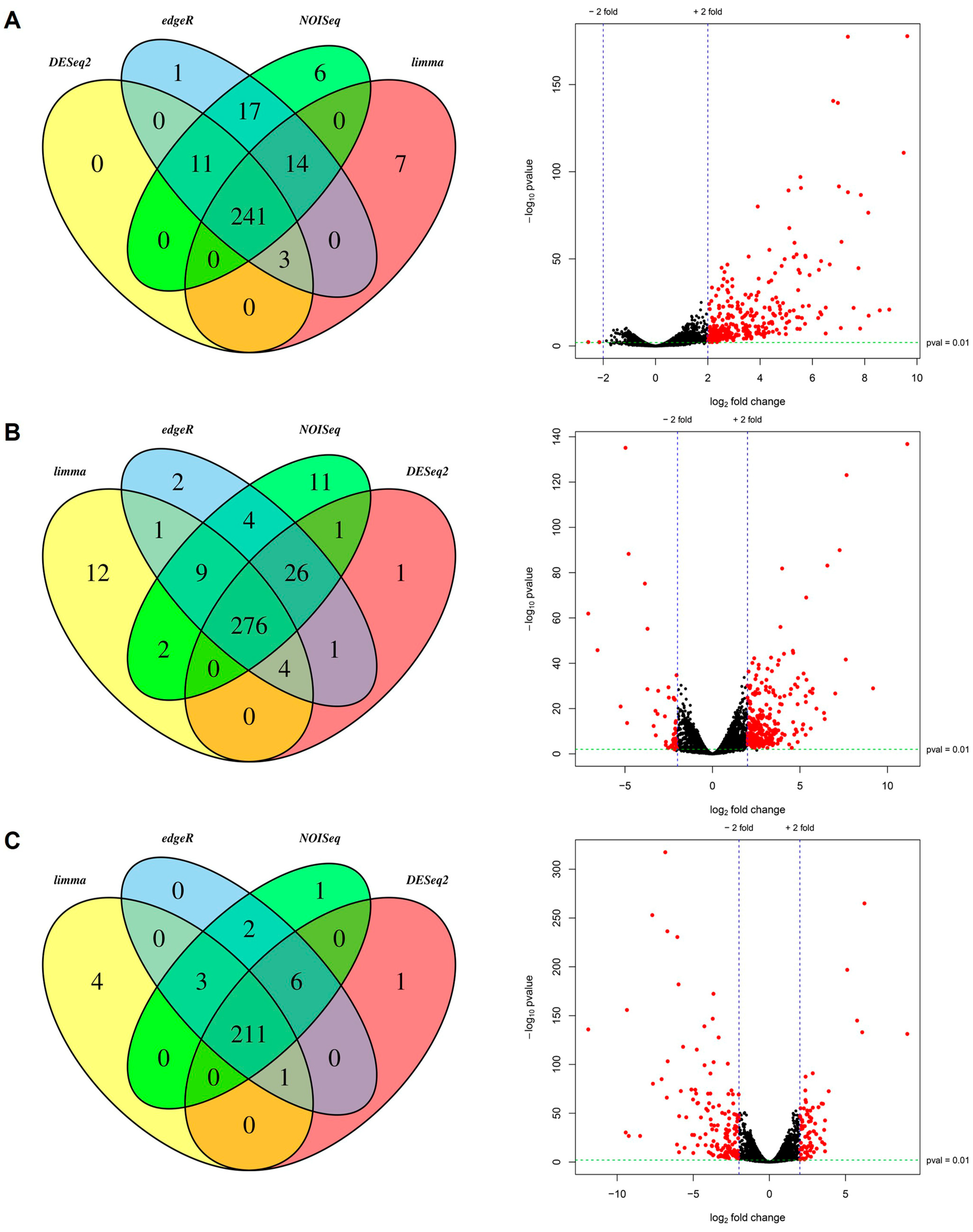
RNA extraction was performed as previously described [25]. For the elaboration and sequencing of cDNA libraries, the services of the Laboratorios de Servicios Genómicos (LabSerGen) de la Unidad de Genómica Avanzada, Laboratorios Nacionales de Genómica para la Biodiversidad (UGA-LANGEBIO) of the Centro de Investigación y de Estudios Avanzados del IPN (CINVESTAV) were used. The cDNA libraries were prepared using the TruSeq RNA Sample Preparation v2 LT kit (Illumina Inc. San Diego, CA, USA), and sequencing was performed on the Illumina NextSeq 500 platform, generating reads of 150 bp.

2.3. Data Analysis

The quality of the RNA-Seq reads was evaluated with the FastQC v0.11.7 software. Trimming of adapters and remotion of poor-quality reads was performed with Trimmomatic v0.36 [27], keeping the read-pairs with values greater than 25 nt in length and quality greater than 30 for the analysis. As part of the Trinity v2.11.0 pipeline [28], reads were aligned to the coding sequences (CDS) of the genome of the 521 strain, downloaded from the Ensembl database (https://ftp.ensemblgenomes.ebi.ac.uk/pub/fungi/release-56/fasta/ustilago_maydis/cds/Ustilago_maydis.Umaydis521_2.0.cds.all.fa accessed on 30 June 2021) using Bowtie2 v2.4.2 [29]. Next, the quantification of transcript abundances was done with the RSEM v1.3.3 program [30]. For the differential expression analysis, the count matrices generated in the previous step were uploaded to the IDEAMEX online platform (http://www.uusmb.unam.mx/ideamex/ accessed on 30 June 2021; [31] to use DESeq2 [32], NOISeq [33], Limma-Voom [34], and EdgeR [35], the cutoff values used to consider the genes as differentially expressed were logFC ≥ 2 and FDR ≤ 0.01. Raw sequencing reads, and expression data were submitted to the Gene Expression Omnibus of NCBI (accession number: GSE225422).

2.4. Annotation and Classification of Differentially Expressed Transcripts

The annotation of the resulting transcripts and products was carried out using the Trinotate v3.2.2 pipeline [36]; the sequences were analyzed by use of the BLAST+ 2.12.0 software packages [37] and the databases UniProt, Gene Ontology (GO), Kyoto Encyclopedia of Genes and Genomes (KEGG), and EggNog, applying the cutoff e-value of 10−5. Next, protein domains were sought in the outcomes employing the hmmer v3.3.1 software of Trinotate suite and the database Pfam-A. Likewise, SignalIP v4.1 and TMHMM v2.0c programs were used to seek the cleavage sites of signal peptides and to predict transmembrane helix [38,39,40]. DEGs were selected, and heatmaps were plotted using the R language. Finally, using the OmicsBox v1.4.12 pipeline (Bioinformatics Made Easy, BioBam Bioinformatics, Valencia, España 3 March 2019, https://www.biobam.com/omicsbox accessed on 30 June 2021), the GO identifiers of DEGs were filtered by taxonomy (fungi) and classified by functional annotation.

3. Results

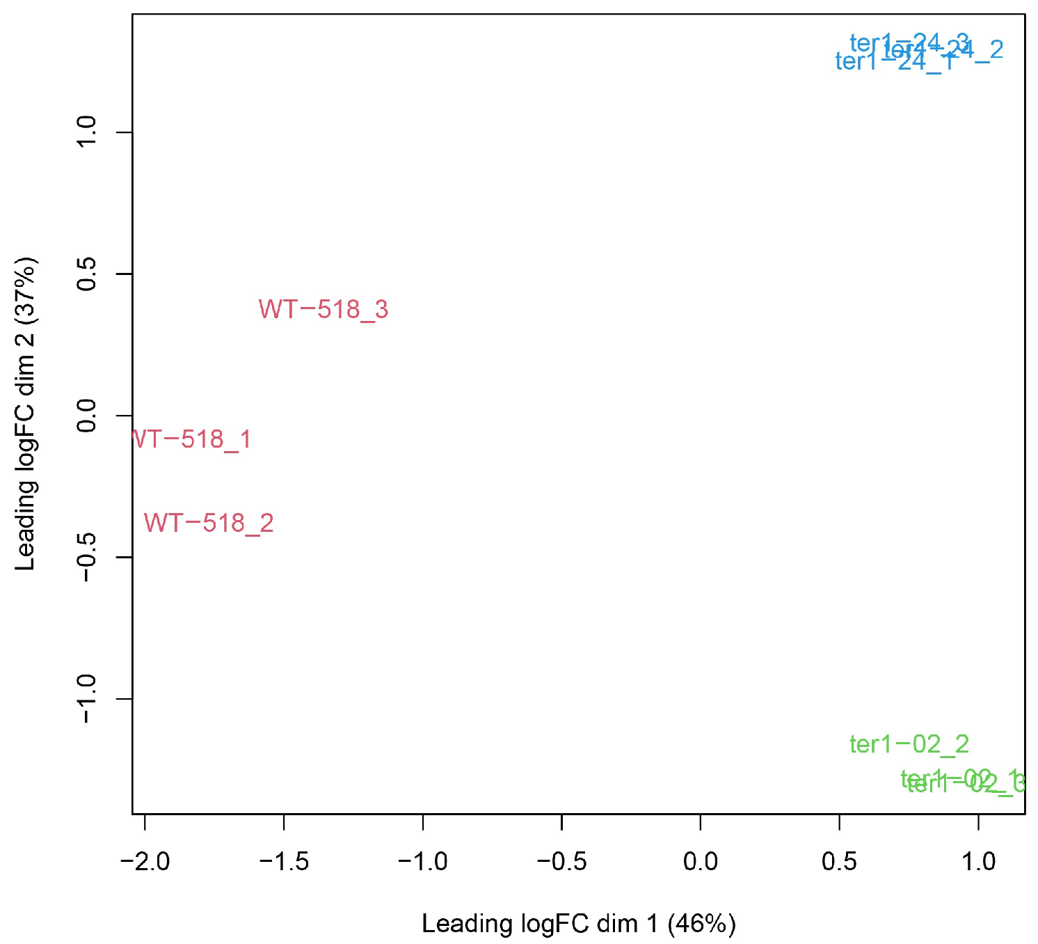
3.1. Sequencing and Quality Control of RNA-Seq Libraries

The global expression profile of TER-deleted mutants at the surviving stage was analyzed using RNA-seq libraries of the parental strain 518 and its telomerase-negative derivatives ter1-02 and ter1-24. The assays were elaborated in independent triplicate repeats and sequenced using the Illumina NextSeq 500 platform (San Diego, CA, USA), yielding a total of 126,587,419 paired reads from nine libraries. After raw read processing, a total of 110,227,520 paired reads were preserved (Table S1). The alignment percentages of the processed reads against the reference sequences with Bowtie 2 are shown in Table S1.

3.2. Changes in the Expression Profile of Telomerase-RNA-Deletion Response (TDR) Genes

To identify the DEGs between the parental strain and ter1Δ mutants, the reads mapping to the reference transcripts were used for the analysis of differential expression on the IDEAMEX platform, setting up cutoff points of: logFC ≥ 2 and FDR ≤ 0.01, and only the genes appearing at the intersection of DESeq2, NOISeq, Limma-Voom, and EdgeR were considered differentially expressed. Likewise, the changes in the expression profile between ter1-02 vs. ter1-24 mutants were compared to find the differences exhibited by both mutants [25].
Concurrently with the differential expression analysis, the input data were analyzed to detect sample variations. In Figure 1, replica sets of each strain analyzed are shown in the multi-dimensional scaling (MDS) graph; as they were grouped in separate clusters, variations in the expression patterns were confirmed for each sample. From 6783 protein-coding genes analyzed, 241 (3.55%) were detected as overexpressed DEGs when compared WT and ter1-02 transcriptomes, whereas 276 DEGs were identified after comparing WT and the ter1-24 transcriptomes. Of these, 246 genes (3.62%) were overexpressed, and 30 (0.44%) were repressed in the mutant strain. To complete the analysis, a comparison of ter1-24 and ter1-02 transcriptomes led us to discover 211 (3.11%) DEGs in the ter1-24 mutant relative to ter1-02, of which 70 genes (1.03%) were overexpressed and 141 (2.07%) were repressed (Figure 2).
Once identified, the DEGs were retrieved and examined to identify the sets of unique and shared genes from each analysis. In U. maydis, deletion of TER causes the deregulation of a core of 90 genes shared by the two TER-mutants, hence named core genes; this represents 1.32% of the total coding genes. Eight of those genes showed clear differences in transcriptional expression among the mutants. When the search for unique DEGs was carried out for each pair of transcriptomes analyzed, 57 genes were identified for ter1-02, and 124 genes were identified for ter1-24. When the differential analysis was conducted against wild-type, 47 unique DEGs were identified when comparing ter1-24 with ter1-02 (Figure 3A).

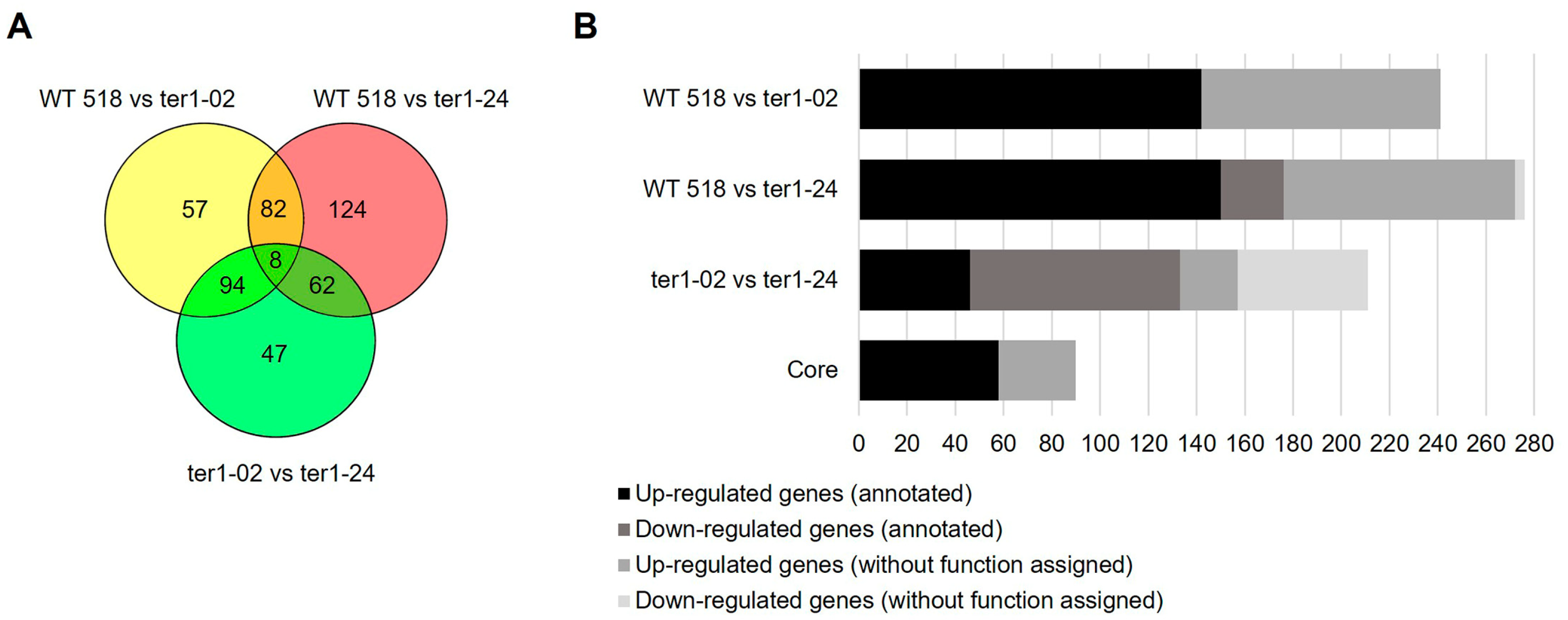
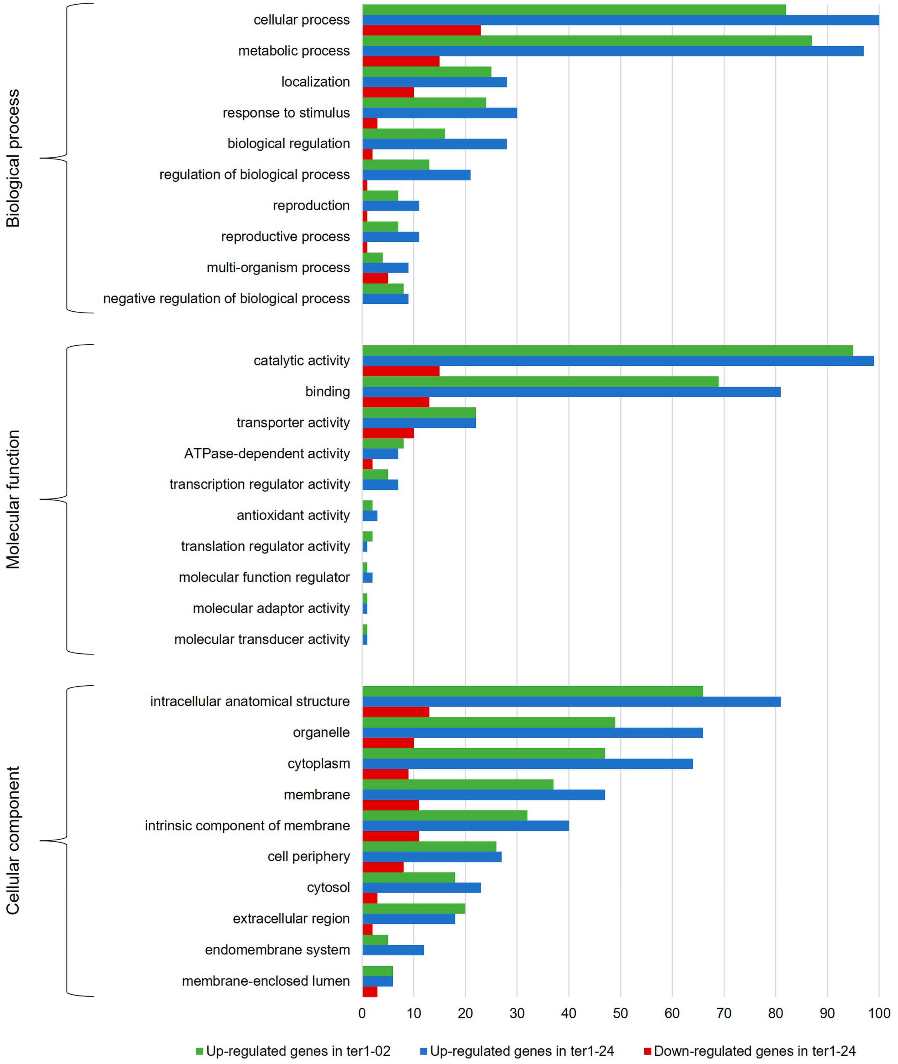
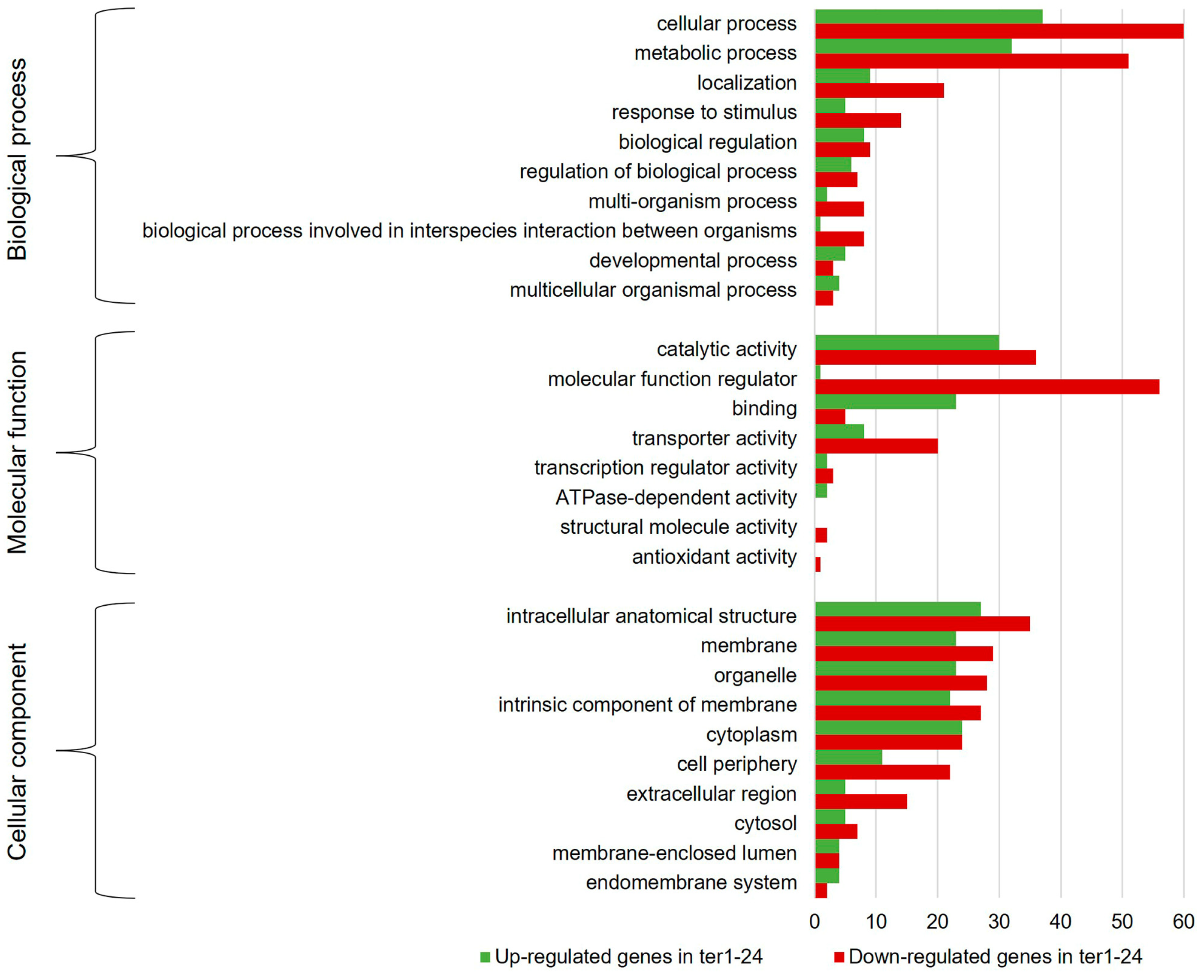
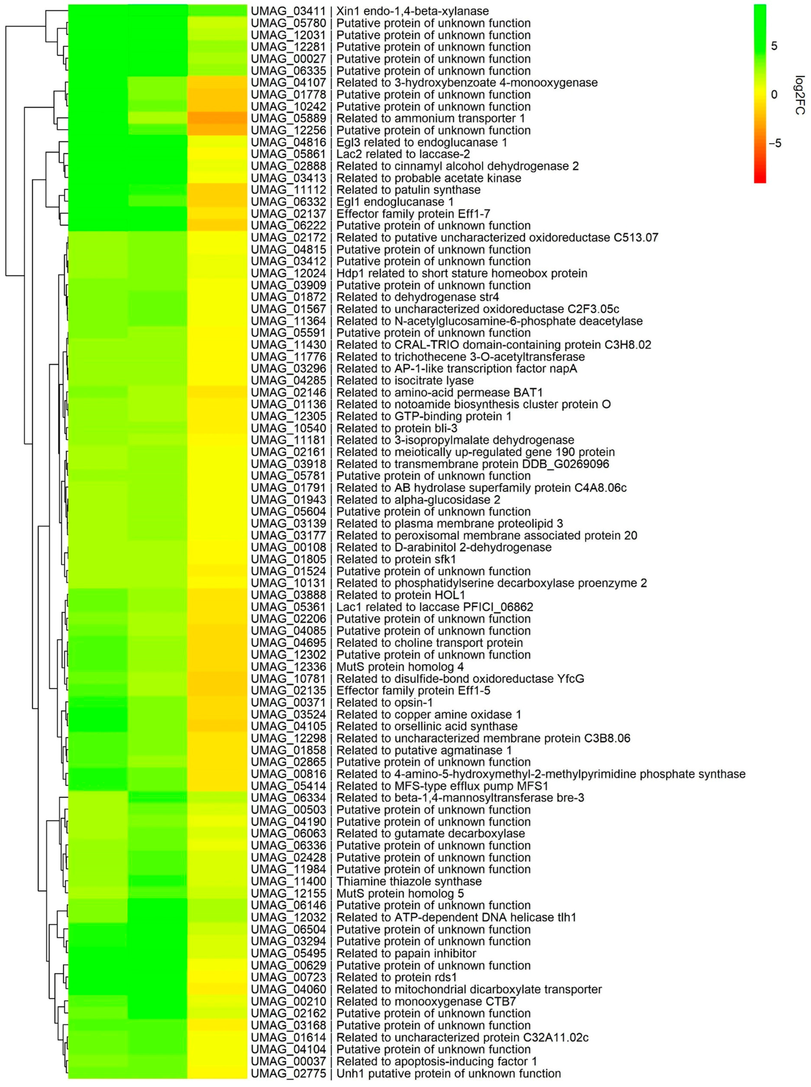
3.3. Annotation and Functional Assignment

To examine how DEGs contribute to the survivor phenotype in U. maydis, the core genes and DEGs recovered from the former analysis were forwarded for functional annotation and GO analysis using the Trinotate pipeline and OmicsBox, respectively. For the DEGs from ter1-24 vs. the WT 518 strain, it was found that ≈36% of the genes do not share similarity with any of the sequences deposited in the protein databases accessed and therefore were considered to not have an assigned function. Regarding the DEGs from a comparison of ter1-02 vs. 518 transcriptomes, the percentage of genes without assigned function increased to ≈41% (Figure 3B). From the annotation of the total set of coding genes, 26% corresponded to genes without any function assigned. Thus, the role of more than a quarter of the coding genes remains to be elucidated; even more interesting is the finding that these genes only represent a small percentage of the global transcriptome of U. maydis previously calculated by [41], and by our group [25].
To determine which of the cellular functions were affected by TER deletion, DEGs were grouped into the three categories of the GO analysis: biological process (BP), molecular function (MF), and cellular component (CC). Figure 4 shows the ten most representative classes of each category of the GO for the DEGs identified in both ter1-disrupted mutants. In the BP category, the highest proportion of overexpressed DEGs matches with genes involved in cellular processes (82 DEGs in ter1-02, 100 DEGs in ter1-24) and metabolic processes (87 DEGs in ter1-02, 97 DEGs in ter1-24) followed by genes involved in localization and response to stimuli (25 and 24 DEGs in ter1-02, and 28 and 30 DEGs in ter1-24, respectively). In the case of downregulated DEGs from ter1-24, GO analysis showed 23 downregulated DEGs, which are involved in cellular processes, 15 DEGs in metabolic processes, and ten DEGs in cellular localization as the most representative categories; remarkably, there also were downregulated genes from multi-organism processes and stimuli response (five and three DEGs, respectively).
Regarding the MF category of the GO analysis, the main enrichment of upregulated genes in ter1-02 occurred in the categories of catalytic activity (95 DEGs), binding capability (69 DEGs), and transporter activity (22 DEGs); likewise, in ter1-24, overexpressed genes also were from catalytic activity (99 DEGs), binding capability (81 DEGs), and transporter activity (22 DEGs). Downregulated DEGs in the same MF categories were found in the ter1-24 strain with catalytic activity (15 DEGs), binding capability (13 DEGs), and transporter activity (10 DEGs). Of the overexpressed genes in ter1-02, it was interesting to observe that 41.05% (39) of those assigned in the status of “catalytic activity” have hydrolase activity, and 38.84% (35 genes) possess oxidoreductase activity; a similar enrichment of these two activities was found in the ter1-24 DEGs reaching 31.31% (31 genes with hydrolase activity) and 38.83% (38 genes with oxidoreductase activity).
In the CC category of upregulated genes, the most enriched DEGs of ter1-02 mutant were intracellular anatomical structure (66 DEGs), organelle (49 DEGs), and cytoplasm (47 DEGs). In the ter1-24 strain, the most enriched DEGs also were intracellular anatomical structure (81 DEGs), organelle (66 DEGs), and cytoplasm (64 DEGs). Likewise, in ter1-24, there were also downregulated DEGs in the same categories; the intracellular anatomical structure was enriched with a count of (13 DEGs), organelle (ten DEGs), and cytoplasm (nine DEGs). Finally, from the DEG analysis for the BP category in both mutants, the most abundant deregulated genes included those from the cellular process, metabolic process, localization, and response to stimuli. Agreeing with the above results, those DEGs were assigned to the catalytic activity group, genes that regulate molecular function, binding and transporter activity in the MF category. Interestingly, genes that regulate molecular function were among the most enriched DEGs (26.54%) obtained from the analysis of both mutant strains. For the CC category, the main enriched DEGs were assigned to the intracellular anatomical structure group, and the number of DEGs assigned to those located in the membrane group increased, as well as those assigned to organelles and intrinsic components of membranes (Figure 5).

3.4. Chromatin Structure

Within the DEGs in ter1-24, we found that UMAG_02709 was repressed (logFC = −2.26), a locus that codes for the histone replication-coupled H3.2; repression of other core histone complex components was also noted (H2B (UMAG_01505), H2A (UMAG_01504), and H4 (UMAG_02710)) and were also found with logFC values of −1.98, −1.93, and −1.74, respectively, in addition to the putative H1 (UMAG_10447) with logFC value −1.83. However, although repression of the same genes was observed in the ter1-02 mutant, such changes were not significant at the cutoff point used, ranging between −0.93 (UMAG_01504) and −0.62 (UMAG_10447). Additionally, in both mutants, there were no significant changes in the expression of H2A.Z (UMAG_00469) and histone replication-independent H3.1 (UMAG_03916). Regarding other components involved in chromatin remodeling, in ter1-24, the changes were subtle, below the threshold in the genes UMAG_02567 (logFC = −1.51) and UMAG_06201 (logFC = −1.19) encoding for the catalytic subunit of histone acetyltransferase type B and RuvB-like helicase1 respectively. However, they need to be further explored.

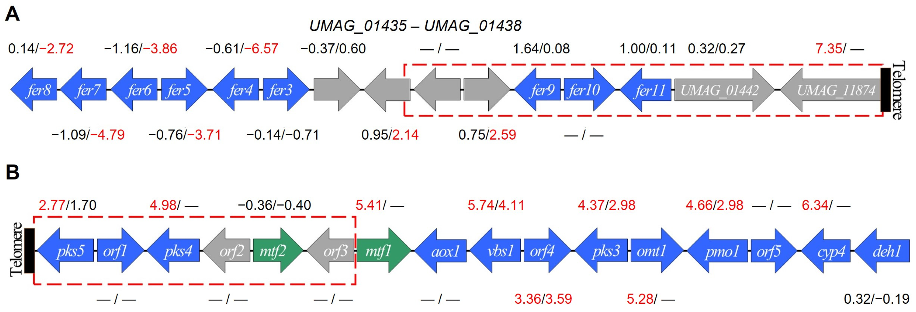
3.5. Subtelomeric Genes

U. maydis has multiple predicted telomere-linked helicase (TLH) isoforms. These include RecQ-like sequences UT5 (AF030886.2) and UT6 (AF030887.1) [42]. UT5 and UT6 have been categorized as UTASa and UTASb, respectively. UTASa is adjacent to telomere repeats and possess the same helicase domain as the RecQ helicases subfamily. UTASb is near telomere repeats or 5’ upstream of UTASa. UTASb may be a truncated segment generated from the homing endonuclease gene HEG [43]. UTASb consists of a highly conserved “core” sequence, an OrsD domain, or both [42]. UT5 exhibits homology with UMAG_12032 and UMAG_12076, while portions of UT6 are conserved in UMAG_06476, UMAG_06474, UMAG_11065, UMAG_04094, and UMAG_04308. UMAG_12032 was upregulated in both mutant strains ter1-02 (logFC = 3.16) and ter1-24 (logFC = 5.20) (Table 1). The other putative proteins displayed heightened expression solely in ter1-02. UMAG_12032, situated at one end of chr 20, encodes an incomplete TLH1-like ATP-dependent DNA helicase. UMAG_12032 may represent a novel secondary group of DEXH helicases that share homology with TLH1 [44]. The disruption of TLHs in telomerase-negative strains is frequently associated with gene de-repression due to its proximity to the shortened telomere [45] (Figure S1B).
Transcriptional expression of genes arranged on the 20 kb adjacent to each of the 46 chromosome termini of U. maydis was analyzed to assess the effect of ter1 disruption on them. These regions contain 146 genes. After analysis of DEGs in ter1-02, it was found that 30.13% of telomere-proximal genes (18.25% of the total DEGs) were differentially expressed, while in ter1-24, 10.27% of telomere-proximal genes were among DEGs (5.43% of DEGs), and from these only UMAG_06259 (peptidyl-prolyl cis-trans isomerase D) was repressed. Seven genes were overexpressed uniquely in ter1-02 (UMAG_05621, UMAG_11183, UMAG_06351, UMAG_11107, UMAG_04305, UMAG_10981, UMAG_04095) with logFC greater than two, without being classified as differentially expressed in ter1-24 at the same cutoff point used. From the sets of DEGs located at the chromosomal ends, four belong to the core genes of the ter1Δ strains; among those are UMAG_12032 and UMAG_04695 that encodes a choline transporter and UMAG_06146 and UMAG_12031 that code for hypothetical proteins.
In addition to the DEGs from subtelomeric regions (Figure S1), we found other DEGs sheltered in three of the four unmapped scaffolds (accession NW_011929455.1 to NW_011929458.1). Set on NW_011929455.1, the UMAG_06490, which encodes a predicted inorganic phosphate transporter, was differentially expressed in ter1-24 (logFC = −2.21); in NW_011929456.1, the UMAG_06503, which encodes a DEG similar to the uncharacterized protein gene C1198.03c, had a logFC = 2.88; finally, placed on NW_011929457.1, the loci UMAG_06504 (encoding a protein of unknown function) and UMAG_12076 (encoding a protein related to ATP-dependent DNA Q5/DEAD/DEAH box helicase) were upregulated in ter1-02. The UMAG_06504 fitted in the core of DEGs, and UMAG_12076, another RecQ-like helicase, was upregulated in ter1-02 with logFC = 4.93 but showed no change in ter1-24 (logFC = 0.22).

3.6. DEGs Related to Stress and DNA-Damage Response (DDR)

DEGs involved in stress and DNA-damage responses are listed in Table 2 and Table 3. Within the stress-response category, DEGs were mainly related to the oxidative stress response and cellular detoxification, except UMAG_03122 (encoding a β-1,3-glucan-binding related protein), located within the first 20 kb of chromosomal ends. Most of these genes could be analogous to stress-induced responses and cellular senescence of S. cerevisiae [7]. The group also included the predicted genes implied in apoptosis, i.e., UMAG_01937, UMAG_02224, and UMAG_03728. In addition, UMAG_04553 (predicted growth hormone-inducible transmembrane protein; logFC = 2.02) was differentially expressed in ter1-24 because of a slight downregulation in ter1-02, whereas UMAG_11483 (predicted protein-L-isoaspartate (D-aspartate) O-methyltransferase) and UMAG_01262 (predicted β-DNA-polymerase) were uniquely upregulated at logFC > 2 in ter1-24 but induced in ter1-02 at logFC = 1.15 and 1.83, respectively.
Among the DEGs in the DDR category (Table 3), the MutS4/Msh4 (UMAG_12336) and MutS5/Msh5 (UMAG_12155) homologs were annotated as members of the DEGs core genes (Figure 6); in yeast, those proteins are part of the ZMM group (Zip1-4, Msh4/Msh5, Mer3), which together with Mlh1 and Mlh3 promote crossover during meiotic recombination [47,48,49].
Table 3. DNA-damage response genes affected by ter1Δ mutation. The DEGs obtained between the various comparisons are indicated in bold.
Gene IdDescriptionWT 518
vs.
ter1-02
WT 518
vs.
ter1-24
ter1-02
vs.
ter1-24
UMAG_12155Msh5—MutS protein homolog 5 22.4263.7971.398
UMAG_12336Msh4—MutS protein homolog 4 23.6742.454−1.205
UMAG_10845Related to G/U mismatch-specific DNA glycosylase1.1832.6081.444
UMAG_05917Related to cryptochrome DASH1.2002.3701.199
UMAG_01262Related to DNA polymerase beta1.8342.3970.585
UMAG_03290Rad51—DNA repair protein RAD51 12.0460.765−1.256
UMAG_11008Mer3—ATP-dependent DNA helicase MER3 22.0112.4270.432
UMAG_04165Related to replication factor A protein 31.741−0.387−2.098
UMAG_01952Related to UV-damage endonuclease1.8122.6830.889
UMAG_00172Related to meiotic recombination protein rec81.7492.1870.481
1 [50], 2 [51].
wAn increase in expression of Mer3 (UMAG_11008) homolog also occurred in both mutants (except for the NOISeq normalization value in ter1-02, logFC = 1.98; Tables S1–S4); Mlh3 (UMAG_03481) and Mlh1 homolog (UMAG_05208) were upregulated more than two-fold in ter1-02. However, no such expression changes of UMAG_05208 occur in ter1-24 (Table S5). In this last strain, upregulation occurred to a poor extent in UMAG_10845 (predicted to encode thymine-DNA glycosylase; logFC = 1.18), UMAG_05917 (predicted to encode deoxyribodipyrimidine photolyase; logFC = 1.20), and UMAG_01262 (predicted to encode β-DNA polymerase; logFC = 1.8), which did not reach significance. Further study is now needed.
Regarding the homologs of the yeast RAD52 epistasis group in U. maydis, only rad51 was differentially expressed (logFC ≥ 2) in the ter1-02 strain, whereas in ter1-24, overexpression of the same gene reaches only a logFC = 0.76. Also, we detected a slight upregulation of the genes encoding the RPA-heterotrimer: RFA1 (UMAG_05156; logFC = 1.56), RFA2 (UMAG_02579; logFC = 1.50) and RFA3 (UMAG_04165; logFC = 1.74).
Homologs of the RAD3 epistasis group were differentially expressed in U. maydis. The putative rad7 (UMAG_00657) showed an upregulation of nearly three times in the ter1-02 strain and of nearly two times in ter1-24 (Table S5), whereas the rad16 homolog (UMAG_03263) did not exhibit expression changes. Finally, in ter1-24, the repression of some genes encoding DDR constituents at values above the logFC threshold of −2 was considered in this report because the interaction of their protein products with those of each other suggests alterations in DNA replication and/or repair among repressed DEGS: PCNA (UMAG_05403, logFC = −1.18), DNA Polε catalytic subunit A (UMAG_01008, logFC = −1.56), and DNA ligase1 (UMAG_11196, logFC = −1.05). Regarding components of the heteropentameric RFC homologs in U. maydis, putative RFC5 (UMAG_00920) was downregulated marginally by logFC = −1.45; RFC4 (UMAG_00729) had a logFC = −1.05, and MCM10 (UMAG_10135) had a logFC = −1.25. On the other hand, the putative MCM2-7 complex exhibited logFC values between −0.62 and −1.01, except MCM5 (UMAG_05064) logFC = −0.10, which could also be considered an ambiguous value. Finally, the following genes encoding the homologous subunits of the DNA-directed RNA polymerase II complex were slightly downregulated: RPB11 (UMAG_02324; logFC = −1.83), RPABC1 (UMAG_10512; logFC = −1.25), RPABC2 (UMAG_10433; logFC = −1.01), and RPABC3 (UMAG_04460; logFC = −1.15) (Table S5). Furthermore, their role in the telomerase-deletion response need to be deciphered using alternative strategies.

3.7. Genes Involved in Telomere Maintenance

3.7.1. Complexes of Shelterin, CST, and MRX/MRN

Except for Tpp1, which showed near to 50% repression in the ter1-24 mutant, no significant changes were found in the expression of genes encoding the components of the shelterin complex (Trf1, UMAG_02326; Rap1, UMAG_04676; Tpp1, UMAG_11538; Pot1, UMAG_05117) [23,26], CST complex (Stn1, UMAG_11687; Ten1, UMAG_11842) [23], or the putative MRX/MRN complex (Mre11, UMAG_04704; Rad50, UMAG_01085) [26].

3.7.2. Putative SM7-like Subunits

A highly conserved motif has been found at the 3′ end of the putative TER of all the members of Ustilaginales studied in [25], which share homology with the binding site for Sm7. This finding, which suggests the requirement of an SM7-like complex for telomerase biogenesis, prompted us to search the Ustilaginales’ genes encoding the protein homologs of B/B’, D1, D2, D3, E, F, and G and analyze their expression changes in the ter1Δ mutants of U. maydis. From those putative homologs of SM7 genes, SMD1 (UMAG_10381, logFC = −1.57), SMD2 (UMAG_04781, logFC = −1.26), and SMD3 (UMAG_11043, logFC = −1.20) were slightly downregulated; a marginal change was found in SMB (UMAG_12244, logFC = −0.75), but no expression changes were found in SME (UMAG_10312), SMF (UMAG_12130), and SMG (UMAG_10805) in the ter1-24 mutant. The same strain also showed a slight downregulation (i.e., logFC < 2.0) of components of the predicted Dyskerin complex: Dyskerin/CBF5 (UMAG_00685, logFC = −1.18), NPH2 (UMAG_03340, logFC = −1.61), GAR1 (UMAG_04573, logFC = −1.03); and NAF1 (UMAG_03271, logFC = −0.87); no expression changes were predicted for NOP10 (UMAG_03354).

3.7.3. Telomere-Linked Helicases (TLH1-like)

As noted above, the participation of middle repeated elements UTASa and UTASb harboring ORFs encoding helicase-like sequences in telomere maintenance has been proposed by some authors [44,52]. In both the ter1-02 and ter1-24 mutant strains, the UMAG_12032 locus was upregulated; this could be associated with a possible gene de-repression of subtelomeric sequences after telomere-repeats loss or a possible role of this RecQ-like helicase in the primary DNA metabolism in the absence of telomerase [52] (Figure S1B). Interestingly, the expression of UMAG_12032 in ter1-24 is sufficiently high compared to ter1-02 to be considered as a DEG (logFC = 2.06) and is the only helicase-related gene overexpressed in this strain in contrast to the seven genes observed in ter-02 (Table 2). The upsurge in transcriptional expression of such genes in U. maydis needs to be surveyed to determine if they play a role in the formation of survivors or if their activity could be dispensable.

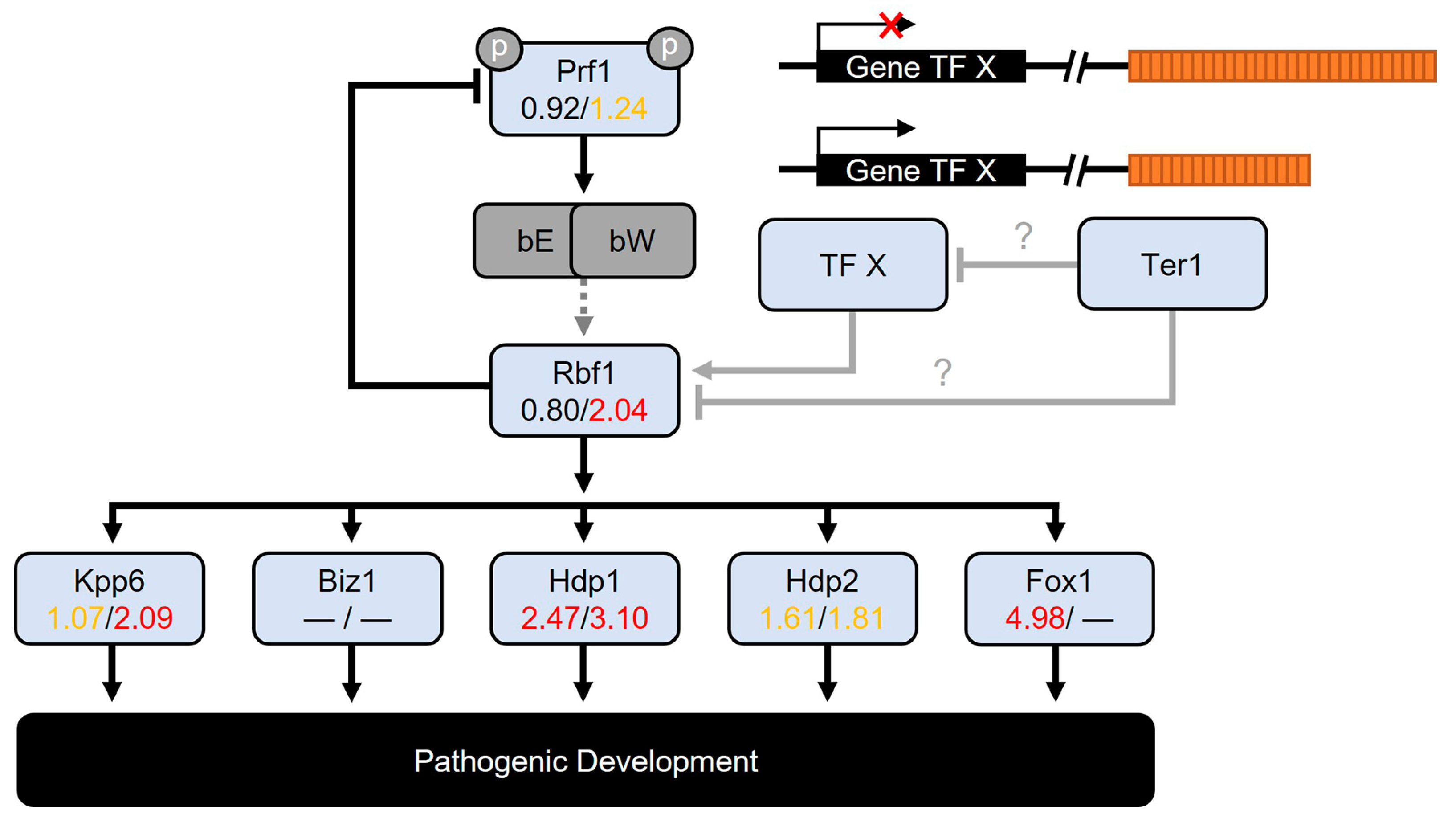
3.8. Genes Involved in Cell Cycle Progression and Pathogenic Development

The dimorphic fungus U. maydis requires in planta development to complete its life cycle. To achieve this, it experiences a morphologic and metabolic transition from saprophytic sporidia (yeast-like) to pathogenic mycelium (hyphae); this lifestyle change requires the reprogramming of gene expression [46,53,54]. Genes engaged in controlling the cell cycle and the fungal growth within the plant tissues, as well as genes encoding effectors required to subdue the plant response, are critical to complete the life cycle of this obligated fungal pathogen. The loss of key genes involved in the development or maintenance of the pathogenic lifestyle could cause growth distress and impairment to complete the life cycle [46,55,56].

3.8.1. The Cell Cycle Progression

In the ter1Δ mutants, there were no expression changes detected in the master regulator cdk1, nor in the cdk5 involved in cell differentiation [57,58], yet Cdk5-related cyclin-partner pcl12 [59] reached the DEG threshold only in ter1-24; the same gene had a logFC = 0.86 in ter1-02; pcl12 and rbf1 are controlled by the heterodimeric master regulator bE/bW; however, its expression pattern suggests an independent mechanism of control [60] (see Table 4).
Table 4. The life cycle and pathogenic development genes are deregulated in response to the ter1Δ mutation. The DEGs obtained between the various comparisons are indicated in bold.
Gene IdDescriptionWT 518
vs.
ter1-02
WT 518
vs.
ter1-24
ter1-02
vs.
ter1-24
UMAG_10529Pcl12—related to PHO85 cyclin-2 100.8603.3882.535
UMAG_00628Putative protein of unknown function 85.109−1.462
UMAG_00876Related to glucan 1,3-beta-glucosidase 10.900−1.435−2.31
UMAG_01130Related to tyrosinase ustQ 7−0.1642.6282.801
UMAG_01237Putative protein of unknown function, cluster 2A 15.519−5.015
UMAG_01238Putative protein of unknown function, cluster 2A 16.249−5.815
UMAG_01431Fer6—multidrug resistance protein fer6 2−1.163−3.861−2.674
UMAG_01432Fer5—acyltransferase fer5 2−0.767−3.711−2.92
UMAG_01433Fer4—enoyl-CoA isomerase/hydratase fer4 2−0.613−6.571−5.924
UMAG_01690Putative protein of unknown function 73.949−2.207
UMAG_01695Stp6—Putative protein of unknown function 105.6150.842
UMAG_01788Related to chitin deacetylase 31.022−2.465−3.476
UMAG_01829Related to alpha-L-arabinofuranosidase A 12.6731.529−1.126
UMAG_01888Probable serine carboxypeptidase, cluster 3A 10.5322.1431.633
UMAG_01945Putative invertase 32.3734.8172.465
UMAG_02135Effector family protein Eff1-5 63.7402.215−1.498
UMAG_02136Effector family protein Eff1-6 67.088−1.341
UMAG_02137Effector family protein Eff1-7 65.3944.716−0.649
UMAG_02138Effector family protein Eff1-8 63.0992.239−0.820
UMAG_02140Effector family protein Eff1-10 62.4262.6920.301
UMAG_02758Putative protein of unknown function 3−0.473−2.133−1.632
UMAG_03023Related to ribonuclease T2-like 1-A 3−0.653−4.881−4.193
UMAG_03382 *Related to 3-phytase A 31.3052.8141.534
UMAG_03411Xin1—endo-1,4-beta-xylanase 15.5629.1753.65
UMAG_03416Putative protein of unknown function 12.4822.6250.160
UMAG_03749Putative protein of unknown function, cluster 10A 11.9442.6010.700
UMAG_03750Putative protein of unknown function, cluster 10A 14.1252.386
UMAG_03751Putative protein of unknown function, cluster 10A 15.5983.721−1.824
UMAG_04282Related to 3-phytase A 31.3363.8692.556
UMAG_04309Probable alpha-L-arabinofuranosidase 14.4690.820−3.625
UMAG_04364Related to glucan 1,3-beta-glucosidase 12.9042.711−0.162
UMAG_04503Probable alpha-galactosidase D 14.467−3.390
UMAG_04816Egl3—Related to endoglucanase 1 1, 55.7176.4150.698
UMAG_05036Related to probable glycosidase C21B10.07 30.7443.0802.360
UMAG_05299Putative protein of unknown function, cluster 19A 10.7562.7892.067
UMAG_05306Putative protein of unknown function, cluster 19A 14.330−1.603
UMAG_05308Putative protein of unknown function, cluster 19A 12.876−2.949
UMAG_05310Putative protein of unknown function, cluster 19A 17.574−9.239
UMAG_05314Putative protein of unknown function, cluster 19A 13.579−3.060
UMAG_05361Lac1—laccase 3, 43.4372.576−0.836
UMAG_05439Related to GlcNAc-binding protein A 73.655−0.031−3.665
UMAG_05495Related to papain inhibitor 74.3555.3721.037
UMAG_05689Related to Fe-regulated protein 8 22.4251.702−0.700
UMAG_05861Lac2—laccase-2 3, 45.3165.3550.064
UMAG_06190Related to chitinase A1 33.479−1.840
UMAG_06221Putative protein of unknown function, cluster 22A 12.2431.030−1.183
UMAG_06222Putative protein of unknown function, cluster 22A 16.2964.759−1.521
UMAG_06274Related to hormone-sensitive lipase 31.2593.2211.985
UMAG_06332Egl1—endoglucanase 1 1, 55.2833.747−1.518
UMAG_10055Related to glutathione hydrolase proenzyme 33.490−2.091
UMAG_10557Putative protein of unknown function, cluster 19A 14.454−3.768
UMAG_11338Fer8—Fe-regulated protein 8 20.141−2.720−2.833
UMAG_11339Fer7—siderophore transporter fer7 2−1.095−4.791−3.672
UMAG_12330Putative protein of unknown function 31.5503.2231.701
UMAG_05528Related to alkali-sensitive linkage protein 1 95.131−4.945
UMAG_02161Related to meiotically upregulated gene 190 protein2.3222.5030.205
UMAG_02517Gpa2—guanine nucleotide-binding protein alpha-2 subunit1.1102.4441.360
UMAG_02994Related to sporulation-specific protein 5−0.0652.5052.591
UMAG_03541Related to meiotic expression upregulated protein 264.233−0.999
UMAG_05467Related to meiotic coiled-coil protein 25.627−3.148
UMAG_11677Related to serine/threonine-protein kinase cek10.8372.5331.720
1 [46]. 2 [61]. 3 [62]. 4 [63]. 5 [64]. 6 [65]. 7 [66]. 8 [67]. 9 [68]. 10 [59]. * Located in subtelomeric region.
The analysis revealed a significant downregulation of Rad21 (logFC = −1.21). This Rad21 downregulation in ter1-24 went near half in the wild-type cell expression. The genes responsible for encoding the postulated cohesin proteins, Smc1 and Smc3, also exhibited substantial deregulation, with their normal expression levels decreasing by nearly half at logFC = −1.15 and logFC = −0.94, respectively. This downregulation could be correlated with the changes in cell morphology and nuclear irregularities observed in ter1Δ mutants [25], possibly indicating alterations in chromatin structure. Modest upregulations were noticed in the checkpoint kinase chk1 (logFC = 1.40) and wee1 kinase (logFC = 1.18) in ter1-02 and ter1-24, respectively, although both were below the logFC threshold of 2.0. Notably, no alterations were detected in the predicted apical kinase atr1 expression within the ter1-02 and ter1-24 mutants (logFC = 0.51 and 0.49). This finding weakens the likelihood of a link between the apical members of DDR and the changes described above [69,70]. Similar instances have been reported in fission yeast and other Rad21-deficient cell lines like mouse embryonic fibroblasts (MEFs) and lymphoblastoid cell lines (LCLs) before [71,72,73].
Since the third portion of the DEGs in mutant strains lacks an assigned function or a defined role, a database of genes involved in pathogenic development was constructed, which included fungal secreted proteins, cell-wall-degrading enzymes, plant component-degrading enzymes, and proteins involved in the organization of the fungal cell wall, to further learn about the influence of chromosome architecture and fungal development [46,62,66,68,74].

3.8.2. The Pathogenic Development

About 10% of the DEGs were involved in pathogenic development in both mutants. Those genes were included in the group of core genes and included some encoding plant cell wall-degrading enzymes such as egl1 and egl3 endoglucanases, xin1 endoxylanase, which is engaged in the fungal cell wall organization, and lac1 and lac2 involved in melanin synthesis [63,64].
Particular gene clusters, which are collectively controlled, were upregulated in both mutants, i.e., the genes encoding the Eff1 protein family (except for eff1) located on the chr 5; those genes whose products have a role in virulence were upregulated [46,65] (Table 4), possibly, the lack of telomerase could cause deregulation of these genes through the modification of the expression pattern of key controllers of pathogenicity. Remarkably, except for fer3, the downregulation of the fer gene cluster, located on the telomere-proximal right end of the chr 2, also occurred in ter1-24 strain of U. maydis (Figure 7A), with fer9, fer10, and fer11 being the most telomere-proximal genes [61].
Both the fer cluster as well as the sid1, sid2, fer1, and fer2 genes are negatively regulated by the Urbs1 factor [61,76], but neither urbs1 nor its target genes outside the fer cluster were downregulated, suggesting chromatin relaxation in the subtelomeric region could facilitate the interaction of Urbs1 with other negative regulators or with the fer-cluster promoters.
Moreover, the UMAG_01436 (related to acetyltransferase MAT1) and UMAG_01438 (predicted Acyl-CoA-dependent acyltransferase gene) loci, which separate fer9-11 from the remaining genes of the fer cluster, were upregulated in ter1-24; whereas strong upregulation (logFC = 7.35) of the UMAG_11874 gene, encoding a hypothetical protein of unknown function, was detected in ter1-02. Moreover, UMAG_11874 is the last gene at the end of chromosome 2 (Figure 7A).
Other DEGs-encoding proteins of unknown function located within clusters 2A, 10A, 19A, and 22A [46] were deregulated in one or both mutants (Table 3). Finally, the genes involved in the evasion of the immune response and release of plant nutrients, such as phytases (UMAG_03382, UMAG_04282), lipases (UMAG_06274), and proteases (UMAG_12330) [62], were identified as DEGs in ter1-24; meanwhile, those in ter1-02 showed slight increases (1< logFC < 2).

3.9. Differentially Expressed Transcription Factors

The DEGs-encoding transcriptional factors (TFs) are listed in Table 5. Among the set TFs-genes altered in the ter1Δ mutants, some were associated with pathogenic development, such as hdp1, which was differentially expressed in both the ter1-02 and ter1-24 mutant strains. The hdp1 is required for filamentous growth and cell cycle arrest [77]; likewise, hdp2 (UMAG_04928), which plays a role in appressorium formation [64], was upregulated (below the threshold of two) in both mutant strains (logFC = 1.61 in ter1-02 and 1.81 ter1-24). The fox1 gene, involved in suppressing plant defenses [78], was highly overexpressed only in ter1-02 (logFC = 4.98). The three TFs (hdp1, hdp2, and fox1) are activated by the Rbf1 master regulator (UMAG_03172), which in turn is activated by the bE/bW heterodimer in a dependent manner and also participates in the activation of biz1 and MAP kinase Kpp6, which are involved in appressorium formation [64,79].
Also, kpp6 was identified as differentially expressed in ter1-24, as was rbf1. Those results in sporidia, which lack the functional bE/bW heterodimer because they are haploid cells, together with the absence of deregulation, or with a slight increase in ter1-24, in the expression of factors upstream of Pfr1 suggest that other unidentified components or possibly Ter1 could be involved in the regulation of rbf1; and hence, some of the target genes of Rbf1 (Figure 8) as occurs when the fungus behaves as a necrotrophic pathogen, and in ter1-24 [60]. Other DEGs which encode transcription factors that are shared by the ter1Δ mutants as core genes were: the homolog of Yap (UMAG_3296) bZIP protein from the AP-1 family, involved in response to oxidative stress and plant–host interaction (Table 5; [82,83]), and unh1, the TF required for formation and pigmentation of teliospores, and completion of meiosis [63]. Intriguingly, none of its five target genes (UMAG_00983, UMAG_06485, UMAG_04827, UMAG_05664, UMAG_11505) were deregulated in either mutant, suggesting the cooperation of other unidentified factors in the regulation of these genes.
Among the TFs upregulated solely in the ter1-24 mutant were: UMAG_01456, UMAG_06308, UMAG_02835, and UMAG_04778, which in natural circumstances are upregulated during pathogenic development [80,84,85], and UMAG_01025, which is progressively repressed during early infection and responds to unfolded proteins [80,81]. Whereas in the same TFs category, the DEGs identified only in the ter1-02 were the UMAG_12304 and UMAG_04101 that encode Mtf1, a transcription regulator of the gene cluster located at the end of chromosome 12 involved in melanin production [75,86]; consistently, its target genes (pks5, pks4, vbs1, orf4, pks3, omt1, pmo1, and cyp4) were also identified as DEGs, while orf1, orf5, aox1, and deh1 do not exhibit changes in transcriptional expression, nor mtf2 or its target orf2 (Table S5).
Although no information about the expression of mtf1 in ter1-24 was registered, upregulation of vbs1, orf4, pks3, and pmo1 supported our notion that a weak upregulation of mtf1 occurred in this mutant, as it was hinted in the analysis of gene expression between both mutants (Table 5). Alternatively, telomere loss could have alleviated the silencing of the subtelomeric gene cluster in ter1-24 (Figure 7B). Another insight was the induction of pks1, pks2, and lac1 expression observed in ter1-02, but solely of lac1 in ter1-24 mutant; those three genes play principal roles in teliospores melanization [87]; paradoxically, expression of ust1, —the TF regulator of pks1, pks2 and lac1— remained unchanged in both mutants.

4. Discussion

Here, we present a transcriptomic DEGs analysis of three strains: the wild-type U. maydis 521 strain and two survivor strains. In both survivor strains, the template domain of ter1 was disrupted. In U. maydis, ter1 encodes the TER subunit of telomerase. In these ter1Δ strains, the template domain was replaced by the selective marker gene hph, as detailed previously [25]. This replacement did not lead to the suppression of transcriptional activity in the flanking genes UMAG_03168 and UMAG_03169. The ter1 transcript is initiated within UMAG_03168, a putative gene (Emi1) situated 5′ upstream from the intergenic region housing the conserved domains of the ter1 gene [24]. Nevertheless, no polar effects were evident in the mutants; the A (unspliced) and B (spliced) isoforms were upregulated (Figure 6).
The UMAG_03169 gene is at the 3′ end of ter1 and encodes a putative ornithine-oxo-acid transaminase. UMAG_03169 was also upregulated in the mutants (logFC = 1.74 in ter1-02 and logFC = 3.0 in ter1-24). This upregulation in these two genes cannot be solely attributed to the transcriptional disruption caused by ter1. Instead, it may be linked to the Telomere Damage Response (TDR). This would be consistent with observations for several other genes.
The putative telomere maintenance genes [26,88] were unaffected in U. maydis. This finding is consistent with previous studies addressing other telomerase-negative mutants [16,18,52,89]. However, rad51 was upregulated (logFC = 2). The rad51 gene is involved in the ALT pathways in S. cerevisiae and in DNA repair through homologous recombination. Conversely, the gene encoding its associated protein Brh2 (UMAG_03200; [69]) showed no changes in expression.
The chromatin modification pattern of telomere-associated sequences (TAS) shifts as telomeres shorten. This shift influences the condensation status of both the TAS chromatin and neighboring genes. Reducing chromatin compaction could lead to a variegated gene expression pattern, impacting genes close to telomeres like UMAG_05721. This gene encodes the srbA homolog in U. maydis, termed srb1, which plays a role in hypoxia adaptation and is a potential co-regulator of tumor formation [80]. Interestingly, in the ter1-02 strain, UMAG_05721 is repressed, whereas srb1 is upregulated in ter1-24 (Table 4).
Other genes within the two terΔ mutants exhibited divergent transcriptional behaviors. However, as these strains share the same mutation, induce similar infection symptoms, and cannot produce tumors in planta, it is likely that a set of “core genes” orchestrates control over these strains during the early stages of their biotrophic development [25]. Additionally, our analysis was extended to several genes situated within a 20-kb range adjacent to the TAS. Some of these genes were deregulated (Figure 7), while others situated at identical distances were unaffected. This variation could be attributed to the penetrance of the ter1Δ mutation or the telomere-position effect (TPE), as previously discussed by [90]. The TPE might involve the repression of sequences neighboring the telomere in a telomere-length dependent fashion; in the case of TPE over long distances (TPE-OLD), it could govern the physical interaction of remote genes (up to 10 Mb away), silencing their expression without affecting neighboring genes [91]. The phenomenon of TPE has been identified in higher eukaryotes and various lower eukaryotes [92], playing pivotal roles in processes such as antigenic variation in protozoa, adaptation to rapid environmental changes in fungi, and replication, repair, and recombination processes [92]. Similar mechanisms could conceivably be at play within U. maydis.
Due to the TPE, it was previously believed that the ends of chromosomes were transcriptionally silent. However, recent findings challenge this notion. In organisms like S. pombe and various eukaryotes, transcripts originating from subtelomeric sequences have been observed. Notable examples include TERRA, ARRET, and α-ARRET, as well as transcripts like ARIA, that initiate from the telomere itself [93]. In both S. pombe and humans, TERRA and ARIA transcripts, which incorporate telomere repeats, display a lesser degree of polyadenylation compared to ARRET and α-ARRET, representing antisense and sense transcripts from subtelomeric regions [94].
For the transcriptomes of the ter1Δ mutants studied here, the poly(A) fraction was extracted from total RNA. Notably, a strand-specific approach was not employed for sequencing, presenting transcripts as homologous to subtelomeric sequences. Previously, in U. maydis, sense and antisense transcripts derived from chromosome ends were amplified using RT-PCR. Although sequences analogous to TERRA, ARRET, and α-ARRET were investigated by our group, their characterization remains incomplete, including determination of polyadenylation extent and percentage. Given that UTASa primarily localizes adjacent to telomere repeats, it is conceivable that their transcripts may lack polyadenylation, akin to the case of TERRA. This absence of poly(A) tail in transcripts could impede their detection in polyadenylated transcriptomes, thereby accounting for their underrepresentation in the analysis. Consequently, this could explain the prevailing abundance of UTASb sequences.
UMAG_12032 and UMAG_12076 are two upregulated UTASa sequences containing RecQ-like motifs. These UTASa sequences lack adjacency to telomeres. This suggests the possibility of these sequences undergoing transcription in a manner akin to ARRET or α-ARRET sequences. Also, UMAG_11065 experiences an approximately 100-fold increase in expression within the ter1-02 strain. Both this sequence and UMAG_06474 share homologous traits with UTASb and are notably detached from telomere repeats.
Moreover, the significance of TERRA extends to its role in telomere lengthening in scenarios where telomerase is absent. This is attributed to TERRA’s involvement in the formation of telomeric R-loops, which represent RNA/DNA hybrids. These structures facilitate recombination-based alternative mechanisms (ALT) that amplify chromosome ends. This phenomenon, in turn, ensures the survival of cells even in the absence of functional telomerase [95].
Non-telomeric functions have also been attributed to TERRA, shelterin proteins, constituents of the CST complex, and TERT, collectively referred to as telomere-associated factors (TAFs). These proteins have been found to intricately modulate the transcriptional activity of critical genes dispersed along the length of the human genome [96]. Additionally, the core components of telomerase have demonstrated functions beyond their telomeric roles across diverse organisms and cell lineages, although the primary focus has been on TERT (as reviewed in [97,98]). However, insights into the non-telomeric roles of TER are steadily emerging.
Consequently, it has come to light that the TER component engages in various non-telomeric activities. For instance, it plays a pivotal role in responding to DNA damage [99], offering protection against oxidative stress in motor neuron cells [100], and potentially regulating apoptosis by promoting cell survival through their intra-TER gene hTERP [101,102]. Furthermore, TER also regulates gene expression and cell differentiation [103,104,105,106]. Notably, a previous investigation unveiled a correlation between the effectiveness of telomerase function and the formation of teliospores [22].
Within ter1Δ mutants, the deregulation of at least 12 pivotal developmental TFs has been identified (Table 5). While the influence of TAFs on TF regulation in lower eukaryotes cannot be definitively ruled out, the attenuation of TPE is plausible, especially for TFs neighboring telomere repeats. As discussed above, chromatin relaxation could facilitate interactions between the repressor Urbs1 [61] and its recognition sites on the fer genes, subsequently downregulating gene expression. Subtle opposing fluctuations in urbs1 expression (logFC of -0.38 in ter1-02, and logFC of 0.31 in ter1-24) appear to exert divergent and substantial impacts on the fer3 to fer8 genes within the strain. This effect is particularly prominent in the ter1-24 strain (Figure 7A).
The polyketide synthase (PKS) gene cluster at the terminal end of chromosome 12 (Figure 7B) encompasses 16 genes responsible for the synthesis of orsellinic acid and a melanin-like compound with dark pigmentation [75]. The regulatory pathways governing PKS biosynthesis are controlled by mtf1, a transcription factor within the PKS cluster. mtf1 shares homology with the human MYB isoform 5. In circumstances where telomeres shorten, resulting in diminished TPE, mtf1 expression is activated, ultimately leading to the upregulation of the entire cluster (Table 4). Consequently, this biochemical pathway might contribute to the distinctive brown pigmentation observed in ter1Δ mutants. However, further investigation is needed.
The bW gene is also influenced by the disruption of ter1. bW and bE play a pivotal role in governing dimorphism and the pathogenic transition in U. maydis. These genes produce proteins that form a heterodimeric TF named bE/bW; both partners are needed for functionality [107]. Encoded within the b locus, these key components are transcribed from divergent promoters. Intriguingly, in the ter1-02 strain, solo expression of bW2 was observed. This occurrence echoes previous reports wherein solo bW and other genes exclusive to the pathogenic lifestyle were de-repressed in sporidia lacking the histone deacetylase hda1Δ [108]. The deregulation of solo bW reinforces the notion of epigenetic factors influencing the development of the pathogenic lifestyle within haploid sporidia mutants. However, beyond epigenetic influence, the atypical transcription at the b locus raises the intriguing question of how its target genes become de-repressed. An unconventional possibility could be the formation of functional homodimers by bW under exceptional circumstances in U. maydis, analogous to the situation in the distantly related basidiomycetous fungus Cystofilobasidium capitatum [109].
One of the prime targets of the bE/bW complex is rbf1, a transcription factor that governs the filamentous and pathogenic transition in U. maydis. In the ter1-02 strain, rbf1 experiences a slight upregulation. Whereas in ter1-24, rbf1 demonstrates more pronounced upregulation (Table 5). Additionally, Rbf1 governs Hdp1 and Fox1 (UMAG_01224 and UMAG_01523, respectively). Within both mutants, Hdp1 displays elevated expression levels. Removal of hdp1 produces filament elongation and G2 cell cycle arrest in wild-type cells [77] akin to the hyper-elongated cells and reduced growth rate observed in the ter1Δ mutants. Conversely, Fox1 regulates genes responsible for suppressing plant defenses through secreted effectors, some of which are harbored in clusters 10 and 19, such as UMAG_03751, UMAG_05308, and UMAG_05314. These genes were upregulated in the ter1Δ mutants, but it has been reported that deletion of Fox1 causes their downregulation [78]. Other members of clusters 10 and 19 are represented among the DEGs in Table 5.
Figure 8 illustrates a schematic representation of the central pathways controlling pathogenic development. We propose roles for the prominent TFs in the transition of U. maydis’s lifestyle and their contributions to the phenotypic changes and DEGs uncovered in this study. Notably, other factors not explicitly depicted, such as unh1 (UMAG_02775; [63]), were upregulated and may have contributed to the inhibition of teliospore formation and meiosis. Similarly, UMAG_01025 and UMAG_12304, implicated in the unfolded protein response (UPR; Table 5), were upregulated. As were UMAG_02835 and UMAG_02775 –linked to conidiophore development and meiosis (Table 5)– and several others [63,81]. Nonetheless, conducting individual analyses for each TF may have limited value, considering that the overexpression of transcriptional factors has been linked to growth inhibition in S. cerevisiae [110], suggesting a similar outcome in the mutants.
Equally noteworthy, in contrast to the trt1Δ mutants, ter1Δ mutants failed to induce tumor galls when mated with wild-type strains. The discovery of this asymmetric effect on teliospore formation following trt1 or ter1 disruption [22,25] is a good reason for further transcriptome analyses to identify the DEGs between these two types of mutants. It will be important to address whether the ter1 subunit of telomerase can undergo regulation during the dimorphic transition in this fungus. Future work should also determine whether core genes are subject to additional telomerase-independent activities within U. maydis.
Data supporting the notion of telomerase deregulation during the dimorphic transition are drawn from [80], wherein slight variations in TERT expression during the shift from sporidia to mycelia development were observed. Those authors also reported the transcriptional repression of UMAG_03168 in the initial stages of biotrophic development [80]. Intriguingly, UMAG_03168 encodes the 5′ end of the bicistronic ter1 transcript [24]. It will be important to establish new cutoff thresholds for determining which genes warrant analysis using RT-qPCR. Additionally, determining suitable housekeeping controls for these genes and devising novel approaches to extract pivotal insights on the regulatory roles of each telomerase subunit in the life cycle of this fungus pose a significant challenge.

5. Conclusions

The absence of the RNA component of telomerase in U. maydis elicits the upregulation of genes associated with ALT, environmental stress response (ESR), and pathogenesis. The concurrent telomere shortening and loss of TPE may account for the observed ALT and ESR patterns. However, the mechanisms underlying the upregulation of genes located outside subtelomeric regions warrant further investigation. The findings of this study underscore the involvement of TER during the dimorphic transition, potentially suggesting a negative regulatory relationship between Ter1 and the master regulator Rbf1 or other factors that influence its functionality. A comparative analysis of the transcriptomes of ter1Δ and trt1Δ mutants could shed additional light on potential non-telomeric roles played by the core telomerase subunits, as well as by genes neighboring ter1, such as UMAG_03168, UMAG_03169, and rbf1.

Supplementary Materials

The following supporting information can be downloaded at: https://www.mdpi.com/article/10.3390/jof9090896/s1, Figure S1: Transcriptional landscape of chromosomal ends of U. maydis; Table S1: Statistics of sequencing and filtering of raw data; Table S2: DEGs identified in ter1-02 with respect to the wild-type strain; Table S3: DEGs identified in ter1-24 with respect to the wild-type strain; Table S4: DEGs identified in ter1-24 with respect to the ter1-02 mutant; Table S5: Counting tables obtained with DESeq2, EdgeR, Limma-Voom, and NOISeq.

Author Contributions

Conceptualization, C.V.-C. and P.S.-A.; Methodology, J.A.S.-L., L.V.-A., C.V.-C. and P.S.-A.; Formal analysis, L.V.-A. and P.S.-A.; Investigation, J.A.S.-L.; Resources, C.V.-C., P.S.-A. and J.A.S.-L.; Writing—original draft, J.A.S.-L.; Writing—review & editing, P.S.-A.; Supervision, L.V.-A. All authors have read and agreed to the published version of the manuscript.

Funding

This research received no external funding.

Institutional Review Board Statement

Not applicable.

Informed Consent Statement

Not applicable.

Data Availability Statement

Not applicable.

Acknowledgments

To Estela Anastacio-Marcelino for help in DNA preparation and to LabSerGen UGA-LANGEBIO for sequencing services.

Conflicts of Interest

The authors declare no conflict of interest.

References

  1. Pfeiffer, V.; Lingner, J. Replication of Telomeres and the Regulation of Telomerase. Cold Spring Harb. Perspect. Biol. 2013, 5, a010405. [Google Scholar] [CrossRef] [PubMed]
  2. Lundblad, V. The End Replication Problem: More Than One Solution. Nat. Med. 1997, 3, 1198–1199. [Google Scholar] [CrossRef]
  3. Blackburn, E.H.; Collins, K. Telomerase: An Rnp Enzyme Synthesizes DNA. Cold Spring Harb. Perspect. Biol. 2011, 3, a003558. [Google Scholar] [CrossRef] [PubMed]
  4. Flores, I.; Benetti, R.; Blasco, M.A. Telomerase Regulation and Stem Cell Behaviour. Curr. Opin. Cell Biol. 2006, 18, 254–260. [Google Scholar] [CrossRef] [PubMed]
  5. Flores, I.; Canela, A.; Vera, E.; Tejera, A.; Cotsarelis, G.; Blasco, M.A. The Longest Telomeres: A General Signature of Adult Stem Cell Compartments. Genes Dev. 2008, 22, 654–667. [Google Scholar] [CrossRef] [PubMed]
  6. Kim, N.W.; Piatyszek, M.A.; Prowse, K.R.; Harley, C.B.; West, M.D.; Ho, P.L.; Coviello, G.M.; Wright, W.E.; Weinrich, S.L.; Shay, J.W. Specific Association of Human Telomerase Activity with Immortal Cells and Cancer. Science 1994, 266, 2011–2015. [Google Scholar] [CrossRef]
  7. Taggart, A.K.; Teng, S.C.; Zakian, V.A. Est1p as a Cell Cycle-Regulated Activator of Telomere-Bound Telomerase. Science 2002, 297, 1023–1026. [Google Scholar] [CrossRef]
  8. Mozdy, A.D.; Cech, T.R. Low Abundance of Telomerase in Yeast: Implications for Telomerase Haploinsufficiency. Rna 2006, 12, 1721–1737. [Google Scholar] [CrossRef]
  9. Dionne, I.; Larose, S.; Dandjinou, A.T.; Abou Elela, S.; Wellinger, R.J. Cell Cycle-Dependent Transcription Factors Control the Expression of Yeast Telomerase RNA. RNA 2013, 19, 992–1002. [Google Scholar] [CrossRef]
  10. Singer, M.S.; Gottschling, D.E. Tlc1: Template RNA Component of Saccharomyces Cerevisiae Telomerase. Science 1994, 266, 404–409. [Google Scholar] [CrossRef]
  11. IJpma, A.S.; Greider, C.W. Short Telomeres Induce a DNA Damage Response in Saccharomyces Cerevisiae. Mol Biol Cell 2003, 14, 987–1001. [Google Scholar] [CrossRef] [PubMed]
  12. Lundblad, V.; Blackburn, E.H. An Alternative Pathway for Yeast Telomere Maintenance Rescues Est7- Senescence. Cell 1993, 73, 347–360. [Google Scholar] [CrossRef] [PubMed]
  13. Teng, S.C.; Zakian, V.A. Telomere-Telomere Recombination Is an Efficient Bypass Pathway for Telomere Maintenance in Saccharomyces Cerevisiae. Mol. Cell Biol. 1999, 19, 8083–8093. [Google Scholar] [CrossRef] [PubMed]
  14. Makovets, S.; Williams, T.L.; Blackburn, E.H. The Telotype Defines the Telomere State in Saccharomyces Cerevisiae and Is Inherited as a Dominant Non-Mendelian Characteristic in Cells Lacking Telomerase. Genetics 2008, 178, 245–257. [Google Scholar] [CrossRef]
  15. Hu, Y.; Tang, H.B.; Liu, N.N.; Tong, X.J.; Dang, W.; Duan, Y.M.; Fu, X.H.; Zhang, Y.; Peng, J.; Meng, F.L.; et al. Telomerase-Null Survivor Screening Identifies Novel Telomere Recombination Regulators. PLoS Genet. 2013, 9, e1003208. [Google Scholar] [CrossRef]
  16. Nautiyal, S.; DeRisi, J.L.; Blackburn, E.H. The Genome-Wide Expression Response to Telomerase Deletion in Saccharomyces Cerevisiae. Proc. Natl. Acad. Sci. USA 2002, 99, 9316–9321. [Google Scholar] [CrossRef]
  17. Chen, D.; Toone, W.M.; Mata, J.; Lyne, R.; Burns, G.; Kivinen, K.; Brazma, A.; Jones, N.; Bähler, J. Global Transcriptional Responses of Fission Yeast to Environmental Stress. Mol. Biol. Cell 2003, 14, 214–229. [Google Scholar] [CrossRef]
  18. Beletsky, A.V.; Malyavko, A.N.; Sukhanova, M.V.; Mardanova, E.S.; Zvereva, M.I.; Petrova, O.A.; Parfenova, Y.Y.; Rubtsova, M.P.; Mardanov, A.V.; Lavrik, O.I.; et al. The Genome-Wide Transcription Response to Telomerase Deficiency in the Thermotolerant Yeast Hansenula Polymorpha Dl-1. BMC Genom. 2017, 18, 492. [Google Scholar] [CrossRef]
  19. Červenák, F.; Juríková, K.; Devillers, H.; Kaffe, B.; Khatib, A.; Bonnell, E.; Sopkovičová, M.; Wellinger, R.J.; Nosek, J.; Tzfati, Y.; et al. Identification of Telomerase RNAs in Species of the Yarrowia Clade Provides Insights into the Co-Evolution of Telomerase, Telomeric Repeats and Telomere-Binding Proteins. Sci. Rep. 2019, 9, 13365. [Google Scholar] [CrossRef]
  20. Niederer, R.O.; Papadopoulos, N.; Zappulla, D.C. Identification of Novel Noncoding Transcripts in Telomerase-Negative Yeast Using RNA-Seq. Sci. Rep. 2016, 6, 19376. [Google Scholar] [CrossRef]
  21. Guzman, P.A.; Sanchez, J.G. Characterization of Telomeric Regions from Ustilago maydis. Microbiology 1994, 140 Pt 3, 551–557. [Google Scholar] [CrossRef]
  22. Bautista-Espana, D.; Anastacio-Marcelino, E.; Horta-Valerdi, G.; Celestino-Montes, A.; Kojic, M.; Negrete-Abascal, E.; Reyes-Cervantes, H.; Vazquez-Cruz, C.; Guzman, P.; Sanchez-Alonso, P. The Telomerase Reverse Transcriptase Subunit from the Dimorphic Fungus Ustilago maydis. PLoS ONE 2014, 9, e109981. [Google Scholar] [CrossRef]
  23. Yu, E.Y.; Kojic, M.; Holloman, W.K.; Lue, N.F. Brh2 and Rad51 Promote Telomere Maintenance in Ustilago maydis, a New Model System of DNA Repair Proteins at Telomeres. DNA Repair. 2013, 12, 472–479. [Google Scholar] [CrossRef]
  24. Logeswaran, D.; Li, Y.; Akhter, K.; Podlevsky, J.D.; Olson, T.L.; Forsberg, K.; Chen, J.J. Biogenesis of Telomerase RNA from a Protein-Coding Mrna Precursor. Proc. Natl. Acad. Sci. USA 2022, 119, e2204636119. [Google Scholar] [CrossRef] [PubMed]
  25. Sanpedro-Luna, J.A.; Jacinto-Vázquez, J.J.; Anastacio-Marcelino, E.; Posadas-Gutiérrez, C.M.; Olmos-Pineda, I.; González-Bernal, J.A.; Carcaño-Montiel, M.; Vega-Alvarado, L.; Vázquez-Cruz, C.; Sánchez-Alonso, P. Telomerase RNA Plays a Major Role in the Completion of the Life Cycle in Ustilago maydis and Shares Conserved Domains with Other Ustilaginales. PLoS ONE 2023, 18, e0281251. [Google Scholar] [CrossRef] [PubMed]
  26. Sanchez-Alonso, P.; Guzman, P. Predicted Elements of Telomere Organization and Function in Ustilago maydis. Fungal. Genet. Biol. 2008, 45 (Suppl. S1), S54–S62. [Google Scholar] [CrossRef] [PubMed]
  27. Bolger, A.M.; Lohse, M.; Usadel, B. Trimmomatic: A Flexible Trimmer for Illumina Sequence Data. Bioinformatics 2014, 30, 2114–2120. [Google Scholar] [CrossRef]
  28. Grabherr, M.G.; Haas, B.J.; Yassour, M.; Levin, J.Z.; Thompson, D.A.; Amit, I.; Adiconis, X.; Fan, L.; Raychowdhury, R.; Zeng, Q.; et al. Full-Length Transcriptome Assembly from RNA-Seq Data without a Reference Genome. Nat. Biotechnol. 2011, 29, 644–652. [Google Scholar] [CrossRef]
  29. Langmead, B.; Salzberg, S.L. Fast Gapped-Read Alignment with Bowtie 2. Nat. Methods 2012, 9, 357–359. [Google Scholar] [CrossRef]
  30. Li, B.; Dewey, C.N. Rsem: Accurate Transcript Quantification from RNA-Seq Data with or without a Reference Genome. BMC Bioinform. 2011, 12, 323. [Google Scholar] [CrossRef]
  31. Jiménez-Jacinto, V.; Sanchez-Flores, A.; Vega-Alvarado, L. Integrative Differential Expression Analysis for Multiple Experiments (Ideamex): A Web Server Tool for Integrated RNA-Seq Data Analysis. Front. Genet. 2019, 10, 279. [Google Scholar] [CrossRef] [PubMed]
  32. Love, M.I.; Huber, W.; Anders, S. Moderated Estimation of Fold Change and Dispersion for Rna-Seq Data with Deseq2. Genome Biol. 2014, 15, 550. [Google Scholar] [CrossRef]
  33. Tarazona, S.; García-Alcalde, F.; Dopazo, J.; Ferrer, A.; Conesa, A. Differential Expression in RNA-Seq: A Matter of Depth. Genome Res. 2011, 21, 2213–2223. [Google Scholar] [CrossRef] [PubMed]
  34. Ritchie, M.E.; Phipson, B.; Wu, D.; Hu, Y.; Law, C.W.; Shi, W.; Smyth, G.K. Limma Powers Differential Expression Analyses for RNA-Sequencing and Microarray Studies. Nucleic Acids Res. 2015, 43, e47. [Google Scholar] [CrossRef] [PubMed]
  35. Robinson, M.D.; McCarthy, D.J.; Smyth, G.K. Edger: A Bioconductor Package for Differential Expression Analysis of Digital Gene Expression Data. Bioinformatics 2010, 26, 139–140. [Google Scholar] [CrossRef] [PubMed]
  36. Bryant, D.M.; Johnson, K.; DiTommaso, T.; Tickle, T.; Couger, M.B.; Payzin-Dogru, D.; Lee, T.J.; Leigh, N.D.; Kuo, T.H.; Davis, F.G.; et al. A Tissue-Mapped Axolotl De Novo Transcriptome Enables Identification of Limb Regeneration Factors. Cell Rep. 2017, 18, 762–776. [Google Scholar] [CrossRef]
  37. Altschul, S.F.; Gish, W.; Miller, W.; Myers, E.W.; Lipman, D.J. Basic Local Alignment Search Tool. J. Mol. Biol. 1990, 215, 403–410. [Google Scholar] [CrossRef]
  38. Finn, R.D.; Clements, J.; Eddy, S.R. Hmmer Web Server: Interactive Sequence Similarity Searching. Nucleic Acids Res. 2011, 39, W29–W37. [Google Scholar] [CrossRef]
  39. Petersen, T.N.; Brunak, S.; von Heijne, G.; Nielsen, H. Signalp 4.0: Discriminating Signal Peptides from Transmembrane Regions. Nat. Methods 2011, 8, 785–786. [Google Scholar] [CrossRef]
  40. Krogh, A.; Larsson, B.; von Heijne, G.; Sonnhammer, E.L. Predicting Transmembrane Protein Topology with a Hidden Markov Model: Application to Complete Genomes. J. Mol. Biol. 2001, 305, 567–580. [Google Scholar] [CrossRef]
  41. Donaldson, M.E.; Ostrowski, L.A.; Goulet, K.M.; Saville, B.J. Transcriptome Analysis of Smut Fungi Reveals Widespread Intergenic Transcription and Conserved Antisense Transcript Expression. BMC Genom. 2017, 18, 340. [Google Scholar] [CrossRef] [PubMed]
  42. Sanchez-Alonso, P.; Guzman, P. Organization of Chromosome Ends in Ustilago maydis. Recq-Like Helicase Motifs at Telomeric Regions. Genetics 1998, 148, 1043–1054. [Google Scholar] [CrossRef] [PubMed]
  43. Dutheil, J.Y.; Münch, K.; Schotanus, K.; Stukenbrock, E.H.; Kahmann, R. The Insertion of a Mitochondrial Selfish Element into the Nuclear Genome and Its Consequences. Ecol. Evol. 2020, 10, 11117–11132. [Google Scholar] [CrossRef] [PubMed]
  44. Gao, W.; Khang, C.H.; Park, S.Y.; Lee, Y.H.; Kang, S. Evolution and Organization of a Highly Dynamic, Subtelomeric Helicase Gene Family in the Rice Blast Fungus Magnaporthe Grisea. Genetics 2002, 162, 103–112. [Google Scholar] [CrossRef] [PubMed]
  45. Hansen, K.R.; Ibarra, P.T.; Thon, G. Evolutionary-Conserved Telomere-Linked Helicase Genes of Fission Yeast Are Repressed by Silencing Factors, Rnai Components and the Telomere-Binding Protein Taz1. Nucleic Acids Res. 2006, 34, 78–88. [Google Scholar] [CrossRef] [PubMed]
  46. Kamper, J.; Kahmann, R.; Bolker, M.; Ma, L.J.; Brefort, T.; Saville, B.J.; Banuett, F.; Kronstad, J.W.; Gold, S.E.; Muller, O.; et al. Insights from the Genome of the Biotrophic Fungal Plant Pathogen Ustilago maydis. Nature 2006, 444, 97–101. [Google Scholar] [CrossRef]
  47. Ross-Macdonald, P.; Roeder, G.S. Mutation of a Meiosis-Specific Muts Homolog Decreases Crossing over but Not Mismatch Correction. Cell 1994, 79, 1069–1080. [Google Scholar] [CrossRef]
  48. Hollingsworth, N.M.; Ponte, L.; Halsey, C. Msh5, a Novel Muts Homolog, Facilitates Meiotic Reciprocal Recombination between Homologs in Saccharomyces Cerevisiae but Not Mismatch Repair. Genes Dev. 1995, 9, 1728–1739. [Google Scholar] [CrossRef]
  49. Lynn, A.; Soucek, R.; Börner, G.V. Zmm Proteins During Meiosis: Crossover Artists at Work. Chromosome Res. 2007, 15, 591–605. [Google Scholar] [CrossRef]
  50. Ferguson, D.O.; Rice, M.C.; Rendi, M.H.; Kotani, H.; Kmiec, E.B.; Holloman, W.K. Interaction between Ustilago maydis Rec2 and Rad51 Genes in DNA Repair and Mitotic Recombination. Genetics 1997, 145, 243–251. [Google Scholar] [CrossRef]
  51. Holloman, W.K.; Schirawski, J.; Holliday, R. The Homologous Recombination System of Ustilago maydis. Fungal Genet. Biol. 2008, 45 (Suppl. S1), S31–S39. [Google Scholar] [CrossRef]
  52. Mandell, J.G.; Goodrich, K.J.; Bähler, J.; Cech, T.R. Expression of a Recq Helicase Homolog Affects Progression through Crisis in Fission Yeast Lacking Telomerase. J. Biol. Chem. 2005, 280, 5249–5257. [Google Scholar] [CrossRef] [PubMed]
  53. Perez-Martin, J.; Castillo-Lluva, S.; Sgarlata, C.; Flor-Parra, I.; Mielnichuk, N.; Torreblanca, J.; Carbo, N. Pathocycles: Ustilago maydis as a Model to Study the Relationships between Cell Cycle and Virulence in Pathogenic Fungi. Mol. Genet. Genom. MGG 2006, 276, 211–229. [Google Scholar] [CrossRef]
  54. Banuett, F.; Herskowitz, I. Discrete Developmental Stages During Teliospore Formation in the Corn Smut Fungus, Ustilago maydis. Development 1996, 122, 2965–2976. [Google Scholar] [CrossRef] [PubMed]
  55. Skibbe, D.S.; Doehlemann, G.; Fernandes, J.; Walbot, V. Maize Tumors Caused by Ustilago maydis Require Organ-Specific Genes in Host and Pathogen. Science 2010, 328, 89–92. [Google Scholar] [CrossRef] [PubMed]
  56. Doehlemann, G.; van der Linde, K.; Aßmann, D.; Schwammbach, D.; Hof, A.; Mohanty, A.; Jackson, D.; Kahmann, R. Pep1, a Secreted Effector Protein of Ustilago maydis, Is Required for Successful Invasion of Plant Cells. PLoS Pathog. 2009, 5, e1000290. [Google Scholar] [CrossRef] [PubMed]
  57. García-Muse, T.; Steinberg, G.; Pérez-Martín, J. Characterization of B-Type Cyclins in the Smut Fungus Ustilago maydis: Roles in Morphogenesis and Pathogenicity. J. Cell Sci. 2004, 117, 487–506. [Google Scholar] [CrossRef]
  58. Castillo-Lluva, S.; Alvarez-Tabarés, I.; Weber, I.; Steinberg, G.; Pérez-Martín, J. Sustained Cell Polarity and Virulence in the Phytopathogenic Fungus Ustilago maydis Depends on an Essential Cyclin-Dependent Kinase from the Cdk5/Pho85 Family. J. Cell Sci. 2007, 120, 1584–1595. [Google Scholar] [CrossRef]
  59. Flor-Parra, I.; Castillo-Lluva, S.; Perez-Martin, J. Polar Growth in the Infectious Hyphae of the Phytopathogen Ustilago maydis Depends on a Virulence-Specific Cyclin. Plant Cell 2007, 19, 3280–3296. [Google Scholar] [CrossRef]
  60. Martinez-Soto, D.; Robledo-Briones, A.M.; Estrada-Luna, A.A.; Ruiz-Herrera, J. Transcriptomic Analysis of Ustilago maydis Infecting Arabidopsis Reveals Important Aspects of the Fungus Pathogenic Mechanisms. Plant Signal. Behav. 2013, 8, e25059. [Google Scholar] [CrossRef]
  61. Eichhorn, H.; Lessing, F.; Winterberg, B.; Schirawski, J.; Kamper, J.; Muller, P.; Kahmann, R. A Ferroxidation/Permeation Iron Uptake System Is Required for Virulence in Ustilago maydis. Plant Cell 2006, 18, 3332–3345. [Google Scholar] [CrossRef]
  62. Mueller, O.; Kahmann, R.; Aguilar, G.; Trejo-Aguilar, B.; Wu, A.; de Vries, R.P. The Secretome of the Maize Pathogen Ustilago maydis. Fungal Genet. Biol. 2008, 45 (Suppl. S1), S63–S70. [Google Scholar] [CrossRef]
  63. Doyle, C.E.; Kitty Cheung, H.Y.; Spence, K.L.; Saville, B.J. Unh1, an Ustilago maydis Ndt80-Like Protein, Controls Completion of Tumor Maturation, Teliospore Development, and Meiosis. Fungal Genet. Biol. 2016, 94, 54–68. [Google Scholar] [CrossRef]
  64. Lanver, D.; Berndt, P.; Tollot, M.; Naik, V.; Vranes, M.; Warmann, T.; Munch, K.; Rossel, N.; Kahmann, R. Plant Surface Cues Prime Ustilago maydis for Biotrophic Development. PLoS Pathog. 2014, 10, e1004272. [Google Scholar] [CrossRef]
  65. Khrunyk, Y.; Munch, K.; Schipper, K.; Lupas, A.N.; Kahmann, R. The Use of Flp-Mediated Recombination for the Functional Analysis of an Effector Gene Family in the Biotrophic Smut Fungus Ustilago maydis. New Phytol. 2010, 187, 957–968. [Google Scholar] [CrossRef]
  66. Schilling, L.; Matei, A.; Redkar, A.; Walbot, V.; Doehlemann, G. Virulence of the Maize Smut Ustilago maydis Is Shaped by Organ-Specific Effectors. Mol. Plant Pathol. 2014, 15, 780–789. [Google Scholar] [CrossRef] [PubMed]
  67. Matei, A.; Ernst, C.; Günl, M.; Thiele, B.; Altmüller, J.; Walbot, V.; Usadel, B.; Doehlemann, G. How to Make a Tumour: Cell Type Specific Dissection of Ustilago maydis-Induced Tumour Development in Maize Leaves. New Phytol. 2018, 217, 1681–1695. [Google Scholar] [CrossRef] [PubMed]
  68. Okmen, B.; Doehlemann, G. Inside Plant: Biotrophic Strategies to Modulate Host Immunity and Metabolism. Curr. Opin. Plant Biol. 2014, 20, 19–25. [Google Scholar] [CrossRef]
  69. Kojic, M.; Kostrub, C.F.; Buchman, A.R.; Holloman, W.K. Brca2 Homolog Required for Proficiency in DNA Repair, Recombination, and Genome Stability in Ustilago maydis. Mol. Cell 2002, 10, 683–691. [Google Scholar] [CrossRef]
  70. Mielnichuk, N.; Sgarlata, C.; Perez-Martin, J. A Role for the DNA-Damage Checkpoint Kinase Chk1 in the Virulence Program of the Fungus Ustilago maydis. J. Cell Sci. 2009, 122, 4130–4140. [Google Scholar] [CrossRef] [PubMed]
  71. Tomonaga, T.; Nagao, K.; Kawasaki, Y.; Furuya, K.; Murakami, A.; Morishita, J.; Yuasa, T.; Sutani, T.; Kearsey, S.E.; Uhlmann, F.; et al. Characterization of Fission Yeast Cohesin: Essential Anaphase Proteolysis of Rad21 Phosphorylated in the S Phase. Genes Dev. 2000, 14, 2757–2770. [Google Scholar] [CrossRef] [PubMed]
  72. Xu, H.; Balakrishnan, K.; Malaterre, J.; Beasley, M.; Yan, Y.; Essers, J.; Appeldoorn, E.; Tomaszewski, J.M.; Vazquez, M.; Verschoor, S.; et al. Rad21-Cohesin Haploinsufficiency Impedes DNA Repair and Enhances Gastrointestinal Radiosensitivity in Mice. PLoS ONE 2010, 5, e12112. [Google Scholar] [CrossRef]
  73. Deardorff, M.A.; Wilde, J.J.; Albrecht, M.; Dickinson, E.; Tennstedt, S.; Braunholz, D.; Mönnich, M.; Yan, Y.; Xu, W.; Gil-Rodríguez, M.C.; et al. Rad21 Mutations Cause a Human Cohesinopathy. Am. J. Hum. Genet 2012, 90, 1014–1027. [Google Scholar] [CrossRef] [PubMed]
  74. Ludwig, N.; Reissmann, S.; Schipper, K.; Gonzalez, C.; Assmann, D.; Glatter, T.; Moretti, M.; Ma, L.S.; Rexer, K.H.; Snetselaar, K.; et al. A Cell Surface-Exposed Protein Complex with an Essential Virulence Function in Ustilago maydis. Nat. Microbiol. 2021, 6, 722–730. [Google Scholar] [CrossRef] [PubMed]
  75. Reyes-Fernández, E.Z.; Shi, Y.M.; Grün, P.; Bode, H.B.; Bölker, M. An Unconventional Melanin Biosynthesis Pathway in Ustilago maydis. Appl. Environ. Microbiol. 2021, 87, e01510-20. [Google Scholar] [CrossRef]
  76. Mei, B.; Budde, A.D.; Leong, S.A. Sid1, a Gene Initiating Siderophore Biosynthesis in Ustilago maydis: Molecular Characterization, Regulation by Iron, and Role in Phytopathogenicity. Proc. Natl. Acad Sci. USA 1993, 90, 903–907. [Google Scholar] [CrossRef]
  77. Pothiratana, C. Functional Characterization of the Homeodomain Transcription Factor Hdp1 in Ustilago maydis. Ph.D. Thesis, Philipps-Universität Marburg, Marburg, Germany, 2007; p. 105. [Google Scholar]
  78. Zahiri, A.R.; Babu, M.R.; Saville, B.J. Differential Gene Expression During Teliospore Germination in Ustilago maydis. Mol. Genet. Genom. 2005, 273, 394–403. [Google Scholar] [CrossRef]
  79. Heimel, K.; Scherer, M.; Vranes, M.; Wahl, R.; Pothiratana, C.; Schuler, D.; Vincon, V.; Finkernagel, F.; Flor-Parra, I.; Kamper, J. The Transcription Factor Rbf1 Is the Master Regulator for B-Mating Type Controlled Pathogenic Development in Ustilago maydis. PLoS Pathog. 2010, 6, e1001035. [Google Scholar] [CrossRef]
  80. Lanver, D.; Muller, A.N.; Happel, P.; Schweizer, G.; Haas, F.B.; Franitza, M.; Pellegrin, C.; Reissmann, S.; Altmuller, J.; Rensing, S.A.; et al. The Biotrophic Development of Ustilago maydis Studied by RNA-Seq Analysis. Plant Cell 2018, 30, 300–323. [Google Scholar] [CrossRef]
  81. Pinter, N.; Hach, C.A.; Hampel, M.; Rekhter, D.; Zienkiewicz, K.; Feussner, I.; Poehlein, A.; Daniel, R.; Finkernagel, F.; Heimel, K. Signal Peptide Peptidase Activity Connects the Unfolded Protein Response to Plant Defense Suppression by Ustilago maydis. PLoS Pathog. 2019, 15, e1007734. [Google Scholar] [CrossRef]
  82. Asano, Y.; Hagiwara, D.; Yamashino, T.; Mizuno, T. Characterization of the Bzip-Type Transcription Factor Napa with Reference to Oxidative Stress Response in Aspergillus Nidulans. Biosci. Biotechnol. Biochem. 2007, 71, 1800–1803. [Google Scholar] [CrossRef]
  83. Lev, S.; Hadar, R.; Amedeo, P.; Baker, S.E.; Yoder, O.C.; Horwitz, B.A. Activation of an Ap1-Like Transcription Factor of the Maize Pathogen Cochliobolus Heterostrophus in Response to Oxidative Stress and Plant Signals. Eukaryot. Cell 2005, 4, 443–454. [Google Scholar] [CrossRef] [PubMed]
  84. León-Ramírez, C.G.; Sánchez-Arreguin, J.A.; Cabrera-Ponce, J.L.; Martínez-Soto, D.; Ortiz-Castellanos, M.L.; Aréchiga-Carvajal, E.T.; Salazar-Chávez, M.F.; Sánchez-Segura, L.; Ruiz-Herrera, J. Tec1, a Member of the Tea Transcription Factors Family, Is Involved in Virulence and Basidiocarp Development in Ustilago maydis. Int. Microbiol. 2022, 25, 17–26. [Google Scholar] [CrossRef]
  85. Donaldson, M.E.; Meng, S.; Gagarinova, A.; Babu, M.; Lambie, S.C.; Swiadek, A.A.; Saville, B.J. Investigating the Ustilago maydis/Zea Mays Pathosystem: Transcriptional Responses and Novel Functional Aspects of a Fungal Calcineurin Regulatory B Subunit. Fungal Genet. Biol. 2013, 58–59, 91–104. [Google Scholar] [CrossRef]
  86. Lee, W. Comprehensive Discovery of Fungal Gene Clusters: Unexpected Co-Work Reflected at the Genomic Level. Ph.D. Thesis, Fakultät Wissenschaftszentrum Weihenstephan, Freising, Germany, 2020; p. 125. [Google Scholar]
  87. Islamovic, E.; García-Pedrajas, M.D.; Chacko, N.; Andrews, D.L.; Covert, S.F.; Gold, S.E. Transcriptome Analysis of a Ustilago maydis Ust1 Deletion Mutant Uncovers Involvement of Laccase and Polyketide Synthase Genes in Spore Development. Mol. Plant-Microbe Interact. 2015, 28, 42–54. [Google Scholar] [CrossRef] [PubMed]
  88. Yu, E.Y.; Perez-Martin, J.; Holloman, W.K.; Lue, N.F. Mre11 and Blm-Dependent Formation of Alt-Like Telomeres in Ku-Deficient Ustilago maydis. PLoS Genet. 2015, 11, e1005570. [Google Scholar] [CrossRef] [PubMed]
  89. Teng, S.C.; Epstein, C.; Tsai, Y.L.; Cheng, H.W.; Chen, H.L.; Lin, J.J. Induction of Global Stress Response in Saccharomyces Cerevisiae Cells Lacking Telomerase. Biochem. Biophys. Res. Commun. 2002, 291, 714–721. [Google Scholar] [CrossRef]
  90. Gottschling, D.E.; Aparicio, O.M.; Billington, B.L.; Zakian, V.A. Position Effect at S. Cerevisiae Telomeres: Reversible Repression of Pol Ii Transcription. Cell 1990, 63, 751–762. [Google Scholar] [CrossRef]
  91. Robin, J.D.; Ludlow, A.T.; Batten, K.; Magdinier, F.; Stadler, G.; Wagner, K.R.; Shay, J.W.; Wright, W.E. Telomere Position Effect: Regulation of Gene Expression with Progressive Telomere Shortening over Long Distances. Genes Dev. 2014, 28, 2464–2476. [Google Scholar] [CrossRef]
  92. Ottaviani, A.; Gilson, E.; Magdinier, F. Telomeric Position Effect: From the Yeast Paradigm to Human Pathologies? Biochimie 2008, 90, 93–107. [Google Scholar] [CrossRef]
  93. Azzalin, C.M.; Lingner, J. Telomere Functions Grounding on Terra Firma. Trends Cell Biol. 2015, 25, 29–36. [Google Scholar] [CrossRef] [PubMed]
  94. Bah, A.; Azzalin, C.M. The Telomeric Transcriptome: From Fission Yeast to Mammals. Int. J. Biochem. Cell Biol. 2012, 44, 1055–1059. [Google Scholar] [CrossRef] [PubMed]
  95. Balk, B.; Maicher, A.; Dees, M.; Klermund, J.; Luke-Glaser, S.; Bender, K.; Luke, B. Telomeric Rna-DNA Hybrids Affect Telomere-Length Dynamics and Senescence. Nat. Struct. Mol. Biol. 2013, 20, 1199–1205. [Google Scholar] [CrossRef] [PubMed]
  96. Vinayagamurthy, S.; Bagri, S.; Mergny, J.L.; Chowdhury, S. Telomeres Expand Sphere of Influence: Emerging Molecular Impact of Telomeres in Non-Telomeric Functions. Trends Genet. 2023, 39, 59–73. [Google Scholar] [CrossRef] [PubMed]
  97. Romaniuk, A.; Paszel-Jaworska, A.; Totoń, E.; Lisiak, N.; Hołysz, H.; Królak, A.; Grodecka-Gazdecka, S.; Rubiś, B. The Non-Canonical Functions of Telomerase: To Turn Off or Not to Turn Off. Mol. Biol. Rep. 2019, 46, 1401–1411. [Google Scholar] [CrossRef]
  98. Ségal-Bendirdjian, E.; Geli, V. Non-Canonical Roles of Telomerase: Unraveling the Imbroglio. Front. Cell Dev. Biol. 2019, 7, 332. [Google Scholar] [CrossRef]
  99. Kedde, M.; le Sage, C.; Duursma, A.; Zlotorynski, E.; van Leeuwen, B.; Nijkamp, W.; Beijersbergen, R.; Agami, R. Telomerase-Independent Regulation of Atr by Human Telomerase RNA. J. Biol. Chem. 2006, 281, 40503–40514. [Google Scholar] [CrossRef]
  100. Eitan, E.; Tamar, A.; Yossi, G.; Peleg, R.; Braiman, A.; Priel, E. Expression of Functional Alternative Telomerase RNA Component Gene in Mouse Brain and in Motor Neurons Cells Protects from Oxidative Stress. Oncotarget 2016, 7, 78297–78309. [Google Scholar] [CrossRef]
  101. Gazzaniga, F.S.; Blackburn, E.H. An Antiapoptotic Role for Telomerase RNA in Human Immune Cells Independent of Telomere Integrity or Telomerase Enzymatic Activity. Blood 2014, 124, 3675–3684. [Google Scholar] [CrossRef]
  102. Rubtsova, M.; Naraykina, Y.; Vasilkova, D.; Meerson, M.; Zvereva, M.; Prassolov, V.; Lazarev, V.; Manuvera, V.; Kovalchuk, S.; Anikanov, N.; et al. Protein Encoded in Human Telomerase RNA Is Involved in Cell Protective Pathways. Nucleic Acids Res. 2018, 46, 8966–8977. [Google Scholar] [CrossRef]
  103. Li, S.; Crothers, J.; Haqq, C.M.; Blackburn, E.H. Cellular and Gene Expression Responses Involved in the Rapid Growth Inhibition of Human Cancer Cells by RNA Interference-Mediated Depletion of Telomerase Rna. J. Biol. Chem. 2005, 280, 23709–23717. [Google Scholar] [CrossRef] [PubMed]
  104. Liu, H.; Yang, Y.; Ge, Y.; Liu, J.; Zhao, Y. Terc Promotes Cellular Inflammatory Response Independent of Telomerase. Nucleic Acids Res. 2019, 47, 8084–8095. [Google Scholar] [CrossRef] [PubMed]
  105. Alcaraz-Pérez, F.; García-Castillo, J.; García-Moreno, D.; López-Muñoz, A.; Anchelin, M.; Angosto, D.; Zon, L.I.; Mulero, V.; Cayuela, M.L. A Non-Canonical Function of Telomerase RNA in the Regulation of Developmental Myelopoiesis in Zebrafish. Nat. Commun. 2014, 5, 3228. [Google Scholar] [CrossRef] [PubMed]
  106. Jose, S.S.; Tidu, F.; Burilova, P.; Kepak, T.; Bendickova, K.; Fric, J. The Telomerase Complex Directly Controls Hematopoietic Stem Cell Differentiation and Senescence in an Induced Pluripotent Stem Cell Model of Telomeropathy. Front. Genet. 2018, 9, 345. [Google Scholar] [CrossRef]
  107. Romeis, T.; Brachmann, A.; Kahmann, R.; Kämper, J. Identification of a Target Gene for the Be-Bw Homeodomain Protein Complex in Ustilago maydis. Mol. Microbiol. 2000, 37, 54–66. [Google Scholar] [CrossRef]
  108. Reichmann, M.; Jamnischek, A.; Weinzierl, G.; Ladendorf, O.; Huber, S.; Kahmann, R.; Kamper, J. The Histone Deacetylase Hda1 from Ustilago maydis Is Essential for Teliospore Development. Mol. Microbiol. 2002, 46, 1169–1182. [Google Scholar] [CrossRef]
  109. Cabrita, A.; David-Palma, M.; Brito, P.H.; Heitman, J.; Coelho, M.A.; Gonçalves, P. Multiple Pathways to Homothallism in Closely Related Yeast Lineages in the Basidiomycota. mBio 2021, 12, 10–128. [Google Scholar] [CrossRef]
  110. Sopko, R.; Huang, D.; Preston, N.; Chua, G.; Papp, B.; Kafadar, K.; Snyder, M.; Oliver, S.G.; Cyert, M.; Hughes, T.R.; et al. Mapping Pathways and Phenotypes by Systematic Gene Overexpression. Mol. Cell 2006, 21, 319–330. [Google Scholar] [CrossRef]
Figure 1. MDS plot. RNA-seq samples are grouped in separate clusters reflecting variations between them.
Jof 09 00896 g001
Figure 2. Venn diagrams and distribution of DEGs. The Venn diagrams show the number of DEGs and intersections between each method used. The volcano plots are the distribution of DEGs (red dots) according to the selected cutoff values. (A) Differential expression analysis between strain WT 518 and mutant ter1-02. (B) Differential expression analysis between strain WT 518 and mutant ter1-24. (C) Differential expression analysis between ter1-02 and ter1-24 mutants.
Jof 09 00896 g002
Figure 3. Venn diagram and statistics of the DEGs annotation. (A) Representation of the unique and shared DEGs among the analyzed transcriptomes. (B) Grouping of DEGs concerning functional annotation assignment.
Jof 09 00896 g003
Figure 4. GO classification histogram of the DEGs identified in the ter1 mutants. The graph shows the ten most representative assignments for the categories of biological process (level 2), molecular function (level 2), and cellular component (level 3).
Jof 09 00896 g004
Figure 5. GO classification histogram of the DEGs identified in the differential expression analysis between ter1-02 and ter1-24 mutants. The graph shows the ten most representative assignments for the categories of biological process (level 2), molecular function (level 2), and cellular component (level 3).
Jof 09 00896 g005
Figure 6. Heat map of the DEGs that comprise the core of the ter1::hph mutants. The map was constructed with the logFC values obtained from the EdgeR analysis. Comparisons: WT vs. ter1-02 (first column), WT vs. ter1-24 (second column), and ter1-02 vs. ter1-24 (third column).
Jof 09 00896 g006
Figure 7. ter1 disruption causes the deregulation of gene clusters involved in pathogenic development. (A) Schematic representation of the iron uptake cluster [61]. (B) Schematic representation of the PKS cluster (adapted from [75]). Arrows indicate the direction of gene transcription but not gene sizes. Red rectangles represent the first 20 kb of the chromosomal end. Blue arrows represent genes that are part of the cluster. Gray arrows represent genes not belonging to the cluster’s co-regulated genes. Green arrows represent transcriptional factors. The logFC values obtained from the differential expression analysis with EdgeR are shown. Values on the left correspond to those of strain ter1-02. Values on the right correspond to those of strain ter1-24. DEGs are represented in red.
Jof 09 00896 g007
Figure 8. Rbf1 pathway expression is altered in ter1::hph mutants. Schematic diagram of the response cascade controlled by the master regulator Rbf1. The logFC values were obtained using the EdgeR analysis, shown below each gene. Values on the left correspond to the ter1-02 strain. Values on the right correspond to the ter1-24 strain. DEGs are represented in red. Genes with an increase in transcriptional expression greater than two-fold are in yellow. ter1-24 shows a significant increase in the expression of Rbf1, independent bWx/bEy heterodimer formation (dotted lines); an increase in mainly the expression of Rbf1-target genes is observed in ter1-02. It is tempting to suggest that due to losing TPE as the telomere shortens, an unidentified transcriptional factor (TF X) upregulated its expression. That TF X may have upregulated Rbf1 and promoted the expression of genes downstream of the regulatory cascade. Alternatively, Ter1 could be the negative regulator of Rbf1 or the unidentified TF X (gray arrows with blunted-end heads and question marks).
Jof 09 00896 g008
Table 1. Telomere-linked RecQ-like helicases are deregulated in ter1Δ mutants. The DEGs obtained between the various comparisons are indicated in bold.
Gene IdDescriptionWT 518
vs.
ter1-02
WT 518
vs.
ter1-24
ter1-02
vs.
ter1-24
UMAG_12032*Related to ATP-dependent DNA helicase 1, 23.1615.2062.067
UMAG_12076 uRelated to ATP-dependent DNA helicase 1, 24.9390.225−4.688
UMAG_06476*Related to RecQ helicase 1, 34.3520.298−4.028
UMAG_0647 *Related to RecQ helicase 1, 32.9240.639−2.259
UMAG_11065*Related to RecQ helicase 1, 36.509−9.438
UMAG_04094*Related to RecQ helicase 1, 38.1360.455−7.651
UMAG_04308*Related to RecQ helicase 1, 37.0130.308−6.679
1.[46], 2.[42], 3.[43]; * Located in subtelomeric region. u Unmapped scaffold.
Table 2. Genes involved in response to environmental stress are deregulated in ter1Δ mutants. The DEGs obtained between the various comparisons are indicated in bold.
Gene IdDescriptionWT 518
vs.
ter1-02
WT 518
vs.
ter1-24
ter1-02
vs.
ter1-24
UMAG_01758Related to multidrug resistance-associated protein 12.7560.040−2.692
UMAG_01937Related to sphingomyelin phosphodiesterase1.0032.1161.136
UMAG_01965Related to solute carrier family 40 member 21.6292.871.263
UMAG_02224Related to palmitoyltransferase ZDHHC16−1.6781.1492.851
UMAG_02753Related to peroxygenase 25.517−3.186
UMAG_02803Related to beta-1,3-glucan-binding protein7.852−11.899
UMAG_03073Related to glutathione S-transferase 31.1762.0270.881
UMAG_03122 *Related to beta-1,3-glucan-binding protein2.1180.938−1.161
UMAG_03177Related to peroxisomal membrane-associated protein 202.2932.7880.519
UMAG_03728Related to indoleamine 2,3-dioxygenase 1−0.8041.5892.421
UMAG_03881Related to 30 kDa heat shock protein4.2071.426−2.755
UMAG_04410Related to MFS siderochrome iron transporter C−0.794−4.970−4.148
UMAG_05600Related to succinate-semialdehyde dehydrogenase [NADP(+)]2.3351.806−0.503
UMAG_06404Related to peroxiredoxin PRX1, mitochondrial0.775−1.938−2.688
UMAG_10131Related to phosphatidylserine decarboxylase proenzyme 22.2342.083−0.127
UMAG_10781Related to disulfide-bond oxidoreductase YfcG3.5182.097−1.394
UMAG_11944Related to glycerol 2-dehydrogenase (NADP(+))1.495−0.928−2.399
UMAG_12161Related to lipase 5−0.163−2.497−2.305
* Located in the subtelomeric region.
Table 5. Transcription factors are deregulated in response to ter1 mutation. The DEGs obtained between the various comparisons are indicated in bold.
Gene IdDescriptionWT 518
vs.
ter1-02
WT 518
vs.
ter1-24
ter1-02
vs.
ter1-24
UMAG_01025Related to probable transcriptional regulatory protein STB42.9532.462−0.429
UMAG_01456Related to regulatory protein CAT8 30.7582.0571.323
UMAG_01523Fox1—related to fork head domain transcription factor Slp1 14.985−3.212
UMAG_02835Related to conidiophore development regulator abaA 3−0.0082.6152.650
UMAG_03296Related to Yap and AP-1-like transcription factor napA2.7512.691−0.026
UMAG_04101 *Mtf1—related to Myb-related protein A 25.413−3.400
UMAG_04778Related to transcriptional repressor XBP1 33.5381.560
UMAG_05721Srb1—related to putative transcription factor sre2 3, 4−1.4941.3862.904
UMAG_06308Related to transcription factor RFX4 30.7272.1381.432
UMAG_12024Hdp1—related to short stature homeobox protein 52.4763.1080.647
UMAG_12304Related to positive regulator of purine utilization4.3300.912−3.390
UMAG_03172Rbf1—related to zinc finger protein 2 60.8032.0411.249
UMAG_02775Unh1 73.3153.281−0.011
1 [78]. 2 [75]. 3 [80]. 4 [81]. 5 [77]. 6 [79]. 7 [63]. * Located in subtelomeric region.
Disclaimer/Publisher’s Note: The statements, opinions and data contained in all publications are solely those of the individual author(s) and contributor(s) and not of MDPI and/or the editor(s). MDPI and/or the editor(s) disclaim responsibility for any injury to people or property resulting from any ideas, methods, instructions or products referred to in the content.

