Article

Cyclic Lipopeptides of Bacillus amyloliquefaciens DHA6 Are the Determinants to Suppress Watermelon Fusarium Wilt by Direct Antifungal Activity and Host Defense Modulation

by
Dhabyan Mutar Kareem Al-Mutar
1,2,3,
Muhammad Noman
1,2,
Noor Salih Abduljaleel Alzawar
4,
Azizullah
1,2,
Dayong Li
1,2 and
Fengming Song
1,2,*
1
Key Laboratory of Crop Diseases and Insect Pests of Ministry of Agriculture, Institute of Biotechnology, Zhejiang University, Hangzhou 310058, China
2
Key Laboratory of Biology of Crop Pathogens and Insects of Zhejiang Province, Institute of Biotechnology, Zhejiang University, Hangzhou 310058, China
3
Basra Agriculture Directorate, Almudaina 61008, Iraq
4
Ministry of Agriculture, Directorate of Agriculture Extension and Training, Albasra 61001, Iraq
*
Author to whom correspondence should be addressed.
J. Fungi 2023, 9(6), 687; https://doi.org/10.3390/jof9060687
Submission received: 23 May 2023 / Revised: 15 June 2023 / Accepted: 17 June 2023 / Published: 19 June 2023

Abstract

:
Fusarium wilt, caused by Fusarium oxysporum f. sp. niveum (Fon), poses a serious threat to watermelon productivity. We previously characterized six antagonistic bacterial strains, including DHA6, capable of suppressing watermelon Fusarium wilt under greenhouse conditions. This study investigates the role of extracellular cyclic lipopeptides (CLPs) produced by strain DHA6 in Fusarium wilt suppression. Taxonomic analysis based on the 16S rRNA gene sequence categorized strain DHA6 as Bacillus amyloliquefaciens. MALDI-TOF mass spectrometry identified five families of CLPs, i.e., iturin, surfactin, bacillomycin, syringfactin, and pumilacidin, in the culture filtrate of B. amyloliquefaciens DHA6. These CLPs exhibited significant antifungal activity against Fon by inducing oxidative stress and disrupting structural integrity, inhibiting mycelial growth and spore germination. Furthermore, pretreatment with CLPs promoted plant growth and suppressed watermelon Fusarium wilt by activating antioxidant enzymes (e.g., catalase, superoxide dismutase, and peroxidase) and triggering genes involved in salicylic acid and jasmonic acid/ethylene signaling in watermelon plants. These results highlight the critical roles of CLPs as determinants for B. amyloliquefaciens DHA6 in suppressing Fusarium wilt through direct antifungal activity and modulation of plant defense responses. This study provides a foundation for developing B. amyloliquefaciens DHA6-based biopesticides, serving as both antimicrobial agents and resistance inducers, to effectively control Fusarium wilt in watermelon and other crops.

1. Introduction

Beneficial microbes offer a promising and sustainable alternative to traditional chemical pesticides for the biological control of crop diseases [1,2]. Among these, bacteria from the genus Bacillus and Pseudomonas as well as fungi such as Trichoderma spp. have been extensively studied as biocontrol agents [1,2,3]. A multitude of microbial biocontrol agents has been developed using Pseudomonas, Trichoderma, and Bacillus species (spp.) and applied to manage fungal and bacterial diseases in crops [4]. Bacillus spp., due to their ubiquitous distribution in soil, rhizosphere, and plant surfaces, are the most exploited beneficial bacteria [1,5,6,7,8,9]. Specifically, Bacillus subtilis, Bacillus velezensis, and Bacillus amyloliquefaciens have demonstrated effective suppression of Fusarium oxysporum (Fo)-caused Fusarium wilt and root rot in a wide range of crop plants [9]. For instance, B. subtilis YB-04 provided remarkable efficacy (>90%) in controlling cucumber Fusarium wilt (Fo f. sp. cucumerinum) [10]. Similarly, B. amyloliquefaciens YN0904 and B. subtillis YN1419 conferred an efficacy of >80% in controlling banana Fusarium wilt (Fo f. sp. cubense tropical race 4) [11]. The application of B. velezensis F21 significantly suppressed watermelon Fusarium wilt (Fo f. sp. niveum; Fon) by 80.35% in the greenhouse and 65.81% in the field [12]. Moreover, seed treatment with Bacillus tequilensis PKDN31 and Bacillus licheniformis PKDL10 reduced (85%) the severity of tomato Fusarium wilt (Fo f. sp. lycopersici) [13]. These studies strongly support the use of beneficial Bacillus spp. as an effective strategy for managing Fusarium wilt in crops [9], a major threat to agriculture sustainability [14,15].
Bacillus spp., as biocontrol agents, can produce various bioactive metabolites that operate distinct mechanisms involved in direct antimicrobial activity, plant growth promotion, and defense response induction [16,17,18]. Among these, cyclic lipopeptides (CLPs), such as iturins, fengycins, bacillomycin, and surfactins, represent the most important class of bioactive compounds produced by Bacillus spp. [16,18]. For example, B. amyloliquefaciens DHA55 produces four CLPs, iturin A, fengycin, surfactin, and bacillomycin, while B. velezensis WB and Bs006 synthesize iturin A, fengycin, surfactin, and bacitracin [19,20,21]. These CLPs possess direct antimicrobial activity, making them potential candidates for controlling plant diseases. For example, iturin A from B. amyloliquefaciens significantly inhibited Fon and other pathogenic fungi [19,22], while fengycins produced by B. amyloliquefaciens PPL showed inhibitory effects against Fo f. sp. lycopersici [23]. CLPs derived from B. velezensis WB have been found to trigger oxidative stress and decrease toxin synthesis in Fon, thus conferring antifungal activity [20]. Moreover, volatile antifungal compounds such as phenylacetic acid and methylphenyl acetate produced by B. mycoides BM02, and myriocin produced by B. amyloliquefaciens LZN01, have been reported to inhibit the growth of Fo f. sp. lycopersici and Fon, respectively [24,25]. These facts suggest that Bacillus spp. can produce a wide range of antimicrobial compounds, including CLPs, which hold potential for development as biopesticides for direct application in the field for managing crop diseases [1,18].
Although the direct antifungal activity of CLPs is recognized as a primary mechanism in controlling plant diseases, they have also been demonstrated to stimulate an innate immune response in plants, called induced systemic resistance (ISR), against multiple biotic stresses [1,8]. For instance, plipastatin, surfactin, and mycosubtilin derived from B. amyloliquefaciens and B. subtilis have been shown to induce disease resistance in strawberry and grapevines against anthracnose (Colletotrichum gloeosporioides) and gray mold (Botrytis cinerea), respectively, via activating defense-related genes [26,27]. Similarly, CLPs produced by B. amyloliquefaciens subsp. plantarum were found to induce defense genes in lettuce against bottom rot fungus Rhizoctonia solani [28]. Further, Bacillus-produced phenylacetic acid activated ISR against Fusarium wilt in tomato and banana [29,30,31,32,33,34]. The CLPs-triggered ISR is generally dependent on the jasmonic acid (JA), ethylene (ET), or brassino-steroid signaling pathways that regulate a sophisticated network of defense-related genes in plants [29,31,35]. CLPs not only modulate defense-related genetic mechanisms but also boost the antioxidant defense capacity in plants. For example, a B. velezensis L-H15-produced CLP, P852, enhanced the basal immunity of faba beans against Fusarium wilt by activating antioxidant enzymes, such as catalase, superoxide dismutase, and peroxidase [36]. However, further investigations are required to elucidate the specific role of CLPs in inducing plant defense signaling pathways.
Watermelon Fusarium wilt, caused by the soil-borne root-infecting fungus Fon, is a devastating disease that can result in serious yield loss [37]. Fon can persist in the soil for extended periods, resulting in severe disease outbreaks, especially when mono-cropping the system. Therefore, it is crucial to implement effective strategies to manage Fusarium wilt and ensure the sustainable development of the watermelon industry. In a previous study, we characterized six antagonistic bacterial strains isolated from the rhizosphere of field-grown watermelon plants, which exhibited plant growth-promoting and Fusarium wilt-suppressing ability in watermelon [19]. In the present study, we aimed to elucidate the role and underlying mechanisms of B. amyloliquefaciens DHA6-derived CLPs in promoting plant growth and suppressing Fusarium wilt in watermelon. Our findings demonstrate that CLPs are critical determinants of the biocontrol capacity of B. amyloliquefaciens DHA6 against Fusarium wilt, which displayed not only direct antifungal activity against Fon but also performed the immunomodulatory role in watermelon plants. Thus, our study highlights the significance of CLPs as key bioactive compounds in the biocontrol of Fusarium wilt in watermelon by B. amyloliquefaciens DHA6, and provides a basis for developing more sustainable and eco-friendly strategies for crop protection.

2. Materials and Methods

2.1. Growth Conditions for Fon, DHA6, and Watermelon Plants

The Fon strain ZJ1 was cultured on potato dextrose agar (PDA; 200 g potato extract, 20 g glucose, 1 L ddH2O) [38], while the spore suspension (3 × 10⁶ spores/mL) used for inoculation was prepared by growing the fungus in mung bean broth (15 g mung bean extract, 1 L ddH2O) at 28 ± 2 °C and 150 rpm for 48 h, as previously described [38]. The bacterial strain DHA6 was maintained on a Luria-Bertani (LB; Sigma-Aldrich, St. Louis, MO, USA) plate. To extract CLPs, a single colony of DHA6 was inoculated into 100 mL LB broth in 250 mL Erlenmeyer flasks and incubated with shaking at 150 rpm and 28 ± 2 °C for 72 h.
Watermelon (Citrullus lanatus L. cv. Zaojia) seeds were surface sterilized by immersing in 3% sodium hypochlorite solution (Sigma-Aldrich, St. Louis, MO, USA) for 3 min, rinsed twice with ddH2O, and germinated under moist conditions at 26 °C. The germinated seeds were transplanted into pots (6 cm × 4 cm) filled with sterilized soil mixture (soil: organic manure: sand = 2:1:1, w/w). The plants were grown in a greenhouse under natural light at 32/18 °C (day/night) temperature and 70% humidity. At the two-true-leaf stage, plants were shifted into soil-filled pots (50 cm length × 25 cm width × 5 cm height) with 25 plants/pot.

2.2. Molecular Characterization of the Bacterial Strain DHA6

Genomic DNA was extracted from DHA6 cells using the MiniBEST Bacterial Genomic DNA Extraction Kit Ver3.0 (Takara, Dalian, China). The universal primer pair 27F (5′-AGAGTTTGATCATGGCTCAG-3′) and 1479R (5′-TACGGTTACCTTGTTACGACTT-3′) [39] was used to amplify a 16S rRNA gene fragment, which was then commercially sequenced (Zhejiang Youkang Biotech, Hangzhou, China). The obtained sequences were trimmed, processed for contig formation, and the final sequence was aligned with its homologues using the ClustalX program [40]. A phylogenetic tree was constructed using the neighbor-joining method in the MEGA 11.0 software package [41].

2.3. Extraction, Purification, and Characterization of CLPs from Strain DHA6

The cell-free supernatant was collected from a 200 mL culture of strain DHA6, acidified using 2 M HCl (pH 2.0), and kept overnight at 4 °C. After incubation, CLPs were precipitated through centrifugation at 14,000 rpm for 20 min at 4 °C, re-dissolved in methanol (pH 7.0), and dried using a rotary vacuum [42]. The dried CLPs were subsequently dissolved in dimethyl sulfoxide (DMSO; Sigma-Aldrich, St. Louis, MO, USA) at 100 μg/mL for further experimental use.
The CLPs produced by strain DHA6 were characterized using matrix-assisted laser desorption ionization-time of flight (MALDI-TOF)-mass spectrometry (MS) method [43,44]. DHA6 was cultured in tryptic soy broth medium at 28 ± 2 °C for 48 h, and a single colony was suspended in a matrix solution (10 mg/mL cyano-4-hydroxycinnamic acid in 70% water, 30% acetonitrile, and 0.1% trifluoroacetic acid). The bacterial samples were homogenized, and 1 μL of the solution was spotted onto a MALDI-TOF MTP 384 objective plate (Bruker Daltonik GmbH, Leipzig, Germany). After air-drying, MALDI-TOF-mass spectra were recorded using a Bruker Ultraflextreme MALDI-TOF instrument (Bruker, Bremen, Germany) equipped with a Smartbeam laser operating at a 2-kHz repetition rate. Spectra were acquired in positive linear ion mode within the mass range of 300 to 3000 Dalton (Da).

2.4. Antifungal Activity of CLPs against Fon

The antifungal activity of CLPs against Fon mycelial growth was determined using the dual culture method [45,46]. PDA plates were prepared with CLPs added at final concentrations of 12.5, 25, 50, and 100 μg/mL, or with 100 μL of DMSO as control. Fon mycelial plugs (0.5 cm in diameter) were inoculated onto the plates, followed by incubation at 28 ± 2 °C for ~5 d. The inhibitory zones were measured to determine the extent of growth inhibition.
To evaluate the effect of CLPs on Fon spore germination, 1 mL Fon conidia suspension (103 conidia/mL) was incubated with different concentrations of CLPs, 21, 24, 27, and 30 µg/mL, or with 100 μL of DMSO as control at 28 ± 2 °C for 24 h with shaking at 180 rpm. The spore germination rate was assessed by observing the appearance and growth of germ tubes.

2.5. Optical and Electronic Microscopy Observation

The viability of Fon mycelia and conidia was assessed using fluorescein diacetate (FDA; Yeasen Biotech, Shanghai, China) and propidium iodide (PI; Yeasen Biotech, Shanghai, China) staining methods [47]. Mycelia and conidia were treated with or without CLPs (30 µg/mL) for 12 h, collected by centrifugation at 1000 rpm for 10 min, and resuspended in 10 mM phosphate buffer saline (PBS, pH 7.4). After staining with FDA and PI at 25 °C for 15 min in the dark, fluorescent signals were observed under a Zeiss LSM 880 confocal laser microscope (Zeiss, Jena, Germany) at excitation/emission wavelengths of 488/500–550 nm for FDA and 561/600–650 nm for PI.
The impact of CLPs on Fon mycelial and conidial morphology was examined using scanning electron microscopy (SEM; Model TM-1000, Hitachi, Japan) and transmission electron microscopy (TEM; Model JEM–1230JEOL, Akishima, Japan) [48,49]. Fon mycelia and conidia were treated with or without CLPs (30 µg/mL) for 12 h and then collected by centrifugation at 1000 rpm for 10 min. The collected mycelia and conidia were pre-fixed overnight in 2.5% glutaraldehyde in 100 mM PBS (pH 7.0), post-fixed with 1% osmic acid for 1 h, and dehydrated using an ethanol gradient (30–100%). For SEM, the samples were incubated in an alcohol and isoamyl acetate (1:1, v/v) mixture for 30 min, treated in pure isoamyl acetate for 1 h, and dehydrated in a critical point dryer (Model HCP-2, Hitachi, Tokyo, Japan) with liquid CO2. After coating with gold-palladium, the samples were examined under SEM at 15 kV. For TEM, the dehydrated mycelia and conidia were embedded in epoxy resin, sectioned into ultrathin sections using an EM UC7 ultramicrotome (Leica Microsystems, Vienna, Austria), stained with uranyl acetate or lead citrate, and examined under TEM. Six samples from each treatment were used for TEM/SEM analysis, and at least 5 fields were examined for each sample.

2.6. Detection of Reactive Oxygen Species (ROS) Accumulation

ROS production was detected using the 2′,7′-dichlorodihydrofluorescein diacetate (DCFH-DA; Sigma-Aldrich, St. Louis, MO, USA) probe [50,51]. Fon mycelia and conidia were treated with or without CLPs (30 µg/mL) for 5 h, collected by centrifugation at 1000 rpm for 10 min, and resuspended in 10 mM PBS (pH 7.4). The samples were then treated with 10 mM DCFH-DA and incubated at 25 °C for 30 min in the dark. ROS levels were visualized using the Zeiss LSM 880 confocal laser microscope (Zeiss, Jena, Germany; excitation wavelength, 488 nm; emission wavelength, 535 nm).

2.7. Effect of CLPs on Plant Growth and Fusarium Wilt in Watermelon

Four treatments, including ddH2O (Mock), CLPs, Fon, and CLPs + Fon, were set up with three replicates in each treatment (25 plants/replicate). To avoid direct contact between CLPs and Fon, a plastic plate (50 cm length × 25 cm width × 5 cm height) system was used. Watermelon plants with two true leaves were transferred into a soil mixture in plastic pots and placed in a container tray for 10 d until the roots reached the tray across the holes on the bottom of the pots. The plants were root-treated with or without CLPs (10 µM) solution in ddH2O. After 24 h post-treatment, one set of the CLPs-treated and untreated plants was inoculated with Fon using the root-dipping method [52]. For this, 10 mL of Fon spore suspension (4 × 10⁶ spore/mL) or ddH2O (as a mock control) was applied to the plants by adding to the tank tray. The inoculated plants were covered with plastic membranes for 3 d to favor disease development [47]. Root samples were taken from three plants/treatment at 3 and 6 d after inoculation for the analysis of antioxidative enzyme activity and defense gene expression. The disease index was assessed using a 4-scale rating standard (0 = no symptoms, 1 = chlorosis, 2 = wilting, and 3 = death) [38] at 2 weeks post-inoculation. Additionally, growth parameters, including plant (aboveground part) height, root length, and fresh and dry weights of plants and roots, were measured as previously described [19].

2.8. Measurement of Antioxidative Enzyme Activity

Root samples were thoroughly washed with ddH2O and homogenized in liquid nitrogen. The homogenate (~100 mg) was then extracted in PBS (pH 7.8) containing 2.0 mM β-mercapto-ethanol (Sigma-Aldrich, St. Louis, MO, USA), 1.0 mM Ethylenediaminetetraacetic acid (EDTA; Sigma-Aldrich, St. Louis, MO, USA), and 1% (w/v) polyvinylpyrrolidone (Sigma-Aldrich, St. Louis, MO, USA). Following centrifugation at 12,000× g for 20 min at 4 °C, the resulting supernatant was collected as enzyme extract, and the activities of superoxide dismutase (SOD), peroxidase (POD), and catalase (CAT) were spectrophotometrically evaluated as described previously [53,54]. Briefly, CAT activity was measured in a reaction containing 2.9 mL 0.1% hydrogen peroxide (H2O2; Sigma-Aldrich, St. Louis, MO, USA) and 100 μL enzyme extract by recording the absorbance at 240 nm at 1 s intervals for 3 min. SOD reactions contained 1.5 mL 50 mM PBS (pH 7.8), 0.3 mL 130 mM methionine, 0.3 mL 1 mM EDTA-Na2, 0.3 mL 0.2 mM riboflavin, 0.3 mL 750 μM nitrogen blue tetrazolium, and 0.3 mL enzyme extract or 0.3 mL ddH2O as a blank. The reactions were exposed to 4000 lx of light for 10 min, and the absorbance at 560 nm was recorded. POD activity was determined in a reaction containing 1.0 mL 50 mM PBS (pH 7.0), 1.0 mL 0.3% H2O2, 0.9 mL 0.2% guaiacol, and 0.1 mL enzyme extract by recording the absorbance at 470 nm at 1 s intervals for 3 min.

2.9. RNA Extraction and RT-qPCR Analysis of Gene Expression

Total RNA was extracted from root samples using Trizol reagent (Vazyme Biotech, Nanjing, China) and then reverse transcribed into cDNA using HiScript super mix (Vazyme Biotech, Nanjing, China). For qPCR reactions, 10 μL 2 × AceQ qPCR SYBR Green Master Mix (Vazyme, Nanjing, China), 0.1 μg cDNA, and 7.5 pmol of each gene-specific primer were combined in a final volume of 20 μL. The qPCR reactions were run on a CFX96 real-time PCR detection system (Bio-Rad, Hercules, CA, USA) using the following reaction conditions: 94 °C for 5 min, followed by 45 cycles of 94 °C for 10 s, 55 °C for 20 s, and 72 °C for 30 s, and end at 72 °C for 5 min. Watermelon ClGAPDH was used as an internal control for data normalization [55], and the relative gene expression level was calculated using the 2−∆∆Ct method. Gene-specific primers used for qPCR are listed in Table 1.

2.10. Experimental Design and Data Analysis

All experiments were conducted three times independently, with three replicates in each treatment. Statistical analysis was performed on the data obtained from three independent experiments using SPSS 14.0 software, and the difference among treatments was considered significant at p-value ≤ 0.05.

3. Results

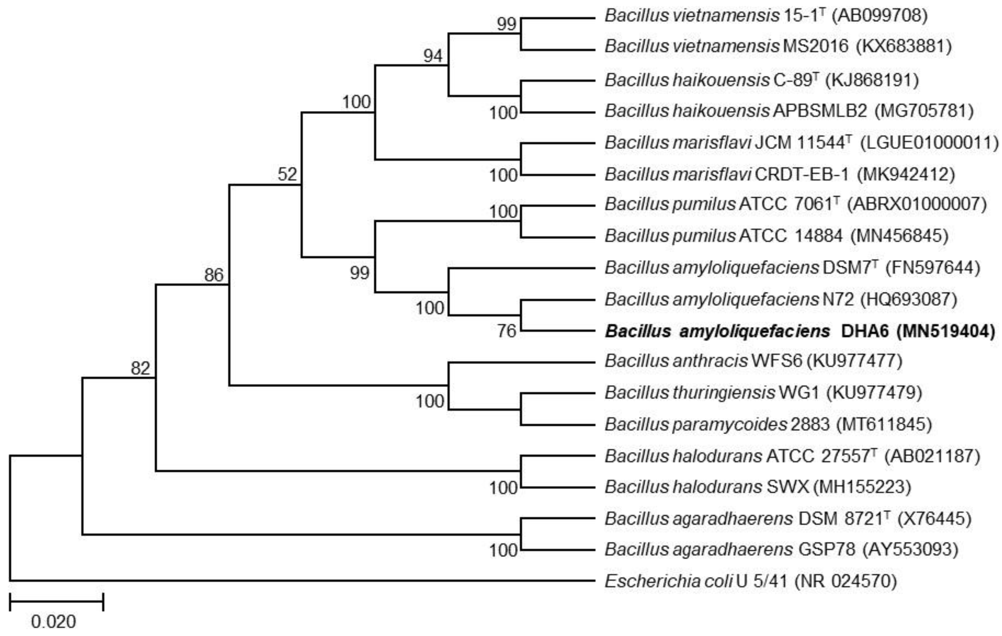
3.1. Molecular Characterization of Strain DHA6

To characterize strain DHA6 taxonomically, a 1023 bp fragment of the 16S rRNA gene was amplified and sequenced. Sequence alignment and phylogenetic analysis revealed a high similarity between the 16S rRNA gene sequence of strain DHA6 (GenBank accession no. MN519404) and other Bacillus spp. The DHA6 16S rRNA sequence closely clustered with B. amyloliquefaciens N72, showing >99% sequence identity to strain N72 (Figure 1). Therefore, it is likely that strain DHA6 belongs to B. amyloliquefaciens.

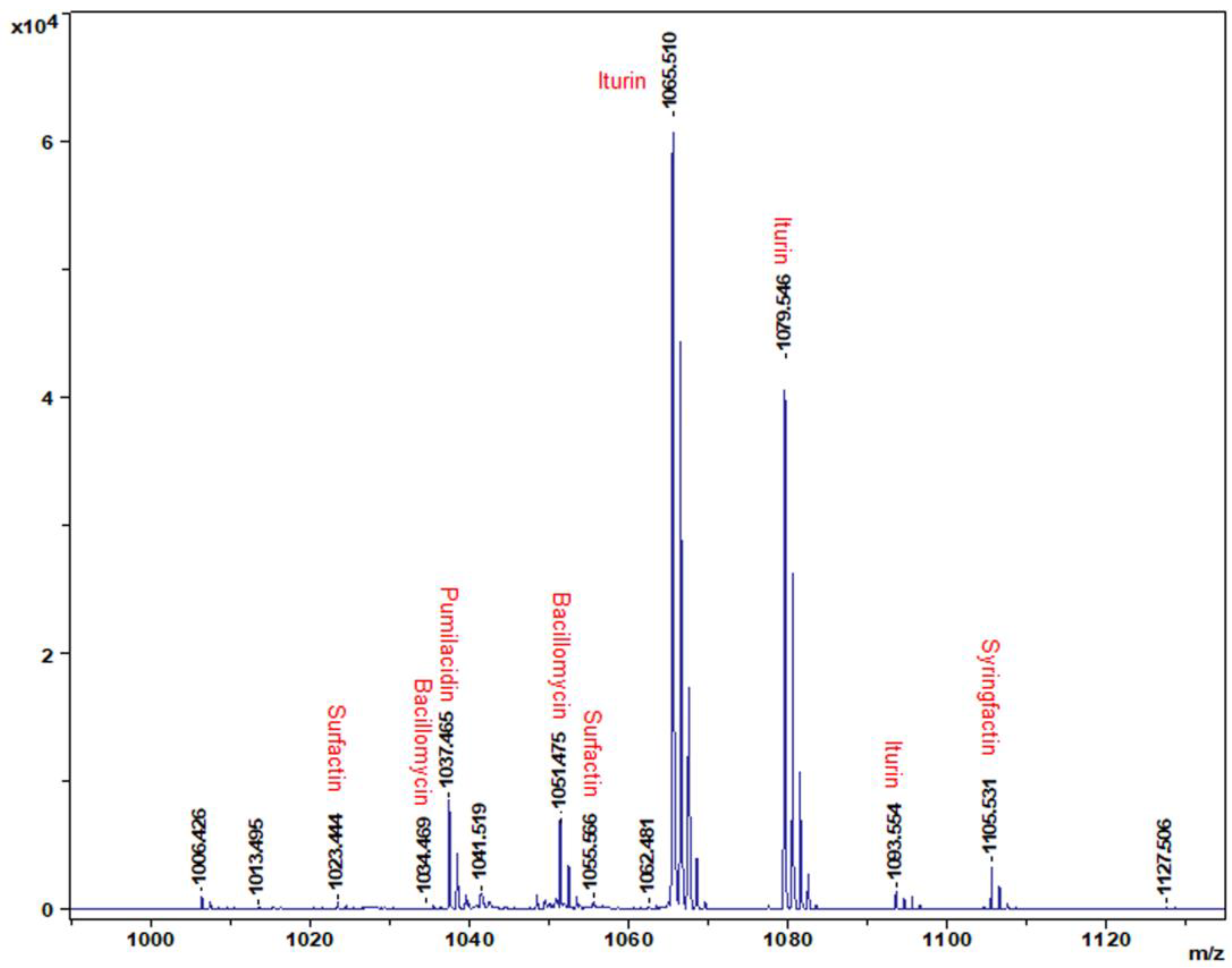
3.2. Identification of CLPs Produced by B. amyloliquefaciens Strain DHA6

MALDI-TOF analysis identified different B. amyloliquefaciens DHA6-produced CLPs, with standard peaks ranging from 1000 to 1120 m/z (Figure 2). The highest peak in the MALDI-TOF profile typically corresponds to non-ribosomal CLPs. B. amyloliquefaciens DHA6 was found to produce five classes of CLPs, namely iturin (m/z:1065.510, 1079.546, and 1093.554), surfactin (m/z:1055.566, and 1023.444), bacillomycin (m/z:1034.469, and 1051.475), pumilacidin (m/z:1037.465), and syringfactin (m/z:1105.531) (Figure 2). These data indicate that B. amyloliquefaciens DHA6 can produce diverse CLPs, which could contribute to its antifungal activity against different phytopathogenic fungi [19].

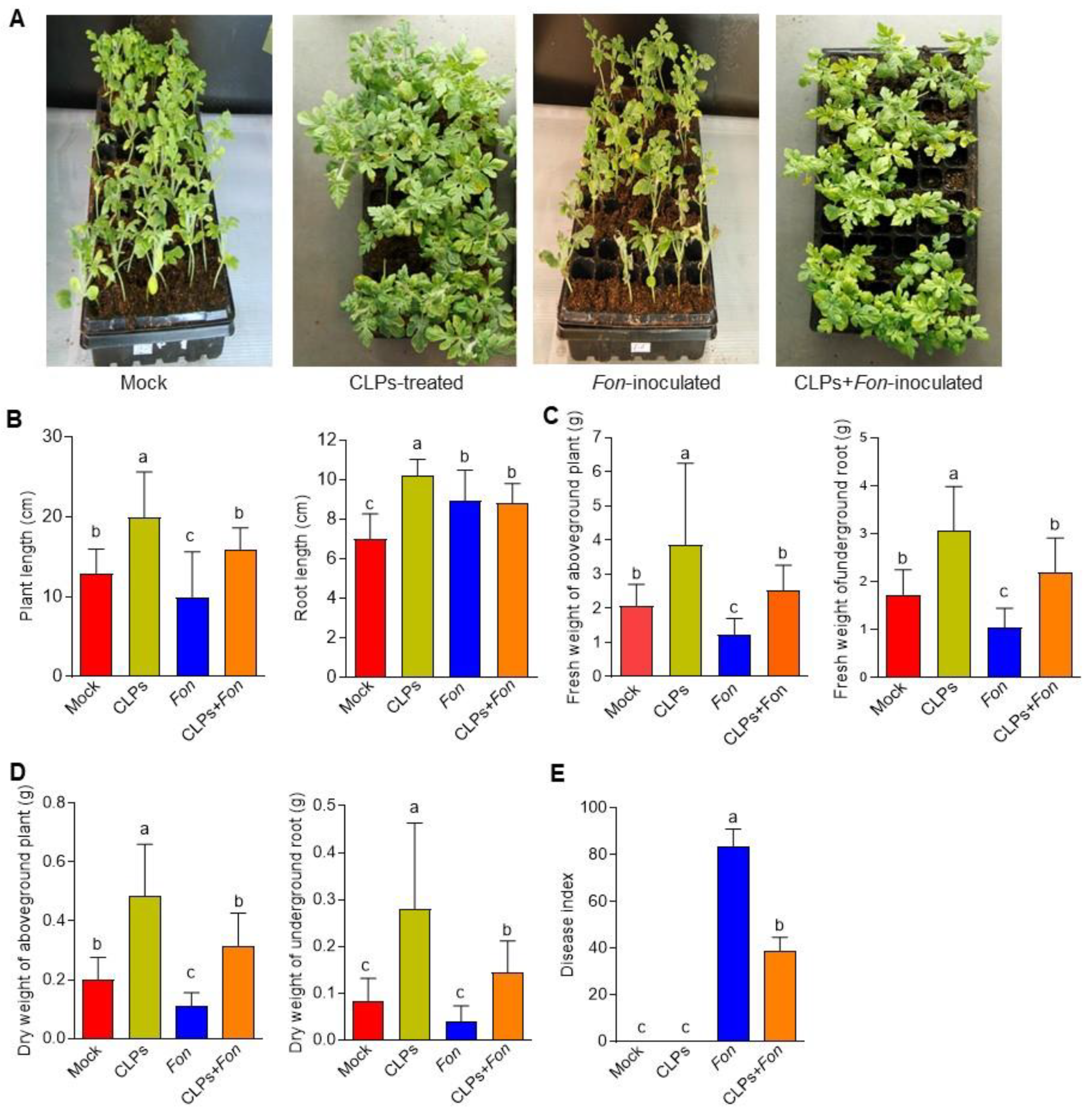
3.3. Plant Growth Promotion and Fusarium Wilt Suppression in Watermelon by CLPs

We previously demonstrated that B. amyloliquefaciens DHA6 could improve plant growth and suppress watermelon Fusarium wilt [19]. In this study, we investigated the potential roles of B. amyloliquefaciens DHA6-produced CLPs in promoting plant growth and suppressing Fusarium wilt in watermelon. Repeated greenhouse experiments showed a significant improvement in the growth performance of uninfected CLPs-treated plants compared to untreated healthy controls (Figure 3A), which was evident from the increased plant height (35.17%), root length (31.37%), as well as the fresh and dry weight of aboveground plants (46.29% and 58.88%, respectively) and roots (43.41% and 69.82%, respectively) (Figure 3B–D).
In disease assays, Fon-inoculated plants showed a higher disease index (83.33) and typical Fusarium wilt disease symptoms, including yellowing and wilting of leaves, while the Fon-inoculated CLPs-pretreated plants displayed significantly less disease severity index (38.67) and disease symptoms, such as slight chlorosis (Figure 3A), leading to a reduction of 54.6% in disease index (Figure 3E). During the experiments, no obvious disease symptom was observed in mock- and CLPs-pretreated plants (Figure 3E). Furthermore, the growth parameters of the Fon-infected plants were dramatically reduced compared to the uninfected control plants (Figure 3B–D). In contrast, the growth of Fon-inoculated CLPs-pretreated plants was comparable to the control healthy plants (Figure 3B–D). These results indicate that B. amyloliquefaciens DHA6-produced CLPs can promote plant growth and suppress Fusarium wilt in watermelon under greenhouse conditions.

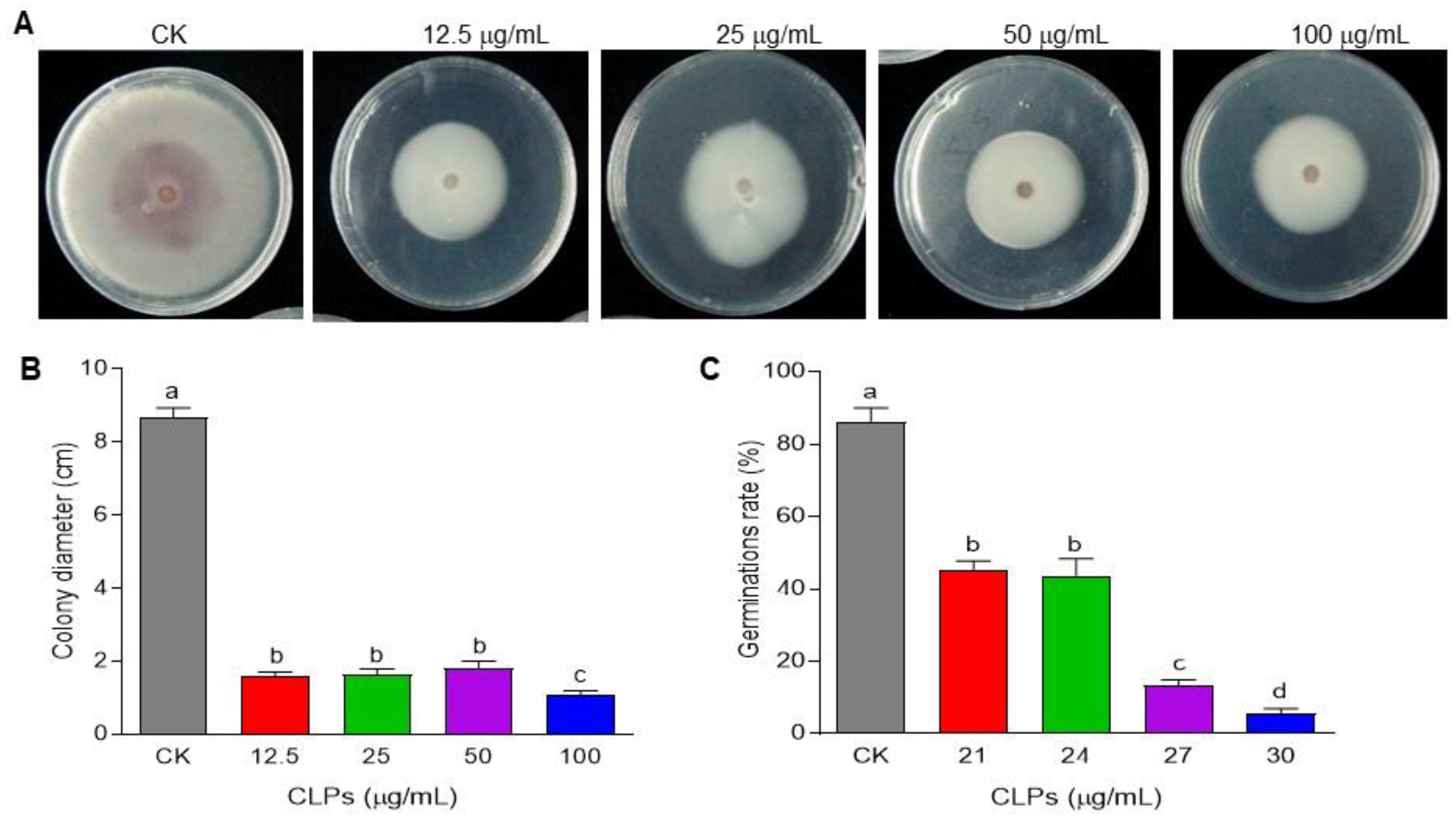
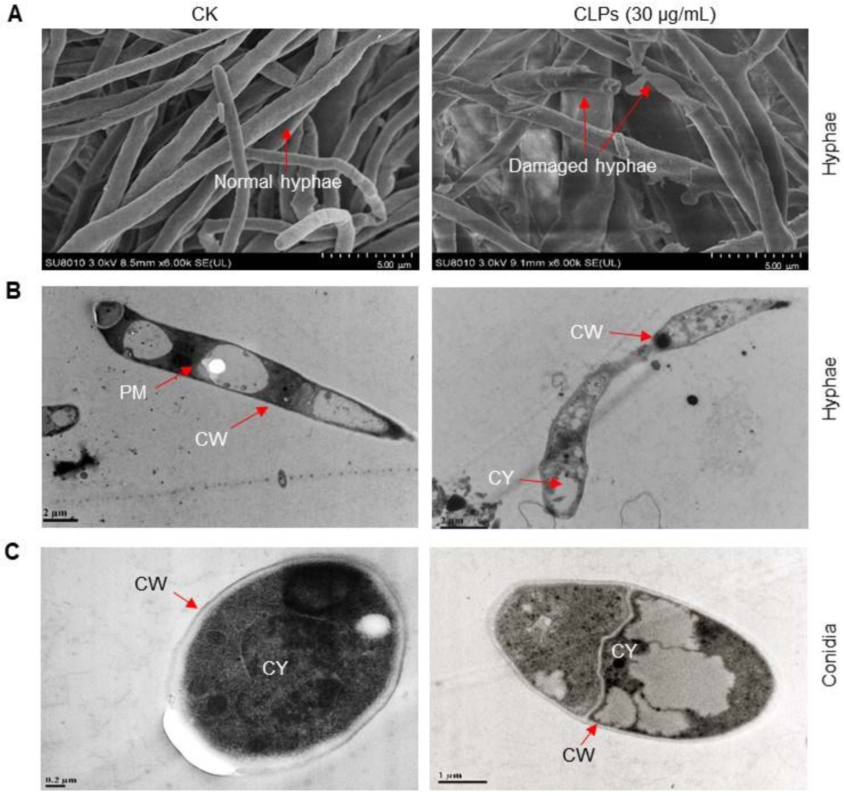
3.4. Antifungal Activity of CLPs against Fon

To explore the mechanisms underlying B. amyloliquefaciens DHA6-mediated suppression of watermelon Fusarium wilt, as previously reported [19], we examined the impact of CLPs on mycelial growth and spore germination in Fon. In these experiments, the specific CLPs concentrations for mycelial growth and spore germination were selected based on pilot experiments that indicated varying sensitivities of Fon mycelia and conidia to CLPs. Overall, CLPs significantly inhibited the mycelial growth of Fon (Figure 4A), resulting in smaller mycelial colonies of Fon on CLPs-supplemented PDA (Figure 4B). CLPs inhibited the mycelial growth of Fon in a dosage-dependent manner, with a maximum antifungal activity observed at 100 μg/mL CLPs, leading to an 87.3% inhibition of Fon mycelial growth (Figure 4A,B). Furthermore, CLPs also inhibited the germination of Fon spores in a dosage-dependent manner. At 30 µg/mL, CLPs inhibited the spore germination, resulting in a 93.8% reduction compared to untreated spores (Figure 4C). These results indicate that B. amyloliquefaciens DHA6-produced CLPs exhibit remarkable antifungal activity against Fon, with the effectiveness varying based on the dosage, and contribute to the biocontrol potential of B. amyloliquefaciens DHA6 in suppressing watermelon Fusarium wilt.

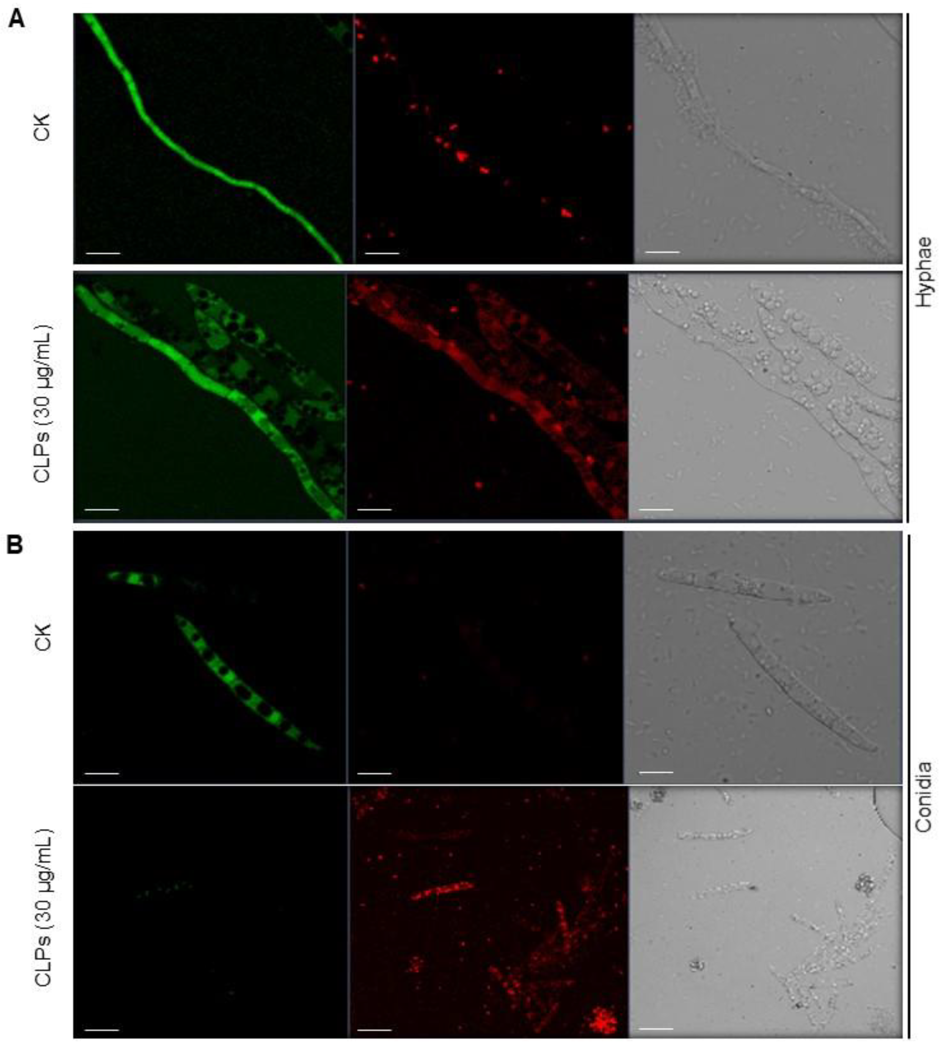
3.5. Inhibitory Effect of CLPs on Fon Viability

To explore the putative causes of the antifungal activity of CLPs, we examined their effect on the viability and ultrastructural changes of Fon through FDA and PI staining. FDA generates green fluorescence after entering living cells, serving as an indicator of living cells, while PI is a nucleus-specific dye for detecting apoptosis, serving as a marker of dead cells [50,56]. Without CLPs treatment, Fon mycelia and conidia showed strong FDA-generated green fluorescence, indicating normal morphology and intact structures, with minimal red fluorescence in mycelia only, representing dead or damaged fungal cells (Figure 5A,B). However, the CLPs-treated Fon mycelia and conidia exhibited bright red fluorescence, indicating disrupted morphology and structure. After staining, the CLPs-treated mycelia showed diffuse red and green fluorescence, but the CLPs-treated conidia no longer exhibited green fluorescence (Figure 5A). These observations suggest that B. amyloliquefaciens DHA6-produced CLPs affect the integrity of Fon cells, leading to cell damage and reduced viability.
To further investigate the effect of CLPs on the morphology and integrity of Fon, we examined ultrastructural changes in mycelia and conidia through SEM and TEM. SEM images revealed that untreated mycelia had regular, intact and column-like trunks, while the CLPs-treated mycelia showed obvious structural damages, such as disintegrated and collapsed mycelial structures (Figure 6A). Similarly, TEM images showed that untreated mycelia and conidia maintained normal cellular morphology and structure, with intact cell walls, plasma membranes, and cytoplasm (Figure 6B,C). In contrast, CLPs-treated mycelia and conidia displayed abnormal morphology and structure, with damaged plasma membrane and cell wall (Figure 6B,C). These results indicate that B. amyloliquefaciens DHA6-produced CLPs can disrupt the cellular structures and compromise the integrity of Fon, thereby influencing its viability.

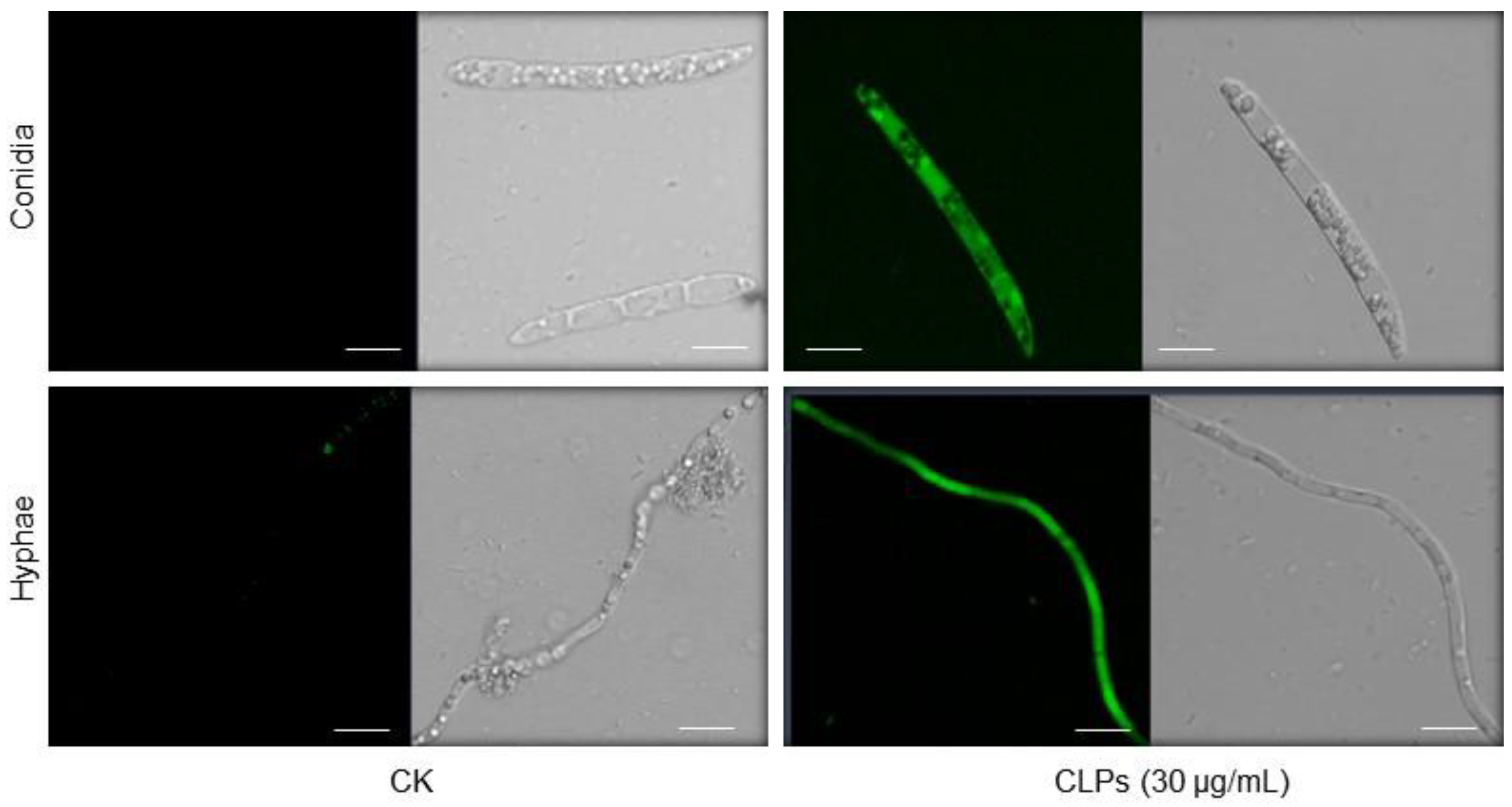
3.6. Induction of ROS Accumulation in Fon by CLPs

To gain insights into the physiological and biochemical mechanisms underlying CLPs-induced structural damages and antifungal activity against Fon, we assessed the effect of CLPs on ROS accumulation in the fungus using the DCFH-DA staining method. Results indicated that untreated Fon mycelia and conidia showed no significant green fluorescence, while CLPs-treated mycelia and conidia exhibited strong green fluorescence, reflecting higher ROS accumulation (Figure 7). These data indicate that B. amyloliquefaciens DHA6-produced CLPs can induce ROS accumulation in Fon, resulting in structural disintegration.

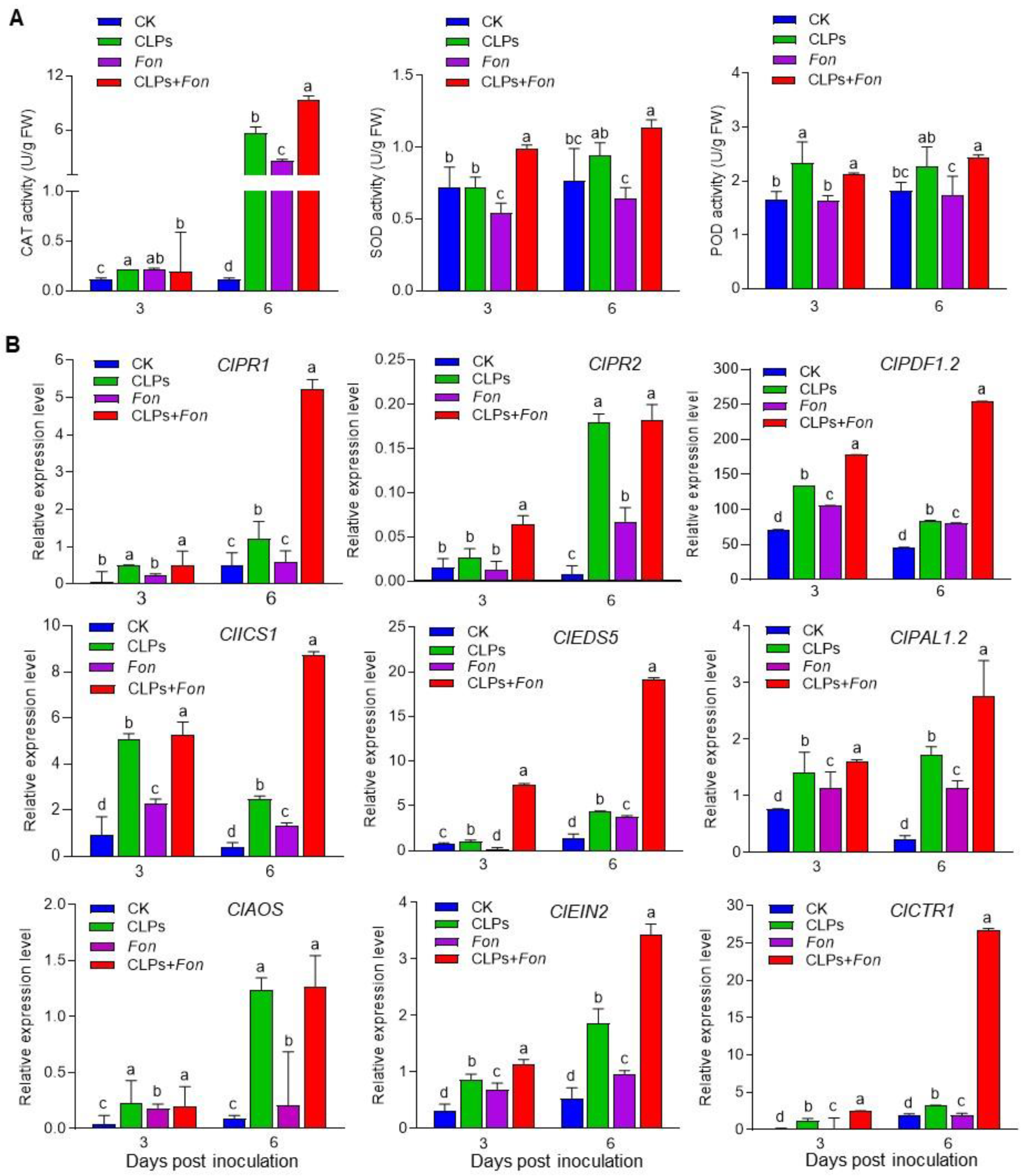
3.7. Enhancement of Antioxidative Capacity and Upregulation of Defense Gene Expression by CLPs

We further investigated the effect of B. amyloliquefaciens DHA6-produced CLPs on the defense response in watermelon plants to decipher the Fusarium wilt-suppressing mechanism. We examined the activities of CAT, SOD, and POD, three main ROS scavenging enzymes, in CLPs-pretreated plants before and after Fon infection. The activities of these antioxidant enzymes were significantly induced in CLPs-pretreated uninfected plants compared to untreated healthy plants at 3 dpi (except SOD), while CLPs-pretreated healthy plants showed significantly higher CAT level (97.98%) than untreated healthy controls at 6 dpi (Figure 8A). In Fon-infected plants, the SOD activity was significantly decreased by 24.65% at 3 dpi, while CAT activity was markedly increased by 95.62% at 6 dpi compared to untreated healthy plants (Figure 8A). In CLPs-pretreated Fon-infected plants, the activities of these enzymatic antioxidants were significantly higher compared to untreated Fon-infected plants at 3 dpi (except CAT) and 6 dpi (Figure 8A). These results indicate that B. amyloliquefaciens DHA6-produced CLPs can strengthen the antioxidative capacity by boosting CAT and SOD activities in watermelon plants, particularly in response to Fon infection.
To understand the molecular mechanism underlying the suppression of Fusarium wilt by B. amyloliquefaciens DHA6-produced CLPs, we analyzed the expression changes of selected defense-related genes in watermelon plants following CLPs treatment and Fon infection. In healthy plants, the expression levels of defense genes (ClPR1, ClPR2, and ClPDF1.2), salicylic acid (SA) signaling genes (ClICS1, ClEDS5, and ClPAL2), and JA/ET signaling genes (ClAOS, ClEIN2, and ClCTR1) were significantly upregulated in CLPs-pretreated healthy plants compared to untreated healthy controls (Figure 8B). In diseased plants, Fon infection induced the expression of these genes compared to untreated healthy plants, but their levels (except ClEDS5, ClPAL1.2, and ClEIN2 at 6 d) were lower than those in CLPs-treated healthy plants (Figure 8B). In CLPs-pretreated infected plants, the expression levels of these genes were significantly upregulated compared to untreated/CLPs-pretreated healthy and untreated Fon-infected plants (Figure 8B). These results suggest that B. amyloliquefaciens DHA6-produced CLPs can induce the expression of genes involved in both SA and JA/ET signaling and defense response, especially upon Fon infection.

4. Discussion

Bacillus spp. employ multiple mechanisms to suppress phytopathogens and improve plant growth [1,2]. These mechanisms include direct and indirect antagonistic approaches, such as competition for nutrients and space, production of antimicrobial agents, and induction of plant defense networks. Among these, the production of antimicrobial metabolites is the primary inhibitory mechanism of Bacillus spp. [1,5]. In a previous study, we characterized six antagonistic watermelon rhizosphere colonizing bacterial strains, DHA4, DHA6, DHA10, DHA12, DHA41, and DHA55, which displayed substantial potential in promoting plant growth and suppressing watermelon Fusarium wilt [19]. In the present study, we investigated the underlying plant growth-promoting and disease-suppressing mechanism of strain DHA6, which previously provided significant protection against Fusarium wilt [19]. Molecular and biochemical characterization revealed that strain DHA6, belonging to B. amyloliquefaciens, produces antifungal CLPs (Figure 1). Our results showed that B. amyloliquefaciens DHA6-produced CLPs suppressed watermelon Fusarium wilt by inhibiting its hyphal growth and spore germination. Moreover, CLPs purified from DHA6 strain induced significant structural damage and ROS accumulation in Fon, reducing viability. These findings suggest that B. amyloliquefaciens DHA6-produced CLPs can be an effective alternative to conventional fungicides for managing Fon-induced wilt disease.
B. amyloliquefaciens strains have previously demonstrated biocontrol potential against Fusarium wilt in cucumber, tomato, and banana [11,57,58]. The production of bioactive compounds, including CLPs, is recognized as a crucial factor contributing to their biocontrol potential against crop diseases [16,17,18]. Bacillus spp. have been found to produce different CLPs, such as iturin, iturinA, bacillomycin D, and fengycin, which are well known for their disease-controlling ability [16,17,18,19]. In the present study, MALDI-TOF MS analysis revealed that B. amyloliquefaciens DHA6 produces five families of CLPs, i.e., iturin, iturinA, bacillomycin D, pumilacidin, and fengycin (Figure 2), consistent with CLPs profiles observed in other B. amyloliquefaciens strains, such as DHA55, NCPSJ7, YN201732, and PPL [19,22,23,59]. Pot experiments revealed that CLPs, similar to their source B. amyloliquefaciens DHA6 [19], possess significant potential in promoting plant growth and suppressing Fusarium wilt in watermelon (Figure 3), indicating that the production of CLPs is a prominent mechanism of B. amyloliquefaciens DHA6 involved in plant growth promotion and biocontrol of Fusarium wilt. Similarly, previous studies have demonstrated that iturin A- and surfactin A-enriched CLPs derived from B. subtilis BS-1 and B. amyloliquefaciens S76-3 have been shown to suppress kiwifruit rot (Botryosphaeria dothidea), rice bakanae disease (Fusarium moniliforme), wheat head scab (Fusarium graminearum), and lettuce Fusarium wilt [47,60,61,62]. Thus, antagonistic bacteria employ different antimicrobial CLPs as a primary mechanism for the biocontrol of crop diseases [16].
Previously, B. amyloliquefaciens DHA6 displayed antifungal activity against five different fungi, including Fon, Didymella bryoniae (causing gummy stem blight on cucurbits), Sclerotinia sclerotiorum (causing stem rot on a wide range of plants), F. graminearum, and R. solani (causing damping-off in a wide range of plants) [19]. This study found that CLPs from B. amyloliquefaciens DHA6 significantly inhibited mycelial growth and conidia germination of Fon (Figure 4), providing mechanistic insights into the direct antifungal activity of B. amyloliquefaciens DHA6 against phytopathogenic fungi. Previous studies have reported that CLPs purified from different B. amyloliquefaciens strains, such as fengycins and iturins, inhibited mycelial growth and spore germination of Fo in vitro [21,22,23,63,64]. Iturins produced by Bacillus spp. have been well known for their strong antifungal activity against Fo [63,64]. B. amyloliquefaciens DHA6 produced at least three types of iturins (Figure 2), which caused damage to the mycelia and conidia, disrupting the plasma membrane and cell wall, affecting Fon viability (Figure 4, Figure 5 and Figure 6). Similarly, CLPs produced by B. velezensis WB caused morphological changes, such as surface subsidence and cytoplasmic shrinkage, in Fon [20], while bacillomycin D, produced by B. amyloliquefaciens FZB42, induced disruptions in the plasma membrane and cell wall, leading to cell death of mycelia and conidia in F. graminearum [50]. The B. amyloliquefaciens DHA6-produced CLPs triggered a significant accumulation of ROS in the mycelia and conidia of Fon (Figure 7). These results align with previous observations indicating that iturins, fengycins, bacillomycin D and other CLPs triggered excessive ROS accumulation in Fon, F. graminearum, Verticillium dahliae, Magnaporthe oryzae, and S. sclerotiorum [20,50,65,66,67]. Although a physiological level of ROS is critical as an intracellular messenger in many biological events, the excessive accumulation of ROS can cause oxidative stress and severe cellular damage by disrupting cellular integrity. Thus, the disruption of ROS homeostasis is one of the physiological and biochemical mechanisms through which CLPs induce cell death and exhibit antifungal activity against different phytopathogenic fungi.
In addition to the direct antimicrobial activity, certain Bacillus spp., including B. subtilis and B. amyloliquefaciens, have been shown to strengthen plant defenses by priming ISR [7,68]. Bioactive compounds produced by different Bacillus spp., including CLPs, have been identified as key elicitors of ISR [7,69]. For example, iturins, surfactins, and fengycins produced by different Bacillus spp. have been found to induce significant ISR-mediated protection in various crop plants against different fungal infections [27,70,71,72]. In the present study, pretreatment with the B. amyloliquefaciens DHA6-produced CLPs, containing iturins, surfactins, pumilacidin, and fengycins (Figure 2), significantly reduced Fusarium wilt in watermelon (Figure 3). This reduction can be attributed to an enhanced defense response primed by CLPs in the plants, as direct contact between CLPs and Fon was avoided using a specific approach. The induction of plant defense responses by CLPs was further confirmed by the increased activity of antioxidative enzymes, i.e., CAT, SOD, and POD (Figure 8A), in watermelon plants upon CLPs pretreatment, supporting the notion that CLPs pretreatment induced plant innate defense mechanisms to suppress Fusarium wilt development. Similar results have been reported in infected watermelon and tomato plants, where B. velezensis F21 and B. amyloliquefaciens Oj-2.16 induced the activities of CAT, POD, and SOD through the production of antioxidant-triggering CLPs [12,73]. The plant immune system is believed to rely on coordinated signaling pathways, including SA, JA, and ET pathways, to mount an effective defense response against pathogen attack [12]. Bacillus-primed ISR has been shown to involve the JA/ET and SA signaling pathways and multiple early signaling events, such as MAPK cascades and ROS [26,28,74,75,76,77]. In lettuce, B. amyloliquefaciens FZB42-produced surfactin, and other immunomodulatory CLPs triggered immunity against R. solani by inducing the expression of defense-related genes, including LsPDF1.2, LsPR1, and LsLOX [28], while fusaricidin, purified from Paenibacillus polymyxa WLY78, induced SA-specific ISR to suppress Fusarium wilt in cucumber [78]. Consistent with these findings, we observed significant upregulation of SA-signaling/responsive, JA/ET signaling, and JA-responsive genes by CLPs, with or without Fon infection (Figure 8B), indicating their potential to activate a broad-spectrum defense response in watermelon plants. Therefore, it is likely that B. amyloliquefaciens DHA6-produced CLPs activate defense responses through the SA and JA/ET signaling pathways, thereby protecting watermelon plants from Fon infection.

5. Conclusions

In this study, the antagonistic bacterium B. amyloliquefaciens DHA6 was found to produce five families of antifungal CLPs, i.e., iturin, surfactin, bacillomycin, syrignfactin, and pumilacidin, which showed inhibitory effects against Fon. These CLPs inhibited mycelial growth and spore germination of Fon by promoting ROS accumulation and disrupting structural integrity of the pathogen. Furthermore, under greenhouse conditions, B. amyloliquefaciens DHA56-produced CLPs displayed plant growth-promoting activity and effectively suppressed Fusarium wilt in watermelon. This was accompanied by the activation of defense responses, as evidenced by enhanced activity of antioxidative enzymes and upregulation of genes involved in SA and JA/ET signaling pathways in watermelon plants. Our findings suggest that CLPs play a crucial role in the biocontrol capacity of B. amyloliquefaciens DHA6 against Fusarium wilt, acting through their direct antifungal activity and ability to induce plant defense responses. The potential of B. amyloliquefaciens DHA6-produced CLPs in promoting plant growth and suppressing Fusarium wilt in watermelon provides a basis for the development of B. amyloliquefaciens DHA6-based biopesticides, which can serve as both antimicrobial agents and resistance inducers, offering sustainable protection to important crops against disease damage under field conditions.

Author Contributions

Conceptualization, F.S. and D.M.K.A.-M.; methodology, D.L. and D.M.K.A.-M.; formal analysis, F.S. and D.M.K.A.-M.; investigation, D.M.K.A.-M., M.N., N.S.A.A. and A.; data curation, D.M.K.A.-M. and F.S.; writing—original draft preparation, D.M.K.A.-M. and F.S.; writing—review and editing, F.S. and M.N.; funding acquisition, F.S. All authors have read and agreed to the published version of the manuscript.

Funding

This research was funded by the China Agriculture Research System of MOF and MARA of China (Grant no. CARS-25) to F. Song.

Institutional Review Board Statement

Not applicable.

Informed Consent Statement

Not applicable.

Data Availability Statement

All the data are present inside manuscript file.

Conflicts of Interest

The authors declare no conflict of interest.

References

  1. Khan, A.R.; Mustafa, A.; Hyder, S.; Valipour, M.; Rizvi, Z.F.; Gondal, A.S.; Yousuf, Z.; Iqbal, R.; Daraz, U. Bacillus spp. as bioagents: Uses and application for sustainable agriculture. Biology 2022, 11, 1763. [Google Scholar] [CrossRef] [PubMed]
  2. Bonaterra, A.; Badosa, E.; Daranas, N.; Francés, J.; Roselló, G.; Montesinos, E. Bacteria as biological control agents of plant diseases. Microorganisms 2022, 10, 1759. [Google Scholar] [CrossRef] [PubMed]
  3. Tyśkiewicz, R.; Nowak, A.; Ozimek, E.; Jaroszuk-Ściseł, J. Trichoderma: The current status of its application in agriculture for the biocontrol of fungal phytopathogens and stimulation of plant growth. Int. J. Mol. Sci. 2022, 23, 2329. [Google Scholar] [CrossRef] [PubMed]
  4. Sarrocco, S. Biological disease control by beneficial (micro)organisms: Selected breakthroughs in the past 50 years. Phytopathology 2023, 113, 732–740. [Google Scholar] [CrossRef] [PubMed]
  5. Luo, L.; Zhao, C.; Wang, E.; Raza, A.; Yin, C. Bacillus amyloliquefaciens as an excellent agent for biofertilizer and biocontrol in agriculture: An overview for its mechanisms. Microbiol. Res. 2022, 259, 127016. [Google Scholar] [CrossRef] [PubMed]
  6. Ngalimat, M.S.; Yahaya, R.S.R.; Baharudin, M.M.A.; Yaminudin, S.M.; Karim, M.; Ahmad, S.A.; Sabri, S. A review on the biotechnological applications of the operational group Bacillus amyloliquefaciens. Microorganisms 2021, 9, 614. [Google Scholar] [CrossRef]
  7. Blake, C.; Christensen, M.N.; Kovács, Á.T. Molecular aspects of plant growth promotion and protection by Bacillus subtilis. Mol. Plant-Microbe Interact. 2021, 34, 15–25. [Google Scholar] [CrossRef]
  8. Miljaković, D.; Marinković, J.; Balešević-Tubić, S. The significance of Bacillus spp. in disease suppression and growth promotion of field and vegetable crops. Microorganisms 2020, 8, 1037. [Google Scholar] [CrossRef]
  9. Khan, N.; Maymon, M.; Hirsch, A.M. Combating Fusarium infection using Bacillus-based antimicrobials. Microorganisms 2017, 5, 75. [Google Scholar] [CrossRef] [Green Version]
  10. Xu, W.; Yang, Q.; Yang, F.; Xie, X.; Goodwin, P.H.; Deng, X.; Tian, B.; Yang, L. Evaluation and genome analysis of Bacillus subtilis YB-04 as a potential biocontrol agent against Fusarium wilt and growth promotion agent of cucumber. Front. Microbiol. 2022, 13, 885430. [Google Scholar] [CrossRef]
  11. Fan, H.; Li, S.; Zeng, L.; He, P.; Xu, S.; Bai, T.; Huang, Y.; Guo, Z.; Zheng, S.J. Biological control of Fusarium oxysporum f. sp. cubense tropical race 4 using natively isolated Bacillus spp. YN0904 and YN1419. J. Fungi 2021, 7, 795. [Google Scholar] [CrossRef]
  12. Jiang, C.H.; Yao, X.F.; Mi, D.D.; Li, Z.J.; Yang, B.Y.; Zheng, Y.; Qi, Y.J.; Guo, J.H. Comparative transcriptome analysis reveals the biocontrol mechanism of Bacillus velezensis F21 against Fusarium wilt on watermelon. Front. Microbiol. 2019, 10, 652. [Google Scholar] [CrossRef]
  13. Karthika, S.; Remya, M.; Varghese, S.; Dhanraj, N.D.; Sali, S.; Rebello, S.; Jose, S.M.; Jisha, M.S. Bacillus tequilensis PKDN31 and Bacillus licheniformis PKDL10-As double headed swords to combat Fusarium oxysporum f. sp. lycopersici induced tomato wilt. Microb. Pathog. 2022, 172, 105784. [Google Scholar]
  14. Fisher, M.C.; Henk, D.A.; Briggs, C.J.; Brownstein, J.S.; Madoff, L.C.; McCraw, S.L.; Gurr, S.J. Emerging fungal threats to animal, plant and ecosystem health. Nature 2012, 484, 186–194. [Google Scholar] [CrossRef] [Green Version]
  15. Gordon, T.R. Fusarium oxysporum and the Fusarium wilt syndrome. Annu. Rev. Phytopathol. 2017, 55, 23–39. [Google Scholar] [CrossRef]
  16. Ongena, M.; Jacques, P. Bacillus lipopeptides: Versatile weapons for plant disease biocontrol. Trends Microbiol. 2008, 16, 115–125. [Google Scholar] [CrossRef]
  17. Caulier, S.; Nannan, C.; Gillis, A.; Licciardi, F.; Bragard, C.; Mahillon, J. Overview of the antimicrobial compounds produced by members of the Bacillus subtilis group. Front. Microbiol. 2019, 10, 302. [Google Scholar] [CrossRef] [Green Version]
  18. Penha, R.O.; Vandenberghe, L.P.S.; Faulds, C.; Soccol, V.T.; Soccol, C.R. Bacillus lipopeptides as powerful pest control agents for a more sustainable and healthy agriculture: Recent studies and innovations. Planta 2020, 251, 70. [Google Scholar] [CrossRef] [Green Version]
  19. Al-Mutar, A.M.A.; Abduljaleel, N.S.; Noman, M.; Azizullah; Li, D.; Song, F. Suppression of Fusarium wilt in watermelon by Bacillus amyloliquefaciens DHA55 through extracellular production of antifungal lipopolypeptides. J. Fungi 2023, 9, 336. [Google Scholar] [CrossRef]
  20. Wang, K.; Wang, Z.; Xu, W. Induced oxidative equilibrium damage and reduced toxin synthesis in Fusarium oxysporum f. sp. niveum by secondary metabolites from Bacillus velezensis WB. FEMS Microbiol. Ecol. 2022, 98, fiac080. [Google Scholar] [CrossRef]
  21. Moreno-Velandia, C.A.; Ongena, M.; Cotes, A.M. Effects of fengycins and iturins on Fusarium oxysporum f. sp. physali and root colonization by Bacillus velezensis Bs006 protect golden berry against vascular wilt. Phytopathology 2021, 111, 2227–2237. [Google Scholar] [CrossRef] [PubMed]
  22. Wang, J.; Qiu, J.; Yang, X.; Yang, J.; Zhao, S.; Zhou, Q.; Chen, L. Identification of lipopeptide produced by Bacillus amyloliquefaciens NCPSJ7 and its antifungal activities against Fusarium oxysporum f. sp. niveum. Foods 2022, 11, 2996. [Google Scholar] [CrossRef] [PubMed]
  23. Kang, B.R.; Park, J.S.; Jung, W.J. Antifungal evaluation of fengycin isoforms isolated from Bacillus amyloliquefaciens PPL against Fusarium oxysporum f. sp. lycopersici. Microb. Pathog. 2020, 149, 104509. [Google Scholar] [CrossRef] [PubMed]
  24. Wu, J.J.; Huang, J.W.; Deng, W.L. Phenylacetic acid and methylphenyl acetate from the biocontrol bacterium Bacillus mycoides BM02 suppress spore germination in Fusarium oxysporum f. sp. lycopersici. Front. Microbiol. 2020, 11, 569263. [Google Scholar] [CrossRef]
  25. Wang, H.; Wang, Z.; Liu, Z.; Wang, K.; Xu, W. Membrane disruption of Fusarium oxysporum f. sp. niveum induced by myriocin from Bacillus amyloliquefaciens LZN01. Microb. Biotechnol. 2021, 14, 517–534. [Google Scholar]
  26. Yamamoto, S.; Shiraishi, S.; Suzuki, S. Are cyclic lipopeptides produced by Bacillus amyloliquefaciens S13-3 responsible for the plant defence response in strawberry against Colletotrichum gloeosporioides? Lett. Appl. Microbiol. 2015, 60, 379–386. [Google Scholar] [CrossRef]
  27. Farace, G.; Fernandez, O.; Jacquens, L.; Coutte, F.; Krier, F.; Jacques, P.; Clément, C.; Barka, E.A.; Jacquard, C.; Dorey, S. Cyclic lipopeptides from Bacillus subtilis activate distinct patterns of defence responses in grapevine. Mol. Plant Pathol. 2015, 16, 177–187. [Google Scholar] [CrossRef]
  28. Chowdhury, S.P.; Uhl, J.; Grosch, R.; Alquéres, S.; Pittroff, S.; Dietel, K.; Schmitt-Kopplin, P.; Borriss, R.; Hartmann, A. Cyclic lipopeptides of Bacillus amyloliquefaciens subsp. plantarum colonizing the lettuce rhizosphere enhance plant defense responses toward the bottom rot pathogen Rhizoctonia solani. Mol. Plant-Microbe Interact. 2015, 28, 984–995. [Google Scholar] [CrossRef] [Green Version]
  29. Pazarlar, S.; Madriz-Ordeñana, K.; Thordal-Christensen, H. Bacillus cereus EC9 protects tomato against Fusarium wilt through JA/ET-activated immunity. Front. Plant Sci. 2022, 13, 1090947. [Google Scholar] [CrossRef]
  30. Ramírez, V.; Martínez, J.; Bustillos-Cristales, M.D.R.; Catañeda-Antonio, D.; Munive, J.A.; Baez, A. Bacillus cereus MH778713 elicits tomato plant protection against Fusarium oxysporum. J. Appl. Microbiol. 2022, 132, 470–482. [Google Scholar] [CrossRef]
  31. Sun, Y.; Huang, B.; Cheng, P.; Li, C.; Chen, Y.; Li, Y.; Zheng, L.; Xing, J.; Dong, Z.; Yu, G. Endophytic Bacillus subtilis TR21 improves banana plant resistance to Fusarium oxysporum f. sp. cubense and promotes root growth by upregulating the jasmonate and brassinosteroid biosynthesis pathways. Phytopathology 2022, 112, 219–231. [Google Scholar] [CrossRef]
  32. Jinal, N.H.; Amaresan, N. Evaluation of biocontrol Bacillus species on plant growth promotion and systemic-induced resistant potential against bacterial and fungal wilt-causing pathogens. Arch. Microbiol. 2020, 202, 1785–1794. [Google Scholar] [CrossRef]
  33. Akram, W.; Anjum, T.; Ali, B.; Ahmad, A. Screening of native bacillus strains to induce systemic resistance in tomato plants against fusarium wilt in split root system and its field applications. Int. J. Agric. Biol. 2013, 15, 1289–1294. [Google Scholar]
  34. Akram, W.; Anjum, T.; Ali, B. Phenylacetic acid is ISR determinant produced by Bacillus fortis IAGS162, which involves extensive re-modulation in metabolomics of tomato to protect against Fusarium wilt. Front. Plant Sci. 2016, 7, 498. [Google Scholar] [CrossRef] [Green Version]
  35. Kawagoe, Y.; Shiraishi, S.; Kondo, H.; Yamamoto, S.; Aoki, Y.; Suzuki, S. Cyclic lipopeptide iturin A structure-dependently induces defense response in Arabidopsis plants by activating SA and JA signaling pathways. Biochem. Biophys. Res. Commun. 2015, 460, 1015–1020. [Google Scholar] [CrossRef]
  36. Zhang, C.; Ou, X.; Wang, J.; Wang, Z.; Du, W.; Zhao, J.; Han, Y. Antifungal peptide P852 controls Fusarium wilt in faba bean (Vicia faba L.) by promoting antioxidant defense and isoquinoline alkaloid, betaine, and arginine biosyntheses. Antioxidants 2022, 11, 1767. [Google Scholar]
  37. Everts, K.L.; Himmelstein, J.C. Fusarium wilt of watermelon: Towards sustainable management of a re-emerging plant disease. Crop Prot. 2015, 73, 93–99. [Google Scholar] [CrossRef]
  38. Gao, Y.; Xiong, X.; Wang, H.; Wang, J.; Bi, Y.; Yan, Y.; Cao, Z.; Li, D.; Song, F. Ero1-Pdi1 module-catalysed dimerization of a nucleotide sugar transporter, FonNst2, regulates virulence of Fusarium oxysporum on watermelon. Environ. Microbiol. 2022, 24, 1200–1220. [Google Scholar] [CrossRef]
  39. Li, B.; Xu, L.; Lou, M.; Li, F.; Zhang, Y.; Xie, G.J. Isolation and characterization of antagonistic bacteria against bacterial leaf spot of Euphorbia pulcherrima. Lett. Appl. Microbiol. 2008, 46, 450–455. [Google Scholar] [CrossRef]
  40. Thompson, J.D.; Gibson, T.J.; Plewniak, F.; Jeanmougin, F.; Higgins, D.G. The CLUSTAL_X windows interface: Flexible strategies for multiple sequence alignment aided by quality analysis tools. Nucleic Acids Res. 1997, 25, 4876–4882. [Google Scholar] [CrossRef] [Green Version]
  41. Tamura, K.; Peterson, D.; Peterson, N.; Stecher, G.; Nei, M.; Kumar, S. MEGA5: Molecular evolutionary genetics analysis using maximum likelihood, evolutionary distance, and maximum parsimony methods. Mol. Biol. Evol. 2011, 28, 2731–2739. [Google Scholar] [CrossRef] [PubMed] [Green Version]
  42. Lin, S.C.; Minton, M.A.; Sharma, M.M.; Georgiou, G. Structural and immunological characterization of a biosurfactant produced by Bacillus licheniformis JF-2. Appl. Environ. Microbiol. 1994, 60, 31–38. [Google Scholar] [CrossRef] [PubMed] [Green Version]
  43. Vater, J.; Gao, X.; Hitzeroth, G.; Wilde, C.; Franke, P. “Whole cell”—matrix-assisted laser desorption ionization-time of flight-mass spectrometry, an emerging technique for efficient screening of biocombinatorial libraries of natural compounds-present state of research. Comb. Chem. High Throughput Screen. 2003, 6, 557–567. [Google Scholar] [CrossRef] [PubMed]
  44. Masum, M.M.I.; Liu, L.; Yang, M.; Hossain, M.M.; Siddiqa, M.M.; Supty, M.E.; Ogunyemi, S.O.; Hossain, A.; An, Q.; Li, B. Halotolerant bacteria belonging to operational group Bacillus amyloliquefaciens in biocontrol of the rice brown stripe pathogen Acidovorax oryzae. J. Appl. Microbiol. 2018, 125, 1852–1867. [Google Scholar] [CrossRef] [PubMed]
  45. Tagg, J.; McGiven, A. Assay system for bacteriocins. Appl. Microbiol. 1971, 21, 943. [Google Scholar] [CrossRef]
  46. Wang, Y.; Sun, Y.; Zhang, Y.; Zhang, X.; Feng, J. Antifungal activity and biochemical response of cuminic acid against Phytophthora capsici Leonian. Molecules 2016, 21, 756. [Google Scholar] [CrossRef] [Green Version]
  47. Gong, A.D.; Li, H.P.; Yuan, Q.S.; Song, X.S.; Yao, W.; He, W.J.; Zhang, J.B.; Liao, Y.C. Antagonistic mechanism of iturin A and plipastatin A from Bacillus amyloliquefaciens S76-3 from wheat spikes against Fusarium graminearum. PLoS ONE 2015, 10, e0116871. [Google Scholar] [CrossRef] [Green Version]
  48. Noman, M.; Ahmed, T.; Ijaz, U.; Shahid, M.; Nazir, M.M.; Azizullah; White, J.C.; Li, D.; Song, F. Bio-functionalized manganese nanoparticles suppress Fusarium wilt in watermelon (Citrullus lanatus L.) by infection disruption, host defense response potentiation, and soil microbial community modulation. Small 2023, 19, e2205687. [Google Scholar] [CrossRef]
  49. Azizullah; Noman, M.; Gao, Y.; Wang, H.; Xiong, X.; Wang, J.; Li, D.; Song, F. The SUMOylation pathway components are required for vegetative growth, asexual development, cytotoxic responses, and programmed cell death events in Fusarium oxysporum f. sp. niveum. J. Fungi 2023, 9, 94. [Google Scholar] [CrossRef]
  50. Gu, Q.; Yang, Y.; Yuan, Q.; Shi, G.; Wu, L.; Lou, Z.; Huo, R.; Wu, H.; Borriss, R.; Gao, X. Bacillomycin D produced by Bacillus amyloliquefaciens is involved in the antagonistic interaction with the plant pathogenic fungus Fusarium graminearum. Appl. Environ. Microbiol. 2017, 83, e01075-17. [Google Scholar] [CrossRef] [Green Version]
  51. Liu, P.; Luo, L.; Guo, J.; Liu, H.; Wang, B.; Deng, B.; Long, C.A.; Cheng, Y. Farnesol induces apoptosis and oxidative stress in the fungal pathogen Penicillium expansum. Mycologia 2010, 102, 311–318. [Google Scholar] [CrossRef]
  52. Dai, Y.; Cao, Z.; Huang, L.; Liu, S.; Shen, Z.; Wang, Y.; Wang, H.; Zhang, H.; Li, D.; Song, F. CCR4-not complex subunit Not2 plays critical roles in vegetative growth, conidiation and virulence in watermelon Fusarium wilt pathogen Fusarium oxysporum f. sp. niveum. Front. Microbiol. 2016, 7, 1449. [Google Scholar] [CrossRef] [Green Version]
  53. Murage, E.N.; Masuda, M. Response of pepper and eggplant to continuous light in relation to leaf chlorosis and activities of antioxidative enzymes. Sci. Hortic. 1997, 70, 269–279. [Google Scholar] [CrossRef]
  54. Wang, C.Y. Effect of temperature preconditioning on catalase, peroxidase, and superoxide dismutase in chilled zucchini squash. Postharvest Biol. Technol. 1995, 5, 67–76. [Google Scholar] [CrossRef]
  55. Rhee, S.J.; Jang, Y.J.; Park, J.Y.; Ryu, J.; Lee, G.P. Virus-induced gene silencing for in planta validation of gene function in cucurbits. Plant Physiol. 2022, 190, 2366–2379. [Google Scholar] [CrossRef]
  56. Zhao, P.C.; Quan, C.S.; Wang, Y.G.; Wang, J.H.; Fan, S.D. Bacillus amyloliquefaciens Q-426 as a potential biocontrol agent against Fusarium oxysporum f. sp. spinaciae. J. Basic Microbiol. 2014, 54, 448–456. [Google Scholar] [CrossRef]
  57. Liu, Y.; Zhang, N.; Qiu, M.; Feng, H.; Vivanco, J.M.; Shen, Q.; Zhang, R. Enhanced rhizosphere colonization of beneficial Bacillus amyloliquefaciens SQR9 by pathogen infection. FEMS Microbiol. Lett. 2014, 353, 49–56. [Google Scholar] [CrossRef] [Green Version]
  58. Wu, K.; Fang, Z.; Wang, L.; Yuan, S.; Guo, R.; Shen, B.; Shen, Q. Biological potential of bioorganic fertilizer fortified with bacterial antagonist for the control of tomato bacterial wilt and the promotion of crop yields. J. Microbiol. Biotechnol. 2016, 26, 1755–1764. [Google Scholar] [CrossRef] [Green Version]
  59. Jiao, R.; Cai, Y.; He, P.; Munir, S.; Li, X.; Wu, Y.; Wang, J.; Xia, M.; He, P.; Wang, G.; et al. Bacillus amyloliquefaciens YN201732 produces lipopeptides with promising biocontrol activity against fungal pathogen Erysiphe cichoracearum. Front. Cell. Infect. Microbiol. 2021, 11, 598999. [Google Scholar] [CrossRef]
  60. Fan, Y.; Liu, K.; Lu, R.; Gao, J.; Song, W.; Zhu, H.; Tang, X.; Liu, Y.; Miao, M. Cell-free supernatant of Bacillus subtilis reduces kiwifruit rot caused by Botryosphaeria dothidea through inducing oxidative stress in the pathogen. J. Fungi 2023, 9, 127. [Google Scholar] [CrossRef]
  61. Sarwar, A.; Hassan, M.N.; Imran, M.; Iqbal, M.; Majeed, S.; Brader, G.; Sessitsch, A.; Hafeez, F.Y. Biocontrol activity of surfactin A purified from Bacillus NH-100 and NH-217 against rice bakanae disease. Microbiol. Res. 2018, 209, 1–13. [Google Scholar] [CrossRef] [PubMed]
  62. Fujita, S.; Yokota, K. Disease suppression by the cyclic lipopeptides iturin A and surfactin from Bacillus spp. against Fusarium wilt of lettuce. J. Gen. Plant Pathol. 2019, 85, 44–48. [Google Scholar] [CrossRef]
  63. Han, Y.; Zhang, B.; Shen, Q.; You, C.; Yu, Y.; Li, P.; Shang, Q. Purification and identification of two antifungal cyclic peptides produced by Bacillus amyloliquefaciens L-H15. Appl. Biochem. Biotechnol. 2015, 176, 2202–2212. [Google Scholar] [CrossRef] [PubMed]
  64. Singh, P.; Xie, J.; Qi, Y.; Qin, Q.; Jin, C.; Wang, B.; Fang, W. A Thermotolerant marine Bacillus amyloliquefaciens S185 producing Iturin A5 for antifungal activity against Fusarium oxysporum f. sp. cubense. Mar. Drugs 2021, 19, 516. [Google Scholar] [CrossRef] [PubMed]
  65. Zhang, L.; Sun, C. Fengycins, cyclic lipopeptides from marine Bacillus subtilis strains, kill the plant-pathogenic fungus Magnaporthe grisea by inducing reactive oxygen species production and chromatin condensation. Appl. Environ. Microbiol. 2018, 84, e00418–e00445. [Google Scholar] [CrossRef] [Green Version]
  66. Han, Q.; Wu, F.; Wang, X.; Qi, H.; Shi, L.; Ren, A.; Liu, Q.; Zhao, M.; Tang, C. The bacterial lipopeptide iturins induce Verticillium dahliae cell death by affecting fungal signalling pathways and mediate plant defence responses involved in pathogen-associated molecular pattern-triggered immunity. Environ. Microbiol. 2015, 17, 1166–1188. [Google Scholar] [CrossRef]
  67. Farzand, A.; Moosa, A.; Zubair, M.; Khan, A.R.; Massawe, V.C.; Tahir, H.A.S.; Sheikh, T.M.M.; Ayaz, M.; Gao, X. Suppression of Sclerotinia sclerotiorum by the induction of systemic resistance and regulation of antioxidant pathways in tomato using fengycin produced by Bacillus amyloliquefaciens FZB42. Biomolecules 2019, 9, 613. [Google Scholar] [CrossRef] [Green Version]
  68. Pieterse, C.M.; Zamioudis, C.; Berendsen, R.L.; Weller, D.M.; Van Wees, S.C.; Bakker, P.A. Induced systemic resistance by beneficial microbes. Annu. Rev. Phytopathol. 2014, 52, 347–375. [Google Scholar] [CrossRef] [Green Version]
  69. Crouzet, J.; Arguelles-Arias, A.; Dhondt-Cordelier, S.; Cordelier, S.; Pršíc, J.; Hoff, G.; Mazeyrat-Gourbeyre, F.; Baillieul, F.; Clément, C.; Ongena, M.; et al. Biosurfactants in plant protection against diseases: Rhamnolipids and lipopeptides case study. Front. Bioeng. Biotechnol. 2020, 8, 1014. [Google Scholar] [CrossRef]
  70. Jourdan, E.; Henry, G.; Duby, F.; Dommes, J.; Barthélemy, J.P.; Thonart, P.; Ongena, M. Insights into the defense-related events occurring in plant cells following perception of surfactin-type lipopeptide from Bacillus subtilis. Mol. Plant-Microbe Interact. 2009, 22, 456–468. [Google Scholar] [CrossRef] [Green Version]
  71. Raaijmakers, J.; De Bruin, I.; Nybroe, O.; Ongena, M. Natural functions of cyclic lipopeptides from Bacillus and Pseudomonas: More than surfactants and antibiotics. FEMS Microbiol. Rev. 2010, 34, 1037–1062. [Google Scholar] [CrossRef] [Green Version]
  72. Ongena, M.; Jourdan, E.; Adam, A.; Paquot, M.; Brans, A.; Joris, B.; Arpigny, J.L.; Thonart, P. Surfactin and fengycin lipopeptides of Bacillus subtilis as elicitors of induced systemic resistance in plants. Environ. Microbiol. 2007, 9, 1084–1090. [Google Scholar] [CrossRef]
  73. Pei, D.; Zhang, Q.; Zhu, X.; Zhang, L. Biological control of Verticillium wilt and growth promotion in tomato by rhizospheric soil-derived Bacillus amyloliquefaciens Oj-2.16. Pathogens 2022, 12, 37. [Google Scholar] [CrossRef]
  74. Niu, D.D.; Liu, H.X.; Jiang, C.H.; Wang, Y.P.; Wang, Q.Y.; Jin, H.L. The plant growth-promoting rhizobacterium Bacillus cereus AR156 induces systemic resistance in Arabidopsis thaliana by simultaneously activating salicylate-and jasmonate/ethylene-dependent signaling pathways. Mol. Plant-Microbe Interact. 2011, 24, 533–542. [Google Scholar] [CrossRef] [Green Version]
  75. Dimopoulou, A.; Theologidis, I.; Liebmann, B.; Kalantidis, K.; Vassilakos, N.; Skandalis, N. Bacillus amyloliquefaciens MBI600 differentially induces tomato defense signaling pathways depending on plant part and dose of application. Sci. Rep. 2019, 9, 19120. [Google Scholar] [CrossRef] [Green Version]
  76. Chuang, C.Y.; Lin, S.T.; Li, A.T.; Li, S.H.; Hsiao, C.Y.; Lin, Y.H. Bacillus amyloliquefaciens PMB05 increases resistance to bacterial wilt by activating mitogen-activated protein kinase and reactive oxygen species pathway crosstalk in Arabidopsis thaliana. Phytopathology 2022, 112, 2495–2502. [Google Scholar] [CrossRef]
  77. Wu, L.; Huang, Z.; Li, X.; Ma, L.; Gu, Q.; Wu, H.; Liu, J.; Borriss, R.; Wu, Z.; Gao, X. Stomatal closure and SA-, JA/ET-signaling pathways are essential for Bacillus amyloliquefaciens FZB42 to restrict leaf disease caused by Phytophthora nicotianae in Nicotiana benthamiana. Front. Microbiol. 2018, 9, 847. [Google Scholar] [CrossRef] [Green Version]
  78. Li, Y.; Chen, S. Fusaricidin produced by Paenibacillus polymyxa WLY78 induces systemic resistance against Fusarium wilt of cucumber. Int. J. Mol. Sci. 2019, 20, 5240. [Google Scholar] [CrossRef] [Green Version]
Figure 1. Molecular characterization of strain DHA6. The phylogenetic tree was constructed using the neighbor-joining method in MEGA11.0 software, comparing the 16S rRNA gene sequence of strain DHA6 with those from other Bacillus species. The 16S rRNA gene sequence of Escherichia coli U 5/41 (NR 024570) was used as an outgroup.
Jof 09 00687 g001
Figure 2. Identification of B. amyloliquefaciens DHA6-derived cyclic lipopeptides (CLPs) by the matrix-assisted laser desorption/ionization-time of flight approach. The spectrum displays the putative CLPs and their corresponding m/z values.
Jof 09 00687 g002
Figure 3. Plant growth promotion and Fusarium wilt suppression by B. amyloliquefaciens DHA6-produced cyclic lipopeptides (CLPs). (A) The growth and disease phenotype of CLPs (30 μg/mL)-treated and untreated watermelon plants after infection with or without F. oxysporum f. sp. niveum (Fon). (B,C) Plant height and root length; (C,D) Fresh and dry weights of aboveground plants and roots. (E) Disease index. The experiments in (A) were independently performed three times, and similar results were obtained. Data presented in (BE) are the means ± standard deviation from three independent experiments, and different letters indicate significant differences among the treatments inferred using one-way analysis of variance (p-value ≤ 0.05).
Jof 09 00687 g003
Figure 4. Inhibition of F. oxysporum f. sp. niveum (Fon) mycelial growth and spore germination by B. amyloliquefaciens DHA6-produced cyclic lipopeptides (CLPs). (A,B) Phenotype (A) and colony size (B) of Fon grown in the presence of different CLPs concentrations. (C) Inhibition of spore germination of Fon by CLPs. The experiments in (A) were independently performed three times, and similar results were obtained. Data presented in (B,C) are the means ± standard deviation from three independent experiments, and different letters indicate a significant difference among the treatments inferred using one-way analysis of variance (p-value ≤ 0.05).
Jof 09 00687 g004
Figure 5. B. amyloliquefaciens DHA6-produced cyclic lipopeptides (CLPs) affect F. oxysporum f. sp. niveum (Fon) viability. Fon mycelia (A) and conidia (B) were treated with or without CLPs (30 μg/mL) and stained with fluorescein diacetate (FDA) and propidium iodide (PI) 12 h after treatment. Green and red fluorescent signals were detected using a confocal laser microscope, with excitation at 488 nm for FDA and 561 nm for PI, respectively. Scale bar, 10 µm. The experiments were independently performed three times with similar results, and data from one representative experiment are shown.
Jof 09 00687 g005
Figure 6. Ultrastructural changes in F. oxysporum f. sp. niveum (Fon) mycelia and conidia caused by B. amyloliquefaciens DHA6-produced cyclic lipopeptides (CLPs). (A) Scanning electron microscopic images showing the impact of CLPs (30 μg/mL) on the mycelial morphology of Fon. (B,C) Transmission electron microscopic images showing the ultrastructural changes in the mycelia (B) and conidia (C) with or without CLPs (30 µg/mL) treatment. The ultrastructural changes in the mycelia and conidia of Fon were examined at 12 h after CLPs treatment. CW, cell wall; CY, cytoplasm; PM, plasma membrane. Scale bars are shown at the bottom right for SEM images and the bottom left for TEM images. The experiments were independently performed three times with similar results, and data from one representative experiment are shown. Red arrows represent normal and damaged Fon hyphal and cellular structures.
Jof 09 00687 g006
Figure 7. Reactive oxygen species (ROS) accumulation in F. oxysporum f. sp. niveum (Fon) mycelia and conidia triggered by B. amyloliquefaciens DHA6-produced cyclic lipopeptides (CLPs). The Fon mycelia and conidia were treated with CLPs (30 µg/mL) or dimethyl sulfoxide (control), and ROS accumulation was detected by 2′,7′-dichlorodihydrofluorescein diacetate staining. Scale bar, 10 µm. The experiments were separately performed three times with similar results, and data from one representative experiment are shown.
Jof 09 00687 g007
Figure 8. Effect of B. amyloliquefaciens DHA6-produced cyclic lipopeptides (CLPs) on antioxidative enzyme activities and expression of defense genes in watermelon plants. (A) Activities of catalase (CAT), superoxide dismutase (SOD), and peroxidase (POD) in F. oxysporum f. sp. niveum (Fon)-infected and uninfected watermelon plants pretreated with or without CLPs (30 μg/mL). (B) Expression levels of selected defense-related genes in Fon-infected and uninfected watermelon plants pretreated with or without CLPs (30 μg/mL). Data presented are the means ± standard deviation from three independent experiments, and different letters indicate a significant difference among the treatments inferred using one-way analysis of variance (p-value ≤ 0.05).
Jof 09 00687 g008
Table 1. Gene-specific primers used in this study.
PrimersSequences (5′–3′)
ClPR1-rt-1FCTTGAGCTTTGCCATGCTGC
ClPR1-rt-1RGCGTTGGTTGGCATATTGTCG
ClPR2-rt-1FAACAACCTTCCAACCCAAAGAG
ClPR2-rt-1RATTCTTTGGAGGTCGGTATTGG
ClICS1-rt-1FTAGGCAAAATTCAGCCACCG
ClICS1-rt-1RAAGCTACGGCTGCTGGAATG
ClEDS5-rt-1FGTGGCTTCACTTCATCTTCGTTC
ClEDS5-rt-1RGCTGAGAAGTGAAGGAAGCGAG
ClAOS-rt-1FTTCAACCCCACTTGCGATTTC
ClAOS-rt-1RGATTGGGCGTAGGGAAACG
ClPDF1.2-rt-1FGCTGCAATTTTGTTGCTCCTC
ClPDF1.2-rt-1RTGTCCTTGCGTCTGTCACCA
ClCTR1-rt-1FCCATTGTTGGCTTCCCTTATTG
ClCTR1-rt-1RCGATGCTTGTGAAGGATGGG
ClEIN2-rt-1FCTGCATACAACTCATCAGTCGGG
ClEIN2-rt-1RCCACTTTCCAGGGTCAACATAAC
ClPAL2-rt-1FTTGCGCCATTACTACTCATCCTG
ClPAL2-rt-1RCGACCATGCGCTTCACCTC
qClGAPDH-FATGGGCAAAGTTAAGATCGGCATCA
qClGAPDH-RCCAATTCGATATCATCACTCTGC
Disclaimer/Publisher’s Note: The statements, opinions and data contained in all publications are solely those of the individual author(s) and contributor(s) and not of MDPI and/or the editor(s). MDPI and/or the editor(s) disclaim responsibility for any injury to people or property resulting from any ideas, methods, instructions or products referred to in the content.

