
Diagnosis of Soybean Diseases Caused by Fungal and Oomycete Pathogens: Existing Methods and New Developments

Abstract
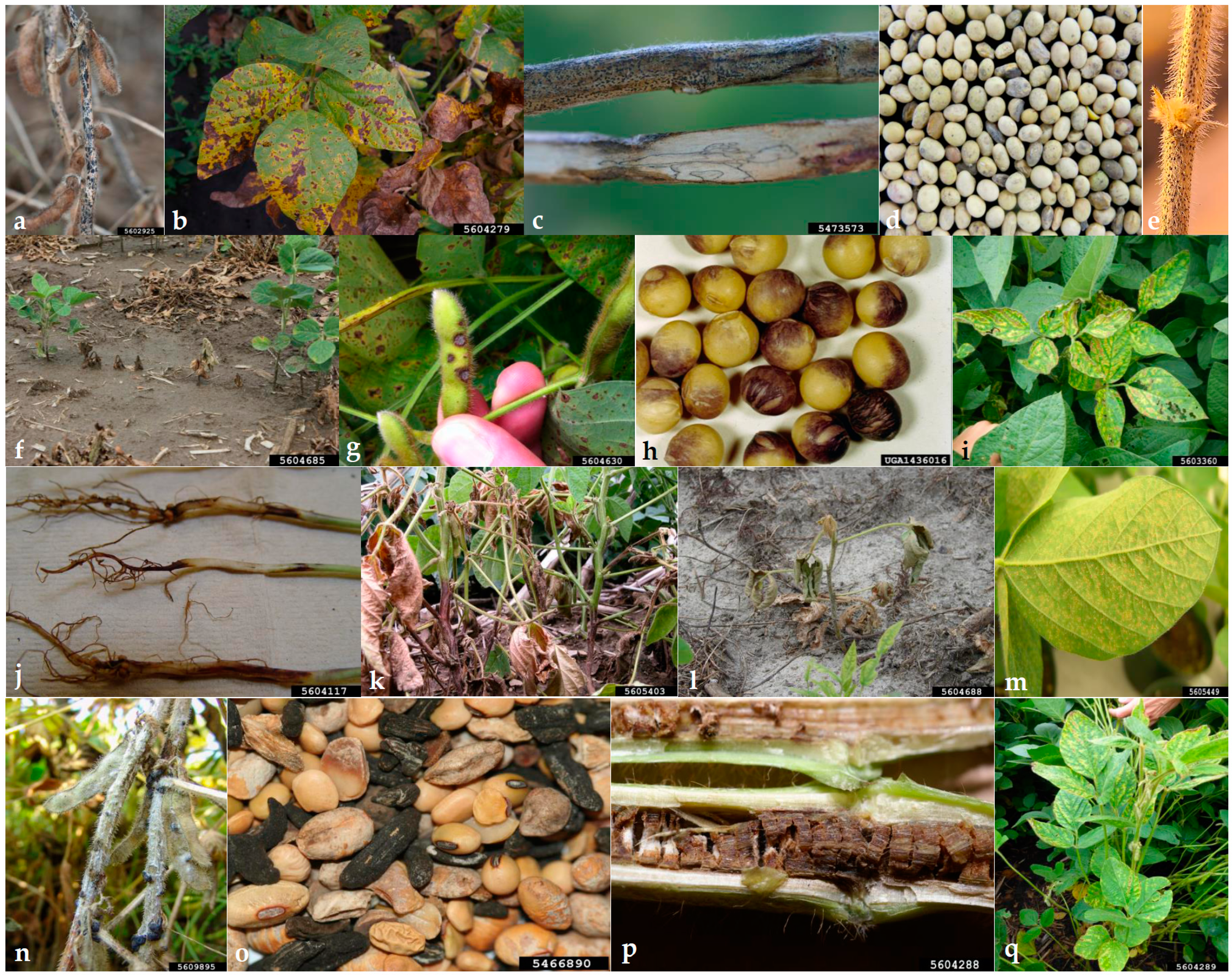
:1. Introduction
2. Common Fungal and Oomycete Soybean Pathogens and Molecular Assays to Detect Them
2.1. Genus Diaporthe
2.2. Genus Sclerotinia
2.3. Genus Colletotrichum
2.4. Genus Fusarium
2.5. Genus Cercospora
2.6. Genus Septoria
2.7. Genus Macrophomina
2.8. Genus Phialophora
2.9. Genus Rhizoctonia
2.10. Genus Phakopsora
2.11. Genus Phytophthora
2.12. Genus Pythium
3. General Steps and Procedures in Establishing Assays for Molecular Diagnosis
3.1. Sequence Determination and Primer Design
3.2. In Vitro Test of the Primers for Efficiency and Specificity
3.3. Test on Different Sample Types, Use of True Samples, Optimizing, Multiplexing
3.3.1. Test on Different Sample Types
3.3.2. Test of True Samples/Field Samples
3.3.3. Optimization
3.3.4. Internal Controls
3.3.5. Multiplexing
4. Potential and Challenges of Using Molecular Diagnosis and Establishing the Assays for Certification Purposes
4.1. Molecular Assays Are Fast and Yield Accurate Results
4.2. qPCR Can Be Used to Quantify Pathogens, Enabling Methods to Test Strains for Aggressiveness or Cultivars for Resistance
4.3. Tests for Certification Purposes Often Require Large Samples or Many Samplings
5. Conclusions
Author Contributions
Funding
Institutional Review Board Statement
Informed Consent Statement
Data Availability Statement
Acknowledgments
Conflicts of Interest
References
- Hartman, G.L.; Rupe, J.C.; Sikora, E.J.; Domier, L.L.; Davis, J.A.; Steffey, K.L. (Eds.) Compendium of Soybean Diseases and Pests, 5th ed.; APS Press: St. Paul, MN, USA, 2015. [Google Scholar]
- USDA-FAS. Oilseeds: World Markets and Trade. Available online: https://www.fas.usda.gov/data/oilseeds-world-markets-and-trade (accessed on 3 March 2023).
- Hartman, G.L.; West, E.D.; Herman, T.K. Crops that feed the world 2. Soybean-worldwide production, use, and constraints caused by pathogens and pests. Food Secur. 2011, 3, 5–17. [Google Scholar] [CrossRef]
- Sinclair, J.B.; Shurtleff, M.C. Compendium of Soybean Diseases, 1st ed.; APS Press: St. Paul, MN, USA, 1975. [Google Scholar]
- Hartman, G.L.; Hill, C.B. Diseases of soybean and their management. In The soybean: Botany, Production, and Uses; Singh, G., Ed.; CABI: Wallingford, UK, 2010; pp. 276–299. [Google Scholar]
- Vidić, M.; Đorđević, V.; Petrović, K.; Miladinović, J. Review of soybean resistance to pathogens. Ratar. Povrt. 2013, 50, 52–61. [Google Scholar] [CrossRef]
- Petrović, K.; Skaltsas, D.; Castlebury, L.A.; Kontz, B.; Allen, T.W.; Chilvers, M.I.; Gregory, N.; Kelly, H.M.; Koehler, A.M.; Kleczewski, N.M.; et al. Diaporthe seed decay of soybean [Glycine max (L.) Merr.] is endemic in the United States, but new fungi are involved. Plant Dis. 2021, 105, 1621–1629. [Google Scholar] [CrossRef]
- Santos, J.M.; Vrandečić, K.; Ćosić, J.; Duvnjak, T.; Phillips, A.J.L. Resolving the Diaporthe species occurring on soybean in Croatia. Persoonia 2011, 27, 9–19. [Google Scholar] [CrossRef]
- Udayanga, D.; Liu, X.; Mckenzie, E.H.C.; Chukeatirote, E.; Bahkali, A.H.A.; Hyde, K.D. The genus Phomopsis: Biology, applications, species concepts and names of common phytopathogens. Fungal Divers. 2011, 50, 189–225. [Google Scholar] [CrossRef]
- Gomes, R.R.; Glienke, C.; Videira, S.I.R.; Lombard, L.; Groenewald, J.Z.; Crous, P.W. Diaporthe: A genus of endophytic, saprobic and plant pathogenic fungi. Persoonia 2013, 31, 1–41. [Google Scholar] [CrossRef]
- Udayanga, D.; Liu, X.; Crous, P.W.; Mckenzie, E.H.C.; Chukeatirote, E.; Hyde, K.D. A multi-locus phylogenetic evaluation of Diaporthe (Phomopsis). Fungal Divers. 2012, 56, 157–171. [Google Scholar] [CrossRef]
- Sinclair, J.B. Phomopsis seed decay of soybeans-a prototype for studying seed disease. Plant Dis. 1993, 77, 329–334. [Google Scholar] [CrossRef]
- Hosseini, B.; El-Hasan, A.; Link, T.; Voegele, R.T. Analysis of the species spectrum of the Diaporthe/Phomopsis complex in European soybean seeds. Mycol. Prog. 2020, 19, 455–469. [Google Scholar] [CrossRef]
- Walcott, R.R. Detection of seedborne pathogens. HortTechnology 2003, 13, 40–47. [Google Scholar] [CrossRef]
- Zhang, A.W.; Hartman, G.L.; Riccioni, L.; Chen, W.D.; Ma, R.Z.; Pedersen, W.L. Using PCR to distinguish Diaporthe phaseolorum and Phomopsis longicolla from other soybean fungal pathogens and to detect them in soybean tissues. Plant Dis. 1997, 81, 1143–1149. [Google Scholar] [CrossRef] [PubMed]
- Zhang, A.W.; Riccioni, L.; Pedersen, W.L.; Kollipara, K.P.; Hartman, G.L. Molecular identification and phylogenetic grouping of Diaporthe phaseolorum and Phomopsis longicolla isolates from soybean. Phytopathology 1998, 88, 1306–1314. [Google Scholar] [CrossRef] [PubMed]
- van Rensburg, J.C.; Lamprecht, S.C.; Groenewald, J.Z.; Castlebury, L.A.; Crous, P.W. Characterisation of Phomopsis spp. associated with die-back of rooibos (Aspalathus linearis) in South Africa. Stud. Mycol. 2006, 55, 65–74. [Google Scholar] [CrossRef] [PubMed]
- Santos, J.; Phillips, A. Resolving the complex of Diaporthe (Phomopsis) species occurring on Foeniculum vulgare in Portugal. Fungal Divers. 2009, 34, 111–125. [Google Scholar]
- Schoch, C.L.; Seifert, K.A.; Huhndorf, S.; Robert, V.; Spouge, J.L.; Levesque, C.A.; Chen, W. Nuclear ribosomal internal transcribed spacer (ITS) region as a universal DNA barcode marker for fungi. Proc. Natl. Acad. Sci. USA 2012, 109, 6241–6246. [Google Scholar] [CrossRef] [PubMed]
- Vechiato, M.H.; Maringoni, A.C.; Martins, E.M.F. Development of primers and method for identification and detection of Diaporthe phaseolorum var. meridionalis in soybean seeds. Summa Phytopathologica 2006, 32, 161–169. [Google Scholar] [CrossRef]
- Zhang, A.W.; Hartman, G.L.; Curio-Penny, B.; Pedersen, W.L.; Becker, K.B. Molecular detection of Diaporthe phaseolorum and Phomopsis longicolla from soybean seeds. Phytopathology 1999, 89, 796–804. [Google Scholar] [CrossRef]
- Hosseini, B.; Voegele, R.T.; Link, T.I. Establishment of a quadruplex real-time PCR assay to distinguish the fungal pathogens Diaporthe longicolla, D. caulivora, D. eres, and D. novem on soybean. PLoS ONE 2021, 16, e0257225. [Google Scholar] [CrossRef]
- Farr, D.F.; Castlebury, L.A.; Rossman, A.Y. Morphological and molecular characterization of Phomopsis vaccinii and additional isolates of Phomopsis from blueberry and cranberry in the eastern United States. Mycologia 2002, 94, 494–504. [Google Scholar] [CrossRef]
- Santos, L.; Alves, A.; Alves, R. Evaluating multi-locus phylogenies for species boundaries determination in the genus Diaporthe. PeerJ 2017, 5, e3120. [Google Scholar] [CrossRef]
- Udayanga, D.; Castlebury, L.A.; Rossman, A.Y.; Chukeatirote, E.; Hyde, K.D. The Diaporthe sojae species complex: Phylogenetic re-assessment of pathogens associated with soybean, cucurbits and other field crops. Fungal Biol. 2015, 119, 383–407. [Google Scholar] [CrossRef]
- Petrović, K.; Riccioni, L.; Vidić, M.; Đorđević, V.; Balešević-Tubić, S.; Đukić, V.; Miladinov, Z. First report of Diaporthe novem, D. foeniculina, and D. rudis associated with soybean seed decay in Serbia. Plant Dis. 2016, 100, 2324. [Google Scholar] [CrossRef]
- Chaisiri, C.; Liu, X.Y.; Lin, Y.; Li, J.B.; Xiong, B.; Luo, C.X. Phylogenetic analysis and development of molecular tool for detection of Diaporthe citri causing melanose disease of citrus. Plants 2020, 9, 329. [Google Scholar] [CrossRef]
- Upchurch, R.G.; Ramirez, M.E. Defense-related gene expression in soybean leaves and seeds inoculated with Cercospora kikuchii and Diaporthe phaseolorum var. meridionalis. Physiol. Mol. Plant Pathol. 2010, 75, 64–70. [Google Scholar] [CrossRef]
- Mena, E.; Stewart, S.; Montesano, M.; Ponce de Leon, I. Soybean Stem Canker caused by Diaporthe caulivora; pathogen diversity, colonization process, and plant defense activation. Front. Plant Sci. 2020, 10, 1733. [Google Scholar] [CrossRef] [PubMed]
- Fernández, F.A.; Hanlin, R.T. Morphological and RAPD analyses of Diaporthe phaseolorum from soybean. Mycologia 1996, 88, 425–440. [Google Scholar] [CrossRef]
- Moleleki, N.; Preisig, O.; Wingfield, M.J.; Crous, P.W.; Wingfield, B.D. PCR-RFLP and sequence data delineate three Diaporthe species associated with stone and pome fruit trees in South Africa. Eur. J. Plant Pathol. 2002, 108, 909–912. [Google Scholar] [CrossRef]
- Brumer, B.B.; Lopes-Caitar, V.S.; Chicowski, A.S.; Beloti, J.D.; Castanho, F.M.; Gregório da Silva, D.C.; de Carvalho, S.; Lopes, I.O.N.; Soares, R.M.; Seixas, C.D.S.; et al. Morphological and molecular characterization of Diaporthe (anamorph Phomopsis) complex and pathogenicity of Diaporthe aspalathi isolates causing stem canker in soybean. Eur. J. Plant Pathol. 2018, 151, 1009–1025. [Google Scholar] [CrossRef]
- Boland, G.J.; Hall, R. Index of plant hosts to Sclerotinia sclerotiorum. Can. J. Plant Pathol. 1994, 16, 93–108. [Google Scholar] [CrossRef]
- Grabicoski, E.M.G.; Jaccoud Filho, D.S.; Pileggi, M.; Henneberg, L.; Pierre, M.L.C.; Vrisman, C.M.; Dabul, A.N.G. Rapid PCR-based assay for Sclerotinia sclerotiorum detection on soybean seeds. Sci. Agric. 2015, 72, 69–74. [Google Scholar] [CrossRef]
- Freeman, J.; Ward, E.; Calderon, C.; McCartney, A. A polymerase chain reaction (PCR) assay for the detection of inoculum of Sclerotinia sclerotiorum. Eur. J. Plant Pathol. 2002, 108, 877–886. [Google Scholar] [CrossRef]
- Botelho, L.S.; Barrocas, E.N.; Machado, J.C.; Martins, R.S. Detection of Sclerotinia sclerotiorum in soybean seeds by conventional and quantitative PCR techniques. J. Seed Sci. 2015, 37, 055–062. [Google Scholar] [CrossRef]
- Yin, Y.; Ding, L.; Liu, X.; Yang, J.; Ma, Z. Detection of Sclerotinia sclerotiorum in planta by a real-time PCR assay. J. Phytopathol. 2009, 157, 465–469. [Google Scholar] [CrossRef]
- Kim, T.G.; Knudsen, G.R. Quantitative real-time PCR effectively detects and quantifies colonization of sclerotia of Sclerotinia sclerotiorum by Trichoderma spp. Appl. Soil Ecol. 2008, 40, 100–108. [Google Scholar] [CrossRef]
- Rogers, S.L.; Atkins, S.D.; West, J.S. Detection and quantification of airborne inoculum of Sclerotinia sclerotiorum using quantitative PCR. Plant Pathol. 2009, 58, 324–331. [Google Scholar] [CrossRef]
- Ziesman, B.R.; Turkington, T.K.; Basu, U.; Strelkov, S.E. A quantitative PCR system for measuring Sclerotinia sclerotiorum in canola (Brassica napus). Plant Dis. 2016, 100, 984–990. [Google Scholar] [CrossRef]
- Marin-Felix, Y.; Groenewald, J.Z.; Cai, L.; Chen, Q.; Marincowitz, S.; Barnes, I.; Bensch, K.; Braun, U.; Camporesi, E.; Damm, U.; et al. Genera of phytopathogenic fungi: GOPHY 1. Stud. Mycol. 2017, 86, 99–216. [Google Scholar] [CrossRef]
- Damm, U.; Sato, T.; Alizadeh, A.; Groenewald, J.Z.; Crous, P.W. The Colletotrichum dracaenophilum, C. magnum and C. orchidearum species complexes. Stud. Mycol. 2019, 92, 1–46. [Google Scholar] [CrossRef]
- Jayawardena, R.S.; Hyde, K.D.; Damm, U.; Cai, L.; Liu, M.; Li, X.H.; Zhang, W.; Zhao, W.S.; Yan, J.Y. Notes on currently accepted species of Colletotrichum. Mycosphere 2016, 7, 1192–1260. [Google Scholar] [CrossRef]
- Cannon, P.F.; Damm, U.; Johnston, P.R.; Weir, B.S. Colletotrichum-current status and future directions. Stud. Mycol. 2012, 73, 181–213. [Google Scholar] [CrossRef]
- Dean, R.; Van Kan, J.A.; Pretorius, Z.A.; Hammond-Kosack, K.E.; Di Pietro, A.; Spanu, P.D.; Rudd, J.J.; Dickman, M.; Kahmann, R.; Ellis, J.; et al. The Top 10 fungal pathogens in molecular plant pathology. Mol. Plant Pathol. 2012, 13, 414–430. [Google Scholar] [CrossRef] [PubMed]
- da Silva, L.L.; Moreno, H.L.A.; Correia, H.L.N.; Santana, M.F.; de Queiroz, M.V. Colletotrichum: Species complexes, lifestyle, and peculiarities of some sources of genetic variability. Appl. Microbiol. Biotechnol. 2020, 104, 1891–1904. [Google Scholar] [CrossRef] [PubMed]
- Sharma, S.K.; Gupta, G.K.; Ramteke, R. Colletotrichum truncatum [(Schw.) Andrus and W.D. Moore], the causal agent of anthracnose of soybean [Glycine max (L.) Merrill.]—A review. Soybean Res. 2011, 9, 31–52. [Google Scholar]
- Riccioni, L.; Conca, G.; Hartman, G.L. First report of Colletotrichum coccodes on soybean in the United States. Plant Dis. 1998, 82, 959. [Google Scholar] [CrossRef]
- Yang, H.C.; Haudenshield, J.S.; Hartman, G.L. First report of Colletotrichum chlorophyti causing soybean anthracnose. Plant Dis. 2012, 96, 1699. [Google Scholar] [CrossRef]
- Mahmodi, F.; Kadir, J.B.; Wong, M.Y.; Nasehi, A.; Puteh, A.; Soleimani, N. First report of anthracnose caused by Colletotrichum gloeosporioides on soybean (Glycine max) in Malaysia. Plant Dis. 2013, 97, 841. [Google Scholar] [CrossRef]
- Yang, H.C.; Haudenshield, J.S.; Hartman, G.L. Colletotrichum incanum sp. nov., a curved-conidial species causing soybean anthracnose in USA. Mycologia 2014, 106, 32–42. [Google Scholar] [CrossRef]
- Barbieri, M.C.G.; Ciampi-Guillardi, M.; Moraes, S.R.G.; Bonaldo, S.M.; Rogério, F.; Linhares, R.R.; Massola Júnior, N.S. First report of Colletotrichum cliviae causing anthracnose on soybean in Brazil. Plant Dis. 2017, 101, 1677. [Google Scholar] [CrossRef]
- Boufleur, T.R.; Castro, R.R.L.; Rogério, F.; Ciampi-Guillardi, M.; Baroncelli, R.; Massola Júnior, N.S. First report of Colletotrichum musicola causing soybean anthracnose in Brazil. Plant Dis. 2020, 104, 1858. [Google Scholar] [CrossRef]
- Shi, X.; Wang, S.; Duan, X.; Gao, X.; Zhu, X.; Laborda, P. First report of Colletotrichum brevisporum causing soybean anthracnose in China. Plant Dis. 2020, 105, 707. [Google Scholar] [CrossRef]
- Damm, U.; Woudenberg, J.H.C.; Cannon, P.F.; Crous, P.W. Colletotrichum species with curved conidia from herbaceous hosts. Fungal Divers. 2009, 39, 45–87. [Google Scholar]
- Damm, U.; Cannon, P.F.; Woudenberg, J.H.C.; Crous, P.W. The Colletotrichum acutatum species complex. Stud. Mycol. 2012, 73, 37–113. [Google Scholar] [CrossRef]
- Damm, U.; Cannon, P.F.; Woudenberg, J.H.C.; Johnston, P.R.; Weir, B.S.; Tan, Y.P.; Shivas, R.G.; Crous, P.W. The Colletotrichum boninense species complex. Stud. Mycol. 2012, 73, 1–36. [Google Scholar] [CrossRef]
- Damm, U.; O’Connell, R.J.; Groenewald, J.Z.; Crous, P.W. The Colletotrichum destructivum species complex-hemibiotrophic pathogens of forage and field crops. Stud. Mycol. 2014, 79, 49–84. [Google Scholar] [CrossRef]
- Liu, F.; Cai, L.; Crous, P.W.; Damm, U. The Colletotrichum gigasporum species complex. Persoonia 2014, 33, 83–97. [Google Scholar] [CrossRef]
- Weir, B.S.; Johnston, P.R.; Damm, U. The Colletotrichum gloeosporioides species complex. Stud. Mycol. 2012, 73, 115–180. [Google Scholar] [CrossRef]
- Vieira, W.A.D.S.; Bezerra, P.A.; Silva, A.C.D.; Veloso, J.S.; Câmara, M.P.S.; Doyle, V.P. Optimal markers for the identification of Colletotrichum species. Mol. Phylogen. Evol. 2020, 143, 106694. [Google Scholar] [CrossRef]
- Chen, L.S.; Chu, C.; Liu, C.D.; Chen, R.S.; Tsay, J.G. PCR-based detection and differentiation of anthracnose pathogens, Colletotrichum gloeosporioides and C. truncatum, from vegetable soybean in Taiwan. J. Phytopathol. 2006, 154, 654–662. [Google Scholar] [CrossRef]
- Pecchia, S.; Caggiano, B.; Da Lio, D.; Cafa, G.; Le Floch, G.; Baroncelli, R. Molecular detection of the seed-borne pathogen Colletotrichum lupini targeting the hyper-variable IGS region of the ribosomal cluster. Plants 2019, 8, 222. [Google Scholar] [CrossRef]
- Ciampi-Guillardi, M.; Ramiro, J.; Moraes, M.H.D.; Barbieri, M.C.G.; Massola, N.S., Jr. Multiplex qPCR assay for direct detection and quantification of Colletotrichum truncatum, Corynespora cassiicola, and Sclerotinia sclerotiorum in soybean seeds. Plant Dis. 2020, 104, 3002–3009. [Google Scholar] [CrossRef]
- Yang, H.C.; Haudenshield, J.S.; Hartman, G.L. Multiplex real-time PCR detection and differentiation of Colletotrichum species infecting soybean. Plant Dis. 2015, 99, 1559–1568. [Google Scholar] [CrossRef]
- Debode, J.; Van Hemelrijck, W.; Baeyen, S.; Creemers, P.; Heungens, K.; Maes, M. Quantitative detection and monitoring of Colletotrichum acutatum in strawberry leaves using real-time PCR. Plant Pathol. 2009, 58, 504–514. [Google Scholar] [CrossRef]
- Garrido, C.; Carbu, M.; Fernandez-Acero, F.J.; Boonham, N.; Colyer, A.; Cantoral, J.M.; Budge, G. Development of protocols for detection of Colletotrichum acutatum and monitoring of strawberry anthracnose using real-time PCR. Plant Pathol. 2009, 58, 43–51. [Google Scholar] [CrossRef]
- Cullen, D.W.; Lees, A.K.; Toth, I.K.; Duncan, J.M. Detection of Colletotrichum coccodes from soil and potato tubers by conventional and quantitative real-time PCR. Plant Pathol. 2002, 51, 281–292. [Google Scholar] [CrossRef]
- Tao, G.; Hyde, K.D.; Cai, L. Species-specific real-time PCR detection of Colletotrichum kahawae. J. Appl. Microbiol. 2013, 114, 828–835. [Google Scholar] [CrossRef]
- Kuan, C.-P.; Wu, M.-T.; Huang, H.C.; Chang, H. Rapid detection of Colletotrichum lagenarium, causal agent of anthracnose of Cucurbitaceous crops, by PCR and real-time PCR. J. Phytopathol. 2011, 159, 276–282. [Google Scholar] [CrossRef]
- Tian, Q.; Lu, C.; Wang, S.; Xiong, Q.; Zhang, H.; Wang, Y.; Zheng, X. Rapid diagnosis of soybean anthracnose caused by Colletotrichum truncatum using a loop-mediated isothermal amplification (LAMP) assay. Eur. J. Plant Pathol. 2017, 148, 785–793. [Google Scholar] [CrossRef]
- Wang, S.; Ye, W.; Tian, Q.; Dong, S.; Zheng, X. Rapid detection of Colletotrichum gloeosporioides using a loop-mediated isothermal amplification assay. Australas. Plant Pathol. 2017, 46, 493–498. [Google Scholar] [CrossRef]
- Wang, H.; Xiao, M.; Kong, F.; Chen, S.; Dou, H.-T.; Sorrell, T.; Li, R.-Y.; Xu, Y.-C. Accurate and practical identification of 20 Fusarium species by seven-locus sequence analysis and reverse line blot hybridization, and an in vitro antifungal susceptibility study. J. Clin. Microbiol. 2011, 49, 1890–1898. [Google Scholar] [CrossRef]
- Munkvold, G.P. Fusarium species and their associated mycotoxins. In Mycotoxigenic Fungi, Methods and Protocols; Moretti, A., Susca, A., Eds.; Methods in Molecular Biology; Humana: New York, NY, USA, 2017; Volume 1542, pp. 51–106. [Google Scholar]
- Armstrong, G.M.; Armstrong, J.K. Biological races of Fusarium causing wilt of cowpeas and soybeans. Phytopathology 1950, 40, 181–193. [Google Scholar]
- Nelson, B.D.; Hansen, J.M.; Windels, C.E.; Helms, T.C. Reaction of soybean cultivars to isolates of Fusarium solani from the Red River Valley. Plant Dis. 1997, 81, 664–668. [Google Scholar] [CrossRef] [PubMed]
- Aoki, T.; O’Donnell, K.; Scandiani, M.M. Sudden death syndrome of soybean in South America is caused by four species of Fusarium: Fusarium brasiliense sp. nov., F. cuneirostrum sp. nov., F. tecumaniae, and F. virguliforme. Mycoscience 2005, 46, 162–183. [Google Scholar] [CrossRef]
- Broders, K.D.; Lipps, P.E.; Paul, P.A.; Dorrance, A.E. Evaluation of Fusarium graminearum associated with corn and soybean seed and seedling disease in Ohio. Plant Dis. 2007, 91, 1155–1160. [Google Scholar] [CrossRef]
- Ellis, M.L.; Diaz Arias, M.M.; Cruz Jimenez, D.R.; Munkvold, G.P.; Leandro, L.F. First report of Fusarium commune causing damping-off, seed rot, and seedling root rot on soybean (Glycine max) in the United States. Plant Dis. 2013, 97, 284. [Google Scholar] [CrossRef] [PubMed]
- Okello, P.N.; Mathew, F.M. Cross pathogenicity studies show South Dakota isolates of Fusarium acuminatum, F. equiseti, F. graminearum, F. oxysporum, F. proliferatum, F. solani, and F. subglutinans from either soybean or corn are pathogenic to both crops. Plant Health Progress 2019, 20, 44–49. [Google Scholar] [CrossRef]
- Ross, J.P. Predispositions of soybeans to Fusarium wilt by Heterodera glycines and Meloidogyne incognita. Phytopathology 1965, 55, 361–364. [Google Scholar]
- O’Donnell, K.; Gray, L.E. Phylogenetic relationships of the soybean sudden death syndrome pathogen Fusarium solani f. sp. phaseoli inferred from rDNA sequence data and PCR primers for its identification. Mol. Plant-Microbe Interact. 1995, 8, 709–716. [Google Scholar] [CrossRef]
- Li, S.; Tam, Y.K.; Hartman, G.L. Molecular differentiation of Fusarium solani f. sp. glycines from other F. solani based on mitochondrial small subunit rDNA sequences. Phytopathology 2000, 90, 491–497. [Google Scholar] [CrossRef]
- Filion, M.; St-Arnaud, M.; Jabaji-Hare, S.H. Quantification of Fusarium solani f. sp. phaseoli in mycorrhizal bean plants and surrounding mycorrhizosphere soil using real-time polymerase chain reaction and direct isolations on selective media. Phytopathology 2003, 93, 229–235. [Google Scholar] [CrossRef]
- Abd-Elsalam, K.A.; Aly, I.N.; Abdel-Satar, M.A.; Khalil, M.S.; Verreet, J.A. PCR identification of Fusarium genus based on nuclear ribosomal-DNA sequence data. Afr. J. Biotechnol. 2003, 2, 82–85. [Google Scholar]
- Roy, K.W.; Rupe, J.C.; Hershman, D.E.; Abney, T.S. Sudden death syndrome of soybean. Plant Dis. 1997, 81, 1100–1111. [Google Scholar] [CrossRef] [PubMed]
- Roy, K.W.; Lawrence, G.W.; Hodges, H.H.; Mclean, K.S.; Killebrew, J.F. Sudden death syndrome of soybean: Fusarium solani as incitant and relation of Heterodera glycines to disease severity. Phytopathology 1989, 79, 191–197. [Google Scholar] [CrossRef]
- Rupe, J.C. Frequency and pathogenicity of Fusarium solani recovered from soybeans with sudden death syndrome. Plant Dis. 1989, 73, 581–584. [Google Scholar] [CrossRef]
- Achenbach, L.A.; Patrick, J.; Gray, L. Use of RAPD markers as a diagnostic tool for the identification of Fusarium solani isolates that cause soybean sudden death syndrome. Plant Dis. 1996, 80, 1228–1232. [Google Scholar] [CrossRef]
- Li, S.; Hartman, G.L. Molecular detection of Fusarium solani f. sp. glycines in soybean roots and soil. Plant Pathol. 2003, 52, 74–83. [Google Scholar] [CrossRef]
- Gao, X.; Jackson, T.A.; Lambert, K.N.; Li, S.; Hartman, G.L.; Niblack, T.L. Detection and quantification of Fusarium solani f. sp. glycines in soybean roots with real-time quantitative polymerase chain reaction. Plant Dis. 2004, 88, 1372–1380. [Google Scholar] [CrossRef]
- Li, S.; Hartman, G.L.; Domier, L.L. Quantification of Fusarium solani f. sp. glycines isolates in soybean roots by colony-forming unit assays and real-time quantitative PCR. Theor. Appl. Genet. 2008, 117, 343–352. [Google Scholar] [CrossRef]
- Aoki, T.; O’Donnell, K.; Homma, Y.; Lattanzi, A. Sudden-death syndrome of soybean is caused by two morphologically and phylogenetically distinct species within the Fusarium solani species complex F. virguliforme in North America and F. tucumaniae in South America. Mycologia 2003, 95, 660. [Google Scholar] [CrossRef]
- O’Donnell, K.; Sink, S.; Scandiani, M.M.; Luque, A.; Colletto, A.; Biasoli, M.; Lenzi, L.; Salas, G.; Gonzalez, V.; Ploper, L.D.; et al. Soybean sudden death syndrome species diversity within North and South America revealed by multilocus genotyping. Phytopathology 2010, 100, 58–71. [Google Scholar] [CrossRef]
- Westphal, A.; Li, C.; Xing, L.; McKay, A.; Malvick, D. Contributions of Fusarium virguliforme and Heterodera glycines to the disease complex of sudden death syndrome of soybean. PLoS ONE 2014, 9, e99529. [Google Scholar] [CrossRef]
- Mbofung, G.C.Y.; Fessehaie, A.; Bhattacharyya, M.K.; Leandro, L.F.S. A new TaqMan real-time polymerase chain reaction assay for quantification of Fusarium virguliforme in soil. Plant Dis. 2011, 95, 1420–1426. [Google Scholar] [CrossRef] [PubMed]
- Wang, J.; Jacobs, J.L.; Byrne, J.M.; Chilvers, M.I. Improved diagnoses and quantification of Fusarium virguliforme, causal agent of soybean sudden death syndrome. Phytopathology 2015, 105, 378–387. [Google Scholar] [CrossRef] [PubMed]
- Roth, M.G.; Oudman, K.A.; Griffin, A.; Jacobs, J.L.; Sang, H.; Chilvers, M.I. Diagnostic qPCR assay to detect Fusarium brasiliense, a causal agent of soybean sudden death syndrome and root rot of dry bean. Plant Dis. 2020, 104, 246–254. [Google Scholar] [CrossRef]
- Rocha, L.F.; Srour, A.Y.; Pimentel, M.; Subedi, A.; Bond, J.P.; Fakhoury, A.; Ammar, H.A. A panel of qPCR assays to detect and quantify soybean soil-borne pathogens. Lett. Appl. Microbiol. 2022, 76, ovac023. [Google Scholar] [CrossRef]
- Orth, C.E.; Schuh, W. Resistance of 17 soybean cultivars to foliar, latent, and seed infection by Cercospora kikuchii. Plant Dis. 1994, 78, 661–664. [Google Scholar] [CrossRef]
- Hartman, G.L.; Sinclair, J.B.; Rupe, J.C. Compendium of Soybean Diseases, 4th ed.; APS Press: St. Paul, MN, USA, 1999. [Google Scholar]
- Matsumoto, T.; Tomoyasu, R. Studies on the purple speck of soybean seed. Ann. Phytopathol. Soc. Jpn. 1925, 1, 1–14. [Google Scholar] [CrossRef]
- Walters, H.J. Soybean leaf blight caused by Cercospora kikuchii. Plant Dis. 1980, 64, 961–962. [Google Scholar] [CrossRef]
- Ehrenshaft, M.; Upchurch, R.G. Host protein(s) induces accumulation of the toxin cercosporin and mRNA in a phytopahtogenic strain of Cercospora kikuchii. Physiol. Mol. Plant Pathol. 1993, 43, 95–107. [Google Scholar] [CrossRef]
- Daub, M.E.; Ehrenshaft, M. The photoactivated Cercospora toxin cercosporin: Contributions to plant disease and fundamental biology. Annu. Rev. Phytopathol. 2000, 38, 461–490. [Google Scholar] [CrossRef]
- Callahan, T.M.; Rose, M.S.; Meade, M.J.; Ehrenshaft, M.; Upchurch, R.G. CFP, the putative cercosporin transporter of Cercospora kikuchii, is required for wild type cercosporin production, resistance, and virulence on soybean. Mol. Plant-Microbe Interact. 1999, 12, 901–910. [Google Scholar] [CrossRef]
- Soares, A.P.G.; Guillin, E.A.; Borges, L.L.; da Silva, A.C.T.; de Almeida, Á.M.R.; Grijalba, P.E.; Gottlieb, A.M.; Bluhm, B.H.; de Oliveira, L.O. More Cercospora species infect soybeans across the Americas than meets the eye. PLoS ONE 2015, 10, e0133495. [Google Scholar] [CrossRef] [PubMed]
- Davidson, R.M.; Hanson, L.E.; Franc, G.D.; Panella, L. Analysis of β-tubulin gene fragments from benzimidazole-sensitive and -tolerant Cercospora beticola. J. Phytopathol. 2006, 154, 321–328. [Google Scholar] [CrossRef]
- Imazaki, I.; Ishikawa, K.; Yasuda, N.; Miyasaka, A.; Kawasaki, S.; Koizumi, S. Incidence of thiophanate-methyl resistance in Cercospora kikuchii within a single lineage based on amplified fragment length polymorphisms in Japan. J. Gen. Plant Pathol. 2006, 72, 77–84. [Google Scholar] [CrossRef]
- Imazaki, I.; Iizumi, H.; Ishikawa, K.; Sasahara, M.; Yasuda, N.; Koizumi, S. Effects of thiophanate-methyl and azoxystrobin on the composition of Cercospora kikuchii populations with thiophanate-methyl-resistant strains. J. Gen. Plant Pathol. 2006, 72, 292–300. [Google Scholar] [CrossRef]
- Groenewald, J.Z.; Nakashima, C.; Nishikawa, J.; Shin, H.D.; Park, J.H.; Jama, A.N.; Groenewald, M.; Braun, U.; Crous, P.W. Species concepts in Cercospora: Spotting the weeds among the roses. Stud. Mycol. 2012, 75, 115–170. [Google Scholar] [CrossRef]
- Chen, H.; Lee, M.H.; Daub, M.E.; Chung, K.R. Molecular analysis of the cercosporin biosynthetic gene cluster in Cercospora nicotianae. Mol. Microbiol. 2007, 64, 755–770. [Google Scholar] [CrossRef]
- Chanda, A.K.; Ward, N.A.; Robertson, C.L.; Chen, Z.-Y.; Schneider, R.W. Development of a quantitative polymerase chain reaction detection protocol for Cercospora kikuchii in soybean leaves and its use for documenting latent infection as affected by fungicide applications. Phytopathology 2014, 104, 1118–1124. [Google Scholar] [CrossRef]
- MacNeill, B.H.; Zalasky, H. Histological study of host–parasite relationships between Septoria glycines Hemmi and soybean leaves and pods. Can. J. Bot. 1957, 35, 501–505. [Google Scholar] [CrossRef]
- Williams, R.F.; Nyvall, D.J. Leaf infection and yield losses caused by brown spot and bacterial blight diseases of soybean. Phytopathology 1980, 70, 900–902. [Google Scholar] [CrossRef]
- Basu, P.K.; Butler, G. Assessment of brown spot (Septoria glycines) alone and in combination with bacterial blight (Pseudomonas syringae pv. glycines) on soybeans in a short-season area. Can. J. Plant Pathol. 1988, 10, 78–82. [Google Scholar] [CrossRef]
- Carmona, M.; Sautua, F.; Perelman, S.; Reis, E.M.; Gally, M. Relationship between late soybean diseases complex and rain in determining grain yield responses to fungicide applications. J. Phytopathol. 2011, 159, 687–693. [Google Scholar] [CrossRef]
- Lin, H.-A.; Mideros, S.X. Accurate quantification and detection of Septoria glycines in soybean using quantitative PCR. Curr. Plant Biol. 2021, 25, 100192. [Google Scholar] [CrossRef]
- Kunwar, I.K.; Singh, T.; Machado, C.C.; Sinclair, J.B. Histopathology of soybean seed and seedling infection by Macrophomina phaseolina. Phytopathology 1986, 76, 532–535. [Google Scholar] [CrossRef]
- Raut, J.G. Transmission of seed borne Macrophomina phaseolina in seed. Sci. Technol. 1983, 11, 807–817. [Google Scholar]
- Jana, T.K.; Singh, N.K.; Koundal, K.R.; Sharma, T.R. Genetic differentiation of charcoal rot pathogen, Macrophomina phaseolina, in to specific groups using URP-PCR. Can. J. Microbiol./Rev. Can. Microbiol. 2005, 51, 159–164. [Google Scholar] [CrossRef]
- Sarr, M.P.; Ndiaye, M.; Groenewald, J.Z.; Crous, P.W. Genetic diversity in Macrophomina phaseolina, the causal agent of charcoal rot. Phytopathol. Mediterr. 2014, 53, 250–268. [Google Scholar] [CrossRef]
- Khan, A.N.; Shair, F.; Malik, K.; Hayat, Z.; Khan, M.A.; Hafeez, F.Y.; Hassan, M. Molecular identification and genetic characterization of Macrophomina phaseolina strains causing pathogenicity on sunflower and chickpea. Front. Microbiol. 2017, 8, 1309. [Google Scholar] [CrossRef]
- Tančić Živanov, S.; Dedić, B.; Dimitrijević, A.; Dušanić, N.; Jocić, S.; Miklič, V.; Kovačević, B.; Miladinović, D. Analysis of genetic diversity among Macrophomina phaseolina (Tassi) Goid. isolates from Euro-Asian countries. J. Plant Dis. Prot. 2019, 126, 565–573. [Google Scholar] [CrossRef]
- Babu, B.K.; Saxena, A.K.; Srivastava, A.K.; Arora, D.K. Identification and detection of Macrophomina phaseolina by using species-specific oligonucleotide primers and probe. Mycol. Helv. 2007, 99, 797–803. [Google Scholar] [CrossRef]
- Babu, B.K.; Mesapogu, S.; Sharma, A.; Somasani, S.R.; Arora, D.K. Quantitative real-time PCR assay for rapid detection of plant and human pathogenic Macrophomina phaseolina from field and environmental samples. Mycologia 2011, 103, 466–473. [Google Scholar] [CrossRef]
- Lu, C.; Song, B.; Zhang, H.; Wang, Y.; Zheng, X. Rapid diagnosis of soybean seedling blight caused by Rhizoctonia solani and soybean charcoal rot caused by Macrophomina phaseolina using LAMP assays. Phytopathology 2015, 105, 1612–1617. [Google Scholar] [CrossRef]
- Gray, L.E. Variation in pathogenicity of Cephalosporium gregatum isolates. Phytopathology 1971, 61, 1410–1411. [Google Scholar] [CrossRef]
- Hughes, T.J.; Chen, W.; Grau, C.R. Pathogenic characterization of genotype A and B of Phialophora gregata f. sp. sojae. Plant Dis. 2002, 86, 729–735. [Google Scholar] [CrossRef]
- Harrington, T.C.; Steimel, J.; Workneh, F.; Yang, X.B. Characterization and distribution of two races of Phialophora gregata in the North-Central United States. Phytopathology 2003, 93, 901–912. [Google Scholar] [CrossRef]
- Chen, W.; Grau, C.R.; Adee, E.A.; Meng, X.-Q. A molecular marker identifying subspecific population of the soybean brown stem rot pathogen, Phialophora gregata. Phytopathology 2000, 90, 875–883. [Google Scholar] [CrossRef]
- Malvick, D.K.; Chen, W.; Kurle, J.E.; Grau, C.R. Cultivar preference and genotype distribution of the brown stem rot pathogen Phialophora gregata in the Midwestern United States. Plant Dis. 2003, 87, 1250–1254. [Google Scholar] [CrossRef]
- Meng, X.-Q.; Grau, C.R.; Chen, W. Cultivar preference exhibited by two sympatric and genetically distinct populations of the soybean fungal pathogen Phialophora gregata f. sp. sojae. Plant Pathol. 2005, 54, 180–188. [Google Scholar] [CrossRef]
- Chen, W.; Gray, L.E.; Grau, C.R. Molecular differentiation of fungi associated with brown stem rot and detection of Phialophora gregata in resistant and susceptible soybean cultivars. Phytopathology 1996, 86, 1140–1148. [Google Scholar] [CrossRef]
- Chen, W.; Gray, L.E.; Kurle, J.E.; Grau, C.R. Specific detection of Phialophora gregata and Plectosporium tabacinum in infected soybean plants using polymerase chain reaction. Mol. Ecol. Notes 1999, 8, 871–877. [Google Scholar] [CrossRef]
- Malvick, D.K.; Impullitti, A.E. Detection and quantification of Phialophora gregata in soybean and soil samples with a quantitative, real-time PCR assay. Plant Dis. 2007, 91, 736–742. [Google Scholar] [CrossRef]
- Hughes, T.J.; Atallah, Z.K.; Grau, C.R. Real-time PCR assays for the quantification of Phialophora gregata f. sp. sojae IGS genotypes A and B. Phytopathology 2009, 99, 1008–1014. [Google Scholar] [CrossRef]
- Parmeter, J.R.; Sherwood, R.T.; Pratt, W.D. Anastomosis grouping among isolates of Thanatephorus cucumeris. Phytopathology 1969, 59, 1270–1278. [Google Scholar]
- Adams, G.C., Jr.; Butler, E.E. Serological relationships among anastomosis groups of Rhizoctonia solani. Phytopathology 1979, 69, 629–633. [Google Scholar] [CrossRef]
- Sneh, B. Anastomosis groups of multinucleate Rhizoctonia spp. In Rhizoctonia Species: Taxonomy, Molecular Biology, Ecology, Pathology and Disease Control; Sneh, B., Jabaji-Hare, S., Neate, S., Dijst, G., Eds.; Kluwer Academic Publishers: Dordrecht, The Netherlands, 1996; pp. 67–75. [Google Scholar]
- Carling, D.E.; Pope, E.J.; Brainard, K.A.; Carter, D.A. Characterization of mycorrhizal isolates of Rhizoctonia solani from an orchid, including AG-12, a new anastomosis group. Phytopathology 1999, 89, 942–946. [Google Scholar] [CrossRef]
- Carling, D.E.; Baird, R.E.; Gitaitis, R.D.; Brainard, K.A.; Kuninaga, S. Characterization of AG-13, a newly reported anastomosis group of Rhizoctonia solani. Phytopathology 2002, 92, 893–899. [Google Scholar] [CrossRef]
- Campion, C.; Chatot, C.; Perraton, B.; Andrivon, D. Anastomosis groups, pathogenicity and sensitivity to fungicides of Rhizoctonia solani isolates collected on potato crops in France. Eur. J. Plant Pathol. 2003, 109, 983–992. [Google Scholar] [CrossRef]
- Matsumoto, M. Trials of direct detection and identification of Rhizoctonia solani AG 1 and AG 2 subgroups using specifically primed PCR analysis. Mycoscience 2002, 43, 185–189. [Google Scholar] [CrossRef]
- Grosch, R.; Schneider, J.H.M.; Peth, A.; Waschke, A.; Franken, P.; Kofoet, A.; Jabaji-Hare, S.H. Development of a specific PCR assay for the detection of Rhizoctonia solani AG 1-IB using SCAR primers. J. Appl. Microbiol. 2007, 102, 806–819. [Google Scholar] [CrossRef]
- Sayler, R.J.; Yang, Y. Detection and quantification of Rhizoctonia solani AG-1 IA, the rice sheath blight pathogen, in rice using real-time PCR. Plant Dis. 2007, 91, 1663–1668. [Google Scholar] [CrossRef]
- Budge, G.E.; Shaw, M.W.; Colyer, A.; Pietravalle, S.; Boonham, N. Molecular tools to investigate Rhizoctonia solani distribution in soil. Plant Pathol. 2009, 58, 1071–1080. [Google Scholar] [CrossRef]
- Ono, Y.; Buritica, P.; Hennen, J.F. Delimitation of Phakopsora, Physopella, and Cerotelium and their species on Leguminosae. Mycol. Res. 1992, 96, 825–850. [Google Scholar] [CrossRef]
- Goellner, K.; Loehrer, M.; Langenbach, C.; Conrath, U.; Koch, E.; Schaffrath, U. Phakopsora pachyrhizi, the causal agent of Asian soybean rust. Mol. Plant Pathol. 2010, 11, 169–177. [Google Scholar] [CrossRef]
- Frederick, R.D.; Snyder, C.L.; Peterson, G.L.; Bonde, M.R. Polymerase chain reaction assays for the detection and discrimination of the soybean rust pathogens Phakopsora pachyrhizi and P. meibomiae. Phytopathology 2002, 92, 217–222. [Google Scholar] [CrossRef] [PubMed]
- Barnes, C.W.; Szabo, L.J. Detection and identification of four common rust pathogens of cereals and grasses using real-time polymerase chain reaction. Phytopathology 2007, 97, 717–727. [Google Scholar] [CrossRef]
- Barnes, C.W.; Szabo, L.J.; Bowersox, V.C. Identifying and quantifying Phakopsora pachyrhizi spores in rain. Phytopathology 2009, 99, 328–338. [Google Scholar] [CrossRef] [PubMed]
- Dorrance, A.E. Phytophthora root and stem rot. In Compendium of Soybean Diseases and Pests; Hartman, G.L., Rupe, J.C., Sikora, E.J., Domier, L.L., David, J.A., Steffey, K.L., Eds.; APS Press: St. Paul, MN, USA, 2015; pp. 73–76. [Google Scholar]
- Jones, J.P.; Johnson, H.W. Lupine, a new host for Phytophthora megasperma var. sojae. Phytopathology 1969, 59, 504–507. [Google Scholar]
- Hamm, P.B.; Hansen, E.M. Host specificity of Phytophthora megasperma from Douglas fir, soybean, and alfalfa. Phytopathology 1981, 71, 65–68. [Google Scholar] [CrossRef]
- Reeser, P.W.; Scott, D.H.; Ruhl, G.E. Recovery of race non-classifiable Phytophthora megasperma f. sp. glycinea from soybean roots in Indiana in 1990. Phytopathology 1991, 81, 1201. [Google Scholar]
- Malvick, D.K.; Grunden, E. Traits of soybean-infecting Phytophthora populations from Illinois agricultural fields. Plant Dis. 2004, 88, 1139–1145. [Google Scholar] [CrossRef]
- Tang, Q.H.; Gao, F.; Li, G.Y.; Wang, H.; Zheng, X.B.; Wang, Y.C. First report of root rot caused by Phytophthora sansomeana on soybean in China. Plant Dis. 2010, 94, 378. [Google Scholar] [CrossRef]
- Zelaya-Molina, L.X.; Ellis, M.L.; Berry, S.A.; Dorrance, A.E. First report of Phytophthora sansomeana causing wilting and stunting on corn in Ohio. Plant Dis. 2010, 94, 125. [Google Scholar] [CrossRef] [PubMed]
- Wang, Y.; Zhang, W.; Wang, Y.; Zheng, X. Rapid and sensitive detection of Phytophthora sojae in soil and infected soybeans by species-specific polymerase chain reaction assays. Phytopathology 2006, 96, 1315–1321. [Google Scholar] [CrossRef] [PubMed]
- Bienapfl, J.C.; Percich, J.A.; Malvick, D.K. Evaluation of PCR-based methods for species specific detection of Phytophthora sojae. Phytopathology 2007, 98, S201. [Google Scholar]
- Bienapfl, J.C.; Malvick, D.K.; Percich, J.A. Specific molecular detection of Phytophthora sojae using conventional and real-time PCR. Fungal Biol. 2011, 115, 733–740. [Google Scholar] [CrossRef] [PubMed]
- Catal, M.; Erler, F.; Fulbright, D.W.; Adams, G.C. Real-time quantitative PCR assays for evaluation of soybean varieties for resistance to the stem and root rot pathogen Phytophthora sojae. Eur. J. Plant Pathol. 2013, 137, 859–869. [Google Scholar] [CrossRef]
- Zhao, W.; Wang, T.; Qi, R. Ypt1 gene-based detection of Phytophthora sojae in a loop-mediated isothermal amplification assay. J. Plant Dis. Prot. 2015, 122, 66–73. [Google Scholar] [CrossRef]
- Dai, T.-T.; Lu, C.-C.; Lu, J.; Dong, S.; Ye, W.; Wang, Y.; Zheng, X. Development of a loop-mediated isothermal amplification assay for detection of Phytophthora sojae. FEMS Microbiol. Lett. 2012, 334, 27–34. [Google Scholar] [CrossRef]
- Bilodeau, G.J.; Martin, F.N.; Coffey, M.D.; Blomquist, C.L. Development of a multiplex assay for genus- and species-specific detection of Phytophthora based on differences in mitochondrial gene order. Phytopathology 2014, 104, 733–748. [Google Scholar] [CrossRef]
- Miles, T.D.; Martin, F.N.; Robideau, G.P.; Bilodeau, G.J.; Coffey, M.D. Systematic development of Phytophthora species-specific mitochondrial diagnostic markers for economically important members of the genus. Plant Dis. 2017, 101, 1162–1170. [Google Scholar] [CrossRef]
- Miles, T.D.; Martin, F.N.; Coffey, M.D. Development of rapid isothermal amplification assays for detection of Phytophthora spp. in plant tissue. Phytopathology 2015, 105, 265–278. [Google Scholar] [CrossRef]
- Rojas, J.A.; Miles, T.D.; Coffey, M.D.; Martin, F.N.; Chilvers, M.I. Development and application of qPCR and RPA genus- and species-specific detection of Phytophthora sojae and P. sansomeana root rot pathogens of soybean. Plant Dis. 2017, 101, 1171–1181. [Google Scholar] [CrossRef]
- Song, J.; Jeon, N.; Li, S.; Kim, H.; Hartman, G.L. Development of PCR assay using species-specific primers for Phytophthora sojae based on the DNA sequence of its transposable element. Phytopathology 2007, 97, S110. [Google Scholar]
- Haudenshield, J.S.; Song, J.Y.; Hartman, G.L. A novel, multiplexed, probe-based quantitative PCR assay for the soybean root- and stem-rot pathogen, Phytophthora sojae, utilizes its transposable element. PLoS ONE 2017, 12, e0176567. [Google Scholar] [CrossRef]
- Judelson, H.S. Sequence variation and genomic amplification of a family of Gypsy-like elements in the Oomycete genus Phytophthora. Mol. Biol. Evol. 2002, 19, 1313–1322. [Google Scholar] [CrossRef]
- Jiang, Y.N.; Haudenshield, J.S.; Hartman, G.L. Characterization of Pythium spp. from soil samples in Illinois. Can. J. Plant Pathol. 2012, 34, 448–454. [Google Scholar] [CrossRef]
- Pimentel, M.F.; Arnao, E.; Warner, A.J.; Rocha, L.F.; Subedi, A.; Elsharif, N.; Chilvers, M.I.; Matthiesen, R.; Robertson, A.E.; Bradley, C.A.; et al. Reduction of Pythium damping-off in soybean by biocontrol seed treatment. Plant Dis. 2022, 106, 2403–2414. [Google Scholar] [CrossRef] [PubMed]
- Kageyama, K.; Ohyama, A.; Hyakumachi, M. Detection of Pythium ultimum using polymerase chain reaction with species-specific primers. Plant Dis. 1997, 81, 1155–1160. [Google Scholar] [CrossRef]
- Wang, P.H.; Wang, Y.T.; White, J.G. Species-specific PCR primers for Pythium developed from ribosomal ITS1 region. Lett. Appl. Microbiol. 2003, 37, 127–132. [Google Scholar] [CrossRef]
- Asano, T.; Senda, M.; Suga, H.; Kageyama, K. Development of multiplex PCR to detect five Pythium species related to turfgrass diseases. J. Phytopathol. 2010, 158, 609–615. [Google Scholar] [CrossRef]
- Shen, D.; Li, Q.; Yu, J.; Zhao, Y.; Zhu, Y.; Xu, H.; Dou, D. Development of a loop-mediated isothermal amplification method for the rapid detection of Pythium ultimum. Australas. Plant Pathol. 2017, 46, 571–576. [Google Scholar] [CrossRef]
- Gafur, A.; Tanaka, C.; Shimizu, K.; Ouchi, S.; Tsuda, M. Molecular analysis and characterization of the Cochliobolus heterostrophus beta-tubulin gene and its possible role in conferring resistance to benomyl. J. Gen. Appl. Microbiol. 1998, 44, 217–223. [Google Scholar] [CrossRef] [PubMed]
- Glass, N.L.; Donaldson, G.C. Development of primer sets designed for use with the PCR to amplify conserved genes from filamentous ascomycetes. Appl. Environ. Microbiol. 1995, 61, 1323–1330. [Google Scholar] [CrossRef]
- Gardes, M.; Bruns, T.D. ITS primers with enhanced specificity for Basidiomycetes—application to the identification of mycorrhizae and rusts. Mol. Ecol. 1993, 2, 113–118. [Google Scholar] [CrossRef]
- White, T.J.; Bruns, T.; Lee, S.; Taylor, J. Amplification and direct sequencing of fungal ribosomal RNA genes for phylogenetics. In PCR Protocols: A Guide to Methods and Applications; Innis, N., Gelfand, D., Sninsky, J., White, T., Eds.; Academic Press Inc.: New York, NY, USA, 1990; pp. 315–322. [Google Scholar]
- Carbone, I.; Kohn, L.M. A method for designing primer sets for speciation studies in filamentous ascomycetes. Mycologia 1999, 91, 553–556. [Google Scholar] [CrossRef]
- Ye, J.; Coulouris, G.; Zaretskaya, I.; Cutcutache, I.; Rozen, S.; Madden, T.L. Primer-BLAST: A tool to design target-specific primers for polymerase chain reaction. BMC Bioinform. 2012, 13, 134. [Google Scholar] [CrossRef] [PubMed]
- Koressaar, T.; Remm, M. Enhancements and modifications of primer design program Primer3. Bioinformatics 2007, 23, 1289–1291. [Google Scholar] [CrossRef] [PubMed]
- Untergasser, A.; Cutcutache, I.; Koressaar, T.; Ye, J.; Faircloth, B.C.; Remm, M.; Rozen, S.G. Primer3--new capabilities and interfaces. Nucleic Acids Res. 2012, 40, e115. [Google Scholar] [CrossRef]
- Haudenshield, J.S.; Hartman, G.L. Exogenous controls increase negative call veracity in multiplexed, quantitative PCR assays for Phakopsora pachyrhizi. Plant Dis. 2011, 95, 343–352. [Google Scholar] [CrossRef]
- ISTA. Detection of Phomopsis complex in Glycine max (soybean, soya bean) seed. ISTA 2023, 7-016. [Google Scholar]
- Ramiro, J.; Ciampi-Guillardi, M.; Caldas, D.G.G.; de Moraes, M.H.D.; Barbieri, M.C.G.; Pereira, W.V.; Massola, N.S., Jr. Quick and accurate detection of Sclerotinia sclerotiorum and Phomopsis spp. in soybean seeds using qPCR and seed-soaking method. J. Plant Pathol. 2019, 167, 273–282. [Google Scholar] [CrossRef]


| Common Name | Causal Organisms |
|---|---|
| Anthracnose | Colletotrichum truncatum |
| Brown spot | Septoria glycines |
| Charcoal rot | Macrophomina phaseolina |
| Pod and stem blight | Diaporthe sojae, Diaporthe spp. |
| Phomopsis seed decay 1 | Diaporthe longicolla, Diaporthe sojae, Diaporthe spp. |
| Stem canker | Diaporthe caulivora 2, Diaporthe aspalathi 3, Diaporthe spp. |
| Pythium damping off and root rot | Pythium ultium, P. aphanidermatum, P. irregolare, P. torulosum |
| Frogeye leaf spot | Cercospora sojina |
| Fusarium root rot | Fusarium spp. |
| Phytophthora root and stem rot | Phytophthora sojae |
| Purple seed stain/Cercospora leaf blight | Cercospora kikuchii |
| Sudden death syndrome | Fusarium virguliforme |
| Rhizoctonia aerial blight | Rhizoctonia solani |
| Asian soybean rust | Phakopsora pachyrhizi |
| Sclerotinia stem rot (white mold) | Sclerotinia sclerotiorum |
| Brown stem rot | Phialophora gregata |
| Target Gene | Target Species (Specificity) | Primer/Probe (Combination) | Sequence (5′-3′) | Tm (°C) | Assay | Ref. |
|---|---|---|---|---|---|---|
| ITS | Diaporthe sp., but based on D. phaseolorum and D. longicolla | Phom.I | GAGCTCGCCACTAGATTTCAGGG | 60 | PCR | [15] |
| Phom.II | GGCGGCCAACCAAACTCTTGT | |||||
| ITS | D. aspalathi | DphLe | TCGGCCTTGGAAGTAGAAGAC | 60 | PCR | [20] |
| DphRi | ACTGAATGCGTTGCGATTCT | |||||
| ITS | D. caulivora | DPC-3F | TTTATGTTTATTTCTCAGAGTTTCAGTGTAA | 60 | qPCR | [21] |
| DPC-3R | GGCGCACCCAGAAACC | |||||
| DPC-3P | FAM-CGGGCTGCTCCCCGTCTCC-TAMRA | |||||
| ITS | D. longicolla | PL-3F | CAGAGATTCACTGTAGAAACAAGAGTTT | 60 | qPCR | [21] |
| PL-3R | CCGGCCTTTTGTGACAAA | |||||
| PL-3P | FAM-CGGGCTGCTCCCTGTCTCCAG-TAMRA | |||||
| ITS | D. longicolla, D. aspalathi, D. sojae | PL-5F | CGAGCTCGCCACTAGATTTCA | 60 | qPCR | [21] |
| PL-5R | CCTCAAGCCTGGCTTGGTGATGG | |||||
| PL-5P | FAM-CCATCACCAAGCCAGGCTTGAGG-TAMRA | |||||
| TEF | D. longicolla | DPCL-F | TGTCGCACCTTTACCACTG | 60 | qPCR a | [22] |
| DPCL-R | GAACGATCCAAAAAGCTCTC | |||||
| DPCL-P | FAM-GCATCACTTTCATTCCCACTTTCTG-BMN-Q535 | |||||
| TEF | D. caulivora | DPCC-F | GCCTGCAAAACCCTGTTAC | 60 | qPCR a | [22] |
| DPCC-R | CATCATGCTTTAAAAATGGGG | |||||
| DPCC-P | Cy5-CTCTTACCACACCTGCCGTCG-BMN-Q620 | |||||
| TEF | D. eres | DPCE-F | ACTCACTCAATCCTTGTCAC | 60 | qPCR a | [22] |
| DPCE-R | GAGGGTCAGCATAATATTCG | |||||
| DPCE-P | ROX-CCATCAACCCCATCGCCTCTTTC-BMN-Q590 | |||||
| TEF | D. novem | DPCN-F | AAAACCCTGCTGGCATTAAC | 60 | qPCR a | [22] |
| DPCN-R | TATTCTTGACAGTTCGTTTCG | |||||
| DPCN-P | HEX-TCTACCACTTTCAACCCTATCAATC-BMN-BMN-Q535 |
| Target Gene | Primer/Probe (Combination) | Sequence (5′-3′) | Tm (°C) | Fragment Length (bp) | Assay | Ref. |
|---|---|---|---|---|---|---|
| ITS | SSFWD | GCTGCTCTTCGGGGCCTTGTATGC | 65 a | 278 | PCR and qPCR | [35] |
| SSREV | TGACATGGACTCAATACCAAGCTG | |||||
| ITS | M13FWD | GTAAAACGACGGCCAGT | 252 | qPCR b | [37] | |
| M13REV | CAGGAAACAGCTATGAC | |||||
| mitochodrial small rRNA | mtSSFor | AGGTAACAAGTCAGAAGATGATCGAAAGAGTT | 125/80? | qPCR | [39] | |
| mtSSRev | GCATTAAGCCTGTCCCTAAAAACAAGG | |||||
| SS1G_00263 | SSBZF | GCTCCAGCAGCCATGGAA | 60 | qPCR | [40] | |
| SSBZR | TGTTGAAGCAGTTGACGAGGTAGT | |||||
| SSBZP | CAGCGCCTCAAGC |
| Target Gene | Target Species (Specificity) | Primer/Probe (Combination) | Sequence (5′-3′) | Tm (°C) | Fragment Length (bp) | Assay | Ref. |
|---|---|---|---|---|---|---|---|
| cox1 | C. chlorophyti | cox1AF | CCTGGTATAAGATTACATAAG | 55 | 115 | qPCR a | [65] |
| cox1AR | CTGTAAGTACCATAGTAATTG | ||||||
| cox1 | C. sojae | cox18AF | ACATTTATCAGGAGTAAGTAG | 55 | 77 | qPCR a | [65] |
| cox18AR | TTCCAGGTGTTCTCATAT | ||||||
| cox1 | C. incanum | cox6AF-2 | ATGAACATTATATCCTCCTT | 55 | 115 | qPCR a | [65] |
| cox6AR-2 | ATTAACTGCTCCTAATAAAC | ||||||
| cox1 | C. truncatum | cox15BF | TTATGCCAGCCTTAATAG | 55 | 117 | qPCR a | [65] |
| cox15BR | AAGATGGTGGTAATAATCA | ||||||
| ITS | C. gloeosporioides | Colg 1 | AACCCTTTGTGAACATACC | 63 | 443 | qPCR | [62] |
| Colg 2 | CCCTCCGGATCCCAG | ||||||
| ITS | C. truncatum | Colg 1 | AACCCTTTGTGAACATACC | 63 | 375 | qPCR a | [62] |
| CT 2 | CTTTAAGGGCCTACGTCAA | ||||||
| ITS | C. acutatum | CaITS_F701 | GGATCATTACTGAGTTACCGC | 60 | 80 | qPCR | [66] |
| CaITS_R699 | GCCCGCGAGAGGCTTC | ||||||
| CaITS_R815 b | GCCCACGAGAGGCTTC | ||||||
| CaITS_P710 | TACCTAACCGTTGCTTCGGCGGG | ||||||
| ITS | C. acutatum | ACUT-F1 | CGGAGGAAACCAAACTCTATTTACA | 60 | 70 | qPCR | [67] |
| ACUT-R1 | CCAGAACCAAGAGATCCGTTG | ||||||
| ACUT-PB | CGTCTCTTCTGAGTGGCACAAGCA | ||||||
| ITS | C. gloeosporioides | GLOE-F1 | GGCGGGTAGGGTCYCCG | 60 | 101 | qPCR | [67] |
| GLOE-R2 | ACTCAGAAGAAACGTCGTTAAATCAG | ||||||
| GLOE-PB | CTCCCGGCCTCCCGCCYC | ||||||
| ITS | Colletotrichum sp. | COL GEN-F1 | TGCCTGTTCGAGCGTCATT | 60 | 111 | qPCR | [67] |
| COL GEN-R2 | CTACGCAAAGGAGGCTCCG | ||||||
| COL GEN-PB | AACCCTCAAGCWCYGCTTGGYKTTGG | ||||||
| IGS | C. lupini | CLF | CCCGAGAAGGCTCCAAGTA | 63 | PCR | [63] | |
| CLR | CATAAACGCCTAAGAACCGC | ||||||
| GAPDH | C. truncatum | ColT-F6 | TTGAGACCAAGTACGCTGTATGTATCAC | 60 | qPCR | [64] | |
| ColT-R5 | TTCTGCCTCACATCGAACTCTC | ||||||
| ColT-P | HEX-CAGCCTTCG/ZEN/ACTCTCGTTGGAAAA-IABkFQ | ||||||
| GS | C. gloeosporioides | F3 | GCTGCAGCCGGAAAATCC | 64 | LAMP | [72] | |
| B3 | GGCAGACTCGGAGAGACC | ||||||
| FIP (F1c + F2) | ACCGGCTCAGCTGCAACGC-ACACGAGCAAAAGGATACGC | ||||||
| BIP (B1c + B2) | TAATGCCTTTCACGACCTGCGG-CCGAGGCAATGATTCCTCAA | ||||||
| LF | CGGGCCAACGCTGGAAAA | ||||||
| LB | GGCGCAACAAAGCTGGG | ||||||
| Rpb1 | C. truncatum | F3 | ACGGAGAATACTCTCTGGGT | 62 | LAMP | [71] | |
| B3 | AGGATGTTGTGTGCCATCTC | ||||||
| FIP (F1c + F2) | GCCTTGTGTCGGACTCTGGG-GCAAGCTCCCGTTAACCA | ||||||
| BIP (B1c + B2) | ACAGCTTGTCGCCAAGTACGAG-GGGTGTGATCTGAGGCTCTT | ||||||
| LF | TGAATGTTGCCACAGCCGC |
| Target Gene | Target Species (Specificity) | Primer/Probe (Combination) | Sequence (5′-3′) | Tm (°C) | Fragment Length (bp) | Assay | Ref. |
|---|---|---|---|---|---|---|---|
| ITS1 | F. solani f. sp. phaseoli | FspF | ACCCCCTAACTCTTGTTATATCC | 60 | 957–958 | PCR | [82] |
| FspR | GCGCAATACCCTGAGGCG | ||||||
| TEF1 | F. solani f. sp. phaseoli | Effp-1 | AACCCCGCCCGAGGACTCA | 72 | 562 | PCR/qPCR | [84] |
| Effp-2 | AGACATGAGCGATGAGAGGCA | ||||||
| TEF1 | F. solani f. sp. glycines | FsgEF1 | GAGTCGGTTAGCTTCTGTC | 66 a/56 | 237 | PCR b | [90] |
| FsgEF2 | GCGCGCCTTGCTATTCTCC | ||||||
| mtSSU | F. solani f. sp. glycines | Fsg1 | GTCTTCTAGGATGGGCTGGT | 66 b/56 | 438 | PCR c | [90] |
| Fsg2 | CATTTAATGCCTAGTCCCCTATCA | ||||||
| mtSSU | F. solani f. sp. glycines | Fsg-q-1F | GATACCCAAGTAGTCTTTGCAGTAAATG | 60 | qPCR | [91] | |
| Fsg-q-1R | TTAATGCCTAGTCCCCTATCAACAT | ||||||
| Fsg-q-1P | 6FAM-TGAATGCCATAGGTCAGAT-MGBNFQ | ||||||
| mtSSU | F. solani f. sp. glycines | FSGq1 | AACCCTTTGTGAACATACC | 60 | qPCR | [92] | |
| FSGq2 | CCCTCCGGATCCCAG | ||||||
| FSG-MGB probe | 6FAM-TCTTCTAGGATGGGCTGGT-MGBNFQ | ||||||
| FvTox1 | F. virguliforme | FV-F | GCAGGCCATGTTGGTTCTGTA | 60 | 200 | qPCR | [96] |
| FV-R | GCACGTAAAGTGAGTCGTCTCATC | ||||||
| FV-MGB probe | 6FAM-ACTCAGCGCCCAGGA-MGBNFQ | ||||||
| IGS | F. virguliforme | FvIGS-F1 | GGTGGTGCGGAAGGTCT | 66 | qPCR | [95] | |
| FvIGS-R3 | CCCTACACCTTTCGTACCAT | ||||||
| FvIGS-Probe2 | 6FAM-ATAGGGTAGGCGGATCTGACTTGGCG-TAMRA | ||||||
| IGS | F. virguliforme | F6-3 | GTAAGTGAGATTTAGTCTAGGGTAGGTGAC | 60 | qPCR | [97] | |
| R6 | GGGACCACCTACCCTACACCTACT | ||||||
| FvPrb-3 | 6FAM-TTTGGTCTAGGGTAGGCCG-MGBNFQ | ||||||
| IGS | F. brasiliense | Fb_F2 | AGGTCAGATTTGGTATAGGGTAGGTGAGA | 67 f | 130 | qPCR d | [98] |
| Fb_R2 | CGGACCATCCGTCTGGGAATTT | 66 f | |||||
| Fb_Prb1 | 5HEX-TGGGATGCCCT+AATTTTT+ACGG-3IABkFQ e | 65 f | |||||
| TEF1 | F. acuminatum | FacuF | TCGCGCACTACATGTCTT | 54 g | 142 | qPCR | [99] |
| FacuR | AGAGAGCGATATCAATGGTGA | 53 g | |||||
| FacuP | FAM-AACCACTGG/ZEN/ACAATAGGAAGCCGC | 61 g | |||||
| TEF1 | F. graminearum | FgraF | CTCTTCCCACAAACCATTCC | 53 g | 104 | qPCR | [99] |
| FgraR | TACTTGAAGGAACCCTTACC | 51 g | |||||
| FgraP | FAM-ACCACCTGT/ZEN/CAATAGGAAGCCGCC | 63 g | |||||
| TEF1 | F. proliferatum | FproF | GCGTTTTTGCCCTTTCCTGT | 57 g | 123 | qPCR | [99] |
| FproR | AACCCAGGCGTACTTGAAGG | 57 g | |||||
| FproP | FAM-AGGAAGCCG/ZEN/CTGAGCTCGGT | 64 g | |||||
| TEF1 | F. solani | FsolF | AAACCCTCATCGCGATCTG | 55 g | 108 | qPCE | [99] |
| FsolR | AGTGACCGGTCTGTAGATGA | 55 g | |||||
| FsolP | FAM-CCTGGTATC/ZEN/TCGGGCGGG | 60 g | |||||
| IGS | F. equiseti | FequiF | TGTTGGGACTCGCGGTAA | 56 g | 94 | qPCR | [99] |
| FequiR | GATTACCAGTAACGAGGTGTA | 51 g | |||||
| FequiP | FAM-CACGTCGAG/ZEN/CTTCCATAGCGTAGT | 60 g | |||||
| IGS | F. oxysporum | FoxyF | CCGTCGATAGGAGTTCCGTC | 57 | 80 | qPCR | [99] |
| FoxyR | TCGAACCGACCATCTCCAAG | 57 | |||||
| FoxyP | FAM-TGGACGGTG/ZEN/CAGGGTAGG | 64 |
| Target Gene | Primer/Probe (Combination) | Sequence (5′-3′) | Tm (°C) | Fragment Length (bp) | Assay | Reference |
|---|---|---|---|---|---|---|
| CTB6 | CKCTB6-2F | CACCATGCTAGATGTGACGACA | qPCR | [113] | ||
| CKCTB6-2R | GGTCCTGGAGGCAGCCA | |||||
| CKCTB6-PRB | CTCGTCGCACAGTCCCGCTTCG |
| Target Gene | Primer/Probe (Combination) | Sequence (5′-3′) | Tm (°C) | Fragment Length (bp) | Assay | Reference |
|---|---|---|---|---|---|---|
| ACT | Ac1(f) | ACAATCCAGGGACCACAATC | 60 a | 90–100 | qPCR | [118] |
| Ac2(r) | ATGGCTGATCGCATACCC | 59 a | ||||
| Ac(probe) | 6FAM-AGAGCTGACCAGGACCCAGCATCCA-TAM | 73 a |
| Target Gene | Primer/Probe (Combination) | Sequence (5′-3′) | Tm (°C) | Fragment Length (bp) | Assay | Ref. |
|---|---|---|---|---|---|---|
| ITS | MpKFI | CCGCCAGAGGACTATCAAAC | 56 | 350 | PCR | [125] |
| MpKRI | CGTCCGAAGCGAGGTGTATT | |||||
| MpKH1 a | GCTCTGCTTGGTATTGGGC | 55 a | dot blot | |||
| Ukn b | MpSyK F | ATCCTGTCGGACTGTTCCAG | 60 | qPCR | [126] | |
| MpSyK R | CTGTCGGAGAAACCGAAGAC | |||||
| MpTqK F | GCCTTACAAGGGTCTCGTCAT | 60 | qPCR | |||
| MpTqK R | CCCTTGGCGATGCCGATA | |||||
| MpTqK P | 6-FAM-CAGGCCACAGGATCTT-MGBNFQ | |||||
| ITS | F3 | GCACATTGCGCCCCTTG | 62 c | LAMP | [127] | |
| B3 | GTTCAGAAGGTTCGTCCGG | |||||
| FIP | AGGACGGTGCCCAATACCAAGCGGGGCATGCCTGTTCGA | |||||
| BIP | CTCAAAGACCTCGGCGGTGGGCTCCGAAGCGAGGTGTA |
| Target Gene | Primer/Probe (Combination) | Sequence (5′-3′) | Tm (°C) | Fragment Length (bp) | Assay | Ref. |
|---|---|---|---|---|---|---|
| ITS | BSR1 | GCTTGCTCCGTGGCGGGCTG | 60 | 480 | PCR | [134] |
| BSR2 | AATTTGGGTGTTGCTGGCATG | |||||
| IGS | BSRIGS1 | GGGGTTCCGGGATTCACAGG | 55 | 1020 a | PCR | [131] |
| BSRIGS2 | GAGTGGTAAATGGGGTAATCAAC | 830 a | ||||
| IGS | BSRqPCRf1 | CAAACCAGGGCCGATCAG | 60 | qPCR | [136] | |
| BSRqPCRr1 | CGGATTCAGCGTAAAAAATGG | |||||
| BSRqPCRpb1 | 6-FAM-CTCCCGTATGGTTTCT-MGBNFQ | |||||
| IGS b | PgsAspF | GGAATTGGTGGGAGAGG | 60 | 69 | qPCR | [137] |
| PgsAspR | GACTTCTAGGGTATGTCTACAGTG | |||||
| PgsAspPR | CAL Flour Red 610-AGGCTACTCTTACAGGCTCTC-BHQ-2 |
| Target Gene | Target Species (Specificity) | Primer/Probe (Combination) | Sequence (5′-3′) | Tm (°C) | Fragment Length (bp) | Assay | Ref. |
|---|---|---|---|---|---|---|---|
| 28S ribosomal DNA | R. solani | AG-common f | CTCAAACAGGCATGCTC | 54 | PCR | [144] | |
| AG 1-IA | AG 1-IA | CAGCAATAGTTGGTGGA | 265 | ||||
| AG 1-IB | AG 1-IB | AAGGTCCTTTGGGGTTGGGG | 300 | ||||
| Unk a | AG 1-IB | N18-rev | AGCGTGCTAACATAGTCACTC | 324 | PCR | [145] | |
| N18-for | ACACTAGAGTAGGTGGTATCA | ||||||
| ITS | AG 1-IA | Rs1F | GCCTTTTCTACCTTAATTTGGCAG | 60 | 137–140 | qPCR | [146] |
| Rs2R | GTGTGTAAATTAAGTAGACAGCAAATG | ||||||
| ITS1 | AG 1-IA | AG-1-1A_F | TTGTTGCTGGCCTTTTCTACCT | 60 | qPCR | [147] | |
| AG-1-1A_R | ATGGAATTAAATCCACCAACTATTGCT | ||||||
| AG-1-1A_P | FAM-CATCACACCCCCTGTGCACTTGTGAGA-TAMRA | ||||||
| ITS | R. solani | F3 | CGAAATGCGATAAGTAATGTGAA | 62 b | LAMP | [127] | |
| B3 | AGAGGAGCAGGTGTGAAG | ||||||
| FIP | GCTCCAAGGAATACCAAGGAGCCAGAATTCAGTGAATCATCGAATC | ||||||
| BIP | TGCCTGTTTGAGTATCATGAATTCTAAAAGACCTCCAATACCAAAG |
| Target Gene | Target Species (Specificity) | Primer/Probe (Combination) | Sequence (5′-3′) | Tm (°C) | Fragment Length (bp) | Assay | Ref. |
|---|---|---|---|---|---|---|---|
| ITS | P. pachyrhizi | Ppm1 | GCAGAATTCAGTGAATCATCAAG | 65 a 60 b | 141 | PCR/qPCR | [150] |
| Ppa2 | GCAACACTCAAAATCCAACAAT | ||||||
| ITS | P. meibomiae | Ppm1 | GCAGAATTCAGTGAATCATCAAG | 139 | |||
| Pme2 | GCACTCAAAATCCAACATGC | ||||||
| ITS | Phakopsora | Ppm1 | GCAGAATTCAGTGAATCATCAAG | 77 | |||
| Ppm2 | CTCAAACAGGTGTACCTTTTGG | ||||||
| ITS | Phakopsora | FAM-probe c | FAM-CCAAAAGGTACACCTGTTTGAGTGTCA-TAMRA | ||||
| VIC-probe d | VIC-TGAACGCACCTTGCACCTTTTGGT-TAMRA | ||||||
| ITS | P. pachyrhizi | ITS1rustF4a e | GAGGAAGTAAAAGTCGTAACAAGGTTTC | 60 | nested qPCR | [151,152] | |
| ITS1rustF10d | TGAACCTGCAGAAGGATCATTA | ||||||
| ITS1rustR3d | TGTGAGAGCCTAGAGATCCATTG | ||||||
| ITS1PhpFAM1 | FAM-TCATTGAT-TGATAAGATCTTTGGGCAATGG-3IABlkFQ |
| Target Gene | Target Species (Specificity) | Primer/Probe (Combination) | Sequence (5′-3′) | Tm (°C) | Fragment Length (bp) | Assay | Ref. |
|---|---|---|---|---|---|---|---|
| ITS | P. sojae a | PS1 | CTGGATCATGAGCCCACT | 66 | 330 | PCR/qPCR b | [160] |
| PS2 | GCAGCCCGAAGGCCAC | ||||||
| ITS | P. sojae | PSOJF1 | GCCTGCTCTGTGTGGCTGT | 50 | 127 | qPCR b | [162] |
| PSOJR1 | GGTTTAAAAAGTGGGCTCATGATC | ||||||
| Ypt1 | P. sojae | F3 | CCTTGTCTGCCCTCTCGA | 65 b | LAMP | [164] | |
| B3 | AGAAGCGTACACCCACCA | ||||||
| FIP | GAATTTTCTGGGCGGGACAACGCCAGGATGGCTAAGGTTTCC | ||||||
| BIP | GAGCTGGACGGCAAGACCATCCATAAGTGCGCTTAACCGG | ||||||
| LF | GCACAATATTGTCAGCAACTGGATC | ||||||
| LB | CAAGCTCCAGATTGTACGTTCA | ||||||
| A3aPro | P. sojae | F3 | GCGTATTGAGGGTTGCTG | 64 c | LAMP | [165] | |
| B3 | GCGTCCTATCACCTAGTGC | ||||||
| FIP | ACGTGGGTTCGGATTGGACC-CTTGGGTACTGTGTACCAG | ||||||
| BIP | CGCCACCGATGATTCGACGA-AATCAACCATCACTCACCG | ||||||
| LB | GTAGGATGATTGGATGAACAC | ||||||
| atp9 | Phytophthora | PhyG_ATP9_2FTail | AATAAATCATAACCTTCTTTACAACAAGAATTAATG | 57 | multiplex qPCR | [166,169] | |
| nad9 | PhyG-R6_Tail | AATAAATCATAAATACATAATTCATTTTTATA | |||||
| atp9-nad9 | Phytophthora genus-specific TaqMan probe | FAM-AAAGCCATC [ZEN] ATTAAACARAATAAAGC-IABkFQ | |||||
| atp9-nad9 | P. sojae | P. sojae species-specific TaqMan probe | HEX-TTGATATAT [ZEN] GAATACAAAGATAGATTTAAGTAAAT-IABkFQ | ||||
| atp9-nad9 | P. sansomeana | P. sansomeana species-specific TaqMan probe | Quasar670-TATTAGTACTAAYTACTAATATGCATTATTTTTAG-BHQ-2 | ||||
| tRNA-M | Phytophthora | TrnM-F | ATGTAGTTTAATGGTAGAGCGTGGGAATC | 39 d | RPA | [168,169] | |
| TrnM-R | GAACCTACATCTTCAGATTATGAGCCTGATAAG | ||||||
| TrnM-P | TAGAGCGTGGGAATCATAATCCTAATGTTG [FAM-dT] A [THF] G [BHQ1-dT] TCAAATCCTACCATCAT [3′-C3SPACER] | ||||||
| atp9 | Atp9-F | CCTTCTTTACAACAAGAATTAATGAGAACCGCTAT | |||||
| atp9-nad9 | P. sojae | Psojae-nad9-R | TTAAATCTATCTTTGTATTCATATATCAA | ||||
| P. sansomeana | Psan-nad9-R | TTAGTAGTTAGTACTAATATAACAAAAATATAATA | |||||
| atp9 | Atp9-P | TTGCTTTATTYTGTTTAATGATGGCWTTY (T-FAM) T [THF] A (T-BHQ1) YTTATTTGCTTTTT [3′-C3SPACER] | |||||
| Ty3/Gypsy retroelement | C. truncatum | Pso12-F | CAGGTTTTCAGCGATCTCATCCAAGTG | 60 | 282 | qPCR | [171] |
| Pso6-R | CACATTGCGGAAAAGGAGGTGATTGCT | ||||||
| Pso-P5 | FAM-TGCCGACTGCGAGGTCAGCAACCACTTCAA-IBFQ |
| Target Gene | Target Species (Specificity) | Primer/Probe (Combination) | Sequence (5′-3′) | Tm (°C) | Fragment Length (bp) | Assay | Ref. |
|---|---|---|---|---|---|---|---|
| ITS | P. ultimum and HS group | K1 | ACGAAGGTTGGTCTGTTG | 55 | PCR | [175] | |
| K3 | TCTCTACGCAACTAAATGC | ||||||
| ITS | P. aphanidermatum | Pa1 | TCCACGTGAACCGTTGAAATC | 67 72 | 210 150 | PCR | [176] |
| none | ITS2 | GCTGCGTTCTTCATCGATGC | |||||
| P. irregulare | Pir1 | AGCGGCGGGTGCTGTTGCAG | |||||
| ITS | P. aphanidermatum | AsAPH2B | GCGCGTTGTTCACAATAAATTGC | 57 a | 163 150 | PCR | [177] |
| Pythium | AsPyF | CTGTTCTTTCCTTGAGGTG | 52 a | ||||
| P. torulosum | AsTOR6 | CGCCTGCCGAAACAGACTAG | 59 a | ||||
| Gene encoding a spore cell wall protein | P. ultimum | F3 | CAACTGGAAAAGCAAGCGG | 64 b | LAMP | [178] | |
| B3 | CCGAAGAACTGTGTCCGC | ||||||
| FIP | GAGCCAGACGGGCCAGTATCAAGTTACAGTGGCGTTGTCA | ||||||
| BIP | TCTCTGTTGCTCGACTGGAGGGTTCCACCTCCTGTAAGACCT | ||||||
| F-Loop | GCTTGCTCCAGTACGAATGC |
| Target | Primer | Sequence (5′-3′) | Tm (°C) | Fragment Length (bp) | Reference |
|---|---|---|---|---|---|
| ITS | ITS1-F | CTTGGTCATTTAGAGGAAGTAA | 54 | 600 | [181] |
| ITS4 | TCCTCCGCTTATTGATATGC | [182] | |||
| TEF1 | EF1-782F | CATCGAGAAGTTCGAGAAGG | 58 | 350 | [183] |
| EF1-986R | TACTTGAAGGAACCCTTACC | ||||
| TUB | Bt-2a | GGTAACCAAATCGGTGCTGCTTTC | 60 | 500 | [180] |
| Bt-2b | ACCCTCAGTGTAGTGACCCTTGGC | ||||
| CAL | CAL-228F | GAGTTCAAGGAGGCCTTCTCCC | 55 | 500 | [183] |
| CAL-737R | CATCTTTCTGGCCATCATGG | ||||
| HIS | H3-1a | ACTAAGCAGACCGCCCGCAGG | 58 | 450 | [180] |
| H3-1b | GCGGGCGAGCTGGATGTCCTT | ||||
| ACT | ACT-512F | ATGTGCAAGGCCGGTTTCGC | 61 | 300 | [183] |
| ACT-783R | TACGAGTCCTTCTGGCCCAT |
| Target | Primer | Sequence (5′-3′) | Tm (°C) | Fragment Length (bp) | Reference |
|---|---|---|---|---|---|
| cox1 | FMPI2b | GCGTGGACCTGGAATGACTA | 57 | [166] | |
| FMPI3b | AGGTTGTATTAAAGTTTCGATCG | ||||
| Plant-IC probe | CalFluorRed610-CTTTTATTATCACTTCCGGTACTGGCAGG-BHQ-2 | ||||
| cox1 | Cox1-IPC-F | CATGCGTGGACCTGGAATGACTATGCATAGA | 39 a | [168] | |
| Cox1-IPC-R | GGTTGTATTAAAGTTTCGATCGGTTAATAACA | ||||
| Cox1-IPC-P | GGTCCGTTCTAGTGACAGCATTCCYACTTTTATTA [TAM-dT] C [THF] C [BHQ2-dT] YCCGGTACTGGC [3′-C3SPACER] | ||||
| GAPDH | GmG-14F | CATCGGAGGGAAGTATGAAAGG | [187] | ||
| GmG-14R | GTACAATGCATGATGGTGGC | ||||
| GmG-14HEX | HEX-TTTGTGGGTGACAACAGGTGATGG-IBFQ | ||||
| HHIC | HHIC-Fwd | CTAGGACGAG AACTCCCACA T | 111 | [187] | |
| HHIC-Rev | CAATCAGCGG GTGTTTCA | ||||
| HHIC-HEX | HEX-TCGGTGTTGA TGTTTGCCAT GGT-IBFQ | ||||
| HHIC b | CACGCCTAGG ACGAGAACTC CCACATCGAG CTTGACGCAA ACGACCACGC CAGGACCATG GCAAACATCA ACACCGAGCG CAACGCCTTG TGCTGAAACA CCCGCTGATT G b |
Disclaimer/Publisher’s Note: The statements, opinions and data contained in all publications are solely those of the individual author(s) and contributor(s) and not of MDPI and/or the editor(s). MDPI and/or the editor(s) disclaim responsibility for any injury to people or property resulting from any ideas, methods, instructions or products referred to in the content. |
© 2023 by the authors. Licensee MDPI, Basel, Switzerland. This article is an open access article distributed under the terms and conditions of the Creative Commons Attribution (CC BY) license (https://creativecommons.org/licenses/by/4.0/).
Share and Cite
Hosseini, B.; Voegele, R.T.; Link, T.I. Diagnosis of Soybean Diseases Caused by Fungal and Oomycete Pathogens: Existing Methods and New Developments. J. Fungi 2023, 9, 587. https://doi.org/10.3390/jof9050587
Hosseini B, Voegele RT, Link TI. Diagnosis of Soybean Diseases Caused by Fungal and Oomycete Pathogens: Existing Methods and New Developments. Journal of Fungi. 2023; 9(5):587. https://doi.org/10.3390/jof9050587
Chicago/Turabian StyleHosseini, Behnoush, Ralf Thomas Voegele, and Tobias Immanuel Link. 2023. "Diagnosis of Soybean Diseases Caused by Fungal and Oomycete Pathogens: Existing Methods and New Developments" Journal of Fungi 9, no. 5: 587. https://doi.org/10.3390/jof9050587