Article

Molecular Variability of the Fusarium solani Species Complex Associated with Fusarium Wilt of Melon in Iran

1
Department of Plant Protection, College of Agriculture, Shiraz University, Shiraz 7144165186, Iran
2
Molecular Plant Pathology, University of Amsterdam, 1098 XH Amsterdam, The Netherlands
3
Department of Agriculture, Food and Environment (Di3A), University of Catania, 95123 Catania, Italy
*
Authors to whom correspondence should be addressed.
J. Fungi 2023, 9(4), 486; https://doi.org/10.3390/jof9040486
Submission received: 5 March 2023 / Revised: 9 April 2023 / Accepted: 14 April 2023 / Published: 18 April 2023

Abstract

:
Species of the Fusarium solani species complex (FSSC) are responsible for the Fusarium wilt disease of melon (Cucumis melo), a major disease of this crop in Iran. According to a recent taxonomic revision of Fusarium based primarily on multilocus phylogenetic analysis, Neocosmospora, a genus distinct from Fusarium sensu stricto, has been proposed to accommodate the FSSC. This study characterized 25 representative FSSC isolates from melon collected in 2009–2011 during a field survey carried out in five provinces of Iran. Pathogenicity assays showed the isolates were pathogenic on different varieties of melon and other cucurbits, including cucumber, watermelon, zucchini, pumpkin, and bottle gourd. Based on morphological characteristics and phylogenetic analysis of three genetic regions, including nrDNA internal transcribed spacer (ITS), 28S nrDNA large subunit (LSU) and translation elongation factor 1-alpha (tef1), Neocosmospora falciformis (syn. F. falciforme), N. keratoplastica (syn. F. keratoplasticum), N. pisi (syn. F. vanettenii), and Neocosmospora sp. were identified among the Iranian FSSC isolates. The N. falciformis isolates were the most numerous. This is the first report of N. pisi causing wilt and root rot disease in melon. Iranian FSSC isolates from different regions in the country shared the same multilocus haplotypes suggesting a long-distance dispersal of FSSC, probably through seeds.

1. Introduction

Melon (Cucumis melo L.) is one of the most economically important horticultural crops among the Cucurbitaceae and comprises diverse varieties, such as C. melo L. var. cantalopensis Naudin (cantaloupe) and C. melo L. var. indorus Naudin (long melon). The top 10 melon producing countries are China, Turkey, India, Kazakhstan, and Iran in Asia, Egypt in Africa, Spain in Europe, United States, Guatemala, and Mexico in America [1]. Iran, where melon cultivation probably dates back more than 5000 years [2], is the fifth producing country in the world. The oldest archaeological finds of melon crop are from China and Iran and are seeds dating back to 3000 B.C. [3]. Consequently, Iran was proposed as a putative center of domestication of melon [4,5]. Like other crops, melon is affected by several fungal and oomycete diseases that reduce yield and fruit quality [6]. Fusarium oxysporum f. sp. melonis [7,8,9,10], Phytophthora melonis [11], Monosporascus cannonballus [12], Paramyrotheium foliicola [13], Plectosphaerella cucumerina [14], and Neoscytalidium hyalinum [15] are among the most important fungal pathogens reported from melons in Iran.
The Fusarium solani species complex (FSSC) comprises filamentous fungi with a worldwide distribution, which causes disease in many economically important crops [16,17]. As a whole, FSSC has a wide host range of over 100 agricultural crops and typically causes foot and root rot in host plants [16,17,18]. The infected plants show wilting, stunting, chlorosis, and stem lesions [17]. This disease is known as Fusarium wilt or Fusarium root rot. The name Fusarium wilt is also used to indicate a vascular disease caused by members of the F. oxysporum species complex [19]. In addition to being plant pathogens, members of FSSC comprise an important group of clinical filamentous fungi, causing infections in humans and animals [20,21,22,23]. Some of the FSSC members, which are known as opportunistic human or animal pathogens, are also plant pathogens [24].
Molecular phylogenetic analyses of internal transcribed spacer (ITS), 28S ribosomal DNA, and TEF-1α gene sequences revealed that the FSSC is highly variable and comprises at least 60 phylogenetic species in three distinct subgroups, clades 1, 2, and 3 [21,22,25,26,27,28]. Clade 1 includes only two known species (F. illucidens and F. plagianthi) from New Zealand. Clade 2, as outlined by O’Donnell [27], includes the soybean sudden death syndrome pathogen [29]. Clade 3, which includes at least 35 phylogenetic species [22], is the most numerous and common group associated with plant diseases and human infections [21]. The most common species in this clade are FSSC 1 (F. petroliphilum), FSSC 2 (F. keratoplasticum), FSSC 3 + 4 (F. falciforme), FSSC 5 (F. solani sensu stricto), and FSSC 6 (F. metavorans sp. nov.) [28,30,31,32,33]. After the introduction of new nomenclatural rules and multilocus phylogenetic analysis as a major taxonomic criterion, both the definition and the concept of Fusarium as a genus have evolved [34,35]. In particular, Neocosmospora, accommodating the FSSC, has been proposed as a genus distinct from Fusarium sensu stricto (s.s.), and the nomenclature of species within this complex has been adapted accordingly [36,37]. Although the segregation of Neocosmospora as a separate genus from Fusarium s.s. has been questioned and is still controversial [38,39], the generic name Neocosmospora for members of the FSSC is gradually entering common use. For instance, it has been used to indicate the fusarioid species associated to Fusarium dry root rot of citrus in South Africa and to Fusarium rot of Cactaceae and other succulent plants in Iran [40,41].
In Iran, FSSC species have been reported as causal agents of diseases in cucurbits. For instance, F. solani f. sp. cucurbitae (Fsc) race 1 was first reported as a causal agent of root, crown, and fruit rot in cucurbits from Khorasan Razavi, Northern Khorasan, and Fars provinces [42]. In 2011, several isolates under the name of F. solani were reported from cucurbits in Kermanshah, Mazandaran, Fars, Khorasan, and Semnan provinces [43,44]. So far, identification of FSSC isolates from cucurbits in Iran has mostly relied on morphological and pathogenic characteristics of the causal agent; although molecular markers such as restriction fragment length polymorphism (RFLP), random amplified polymorphic DNAs (RAPDs), and fluorescent amplified fragment length polymorphism (FAFLP) were also used to explore the genetic diversity of Fsc race 1 [45,46]. Hence, detailed information on the geographic distribution, host range, and phylogenetic position of FSSC isolates recovered from melons in Iran is mostly missing. Considering the economic importance of melon cultivation in Iran, a better molecular characterization of the FSSC as a causal agent of wilt and root rot is warranted. The objectives of the present study were to (i) obtain FSSC isolates from melon plants with symptoms of Fusarium wilt sampled in major melon growing regions in Iran, (ii) assess the pathogenicity and host range of these FSSC isolates on a set of species of the Cucurbitaceae family, and (iii) analyze phylogenetically and identify these isolates.

2. Materials and Methods

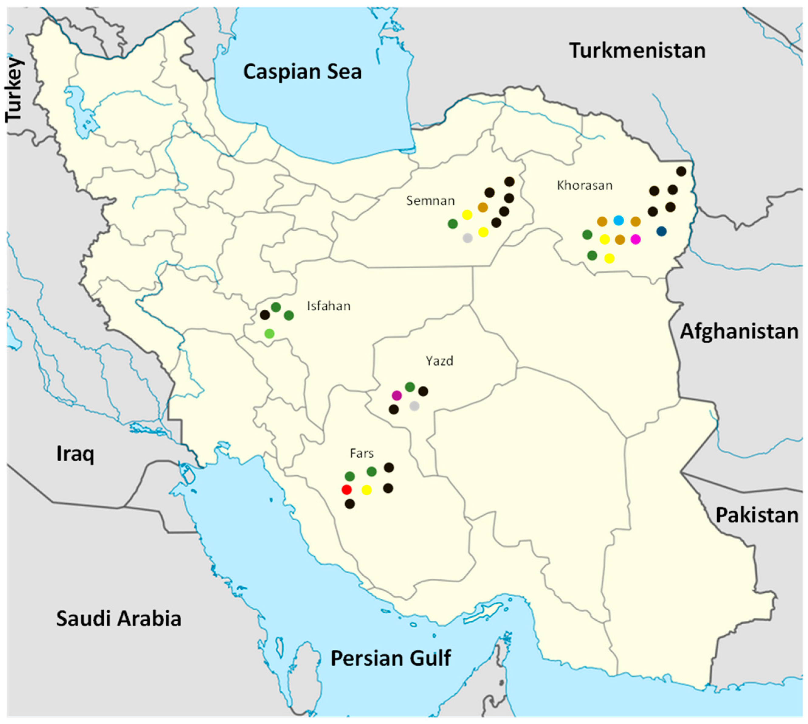
2.1. Survey, Sampling, and Fungal Isolation

From 2009 to 2011, a field survey was performed across melon growing regions of Central (Isfahan and Yazd), Eastern (Khorasan), Northern (Semnan), and Southern (Fars) provinces in Iran. Muskmelon plants showing wilting, crown, and root rot symptoms were sampled and brought to the laboratory for further analyses. Lower stems and upper roots of both severely decayed and wilted plants were cut into 0.5–1 cm segments, surface-sterilized by dipping into 1% sodium hypochlorite for 2 min, rinsed three times in sterile distilled water (SDW), and placed in Petri dishes onto acidified potato dextrose agar (PDA; potato extract 300 g/L, dextrose 20 g/L, agar 15 g/L). Dishes were incubated at 25 °C for 3–5 days in the dark, and the resulting colonies were purified by single conidium isolation. All recovered Fusarium isolates were preserved by both the sterile soil and cellulose filter paper methods [47] for further use and deposited in the fungal collection of the Plant Protection Department (Shiraz University, Iran). Furthermore, a FSSC isolate obtained from J. Armengol, Universidad Politécnica de Valencia, Spain, was included as a reference in the pathogenicity assays and molecular phylogenetic analysis.

2.2. Morphological Characterization

In order to study the in vitro pigmentation and growth rates of isolates, single-conidium subcultures were grown on fresh PDA dishes and incubated under alternating dark and light with a 12-h photoperiod at 25 °C for two weeks. For microscopic examination, all strains were grown on carnation leaf-piece agar (CLA) [48,49], potassium chloride agar (KCL-Agar) [50], and spezieller nahrstoffarmer agar (SNA) [51] Petri dishes, which were incubated at 25 °C for 4 days (KCL-Agar dishes) and also for 14 days (CLA and SNA dishes) under alternating dark and light with a 12-h photoperiod. Colony and conidia characteristics including the colony growth rate, pigmentation, mean size of 30 randomly selected well-developed macroconidia, and number of septa were recorded. Finally, the descriptions by Summerbell and Schroers [33], Leslie and Summerell [52], Nalim et al. [28], and Short et al. [32] were used for morphological species determination.

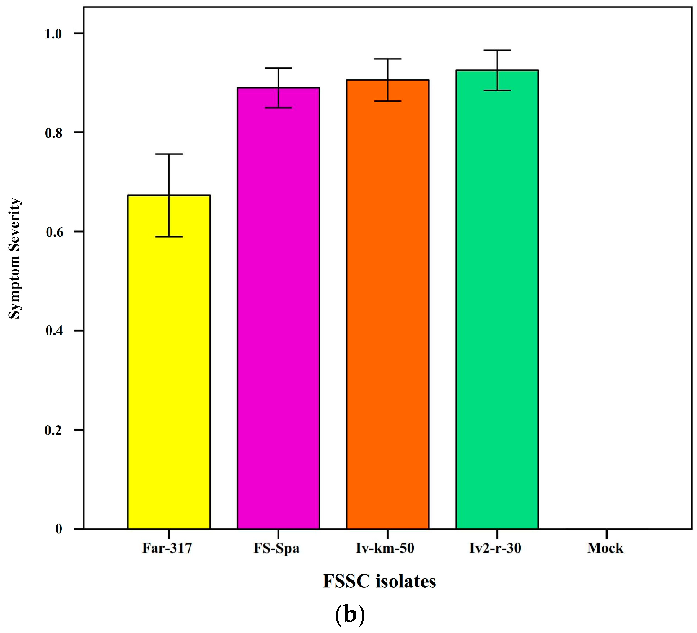
2.3. Pathogenicity Tests

Overall, 25 isolates were evaluated for their pathogenicity on their host of origin. Moreover, four of these isolates, Iv-Km50, Iv2r30, Far-317, and FS-Spa, were assessed for their pathogenicity on 11 different cucurbit varieties and species, including different varieties of melon, i.e., Garmak-Ahmadabadi (Cucumis melo var. reticulatus), Shahd-e-Shiraz (C. melo var. cantalopensis), Kharboz-e-Mashhadi (C. melo var. indorous), Snake melon (C. melo var. flexusus), Til-Mashhad (C. melo L.), and Semsoori (C. melo L.), as well as five other species of cucurbits, cucumber (C. sativus L.), watermelon (Citrullus lanatus L.), zucchini (Cucurbita pepo L.), pumpkin (C. moschata L.), and bottle gourd (Lagenaria siceraria L.). Inoculum was prepared by adding five discs (4 mm diameter) from 7-day-old Fusarium colonies grown on PDA to flasks containing sterile wheat seeds as described by Sabahi et al. [13]. Sterile agar plugs were added to wheat seeds to inoculate control plants. Flasks were incubated at room temperature for 15 days to ensure complete colonization of the grains. Inoculation of 15-day-old cucurbit seedlings was performed using a procedure described previously [13,53]. Inoculated plants were kept under greenhouse conditions (25 °C and 65% relative humidity) for symptoms’ development until 30 days post-inoculation (dpi). To determine the virulence and pathogenicity of FSSC isolates, the seedlings were examined 30 dpi; they were cut at cotyledon level, and the symptom severity (SS) was scored according to a scale from 0 to 5 as described by Nagao et al. [54]. Nine plants of each host were inoculated per fungal isolate, and the same number of plants inoculated with water was used as a control. SS measurements were converted from original ordinal scale to ratio scale and normalized (from 0 to 1) using the following formula:
SS = [(0 × n0) + (1 × n1) + (2 × n2) + (3 × n3) + (4 × n4) + (5 × n5)]/N × Mi
where n0, n1, n2, n3, n4, and n5 are the number of symptomatic plants per each scale level (from 0 to 5), N is total number of plants examined, and Mi is the highest score scale. SS data were analyzed using analysis of variance (ANOVA), and differences between the strains were analyzed using the GLM procedure of the SPSS software (SPSS, version 16). Tukey’s test was used for pairwise mean comparisons.
After symptom scoring, re-isolation was performed from symptomatic seedlings using PDA. The fungal isolates were identified based on morphological characteristics and sequencing of three genetic regions (ITS, LSU, and tef1) to fulfill Koch’s postulates. The experiments were repeated once with similar results.

2.4. DNA Extraction, PCR Amplification and Phylogenetic Analysis

Genomic DNA extraction and purification were performed using the procedures described by Sabahi et al. [10]. Internal transcribed spacer (ITS), nuclear large-subunit (28S) rDNA (LSU), and translation elongation factor 1-alpha (tef1) gene were amplified using the primer pairs ITS1/ITS4 [55], NL1/NL4, [56], and EF1-728F/EF2 [57], respectively. For PCR reactions, the Universal PCR Kit Ampliqon® Taq DNA Polymerase Master Mix Red (Ampliqon A/S, Odense, Denmark) was used according to the manufacturer’s recommendations. For each fungal isolate, a 50 µL PCR reaction including 50 ng total DNA and 1 µL of each primer (10 pmol µL−1) was used. The PCR amplification conditions were 30 cycles of 95 °C for 5 min, 95 °C for 50 s, 54 °C for 1 min, 72 °C for 50 s, and final step of 72 °C for 10 min. PCR products were sent to Bioneer Corporation (http://www.Bioneer.com (accessed on 4 March 2023)) to be sequenced via Sanger sequencing technology.
Newly obtained sequences were blasted against databases available at BLAST [58] on NCBI-GenBank database. Three loci in a set of worldwide FSSC strains were retrieved from the GenBank database and included in the phylogenetic analysis (Table S1). Sequences were aligned using the CLUSTAL W program, and concatenated following the alphabetic order of the genes, ending in a sequence of 1778 base pairs: nucleotides 1 to 667 for ITS, 668 to 1111 for LSU, and 1112 to 1778 for tef1.
Phylogenetic trees were constructed using the concatenated sequences of three loci via maximum likelihood with MEGA 6.06 [59]. The best model of evolution was determined using the Modeltest option from MEGA 6.06, and the phylogenetic tree was constructed with bootstrapping (1000 replications). Fusarium staphyleae (NRRL 22316) was used as an outgroup, and the final tree was drawn using infix pdf editor [60].

2.5. Genetic Diversity and Haplotype Network Analysis

Nucleotide diversity, number of haplotypes, haplotype frequency, haplotype diversity, number of segregating sites, number of mutations, percentage of polymorphism site, and the minimum number of recombination events were estimated using DnaSP v. 5.10 software for the sequences of each gene as well as concatenated sequences of FSSC isolates. The class I neutrality test (Tajima’s D, Fu and Li’D*, and Fu and Li’s F* statics) were also calculated for detecting departure from the mutation-drift equilibrium [61]. NeighborNet networks were constructed, and the pairwise homoplasy index (PHI-Test) was estimated for each gene region and the combined data set by SplitsTree v. 4.18.2 [62]. To see the phylogeographic relationship between the FSSC isolates, a haplotype network was generated for the concatenated sequences data set using TCS (Tata Consultancy Services) algorithm [63], which is implemented in Population Analysis with Reticulate Trees (PopART v. 1.7) [64]. The geographic origin of FSSC isolates investigated in this study and the FSSC strains retrieved from the GenBank database (Table 1) were allocated into the haplotype network as described by Leigh and Bryant [64].

3. Results

3.1. FSSC Isolates Obtained from Symptomatic Melon Plants in Iran

Melon growing regions in five provinces of Iran, including Fars, Isfahan, Khorasan, Semnan, and Yazd, were surveyed for the occurrence of Fusarium wilt. Melons with the Fusarium wilt syndrome consisting in wilt associated to crown and root rot were observed in all surveyed provinces (Figure 1).
A total of 41 Fusarium-like isolates were recovered from symptomatic melon plants of two varieties, C. melo L. var. cantalopensis Naudin (cantaloupe) and C. melo L. var. inodorous Naudin (long melon). All isolates were morphologically identified as F. solani sensu Leslie and Summerell [51]. Among the FSSC isolates examined in this study, 15 and 10 were from Khorasan and Semnan provinces, respectively, seven and five from Fars and Yazd provinces, respectively, and four from Isfahan province.
Based on the geographical origins, host, and morphological characteristics, 25 isolates representing the overall diversity of the original set of isolates were selected for in depth investigation (Table 1). An isolate of FSSC from Spain (FS-Spa) was included in this study as a reference isolate. According to the sequencing data of three genetic regions (ITS, LSU, and tef1) and morphological characteristics, four phylogenetic species were identified among the 25 Iranian isolates (Table 1 and Table 2, Figure S1). Of these, F. falciforme (syn. Neocosmospora falciformis) (18 isolates), F. keratoplasticum (syn. N. keratoplastica) (a single isolate), and F. vanettenii (syn. N. pisi) (five isolates) are known species. One isolate and the reference isolate from Spain were of an undescribed phylogenetic species of Fusarium sensu lato, tentatively named FSSC 5 or Neocosmospora sp.
Significant differences between distinct phylogenetic species were noticed in some morphometric and cultural characteristics, such as the shape of macroconidia and the growth rate on PDA (Table 2). In particular, the shape index (length to width ratio) of macroconidia of F. falciforme isolates ranged from 7.9 to 8.3, with a mean ± SD of 8.1 ± 0.12, while the same index for macroconidia of F. venattenii isolates ranged from 10.2 to 10.6, with a mean ± SD of 10.4 ± 0.23. The value of shape index of macroconidia of the only F. keratoplasticum isolate was 7.9, while the corresponding values for the two FSSC 5 isolates (Tay-r2-r, from Iran, and FS-Spa, from Spain) were 7.9 and 7.0, respectively. The growth rate on PDA of F. falciforme isolates ranged from 7.0 to 9.0 mm/day, with a mean ± SD of 8.1 ± 0.75, while the growth rate of F. venattenii isolates ranged from 5.0 to 6.0 mm/day, with a mean ± SD of 5.6 ± 0.42 mm/day. The growth rate of the F. keratoplasticum isolate was 8.5 mm/day, while the corresponding values for the two FSSC 5 isolates (Tay-r2-r, from Iran, and FS-Spa, from Spain) were 8.5 and 8.0 mm/day, respectively. Other morphological characteristics overlapped among different phylogenetic species or were not enough discriminant, as they showed a great intraspecific variability.

3.2. Pathogenicity and Host Range of FSSC Strains

Under greenhouse conditions, all 25 Iranian isolates and the reference isolate from Spain evaluated in this study were shown to be pathogenic on the host plant from which they were isolated; symptoms of wilting and crown- and root-rot were observed in artificially inoculated plants (Figure S1). In the host range assays, performed with four strains, each representing a distinct phylospecies, all melon varieties were severely affected by Iv2-r-30 and Iv-km-50 isolates, with SS values around 1, while the other cucurbit crops were less severely affected with the least severe symptoms being observed on zucchini plants with an SS value of 0.7 (Figure 2a).
The symptoms induced by Far-317 and FS-Spa isolates on six varieties of melons were less severe than those induced by Iv2-r-30 and Iv-km-50 isolates. Conversely, the degree of aggressiveness of FS-Spa strain on cucumber, watermelon, zucchini, pumpkin, and bottle gourd plants was higher than the other FSSC isolates evaluated in this study. Among all four isolates tested, Far-317 was the least aggressive on cucurbits (Figure 2b).
The inoculated fungi were re-isolated from symptomatic, artificially infected plants on PDA medium and their identity was confirmed by their morphological characteristics on SNA, CLA, and KCL-Agar media, as well as the sequencing of three genetic regions (ITS, LSU, and tef1). The negative (mock-inoculated) control plants remained healthy and did not develop any symptoms. The same results were observed in a replication of the pathogenicity and host range assays.

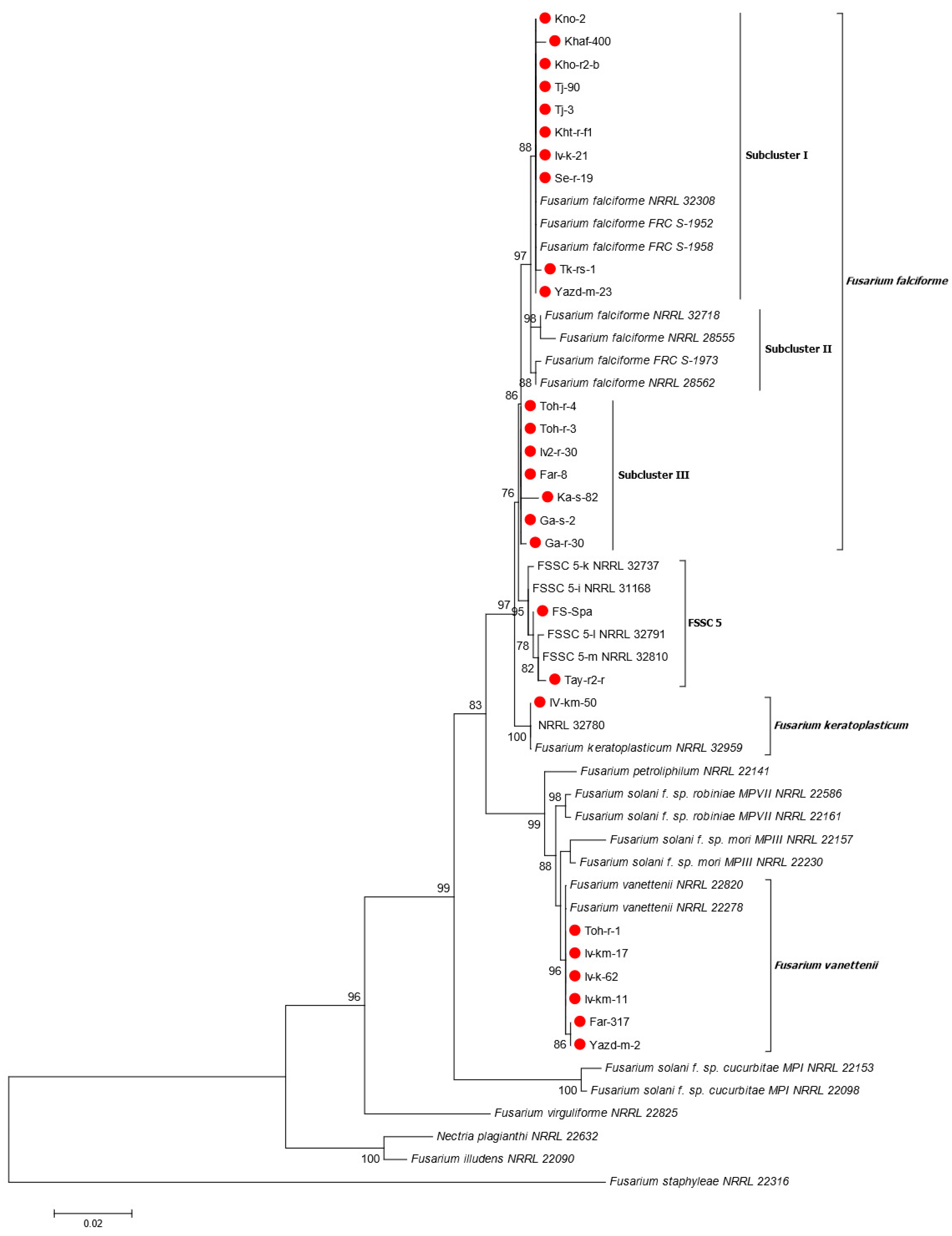
3.3. Phylogenetic Analyses

Sequence analysis of three genetic regions (ITS, LSU, and tef1) was conducted on all 25 selected Iranian FSSC isolates and the reference isolate from Spain. The nucleotide sequences of FSSC isolates were deposited in GenBank database (see Table 1).
Based on BLASTn searches using the sequence of tef1 gene on the NCBI GenBank database, the 26 strains were identified as either F. falciforme, F. keratoplasticum, F. vanettenii, or an undescribed species belonging to the FSSC. Overall, 17 of the 25 isolates from Iran clustered with the F. falciforme group with high bootstrap support (86%). Fusarium falciforme strains are divided into three subclusters, two of which, subcluster I and III, were found in Iran (Figure 3). The isolates of subcluster I showed two and three nucleotide differences in the sequences of ITS and tef1 gene regions, respectively, compared to the sequence of the subcluster III isolates. However, no nucleotide differences in the sequences of the LSU region between isolates of subclasters I and III were observed. The phylogenetic tree further showed a strongly-supported relationship (96% bootstrap support) between F. vanettenii (NRRL 22820 and NRRL 22278) obtained from GenBank and six isolates from melon plants in Iran. In addition, the tree showed that one isolate from Iran (Tay-r2-r) and the reference isolate from Spain (FS-Spa) belong to the undescribed species FSSC 5 (Neocosmospora sp.) with high bootstrap value (95%), while another Iranian isolate (Iv-km-50) was placed in F. keratoplasticum with strong phylogenetic affinity (100% bootstrap support) (Figure 3). Similar results were obtained when the sequences of gene tef1 were analyzed separately (Figure S3), while results of the phylogenetic analysis of the ITS and LSU regions were not consistent (Figures S2 and S4). Based on ITS maximum likelihood phylogeny, all F. falciforme isolates were placed in one group, while F. solani f. sp. robiniae and F. petroliphilum isolates from GenBank clustered together. As well, F. vanettenii and F. solani f. sp. mori isolates retrieved from GenBank clustered in one group, and Iranian isolates clustered in a separate group (Figure S2). In the LSU-based maximum likelihood phylogeny, all F. falciforme isolates clustered together, while F. vanettenii isolates clustered with the F. solani f. sp. mori and F. solani f. sp. robiniae isolates retrieved from GenBank (Figure S4).

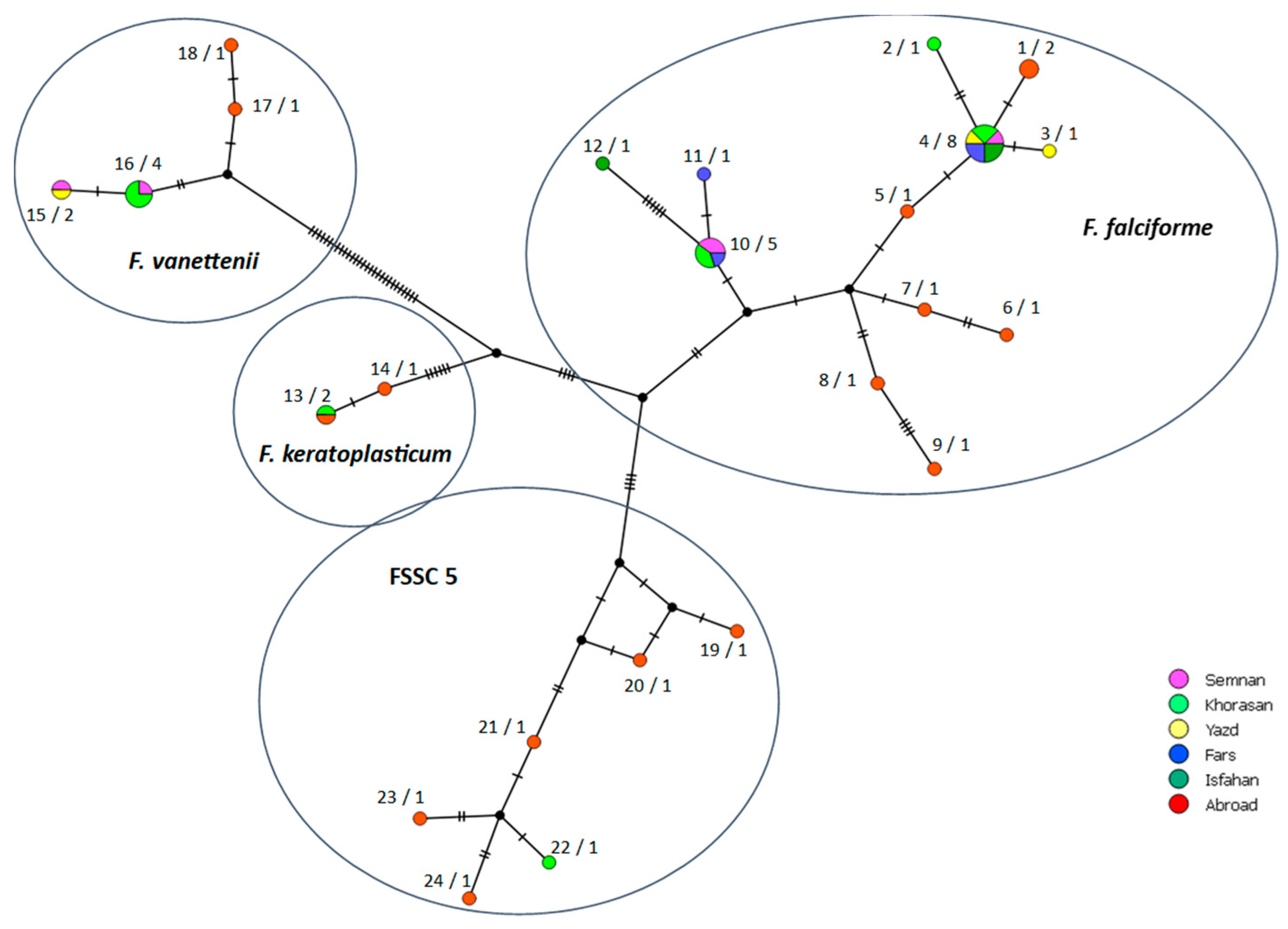
3.4. Genetic Diversity

The FSSC isolates recovered from melons in Iran carried different allelic forms and sequence types (STs) and corresponded to 10 multilocus haplotypes (MHs) based on the concatenated sequences of the three gene regions. Six, one, two, and one MHs belonged to F. falciforme, F. keratoplasticum, F. vanettenii, and FSSC 5 isolates, respectively (Table 1; Figure 4). As for the individual gene regions, 4, 10, and 5 STs were detected for the ITS, tef1 and LSU, respectively, in the Iranian isolates while 8, 13, and 4 STs were identified for these gene regions in the FSSC isolates from other countries (Table 1; Figure S5). Sequence variation statistics and diversity parameters were estimated by DnaSP v. 5.10 software for three individual gene regions, as well as concatenated sequences in all evaluated FSSC isolates in this study (Table 3). Similarly, the diversity parameters were calculated among the F. falciforme isolates recovered in Iran and compared with F. falciforme isolates from other countries (Table 3). As for the F. falciforme isolates from Iran, there were 1, 6, and 2 STs in the sequences of ITS, tef1, and LSU gene regions, respectively, while 1, 1, and 1 STs were found among the F. keratoplasticum isolates, 1, 2, and 1 STs among the F. vanettenii isolates, and 1, 1, and 1 STs among the isolates of the undescribed species FSSC 5 for the sequences of ITS, Tef-1α, and LSU gene regions, respectively (Table 1; Figure S5). Based on the concatenated sequences of three gene regions, the haplotype frequency (HF) and haplotype diversity (HD) indices were 0.352, and 0.721, respectively, for F. falciforme isolates from Iran and 0.857, and 0.952, respectively, for F. falciforme isolates from other countries, indicating higher genetic diversity among isolates from other countries (Table 3).
Most isolates from different regions of Iran were placed in separate MHs compared to isolates from other countries. Only one isolate, Iv-km-50 (F. keratoplasticum), recovered in Khorasan (Iran), shared the same MH as isolate NRRL 32780 from USA (Figure 4). Some MHs were represented by several Iranian isolates. For instance, isolates Se-r-19, Iv-k-21, Yazd-m-23, Tj-90, Tj-3, Kht-r-f1, Kno-2, and Kho-r2-b of F. falciforme belonged to the same MH. Conversely, several MHs were unique and represented by only a single Iranian isolate. This was the case of isolate Tk-rs-1, Khaf-400, Ka-s-82, and Ga-r-30 of F. falciforme, and isolate Tay-r2-r of FSSC 5 (Figure 4; Table 1).
A phylogenetic network was constructed by the NeighborNet method using the concatenated sequences of three gene regions. While the minor reticulations observed in the NeighborNet network indicate possible recombination events within the Iranian FSSC population, the PHI-Test did not find statistically significant evidence for recombination either for each gene region or in concatenated gene regions. The results of PHI-Test were in general accordance with those of the DnaSP results and did not show statistically significant evidence for recombination. Population neutrality indices (i.e., Fu and Li’ D*, and Fu and Li’s F*) were significantly negative for the LSU gene region of F. falciforme isolates (Table 3), indicating a recent selective sweep and/or population expansion after a recent bottleneck. However, considering the nonsignificant results obtained in sequences of ITS, and tef1 gene regions, further investigation by sequences of additional gene regions is needed to confirm these observations.

4. Discussion

This study discloses the genetic variability, potential host range, and geographic distribution of the FSSC population associated with Fusarium wilt of melon in Iran. Field surveys for three consecutive years (2009–2011) showed the disease occurred across five major melon-producing provinces of the country. Both molecular and morphological data were used to identify species of the FSSC recovered from symptomatic melon plants. In an earlier phylogenetic study, it was shown that DNA sequences of the LSU, ITS and tef1 gene regions can resolve evolutionary relationships within the FSSC [28]. Therefore, in the present study the combined data set of these three loci was used to identify FSSC isolates from melon at the species level. FSSC strains have been previously divided into three distinct subgroups, termed clades 1, 2, and 3 [25,26,27]. All Iranian FSSC isolates from melons, as well as the FS-Spa reference isolate from Spain, were found to be members of clade 3. Phylogenetic analyses of the concatenated three gene regions revealed that Iranian isolates grouped into five lineages, three of which had been identified earlier as F. vanettenii, F. keratoplasticum, and the undescribed species FSSC 5. According to the taxonomic criterion proposed by Sandoval-Denis and Crous [36] and Sandoval et al. [37], assigning the whole FSSC to Neocosmospora as a genus distinct from Fusarium s.s., the three above mentioned species should be named N. pisi, N. keratoplastica, and Neocosmospora sp., respectively, while F. falciforme is a synonym of N. falciformis. Here, both nomenclatural criteria have been interchangeably adopted to bypass the dispute concerning the taxonomy of the genus Fusarium and to make it easier to compare results from studies of diverse authors. The majority of Iranian FSSC isolates from melon fell into F. falciforme (FSSC 3 + 4) (syn. N. falciformis) and grouped into two subclusters. Similar results were obtained when sequences of tef1 were used individually for phylogenetic analyses, while all Iranian F. falciforme isolates were grouped into only one cluster based on phylogenetic analysis of ITS and LSU sequences. In ITS and LSU-based phylogenetic analysis, the F. vanettenii isolates clustered together with the isolates of F. solani f. sp. mori and F. solani f. sp. robiniae, indicating these gene regions were not suitable to distinguish these lineages.
The F. falciforme and F. vanettenii phylogenetic species showed distinct cultural and morphometric characteristics, such as the growth rate on agar medium and the shape of macroconidia. Conversely, no significant difference in these morphological traits was noticed between the F. falcforme isolates and either the F. vanettenii or the FSSC 5 isolates.
All FSSC isolates evaluated in this study were pathogenic on melon plants. Although N. keratoplastica and N. falciformis have been reported previously to cause root rot of muskmelons in Spain [65,66], to our knowledge, this is the first report of F. vanettenii and FSSC 5 causing wilt and root rot of melons in Iran. Fusarium vanettenii (synonym: Neocosmospora pisi), formerly Fusarium solani f. sp. pisi, is a recently recognized species in the FSSC [38]. This fungus has been reported as a destructive pathogen of legumes in different parts of the world, including Canada, Czech Republic, India, Iran, New Zealand, Southern Scandinavia, United Kingdom, and USA [47,67]. Šišić et al. [68] demonstrated this pathogen has a broad host range and raised doubts about the definition of it as a forma specialis. Consistently with this hypothesis, F. vanettenii was recently reported as a causal agent of tomato root rot in India [69]. Results of the present study confirm the host range of this species also encompasses non-leguminous hosts. It is the first time N. pisi is reported as a pathogen of melon worldwide. Members of FSSC 5 have been mostly reported as clinical opportunistic pathogens associated with infectious diseases of humans and animals [22]. Moreover, this phylogenetic species is a soil inhabitant and has been reported to be associated to the dry rot of potato [31,70]. In the present study, the FSSC 5 isolates were proved to be pathogenic on melon plants and other cucurbits, further expanding the known, already broad, host range of this Fusarium lineage. The melon varieties tested in this study, all widely grown in Iran, were susceptible to FSSC isolates. In greenhouse assays, besides melon, other crops of the Cucurbitaceae family, including watermelon, cucumber, zucchini, pumpkin, and bottle gourd, showed wilting and root rot symptoms when inoculated with FSSC isolates recovered from melon plants with natural infections. Although there was a noticeable variability in virulence among the isolates, no differences were observed in their host range on cucurbits.
FSSC species are considered cosmopolitan pathogens, as they occur in all climatic regions [71,72]. However, they prefer tropical hot areas [73,74]. In all provinces surveyed in this study, melon crops were in plains with warm to hot springs and summers and were planted in spring (March to April), so climatic conditions were relatively uniform. Despite this, the incidence of the disease was higher in Khorasan and Semnan. Moreover, a certain geographical structure in the Iranian FSSC population associated to Fusarium wilt of melon seems to exist. However, these aspects need to be further investigated to be confirmed.
A total of 24 MHs were identified among the FSSC isolates analyzed in this study; nine of them were only found in Iranian isolates, and one was shared between Iranian isolate and isolates from other country. Haplotypes MH2 and MH22 were only found in the Khorasan province, the first producer of melon in Iran with more than 48% of the national production [75].
Overall, 12 MHs were found among the isolates of F. falciforme (syn. N. falciformis). According to Posada and Crandall [76], the most frequent haplotype is probably the oldest in a given population. MH4 was the most common and widely distributed haplotype among Iranian F. falciforme isolates and included eight strains from five different provinces. The wide geographic distribution of this haplotype suggests effective mechanisms of dispersion over long distances. Very probably, in melon crops fungi of the FSSC have been transmitted prevalently by seeds, as F. solani s.l. is known to be a seed-borne pathogen [77]. A patchy distribution of symptomatic plants in surveyed melon crops, which is typical of seed-borne pathogens, would reinforce this hypothesis.
Interestingly, haplotype MH13 included two isolates of F. keratoplasticum (syn. N. keratoplastica) recovered from melon in Iran and sea turtles in the USA. This is not so surprising as several taxa of the FSSC have been associated with both clinical infections and plant diseases [41,65]. For instance, N. keratoplastica, a relevant pathogen of animals (including humans), has been recently reported as causal agent of wilt and root rot of muskmelon and watermelon crops [65]. Similarly, N. petroliphila (syn. F. petroliphilum), responsible for human keratitis, has been formerly known among plant pathologists as F. solani f. sp. cucurbitae race 1, a causal agent of fruit, stem, and root rot of cucurbits [65,78]. Neocosmospora falciformis (syn. F. falciforme), which is frequently responsible for clinical infections on humans, has been reported as a pathogen of several host plants, including species of the Cucurbitaceae family, confirming a wide host and ecological range of members of the FSSC [41,66].

5. Conclusions

This study provides preliminary information on the genetic variability of FSSC populations associated with Fusarium wilt of melon in Iran. Four phylogenetic species, including N. falciformis (syn. F. falciforme), N. pisi (syn. F. vanettenii), N. keratoplastica (syn. F. keratoplasticum), and Neocosmospora sp. (FSSC 5), and 10 diverse haplotypes have been identified in a set of isolates collected in major melon producing provinces of the country. Isolates of diverse genotypes differed in virulence, but all were pathogenic on the most common melon varieties grown in Iran and on a wide range of other cucurbits. The diversity of FSSC genotypes associated with Fusarium wilt of melon as well as their broad host range and ecological plasticity have implications for epidemiology, disease management strategies, breeding programs for disease resistance, and quarantine measures. The study of the genetic variability of FSSC populations would benefit from the use of additional markers. Moreover, targeting pathogenesis-related genes could provide a better insight into the biology and epidemiology of members of this complex. Focusing on taxa with a very broad host range that comprises both plant and animal pathogens might be helpful to understand the pathogenesis mechanisms of these fungi and in particular genetic determinants of both their polyphagia and ability to switch from a saprophytic to a parasitic lifestyle.

Supplementary Materials

The following supporting information can be downloaded at: https://www.mdpi.com/article/10.3390/jof9040486/s1, Table S1: Details on FSSC isolates and GenBank accessions for their sequence data. Figure S1: Symptoms of wilt (a), crown (b,c), and root (d) rot observed in pathogenicity test of FSSC isolate Iv2-r-30 on Cucumis melo plants 30 days after inoculation. Colony morphology of FSSC isolates grown on PDA for two weeks under 12-h alternating dark and light at 25 °C, front (left) and back (right) side, macroconidia and microconidia of FSSC isolates: F. keratoplasticum (isolate Iv-km50) (e–g), F. falciforme (isolate Iv2-r-30) (h–j), F. vanettenii (isolate Far-317) (k–m), and FSSC 5 (isolate FS-Spa) (n–p). (Scale bars = 10 μm). Figure S2: Maximum likelihood phylogeny of FSSC isolates recovered from melons in Iran based on ITS sequencing data. Scale bar indicates number of substitutions per site. Bootstrap values higher than 70 are shown. Fusarium staphyleae (NRRL 22316) was used as the outgroup. Red dots indicate strains isolated in this study. Figure S3: Maximum likelihood phylogeny of FSSC isolates recovered from melons in Iran based on tef1sequencing data. Scale bar indicates number of substitutions per site. Bootstrap values higher than 70 are shown. Fusarium staphyleae (NRRL 22316) was used as the outgroup. Red dots indicate strains isolated in this study. Figure S4: Maximum likelihood phylogeny of FSSC isolates recovered from melons in Iran based on LSU sequencing data. Scale bar indicates number of substitutions per site. Bootstrap values higher than 70 are shown. Fusarium staphyleae (NRRL 22316) was used as the outgroup. Red dots indicate strains isolated in this study. Figure S5: TCS multilocus haplotype network generated using the POPArt program from the partial sequences of three gene regions (ITS, LSU, and tef1) in FSSC isolates recovered from melons in Iran. The size of the circles indicates the relative frequency of sequences of a given haplotype. Hatch marks along the branches indicate the number of mutations. Each color represents one of the five provinces where the FSSC isolates were recovered. The isolates of F. falciforme, F. keratoplasticum, F. vanettenii, and FSSC 5 are framed by a blue, black, red, and grey boxes, respectively. The isolates retrieved from GenBank and the reference isolate FS-Spa received from Spain are assigned to the category ‘Abroad’. The numbers of the left of a slash indicates the MH number (Table 1), while the numbers of the right of the slash indicates the number of isolates in a given MH.

Author Contributions

Conceptualization, F.S., Z.B., and M.M.; M.R. and S.O.C.; methodology, F.S., Z.B., and M.M; software, F.S.; formal analysis, F.S.; investigation, F.S. and M.M.; resources, Z.B. and S.O.C.; data curation, F.S.; writing—original draft preparation, F.S.; writing—review and editing, F.S., Z.B., M.R., and S.O.C.; supervision, Z.B., M.R., and S.O.C.; project administration, F.S., Z.B., and S.O.C.; funding acquisition, F.S., Z.B., M.M., and S.O.C. All authors have read and agreed to the published version of the manuscript.

Funding

This research was funded by the Centre for International Scientific Studies & Collaboration (CISSC), Ministry of Science, Research & Technology, Iran. This study was also funded by the Iran National Science Foundation (INSF), award number 92033695. This research was also funded by the University of Catania, Italy “Investigation of phytopathological problems of the main Sicilian productive contexts and eco-sustainable defense strategies (ME-DIT-ECO)” PiaCeRi-PIAno di inCEntivi per la Ricerca di Ateneo 2020-22 linea 2” “5A722192155”, by PON “RICERCA E INNOVAZIONE” 2014–2020, Azione II-Obiettivo Specifico 1b-Progetto “Miglioramento delle produzioni agroalimentari mediterranee in condizioni di carenza di risorse idriche-WATER4AGRIFOOD”, B64I2000016000500, by the project “Smart and innovative packaging, postharvest rot management, and shipping of organic citrus fruit (BiOrangePack)” under the Partnership for Research and Innovation in the Mediterranean Area (PRIMA)—H2020 (E69C20000130001), by the “Italie–Tunisie Cooperation Program 2014–2020” project “PROMETEO «Un village transfrontalier pour protéger les cultures arboricoles méditerranéennes en partageant les connaissances» cod. C-5-2.1-36, CUP 453E25F2100118000 and by the by European Union (NextGeneration EU), through the MUR-PNRR project SAMOTHRACE (ECS00000022).

Institutional Review Board Statement

Not applicable.

Informed Consent Statement

Not applicable.

Data Availability Statement

The data that support the findings of this study are available GenBank database (see Table 1).

Conflicts of Interest

The authors declare no conflict of interest.

References

  1. Flores-León, A.; García-Martínez, S.; González, V.; Garcés-Claver, A.; Martí, R.; Julián, C.; Sifres, A.; Pérez de Castro, A.; Díez, M.J.; Lòpez, C.; et al. Grafting snake melon [Cucumis melo L. subsp. melo Var. flexuosus (L.) Naudin] in organic farming: Effects on agronomic performance; resistance to pathogens; sugar, acid, and VOC profiles; and consumer acceptance. Front. Plant Sci. 2021, 12, 613845. [Google Scholar] [CrossRef] [PubMed]
  2. Zohary, D.; Hopf, M. Domestication of Plants in the Old World, 3rd ed.; Oxford University Press: New York, NY, USA, 2000; p. 316. [Google Scholar]
  3. Watson, W. Early cereal cultivation in China. In The Domestication and Exploitation of Plants and Animals; Ucko, P.J., Dimbleby, G.W., Eds.; Gerald Duckworth and Co.: London, UK, 1969; pp. 397–402. [Google Scholar]
  4. Mallick, M.F.R.; Masui, M. Origin, distribution and taxonomy of melons. Sci. Hortic.-Amsterdam 1986, 28, 251–261. [Google Scholar] [CrossRef]
  5. Kerje, T.; Grum, M. The origin of melon, Cucumis melo: A review of the literature. Acta Hortic. 2000, 510, 37–44. [Google Scholar] [CrossRef]
  6. Oumouloud, A.; El-Otmani, M.; Chikh-Rouhou, H.; Claver, A.G.; Torres, R.G.; Perl-Treves, R.; Alvarez, J.M. Breeding melon for resistance to Fusarium wilt: Recent developments. Euphytica 2013, 192, 155–169. [Google Scholar] [CrossRef]
  7. Banihashemi, Z. The existence of Fusarium wilt of melon in Iran. Proc. First Nat. Cong. Plant Med. Iran 1968, 47–48. [Google Scholar]
  8. Banihashemi, Z. A new physiological race of Fusarium oxysporum f. sp. melonis in Iran. Iran. J. Plant Pathol. 1982, 18, 1–6. [Google Scholar]
  9. Sabahi, F.; Banihashemi, Z. Isolation, race determination and molecular characterization of Fusarium oxysporum f. sp. melonis in Bushehr province. Iran. J. Plant Pathol. 2020, 56, 359–369. [Google Scholar]
  10. Sabahi, F.; de Sain, M.; Banihashemi, Z.; Rep, M. Comparative genomics of Fusarium oxysporum f. sp. melonis strains reveals nine lineages and a new sequence type of AvrFom2. Environ. Microbiol. 2021, 23, 2035–2053. [Google Scholar] [CrossRef]
  11. Banihashemi, Z. Distribution of mating types of Phytophthora drechsleri the incitant of cucurbit root rot in Fars province. In Proceedings of the 9th Iranian Plant Protection Congress, Karaj, Iran; 1983; p. 102. [Google Scholar]
  12. Sarpeleh, A. The role of Monosporascus cannonballus in melon collapse in Iran. Australas. Plant Dis. Notes 2008, 3, 162–164. [Google Scholar]
  13. Sabahi, F.; Mirtalebi, M.; Banihashemi, Z. Paramyrothecium foliicola causes crown dry rot of melon (Cucumis melo) in Iran. J. Phytopathol. 2022, 170, 655–660. [Google Scholar] [CrossRef]
  14. Mirtalebi, M.; Sabahi, F.; Banihashemi, Z. First report of melon decline caused by Plectosphaerella cucumerina in Iran. Mycol. Iran. 2022, 9, 97–103. [Google Scholar]
  15. Mirtalebi, M.; Sabahi, F.; Banihashemi, Z. Fruit rot caused by Neoscytalidium hyalinum on melon in Iran. Australas. Plant Dis. Notes 2019, 14, 1–4. [Google Scholar] [CrossRef]
  16. Homa, M.; Galgóczy, L.; Manikandan, P.; Narendran, V.; Sinka, R.; Csernetics, Á.; Csaba, V.; László, K.; Papp, T. South Indian isolates of the Fusarium solani species complex from clinical and environmental samples: Identification, antifungal susceptibilities, and virulence. Front. Microbiol. 2018, 9, 1052. [Google Scholar] [CrossRef]
  17. Coleman, J.J. The Fusarium solani species complex: Ubiquitous pathogens of agricultural importance. Mol. Plant Pathol. 2016, 17, 146–158. [Google Scholar] [CrossRef] [PubMed]
  18. Kolattukudy, P.E.; Gamble, D.L. Nectria haematococca: Pathogenesis and host specificity in plant diseases. In Pathogenesis and Host Specificity in Plant Pathogenic Fungi and Nematodes; Kohmoto, K., Singh, U.S., Singh, R.P., Eds.; Elsevier Science Limited: Amsterdam, The Netherlands, 1995; pp. 83–102. [Google Scholar]
  19. Gordon, T.R. Fusarium oxysoporum and the Fusarium wilt syndrome. Annu. Rev. Phytopathol. 2017, 55, 23–39. [Google Scholar] [CrossRef] [PubMed]
  20. Dignani, M.C.; Anaissie, E.J. Human fusariosis. Clin. Microbiol. Infec. 2004, 10, 67–75. [Google Scholar] [CrossRef]
  21. Zhang, N.; O’Donnell, K.; Sutton, D.A.; Nalim, F.A.; Summerbell, R.C.; Padhye, A.A.; Geiser, D.M. Members of the Fusarium solani species complex that cause infections in both humans and plants are common in the environment. J. Clin. Microbiol. 2006, 44, 2186–2190. [Google Scholar] [CrossRef]
  22. O’Donnell, K.; Sutton, D.A.; Fothergill, A.; McCarthy, D.; Rinaldi, M.G.; Brandt, M.E.; Zhang, N.; Geiser, D.M. Molecular phylogenetic diversity, multilocus haplotype nomenclature, and in vitro antifungal resistance within the Fusarium solani species complex. J. Clin. Microbiol. 2008, 46, 2477–2490. [Google Scholar] [CrossRef]
  23. Muhammed, M.; Anagnostou, T.; Desalermos, A.; Kourkoumpetis, T.K.; Carneiro, H.A.; Glavis-Bloom, J.; Cleman, J.J.; Mylonakis, E. Fusarium infection: Report of 26 cases and review of 97 cases from the literature. Medicine 2013, 92, 305. [Google Scholar] [CrossRef]
  24. Mehl, H.L.; Epstein, L. Fusarium solani species complex isolates conspecific with Fusarium solani f. sp. cucurbitae race 2 from naturally infected human and plant tissue and environmental sources are equally virulent on plants, grow at 37 °C and are interfertile. Environ. Microbiol. 2007, 9, 2189–2199. [Google Scholar] [CrossRef]
  25. Guadet, J.; Julien, J.; Lafay, J.F.; Brygoo, Y. Phylogeny of some Fusarium species, as determined by large-subunit rRNA sequence comparison. Mol. Biol. Evol. 1989, 6, 227–242. [Google Scholar] [PubMed]
  26. O’Donnell, K. Progress towards a phylogenetic classification of Fusarium. Sydowia 1996, 48, 57–70. [Google Scholar]
  27. O’Donnell, K. Molecular phylogeny of the Nectria haematococca-Fusarium solani species complex. Mycologia 2000, 92, 919–938. [Google Scholar] [CrossRef]
  28. Nalim, F.A.; Samuels, G.J.; Wijesundera, R.L.; Geiser, D.M. New species from the Fusarium solani species complex derived from perithecia and soil in the old world tropics. Mycologia 2011, 103, 1302–1330. [Google Scholar] [CrossRef] [PubMed]
  29. Aoki, T.; O’Donnell, K.; Homma, Y.; Lattanzi, A.R. Sudden-death syndrome of soybean is caused by two morphologically and phylogenetically distinct species within the Fusarium solani species complex—F. virguliforme in North America and F. tucumaniae in South America. Mycologia 2003, 95, 660–684. [Google Scholar] [CrossRef] [PubMed]
  30. Al-Hatmi, A.M.; Ahmed, S.A.; Van Diepeningen, A.D.; Drogari-Apiranthitou, M.; Verweij, P.E.; Meis, J.F.; De Hoog, G.S. Fusarium metavorans sp. nov.: The frequent opportunist ‘FSSC6’. Med. Mycol. 2018, 56, S144–S152. [Google Scholar] [CrossRef]
  31. Schroers, H.J.; Samuels, G.J.; Zhang, N.; Short, D.P.; Juba, J.; Geiser, D.M. Epitypification of Fusisporium (Fusarium) solani and its assignment to a common phylogenetic species in the Fusarium solani species complex. Mycologia 2016, 108, 806–819. [Google Scholar] [CrossRef]
  32. Short, D.P.G.; O’Donnell, K.; Thrane, U.; Nielsen, K.F.; Zhang, N.; Juba, J.H.; Geiser, D.M. Phylogenetic relationships among members of the Fusarium solani species complex in human infections and the descriptions of F. keratoplasticum sp. nov. and F. petroliphilum stat. nov. Fungal Genet. Biol. 2013, 53, 59–70. [Google Scholar] [CrossRef]
  33. Summerbell, R.C.; Schroers, H.J. Analysis of phylogenetic relationship of Cylindrocarpon lichenicola and Acremonium falciforme to the Fusarium solani species complex and a review of similarities in the spectrum of opportunistic infections caused by these fungi. J. Clin. Microbiol. 2002, 40, 2866–2875. [Google Scholar] [CrossRef]
  34. Geiser, D.M.; Aoki, T.; Bacon, C.W.; Baker, S.E.; Bhattacharyya, M.K.; Brandt, M.E.; Brown, D.W.; Burgess, L.W.; Chulze, S.; Coleman, J.J.; et al. One fungus, one name: Defining the genus Fusarium in a scientifically robust way that preserves longstanding use. Phytopathology 2013, 103, 400–408. [Google Scholar] [CrossRef]
  35. O’Donnell, K.; Ward, T.J.; Robert, V.A.R.G.; Crous, P.W.; Geiser, D.M.; Kang, S. DNA sequence-based identification of Fusarium: Current status and future directions. Phytoparasitica 2015, 43, 583–595. [Google Scholar] [CrossRef]
  36. Sandoval-Denis, M.; Crous, P.W. Removing chaos from confusion assigning names to common human an animal pathogens in Neocosmospora. Persoonia 2018, 41, 109–129. [Google Scholar] [CrossRef] [PubMed]
  37. Sandoval-Denis, M.; Lombard, L.; Crous, P.W. Back to the roots: A reappraisal of Neocosmospora. Persoonia 2019, 43, 90–185. [Google Scholar] [CrossRef] [PubMed]
  38. O’Donnell, K.; Al-Hatmi, A.M.S.; Aoki, T.; Brankovics, B.; Cano-Lira, J.F.; Coleman, J.J.; de Hoog, S.G.; Di Pietro, A.; Frandsen, R.J.N.; Geiser, D.M.; et al. No to Neocosmospora: Phylogenomic and practical reasons for continued inclusion of the Fusarium solani species complex in the genus Fusarium. mSphere 2020, 5, e00810–e00820. [Google Scholar] [CrossRef]
  39. Geiser, D.M.; Al-Hatmi, A.M.S.; Aoki, T.; Arie, T.; Balmas, V.; Barnes, I.; Bergstrom, G.C.; Bhattacharyya, M.K.; Blomquist, C.L.; Bowden, R.L.; et al. Phylogenomic analysis of a 55.1 kb 19-gene dataset resolves a monophyletic Fusarium that includes the Fusarium solani species complex. Phytopathology 2021, 111, 1064–1079. [Google Scholar] [CrossRef]
  40. Guarnaccia, V.; van Niekerk, J.; Crous, P.W.; Sandoval-Denis, M. Neocosmospora spp. associated with dry root rot of citrus in South Africa. Phytopathol. Mediterr. 2021, 60, 79–100. [Google Scholar] [CrossRef]
  41. Kamali-Sarvestani, S.; Mostowfizadeh-Ghalamfarsa, R.; Salmaninezhad, F.; Cacciola, S.O. Fusarium and Neocosmospora species associated with rot of Cactaceae and other succulent plants. J. Fungi 2022, 8, 364. [Google Scholar] [CrossRef]
  42. Alymanesh, M.; Falahati, R.M.; Jafarpour, B.; Mahdikhanimoghadam, E. Identification of Fusarium solani f. sp. cucurbitae race 1 on cucurbit plants from Khorasan Razavi, northern Khorasan and some regions of Fars provinces, using classic and molecular methods. Hortic. Sci. 2007, 21, 49–56. [Google Scholar]
  43. Chehri, K.; Salleh, B.; Yli-Mattila, T.; Reddy, K.R.N.; Abbasi, S. Molecular characterization of pathogenic Fusarium species in cucurbit plants from Kermanshah province, Iran. Saudi J. Biol. Sci. 2011, 18, 341–351. [Google Scholar] [CrossRef]
  44. Zhian Jahangir, A.; Banihashemi, Z. Etiology of Fusarium Root Rot of Cucurbits with Emphasis on Fusarium solani f. sp. cucurbitae and its Vegetative Compatibility Groups and Host Range. Master’s Thesis in Agrometeorology, Shiraz University, Shiraz, Iran, 2011. [Google Scholar]
  45. Alymanesh, M.R.; Falahatirastegar, M.; Jafarpour, B.; Mahdikhanimoghadam, E. Genetic diversity in the fungus Fusarium solani f. sp. cucurbitae race 1, the causal agent of root and crown rot of cucurbits in Iran, using molecular markers. Pak. J. Biol. Sci. 2009, 12, 836–843. [Google Scholar] [CrossRef]
  46. Moradzadeh Eskandari, M.; Javan-Nikkhah, M.; Zare, R.A.S.O.U.L.; Okhowat, M.; Moretti, A.; Somma, S.; Stea, G. Study on genetic structure of Fusarium solani populations isolated from potato, cucurbit, bean and chickpea based on FAFLP markers. Iran. J. Plant Pathol. 2010, 45, 189–197. [Google Scholar]
  47. Porter, L.D.; Pasche, J.S.; Chen, W.; Harveson, R.M. Isolation, identification, storage, pathogenicity tests, hosts, and geographic range of Fusarium solani f. sp. pisi causing Fusarium root rot of pea. Plant Health Prog. 2015, 16, 136–145. [Google Scholar] [CrossRef]
  48. Snyder, W.C.; Hansen, H.N. Advantages of natural media and environments in the culture of fungi. Phtopathology 1974, 37, 420–421. [Google Scholar]
  49. Fisher, N.L.; Burgess, L.W.; Toussoun, T.A.; Nelson, P.E. Carnation leaves as a substrate and for preserving cultures of Fusarium species. Phytopathology 1982, 72, 151–153. [Google Scholar] [CrossRef]
  50. Pascoe, I.G. Fusarium morphology identification and characterization of a third conidial type, the mesoconidium. Mycotaxon 1990, 37, 121–160. [Google Scholar]
  51. Nirenberg, H.L. Untersuchungen Uber Die Morphologische Und Biologische Differenzierung in der FUSARIUM-Section Liseola; Kommissionsverlag Paul Parey: Berlin, Germany, 1976; pp. 1–117. [Google Scholar]
  52. Leslie, J.F.; Summerell, B.A.; Bullock, S. The Fusarium Laboratory Manual; Blackwell Publishing: Ames, IA, USA, 2006; pp. 1–388. [Google Scholar]
  53. Zhang, X.Y.; Yu, X.X.; Yu, Z.; Xue, Y.F.; Qi, L.P. A simple method based on laboratory inoculum and field inoculum for evaluating potato resistance to black scurf caused by Rhizoctonia solani. Breeding Sci. 2014, 64, 156–163. [Google Scholar] [CrossRef] [PubMed]
  54. Nagao, H.; Sato, K.; Ogiwara, S. Susceptibility of Cucurbita spp. to the cucurbit root-rot fungus, Fusarium solani f. sp. cucurbitae race 1. Agronomie 1994, 14, 95–102. [Google Scholar] [CrossRef]
  55. White, T.J.; Bruns, T.D.; Lee, S.B.; Taylor, J.W. Amplification and direct sequencing of fungal ribosomal RNA genes for phylogenetics. In PCR Protocols: A Guide to Methods and Applications; Innis, N., Gelfand, D., Sninsky, J., White, T., Eds.; Academic Press: New York, NY, USA, 1990; pp. 315–322. [Google Scholar]
  56. O’Donnell, K. Fusarium and its near relatives. In The Fungal Holomorph: Mitotic, Meiotic and Pleomorphic Speciation in Fungal Systematics; Reynolds, D.R., Taylor, J.W., Eds.; CAB International: Wallingford, UK, 1993; pp. 225–233. [Google Scholar]
  57. O’Donnell, K.; Kistler, H.C.; Cigelnik, E.; Ploetz, R.C. Multiple evolutionary origins of the fungus causing Panama disease of banana: Concordant evidence from nuclear and mitochondrial gene genealogies. Proc. Natl. Acad. Sci. USA 1998, 95, 2044–2049. [Google Scholar] [CrossRef]
  58. National Library of Medicine. BLAST®. Available online: https://blast.ncbi.nlm.nih.gov/ (accessed on 10 February 2023).
  59. Tamura, K.; Stecher, G.; Peterson, D.; Filipski, A.; Kumar, S. MEGA6: Molecular evolutionary genetics analysis version 6.0. Mol. Biol. Evol. 2013, 30, 2725–2729. [Google Scholar] [CrossRef]
  60. Iceni Technology. Available online: https://www.iceni.com/infix.htm (accessed on 10 February 2023).
  61. Librado, P.; Rozas, J. DnaSP v5: A software for comprehensive analysis of DNA polymorphism data. Bioinformatics 2009, 25, 1451–1452. [Google Scholar] [CrossRef]
  62. Huson, D.H.; Bryant, D. Application of phylogenetic networks in evolutionary studies. Mol. Biol. Evol. 2006, 23, 254–267. [Google Scholar] [CrossRef]
  63. Clement, M.; Snell, Q.; Walker, P.; Posada, D.; Crandall, K. TCS: Estimating gene genealogies. Int. Parallel Distrib. Process. Symp. 2002, 2, 184. [Google Scholar]
  64. Leigh, J.W.; Bryant, D. POPART: Full-feature software for haplotype network construction. Methods Ecol. Evol. 2015, 6, 1110–1116. [Google Scholar] [CrossRef]
  65. González, V.; García-Martínez, S.; Flores-León, A.; Ruiz, J.J.; Picó, B.; Garcés-Claver, A. Neocosmospora keratoplastica, a relevant human fusarial pathogen is found to be associated with wilt and root rot of muskmelon and watermelon crops in Spain: Epidemiological and molecular evidences. Eur. J. Plant Pathol. 2020, 156, 1189–1196. [Google Scholar] [CrossRef]
  66. González, V.; Ruiz, J.J.; Picó, B.; García-Martínez, S.; Garcés-Claver, A.; Flores-León, A. First report of Neocosmospora falciformis causing wilt and root rot of muskmelon in Spain. Plant Dis. 2020, 104, 1256. [Google Scholar] [CrossRef]
  67. Mohammadi, H.; Banihashemi, Z. Distribution, pathogenicity and survival of Fusarium spp. the causal agents of chickpea wilt and root rot in the Fars province of Iran. Iran. J. Plant Pathol. 2005, 41, 687–708. [Google Scholar]
  68. Šišić, A.; Baćanović-Šišić, J.; Al-Hatmi, A.M.S.; Karlovsky, P.; Ahmed, S.A.; Maier, W.; Sybren De Hoog, G.; Finckh, M.R. The ‘forma specialis’ issue in Fusarium: A case study in Fusarium solani f. sp. pisi. Sci. Rep.-UK 2018, 8, 1252. [Google Scholar] [CrossRef]
  69. Debbarma, R.; Kamil, D.; Maya Bashyal, B.; Choudhary, S.P.; Thokla, P. First report of root rot disease on Solanum lycopersicum L. caused by Fusarium vanettenii in India. J. Phytopathol. 2021, 169, 752–756. [Google Scholar] [CrossRef]
  70. Chehri, K.; Ghasempour, H.R.; Karimi, N. Molecular phylogenetic and pathogenetic characterization of Fusarium solani species complex (FSSC), the cause of dry rot on potato in Iran. Microb. Pathogenesis 2014, 67, 14–19. [Google Scholar] [CrossRef]
  71. Backhouse, D.; Burgess, L.W. Mycogeography of Fusarium: Climatic analysis of the distribution within Australia of Fusarium species in section Gibbosum. Mycol. Res. 1995, 99, 1218–1224. [Google Scholar] [CrossRef]
  72. Burgess, L.W.; Nelson, P.E.; Toussoun, T.A.; Forbes, G.A. Distribution of Fusarium species in sections Roseum, Arthrosporiella, Gibbosum and Discolor recovered from grassland, pasture and pine nursery soils of eastern Australia. Mycologia 1988, 80, 815–824. [Google Scholar] [CrossRef]
  73. Sangalang, A.E.; Backhouse, D.; Burgess, L.W. Survival and growth in culture of four Fusarium species in relation to occurrence in soils from hot climatic regions. Mycol. Res. 1995, 99, 529–533. [Google Scholar] [CrossRef]
  74. Saremi, H.; Burgess, L.W.; Backhouse, D. Temperature effects on the relative abundance of Fusarium species in a model plant–soil ecosystem. Soil Biol. Biochem. 1999, 31, 941–947. [Google Scholar] [CrossRef]
  75. Narouirad, M.; Bakhshi, B.; Kohpalekani, A.J.; Ghasemi, A.; Fazeli, R.M. Response of Iranian melon populations to deficit irrigation condition. Hort. Sci. 2020, 34, 161–169. [Google Scholar]
  76. Posada, D.; Crandall, K.A. Evaluation of methods for detecting recombination from DNA sequences: Computer simulations. Proc. Natl. Acad. Sci. USA 2001, 98, 13757–13762. [Google Scholar] [CrossRef] [PubMed]
  77. Pareek, V.; Varma, R. Fusarium solani a dominant seed borne pathogen in seeds of cluster bean grown in Rajasthan. Biosci. Biotech. Res. Commun. 2015, 8, 29–34. [Google Scholar]
  78. González, V.; Armengol, J.; Garcés-Claver, A. First report of Fusarium petroliphilum causing fruit root of butternut squash in Spain. Plant Dis. 2018, 102, 1662. [Google Scholar] [CrossRef]
Figure 1. Distribution of FSSC isolates recovered from melons in Iran and examined in this study. Each isolate was recovered from a distinct field. In the map, each dot represents an isolate and each color represents a multilocus haplotype (MH). Dark green: MH 4; Light green: MH 12; Dark blue: MH 2; Yellow: MH 10; Pink: MH 22; Brown: MH 16; Light blue: MH 13; Red: MH 11; Gray: MH 15; Purple: MH 3.
Jof 09 00486 g001
Figure 2. Results of pathogenicity assays of four isolates of diverse FSSC species on different cucurbits. The FSSC species tested included F. keratoplasticum (isolate Iv-km-50), F. falciforme (isolate Iv2-r-30), F. vanettenii (isolate Far-317), and FSSC 5 (isolate FS-Spa). Nine plants of each cucurbit species or variety were inoculated per fungal isolate, and the same number of plants was mock-inoculated with water. Symptom severity was evaluated 30 days after inoculation. (a) Symptom severity induced by each Fusarium isolate on 11 diverse cucurbits (means of nine values ± SE). (b) Values of symptoms severity induced by each Fusarium isolate on 11 diverse cucurbits were pooled together to compare the virulence of isolates (means of 99 values ± SE).
Jof 09 00486 g002aJof 09 00486 g002b
Figure 3. Maximum likelihood phylogeny of FSSC isolates from melons based on concatenated sequencing data of three gene regions (ITS, LSU, and tef1). Scale bar indicates number of substitutions per site. Bootstrap values higher than 70 are shown. Fusrium staphyleae (NRRL 22316) was used as the outgroup. Red dots indicate Iranian isolates recovered in this study.
Jof 09 00486 g003
Figure 4. TCS multilocus haplotype network generated using the POPArt program by combining three gene regions (ITS, LSU, and tef1) of FSSC isolates recovered from melons in Iran. The size of the circles indicates the relative frequency of sequences of a given multilocus haplotype (MH). Hatch marks along the branches indicate the number of mutations. Each color represents one of the five provinces where the FSSC isolates were recovered. The isolates retrieved from GenBank and FS-Spa reference isolate received from Spain are assigned to the category ‘Abroad’. The numbers on the left of a slash indicates the MH number (Table 1), while the numbers on the right of the slash indicates the number of isolates in a given MH.
Jof 09 00486 g004
Table 1. FSSC isolates recovered from melons in Iran, their host and geographic area of origin, date of isolation, and multilocus haplotypes (MHs) and sequence types (STs) based on the partial sequences of three gene regions (ITS, LSU, and tef1).
IsolateSpeciesYear aSourceCollection b LocalitySequence Type (ST) c Detected in:Multilocus Haplotype (MH)
ITSLSUtef1
Se-r-19F. falciforme2010long melonSe, Is, Ir72124
Khaf-400F. falciforme2009long melonKha, Kho, Ir72132
Toh-r-3F. falciforme2009long melonToh, Kho, Ir72610
Toh-r-4F. falciforme2009long melonToh, Kho, Ir72610
Iv-k-21F. falciforme2011long melonEy, S, Ir72124
Iv2-r-30F. falciforme2009long melonEy, S, Ir72610
Yazd-m-23F. falciforme2010cantaloupeMey, Y, Ir72124
Tj-90F. falciforme2009long melonToj, Kho, Ir72124
Tk-rs-1F. falciforme2011cantaloupeTa, Y, Ir72143
Ga-r-30F. falciforme2009cantaloupeGa, Fa, Ir72711
Ga-s-2F. falciforme2011cantaloupeGa, Fa, Ir72610
Ka-s-82F. falciforme2010long melonKa, Is, Ir71512
Tj-3F. falciforme2010long melonToj, Kho, Ir72124
Far-8F. falciforme2011long melonFa, S, Ir72610
Kht-r-f1F. falciforme2009long melonKhan, Is, Ir72124
Kno-2F. falciforme2009cantaloupeKhaf, Fa, Ir72124
Kho-r2-bF. falciforme2011cantaloupeKhaf, Fa, Ir72124
Tay-r2-rFSSC 52009long melonTay, Kho, Ir131822
Iv-k-62F. vanettenii2009long melonEy, S, Ir35216
Far-317F. vanettenii2009long melonFar, S, Ir35115
Iv-km-11F. vanettenii2010long melonKash, Kho, Ir35216
Iv-km-17F. vanettenii2010long melonKash, Kho, Ir35216
Toh-r-1F. vanettenii2009long melonToh, Kho, Ir35216
Yazd-m-2F. vanettenii2010long melonMey, Y, Ir35115
Iv-km-50F. keratoplasticum2009long melonKash, Kho, Ir64313
Strains from different countries and sources
NRRL 32718F. falciforme2006human eyeUSA72108
FRC S-1973F. falciforme2011soilAust7296
FRC S-1958F. falciforme2011soilAust92121
FRC S-1952F. falciforme2011soilAust92121
NRRL 28555F. falciforme2006human wristUSA82119
NRRL 28562F. falciforme2006human boneUSA7287
NRRL 32308F. falciforme2006human footSA72125
FS-SpaFSSC 5NDNDSpain131923
NRRL 31168FSSC 52006human toe leukemiaUSA231520
NRRL 32810FSSC 52006human eyeUSA131621
NRRL 32737FSSC 52006human eyeUSA232019
NRRL 32791FSSC 52006humanUSA231724
NRRL 22820F. vanetteniiNDNDUSA55218
NRRL 22278F. vanetteniiNDNDUSA45217
NRRL 32780F. keratoplasticum2006sea turtleUSA64313
NRRL 32959F. keratoplasticum2006human skinUSA64414
a ND: not determined. b Se: Sefidshahr; Is: Isfahan; Ir: Iran; Kha: Khaf; Kho: Khorasan; Toh: Torbat-e Heydariyeh; Ey: Eyvanekey; S: Semnan; Mey: Meybod; Y: Yazd; Toj: Torbat-e Jam; Ta: Tabas; Ga: Galehdar; Fa: Fars; Ka: Kashan; Far: Faravan; Khan: Khansar; Khaf: Khafr; Tay: Taybad; Kash: Kashmar; Aust: Australia; SA: Saudi Arabia. c Sequence type is based on TCS sequence type network as shown in Figure S4.
Table 2. Morphological characteristics of FSSC isolates obtained from melon plants in Iran.
IsolateSpeciesColony Growth Rate (mm/day)Pigment on PDA aChlamydospores bShape/No. of Septa
of Microconidia a
Shape of Basal and Apical Cell aLength × Width of Macroconidia (µm) c
3 and 4 Septate5 Septate
Se-r19F. falciforme7.5WY or G+EO, OT/0-1BNP, Cu40.5 ± 1.5 × 5.0 ± 0.5-
Khaf-400F. falciforme7.5WY or G+EO, OT/0-1BNP, Cu41.5 ± 2.5 × 5.1 ± 0.5-
Toh-r3F. falciforme9.0WY or G+EO, OT/0-1BNP, Cu42.5 ± 2.5 × 5.2 ± 0.5-
Yazd-m23F. falciforme8.0WY or G+EO, OT/0-1BNP, Cu41.0 ± 1.5 × 5.2 ± 0.5-
Tj-90F. falciforme8.5WY or G+EO, OT/0-1BNP, Cu42.0 ± 2.5 × 5.1 ± 0.5-
Tk-rs-1F. falciforme9.0WY or G+EO, OT/0-1BNP, Cu41.5 ± 2.5 × 5.0 ± 0.5-
Ga-r30F. falciforme9.0WY or G+EO, OT/0-1BNP, Cu40.5 ± 1.5 × 5.0 ± 0.5-
Ga-s2F. falciforme8.5WY or G+EO, OT/0-1BNP, Cu42.5 ± 2.5 × 5.2 ± 0.5-
Ka-s-82F. falciforme9.0WY or G+EO, OT/0-1BNP, Cu40.5 ± 1.5 × 5.0 ± 0.5-
Tj-3F. falciforme7.0WY or G+EO, OT/0-1BNP, Cu41.5 ± 2.5 × 5.1 ± 0.5-
Far-8F. falciforme7.5WY or G+EO, OT/0-1BNP, Cu41.5 ± 2.5× 5.1 ± 0.5-
Yazd-m2F. falciforme8.0WY or G+EO, OT/0-1BNP, Cu42.0 ± 2.5 × 5.3 ± 0.5-
Kho-r2-bF. falciforme8.0WY or G+EO, OT/0-1BNP, Cu42.5 ± 2.5 × 5.1 ± 0.5-
Kht-r-f1F. falciforme7.5WY or G+EO, OT/0-1BNP, Cu42.5 ± 2.5 × 5.2 ± 0.5-
Toh-r4F. falciforme7.0WY or G+EO, OT/0-1BNP, Cu40.5 ± 1.5 × 5.0 ± 0.5-
iv-k-21F. falciforme9.0WY or G+EO, OT/0-1BNP, Cu40.5 ± 1.5 × 5.0 ± 0.5-
iv2-r30F. falciforme8.5WY or G+EO, OT/0-1BNP, Cu41.5 ± 2.5 × 5.1 ± 0.5-
Kno2F. falciforme7.0WY or G+EO, OT/0-1BNP, Cu41.0 ± 2.5 × 5.2 ± 0.5-
iv-km-50F. keratoplasticum8.5WY+O, PC, S/0-1NB36.5 ± 2.3 × 4.8 ± 0.3-
Far-317F. venattenii5.5WB+OR and T/0-1PR46.2 ± 2.0 × 4.35 ± 0.4-
iv-km-11F. venattenii6.0WB+OR and T/0-1PR45.2 ± 1.5 × 4.35 ± 0.4-
iv-km-17F. venattenii6.0WB+OR and T/0-1PR46.2 ± 2.5 × 4.33 ± 0.1-
Toh-r1F. venattenii5.0WB+OR and T/0-1PR44.1 ± 2.3 × 4.34 ± 0.2-
iv-k-62F. venattenii5.5WB+OR and T/0-1PR44.2 ± 2.0 × 4.35 ± 0.3-
Tay-r2-rFSSC 58.0WY or G+EO, OT/0-1BNP, Cu40.5 ± 1.5 × 5.1 ± 0.5-
FS-SpaFSSC 58.5WC+O, EO, C, R/0-1BN, P Cu38.6 ± 1.5 × 5.5 ± 0.342.5 ± 2.5 × 5.8 ± 0.3
a WY: white to yellow; G: green; WB: white to brown; WC: white to cream; EO: elongated oval; OT: obovoid with a basal truncation; O: oval; PC: pyriform to cylindrical; S: straight; OR: ovoidal rounded apex; T: basal truncation; C: clavate; R: reniform; BNP: barely notched and pointed; Cu: curved; NB: notched and blunt; PR: pedicellate and rounded; BN: barely notched; P Cu: papillate curved. b +: Present. c Mean value of 30 random selected conidia ± SD.
Table 3. Sequence variation statistics of the partial sequences of three gene regions (ITS, LSU, and tef1) among the FSSC isolates examined in this study.
No. of Value for Indicated Neutrality Test b
Region, FSSC SpeciesGeneStrainsNucleotidesHaplotypesHaplotype Frequency aNumber of Segregating SitesPolymorphic Sites
(%)
Nucleotide Diversity (π)Number of Mutation (ղ)Haplotype (Gene) DiversityTajima’s DFu and Li’s D*Fu and Li’s F*Minimum No. of Recombination Events b
F. falciformeConcatenated241556120.50241.5420.00262240.859−1.47289 NS−2.27745 NS−2.37548 NS0
ITS2443940.16630.6830.0018130.634−0.12171 NS−0.18894 NS−0.19630 NS0
tef124668100.416182.6940.00452180.764−1.46801 NS−2.18497 NS−2.29803 NS0
LSU2443520.08330.6890.0005930.083−1.73253 NS−2.52572*
(p < 0.05)
−2.65835*
(p < 0.05)
0
F. keratoplasticumConcatenated3155620.66610.0640.0004510.667NANANA0
ITS343910.33300.0000.00000.00NANANA0
tef1366820.66610.1490.0010510.667NANANA0
LSU343510.33300.0000.00000.000NPNPNPNP
FSSC 5Concatenated6155661.000100.6420.00297101.0000.11945 NS0.7918 NS0.9479 NS2
ITS643920.33310.2270.0014310.6001.44510 NS1.05247 NS1.15768 NS0
tef1666861.00091.3470.0059591.000−0.11324 NS−0.09221 NS−0.10424 NS1
LSU643510.16600.0000.00000.000NPNPNPNP
F. vanetteniiConcatenated8155640.5050.3210.0013150.7500.08445 NS0.74709 NS0.65390 NS0
ITS843930.37540.9110.0036740.464−0.01957 NS0.56807 NS0.47502 NS0
Tef-1α866820.2510.1490.0006610.4290.33350 NS0.88779 NS0.82528 NS0
LSU843510.12500.0000.00000.000NPNPNPNP
Iran
F. falciformeConcatenated17155660.352140.8990.00218140.721−0.83607 NS−1.90361 NS−1.85065 NS0
ITS1743920.11710.2270.0012210.5151.43020 NS0.67700 NS0.99442 NS0
tef11766860.352101.4970.00372100.721−0.73074 NS−1.30215 NS−1.68143 NS0
LSU1743520.11730.6890.0008330.118−1.70573 NS−2.25481 NS−2.41419 NS0
Non-Iran
F. falciformeConcatenatedConcatenated7155660.857120.7710.00313120.952−0.25712 NS−0.07864 NS−0.13028 NS
ITSITS743930.42830.6830.0029130.6670.05031 NS0.38925 NS0.33832 NS
tef1766850.71491.3470.0054290.857−0.35433 NS−0.25556 NS−0.30404 NS0
LSU743510.14200.0000.00000.000NPNPNPNP
a Number of haplotype/number of isolates. b NA: not applicable; NS: not significant; NP: not polymorphism.
Disclaimer/Publisher’s Note: The statements, opinions and data contained in all publications are solely those of the individual author(s) and contributor(s) and not of MDPI and/or the editor(s). MDPI and/or the editor(s) disclaim responsibility for any injury to people or property resulting from any ideas, methods, instructions or products referred to in the content.

