Review

Interactions between Entomopathogenic Fungi and Insects and Prospects with Glycans

1
Institute of Entomology, Guizhou University, Guiyang 550025, China
2
Institute of Plant Health and Medicine, Guizhou University, Guiyang 550025, China
*
Authors to whom correspondence should be addressed.
J. Fungi 2023, 9(5), 575; https://doi.org/10.3390/jof9050575
Submission received: 4 April 2023 / Revised: 11 May 2023 / Accepted: 12 May 2023 / Published: 15 May 2023

Abstract

:
Concerns regarding the ecological and health risks posed by synthetic insecticides have instigated the exploration of alternative methods for controlling insects, such as entomopathogenic fungi (EPF) as biocontrol agents. Therefore, this review discusses their use as a potential alternative to chemical insecticides and especially focuses on the two major ones, Beauveria bassiana and Metarhizium anisopliae, as examples. First, this review exemplifies how B. bassiana- and M. anisopliae-based biopesticides are used in the world. Then, we discuss the mechanism of action by which EPF interacts with insects, focusing on the penetration of the cuticle and the subsequent death of the host. The interactions between EPF and the insect microbiome, as well as the enhancement of the insect immune response, are also summarized. Finally, this review presents recent research that N-glycans may play a role in eliciting an immune response in insects, resulting in the increased expression of immune-related genes and smaller peritrophic matrix pores, reducing insect midgut permeability. Overall, this paper provides an overview of the EPF in insect control and highlights the latest developments relating to the interaction between fungi and insect immunity.

1. Introduction

Insects are incredibly ubiquitous organisms in nature and are found in virtually every corner of the planet. Insect pests can reduce crop yields and, as a result, impact agricultural productivity and horticultural output, posing a formidable threat to food supply and economic stability. Over the past several decades, the employment of chemical pesticides has become increasingly widespread for pest management globally. While these substances offer quick pest eradication, their utilization presents hazards to both human health and the agroecosystem, leading to undesirable effects on natural enemies or pesticide residue [1]. Furthermore, persistent exposure to pesticides has resulted in the development of pest resistance to various chemicals [2,3]. Consequently, concerns regarding the negative effects of chemical insecticides have prompted a focus on eco-friendly and alternative strategies for pest control. Numerous efforts have been made to develop biological control agents (BCAs) as alternatives or supplements to these chemicals. These include the utilization of microbial control agents against insect pests, such as bacteria, viruses and fungi [4,5,6,7,8,9,10]. In 2022, at the Annual Biocontrol Industry Meeting (ABIM) held in Basel, Switzerland, Marrone provided an update on the share of the biological market [11]. The report showed that the fastest-growing segment of the biocontrol market was pest biocontrol, with a compound annual growth rate (CAGR) of 13.6%. Biostimulants and biofertilizers are estimated to have CAGRs of 12.0% and 12.5%, respectively. The biocontrol market is expected to increase from its current value of around $5 billion to $15 billion USD by 2029. Currently, the largest biocontrol category is biochemicals, which include pheromones, plant extracts, and plant growth regulators. However, by 2029, microbial biopesticides are expected to be almost equally significant [11]. In addition, the markets in Latin America and North America are dominated by microbials, with the exception of Europe, where regulatory policies hinder microbial introduction. Direct statistics reveal that biofungicides alone generated $58.8 million in sales during the 2021–2022 harvest season in Brazil [12].
Except for species such as Lecanicillium sp., which mostly occurs on phylloplane, entomopathogenic fungi (EPF) are a special group of soil-dwelling microorganisms that possess the ability to infect and kill arthropods through cuticle penetration and proliferation in the hemolymph [13,14]. Some of these fungi are currently used as BCA against insect plant pests and play a vital role in their management, including Beauveria bassiana (Cordycipitaceae) Vuillemin and Metarhizium anisopliae (Metschnikoff) Sorokin. Beauveria bassiana and M. anisopliae have shown potential for controlling many economically important insect pests and have been developed as BCAs for agricultural applications (for inundation and inoculation biological control). Table 1 presents an overview of these commercial B. bassiana- and M. anisopliae-based biopesticides. Since different strains show varied virulence in fields of different regions, we considered the strains and their use against different pest insects in different regions. Additionally, in this review, we utilized B. bassiana and M. anisopliae as major examples for illustrating the potential of EPF.
There are several reasons why B. bassiana and M. anisopliae are recognized as the two most known and significant entomopathogens. Apart from their advantages with a broad distribution and host range, which can be used in different agricultural crops and agricultural fields, other characteristics can be shown in terms of their field application and ease of mass production. First, we provide some descriptions of these two entomopathogens. Metarhizium anisopliae, which was first described by Metschnikoff in 1879 on infected larvae of the wheat cockchafer Anisoplia austriaca (Coleoptera) and later established by Sorokin in 1883 as the green muscardine fungus [15], occurs on a broad range of insect hosts [16,17,18,19,20]. The most comprehensive list of host insects was presented by Veen, who recorded 204 naturally infected insect species from seven orders [21]. Notably, studies by Hussein et al. [22] have reported the prevalence of M. anisopliae in cultivated soils. Of all the biopesticides investigated, M. anisopliae is the most extensively researched, and its insecticidal properties have been widely investigated in recent studies [23,24,25]. Additionally, its effectiveness in controlling agricultural insect pests has made it a popular BCA alongside the microbial insecticide Bacillus thuringiensis Berliner (Bacillales: Bacillaceae) [26]. B. bassiana, which was originally isolated from silkworm cadavers by Agostino Bassi in the 19th century, exhibits a broad host range as M. anisopliae, infecting more than 200 insect species across six orders and fifteen families [27,28], inhabiting diverse ecosystems such as stored product insects [29,30], bees [31,32], moths [33,34,35,36] and mosquitos [37,38,39] among others [40,41], making it a highly versatile and effective biopesticide.
The key advantage of using EPF as a biopesticide is its specific mode of action, which primarily involves the production of a hypha/penetration peg that can penetrate the insect host, leading to the invasion of the insect’s body and eventual death without further producing toxins that are harmful to non-insect organisms such as mammals or birds. This characteristic makes it a safe and environmentally friendly alternative or supplement to chemical pesticides for controlling insect pests in agricultural settings. The incorporation of EPF with insecticides can potentially reduce the use of chemical insecticides, thereby improving pesticide efficacy and reducing chemical residues and negative side effects in agriculture [42]. Several studies have indicated that the underlying mechanism for this phenomenon could be that insecticides may act as a general stressor by weakening the insect cuticle, reducing the mobility of the target pest due to paralysis, disrupting the removal of fungal conidia via grooming behavior and making the insect more susceptible to the attachment and entry of EPF [43,44]. For now, numerous EPF collections are available, including those maintained by organizations in the USA [45], Europe [46], China [47], and Brazil [48], that could support academic research and field applications. In addition, experiences in mass production have been accumulated; therefore, several mycoinsecticides have been successfully mass-produced for the control of pests [49,50,51] in the formulation of M. anisopliae or B. bassiana. These fungi can be easily cultured on a large scale and can be formulated into various formulations such as sprays, dust, and granules. Observations of epizootics among insect populations are common, indicating the significant potential of these microorganisms for the regulation of pest species.
Indeed, biological pest control by EPF has immense advantages, but the application of EPF also has some limitations. Firstly, there are still challenges in the isolation and identification of fungal endophytes. Several fungal strains need specific media for their growth and recovery rate, and many fungal strains have been found to be unculturable. Hence, measuring and identifying the endophyte community structure, composition and diversity have been difficult tasks [52]. Secondly, the proper function of EPF needs favorable environmental conditions (a favorable temperature, relative humidity, and pH) for germination and infection in the fields. Additionally, their persistence and infection rate is also highly dependent on changing environmental conditions. Moreover, the mass production of most EPF is costly, which makes the field application of EPF not so cost-effective when compared to the use of chemical insecticides. In addition, the successful application of EPF requires excellent technical expertise for smooth spray coverage before and during its application. Aside from these, there are still other factors that may influence the successful application of EPF, and readers are recommended to refer to the contents in [53,54].
Insects and fungi have been interacting for millions of years; therefore, except for antagonistic relationships, the interactions between these two could also be mutualistic. One case is that fungi could represent a food source for some insects. Studies on species such as ants, termites and some Coleoptera (e.g., the ambrosia beetles and the ship-timber beetles), have shown that these insects could cultivate fungi in their nests as their main food source [55]. As a result of these interactions, many insects have arisen to evolve external cuticular modifications to house fungal symbionts, such as mycangia in beetle-fungus symbioses [56]. Following the paradigm of fungi, host plants and insects in the ecosystem, associated plants are also part of these relationships, and they mediate fungi and insect interactions. Plant-mediated interactions between fungi and insects can also be mutualistic. An extraordinary case of this interaction is that flower organs and nectar are commonly inhabited by yeasts which have a significant impact on the foraging behavior of pollinators and parasitoid attraction. The consumption of nectar colonized by yeasts has been shown to improve bee fitness [57,58,59]. While beneficial microbes could act as plant defense elicitors that confer plant resistance against pests and pathogens. In other aspects, plants could take advantage of fungi to protect themselves from herbivores. One example of this is in the protection of tomato plants against the two-spotted spider mite Tetranychus urticae (Acari: Tetranychidae). When the tested fungal strains were applied, T. urtica’s survival, egg production and feeding were severely reduced. In this study, all fungal strains studied were shown to negatively affect the spider mite’s performance when applied as a water drench, while arbuscular mycorrhizal fungi-Rhizoglomus irregularis (Glomerales) strains were the most promising of all [60]. More examples can be seen in the area of mycorrhiza and endophyte, where “mycorrhiza” describes a type of fungus that has a mutualistic relationship with plant roots, while “endophyte” describes a fungus that lives within above-ground healthy plant tissue and does not seem to harm it. Root-colonizing and endophytic fungi interact with herbivore insects by modulating plant defenses and stimulating the production of plant volatile organic compounds, which attract the natural antagonists of pests [59,61,62,63].
In other aspects, plants can be considered as an active bridge between above- and below-ground organisms, including fungi, insects, and other vertebrate and invertebrate species [64,65]. Plant hormones, including jasmonic acid (JA) and salicylic acid (SA), are the key hormones regulating plant defenses against biotrophic pathogens and insect herbivores with a piercing-sucking feeding mode, such as aphids and whiteflies [66,67]. At present, increasing evidence has shown that the final outcome of plant defenses against various attackers is also dependent on hormones other than JA and SA, such as auxin, ethylene, brassinosteroid, and strigolactone, all of which are important in many aspects of plant growth and development [68,69,70,71,72,73]. The review by Nurmi et al. [74] in this issue provides valid examples of how these hormones influence the interactions between insects and fungi. Additionally, all these interactions hold promise for the application of microbes in the control of pests that are above- and below-ground.
In all, in this review, we present B. bassiana and M. anisopliae as examples and provide a synopsis of the utilization of EPF for insect control. We explain the fungus’ pathogenicity by delving into the intricacies of the fungus–insect interaction with special emphasis on immune responses. Notably, we also draw attention to a recent discovery, positing that the accumulation of fungal N-glycans may function as a mechanism for the invading fungi to elicit an immune response in insects.

2. EPF Pathogenicity and the Interaction between Fungi and Insect

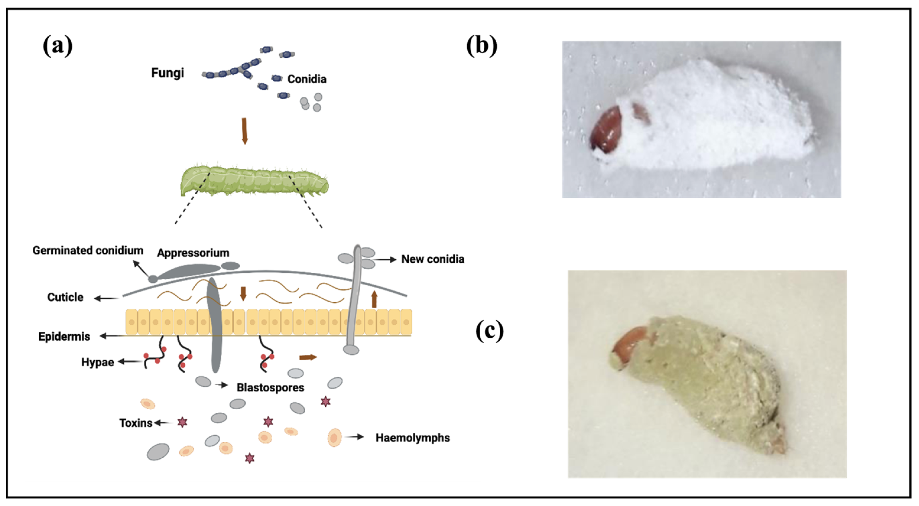
2.1. Fungi’s Infection Mode of Action in Insects: Penetration through the Cuticle

On the mode of action, the primary means of entry for most EPFs are via penetration through the host cuticle. A number of steps occur during fungal infection, with the initial adhesion of fungal conidia (asexual spores or fungal seeds) to the host cuticle preceding penetration (Figure 1a). These fungal pathogens are capable of infecting both hard- and soft-bodied insects, as well as a range of other arthropods such as Acari (i.e., ticks and mites) [75]. Among sucking insects, such as aphids and whiteflies, EPF is the primary pathogen as these hosts cannot ingest other pathogens that infect the gut wall [76].
During the course of insect infection, B. bassiana and M. anisopliae encounter a variety of niches within the host, which are highly variable with respect to the types and abundances of available nutrients [77]. First, the spores adhere to the cuticle of the insects. Additionally, the degree of attachment and the ability of the fungi to penetrate inside the host exoskeleton are crucial determinants for the success rate or extent of infection. Germination represents the second step of the infection process (Figure 1a). The fungus produces an appressorium and hyphal penetration through the cuticle, where primary nutrients, such as the protein and chitin co-polymers, are bound in the cuticle matrix. Indeed, the process of appressorium production is not for all fungal species; an exception can be seen in the example of Conidiobolus coronatus (Costantin) Batko (Entomophthorales: Ancylistaceae), which does not produce an appressorium during its infection process. Penetration constitutes the third step, whereby the fungus enters the nutrient-rich insect hemolymph containing accessible sugars, proteins, and lipids. During this process, the fungus spreads within the hemolymph and produces toxins. After this, the mycelium grows on the cadaver and produces new conidia (Figure 1a). The conidia of B. bassiana are typically cylindrical or oval in shape, measuring approximately 3–4 microns in length, and are borne on long, slender stalks known as conidiophores. The infected insect may also exhibit deformities and mummification as the disease progresses. These features are critical for accurate diagnosis and the identification of infected insects in the field (Figure 1b). For M. anisopliae, infections of insects are easily recognized a few days after death. Initially, the fungal hyphae appear white, but as conidia form and mature, they often take on a characteristic olive-green color (Figure 1c).
Figure 1. A schematic overview of the infection process of EPF by penetration through the cuticle, and photos of pathogen infection. (a) The process of fungal infection on the host cuticle. First, adhesion of conidium to the insect epicuticle; second, spore germination results in appressorium (penetration pegs) penetrating the host cuticle and this follows the hypha/penetration peg entering inside the insect hemocoel; next, the fungal proliferate in the host and produce toxins; to accomplish the infection, the fungal cells differentiate into yeast-like cells called blastospores, and later mycelium grows on cadaver and produces conidia that come out of the insect under suitable environmental conditions and spread to other insects. The arrows indicate the direction of fungal growth. The figure is drawn with BioRender.com. (b,c) Photo of red palm weevil (Rhynchophorus ferrugineus) killed by B. bassiana and M. anisopliae, respectively. Photos from [78].
Jof 09 00575 g001

2.2. Ways of Interaction between Fungi and Insects

2.2.1. Interaction of the Fungus with the Microbiome of the Insect

Insects harbor microbes in various parts of their bodies, including their cuticle surfaces, digestive tract, and tissues or cells. Interactions between insects and their microbes can be classified into two categories: interactions with cuticle surface microbes and interactions with gut microbes. The consequence of insect microbes on the development and survival of EPF can vary depending on the diversity of the insect microbiome, which can differ between developmental stages and in response to environmental conditions. Different studies have shown that both B. bassiana and M. anisopliae can invade the cuticle and survive within insect cells, potentially coming into direct contact with intracellular endosymbionts [37,38]. Examples of interactions between fungi and insect cuticle microbes are summarized in Boucias’s review [79]. Overall, the cuticle is generally unsuitable for microbes due to its hydrophobic nature, but specialized structures in the exoskeleton may provide habitats for specific microbial taxa. Additionally, for mutualistic relations between fungi and insects, cuticles have evolved to inhabit specific fungi [80,81].
The insect gut has long been targeted for insect control, and research has recently focused on gut microbes and their interactions with EPF. The number and complexity of the gut microbiome may vary in different regions and depend on the insect’s developmental stage and taxa. All insect orders examined to date possess microbial symbionts that contribute to the defense against other microbes. Recent studies have reported that gut microbiota can suppress or promote infections by B. bassiana. For example, B. bassiana can interact with gut microbiota and accelerate mosquito mortality. Mosquitoes with gut microbiota died significantly faster than those without microbiota after topical fungal infection [82]. Furthermore, the fungal infection caused the dysbiosis of the mosquito gut microbiota, with a significant increase in their gut bacterial load and a decrease in bacterial diversity. The interplay between B. bassiana and the gut microbiota of bark beetles (Dendroctonus valens) also accelerated mortality, and the gut bacterial community was altered by B. bassiana [83]. In a more recent study, Wang et al. [84] found dramatic changes in the gut bacterial community structure in the brown planthopper (Nilaparvata lugens) after M. anisopliae infection. There was a significant increase in the bacterial load, a decrease in bacterial community evenness, and significant shifts in dominant bacterial abundance at the taxonomic level below the class.

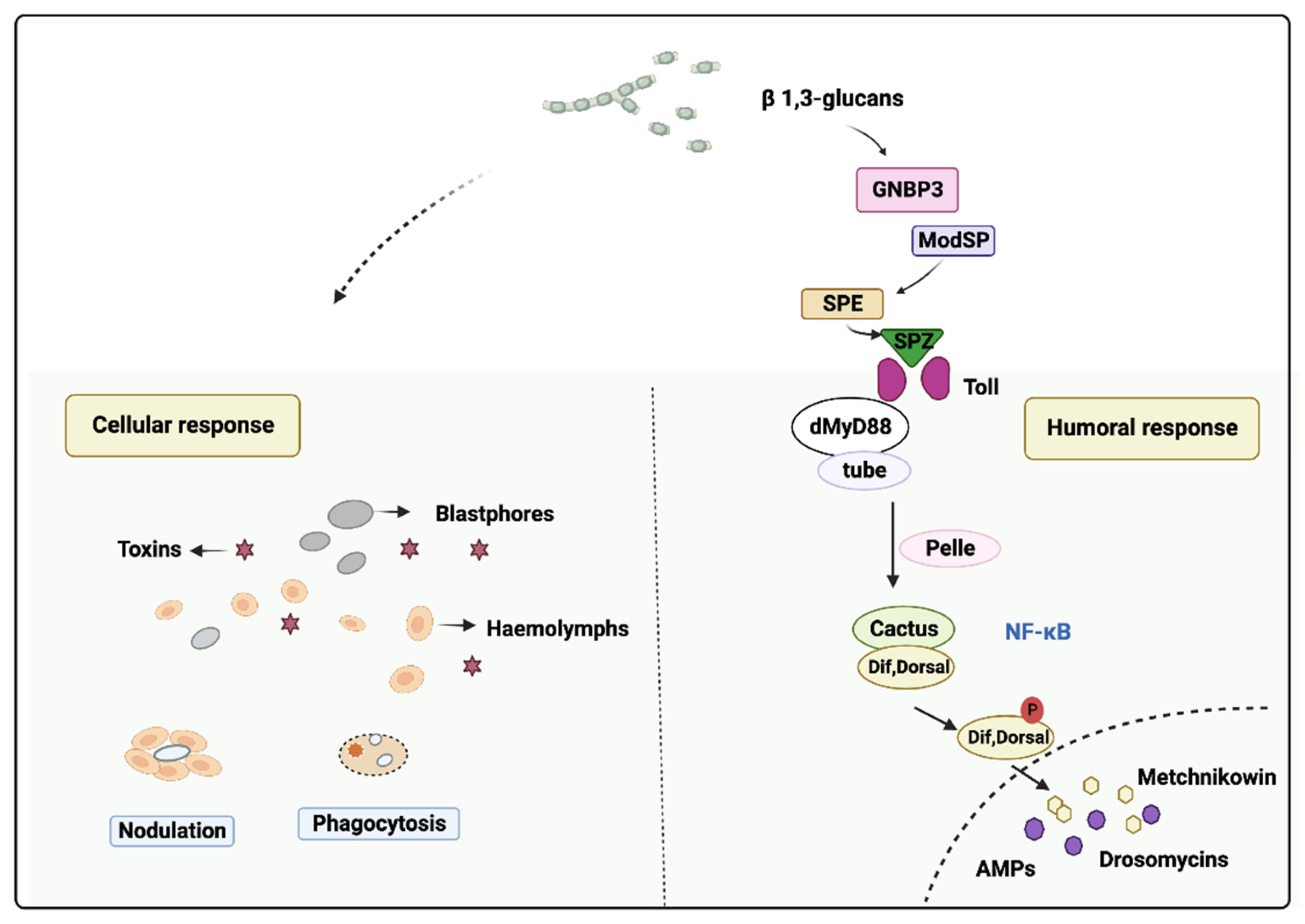
2.2.2. Stimulation of the Insect Immune-Competence by EPF

Fungi can express highly conserved pathogen-associated molecular patterns (PAMPs) that are recognized by pathogen recognition receptors (PRRs) and expressed on host phagocytes. For instance, glucans on the fungal cell surface can serve as PAMPs and be recognized by PRRs expressed on hosts, inducing the humoral and cellular immune responses in insects [85]. However, behavioral avoidance is considered the most effective defense against pathogens. Examples of this include social insects, such as the termite Macrotermes michaelseni that can ascertain the virulence of Metarhizium and Beauveria strains (Table 1) from a distance and is, thus, more strongly repelled by more virulent strains [86]; additionally, the bug Anthocoris nemorum avoids foraging and ovipositing on plants contaminated with Beauveria spores [87].
Before the pathogen encounters the host immune system, the infection begins with the attachment of single-cell dispersive forms of the fungus, e.g., conidia or blastospores, to the insect cuticle, as we discussed in the former section (Figure 1). The insect cuticle itself is a highly heterogeneous structure that can vary greatly in composition even during the various life stages of a particular insect. As summarized in [88], the epicuticle or outermost layer provides a hydrophobic barrier that is rich in lipids and is followed by the procuticle that contains chitin and sclerotized protein, which can typically be divided into the exo-, meso-, and endo-cuticular layers. The procuticle, in turn, is followed by the cells that constitute the epidermis and surrounds the internal structures of the insect. When fungi come into contact with an insect, they use various mechanisms to breach the insect’s cuticle and enter the body cavity [89]. This includes the expression of a variety of hydrolytic enzymes, e.g., proteases, chitinases, and lipases, and other factors, that promote the germination and growth of the fungus across the surface of the host and the subsequent penetration of cuticular layers [90].
Once inside, the fungi proliferate and release enzymes and toxins that break down the insect’s tissues and organs, ultimately leading to the death of the insect. B. bassiana synthesizes various secondary metabolites upon the invasion of insect hosts, including beauvericin, bassianin, bassianolide, beauverolides, tenellin, oosporein, and oxalic acid. These toxins facilitate host parasitism and cause mortality. For instance, oosporein, a dibenzoquinone toxin secreted by B. bassiana belonging to Sordariomycetes, Cordycipitaceae, represses immune responses in the mosquitoes’ midgut, causing dysbiosis, and subsequently triggers bacterial translocation from the gut into the hemocoel [82]. In the case of M. anisopliae, the primary toxic metabolites produced by this species include destruxins (six types) and cytochalasins (C and D), which were identified by Roberts and Hajek [91]. Recent studies have also isolated hydroxyfungerins A and B from Metarhizium sp.’s culture broth [92,93]. Additionally, both B. bassiana and M. anisopliae are believed to employ various extracellular proteases to facilitate pathogenic processes [94,95].
When the fungus breaches the cuticle and penetrates into the host integument by the penetration pegs and/or appressoria, which enable the growing hyphae to penetrate into the host integument, it reaches the insect’s innate immune system. The humoral response of the insect involves the production of antimicrobial peptides (AMPs) in the Toll immune pathway, which is secreted into the body cavity upon infection [96,97,98,99]. For details, the Toll receptor signaling pathway is initiated by the binding of an endogenous peptide ligand termed Spätzle. Spätzle is synthesized as an inactive precursor protein that is cleaved by the protease Easter. Easter is also generated from the precursor protein in a series of protease activation cascades. The binding of the Toll receptor causes recruitment to the membrane of the adapter Tube and the protein kinase Pelle, and finally, this leads to the degradation of the Cactus and the nuclear localization of NF-κB transcription factors Dif and Dorsal. These transcription factors induce the expression of antifungal genes such as Drosomycin (Drs) and Metchnikowin (Mtk) (Figure 2). For Drs, it is a member of the cysteine-stabilized α-helical and β-sheet (CSαβ) superfamily, consisting of 44 amino acid residues with an α-helix and a three-stranded β-sheet. This peptide is fortified by four disulfide bridges and has a limited antimicrobial range, specifically showing significant antifungal properties [100].
Cellular immunity is mediated by hemocytes, including phagocytosis, nodulation and encapsulation. Cellular responses, which involve hemocytes present in the body cavity, are not fully understood compared with the humoral immune response, but they are thought to play a significant role in conjunction with humoral defenses in eliminating insect-invaded bacteria. In Drosophila, cellular and humoral responses act together to combat infection [102], with cellular defenses playing a bigger role through nodulation and phagocytosis to eliminate the bacteria (Figure 2). In the later stage of the infection, this bacteria elimination could minimize the infection by the pathogen and facilitate microbe clearance by a later humoral response [103]. For example, in the study with the infection of B. bassiana in a yellow peach moth, Conogethes punctiferalis, significant decreases in the total and differential hemocyte counts were recorded over time in the larvae after they were injected with B. bassiana conidia. Additionally, hemocyte-mediated phagocytosis and nodulation were initiated in the hemolymph of larvae from the B. bassiana conidia challenge [104]. In another moth, Spodoptera frugiperda, the injection of Metarhizium rileyi (Farlow) Samson (Hypocreales: Clavicipitaceae) blastospores decreased the number of S. frugiperda hemocytes and impaired host cellular reactions such as nodulation, encapsulation and phagocytosis [105].
In other aspects, the activation of host defenses, in addition to impacting resident microbes, may suppress both the host and the invasive mycopathogen. For example, exposure to a high number of conidia may cause a massive upregulation in the phenoloxidase cascade, leading to the production of toxic quinones that suppress fungal development and/or kill the host [106]. To obtain additional information regarding the relationship between immunity and fungi, the readers are referred to Lu and Leger [107].

3. New Interesting Findings on the Interaction of Fungi in Insects

Fungal cell walls are complex and dynamic structures that are essential for fungal growth, development, and survival. They play a critical role in protecting fungal cells from environmental stresses and providing structural support [108,109]. The fungal cell wall is composed of several layers, with the composition and organization of each layer varying depending on the fungal species, morphotype, and growth stage. Fungal cell walls are primarily comprised chitin and β-glucans, which form an inner rigid core (Figure 3) [110]. Chitin is a polymer of N-acetylglucosamine (GlcNAc), while β-glucans are polysaccharides composed of glucose monomers that are linked by β-1,3- or β-1,6-glycosidic bonds. These polysaccharides provide strength and rigidity to the cell wall, making it resistant to mechanical stress [111,112]. In addition to chitin and β-glucans, the outer layer of the fungal cell wall contains various components, including polysaccharides and glycoproteins. In yeast cells, for example, the outer layer of the cell wall is enriched in mannosylated glycoproteins, also known as mannoproteins. These glycoproteins form a top layer on the cell surface and play a critical role in fungal cell adhesion and recognition (Figure 3). Mannoproteins are N-glycosylated or O-glycosylated proteins that are modified with various types of glycans [113,114]. The N-glycosylation pathway in fungi is conserved and produces a wide range of glycans [115]. These glycans can be classified into four main categories based on their structure: pauci-mannose glycans, high-mannose glycans, hybrid glycans, and complex glycans [116,117]. Pauci-mannose glycans are composed of one to three mannose residues and two GlcNAc residues at their base. High-mannose glycans are composed of four or more mannose residues and two GlcNAc residues at their base. Hybrid glycans contain a core structure of two GlcNAc and three mannose residues, with branches consisting of additional sugars such as galactose, sialic acid, and fucose. Finally, complex glycans are branched N-glycans that contain a variety of different sugar residues, including GlcNAc, galactose, and sialic acid. Mannoproteins on the cell surface of yeast cells belong to the high-mannose category, which contains four or more mannose residues and two GlcNAc residues at its base. The exact composition of these glycans can vary depending on the type and number of sugar molecules and the location of the branches (Figure 3).
The presence of N-glycans on the surface of fungal cells is an important factor in fungal biology and pathogenicity [119]. For example, glycoconjugates containing mannose, fucose, or GlcNAc on the surface of some fungal pathogens can be recognized by host mannose receptors on the cell, triggering signaling pathways that are involved in the induction of cytokine production [120,121]. In accordance with this, in one newest research on the glycans and immune response in insects, the authors found that in larvae of the Colorado potato beetle (Leptinotarsa decemlineata), after RNAi of the expression of Mannosidase-Ia (ManIa), which is responsible for the transition from high-mannose to paucimannose glycans [122], the peritrophic matrix pore size width in the ManIaRNAi insects was decreased by nearly 10 percent when compared to the control GFPRNAi insects (Figure 4). Interestingly, these smaller pores were connected to the observation of thinner microvilli on the epithelial cells of the midgut of ManIaRNAi insects, and in addition, these observations in the insect midgut agreed with an accumulation of high-mannose N-glycans in ManIa-silenced insects [123]. The peritrophic matrix is a physical barrier in the gut of insects and functions to protect the midgut epithelium from mechanical damage and harm from pathogens, toxins, and other damaging chemicals [124]. Based on the functions of the peritrophic matrix, the authors brought up the hypothesis that accumulated high-mannose glycans could simulate the cell wall structure of the fungi to trigger an immune response in the insect. They also made the speculation that a decreased pore size could be a protective response to prevent potential pathogens from gaining access to the midgut epithelium. This hypothesis was co-supported by the strong increase in transcription levels of the anti-fungal peptide drosomycin-like in the ManIaRNAi insects [123], although further research is required to elucidate this possibility. Nonetheless, we believe that such information may lead to novel approaches to improve the efficacy of pest control [125] and could be used for the rational design of strategies to increase the effectiveness of EPF for pest control applications.

4. Conclusions

The cosmopolitan existence and rich diversity of EPF contribute significantly to insect population regulation. In the agricultural sector, commercial formulations of EPF have shown efficacy as alternative control agents to chemical pesticides. Among the commercially produced entomopathogens, B. bassiana and M. anisopliae are the most important bioinsecticides. This review provided examples of Beauveria- and Metarhizium-based bioinsecticides, information on their infection mechanism and the possible modes of action for these two EPF insects. EPF penetrates their insect hosts through the spore adhesion of the cuticle and germination of the conidia, followed by the formation of an appressorium, and after that, the fungus penetrates the cuticle. Later, the fungus grows/proliferates in the hemocoel and produces blastospore toxins, ultimately leading to insect death. Additionally, we hypothesized that the glycans present on the fungal cell surface could act as motifs that elicit immune responses during the infection process. We also think that cellular responses are important, but we do not know so much about these defense systems. Here, we believe that modern omics-research and RNAi and CRISPR/Cas technologies could help elucidate the function of the many defense genes involved [126,127].
Overall, this review emphasizes the potential of EPF as bioinsecticides and underscores the significance of comprehending the mechanisms underlying their infection processes and immune responses elicited while also highlighting a recent study related to glycosylation and insect biocontrol, which suggests a new way of controlling insect pests by manipulating glycosylation. The study of glycosylation and its role in insect biocontrol represents a promising avenue for future research and has the potential to advance our understanding of fungi.

Author Contributions

Writing—original draft preparation, D.L.; writing—review and editing, G.S. and T.-X.L. All authors have read and agreed to the published version of the manuscript.

Funding

The authors thank the high talents program of Guizhou University.

Institutional Review Board Statement

Not applicable.

Informed Consent Statement

Not applicable.

Data Availability Statement

Not applicable.

Conflicts of Interest

The authors declare no conflict of interest.

References

  1. Naqqash, M.N.; Gökçe, A.; Bakhsh, A.; Salim, M. Insecticide resistance and its molecular basis in urban insect pests. Parasitol. Res. 2016, 115, 1363–1373. [Google Scholar] [CrossRef]
  2. Venkatesan, T.; Chethan, B.R.; Mani, M. Insecticide resistance and its management in the insect pests of horticultural crops. In Trends in Horticultural Entomology; Mani, M., Ed.; Springer: Singapore, 2022; pp. 455–490. [Google Scholar] [CrossRef]
  3. Sparks, T.C.; Storer, N.; Porter, A.; Slater, R.; Nauen, R. Insecticide resistance management and industry: The origins and evolution of the Insecticide Resistance Action Committee (IRAC) and the mode of action classification scheme. Pest Manag. Sci. 2021, 77, 2609–2619. [Google Scholar] [CrossRef] [PubMed]
  4. Lacey, L.A.; Grzywacz, D.; Shapiro-Ilan, D.I.; Frutos, R.; Brownbridge, M.; Goettel, M.S. Insect pathogens as biological control agents: Back to the future. J. Invertebr. Pathol. 2015, 132, 1–41. [Google Scholar] [CrossRef]
  5. Wakil, W.; Gulzar, S.; Prager, S.M.; Ghazanfar, M.U.; Shapiro-Ilan, D.I. Efficacy of entomopathogenic fungi, nematodes and spinetoram combinations for integrated management of Thrips tabaci: A two-year onion field study. Pest Manag. Sci. 2023. [Google Scholar] [CrossRef] [PubMed]
  6. Wakil, W.; Ghazanfar, M.U.; Usman, M.; Hunter, D.; Shi, W. Fungal-based biopesticide formulations to control nymphs and adults of the desert locust, Schistocerca gregaria Forskål (Orthoptera: Acrididae): A laboratory and field cage study. Agronomy 2022, 12, 1160. [Google Scholar] [CrossRef]
  7. Wakil, W.; Usman, M.; Piñero, J.C.; Wu, S.; Toews, M.D.; Shapiro-Ilan, D.I. Combined application of entomopathogenic nematodes and fungi against fruit flies, Bactrocera zonata and B. dorsalis (Diptera: Tephritidae): Laboratory cups to field study. Pest Manag. Sci. 2022, 78, 2779–2791. [Google Scholar] [CrossRef]
  8. Wakil, W.; Tahir, M.; Al-Sadi, A.M.; Shapiro-Ilan, D. Interactions between two invertebrate pathogens: An endophytic fungus and an externally applied bacterium. Front. Microbiol. 2020, 11, 2624. [Google Scholar] [CrossRef]
  9. Qayyum, M.A.; Saleem, M.A.; Saeed, S.; Wakil, W.; Ishtiaq, M.; Ashraf, W.; Ahmed, N.; Ali, M.; Ikram, R.M.; Yasin, M.; et al. Integration of entomopathogenic fungi and eco-friendly insecticides for management of red palm weevil, Rhynchophorus ferrugineus (Olivier). Saudi J. Biol. Sci. 2020, 27, 1811–1817. [Google Scholar] [CrossRef]
  10. Gulzar, S.; Wakil, W.; Shapiro-Ilan, D.I. Combined effect of entomopathogens against Thrips tabaci Lindeman (Thysanoptera: Thripidae): Laboratory, greenhouse and field trials. Insects 2021, 12, 456. [Google Scholar] [CrossRef]
  11. Marrone, P.G. Status of the biopesticide market and prospects for new bioherbicides. Pest Manag. Sci. 2023. [Google Scholar] [CrossRef]
  12. AgroPages. Bioinput Market Grows 67% during 2021–22 Crop in Brazil-Agricultural News. Available online: https://news.agropages.com/News/NewsDetail---45387.htm (accessed on 29 March 2023).
  13. Shah, P.A.; Pell, J.K. Entomopathogenic fungi as biological control agents. Appl. Microbiol. Biotechnol. 2003, 61, 413–423. [Google Scholar] [CrossRef]
  14. Pucheta, D.M.; Macias, A.F.; Navarro, S.R.; Mayra, D.L.T. Mechanism of action of entomopathogenic fungi. Interciencia 2016, 156, 2164–2171. [Google Scholar]
  15. Tulloch, M. The genus Metarhizium. Trans. Br. Mycol. Soc. 1976, 3, 407–411. [Google Scholar] [CrossRef]
  16. Dolinski, C.; Lacey, L.A. Microbial control of arthropod pests of tropical tree fruits. Neotrop. Entomol. 2007, 36, 161–179. [Google Scholar] [CrossRef]
  17. Lacey, L.A.; Shapiro-Ilan, D.I. Microbial control of insect pests in temperate orchard systems: Potential for incorporation into IPM. Annu. Rev. Entomol. 2008, 53, 121–144. [Google Scholar] [CrossRef]
  18. Qayyum, M.A.; Wakil, W.; Arif, M.J.; Sahi, S.T.; Dunlap, C.A. Infection of Helicoverpa armigera by endophytic Beauveria bassiana colonizing tomato plants. Biol. Control 2015, 90, 200–207. [Google Scholar] [CrossRef]
  19. Roberts, D.W.; St Leger, R. Metarhizium spp., cosmopolitan insect-pathogenic fungi: Mycological aspects. Adv. Appl. Microbiol. 2004, 54, 1–70. [Google Scholar] [CrossRef]
  20. Tahir, M.; Wakil, W.; Ali, A.; Gen, S.S.E. Pathogenicity of Beauveria bassiana and Metarhizium anisopliae isolates against larvae of the polyphagous pest Helicoverpa armigera. Entomol. Gen. 2019, 38, 225–242. [Google Scholar] [CrossRef]
  21. Veen, K.H. Recherches Sur La Maladie, Due Al Metarrhizium anisopliae Chez Le Criquet Pelerin; Wageningen University and Research: Wageningen, The Netherlands, 1968. [Google Scholar]
  22. Hussein, K.A.; Abdel Rahman, M.A.A.; Abdel-Mallek, A.Y.; El Maraghy, S.S.; Joo, J.H. Climatic factors interference with the occurrence of Beauveria bassiana and Metarhizium anisopliae in cultivated soil. Afr. J. Biotechnol. 2010, 9, 7674–7682. [Google Scholar] [CrossRef]
  23. Khan, S.; Nadir, S.; Lihua, G.; Xu, J.; Holmes, K.A.; Dewen, Q. Identification and characterization of an insect toxin protein, Bb70p, from the entomopathogenic fungus, Beauveria bassiana, using Galleria mellonella as a model system. J. Invertebr. Pathol. 2016, 133, 87–94. [Google Scholar] [CrossRef]
  24. Wang, J.; Ying, S.H.; Hu, Y.; Feng, M.G. Mas5, a homologue of bacterial DnaJ, is indispensable for the host infection and environmental adaptation of a filamentous fungal insect pathogen. Environ. Microbiol. 2015, 18, 1037–1047. [Google Scholar] [CrossRef] [PubMed]
  25. Chu, Z.J.; Sun, H.H.; Zhu, X.G.; Ying, S.H.; Feng, M.G. Discovery of a new intravacuolar protein required for the autophagy, development and virulence of Beauveria bassiana. Environ. Microbiol. 2017, 19, 2806–2818. [Google Scholar] [CrossRef] [PubMed]
  26. Sufyan, M.; Abbasi, A.; Wakil, W.; Gogi, M.D.; Arshad, M.; Nawaz, A.; Shabbir, Z. Efficacy of Beauveria bassiana and Bacillus thuringiensis against maize stem borer Chilo partellus (Swinhoe) (Lepidoptera: Pyralidae). Gesunde Pflanzen. 2019, 71, 197–204. [Google Scholar] [CrossRef]
  27. Nakahara, Y.; Shimura, S.; Ueno, C.; Kanamori, Y.; Mita, K.; Kiuchi, M.; Kamimura, M. Purification and characterization of silkworm hemocytes by flow cytometry. Dev. Comp. Immunol. 2009, 33, 439–448. [Google Scholar] [CrossRef] [PubMed]
  28. Chelico, L.; Khachatourians, G.G. Isolation and characterization of nucleotide excision repair deficient mutants of the entomopathogenic fungus, Beauveria bassiana. J. Invertebr. Pathol. 2008, 98, 93–100. [Google Scholar] [CrossRef]
  29. Abdel-Raheem, M.A.; Ismail, I.A.; Abdel Rahman, R.S.; Farag, N.A.; Abdel Rhman, I.E. Entomopathogenic fungi, Beauveria bassiana (Bals.) and Metarhizium anisopliae (Metsch.) as biological control agents on some stored product insects. J. Entomol. Zool. Stud. 2015, 3, 316–320. [Google Scholar]
  30. Mantzoukas, S.; Lagogiannis, I.; Kitsiou, F.; Eliopoulos, P.A. Entomopathogenic action of wild fungal strains against stored product beetle pests. Insects 2023, 14, 91. [Google Scholar] [CrossRef]
  31. Meikle, W.; Mercadier, G.; Holst, N.; Nansen, C.; Girod, V. Impact of a treatment of Beauveria bassiana (Deuteromycota: Hyphomycetes) on honeybee (Apis mellifera) colony health and on Varroa destructor mites. Apidologie 2008, 39, 247–259. [Google Scholar] [CrossRef]
  32. Kanga, L.H.B.; Jones, A.W.; James, R.R. Field trials using the fungal pathogen, Beauveria bassiana (Deuteromycetes: Hyphomycetes) to control the ectoparasitic mite, Varroa destructor (Acari: Varroidae) in honey bee, Apis mellifera (Hymenoptera: Apidae) colonies. J. Econ. Entomol. 2003, 96, 1091–1099. [Google Scholar] [CrossRef]
  33. Ummidi, V.R.S.; Vadlamani, P. Preparation and use of oil formulations of Beauveria bassiana and Metarrhizium anisopliae against Spodoptera litura larvae. Afr. J. Microbiol. Res. 2014, 8, 1638–1644. [Google Scholar] [CrossRef]
  34. Freed, S.; Saleem, M.; Khan, M.; Naeem, M. Prevalence and effectiveness of Metarhizium anisopliae against Spodoptera exigua (Lepidoptera: Noctuidae) in southern Punjab, Pakistan. Pak. J. Zool. 2012, 44, 753–758. [Google Scholar]
  35. Petlamul, W.; Prasertsan, P. Evaluation of strains of Metarhizium anisopliae and Beauveria bassiana against Spodoptera litura on the basis of their virulence, germination rate, conidia production, radial growth and enzyme activity. Mycobiology 2012, 40, 111–116. [Google Scholar] [CrossRef]
  36. Hussein, K.A.; Abdel-Rahman, M.A.A.; Abdel-Mallek, A.Y.; El-Maraghy, S.S.; Joo, J.H. Pathogenicity of Beauveria bassiana and Metarhizium anisopliae against Galleria mellonella. Phytoparasitica 2012, 40, 117–126. [Google Scholar] [CrossRef]
  37. Bukhari, T.; Takken, W.; Koenraadt, C.J.M. Development of Metarhizium anisopliae and Beauveria bassiana formulations for control of Malaria mosquito larvae. Parasites Vectors 2011, 4, 23. [Google Scholar] [CrossRef]
  38. Lee, J.Y.; Woo, R.M.; Choi, C.J.; Shin, T.Y.; Gwak, W.S.; Woo, S.D. Beauveria bassiana for the simultaneous control of Aedes albopictus and Culex pipiens mosquito adults shows high conidia persistence and productivity. AMB Express 2019, 9, 206. [Google Scholar] [CrossRef]
  39. Bitencourt, R.D.O.B.; Santos-Mallet, J.R.D.; Lowenberger, C.; Ventura, A.; Gôlo, P.S.; Bittencourt, V.R.E.P.; Angelo, I.D.C. A novel model of pathogenesis of Metarhizium anisopliae propagules through the midguts of Aedes aegypti larvae. Insect 2023, 14, 328. [Google Scholar] [CrossRef]
  40. Dong, C.; Zhang, J.; Huang, H.; Chen, W.; Hu, Y. Pathogenicity of a new China variety of Metarhizium anisopliae (M. anisopliae Var. Dcjhyium) to subterranean termite Odontotermes formosanus. Microbiol. Res. 2009, 164, 27–35. [Google Scholar] [CrossRef]
  41. Rios-Velasco, C.; Pérez-Corral, D.A.; Salas-Marina, M.Á.; Berlanga-Reyes, D.I.; Ornelas-Paz, J.J.; Muñiz, C.H.A.; Cambero-Campos, J.; Jacobo-Cuellar, J.L. Pathogenicity of the hypocreales fungi Beauveria bassiana and Metarhizium anisopliae against insect pests of tomato. Southwest. Entomol. 2014, 39, 739–750. [Google Scholar] [CrossRef]
  42. Rivero-Borja, M.; Guzmán-Franco, A.W.; Rodríguez-Leyva, E.; Santillán-Ortega, C.; Pérez-Panduro, A. Interaction of Beauveria bassiana with chlorpyrifos ethyl and spinosad in Spodoptera frugiperda larvae. Pest Manag. Sci. 2018, 74, 2047–2052. [Google Scholar] [CrossRef]
  43. Quintela, E.; Mccoy, C.W. Pathogenicity enhancement of Metarhizium anisopliae and Beauveria bassiana to first instars of Diaprepes abbreviatus (Coleoptera: Curculionidae) with sublethal doses of imidacloprid. Environ. Entomol. 1997, 26, 1173–1182. [Google Scholar] [CrossRef]
  44. Brito, E.S.; De Paula, A.R.; Vieira, L.P.; Dolinski, C.; Samuels, R.I. Combining vegetable oil and sub-lethal concentrations of imidacloprid with Beauveria bassiana and Metarhizium anisopliae against adult Guava weevil Conotrachelus psidii (Coleoptera: Curculionidae). Biocontrol Sci. Technol. 2008, 18, 665–673. [Google Scholar] [CrossRef]
  45. Wendel, J.; Cisneros, J.; Jaronski, S.; Vitek, C.; Ciomperlik, M.; Flores, D. Screening commercial entomopathogenic fungi for the management of Diaphorina citri Populations in the lower Rio Grande Valley, Texas, USA. BioControl 2022, 67, 225–235. [Google Scholar] [CrossRef]
  46. Bava, R.; Castagna, F.; Piras, C.; Musolino, V.; Lupia, C.; Palma, E.; Britti, D.; Musella, V. Entomopathogenic fungi for pests and predators control in beekeeping. Vet. Sci. 2022, 9, 95. [Google Scholar] [CrossRef] [PubMed]
  47. Chang, J.C.; Wu, S.S.; Liu, Y.C.; Yang, Y.H.; Tsai, Y.F.; Li, Y.H.; Tseng, C.T.; Tang, L.C.; Nai, Y.S. Construction and selection of an entomopathogenic fungal library from soil samples for controlling Spodoptera litura. Front. Sustain. Food Syst. 2021, 5, 596316. [Google Scholar] [CrossRef]
  48. De Souza, D.A.; Lopes, R.B.; Humber, R.; Faria, M. Assessment of the diversity of Brazilian entomopathogenic fungi in the genus Beauveria. J. Invertebr. Pathol. 2020, 171, 107339. [Google Scholar] [CrossRef]
  49. Mathulwe, L.L.; Malan, A.P.; Stokwe, N.F. Mass production of entomopathogenic fungi, Metarhizium robertsii and Metarhizium pingshaense, for commercial application against insect pests. J. Vis. Exp. 2022, 181, e63246. [Google Scholar] [CrossRef]
  50. Rice, S.J.; Baker, D.K.; Mayer, D.G.; Leemon, D.M. Mycoinsecticide formulations of Beauveria bassiana and Metarhizium anisopliae reduce populations of lesser mealworm, Alphitobius diaperinus, in chicken-broiler houses. Biol. Control 2020, 144, 104234. [Google Scholar] [CrossRef]
  51. Kassa, A.; Brownbridge, M.; Parker, B.L.; Skinner, M.; Gouli, V.; Gouli, S.; Guo, M.; Lee, F.; Hata, T. Whey for mass production of Beauveria bassiana and Metarhizium anisopliae. Mycol. Res. 2008, 112, 583–591. [Google Scholar] [CrossRef]
  52. Fadiji, A.E.; Babalola, O.O. Exploring the potentialities of beneficial endophytes for improved plant growth. Saudi J. Biol. Sci. 2020, 27, 3622–3633. [Google Scholar] [CrossRef]
  53. Bamisile, B.S.; Siddiqui, J.A.; Akutse, K.S.; Aguila, L.C.R.; Xu, Y. General limitations to endophytic entomopathogenic fungi use as plant growth promoters, pests and pathogens biocontrol agents. Plants 2021, 10, 2119. [Google Scholar] [CrossRef]
  54. Bamisile, B.S.; Akutse, K.S.; Siddiqui, J.A.; Xu, Y. Model application of entomopathogenic fungi as alternatives to chemical pesticides: Prospects, challenges, and insights for next-generation sustainable agriculture. Front. Plant Sci. 2021, 12, 2132. [Google Scholar] [CrossRef]
  55. Biedermann, P.H.W. Cooperative breeding in the Ambrosia beetle Xyleborus affinis and management of its fungal symbionts. Front. Ecol. Evol. 2020, 8, 518954. [Google Scholar] [CrossRef]
  56. Hulcr, J.; Stelinski, L.L. The ambrosia symbiosis: From evolutionary ecology to practical management. Annu. Rev. Entomol. 2017, 62, 285–303. [Google Scholar] [CrossRef]
  57. Sobhy, I.S.; Baets, D.; Goelen, T.; Herrera-Malaver, B.; Bosmans, L.; Van den Ende, W.; Verstrepen, K.J.; Wäckers, F.; Jacquemyn, H.; Lievens, B. Sweet scents: Nectar specialist yeasts enhance nectar attraction of a generalist aphid parasitoid without affecting survival. Front. Plant Sci. 2018, 9, 1009. [Google Scholar] [CrossRef]
  58. Jacquemyn, H.; Pozo, M.I.; ÁLvarez-Pérez, S.A.; Lievens, B.; Fukami, T. Yeast–nectar interactions: Metacommunities and effects on pollinators. Curr. Opin. Insect Sci. 2021, 44, 35–40. [Google Scholar] [CrossRef]
  59. Nicoletti, R.; Becchimanzi, A. Ecological and molecular interactions between insects and fungi. Microorganisms 2022, 10, 96. [Google Scholar] [CrossRef]
  60. Samaras, K.; Mourtiadou, S.; Arampatzis, T.; Kakagianni, M.; Feka, M.; Wäckers, F.; Papadopoulou, K.K.; Broufas, G.D.; Pappas, M.L. Plant-mediated effects of beneficial microbes and a plant strengthener against spider mites in tomato. Plants 2023, 12, 938. [Google Scholar] [CrossRef]
  61. Coppola, M.; Cascone, P.; Di Lelio, I.; Woo, S.L.; Lorito, M.; Rao, R.; Pennacchio, F.; Guerrieri, E.; Digilio, M.C. Trichoderma atroviride P1 colonization of tomato plants enhances both direct and indirect defense barriers against insects. Front. Physiol. 2019, 10, 813. [Google Scholar] [CrossRef]
  62. Coppola, M.; Cascone, P.; Chiusano, M.L.; Colantuono, C.; Lorito, M.; Pennacchio, F.; Rao, R.; Woo, S.L.; Guerrieri, E.; Digilio, M.C. Trichoderma harzianum enhances tomato indirect defense against aphids. Insect Sci. 2017, 24, 1025–1033. [Google Scholar] [CrossRef]
  63. González-Mas, N.; Gutiérrez-Sánchez, F.; Sánchez-Ortiz, A.; Grandi, L.; Turlings, T.C.J.; Manuel Muñoz-Redondo, J.; Moreno-Rojas, J.M.; Quesada-Moraga, E. Endophytic colonization by the entomopathogenic fungus Beauveria bassiana affects plant volatile emissions in the presence or absence of chewing and sap-sucking insects. Front. Plant Sci. 2021, 12, 660460. [Google Scholar] [CrossRef]
  64. Dicke, M.; Baldwin, I.T. The evolutionary context for herbivore-induced plant volatiles: Beyond the ‘cry for help’. Trends Plant Sci. 2010, 15, 167–175. [Google Scholar] [CrossRef]
  65. Bezemer, T.M.; van Dam, N.M. Linking aboveground and belowground interactions via induced plant defenses. Trends Ecol. Evol. 2005, 20, 617–624. [Google Scholar] [CrossRef] [PubMed]
  66. Zarate, S.I.; Kempema, L.A.; Walling, L.L. Silverleaf whitefly induces salicylic acid defenses and suppresses effectual jasmonic acid defenses. Plant Physiol. 2007, 143, 866–875. [Google Scholar] [CrossRef]
  67. Kuśnierczyk, A.; Winge, P.E.R.; Jørstad, T.S.; Troczyńska, J.; Rossiter, J.T.; Bones, A.M. Towards global understanding of plant defence against aphids–timing and dynamics of early Arabidopsis defence responses to cabbage aphid (Brevicoryne brassicae) attack. Plant Cell Environ. 2008, 31, 1097–1115. [Google Scholar] [CrossRef] [PubMed]
  68. Robert-Seilaniantz, A.; Grant, M.; Jones, J.D.G. Hormone crosstalk in plant disease and defense: More than just more than just jasmonate-salicylate antagonism. Annu. Rev. Phytopathol. 2011, 49, 317–343. [Google Scholar] [CrossRef] [PubMed]
  69. Giron, D.; Frago, E.; Glevarec, G.; Pieterse, C.M.; Dicke, M. Cytokinins as key regulators in plant–microbe–insect interactions: Connecting plant growth and defence. Funct. Ecol. 2013, 27, 599–609. [Google Scholar] [CrossRef]
  70. Navarro, L.; Bari, R.; Achard, P.; Lisón, P.; Nemri, A.; Harberd, N.P.; Jones, J.D. DELLAs control plant immune responses by modulating the balance of jasmonic acid and salicylic acid signaling. Curr. Biol. 2008, 18, 650–655. [Google Scholar] [CrossRef]
  71. Ryu, H.; Cho, H.; Choi, D.; Hwang, I. Plant hormonal regulation of nitrogen-fixing nodule organogenesis. Mol. Cells 2012, 34, 117–126. [Google Scholar] [CrossRef]
  72. Cosme, M.; Wurst, S. Interactions between arbuscular mycorrhizal fungi, rhizobacteria, soil phosphorus and plant cytokinin deficiency change the root morphology, yield and quality of tobacco. Soil Biol. Biochem. 2013, 57, 436–443. [Google Scholar] [CrossRef]
  73. Liu, J.; Lovisolo, C.; Schubert, A.; Cardinale, F. Signaling role of strigolactones at the interface between plants, (micro)organisms, and a changing environment. J. Plant Interact. 2013, 8, 17–33. [Google Scholar] [CrossRef]
  74. Pangesti, N.; Pineda, A.; Pieterse, C.M.J.; Dicke, M.; Loon, J.J.A. Two-way plant-mediated interactions between root-associated microbes and insects: From ecology to mechanisms. Front. Plant Sci. 2013, 4, 414. [Google Scholar] [CrossRef]
  75. Sharma, A.; Sharma, S.; Yadav, P.K. Entomopathogenic fungi and their relevance in sustainable agriculture: A review. Cogent Food Agric. 2023, 9, 2180857. [Google Scholar] [CrossRef]
  76. Halder, J.; Rai, A.B.; Kodandaram, M.H. Compatibility of neem oil and different entomopathogens for the management of major vegetable sucking pests. Natl. Acad. Sci. Lett. 2013, 36, 19–25. [Google Scholar] [CrossRef]
  77. Padilla-Guerrero, I.E.; Barelli, L.; González-Hernández, G.A.; Torres-Guzmán, J.C.; Bidochka, M.J. Flexible metabolism in Metarhizium anisopliae and Beauveria bassiana: Role of the glyoxylate cycle during insect pathogenesis. Microbiology 2011, 157, 199–208. [Google Scholar] [CrossRef]
  78. Sutanto, K.D.; Husain, M.; Rasool, K.G.; Al-Qahtani, W.H.; Aldawood, A.S. Pathogenicity of local and exotic entomopathogenic fungi isolates against different life stages of red palm weevil (Rhynchophorus ferrugineus). PLoS ONE 2021, 16, e0255029. [Google Scholar] [CrossRef]
  79. Boucias, D.G.; Zhou, Y.; Huang, S.; Keyhani, N.O. Microbiota in insect fungal pathology. Appl. Microbiol. Biotechnol. 2018, 102, 5873–5888. [Google Scholar] [CrossRef]
  80. You, L.; Simmons, D.R.; Bateman, C.C.; Short, D.P.G.; Kasson, M.T.; Rabaglia, R.J.; Hulcr, J. New fungus-insect symbiosis: Culturing, molecular, and histological methods determine Saprophytic Polyporales mutualists of Ambrosiodmus Ambrosia beetles. PLoS ONE 2015, 11, e0147305. [Google Scholar] [CrossRef]
  81. Blackwell, M. Made for each other: Ascomycete yeasts and insects. Microbiol. Spectr. 2017, 5, FUNK-0081-2016. [Google Scholar] [CrossRef]
  82. Wei, G.; Lai, Y.; Wang, G.; Chen, H.; Li, F.; Wang, S. Insect pathogenic fungus interacts with the gut microbiota to accelerate mosquito mortality. Proc. Natl. Acad. Sci. USA 2017, 114, 5994–5999. [Google Scholar] [CrossRef]
  83. Xu, L.; Deng, J.; Zhou, F.; Cheng, C.; Zhang, L.; Zhang, J.; Lu, M. Gut microbiota in an invasive bark beetle infected by a pathogenic fungus accelerates beetle mortality. J. Pest Sci. 2019, 92, 343–351. [Google Scholar] [CrossRef]
  84. Wang, Z.; Cheng, Y.; Wang, Y.; Yu, X. Topical fungal infection induces shifts in the gut microbiota structure of brown planthopper, Nilaparvata lugens (Homoptera: Delphacidae). Insects 2022, 13, 528. [Google Scholar] [CrossRef] [PubMed]
  85. Stączek, S.; Zdybicka-barabas, A.; Wojda, I.; Wiater, A.; Mak, P.; Suder, P.; Skrzypiec, K.; Cytryńska, M. Fungal α-1,3-Glucan as a new pathogen-associated molecular pattern in the insect model host Galleria mellonella. Molecules 2021, 26, 5097. [Google Scholar] [CrossRef] [PubMed]
  86. Mburu, D.M.; Ochola, L.; Maniania, N.K.; Njagi, P.G.N.; Gitonga, L.M.; Ndung’u, M.W.; Wanjoya, A.K.; Hassanali, A. Relationship between virulence and repellency of entomopathogenic isolates of Metarhizium anisopliae and Beauveria bassiana to the termite Macrotermes michaelseni. J. Insect Physiol. 2009, 55, 774–780. [Google Scholar] [CrossRef] [PubMed]
  87. Meyling, N.V.; Pell, J.K.; Eilenberg, J. Dispersal of Beauveria bassiana by the activity of nettle insects. J. Invertebr. Pathol. 2006, 93, 121–126. [Google Scholar] [CrossRef] [PubMed]
  88. Blomquist, G.J.; Tillman, J.A.; Mpuru, S.; Seybold, S.J. The cuticle and cuticular hydrocarbons of insects: Structure, function, and biochemistry. In Pheromone Communication in Social Insects; Vander Meer, R.K., Breed, M.D., Winston, M., Espelie, K.E., Eds.; CRC Press: New York, NY, USA, 1998; pp. 34–54. [Google Scholar]
  89. Ortiz-Urquiza, A.; Keyhani, N.O. Action on the surface: Entomopathogenic fungi versus the insect cuticle. Insects 2013, 4, 357–374. [Google Scholar] [CrossRef]
  90. Charnley, A.K. Fungal pathogens of insects: Cuticle degrading enzymes and toxins. Adv. Bot. Res. 2003, 40, 241–321. [Google Scholar] [CrossRef]
  91. Roberts, D.W.; Hajek, A.E. Entomopathogenic fungi as bioinsecticides. In Frontiers in Industrial Mycology; Leatham, G.F., Ed.; Springer Science & Business Media: Berlin/Heildelberg, Germany, 1992; pp. 144–159. [Google Scholar]
  92. Uchida, R.; Imasato, R.; Yamaguchi, Y.; Masuma, R.; Shiomi, K.; Tomoda, H.; Ōmura, S. New insecticidal antibiotics, hydroxyfungerins A and B, produced by Metarhizium sp. FKI-1079. J. Antibiot. 2005, 58, 804–809. [Google Scholar] [CrossRef]
  93. Zimmermann, G. Review on safety of the entomopathogenic fungus Metarhizium anisopliae. Biocontrol Sci. Technol. 2007, 17, 879–920. [Google Scholar] [CrossRef]
  94. Stleger, R.; Bidochka, M.J.; Roberts, D.W. Isoforms of the cuticle-degrading Pr1 proteinase and production of a metalloproteinase by Metarhizium anisopliae. Arch. Biochem. Biophys. 1994, 313, 1–7. [Google Scholar] [CrossRef]
  95. Schrank, A.; Vainstein, M.H. Beauveria bassiana enzymes and toxins. Toxicon 2010, 56, 1267–1274. [Google Scholar] [CrossRef]
  96. Strand, M.R. The insect cellular immune response. Insect Sci. 2008, 15, 1–14. [Google Scholar] [CrossRef]
  97. Ali Mohammadie Kojour, M.; Han, Y.S.; Jo, Y.H. An overview of insect innate immunity. Entomol. Res. 2020, 50, 282–291. [Google Scholar] [CrossRef]
  98. Kingsolver, M.B.; Huang, Z.; Hardy, R.W. Insect antiviral innate immunity: Pathways, effectors, and connections. J. Mol. Biol. 2013, 425, 4921–4936. [Google Scholar] [CrossRef]
  99. Tsakas, S.; Marmaras, V.J. Insect immunity and its signalling: An overview. Invertebr. Surviv. J. 2010, 7, 228–238. [Google Scholar]
  100. Zhang, Z.T.; Zhu, S.Y. Drosomycin, an essential component of antifungal defence in Drosophila. Wiley Online Libr. 2009, 18, 549–556. [Google Scholar] [CrossRef]
  101. Cherry, S.; Silverman, N. Host-pathogen interactions in Drosophila: New tricks from an old friend. Nat. Immunol. 2006, 7, 911–917. [Google Scholar] [CrossRef]
  102. Elrod-Erickson, M.; Mishra, S.; Schneider, D. Interactions between the cellular and humoral immune responses in Drosophila. Curr. Biol. 2000, 10, 781–784. [Google Scholar] [CrossRef]
  103. Haine, E.R.; Moret, Y.; Siva-Jothy, M.T.; Rolff, J. Antimicrobial defense and persistent infection in insects. Science 2008, 322, 1257–1259. [Google Scholar] [CrossRef]
  104. Wang, J.L.; Yang, K.H.; Wang, S.S.; Li, X.L.; Liu, J.; Yu, Y.X.; Liu, X.S. Infection of the entomopathogenic fungus Metarhizium rileyi suppresses cellular immunity and activates Humoral antibacterial immunity of the host Spodoptera frugiperda. Pest Manag. Sci. 2022, 78, 2828–2837. [Google Scholar] [CrossRef]
  105. Li, S.; Liu, F.; Kang, Z.; Li, X.; Lu, Y.; Li, Q.; Pang, Y.; Zheng, F.; Yin, X. Cellular immune responses of the yellow peach moth, Conogethes punctiferalis (Lepidoptera: Crambidae), to the entomopathogenic fungus, Beauveria bassiana (Hypocreales: Cordycipitaceae). J. Invertebr. Pathol. 2022, 194, 107826. [Google Scholar] [CrossRef]
  106. Söderhäll, K.; Ajaxon, R. Effect of quinones and melanin on mycelial growth of Aphanomyces spp. and extracellular protease of Aphanomyces astaci, a parasite on crayfish. J. Invertebr. Pathol. 1982, 39, 105–109. [Google Scholar] [CrossRef]
  107. Lu, H.L.; Leger, R.S. Insect immunity to entomopathogenic fungi. Adv. Genet. 2016, 94, 251–285. [Google Scholar] [CrossRef] [PubMed]
  108. Thak, E.J.; Lee, S.B.; Xu-Vanpala, S.; Lee, D.J.; Chung, S.Y.; Bahn, Y.S.; Oh, D.B.; Shinohara, M.L.; Kang, H.A. Core N-glycan structures are critical for the pathogenicity of Cryptococcus neoformans by modulating host cell death. mBio 2020, 11, 711–720. [Google Scholar] [CrossRef] [PubMed]
  109. Gow, N.A.R.; Lenardon, M.D. Architecture of the dynamic fungal cell wall. Nat. Rev. Microbiol. 2022, 21, 248–259. [Google Scholar] [CrossRef] [PubMed]
  110. Ruiz-Herrera, J.; Ortiz-Castellanos, L. Cell wall glucans of fungi. A review. Cell Surf. 2019, 5, 100022. [Google Scholar] [CrossRef]
  111. Fernando, L.D.; Dickwella Widanage, M.C.; Penfield, J.; Lipton, A.S.; Washton, N.; Latgé, J.P.; Wang, P.; Zhang, L.; Wang, T. Structural polymorphism of chitin and chitosan in fungal cell walls from solid-state NMR and principal component analysis. Front. Mol. Biosci. 2021, 8, 814. [Google Scholar] [CrossRef]
  112. Gow, N.A.R.; Latge, J.P.; Munro, C.A. The fungal cell wall: Structure, biosynthesis, and function. Microbiol. Spectr. 2017, 5, 5–13. [Google Scholar] [CrossRef]
  113. Lipke, P.N.; Ovalle, R. Cell wall architecture in yeast: New structure and new challenges. J. Bacteriol. 1998, 180, 3735–3740. [Google Scholar] [CrossRef]
  114. Schäffer, C.; Messner, P. Emerging facets of prokaryotic glycosylation. FEMS Microbiol. Rev. 2017, 41, 49–91. [Google Scholar] [CrossRef]
  115. De Pourcq, K.; De Schutter, K.; Callewaert, N. Engineering of glycosylation in yeast and other fungi: Current state and perspectives. Appl. Microbiol. Biotechnol. 2010, 87, 1617–1631. [Google Scholar] [CrossRef]
  116. Chung, C.Y.; Majewska, N.I.; Wang, Q.; Paul, J.T.; Betenbaugh, M.J. SnapShot: N-glycosylation processing pathways across kingdoms. Cell 2017, 171, 258. [Google Scholar] [CrossRef]
  117. Yan, A.; Lennarz, W.J. Unraveling the mechanism of protein N-glycosylation. J. Biol. Chem. 2005, 280, 3121–3124. [Google Scholar] [CrossRef]
  118. Garcia-Rubio, R.; De Oliveira, H.C.; Rivera, J.; Trevijano-Contador, N. The Fungal cell wall: Candida, Cryptococcus, and Aspergillus Species. Front. Microbiol. 2020, 10, 2993. [Google Scholar] [CrossRef]
  119. Liu, D.D.; De Schutter, K.; Smargiass, N.; De Pauw, E.; Van Damme, E.J.M.; Smagghe, G. The N-glycan profile of the peritrophic membrane in the Colorado potato beetle larvae (Leptinotarsa decemlineata). J. Insect Physiol. 2019, 115, 27–32. [Google Scholar] [CrossRef]
  120. Van de Veerdonk, F.L.; Marijnissen, R.J.; Kullberg, B.J.; Koenen, H.J.; Cheng, S.C.; Joosten, I.; Van den Berg, W.B.; Williams, D.L.; Van der Meer, J.W.; Joosten, L.A.; et al. The macrophage mannose receptor induces IL-17 in response to Candida albicans. Cell Host Microbe 2009, 5, 329–340. [Google Scholar] [CrossRef]
  121. Schirmer, M.; Smeekens, S.P.; Vlamakis, H.; Jaeger, M.; Oosting, M.; Franzosa, E.A.; Jansen, T.; Jacobs, L.; Bonder, M.J.; Kurilshikov, A.; et al. Linking the human gut microbiome to inflammatory cytokine production capacity. Cell 2016, 167, 1125–1136. [Google Scholar] [CrossRef]
  122. Walski, T.; Van Damme, E.J.M.; Smargiasso, N.; Christiaens, O.; De Pauw, E.; Smagghe, G. Protein N-glycosylation and N-glycan trimming are required for postembryonic development of the pest beetle Tribolium castaneum. Sci. Rep. 2016, 6, 35151. [Google Scholar] [CrossRef]
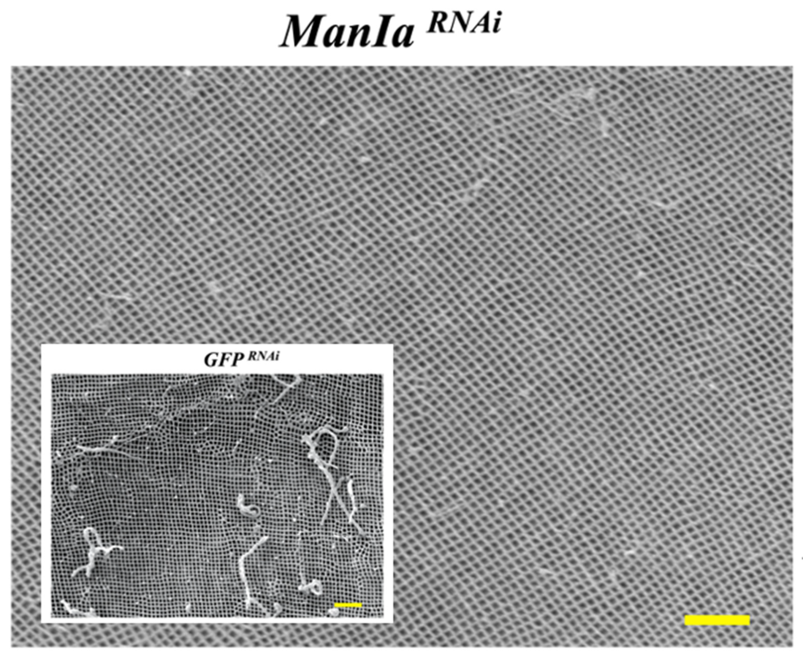
  123. Liu, D.; De Schutter, K.; Far, J.; Staes, A.; Dewettinck, K.; Quinton, L.; Gevaert, K.; Smagghe, G. RNAi of Mannosidase-Ia in the Colorado potato beetle and changes in the midgut and peritrophic membrane. Pest Manag. Sci. 2022, 78, 5071–5079. [Google Scholar] [CrossRef]
  124. Hakim, R.S.; Baldwin, K.; Smagghe, G. Regulation of midgut growth, development, and metamorphosis. Annu. Rev. Entomol. 2010, 55, 593–608. [Google Scholar] [CrossRef]
  125. Liu, D.; De Schutter, K.; Chen, P.; Smagghe, G. The N-glycosylation-related genes as potential targets for RNAi-mediated pest control of the Colorado potato beetle (Leptinotarsa decemlineata). Pest Manag. Sci. 2021, 78, 3815–3822. [Google Scholar] [CrossRef]
  126. Huvenne, H.; Smagghe, G. Mechanisms of dsRNA uptake in insects and potentials of RNAi for pest control: A review. J. Insect Physiol. 2010, 56, 227–235. [Google Scholar] [CrossRef] [PubMed]
  127. Li, J.-J.; Shi, Y.; Wu, J.-N.; Li, H.; Smagghe, G.; Liu, T.-X. CRISPR/Cas9 in Lepidopteran insects: Progress, application and prospects. J. Insect Physiol. 2021, 135, 104325. [Google Scholar] [CrossRef] [PubMed]
Figure 2. The process of humoral and cellular immune response. The Toll pathway is activated by Gram-positive bacteria and fungi. The β-1,3-glucans of fungi cell wall is recognized by GNBP3, then these interactions initiate protease cascades that converge at the level of the serine protease ModSP, which activates the protease Grass, in turn, activates the Spätzle processing enzyme (SPE). The SPE binding of the Toll receptor causes recruitment to the membrane of the adapter Tube and the protein kinase Pelle, and finally this leads the degradation of the Cactus and nuclear localization of NF-κB transcription factors Dif and Dorsal. The process of cellular response eliminates the pathogens by means of nodulation and phagocytosis. The figure was modified from [101] and drawn with BioRender.com.
Jof 09 00575 g002
Figure 3. Illustration of yeast cell wall structure with mannosylated glycoproteins forming the top layer. Jof 09 00575 i001—GlcNAc, N-acetylglucosamine, Jof 09 00575 i002—Man, mannose, Jof 09 00575 i003
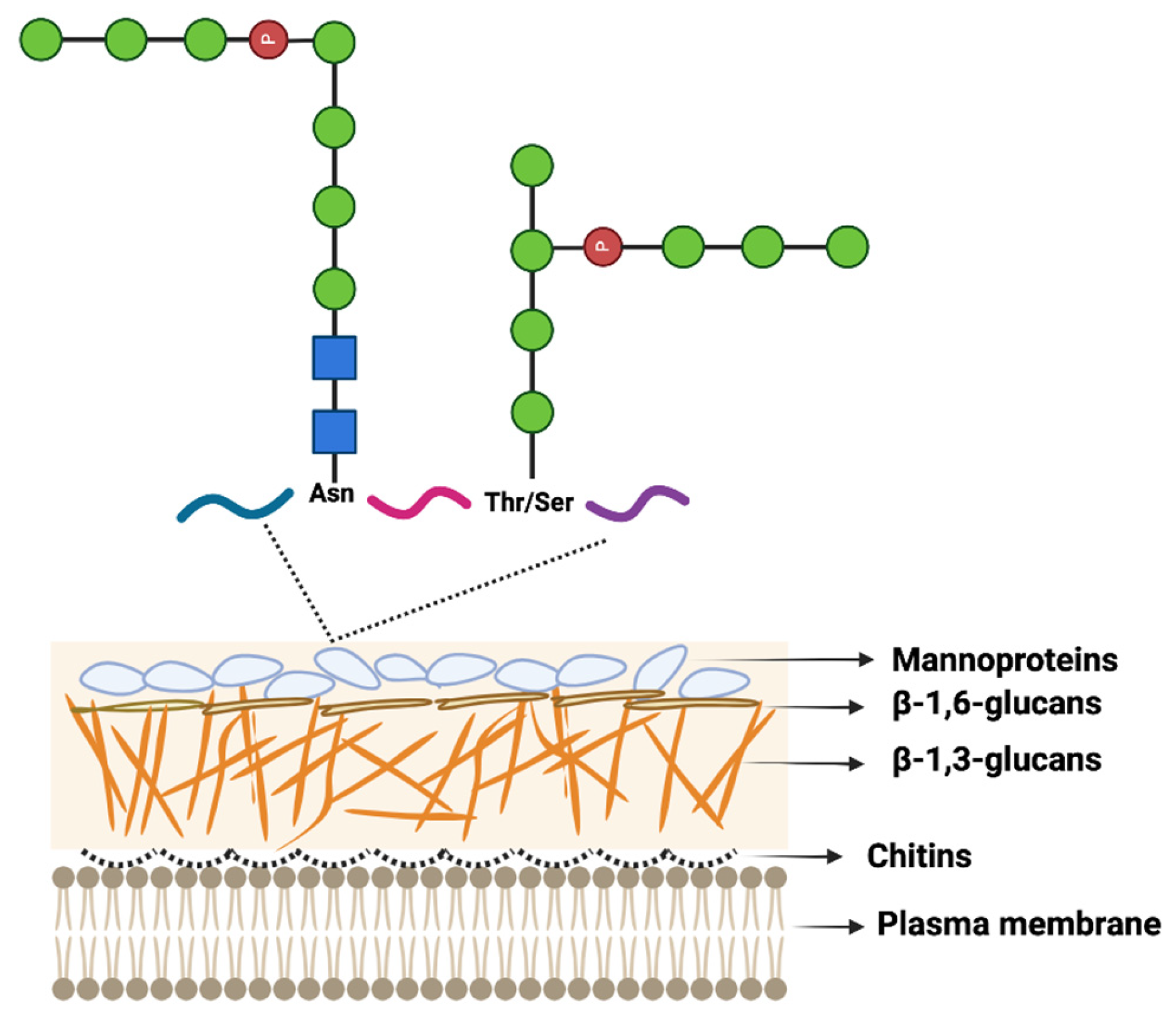
—phosphate group. The fungal cell wall is composed of chitins (1.5–6% by weight), β-linked glucans (β-1,3-glucans (30–45% by weight), β-1,6 glucan (5–10% by weight)), and mannoproteins (30–50% by weight). Chitin is a structurally important component of the fungal cell wall located closest to the plasma membrane. Branched β-1,3 glucan cross-links to chitin and is covalently linked to other polysaccharides (e.g., galactomannan and β-1,6 glucan). Mannoproteins are N- and O-glycosylated proteins. The Figure was modified from [118] and drawn with BioRender.com.
Jof 09 00575 g003
Figure 4. Scanning electron microscopic micrograph of the peritrophic matrix of larvae of the Colorado potato beetle (Leptinotarsa decemlineata) after the silencing of the ManIa gene expression (ManIaRNAi). The peritrophic matrix is a physical barrier in the gut of insects that functions to protect the midgut epithelium from mechanical damage and harm from pathogens, toxins, and other damaging chemicals. The pore width of the peritrophic matrix in ManIaRNAi-treated insects was nearly 10% smaller (83 ± 2 nm) when compared to the controls (GFPRNAi; inset micrograph; 89 ± 1 nm; significance at p < 0.05). Photo and data from [124]. The scale bars are 1 µm.
Jof 09 00575 g004
Table 1. Examples of B. bassiana- and M. anisopliae-based biopesticides: Overview of the strains used, commercial names and producers, target pest insects, and region of usage. The list is a representation and is not the complete list of biopesticides in the market.
EPF SpeciesCommercial NameStrainProducerTarget PestsRegions to Use
B. bassiana *BotaniGardTM ESGHALaverlam Whiteflies, aphids, weevils, mealybugsWorldwide
BotaniGardTM MAXX
MycotrolTM ESOCertis USAUSA
MycotrolTM WPOUSA
AprehendTMConidia BioscienceUSA and Canada
BroadbandTMBioworksWhiteflies, thrips, moths, stinkbugs, red spider mite, red scaleWorldwide
BioBeeTMPPRI 5339BioBee Biological Systems Worldwide
BioforestTMANT-03BioForest TechnologiesSpruce budwormUSA and Canada
BioPalmTMPL11BioPalm Manufacturing BagwormsMalaysia
BoverinTMATCC 74040Boverin Europe Europe
Boveril TMESALQ TecWhiteflies, weevilsBrazil
NaturalisTMKoppert Biological Systems Whiteflies, thrips, mites, aphids, tingidsWorldwide
BioCeresTMANT-03Bioceres Whiteflies, aphids, thripsArgentina, Chile, Peru, Mexico
M. anisopliae *ESALQ-E9 MetarrilTMESALQ-E9EMBRAPASeveral insect and mitesBrazil
NCIM 1311 PacerTMNCIM 1311Chema IndustriesTermites, root grub, locusts, root weevils, ants, beetles, caterpillarWorldwide
Bio-Blast Biological TermiticideTMF52BioProdex Ticks, weevils, mites, thripsUSA
Tick-Ex TM ECTMICIPE 69Kenyan BiologicsWorldwide
Met52 TM ECTMF52Novozymes BioAgWorldwide
MAS-01TMSor-1Bio-Insumos Naturales Coffee berry borerUSA
Green MuscleTMF52Bioworks Incthrips, whiteflies, and aphidsWorldwide
Metarhizium ICIPE 7TMICIPE 7Kenya Biologics East Africa
Metarhizium AQTMCQMa421SinochemCitrus root weevilChina
* The virulence of Beauveria and Metarhizium strains can be ascertained by targeted insects from a distance and the immune response is more strongly triggered by more virulent strains.
Disclaimer/Publisher’s Note: The statements, opinions and data contained in all publications are solely those of the individual author(s) and contributor(s) and not of MDPI and/or the editor(s). MDPI and/or the editor(s) disclaim responsibility for any injury to people or property resulting from any ideas, methods, instructions or products referred to in the content.

