Article

Anastomosis Groups and Mycovirome of Rhizoctonia Isolates Causing Sugar Beet Root and Crown Rot and Their Sensitivity to Flutolanil, Thifluzamide, and Pencycuron

1
College of Plant Protection, China Agricultural University, Beijing 100193, China
2
College of Horticulture, China Agricultural University, Beijing 100193, China
3
Institute of Plant Protection, Xinjiang Academy of Agricultural Science, Urumqi 830091, China
*
Author to whom correspondence should be addressed.
J. Fungi 2023, 9(5), 545; https://doi.org/10.3390/jof9050545
Submission received: 21 February 2023 / Revised: 26 April 2023 / Accepted: 5 May 2023 / Published: 9 May 2023
(This article belongs to the Special Issue Mycoviruses: Emerging Investigations on Virus-Fungal Host Interaction)

Abstract

:
Anastomosis groups (AGs) or subgroups of 244 Rhizoctonia isolates recovered from sugar beet roots with symptoms of root and crown rot were characterized to be AG-A, AG-K, AG-2-2IIIB, AG-2-2IV, AG-3 PT, AG-4HGI, AG-4HGII, and AG-4HGIII, with AG-4HGI (108 isolates, 44.26%) and AG-2-2IIIB (107 isolates, 43.85%) being predominate. Four unclassified mycoviruses and one hundred and one putative mycoviruses belonging to six families, namely Mitoviridae (60.00%), Narnaviridae (18.10%), Partitiviridae (7.62%), Benyviridae (4.76%), Hypoviridae (3.81%), and Botourmiaviridae (1.90%), were found to be present in these 244 Rhizoctonia isolates, most of which (88.57%) contained positive single-stranded RNA genome. The 244 Rhizoctonia isolates were all sensitive to flutolanil and thifluzamide, with average median effective concentration (EC50) value of 0.3199 ± 0.0149 μg·mL−1 and 0.1081 ± 0.0044 μg·mL−1, respectively. Among the 244 isolates, except for 20 Rhizoctonia isolates (seven isolates of AG-A and AG-K, one isolate of AG-4HGI, and 12 isolates of AG-4HGII), 117 isolates of AG-2-2IIIB, AG-2-2IV, AG-3 PT, and AG-4HGIII, 107 isolates of AG-4HGI, and six isolates of AG-4HGII were sensitive to pencycuron, with average EC50 value of 0.0339 ± 0.0012 μg·mL−1. Correlation index (ρ) of cross-resistance level between flutolanil and thifluzamide, flutolanil and pencycuron, and thifluzamide and pencycuron was 0.398, 0.315, and 0.125, respectively. This is the first detailed study on AG identification, mycovirome analysis, and sensitivity to flutolanil, thifluzamide, and pencycuron of Rhizoctonia isolates associated with sugar beet root and crown rot.

1. Introduction

Sugar beet (Beta vulgaris L.) is the second largest sugar crop worldwide, which is mainly cultivated in Xinjiang Uygur and Inner Mongolia autonomous regions, and Hebei, Heilongjiang, Gansu, Jilin, and Shanxi provinces of China [1]. Seedling damping-off, root and crown rot, foliar blight, as well as dry rot canker are common diseases that occur in the growing season or at the storage period of sugar beet, and all these diseases can be caused by the phytopathogen Rhizoctonia [2]. Of these diseases, sugar beet root and crown rot is the most serious and important disease, which causes decline in yield and saccharinity directly, and has an impact on storage of sugar beet [3,4,5].
It was widely known that anastomosis group (AG)-2-2IIIB and AG-2-2IV of R. solani were the predominant AGs causing sugar beet root and crown rot [5,6,7], while AG-4HGI, AG-4HGII, and AG-4HGIII were mainly attributed to sugar beet seedling damping-off [8,9,10]. Besides AG-2-2IIIB and AG-2-2IV, binucleate Rhizoctonia (BNR) (including AG-A, AG-D, AG-E, and AG-K) and multinucleate Rhizoctonia (MNR) (including AG-4HGI and AG-4HGII) were also occasionally isolated from sugar beet roots with symptoms of root and crown rot in Iran and the United States of America (USA) [7,11]. However, only two reports related to sugar beet root and crown rot had been documented in China; one was reported in our previous study that AG-2-2IIIB was the causal agent of sugar beet root and crown rot in Datong city of Shanxi province [12]; the other was recorded by Zhang et al. [13] that AG-4HGI was associated with sugar beet root and crown rot in Hohohot city of Inner Mongolia autonomous region.
Because of the broad host range of Rhizoctonia and the limitation of the high resistant commercial cultivars with high yield, management of sugar beet root and crown rot caused by Rhizoctonia mainly relied on the timely application of fungicides. It was reported that the quinone outside inhibitor (QoI), azoxystrobin, was the most widely used fungicide for controlling R. solani on sugar beet since its registration in USA in 1997 [14]. However, the resistance for Rhizoctonia isolates to azoxystrobin had been reported due to the long-time use of this fungicide [15,16]. In addition, the succinate dehydrogenase inhibitor (SDHI), penthiopyrad, was recorded to be able to effectively control R. solani on sugar beet in USA [17]. Flutolanil, polyoxin-D, and azoxystrobin were also documented to provide a high level of disease suppression to suppress sugar beet disease developed by AG-2-2IIIB and AG-2-2IV in the Red River Valley of USA [18]. There were only two fungicides, namely fenaminosulf and thiram, that had been registered in China for controlling sugar beet root and crown rot (China Pesticide Information Network, www.chinapesticide.gov.cn, accessed on 8 January 2021). Repeated use of fungicides with a single-site mode-of-action favors the development of resistance in fungal populations. Therefore, it is of importance to screen new fungicides for controlling sugar beet root and crown rot caused by Rhizoctonia.
Both flutolanil and thifluzamide are SDHI fungicides, which are effectively against Rhizoctonia spp., and were used to control rice sheath blight and wheat sharp eyespot in China [19,20,21,22]. Previously, our study demonstrated that both flutolanil and thifluzamide had a great efficiency on the Rhizoctonia isolates associated with sugar beet seedling damping-off, predominant AGs or subgroups of which were AG-4HGI, AG-4HGII, and AG-4HGIII [23,24]. Pencycuron is a phenylurea fungicide, which also has specific activity against Rhizoctonia, and is usually used to control rice sheath blight and potato stem canker or black scurf caused by Rhizoctonia [25,26]. However, there were no reports related to the effect of flutolanil, thifluzamide, and pencycuron on Rhizoctonia associated with sugar beet root and crown rot.
Mycoviruses are viruses that replicate in fungi and widespread in all major taxonomic groups of fungi [27,28]. To date, approximately 100 mycoviruses were found in Rhizoctonia, including members of established families accommodating double-stranded RNA (dsRNA), positive single-stranded RNA (+ssRNA), or negative single-stranded RNA (-ssRNA) viruses, together with members of proposed families and some unclassified RNA elements [29]. Mycoviruses were reported to be present in AG-1-IA [30,31], AG-2-2IIIB [32], AG-2-2IV [33,34], AG-2-2LP [35], AG-3 PT [36,37], AG-4HGI [38], AG-4HGIII [39], R. oryzae-sativae [40], R. cerealis [41], and R. fumigata [42], and the majority of identified mycoviruses were from R. solani AG-1-IA, the predominate pathogen causing rice sheath blight worldwide [29].
The metatranscriptomic and metagenomic sequencing technology is currently the most efficient approach for facilitating the identification of known and unreported mycoviruses in the target fungi [43,44]. In the last decade, many novel viruses were discovered using viral metagenomics [44]. However, only four studies concerning mycovirome of Rhizoctonia, which causes destructive economically important diseases on numerous crops, were reported. The mycoviral diversity of 84 isolates of R. solani (whose anastomosis groups or subgroups were unknown) collected from USA was investigated, and 27 mycoviruses with +ssRNA, -ssRNA, and dsRNA genomes were found [43]. Forty-seven partial or complete viral unique RNA-dependent RNA polymerase (RdRp) sequences with a high prevalence of -ssRNA viruses from eight strains of AG-2-2LP were gained using metatranscriptomic analysis [35]. A metatranscriptomic analysis of 43 isolates of R. solani AG-1-IA infecting rice that sampled in southern China was performed, and 10 mycovirus-related contigs composing five mycoviruses were obtained [45]. Recently, the diversity of putative mycoviruses containing +ssRNA, dsRNA, and -ssRNA genomes present in 66 strains of BNR (including AG-A, AG-Fa, AG-K, and AG-W) and 192 strains of MNR (including AG-1-IA, AG-2-1, AG-3 PT, AG-4HGI, AG-4HGII, AG-4HGIII, and AG-5) inciting potato stem canker or black scurf was studied using metatranscriptome sequencing, with four new parititviruses, 39 novel mitoviruses, and four new hypoviruses with nearly whole genome being detected in these 258 strains of BNR and MNR [46].
Until now, there are no detailed reports related to AGs or subgroups composition of Rhizoctonia isolates associated with sugar beet root and crown rot across China, let alone the diversity of mycoviruses present in these Rhizoctonia isolates as well as the sensitivity of them to the three fungicides (flutolanil, thifluzamide, and pencycuron). In the current study, the AGs or subgroups composition of Rhizoctonia isolates associated with sugar beet root and crown rot were characterized based on morphological traits and sequence analysis of internal transcribed spacer region of ribosomal DNA (rDNA-ITS) [10], the diversity of mycoviruses infecting these Rhizoctonia isolates were explored and analyzed by metatranscriptome sequencing [46], and the sensitivity of these Rhizoctonia isolates to the three fungicides (flutolanil, thifluzamide, and pencycuron) was detected using the mycelium growth inhibition method [23,24,47]. The results obtained in this study will be of great significance for understanding the AGs or subgroups composition of Rhizoctonia causing sugar beet rot and crown rot, revealing the diversity of mycoviruses discovered in Rhizoctonia, and screening efficient fungicides used for controlling diseases caused by Rhizoctonia.

2. Materials and Methods

2.1. Rhizoctonia Isolation and Identification

From 2009 to 2016, sugar beet roots with the symptoms of root and crown rot were collected from the sugar beet-growing regions across China, including Inner Mongolia and Xinjiang Uygur autonomous regions, Hebei, Heilongjiang, Gansu, Jilin, and Shanxi provinces, and Beijing municipality, which were used to isolate Rhizoctonia. Isolation, purification, and identification of Rhizoctonia were conducted according to the methods described in our previous study [10].

2.2. Pathogenicity Test

The pathogenicity of 61 representative Rhizoctonia isolates covering all the AGs or subgroups from different geographic origins was tested on eight-week-old sugar beet plants (cv. HI0305) under greenhouse conditions following the procedure described by Strausbaugh et al. [11] with a slight modification. The preparations of inoculum, planting soil, and sugar beet seedling were performed as previously described [10]. Two infested wheat seeds were placed at a depth of 10 mm into the soil next to the root of each eight-week-old sugar beet plant. Negative controls were inoculated with two un-infested autoclaved wheat seeds. The sugar beet plants were incubated in a greenhouse maintained at 25 to 27 °C with a 12 h photoperiod and watered whenever the surface soil appeared dry. Seven days later, all the plants including the control plants were harvested and used to assess disease incidence and disease index. Each treatment was conducted with three replicates, and ten sugar beet plants were used in each replicate. The experiment was arranged in a randomized block design and repeated twice.
Disease severity was rated using a 0–4 scale according to Harveson [48], which was described as follows: 0 = no disease, l = small, localized lesions with up to 25% of root surface affected, 2 = lesions coalescing with 26–50% of root affected, 3 = 51–75% of root covered with lesions but no internal discoloration, and 4 = more than 75% of beet surface covered with lesions and internal discoloration. The calculation of disease incidence and disease index was performed as described in our previous study [10]. All the sugar beet plants including the control plants were used to re-isolate Rhizoctonia and the resulting Rhizoctonia isolates were identified as the methods described in our previous study [10], fulfilling Koch’s postulates.

2.3. RNA Extraction, Metatranscriptomic Sequencing, and Sequence Analysis

For extracting total RNA, the 244 Rhizoctonia isolates identified above were cultured on potato dextrose agar (PDA) plates covered with cellophane film membranes (PDA-CF) at 25 °C in the dark for five days. Then, approximately 0.5 g of fresh mycelia were harvested and total RNAs were extracted using TRIzol Reagent (Invitrogen, Carlsbad, CA, USA) according to the manufacturer’s protocol. The extracted total RNA was further treated with RNase-Free DNase I to remove DNA contamination from RNA samples. The concentration and quality of RNA samples were measured using an ultramicro spectrophotometer (Nanodrop 2000, Thermo Scientific, Waltham, MA, USA), and the RNA integrity was further confirmed using 1.0% (w/v) gel electrophoresis. Finally, RNA samples were pooled by mixing 1 µg of RNA from each fungal sample to obtain one single sample with a final concentration (~200 ng/µL), which was sent to Shanghai Biotechnology Corporation (Shanghai, China) for metatranscriptomic sequencing using an Illumina X-TEN instrument with paired-end program.
TruSeq Stranded Total RNA LT Sample Prep Kit (Illumina, San Die-go, CA, USA) was used to establish sequencing library of the 244 Rhizoctonia isolates from rRNA-depleted total RNA. Library quality was checked using Qubit® 2.0 Fluorometer (Invitrogen, Q32866) and Agilent Technologies 2100 Bioanalyzer (Agilent Technologies, Santa Clara, CA, USA). Clean data with high quality were obtained by filtering low quality reads (Q ≤ 20 bases accounted for more than 50% of the total bases), joint contamination reads (>5 bp) and reads containing more than 5% N in the original data using Trimmomatic. These clean reads were first matched against the genome sequences of Rhizoctonia using the Bowtie (1.0) software. The unmatched RNAs were next assembled into longer contiguous sequences (contigs) in the Velvet software. Subsequently, Cap3 and CD-Hit-Est (version 4.8.1) were used for splicing primary contigs and clustering them with 95% homologous data, respectively. Finally, the contigs obtained were employed for searching in non-redundant protein sequences (Nr) of GenBank database (http://www.ncbi.nlm.nih.gov/, accessed on 8 January 2021) in BLASTx program using the Diamond software (version 0.9.25) under default parameters with the exception of the e-value lower than 1 × 10⁻⁵ [49]. At last, the unmatched contigs with homologous to viral amino acid sequences, and with size of nucleotides over 1.0 kb or encoding a protein of at least 150 amino acids (aa), were regarded as potential viral sequences [50,51].

2.4. Open Reading Frame (ORF) Prediction and Phylogenetic Analysis

Nucleotide sequence of each contig was first analyzed for ORF prediction, which was performed using the ORF finder tool from NCBI (https://www.ncbi.nlm.nih.gov/orffinder/, accessed on 8 January 2021). Except for the contigs closely related to mitoviruses generally hosted in the mitochondria, which were analyzed using the “mold, protozoan and coelenterate mitochondrial” genetic code, predictions of the contigs related to other mycoviruses were conducted using the “standard” genetic code. The sequences which could encode an ORF that begins at the start codon and ends at the termination codon, were predicted to contain a complete ORF. Then, Conserved Domain Database (CDD) (with e-value of 0.01) (http://www.ncbi.nlm.nih.gov/Structure/cdd/wrpsb.cgi, accessed on 8 January 2021), Protein Family (Pfam) database (http://pfam.sanger.ac.uk/, accessed on 8 January 2021), and PROSITE database (http://www.expasy.ch/, accessed on 8 January 2021) were used to find conserved motifs of amino acid (aa) sequences of putative mycoviruses [37,46,52]. CLUSTAL_X was used to perform sequence alignments [53]. For phylogenetic analysis, the mycoviruses with complete RdRp domains were aligned with MUSCLE implemented in MEGA 6, and maximum likelihood (ML) trees were constructed in Jones–Taylor–Thornton (JTT) model with 1000 bootstrap replicates [54]. The reference sequences used in the phylogenetic tree were retrieved from GenBank.

2.5. Confirmation of Identified Mycoviruses in Rhizoctonia Populations

In order to confirm the potential viral sequences from metatranscriptomic sequencing and distribution of each mycovirus among the tested Rhizoctonia populations, the reverse transcription-polymerase chain reaction (RT-PCR) was conducted. The viral-specific primers (Table S1) were designed based on the virus sequences from metatranscriptomic sequencing, and the complementary DNA (cDNA) was synthesized using the High-Capacity cDNA Reverse Transcription Kit with RNase inhibitor (TaKaRa) from the total RNA that was extracted from the 244 Rhizoctonia isolates characterized. The RT-PCR was performed with a 25 µL PCR mixture consisted of 8.5 µL ddH2O, 12.5 µL 2 × Pfu Master Mix [PC1102 (Aidlab Biotechnology, Beijing, China), containing 0.05 U·µL−1 Pfu DNA polymerase, 400 μM dNTP, and 4 mM Mg2+], 1 µL each of viral-specific primers (10 µM), and 2 µL cDNA. The PCR products were verified using 1% agarose gel electrophoresis, and the amplified sequence with right size was sent to Beijing Tianyihuiyuan Co., Ltd. (Beijing, China) for sequencing. The amplification for each potential viral sequence was conducted three times and the amplification products of these three repetitions were sequenced separately.

2.6. Virus Name

The name of a novel putative mycovirus identified for the first time in this study is named according to previous references [46,55], which consists of three parts: (1) the first part is the source of the virus; (2) the second part shows the virus taxonomical group; and (3) the third part is a progressive number. For example, “Rhizoctonia solani [part 1] mitovirus [part 2] 41 [part 3]” presents a new mitovirus and the forty-first mitovirus found in R. solani. The threshold for identification of a new mycovirus was based on the ICTV report, which declared the species demarcation criteria of mycoviruses from each mycovirus family. For example, when the putative RdRp aa identity of a mitovirus was lower than 90%, the mitovirus was regarded as a new species in the family Mitoviridae. When a mycovirus was reported previously and identified also in this study, “strain beet” was labeled to indicate its host source in this study. For example, “Rhizoctonia solani partitivirus 2 strain beet” presents a strain of Rhizoctonia solani partitivirus 2 reported previously [56] and is identified from a strain of R. solani that isolated from sugar beet roots with the symptoms of root and crown rot in this study.

2.7. Sensitivity of Rhizoctonia Isolates to Flutolanil, Thifluzamide, and Pencycuron In Vitro

The sensitivity of all the 244 identified Rhizoctonia isolates to flutolanil, thifluzamide, and pencycuron were evaluated in vitro. Flutolanil [Active ingredient (AI): 99.50%], thifluzamide (AI: 96.00%), and pencycuron (AI: 99.50%) were provided by the Institute for the Control of Agrochemicals, Ministry of Agriculture and Rural Affairs (ICAMOARA), and dissolved in analytically pure methanol at 10.00 mg·mL−1 to make a stock solution. Then, the stock solutions of 10.00 mg·mL−1 of flutolanil, thifluzamide, and pencycuron were diluted with analytically pure methanol to obtain the gradient concentrations of 0.05, 0.10, 0.20, 0.40, and 0.80 mg·mL−1, 0.01, 0.02, 0.04, 0.08, and 0.16 mg·mL−1, and 0.005, 0.01, 0.02, 0.04, and 0.08 mg·mL−1, respectively. Finally, by adding appropriate volumes of each gradient concentration into the autoclaved PDA culture media prior to solidification (approximately 45 °C), the PDA culture media was amended with flutolanil, thifluzamide, and pencycuron to obtain final concentrations of 0.05, 0.10, 0.20, 0.40, and 0.80 μg·mL−1, 0.01, 0.02, 0.04, 0.08, and 0.16 μg·mL−1, and 0.005, 0.01, 0.02, 0.04, and 0.08 μg·mL−1, respectively, for testing its ability to inhibit the mycelial growth of the Rhizoctonia isolates. An equivalent amount of analytically pure methanol was added into non-amended PDA culture media, which was used as control. Isolates that showed an EC50 value ≥ 0.8 μg·mL−1, as was the case for pencycuron, were tested again using fungicide concentrations of 1.60, 3.20, 6.40, 15.00, and 30.00 μg·mL−1. The incubation of fungi and calculation of median effective concentration (EC50) values were conducted as described in our previous study [23,24].

2.8. Statistical Analysis

The statistical significance of disease incidence and disease index on sugar beet plants incited by the testing AGs or subgroups of Rhizoctonia and EC50 values of flutolanil, thifluzamide, and pencycuron to the testing AGs or subgroups of Rhizoctonia were analyzed with SPSS (Statistical Product and Service Solutions) software (version 20.0). Homogeneity of variance was assessed using Levene’s test. As variances and sample size were unequal, differences among different AGs or subgroups were tested via the non-parametric tests of Kruskal–Wallis H. The results were further examined by one-way analysis of variance (ANOVA) with Dunnett’s T3 tests (p = 0.05). Spearman’s rank correlation coefficients were used to determine whether logarithmic EC50 values between two tested fungicides were correlated with each other.

3. Results

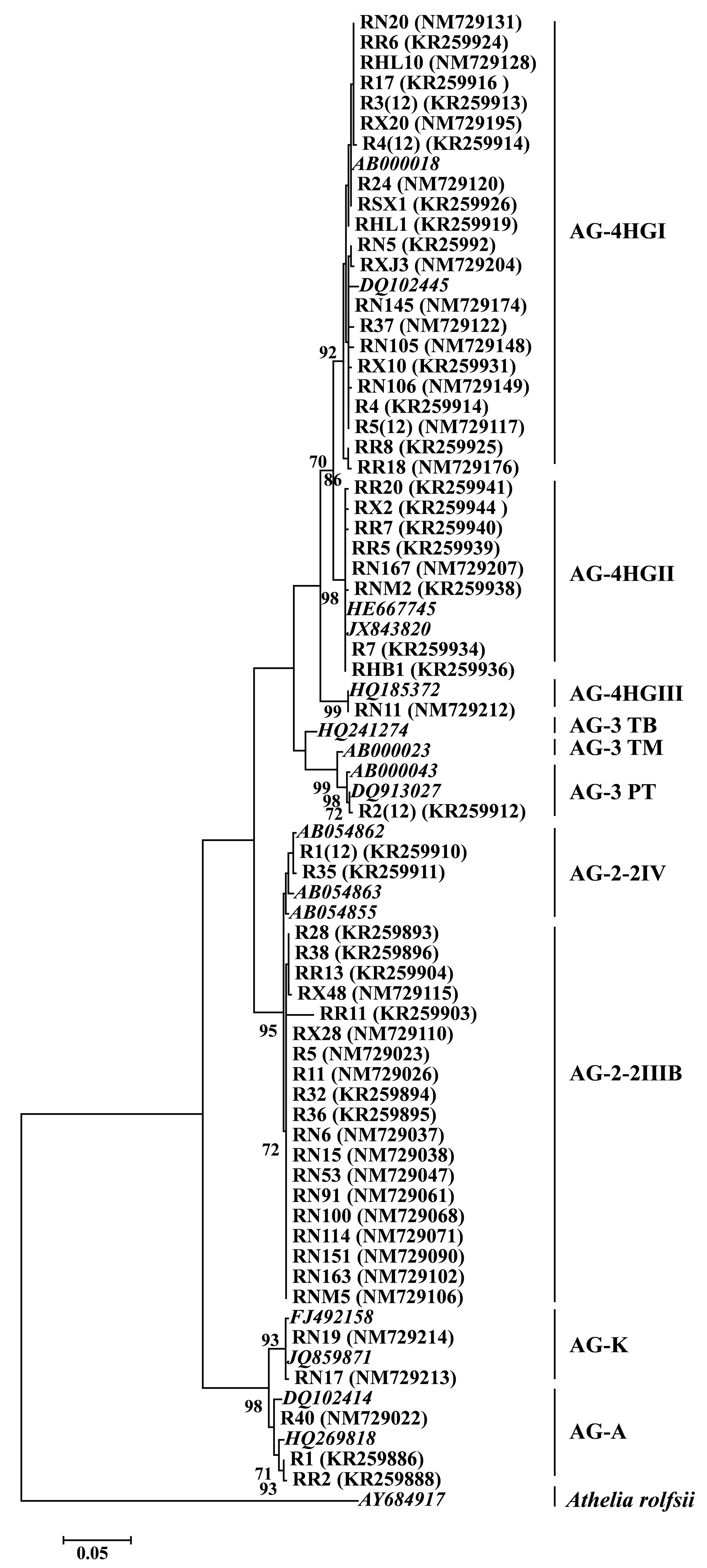
3.1. AGs or Subgroups Determination of Rhizoctonia

In total, 244 Rhizoctonia isolates were successfully recovered from the diseased sugar beet roots with the symptoms of root and crown rot collected from Xinjiang Uygur and Inner Mongolia autonomous regions, Hebei, Heilongjiang, Gansu, Jilin, and Shanxi provinces, and Beijing municipality across China (Table S2) and identified to be AG-A, AG-K, AG-2-2IIIB, AG-2-2IV, AG-3 PT, AG-4HGI, AG-4HGII, and AG-4HGIII. Among the eight AGs or subgroups, the most predominant AGs or subgroups were AG-4HGI (108 isolates, 44.26%) and AG-2-2IIIB (107 isolates, 43.85%), followed by AG-4HGII (18 isolates, 7.38%), AG-A (five isolates, 2.05%), AG-K (two isolates, 0.82%), AG-2-2IV (two isolates, 0.82%), AG-3 PT (one isolate, 0.41%), and AG-4HGIII (one isolate, 0.41%) (Table 1).
Cluster analysis was performed using the sequences of rDNA-ITS of eight AGs or subgroups in this study and the reference AGs or subgroups retrieved from GenBank. Two distinct clades were mainly divided in the phylogenetic tree (Figure 1); one clade was composed of the BNR isolates belonging to AG-A and AG-K, and the other clade consisted of the MNR isolates belonging to AG-2, AG-3, and AG-4. Moreover, the Rhizoctonia isolates belonging to AG-2 and AG-4 in this study were further divided into two sub-clades (AG-2-2IIIB and AG-2-2IV) and three sub-clades (AG-4HGI, AG-4HGII, and AG-4HGIII), respectively.

3.2. Pathogenicity on Sugar Beet

The results of pathogenicity test indicated that except for R. solani AG-3 PT which could only form sclerotia on the surface of sugar beet roots, the remaining 60 tested Rhizoctonia isolates could cause mild or moderate root and crown rot symptoms on sugar beet roots, with brown and dry lesions on sugar beet roots or crowns. The disease incidence and the disease index of the sugar beet roots caused by the tested 60 Rhizoctonia isolates ranged from 29.10% to 100.00% and from 16.82 to 73.85, respectively (Table 2). Collectively, the BNR isolates (whose average disease index ranged from 16.82 to 19.98) were less aggressive than the MNR isolates (whose average disease index ranged from 61.57 to 73.85). No significant differences in disease index were detected among isolates representing the four AGs or subgroups of Rhizoctonia, namely AG-2-2IIIB, AG-2-2IV, AG-4HGI, and AG-4HGII, which were the most aggressive on sugar beet plants.
Rhizoctonia isolates were consistently re-isolated from symptomatic sugar beet plants which were inoculated with Rhizoctonia, and their identities were confirmed as the methods described above, fulfilling Koch’s postulates, whereas no Rhizoctonia isolates were re-isolated from the control plants which were asymptomatic.

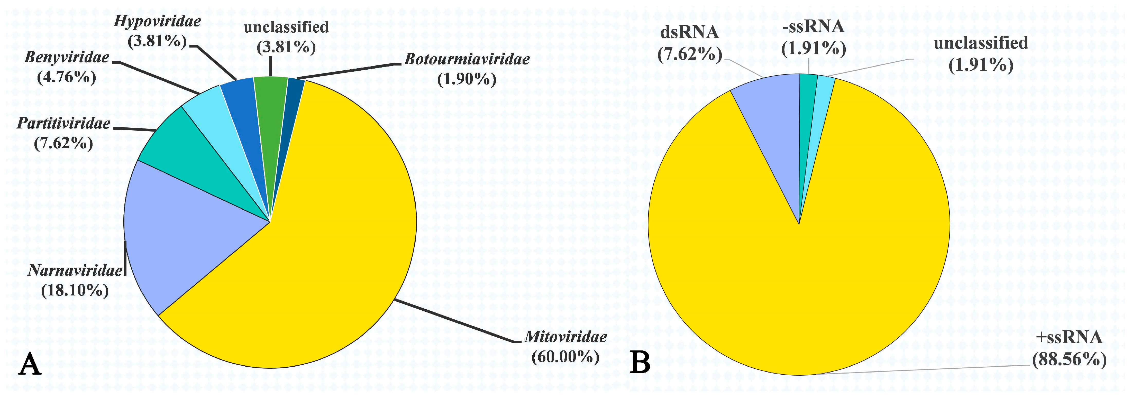
3.3. Mycovirus Diversity in Rhizoctonia Isolates

A total of 67,270,624 raw reads yielding 10.09 GB data were obtained from the 244 Rhizoctonia isolates by metatranscriptome sequencing on an Illumina HiSeq 2500 platform, and were deposited in Sequence Read Archive (SRA) on the NCBI server (BioProject PRJNA766298). After filtering, the resulting clean reads were de novo assembled into 66,082 contigs with length >200 nt using CLC Genomics Workbench version 6.0.4 software. As a result of sequence analysis, 106 contigs with nearly complete or partial genomic sequences of viruses were obtained and composed 105 putative mono- or bi-segmented mycoviruses, more than half (56.19%) of which exhibited an amino acid (aa) identity less than 60% with the counterpart of their closest relatives (Table S3).
Except for four unclassified mycoviruses (3.81%), the remaining 101 putative viral genomes showed affinity with six distinct families, namely Mitoviridae (63 putative viruses, 60.00%), Narnaviridae (19 putative viruses, 18.10%), Partitiviridae (eight putative viruses, 7.62%), Benyviridae (five putative viruses, 4.76), Hypoviridae (four putative viruses, 3.81%), and Botourmiaviridae (two putative viruses, 1.90%) (Figure 2A). Moreover, the genomes of 105 putative mycoviruses contained three nucleic acid types, including +ssRNA (Mitoviridae, Narnaviridae, Benyviridae, Hypoviridae, Botourmiaviridae, and two unclassified mycoviruses), dsRNA (Partitiviridae), and –ssRNA (two unclassified mycoviruses), with +ssRNA (accounting for 88.57% of the total viruses) being the most predominant (Figure 2B).
Each of the tested 244 Rhizoctonia isolates carried at least one mycovirus. Among the 244 isolates, 54, 39, 31, 24, 11, 7, 5, 4, and 2 isolates were infected simultaneously by two, three, four, five, six, seven, eight, nine, and ten mycoviruses, respectively. Four isolates, RN136, RN137, RN138, and RN141, even carried 13, 18, 17, and 20 mycoviruses, respectively (Table S4). In addition, RsNV16 and RsPV2B were confirmed to be present in all the tested 244 Rhizoctonia isolates and 37 of the tested 244 Rhizoctonia isolates, respectively (Table S3).

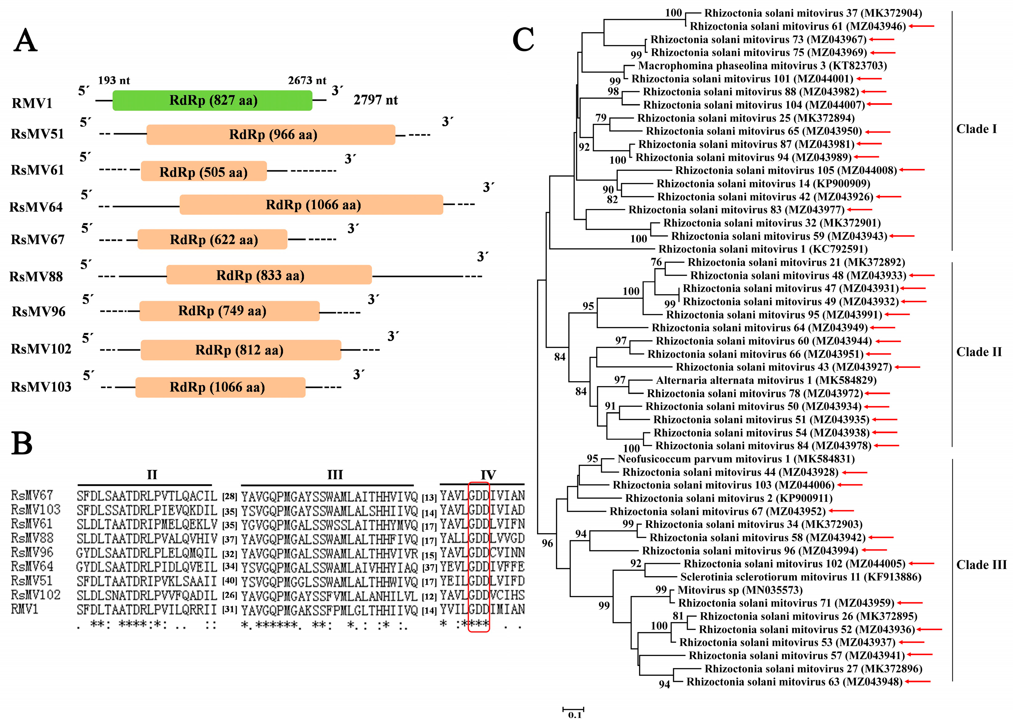
3.4. Sequences Related to Putative Members of the Family Mitoviridae

Sixty-three contigs (the length of nucleotide ranging from 1035 nt to 4397 nt) related to mycoviruses belonging to the family Mitoviridae were found in the 244 Rhizoctonia isolates, whose aa sequence identities with those of mycoviruses reported previously in the NCBI Nr database ranged from 39.83% to 89.66%, which were considered as novel viral species and named Rhizoctonia solani mitovirus 41 to 78, 80 to 96, and 98 to 105 (RsMV41–78, RsMV80–96, and RsMV98–105; Table S3). Among the sixty-three contigs, thirty-seven contigs related to mycoviruses were predicted to contain a complete ORF encoding RdRp, with the length of aa sequence ranging from 502 aa to 1066 aa. Contig ID, size, mycovirus name, best match, identity, query cover, E-value, and accession number were listed in Table 3. Eight contigs (Table 3) were chosen to carry out genome organization and multiple alignment. Based on mitochondrial codon usage, all the eight contigs related to mitoviruses contained an ORF encoding RdRp (Figure 3A). The results of multiple alignment indicated that three conserved motifs (motif II to motif IV) containing the GDD tripeptide in motif IV (characteristics of RdRp in mitoviruses) were found in the domain of RdRp in these eight viruses (Figure 3B).
The results of phylogenetic tree constructed based on the aa sequence of RdRps of the 37 putative mitoviruses identified in this study and 15 reference viruses belonging to the family Mitoviridae retrieved from NCBI database showed that the 37 putative mitoviruses belonged to the family Mitoviridae and were divided into three clades (clade I, II, and III) (Figure 3C).

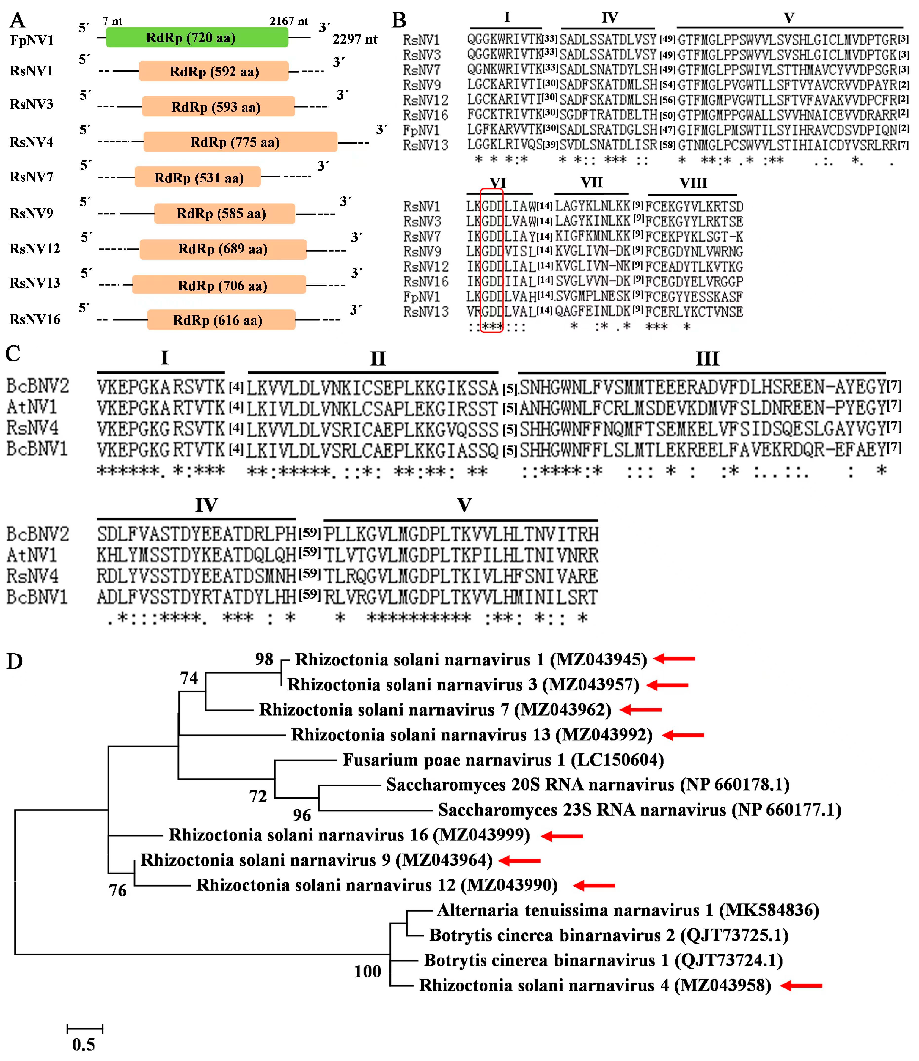
3.5. Sequences Related to Putative Members of the Family Narnaviridae

Nineteen contigs related to mycoviruses in the family Narnaviridae were discovered in the 244 Rhizoctonia isolates, with the length of nucleotide sequence ranging from 1031 nt to 2408 nt, whose aa sequence identities with the reported viruses previously in the NCBI Nr database ranged from 27.97% to 58.50%. According to the classification criteria of the family Narnaviridae provided by International Committee on Taxonomy of Viruses (ICTV, https://ictv.global/report/chapter/narnaviridae/narnaviridae, accessed on 8 January 2021), these nineteen mycoviruses were considered as new viral species and named Rhizoctonia solani narnavirus 1 to 19 (RsNV1–19; Table S3). Among the 19 contigs, eight contigs (Contig 1113, Contig 8527, Contig 8560, Contig 11100, Contig 19187, First_Contig 917, First_Contig 1098, and First_Contig 5037) composing the eight putative mycoviruses, RsNV1, RsNV3, RsNV4, RsNV7, RsNV9, RsNV12, RsNV13, and RsNV16, respectively (Table 4), were predicted to have a complete ORF, with the length of aa sequence ranging from 531 aa to 775 aa, which were chosen to conduct genome organization (Figure 4A) and multiple alignment (Figure 4B,C). Contig ID, size, mycovirus name, best match, identity, query cover, E-value, and accession number are listed in Table 4. The results of multiple alignment revealed that the seven narnaviruses, namely RsNV1, RsNV3, RsNV7, RsNV9, RsNV12, RsNV13, and RsNV16, had the closest relationship to Fusarium poae narnairus 1 (FpNV1, GenBank accession number LC150604) and contained six conserved motifs (motif I, and motif IV to VIII) with a GDD tripeptide in motif VI (Figure 4B); however, RsNV4 was the most closely related to Alternaria tenuisimma narnavirus 1 (AtNV1, GenBank accession number MK584836), which contained five conserved motifs (motif I to motif V) without a GDD tripeptide (Figure 4C).
The results of phylogenetic tree constructed based on the aa sequence of RdRps of eight putative narnaviruses identified in this study and six reference viruses belonging to the family Narnaviridae retrieved from NCBI database showed that the eight putative narnaviruses were divided into two clades (Figure 4D). RsNV 1, 3, 7, 9, 12, 13, and 16 were clustered together with FpNV1, Saccharomyces 20S RNA narnavirus (ScNV-20S, GenBank accession number NP660178.1), and Saccharomyces 23S RNA narnavirus (ScNV-23S, GenBank accession number NP660177.1) (Figure 4D), while RsNV4 was clustered together with AtNV1, Botrytis cinerea binarnavirus 1 (BcBNV1, GenBank accession number QJT73724.1), and Botrytis cinerea binarnavirus 2 (BcBNV2, GenBank accession number QJT73725.1), the latter two of which was suggested to the members of a new proposed family Binarnaviridae [51] (Figure 4D).

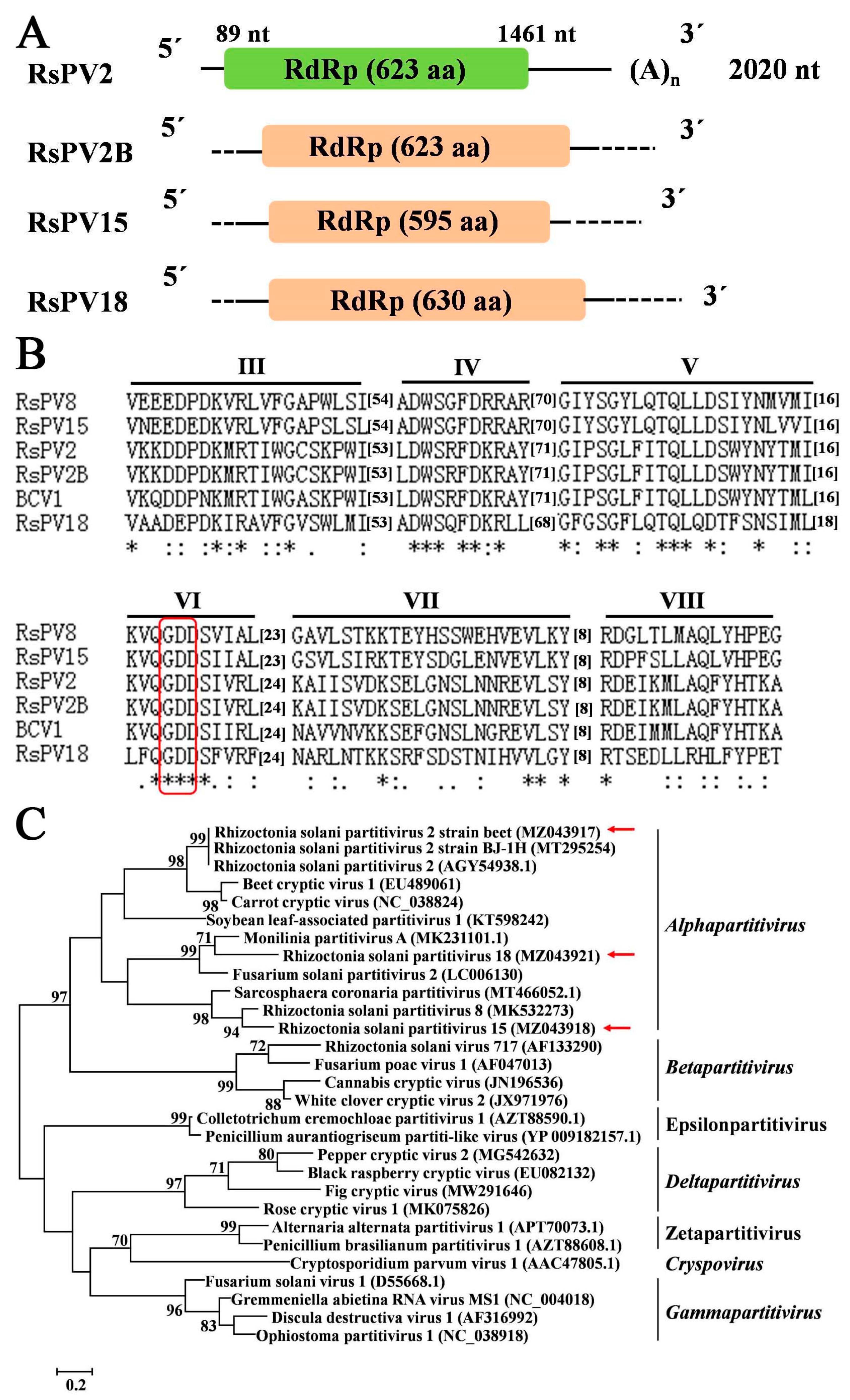
3.6. Sequences Related to Putative Members of the Family Partitiviridae

Nine contigs composing eight mycoviruses which belonged to the family Partitiviridae were found in the 244 Rhizoctonia isolates, with the length of nucleotide sequence ranging from 930 nt to 2011 nt (Table S3). The aa sequences of coat protein (CP, Contig 2073) and RdRp (Contig 3529) of Rhizoctonia solani partitivirus 2 strain beet (RsPV2B) had identities of 99.39% and 99.52% with those of Rhizoctonia solani partitivirus (RsPV2) [55], respectively. With the exception of RsPV2B, seven other mycoviruses identified in this study had low aa sequence identities (47.52%–67.68%) with CP or RdRp domain of mycoviruses reported previously in the NCBI Nr databases, which were considered as new viral species and, thus, named Rhizoctonia solani partitivirus 15–21 (RsPV15–21).
Among the nine contigs, three contigs (Contig 3073, Contig 3529, and Contig 14630) composing the three putative mycoviruses, RsPV15, RsPV2B, and RsPV18, respectively, were predicted to have a complete ORF encoding RdRp, with the length of aa sequence ranging from 595 aa to 630 aa, which were chosen to perform genome organization (Figure 5A), multiple alignment (Figure 5B), and phylogenetic analysis (Figure 5C). Contig ID, size, mycovirus name, best match, identity, query cover, E-value, and accession number were listed in Table 5. Multiple alignment of the three partitiviruses (RsPV15, RsPV2B, and RsPV18) and three reference viruses revealed that five conserved motifs (motif III to motif VII) containing the GDD tripeptide were found to be present in RdRp domain of these six viruses (Figure 5B). Results of the phylogenetic tree constructed based on the RdRp aa sequence of these three partitiviruses in this study and representative members from five genera (Alphapartitivirus, Betapartitivirus, Gammapartitivirus, Deltapartitivirus, and Cryspovirus) and two proposed genera (Epsilonpartitivirus and Zetapartitivirus) within the family Partitiviridae indicated that the three mycoviruses (RsPV15, RsPV2B, and RsPV18) belonged to the genus Alphapartitivirus (Figure 5C); moreover, RsPV2B was clustered together with RsPV2 and Rhizoctonia solani partitivirus strain BJ-1H (RsPV2-BJ) [38] (Figure 5C).

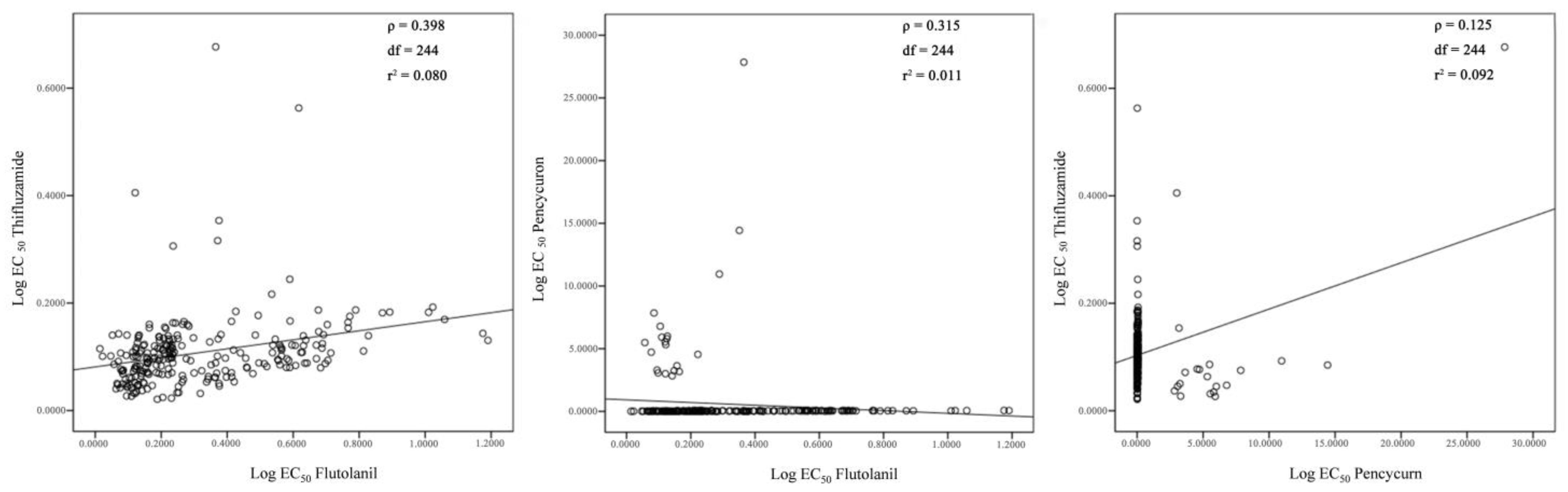
3.7. Sensitivity of 244 Rhizoctonia Isolates to Flutolanil, Thifluzamide, and Pencycuron

The sensitivity of 244 Rhizoctonia isolates belonging to eight AGs or subgroups to flutolanil, thifluzamide, and pencycuron was evaluated (Table 6, p = 0.05). All the tested 244 Rhizoctonia isolates were highly sensitive to flutolanil and thifluzamide, with an average EC50 value of 0.3199 ± 0.0149 μg·mL−1 and 0.1081 ± 0.0044 μg·mL−1, respectively (Table S4). Rhizoctonia isolates belonging to AG-2-2IIIB, the most prevalent AG associated with sugar beet root and crown rot, was significantly less sensitive to flutolanil and thifluzamide than Rhizoctonia isolates belonging to AG-4HGI (Table 6, p = 0.05). All the Rhizoctonia isolates belonging to AG-2-2IIIB, AG-2-2IV, AG-3 PT, and AG-4HGIII, 107 of 108 Rhizoctonia isolates belonging to AG-4HGI, and six of eighteen Rhizoctonia isolates belonging to AG-4HGII were sensitive to pencycuron, with the average EC50 value of 0.0339 ± 0.0012 μg·mL−1 (Table S4). However, Rhizoctonia isolates belonging to AG-A and AG-K, one of 108 Rhizoctonia isolates belonging to AG-4HGI (R4), and 12 of 18 Rhizoctonia isolates belonging to AG-4HGII presented reduced sensitivity to pencycuron, with the average EC50 values of 6.6728 ± 1.2863 μg·mL−1 (Table S4).
In addition, the cross-resistance between the tested three fungicides was analyzed, and positive correlation between flutolanil and thifluzamide, flutolanil and pencycuron, and pencycuron and thifluzamide was low, with correlation index (ρ) of 0.398, 0.315, and 0.125, respectively (Figure 6).

4. Discussion

In this study, 244 Rhizoctonia isolates associated with sugar beet root and crown rot were identified and belonged to eight AGs or subgroups, including AG-A, AG-K, AG-2-2IIIB, AG-2-2IV, AG-3 PT, AG-4HGI, AG-4HGII, and AG-4HGIII. Among these eight AGs or subgroups, AG-4HGI (108 isolates, 44.26%) and AG-2-2IIIB (107 isolates, 43.85%) were the most prevalent. It is the first report that AG-A, AG-K, AG-4HGII, and AG-4HGIII can cause sugar beet root and crown rot in China. It was demonstrated that the eight AGs or subgroups mentioned above could cause sugar beet seedling damping-off in our previous study [10], inferring that effective management of seedling damping-off incited by these eight AGs or subgroups of Rhizoctonia could be beneficial to prevent sugar beet plants in the later growing season from infection of them. R. solani AG-2-2IIIB was proven to be the dominant AG associated with sugar beet root and crown rot in the present study, which was in accordance with the results reported in previous studies [5,6]. Generally, R. solani AG-4HGI was regarded as the dominant pathogen associated with sugar beet seedling damping-off [8,9,10], but the results in the current study confirmed that R. solani AG-4HGI was also the predominate AG causing sugar beet root and crown rot. In 2015, there was one report that sugar beet root and crown rot could be caused by R. solani AG-4HGI in China [13]. Interestingly, our previous study demonstrated that R. solani AG-3 TB could cause sugar beet seedling damping-off [10], which was reported as the causal agent of tobacco target leaf spot [57]. However, AG-3 PT, the predominate pathogen causing potato stem canker or black scurf [58], could not incite root and crown rot on sugar beet roots in this study, which only formed sclerotia on the surface of eight-week-old sugar beet roots.
To date, more than 100 mycoviruses were found in Rhizoctonia, most of which belong to the nine families, namely Barnaviridae, Botourmiaviridae, Deltaflexiviridae, Endornaviridae, Hypoviridae, Megabirnaviridae, Mitoviridae, Partitiviridae, and Fusariviridae [29,32,37]. There were only two publications which reported mycoviruses identified from Rhizoctonia isolates causing sugar beet root and crown rot; one publication was that a R. solani AG-2-2IV strain DC17 was infected by 17 viral species through deep sequencing analysis, and complete genome of Rhizoctonia solani flexivirus 1 (RsFV-1) was further sequenced and analyzed to confirm that it belonged to the order Tymovirales [33,34]; the other publication was that a new mitovirus, Rhizoctonia solani mitovirus 39 (RsMV-39), was isolated from R. solani AG-2-2IIIB strain RR17 in our previous study [32]. In the current study, 105 putative mycoviruses were found in 244 Rhizoctonia isolates; excluding four unclassified mycoviruses, the remaining 101 putative mycoviruses belonged to the six families, including Benyviridae, Botourmiaviridae, Hypoviridae, Mitoviridae, Narnaviridae, and Partitiviridae, with 63 mycoviruses (62.38%) being assigned to the family Mitoviridae, which was approximately consistent with the previous studies [33,35,43]. This was the first detailed record of the putative mycoviruses associated with Rhizoctonia causing sugar beet root and crown rot in China and perhaps worldwide using metatranscriptome sequencing.
Mitoviridae is a newly established family designated by the ICTV (2019) and comprises mitoviruses from plants and fungi, which typically replicate and persist in the mitochondrion of a host [59]. Viruses of the family Mitoviridae are known to be the simplest naked mycoviruses and exist as RNA–RdRp nucleoprotein complexes, whose genomes only encode an RdRp protein [28]. In the present study, 37 mitoviruses predicted to have a complete ORF were divided into three clades (clade I, clade II, and clade III) in the phylogenetic tree (Figure 3), whose aa sequence identities of RdRp with those of the corresponding mitoviruses in the NCBI Nr database ranged from 39.83% to 89.66%; moreover, the result of phylogenetic analysis is consistent with that of the previous reports [37,46,60,61].
Only two narnaviruses, namely Saccharomyces 20S RNA narnavirus (ScNV-20S) and Saccharomyces 23S RNA narnavirus (ScNV-23S) [62], were recognized by the ICTV. In the present study, 19 putative narnaviruses were obtained, eight of which were predicted to encode a complete ORF; furthermore, it was the first record of narnaviruses discovered in Rhizoctonia. It is noteworthy that RsNV4 recorded in this study as well as three reference viruses (AtNV1, BcBNV1, and BcBNV1) does not contain a typical “GDD” motif and is the same as the narnavius, Magnaporthe oryzae narnavirus virus 1 (MoNV1), isolated from Magnaporthe oryzae [63], which is highly conserved in almost all viral RdRps and was deduced to be part of the catalytic site of RdRp [64,65].
Partitiviruses, with bi-segmented genomes, have broad host ranges and are usually associated with latent infections in fungi, plants, and protozoa [27,66]. In general, five genera, namely Alphapartitivirus, Betapartitivirus, Gammapartitivirus, Deltapartitivirus, and Cryspovirus, were recognized in the family Partitiviridae. The members in each of these five genera of the family Partitiviridae are corresponding to typical hosts: alphapartitiviruses and betapartitiviruses infect plants and fungi, gammapartitiviruses and deltapartitiviruses only infect fungi and plants, respectively, and cryspoviruses only comprise viruses isolated from protozoa by now [67]. Recently, two new genera, “Epsilonpartitivirus” and “Zetapartitivirus”, were proposed to be established in the family Partitiviridae [68,69]. In this study, nine contigs related to partitiviruses were obtained, three of which could encode a complete RdRp and were clustered into the genus Alphapartitivirus (Figure 5).
R. solani AG-2-2IV isolate DC17 was reported to harbor 17 mycoviruses, including six mitoviruses, three Sclerotinia sclerotiorum RNA virus L-like viruses, two putative members of the order Tymovirales, one endornavirus, one partitivirus, one megabirnavirus, one Aspergillus foetidus slow virus 2-like virus, and one Rhizoctonia solani dsRNA virus 1-like virus [33]. Chen et al. [70] reported that at least three novel betapartitiviruses co-infected the phytopathogenic fungus R. solani. R. solani AG-3 PT RS002 isolate infecting potato harbored an endornavirus and a mitovirus [36,71]. Recently, we reported that six novel mycoviruses containing +ssRNA and dsRNA genomes co-infected a single strain of R. solani AG-3 PT [37]. In this study, each of the tested 244 Rhizoctonia isolates were determined to carry at least one mycovirus; 181 (74.18%) out of these 244 Rhizoctonia isolates were infected by at least two mycoviruses simultaneously; moreover, four isolates even carried more than ten mycoviruses.
Flutolanil and thifluzamide are SDHI fungicides, and have high efficiency on controlling diseases caused by Rhizoctonia. Flutolanil was reported to control rice sheath blight [20], wheat sharp eyespot [21], potato stem canker or black scurf [72], peanut stem rot [73], tall fescue brown patch [74], sugar beet seedling damping-off [23], and sugar beet root and crown rot [18]. Thifluzamide was recorded to control rice sheath blight, wheat sharp eyespot, and sugar beet seedling damping-off [19,22,24]. As a phenylurea fungicide, pencycuron had specific activity against Rhizoctonia, and was usually used to control seedling diseases of crops, such as rice sheath blight and potato stem canker or black scurf caused by Rhizoctonia [25,26]. In this study, all the 244 tested Rhizoctonia isolates were highly sensitive to flutolanil and thifluzamide; 224 Rhizoctonia isolates (including 107 isolates of AG-2-2IIIB, two isolates of AG-2-2IV, one isolate of AG-3 PT, 107 isolates of AG-4HGI, six isolates of AG-4HGII, and one isolate of AG-4HGIII) were sensitive to pencycuron, but the remaining 20 Rhizoctonia isolates (including seven isolates of AG-A and AG-K, one isolate of AG-4HGI, and 12 isolates of AG-4HGII) presented a reduced sensitivity to pencycuron, with EC50 of pencycuron on these 20 isolates being more than 2.00 μg·mL−1. It was previously reported that R. solani AG-2 and AG-3 were highly sensitive to pencycuron, while AG-4, AG-5, and AG-7 only presented moderate to low level of sensitivity to pencycuron [75,76]. R. solani AG-3, AG-4HGI, and AG-4HGII associated with potato black scarf disease were sensitive to pencycuron, with AG-3 being more sensitive than AG-4HGI and AG-4HGII [77]. Therefore, it was concluded that flutolanil and thifluzamide would be the suitable fungicides for controlling sugar beet root and crown rot caused by Rhizoctonia in China. Since different AGs presented different level of sensitivity to pencycuron and AGs composition in each producing region of sugar beet in China was various, AGs composition in a certain production region of sugar beet should be identified at first, when pencycuron was chosen to control root and crown rot caused by Rhizoctonia in this region. In addition, positive correlation between flutolanil and thifluzamide, flutolanil and pencycuron, and thifluzamide and pencycuron was low, which inferred that these three fungicides could be used for controlling sugar beet root and crown rot caused by Rhizoctonia with rotation or mixture.
Previous studies reported that mycoviruses could affect the sensitivity of their host fungi to fungicides. Niu et al. [78] found that the fungicide-resistant Penicillium digitatum strains HS-F6 and HS-E9 co-infected by Penicillium digitatum polymycovirus 1 and Penicillium digitatum Narna-like virus 1 exhibited obvious reduction in resistance to the demethylation inhibitor (DMI)-fungicide, prochloraz. Wang et al. [79] reported that when P. crustosum strain HS-CQ15 was infected by Penicillium crustosum chrysovirus 1, the resistance of it to prochloraz decreased. In our previous study, the sensitivity of Alternaria alternata strain SD-BZF-12 infected by Alternaria alternata chrysovirus 1-AT1 to difenoconazole and tebuconazole reduced [80]. Our recent study demonstrated that Alternaria alternata botybirnavirus 1-AT1 decreased the sensitivity of its host A. tenuissima strain TJ-NH-51S-4 to difenoconazole [81]. In the present study, each of the tested 244 Rhizoctonia isolates presented different sensitivity to the three fungicides (flutolanil, thifluzamide, and pencycuron). For example, among the 108 isolates of AG-4HGI, the EC50 of pencycuron on isolate R4 was 3.2941 μg·mL−1, while the EC50 of pencycuron on the remainder 107 isolates ranged from 0.0049 μg·mL−1 to 0.0581 μg·mL−1. Moreover, the number and species of mycoviruses associated with each of these 244 isolates were diverse. Collectively, it was inferred that the varied sensitivity of these isolates to fungicides might be related to the mycoviruses associated with them. In the future, whether mycoviruses could affect the sensitivity of their host fungi to fungicides or not needs to be further studied.

Supplementary Materials

The following are available online at https://www.mdpi.com/article/10.3390/jof9050545/s1: Table S1: The primer pairs used to verify the 105 putative mycoviruses identified in this study and to detect the distribution of them in the 244 tested Rhizoctonia isolates. Table S2: Rhizoctonia isolates recovered from sugar beet roots with the symptoms of root and crown rot in China from 2009 to 2016. Table S3: Assembled sequences of mycoviruses found in Rhizoctonia isolates associated with sugar beet root and crown rot and their amino acid (aa) identities to those of viruses described previously. Table S4: Mycoviruses associated with 244 Rhizoctonia isolates recovered from sugar beet roots with the symptoms of root and crown rot in China from 2009 to 2016 and the median effective concentration (EC50) of flutolanil, thifluzamide, and pencycuron to these Rhizoctonia isolates.

Author Contributions

Conceptualization, X.W.; methodology, C.Z., S.L. and Z.M.; software, C.Z. and S.L.; validation, C.Z., S.L. and Z.M.; formal analysis, C.Z., Z.M. and W.W.; investigation, C.Z., S.L. and A.Y.; resources, C.H., X.W. and A.Y.; data curation, C.Z. and S.L.; writing—original draft preparation, C.Z.; writing—review and editing, L.G. and X.W.; visualization, C.Z. and W.W.; supervision, X.W.; project administration, X.W.; funding acquisition, C.H., X.W. and A.Y. All authors have read and agreed to the published version of the manuscript.

Funding

This work was funded by the earmarked fund for China Agriculture Research System (CARS-170304) and Science & Technology Assisting Xinjiang Project of Xinjiang Uygur Autonomous Region (2022E02072).

Institutional Review Board Statement

Not applicable.

Informed Consent Statement

Not applicable.

Data Availability Statement

The sequences of internal transcribed spacer region of ribosomal DNA (rDNA-ITS) of 244 Rhizoctonia isolates reported in this study have been deposited in the GenBank database under accession numbers of KP259886, KP259888 to KP259897, KP259899 to KP259904, KP259906, KP259908, KP259910 to KP259916, KP259918 to KP259920, KP259922 to KP259932, KP259934 to KP259944, NM729020 to NM729029, and NM729031 to NM729214. The sequences of 105 putative mycoviruses reported in this study have been deposited in the GenBank database under accession numbers of MZ043901 to MZ043972, MZ043974 to MZ043994, MZ043996 to MZ44008. All raw data of RNA-seq are available at NCBI Sequence Read Archive (BioProject PRJNA766298).

Acknowledgments

Mention of trade names or commercial products in this report is solely for the purpose of providing specific information and does not imply recommendation or endorsement.

Conflicts of Interest

The authors declare no conflict of interest.

References

  1. Geng, G.; Yang, J. Sugar beet production and industry in China. Sugar Tech. 2015, 17, 13–21. [Google Scholar] [CrossRef]
  2. Harveson, R.M.; Hanson, L.E.; Hein, G.L. (Eds.) Root diseases caused by fungi and oomycetes. In Compendium of Beet Diseases and Pests, 2nd ed.; American Phytopathological Society: Saint Paul, MN, USA, 2009; pp. 21–24. [Google Scholar]
  3. Campbell, L.G.; Windels, C.E.; Fugate, K.K.; Brantner, J.R. Postharvest losses associated with severity of Rhizoctonia root and crown rot of sugar beet at harvest. J. Sugar Beet Res. 2014, 51, 31–51. [Google Scholar] [CrossRef]
  4. Inokuti, E.M.; Thiery-Lanfranchi, D.; Edel-Hermann, V.; Gautheron, N.; Fayolle, L.; Michereff, S.J.; Steinberg, C. Genetic and pathogenic variability of Rhizoctonia solani causing root and crown rot on sugar beet in France. J. Plant Pathol. 2019, 101, 907–916. [Google Scholar] [CrossRef]
  5. Stojsin, V.; Budakov, D.; Jacobsen, B.; Bagi, F.; Grimme, E.; Neher, O. Analysis of Rhizoctonia solani isolates associated with sugar beet root and crown rot from Serbia. Afr. J. Biotechnol. 2011, 10, 19049–19055. [Google Scholar]
  6. Berdugo, C.; Hillnhütter, C.; Sikora, R.; Oerke, E.C. A resistance bioassay for Rhizoctonia root and crown rot and damping-off caused by the anastomosis groups AG-2-2IIIB and AG-4 in sugar beet. J. Agric. Sci. Technol. 2012, 2, 294–302. [Google Scholar]
  7. Taheri, P.; Tarighi, S. Genetic and virulence analysis of Rhizoctonia spp. associated with sugar beet root and crown rot in the Northeast region of Iran. Plant Dis. 2012, 96, 398–408. [Google Scholar] [CrossRef] [PubMed]
  8. Nagendran, S.; Hammerschmidt, R.; McGrath, J.M. Identification of sugar beet germplasm EL51 as a source of resistance to post-emergence Rhizoctonia damping-off. Eur. J. Plant Pathol. 2009, 123, 461–471. [Google Scholar] [CrossRef]
  9. Hanson, L.E.; McGrath, J.M. Rhizoctonia seedling disease on sugar beet. Int. Sugar J. 2011, 113, 584–589. [Google Scholar]
  10. Zhao, C.; Li, Y.; Wu, S.; Wang, P.; Han, C.; Wu, X. Anastomosis group and pathogenicity of Rhizoctonia spp. associated with seedling damping-off of sugar beet in China. Eur. J. Plant Pathol. 2019, 153, 869–878. [Google Scholar] [CrossRef]
  11. Strausbaugh, C.A.; Eujayl, I.A.; Panella, L.W.; Hanson, L.E. Virulence, distribution and diversity of Rhizoctonia solani from sugar beet in Idaho and Oregon. Can. J. Plant Pathol. 2011, 33, 210–226. [Google Scholar] [CrossRef]
  12. Zhao, C.; Wu, X.H. First report of sugar beet Rhizoctonia root and crown rot caused by Rhizoctonia solani AG-2-2IIIB in Shanxi province of China. Plant Dis. 2014, 98, 419. [Google Scholar] [CrossRef] [PubMed]
  13. Zhang, X.; Dong, B.; Zhou, H.; Xie, J.; Hao, J.J. First report of Rhizoctonia solani AG4-HG-I infecting sugar beet in China. Plant Dis. 2015, 99, 1185. [Google Scholar] [CrossRef]
  14. Khan, A.F.; Liu, Y.; Khan, M.F.R. Efficacy and safety of generic azoxystrobin at controlling Rhizoctonia solani in sugar beet. Crop Prot. 2017, 93, 77–81. [Google Scholar] [CrossRef]
  15. Arabiat, S.; Khan, M.F.R. Sensitivity of Rhizoctonia solani AG-2-2 from sugar beet to fungicides. Plant Dis. 2016, 100, 2427–2433. [Google Scholar] [CrossRef]
  16. Sharma, P.; Malvick, D.K.; Chanda, A.K. Sensitivity of Rhizoctonia solani anastomosis group 2-2 isolates from soybean and sugar beet to selected SDHI and QoI fungicides. Plant Dis. 2021, 105, 3573–3579. [Google Scholar] [CrossRef]
  17. Liu, S.; Xie, J.T.; Cheng, J.S.; Li, B.; Chen, T.; Fu, Y.P.; Li, G.Q.; Wang, M.Q.; Jin, H.A.; Wan, H.; et al. Fungal DNA virus infects a mycophagous insect and utilizes it as a transmission vector. Proc. Natl. Acad. Sci. USA 2016, 113, 12803–12808. [Google Scholar] [CrossRef]
  18. Bolton, M.D.; Panella, L.; Campbell, L.; Khan, M.F.R. Temperature, moisture, and fungicide effects in managing Rhizoctonia root and crown rot of sugar beet. Phytopathology 2010, 100, 689–697. [Google Scholar] [CrossRef]
  19. Chen, Y.; Zhang, A.F.; Wang, W.X.; Zhang, Y.; Gao, T.C. Baseline sensitivity and efficacy of thifluzamide in Rhizoctonia solani. Ann. Appl. Biol. 2012, 161, 247–254. [Google Scholar] [CrossRef]
  20. Li, S.D.; Hou, Y.P.; Peng, D.; Meng, L.; Wang, J.X.; Zhou, M.G.; Chen, C.J. Baseline sensitivity and control efficacy of flutolanil in Rhizoctonia solani. Australas. Plant Pathol. 2014, 43, 313–320. [Google Scholar] [CrossRef]
  21. Peng, D.; Li, S.D.; Chen, C.J.; Zhou, M.G. Combined application of Bacillus subtilis NJ-18 with fungicides for control of sharp eyespot of wheat. Biol. Control 2014, 70, 28–34. [Google Scholar] [CrossRef]
  22. Zhang, Y.; Lu, J.L.; Wang, J.X.; Zhou, M.G.; Chen, C.J. Baseline sensitivity and resistance risk assessment of Rhizoctonia cerealis to thifluzamide, a succinate dehydrogenase inhibitor. Pestic. Biochem. Physiol. 2015, 124, 97–102. [Google Scholar] [CrossRef] [PubMed]
  23. Zhao, C.; Zhang, X.F.; Hua, H.H.; Han, C.G.; Wu, X.H. Sensitivity of Rhizoctonia spp. to flutolanil and characterization of the point mutation in succinate dehydrogenase conferring fungicide resistance. Eur. J. Plant Pathol. 2019, 155, 13–23. [Google Scholar] [CrossRef]
  24. Zhao, C.; Li, Y.T.; Liang, Z.J.; Gao, L.H.; Han, C.G.; Wu, X.H. Molecular mechanisms associated with the resistance of Rhizoctonia solani AG-4 isolates to the succinate dehydrogenase inhibitor, thifluzamide. Phytopathology 2022, 112, 567–578. [Google Scholar] [CrossRef]
  25. Evan, E.; Rush, M.C.; Blackwell, M. Effects of rate and timing of fungicide applications on incidence and severity of sheath blight and grain yield of rice. Plant Dis. 1991, 75, 1254–1261. [Google Scholar]
  26. Moszczynska, E.; Pytlarz-Kozicka, M. The effect of pencycuron on health of potato tubers. Przem. Chem. 2017, 96, 406–408. [Google Scholar]
  27. Ghabrial, S.A.; Castón, J.R.; Jiang, D.H.; Nibert, M.L.; Suzuki, N. 50-plus years of fungal viruses. Virology 2015, 479, 356–368. [Google Scholar] [CrossRef]
  28. Ghabrial, S.A.; Suzuki, N. Viruses of plant pathogenic fungi. Annu. Rev. Phytopathol. 2009, 47, 353–384. [Google Scholar] [CrossRef]
  29. Abdoulaye, A.H.; Foda, M.F.; Kotta-Loizou, I. Viruses infecting the plant pathogenic fungus Rhizoctonia solani. Viruses 2019, 11, 1113. [Google Scholar] [CrossRef]
  30. Sun, Y.; Li, Y.Q.; Dong, W.H.; Sun, A.L.; Chen, N.W.; Zhao, Z.F.; Li, Y.Q.; Li, C.Y.; Yang, G.H. Molecular characterization of a novel mycovirus isolated from Rhizoctonia solani AG-1 IA strain 9-11. Arch. Virol. 2021, 166, 3229–3232. [Google Scholar] [CrossRef]
  31. Zhang, M.L.; He, Z.R.; Huang, X.T.; Shu, C.W.; Zhou, E.X. Genome organizations and functional analyses of a novel gammapartitivirus from Rhizoctonia solani AG-1 IA strain D122. Viruses 2021, 13, 2254. [Google Scholar] [CrossRef]
  32. Li, S.W.; Li, Y.T.; Hu, C.H.; Han, C.G.; Zhou, T.; Zhao, C.; Wu, X.H. Full genome sequence of a new mitovirus from the phytopathogenic fungus Rhizoctonia solani. Arch. Virol. 2020, 165, 1719–1723. [Google Scholar] [CrossRef] [PubMed]
  33. Bartholomaus, A.; Wibberg, D.; Winkler, A.; Puhler, A.; Schluter, A.; Varrelmann, M. Deep sequencing analysis reveals the mycoviral diversity of the virome of an avirulent isolate of Rhizoctonia solani AG-2-2 IV. PLoS ONE 2016, 11, e0165965. [Google Scholar] [CrossRef] [PubMed]
  34. Bartholomaus, A.; Wibberg, D.; Winkler, A.; Puhler, A.; Schluter, A.; Varrelmann, M. Identification of a novel mycovirus isolated from Rhizoctonia solani (AG-2-2 IV) provides further information about genome plasticity within the order Tymovirales. Arch. Virol. 2017, 162, 555–559. [Google Scholar] [CrossRef]
  35. Picarelli, M.A.S.; Forgia, M.; Rivas, E.B.; Nerva, L.; Chiapello, M.; Turina, M.; Colariccio, A. Extreme diversity of mycoviruses present in isolates of Rhizoctonia solani AG2-2 LP from Zoysia japonica from Brazil. Front. Cell Infect. Microbiol. 2019, 9, 244. [Google Scholar] [CrossRef] [PubMed]
  36. Das, S.; Falloon, R.E.; Stewart, A.; Pitman, A.R. Novel mitoviruses in Rhizoctonia solani AG-3 PT infecting potato. Fungal Biol. 2016, 120, 338–350. [Google Scholar] [CrossRef]
  37. Li, Y.T.; Li, S.W.; Zhao, Y.M.; Zhou, T.; Wu, X.H.; Zhao, C. Six novel mycoviruses containing positive single-stranded RNA and double-stranded RNA genomes co-infect a single strain of the Rhizoctonia solani AG-3 PT. Viruses 2022, 14, 813. [Google Scholar] [CrossRef]
  38. Li, Y.T.; Li, S.W.; Liang, Z.J.; Cai, Q.N.; Zhou, T.; Zhao, C.; Wu, X.H. RNA-seq analysis of Rhizoctonia solani AG-4HGI strain BJ-1H infected by a new viral strain of Rhizoctonia solani partitivirus 2 reveals a potential mechanism for hypovirulence. Phytopathology 2022, 112, 1373–1385. [Google Scholar] [CrossRef]
  39. Li, Y.Q.; Chen, W.R.; Niu, Y.F.; Xu, P.; Zhang, L.F.; Yu, S.H.; Yang, G.H.; Mo, X.H. Complete nucleotide sequence of a novel alphapartitivirus from Rhizoctonia solani AG-4HGIII isolate SM03. Arch. Virol. 2022, 167, 953–957. [Google Scholar] [CrossRef]
  40. Abdoulaye, A.H.; Cheng, J.S.; Fu, Y.P.; Jiang, D.H.; Xie, J.T. Complete genome sequence of a novel mitovirus from the phytopathogenic fungus Rhizoctonia oryzae-sativae. Arch. Virol. 2017, 162, 1409–1412. [Google Scholar] [CrossRef]
  41. Zhang, T.; Li, W.; Chen, H.G.; Yu, H.S. Full genome sequence of a putative novel mitovirus isolated from Rhizoctonia cerealis. Arch. Virol. 2015, 160, 1815–1818. [Google Scholar] [CrossRef]
  42. Li, Y.Q.; Xu, P.; Zhang, L.F.; Xia, Z.Y.; Qin, X.Y.; Yang, G.H.; Mo, X.H. Molecular characterization of a novel mycovirus from Rhizoctonia fumigata AG-Ba isolate C-314 Baishi. Arch. Virol. 2015, 160, 2371–2374. [Google Scholar] [CrossRef] [PubMed]
  43. Marzano, S.Y.L.; Nelson, B.D.; Ajayi-Oyetunde, O.; Bradley, C.A.; Hughes, T.J.; Hartman, G.L.; Eastburn, D.M.; Domier, L.L. Identification of diverse mycoviruses through metatranscriptomics characterization of the viromes of five major fungal plant pathogens. J. Virol. 2016, 90, 6846–6863. [Google Scholar] [CrossRef] [PubMed]
  44. Mokili, J.L.; Rohwer, F.; Dutilh, B.E. Metagenomics and future perspectives in virus discovery. Curr. Opin. Virol. 2012, 2, 63–77. [Google Scholar] [CrossRef]
  45. He, Z.R.; Huang, X.T.; Fan, Y.; Yang, M.; Zhou, E.X. Metatranscriptomic analysis reveals rich mycoviral diversity in three major fungal pathogens of rice. Int. J. Mol. Sci. 2022, 23, 9192. [Google Scholar] [CrossRef]
  46. Li, Y.T.; Yang, N.B.; Mu, T.Y.; Wu, X.H.; Zhao, C. Diversity of mycoviruses present in strains of binucleate Rhizoctonia and multinucleate Rhizoctonia, causal agents for potato stem canker or black scurf. J. Fungi 2023, 9, 214. [Google Scholar] [CrossRef]
  47. Sun, H.Y.; Lu, C.Q.; Li, W.; Deng, Y.Y.; Chen, H.G. Homozygous and heterozygous point mutations in succinate dehydrogenase subunits b, c and d of Rhizoctonia cerealis conferring resistance to thifluzamide. Pest Manag. Sci. 2017, 73, 896–903. [Google Scholar] [CrossRef]
  48. Harveson, R.M. Evaluation of fumigation and Rhizomania-tolerant cultivars for control of a root disease complex of sugar beets. Plant Dis. 1994, 78, 1197. [Google Scholar] [CrossRef]
  49. Buchfink, B.; Xie, C.; Huson, D.H. Fast and sensitive protein alignment using DIAMOND. Nat. Methods 2015, 12, 59–60. [Google Scholar] [CrossRef]
  50. Crucitti, D.; Chiapello, M.; Oliva, D.; Forgia, M.; Turina, M.; Carimi, F.; La Bella, F.; Pacifico, D. Identification and molecular characterization of novel mycoviruses in Saccharomyces and Non-Saccharomyces yeasts of oenological interest. Viruses 2021, 14, 52. [Google Scholar] [CrossRef]
  51. Ruiz-Padilla, A.; Rodríguez-Romero, J.; Gómez-Cid, I.; Pacifico, D.; Ayllón, M.A. Novel mycoviruses discovered in the mycovirome of a necrotrophic fungus. mBio 2021, 12, e03705-20. [Google Scholar] [CrossRef]
  52. Mistry, J.; Chuguransky, S.; Williams, L.; Qureshi, M.; Salazar, G.A.; Sonnhammer, E.L.L.; Tosatto, S.C.E.; Paladin, L.; Raj, S.; Richardson, L.J.; et al. Pfam: The protein families database in 2021. Nucleic Acids Res. 2021, 49, D412–D419. [Google Scholar] [CrossRef] [PubMed]
  53. Thompson, J.D.; Gibson, T.J.; Plewniak, F.; Jeanmougin, F.; Higgins, D.G. The CLUSTAL_X windows interface: Flexible strategies for multiple sequence alignment aided by quality analysis tools. Nucleic Acids Res. 1997, 25, 4876–4882. [Google Scholar] [CrossRef] [PubMed]
  54. Tamura, K.; Stecher, G.; Peterson, D.; Filipski, A.; Kumar, S. MEGA6: Molecular evolutionary genetics analysis version 6.0. Mol. Biol. Evol. 2013, 30, 2725–2729. [Google Scholar] [CrossRef] [PubMed]
  55. Chiapello, M.; Rodriguez-Romero, J.; Ayllon, M.A.; Turina, M. Analysis of the virome associated to grapevine downy mildew lesions reveals new mycovirus lineages. Virus Evol. 2020, 6, veaa058. [Google Scholar] [CrossRef] [PubMed]
  56. Zheng, L.; Zhang, M.L.; Chen, Q.G.; Zhu, M.H.; Zhou, E.X. A novel mycovirus closely related to viruses in the genus Alphapartitivirus confers hypovirulence in the phytopathogenic fungus Rhizoctonia solani. Virology 2014, 456, 220–226. [Google Scholar] [CrossRef]
  57. Xu, S.S.; Liu, X.R.; Zhang, J.; Xiao, L.Y.; Zhao, L.J.; Li, C.Y.; Dong, W.H.; Yang, G.H. Morphological and molecular characterization of Rhizoctonia solani AG-3 associated with tobacco target leaf spot in China. J. Basic Microbiol. 2023, 63, 200–209. [Google Scholar] [CrossRef]
  58. Yang, Y.G.; Zhao, C.; Guo, Z.J.; Wu, X.H. Anastomosis group and pathogenicity of Rhizoctonia solani associated with stem canker and black scurf of potato in China. Eur. J. Pathol. 2015, 143, 99–111. [Google Scholar] [CrossRef]
  59. Nerva, L.; Vigani, G.; di Silvestro, D.; Ciuffo, M.; Forgia, M.; Chitarra, W.; Turina, M. Biological and molecular characterization of Chenopodium quinoa mitovirus 1 reveals a distinct small RNA response compared to those of cytoplasmic RNA viruses. J. Virol. 2019, 93, e01998-18. [Google Scholar] [CrossRef]
  60. Mizutani, Y.; Abraham, A.; Uesaka, K.; Kondo, H.; Suga, H.; Suzuki, N.; Chiba, S. Novel mitoviruses and a unique Tymo-like virus in hypovirulent and virulent strains of the Fusarium head blight fungus, Fusarium boothii. Viruses 2018, 10, 584. [Google Scholar] [CrossRef]
  61. Xu, Z.Y.; Wu, S.S.; Liu, L.J.; Cheng, J.S.; Fu, Y.P.; Jiang, D.H.; Xie, J.T. A mitovirus related to plant mitochondrial gene confers hypovirulence on the phytopathogenic fungus Sclerotinia sclerotiorum. Virus Res. 2015, 197, 127–136. [Google Scholar] [CrossRef]
  62. Hillman, B.I.; Esteban, R. Family Narnaviridae. In Virus Taxonomy: Ninth Report of the International Committee for the Taxonomy of Viruses; King, A.M.Q., Adams, M.J., Carstens, E.B., Lefkowitz, E.J., Eds.; Elsevier Inc.: Amsterdam, The Netherlands, 2011; pp. 1025–1030. [Google Scholar]
  63. Lin, Y.; Zhou, J.; Zhou, X.; Shuai, S.; Zhou, R.; An, H.; Fang, S.; Zhang, S.; Deng, Q. A novel narnavirus from the plant-pathogenic fungus Magnaporthe oryzae. Arch. Virol. 2020, 165, 1235–1240. [Google Scholar] [CrossRef] [PubMed]
  64. Kamer, G.; Argos, P. Primary structural comparison of RNA-dependent polymerases from plant, animal and bacterial viruses. Nucleic Acids Res. 1984, 12, 7269–7282. [Google Scholar] [CrossRef] [PubMed]
  65. Poch, O.; Sauvaget, I.; Delarue, M.; Tordo, N. Identification of four conserved motifs among the RNA-dependent polymerase encoding elements. EMBO J. 1989, 8, 3867–3874. [Google Scholar] [CrossRef] [PubMed]
  66. Nibert, M.L.; Ghabrial, S.A.; Maiss, E.; Lesker, T.; Vainio, E.J.; Jiang, D.H.; Suzuki, N. Taxonomic reorganization of family Partitiviridae and other recent progress in partitivirus research. Virus Res. 2014, 188, 128–141. [Google Scholar] [CrossRef]
  67. Vainio, E.J.; Chiba, S.; Ghabrial, S.A.; Maiss, E.; Roossinck, M.; Sabanadzovic, S.; Suzuki, N.; Xie, J.T.; Nibert, M.L.; Consortium, I.R. ICTV virus taxonomy profile: Partitiviridae. J. Gen. Virol. 2018, 99, 17–18. [Google Scholar] [CrossRef] [PubMed]
  68. Jiang, Y.H.; Wang, J.X.; Yang, B.; Wang, Q.R.; Zhou, J.J.; Yu, W.F. Molecular characterization of a debilitation-associated partitivirus infecting the pathogenic fungus Aspergillus flavus. Front. Microbiol. 2019, 10, 626. [Google Scholar] [CrossRef]
  69. Nerva, L.; Silvestri, A.; Ciuffo, M.; Palmano, S.; Varese, G.C.; Turina, M. Transmission of Penicillium aurantiogriseum partiti-like virus 1 to a new fungal host (Cryphonectria parasitica) confers higher resistance to salinity and reveals adaptive genomic changes. Environ. Microbiol. 2017, 19, 4480–4492. [Google Scholar] [CrossRef]
  70. Chen, Y.; Gai, X.T.; Chen, R.X.; Li, C.X.; Zhao, G.K.; Xia, Z.Y.; Zou, C.M.; Zhong, J. Characterization of three novel betapartitiviruses co-infecting the phytopathogenic fungus Rhizoctonia solani. Virus Res. 2019, 270, 197649. [Google Scholar] [CrossRef]
  71. Das, S.; Falloon, R.E.; Stewart, A.; Pitman, A.R. Molecular characterization of an endornavirus from Rhizoctonia solani AG-3 PT infecting potato. Fungal Biol. 2014, 118, 924–934. [Google Scholar] [CrossRef]
  72. Campion, C.; Chatot, C.; Perraton, B.; Andrivon, D. Anastomosis groups, pathogenicity and sensitivity to fungicides of Rhizoctonia solani isolates collected on potato crops in France. Eur. J. Plant Pathol. 2003, 109, 983–992. [Google Scholar] [CrossRef]
  73. Brenneman, T.B.; Sumner, D.R.; Baird, R.E.; Burton, G.W.; Minton, N.A. Suppression of foliar and soilborne peanut diseases in bahiagrass rotations. Phytopathology 1995, 85, 948–952. [Google Scholar] [CrossRef]
  74. Koehler, A.M.; Shew, H.D. Seasonal dynamics and fungicide sensitivity of organisms causing brown patch of tall fescue in North Carolina. Mycologia 2017, 109, 667–675. [Google Scholar] [CrossRef] [PubMed]
  75. Tsror, L. Biology, epidemiology and management of Rhizoctonia solani on potato. J. Phytopathol. 2010, 158, 649–658. [Google Scholar] [CrossRef]
  76. Virgen-Calleros, G.; Olalde-Portugal, V.; Carling, D.E. Anastomosis groups of Rhizoctonia solani on potato in central Mexico and potential for biological and chemical control. Am. J. Potato Res. 2000, 77, 219–224. [Google Scholar] [CrossRef]
  77. Kanetis, L.; Tsimouris, D.; Christoforou, M. Characterization of Rhizoctonia solani associated with black scurf in Cyprus. Plant Dis. 2016, 100, 1591–1598. [Google Scholar] [CrossRef]
  78. Niu, Y.H.; Yuan, Y.Z.; Mao, J.L.; Yang, Z.; Cao, Q.W.; Zhang, T.F.; Wang, S.Q.; Liu, D.L. Characterization of two novel mycoviruses from Penicillium digitatum and the related fungicide resistance analysis. Sci. Rep. 2018, 8, 5513. [Google Scholar] [CrossRef]
  79. Wang, S.Q.; Yang, Z.; Zhang, T.F.; Li, N.; Cao, Q.W.; Li, G.Q.; Yuan, Y.Z.; Liu, D.L. Molecular characterization of a chrysovirus isolated from the citrus pathogen Penicillium crustosum and related fungicide resistance analysis. Front. Cell Infect. Microbiol. 2019, 9, 156. [Google Scholar] [CrossRef]
  80. Ma, G.P.; Zhang, X.F.; Hua, H.H.; Zhou, T.; Wu, X.H. Molecular and biological characterization of a novel strain of Alternaria alternata chrysovirus 1 identified from the pathogen Alternaria tenuissima causing watermelon leaf blight. Virus Res. 2020, 280, 197904. [Google Scholar] [CrossRef]
  81. Liang, Z.J.; Hua, H.H.; Wu, C.Y.; Zhou, T.; Wu, X.H. A botybirnavirus isolated from Alternaria tenuissima confers hypervirulence and decreased sensitivity of its host fungus to difenoconazole. Viruses 2022, 14, 2093. [Google Scholar] [CrossRef]
Figure 1. Phylogenetic tree constructed based on the sequences of internal transcribed spacer region of ribosomal DNA (rDNA-ITS) of 57 representative Rhizoctonia isolates in this study and 17 reference strains of Rhizoctonia retrieved from GenBank using neighbor-joining (NJ) method. Bootstrap values (1000 replicates) greater than 70 are shown above the branches. Scale bar represents a genetic distance of 0.05 for horizontal branch length. The sequence of rDNA-ITS of Athelia rolfsii FSR-052 (GenBank accession number AY684917) was used as outgroup.
Jof 09 00545 g001
Figure 2. The diversity of mycoviruses present in 244 Rhizoctonia isolates associated with sugar beet root and crown rot. (A) Viral families identified in this study. Percentage in parentheses indicates the proportion of each virus family. (B) Nucleic acid types of mycoviruses identified in this study. Percentage in parentheses indicates the proportion of each type of nucleic acid.
Jof 09 00545 g002
Figure 3. Genome organization, multiple alignment of amino acid (aa) sequence of RNA-dependent RNA polymerase (RdRp), and phylogenetic analysis of mitoviruses found in 244 Rhizoctonia isolates associated with sugar beet root and crown rot. (A) Schematic diagrams of genome organization of eight representative mitoviruses identified in this study and one reference virus, Rhizoctonia mitovirus 1 (RMV1, GenBank accession number KC792591). The colored boxes indicate hypothetical ORFs. Dotted lines represent undetermined untranslated regions (UTRs). (B) Multiple alignment of aa sequence of RdRp of eight representative mitoviruses identified in this study and one reference mitovirus, Rhizoctonia mitovirus 1 (RMV1, GenBank accession number KC792591). Three conserved motifs (motif II to IV) are indicated. The highly conserved GDD tripeptide present in mitoviruses is indicated by red boxes. “*” indicates identical aa, “:” indicates high chemically similar aa, and “.” indicates low chemically similar aa. (C) Phylogenetic tree constructed based on amino acid (aa) sequence of RNA-dependent RNA polymerase (RdRp) of 37 mitoviruses reported in this study and 15 reference viruses using the maximum-likelihood (ML) method in Jones–Taylor–Thornton (JTT) model with 1000 bootstrap replicates. Mycoviruses identified in this study are indicated by red arrows.
Jof 09 00545 g003
Figure 4. Genome organization, multiple alignment of amino acid (aa) sequence of RNA-dependent RNA polymerase (RdRp), and phylogenetic analysis of narnaviruses found in 244 Rhizoctonia isolates associated with sugar beet root and crown rot. (A) Schematic diagrams of genome organization of eight narnaviruses identified in this study and one reference virus, Fusarium poae narnavirus 1 (FpNV1, GenBank accession number LC150604). The colored boxes indicate hypothetical ORFs. Dotted lines represent undetermined untranslated regions (UTRs). (B) Multiple alignment of aa sequence of RdRp of seven narnaviruses identified in this study and one reference narnavirus, Fusarium poae narnavirus 1 (FpNV1, GenBank accession number LC150604). Six conserved motifs (motif I, IV to VIII) are indicated. The highly conserved GDD tripeptide present in narnaviruses is indicated by red boxes. “*” indicates identical aa, “:” indicates high chemically similar aa, and “.” indicates low chemically similar aa. (C) Multiple alignment of aa sequence of RdRp of Rhizoctonia solani narnavirus 4 (RsNV4) identified in this study and three reference viruses, namely Alternaria tenuisimma narnavirus 1 (AtNV1, GenBank accession number MK584836), Botrytis cinerea binarnavirus 1 (BcBNV1, GenBank accession number QJT73724.1), and Botrytis cinerea binarnavirus 2 (BcBNV2, GenBank accession number QJT73725.1). Five conserved motifs (motif I to V) are shown. “*” indicates identical aa, “:” indicates high chemically similar aa, and “.” indicates low chemically similar aa. (D) Phylogenetic tree constructed based on amino acid (aa) sequence of RNA-dependent RNA polymerase (RdRp) of eight narnaviruses reported in this study and six reference viruses using the ML method in Jones–Taylor–Thornton (JTT) model with 1000 bootstrap replicates. Mycoviruses identified in this study are indicated by red arrows.
Jof 09 00545 g004
Figure 5. Genome organization, multiple alignment of amino acid (aa) sequence of RNA-dependent RNA polymerase (RdRp), and phylogenetic analysis of partitiviruses found in 244 Rhizoctonia isolates associated with sugar beet root and crown rot. (A): Schematic diagrams of genome organization of three partitiviruses identified in this study and one reference partitivirus, Rhizoctonia solani partitivirus 2 (RsPV2, GenBank accession number AGY54938.1). (B) Multiple alignment of aa sequence of RdRp of three partitiviruses identified in this study and three reference viruses, namely RsPV2, Rhizoctonia solani partitivirus 2 strain BJ-IH (RsPV2-BJ, GenBank accession number MT295254), and Beet cryptic virus1 (BCV1, GenBank accession number EU489061). The highly conserved GDD tripeptide present in partitiviruses is indicated by red boxes. “*” indicates identical aa, “:” indicates high chemically similar aa, and “.” indicates low chemically similar aa. (C) Phylogenetic tree constructed based on the aa sequences of RdRp of three partitiviruses identified in this study and 26 reference viruses using the ML method in Jones–Taylor–Thornton (JTT) model with 1000 bootstrap replicates. Mycoviruses identified in this study are indicated by red arrows.
Jof 09 00545 g005
Figure 6. Cross-resistance of Rhizoctonia isolates to flutolanil, thifluzamide, and pencycuron was determined by a Spearman’s rank correlation analysis. Presented data represent the logarithmic values of the median effective concentrations (Log EC50) of the tested fungicides on mycelial growth of Rhizoctonia isolates.
Jof 09 00545 g006
Table 1. Geographic origin and number of Rhizoctonia isolates isolated from sugar beet roots with the symptoms of root and crown rot in China.
Geographic OriginNumber of Isolates in Different Anastomosis Groups (AGs) or Subgroups of Rhizoctonia
AG-AAG-KAG-2-2IIIBAG-2-2IVAG-3 PTAG-4HGIAG-4HGIIAG-4HGIII
Beijing municipality0001 (1) z0000
Gansu province000001 (1)1 (1)0
Hebei province0000004 (1)0
Heilongjiang province5 (5)04 (3)1 (1)1 (1)18 (5)3 (2)0
Inner Mongolia autonomous region02 (2)93 (12)0050 (7)8 (3)1 (1)
Jilin province001 (1)00000
Shanxi province002 (2)007 (2)00
Xinjiang Uygur autonomous region007 (4)0032 (6)1 (1)0
Total5 (5)2 (2)107 (22)2 (2)1 (1)108 (21)18 (8)1 (1)
Ratio (%)2.050.8243.850.820.4144.267.380.41
Note: z Number in parentheses was the number of corresponding AG or subgroup of Rhizoctonia isolates used for pathogenicity tests.
Table 2. Disease incidence and disease index on eight-week-old sugar beet plants caused by different anastomosis groups (AGs) or subgroups of Rhizoctonia isolates.
AGs or Subgroups of RhizoctoniaDisease Incidence (%) yDisease Index y
AG-A29.10 ± 6.28 b19.98 ± 3.87 b
AG-K54.35 ± 24.55 b16.82 ± 7.80 b
AG-2-2IIIB80.83 ± 6.60 ab61.57 ± 4.59 a
AG-2-2IV95.70 ± 4.30 a68.20 ± 18.20 a
AG-3 PT z0.000.00
AG-4HGI96.69 ± 1.42 a73.85 ± 3.39 a
AG-4HGII98.12 ± 1.26 a68.70 ± 10.29 a
AG-4HGIII z100.0072.73
Note: z Only one isolate of Rhizoctonia was obtained in AG-3 PT or AG-4HGIII. y Letters following the disease incidence and the disease index in the same column indicate significantly different averages (p = 0.05) based on one-way analysis of variance (ANOVA) with Dunnett’s T3 tests.
Table 3. The information of thirty-seven mitoviruses with complete open reading frame (ORF) encoding RNA dependent-RNA polymerases (RdRp) found in Rhizoctonia isolates associated with sugar beet root and crown rot.
Contig IDSize
(Amino Acid)
Mycovirus NameBest MatchIdentity
(Amino Acid)
Query
Cover
E-ValueAccession
Number
Contig 1901001Rhizoctonia solani mitovirus 42Rhizoctonia solani mitovirus 1457.91%70%0.0MZ043926
Contig 195879Rhizoctonia solani mitovirus 43Epicoccum nigrum mitovirus 153.20%66%0.0MZ043927
Contig 249697Rhizoctonia solani mitovirus 44Neofusicoccum parvum mitovirus 188.52%78%0.0MZ043928
Contig 370898Rhizoctonia solani mitovirus 47Macrophomina phaseolina mitovirus 341.43%80%0.0MZ043931
Contig 4661066Rhizoctonia solani mitovirus 48Rhizoctonia solani mitovirus 2158.70%70%0.0MZ043932
Contig 4681041Rhizoctonia solani mitovirus 49Rhizoctonia solani mitovirus 2176.89%71%0.0MZ043933
Contig 508967Rhizoctonia solani mitovirus 50Mitovirus sp.42.71%57%2 × 10−179MZ043934
Contig 509966Rhizoctonia solani mitovirus 51 zMitovirus sp.43.61%57%0.0MZ043935
Contig 538781Rhizoctonia solani mitovirus 52Rhizoctonia solani mitovirus 2684.54%89%0.0MZ043936
Contig 539783Rhizoctonia solani mitovirus 53Rhizoctonia solani mitovirus 2685.06%77%0.0MZ043937
Contig 604844Rhizoctonia solani mitovirus 54Mitovirus sp.48.70%51%5 × 10−168MZ043938
Contig 851811Rhizoctonia solani mitovirus 57Rhizoctonia solani mitovirus 872.80%81%0.0MZ043941
Contig 997869Rhizoctonia solani mitovirus 58Rhizoctonia solani mitovirus 3480.92%76%0.0MZ043942
Contig 1038887Rhizoctonia solani mitovirus 59Rhizoctonia solani mitovirus 3270.90%64%0.0MZ043943
Contig 1104850Rhizoctonia solani mitovirus 60Epicoccum nigrum mitovirus 175.54%70%0.0MZ043944
Contig 1212505Rhizoctonia solani mitovirus 61 zRhizoctonia solani mitovirus 3777.25%83%0.0MZ043946
Contig 1830855Rhizoctonia solani mitovirus 63Rhizoctonia solani mitovirus 2777.09%76%0.0MZ043948
Contig 23771066Rhizoctonia solani mitovirus 64 zRhizoctonia solani mitovirus 172.18%35%0.0MZ043949
Contig 3007835Rhizoctonia solani mitovirus 65Rhizoctonia solani mitovirus 2562.19%65%0.0MZ043950
Contig 3241838Rhizoctonia solani mitovirus 66Epicoccum nigrum mitovirus 185.00%71%0.0MZ043951
Contig 3312622Rhizoctonia solani mitovirus 67 zNeofusicoccum parvum mitovirus 146.21%87%0.0MZ043952
Contig 10147804Rhizoctonia solani mitovirus 71Mitovirus sp.89.66%77%0.0MZ043959
First_Contig 14715Rhizoctonia solani mitovirus 73Macrophomina phaseolina mitovirus 342.95%82%5 × 10−175MZ043967
First_Contig 20715Rhizoctonia solani mitovirus 75Macrophomina phaseolina mitovirus 342.97%80%1 × 10−169MZ043969
First_Contig 27847Rhizoctonia solani mitovirus 78Alternaria alternata mitovirus 180.87%71%0.0MZ043972
First_Contig 139949Rhizoctonia solani mitovirus 83Rhizoctonia solani mitovirus 770.06%72%0.0MZ043977
First_Contig 203876Rhizoctonia solani mitovirus 84Epicoccum nigrum mitovirus 147.54%57%8 × 10−161MZ043978
First_Contig 275864Rhizoctonia solani mitovirus 87Rhizoctonia solani mitovirus 2547.64%77%0.0MZ043981
First_Contig 345833Rhizoctonia solani mitovirus 88 zRhizoctonia solani mitovirus 3362.34%21%2 × 10−122MZ043982
First_Contig 740730Rhizoctonia solani mitovirus 94Rhizoctonia solani mitovirus 2545.35%93%0.0MZ043989
First_Contig 919809Rhizoctonia solani mitovirus 95Rhizoctonia solani mitovirus 2155.36%90%0.0MZ043991
First_Contig 1173749Rhizoctonia solani mitovirus 96 zRhizoctonia solani mitovirus 1141.36%79%2 × 10−164MZ043994
Second_Contig 58785Rhizoctonia solani mitovirus 101Macrophomina phaseolina mitovirus 386.19%93%0.0MZ044001
Contig 11664812Rhizoctonia solani mitovirus 102 zSclerotinia sclerotiorum mitovirus 1174.05%38%0.0MZ044005
Contig 163731Rhizoctonia solani mitovirus 103 zRhizoctonia solani mitovirus 256.04%75%0.0MZ044006
Contig 1141803Rhizoctonia solani mitovirus 104Rhizoctonia solani mitovirus 2548.38%66%3 × 10−175MZ044007
Contig 1714866Rhizoctonia solani mitovirus 105Rhizoctonia solani mitovirus 1452.10%69%0.0MZ044008
Note: ‘z’ indicated mycoviruses selected for genome organization and multiple alignment analyses.
Table 4. The information of eight narnaviruses with complete open reading frame (ORF) encoding RNA dependent-RNA polymerases (RdRp) found in Rhizoctonia isolates associated with sugar beet root and crown rot.
Contig IDSize
(Amino Acid)
Mycovirus NameBest MatchIdentity
(Amino Acid)
Query
Cover
E-ValueAccession
Number
Contig 1113592Rhizoctonia solani narnavirus 1Fusarium poae narnavirus 130.83%45%4 × 10⁻²⁷MZ043945
Contig 8527593Rhizoctonia solani narnavirus 3Fusarium poae narnavirus 130.71%44%3 × 10⁻²⁸MZ043957
Contig 8560775Rhizoctonia solani narnavirus 4Alternaria tenuissima narnavirus 145.55%88%0.0MZ043958
Contig 11100531Rhizoctonia solani narnavirus 7Fusarium poae narnavirus 131.72%45%1 × 10⁻²⁴MZ043962
Contig 19187585Rhizoctonia solani narnavirus 9Fusarium poae narnavirus 128.98%54%8 × 10⁻³⁰MZ043964
First_Contig 917689Rhizoctonia solani narnavirus 12Fusarium poae narnavirus 128.68%60%1 × 10−36MZ043990
First_Contig 1098706Rhizoctonia solani narnavirus 13Fusarium poae narnavirus 128.29%64%3 × 10⁻¹⁸MZ043992
First_Contig 5037616Rhizoctonia solani narnavirus 16Fusarium poae narnavirus 128.96%55%3 × 10⁻³⁴MZ043999
Table 5. The information of three partitiviruses with complete open reading frame (ORF) encoding RNA dependent-RNA polymerases (RdRp) found in Rhizoctonia isolates associated with sugar beet root and crown rot.
Contig IDSize
(Amino Acid)
Mycovirus NameBest MatchIdentity
(Amino Acid)
Query
Cover
E-ValueAccession
Number
Contig 3529623Rhizoctonia solani partitivirus 2 strain beetRhizoctonia solani dsRNA virus 299.52%95%0.0MZ043917
Contig 3073595Rhizoctonia solani partitivirus 15Rhizoctonia solani partitivirus 867.68%94%0.0MZ043918
Contig 14630630Rhizoctonia solani partitivirus 18Fusarium solani partitivirus 247.52%88%0.0MZ043921
Table 6. Median effective concentration (EC50, μg·mL−1) of flutolanil, thifluzamide, and pencycuron on different anastomosis groups (AGs) or subgroups of Rhizoctonia isolates associated with sugar beet root and crown rot.
AGs or Subgroups of RhizoctoniaNumber of IsolatesMean of EC50 (μg·mL−1) of Fungicide on Rhizoctonia y
FlutolanilThifluzamidePencycuron
AG-A50.1568 ± 0.1235 b0.2797 ± 0.2588 a8.8470 ± 10.6742 b
AG-K20.3201 ± 0.0440 ab0.0889 ± 0.0055 c12.6914 ± 2.4621 a
AG-2-2IIIB1070.5071 ± 0.2288 a0.1200 ± 0.0714 b0.0372 ± 0.0200 d
AG-2-2IV20.3220 ± 0.0651 ab0.0828 ± 0.0189 c0.0124 ± 0.0061 d
AG-3 PT z10.23300.09790.0109
AG-4HGI1080.1754 ± 0.0833 b0.0958 ± 0.0328 c0.0623 ± 0.3142 d
AG-4HGII170.1309 ± 0.0393 b0.0728 ± 0.0402 c3.5671 ± 2.7024 c
AG-4HGIII z10.12880.04760.0238
Note: z Only one isolate of Rhizoctonia was obtained in AG-3 PT or AG-4HGIII. y Letters following the mean of median effective concentration (EC50, μg·mL−1) in the same column indicate significantly different averages (p = 0.05) based on one-way analysis of variance (ANOVA) with Dunnett’s T3 tests.
Disclaimer/Publisher’s Note: The statements, opinions and data contained in all publications are solely those of the individual author(s) and contributor(s) and not of MDPI and/or the editor(s). MDPI and/or the editor(s) disclaim responsibility for any injury to people or property resulting from any ideas, methods, instructions or products referred to in the content.

