Article

Hyphopodium-Specific Signaling Is Required for Plant Infection by Verticillium dahliae

1
State Key Laboratory of Plant Genomics, Institute of Microbiology, Chinese Academy of Sciences, Beijing 100101, China
2
CAS Center for Excellence in Biotic Interactions, University of Chinese Academy of Sciences, Beijing 100101, China
3
School of Life Sciences, Hebei University, Baoding 071000, China
4
Qilu Zhongke Academy of Modern Microbiology Technology, Jinan 250022, China
*
Author to whom correspondence should be addressed.
These authors contributed equally to this work.
J. Fungi 2023, 9(4), 484; https://doi.org/10.3390/jof9040484
Submission received: 17 March 2023 / Revised: 10 April 2023 / Accepted: 12 April 2023 / Published: 18 April 2023
(This article belongs to the Special Issue The Role of Fungi in Plant Defense Mechanisms)

Abstract

:
For successful colonization, fungal pathogens have evolved specialized infection structures to overcome the barriers present in host plants. The morphology of infection structures and pathogenic mechanisms are diverse according to host specificity. Verticillium dahliae, a soil-borne phytopathogenic fungus, generates hyphopodium with a penetration peg on cotton roots while developing appressoria, that are typically associated with leaf infection on lettuce and fiber flax roots. In this study, we isolated the pathogenic fungus, V. dahliae (VdaSm), from Verticillium wilt eggplants and generated a GFP-labeled isolate to explore the colonization process of VdaSm on eggplants. We found that the formation of hyphopodium with penetration peg is crucial for the initial colonization of VdaSm on eggplant roots, indicating that the colonization processes on eggplant and cotton share a similar feature. Furthermore, we demonstrated that the VdNoxB/VdPls1-dependent Ca2+ elevation activating VdCrz1 signaling is a common genetic pathway to regulate infection-related development in V. dahliae. Our results indicated that VdNoxB/VdPls1-dependent pathway may be a desirable target to develop effective fungicides, to protect crops from V. dahliae infection by interrupting the formation of specialized infection structures.

1. Introduction

For any successful fungal-host interaction, including commensalism, symbiosis and pathogenesis, the most crucial event is the attachment and penetration of the plant surface [1]. During their colonization [1,2], fungi have evolved various strategies to overcome the barriers present in the host, such as by forming specialized infection structures [3,4,5,6] and secreting cell-wall-degrading enzymes [7]. A subset of fungal pathogens has evolved specialized infection structures to facilitate their penetration, whose infection strategies and morphology of the infection process have been well documented [1,2,3,5,8,9].
Appressoria have been thoroughly studied in Magnaporthe oryzae, which causes the most serious foliar fungal disease of cultivated rice [10]. Leaf infection by M. oryzae initiates from conidia that adhere to the leaf surface [10,11]. Conidia germinate and produce germ tubes that differentiate into heavily melanized penetration structures, known as appressoria. The appressoria then build up the tremendous turgor pressure to pierce the tough leaf surface [10,12]. It is noteworthy that M. oryzae also initiates root infection by forming the typical root pathogen hyphopodia, where the melanin layer is not observed. Moreover, root infection can lead to systemic invasion and classical disease symptoms on the aerial parts of the plant under laboratory conditions [10,12]. These results provide initial evidence for tissue-adapted fungal infection strategies [10]. In addition, a previous study showed that three M. oryzae genes essential for appressoria maturation in rice played only limited roles in the infection of Arabidopsis, suggesting that the pathogenic mechanisms are distinct from those in different hosts [13].
The infection structures of the soil-borne fungus Verticillium dahliae, which poses a major threat to more than 400 plant species by causing Verticillium wilt [14,15], are variable in different host species. In lettuce and fiber flax, V. dahliae developed an infection structure called appressoria at the junction of epidermal cells of host roots [16,17]. During the initial infection, conidia germinated on the root surface. Germinating hyphae grew parallel to the longitudinal axis of the root. Few hyphae developed appressoria along the junctions of root epidermal cells and penetrated an adjacent epidermal cell directly [16,17]. However, in N. benthamiana, hyphae invaded the host quickly through root wounds and the lateral root primordium without the formation of special infection structures [18]. When V. dahliae invaded oilseed rape [19] and sunflower [20], only slight hyphal swelling without penetration peg was observed before penetration. During oilseed rape infection, random growth of V. dahliae hyphae on the oilseed rape root surface was observed but not following any pattern [19]. For successfully colonizing the sunflower root, hyphae grow along the longitudinal grooves of epidermal cells, a process similar to that in lettuce. Additionally, abundant hyphae were observed at the protrusion sites of taproots, suggesting that these sites may facilitate V. dahliae sunflower root colonization [20]. For a cotton isolate of V. dahliae, strain V592, we incipiently observed slight hyphal swelling followed by a narrow penetration peg during infection of Arabidopsis roots [21]. Subsequently, the slight hyphal swelling cell was molecular, characterized as a typical infection structure called hyphopodium, which develops the penetration peg required for V592 to breach the cotton root cell wall during the initial colonization [22]. Hyphopodium-specific VdNoxB/VdPls1-mediated reactive oxygen species (ROS) production elevates Ca2+ accumulation in hyphopodia and then activates VdCrz1 signaling to form penetration pegs [22]. NADPH oxidase (Nox) is the major enzymatic producer of ROS, which has been shown to be crucial for fungal hyphal tip growth and fungal virulence [23,24,25,26,27]. Endogenous ROS elevation produced by Nox-activated Ca2+ channels facilitate Ca2+ influx for fungal cell polarity [28,29,30]. We found that ROS-Ca2+ signal plays a pivotal role in the fungal pathogenicity by regulating the penetration peg formation [22]. Furthermore, we demonstrated that the V592 infection structure not only functions as a colonization apparatus but also provides a unique interface for the secretion of fungal effectors [31]. Whether this hyphopodium molecular feature exists in other V. dahliae strains remains unknown.
In this study, we collected Verticillium wilt eggplants (Solanum melongena L.) from Hebei, China, and isolated the pathogenic fungus. Internal transcribed spacer (ITS) sequencing results indicated that the pathogenic fungus was V. dahliae (VdaSm). The objectives of this study were to characterize the infection process of VdaSm, determine whether VdaSm evolves infection structure, and identify the molecular features and the role of infection structure in the pathogenicity of VdaSm.

2. Materials and Methods

2.1. Fungal Recovery, Culture Conditions, ITS Analysis and Infection Assays

The eggplants with Verticillium wilt symptoms were collected from a field in Guan County (39.43° N, 116.23° E), Langfang City, Hebei Province, China. The stems of the pathogenetic eggplants were placed in paper bags. The stems were cross-sectioned into 1 cm slices and soaked in 75% alcohol for approximately 10 min. Then, these slices were transferred to 30% sodium hypochlorite and soaked for approximately 20 min for surface sterilization. After rinsing three times with sterile water, the samples were cultured at 26 °C on potato dextrose agar (PDA) medium. Isolates were purified using single spore isolation [32]. Individual colonies were picked up and transferred to another PDA plate to continue growth for morphological observation or DNA extraction. For ITS analysis, fungal DNA extraction followed a previous description [33]. The ITS region was amplified with conserved primers [34] and sequenced by Suzhou Ribo Life Science Co., Ltd. (Ribo, Suzhou, China) (https://www.ribolia.com/en, accessed on 13 April 2023). Blastn (https://blast.ncbi.nlm.nih.gov/Blast.cgi, accessed on 13 April 2023) with default parameters was used to search the homologous sequences of ITS.
This isolate was stored at −80 °C and reactivated on PDA for pathogenicity assays. For the eggplant infection assays, the conidia were cultured in liquid Czapek–Dox medium, shaking at 200 rpm and 26 °C in the dark. Twelve-day-old seedlings of eggplants were irrigated with 1 × 10⁷ cfu/mL spores of VdaSm for infection. Disease progression in eggplants was recorded over time for at least 20 days. Disease symptom was classified into five grades: 0 (asymptomatic), 1 (0–25% leaf wilted or dropped off), 2 (25–50% leaves wilted or dropped off), 3 (50–75% leaves wilted or dropped off), and 4 (75–100% leaves wilted or dropped off or plant died).

2.2. Penetration Assays and Confocal Laser Scanning Microscopy (CLSM)

Minimal medium (MM) [22] was used for penetration assays. The VdaSm cultures were incubated on a cellophane membrane (DINGGUO, Beijing, China), which was overlaid onto MM. To determine if any VdaSm penetrated the cellophane, the hyphae were observed in the medium after removing the membranes. The experiments for each colony were repeated independently at least three times. For hyphopodium detection, the mycelium was grown on cellophane for 2 days and observed as previously described [22]. The protocol of plasma membrane staining using FM4-64 (ThermoFisher, Shanghai, China), ER-Tracker (ThermoFisher, Shanghai, China) staining and protein localization assays have been described [22,31].
To observe the infection process of VdaSm, eggplant roots (Hang No. 1) were inoculated for 8 days and sectioned. Fluorescent photographs were captured using a Leica SP8 confocal laser scanning microscope system [21,22].

2.3. Construction and Transformation

To obtain GFP-labeled VdaSm-GFP, the pNEO-olic GFP plasmid (stored in our laboratory) was transferred to VdaSm. G418 was used to select the transformants on PDA medium.
To generate the knockout plasmids, pKOVsmNoxB, pKOVsmPls1 and pKOVsmCrz1, upstream and downstream genomic sequences of these genes were amplified with the corresponding primers (Table S1). The paired sequences were inserted into a position flanking the hygromycin resistant cassette of the vector pGKO-HPT with the Exnase MultiS (Vazyme, Nanjing, China), and then the knock-out plasmids were transformed as previously described [35].
To produce the complemented strains, including VdΔnoxb/VdNoxB and VdΔpls1/VdPls1, GFP-fused VdNoxB and VdPls1 under the native promoter were introduced into VdΔnoxb and VdΔpls1, respectively. RFP-fused VdKar2 under the Tef promoter were introduced into VdΔnoxb/VdNoxB and VdΔpls1/VdPls1, to detect the location of VdNoxB and VdPls1, respectively. The fusion plasmids were constructed as previously described [22], and the primers are listed in Table S1.

2.4. Detection of ROS and Ca2+

For ROS detection, DAB staining solution (1 mg/mL, pH adjusted to 6.5–7.0) was prepared in PBS buffer. After culturing on cellophane membranes for 2 days, the colonies of VdaSm were cut and floated on the staining solution (1 mL). These colonies were cultured for 8 h in the dark at room temperature, rinsed twice and observed under a microscope.
Ca2+ detection followed our previous description [22].

2.5. Quantitative Real-Time PCR (qRT-PCR)

For qRT-PCR, total RNA was isolated from fungi using hot-phenol extraction [36]. gDNA wiper (Vazyme, Nanjing, China) was used to remove the residual DNA. Then, the RNA was reverse transcribed into cDNA using HiScript II Q RT Supermix (Vazyme, Nanjing, China). cDNA was subjected to qRT-PCR with a BioRad CFX96 Real-Time system using ChamQ SYBR qPCR MasterMix (Vazyme, Nanjing, China). β-Tubulin was included in the assay for normalization. The relative quantification was analyzed using the 2−ΔΔCT method. For each sample, at least three biological replicates and three technical replicates were performed. The primers are listed in Table S1.

2.6. Southern Blot

A total of 20 μg of genomic DNA was completely digested by proper restriction enzymes, and separated using agarose gel electrophoresis as previous described [22]. Gene-specific probes were amplified with primers listed in Table S1 and labeled with Biotin-11-dUTP (Thermo Fisher, Waltham, MA, USA, R0081). The chemiluminescence image analysis system (Tanon, St Andrew, UK, Tanon-4600SF) was used to detect the hybridization signals.

3. Results

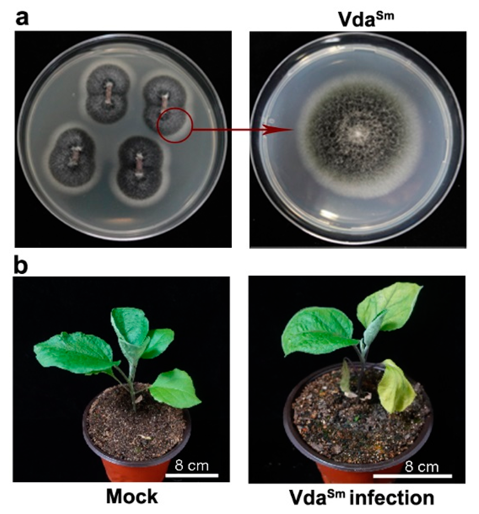
3.1. Isolation and Identification of Pathogens Causing Verticillium Wilt in Eggplants

Based on plant symptoms, we collected diseased eggplants with the typical symptom of Verticillium wilt in the field. To isolate the pathogen, the stem sections of diseased plants were surface-sterilized and transferred to potato dextrose agar (PDA) medium (Figure 1a). The hyphae that grew around the tissue slices of eggplant were transferred to another PDA medium to continue growth (Figure 1a). In the laboratory, the fungus caused typical Verticillium wilt symptoms in healthy plants, such as leaf wilting and chlorosis and plant stunting (Figure 1b).
For molecular identification, the ITS region of the fungus was sequenced using the primer pairs ITS1/ITS4 [34]. Blastn analysis revealed that the ITS sequence was 99–100%, identical to the V. dahliae reference sequences. PCR assays, with two primer pairs which were designed to differentiate Verticillium species [37], further confirmed that the isolate is V. dahliae (Figure S1a). Therefore, we conclude that the V. dahliae recovered from diseased eggplants (VdaSm) was the pathogenic fungus causing Verticillium wilt.

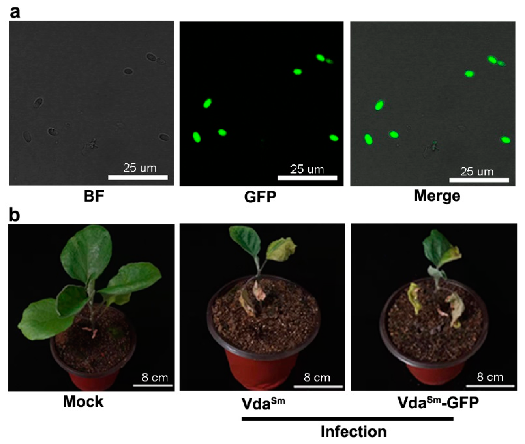
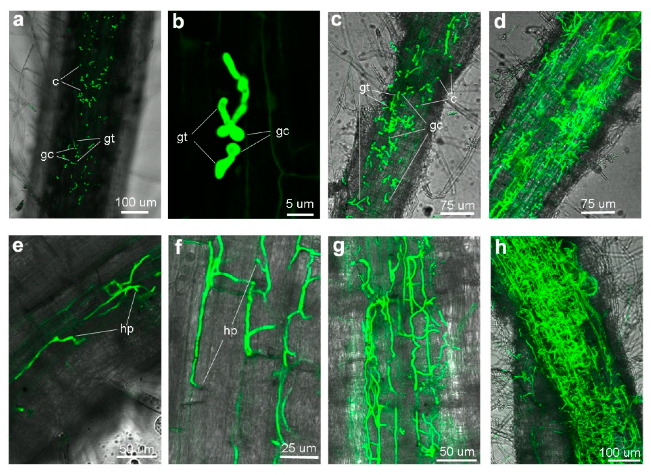
3.2. Colonization Process on Eggplant Roots by GFP-Labeled VdaSm

To explore the colonization process of VdaSm, we generated a GFP-labeled isolate, VdaSm-GFP. Green fluorescence was detected under a microscope to confirm the stable expression of GFP (Figure 2a). When cultured on PDA, both VdaSm and VdaSm-GFP were similar in colony morphology and growth rate (Figure S1b,c). Moreover, disease severity on eggplants caused by VdaSm and VdaSm-GFP did not show any obvious difference (Figure 2b). Given that its pathogenicity was unaffected, VdaSm-GFP was used to study the infection processes on eggplants.
The roots of infected eggplant were observed using the confocal laser scanning microscopy (CLSM). After 12 h inoculation (hpi), a small fraction of conidia germinated on the root surface at random sites (Figure 3a,b). Massive conidia that had germinated were observed at 24 hpi (Figure 3c). By 3 dpi, mycelium expanded, and hyphae covered the root surface. A fraction of hyphae expanded in parallel along the epidermal cells (Figure 3d). Only a few hyphae, tightly adhering to the surface, penetrate intercellularly into the epidermal cells (Figure 3e). At the site of penetration, we observed slight swelling of the elongating hyphae (Figure 3e). After successfully invading the root, the hyphae elongated parallelly along the longitudinal axis. The intercellular hyphal swelling between the epidermal cell junctions at the site of penetration to an adjacent cell was observed at 4 dpi (Figure 3f). By 6 dpi, the hyphae reached the vascular tissue and continued to grow and ramify, forming a hyphal net within the xylem vessels (Figure 3g). The root xylem vessels filled with hyphae were observed by 8 dpi (Figure 3h). Our results indicated that the invasion of eggplant roots by VdaSm shares a colonization process similar to that of V592 on cotton plants [21].

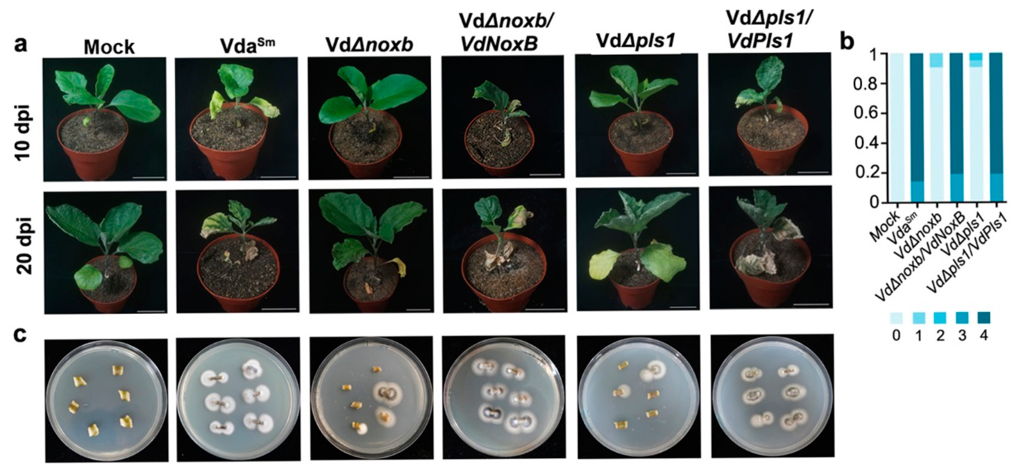
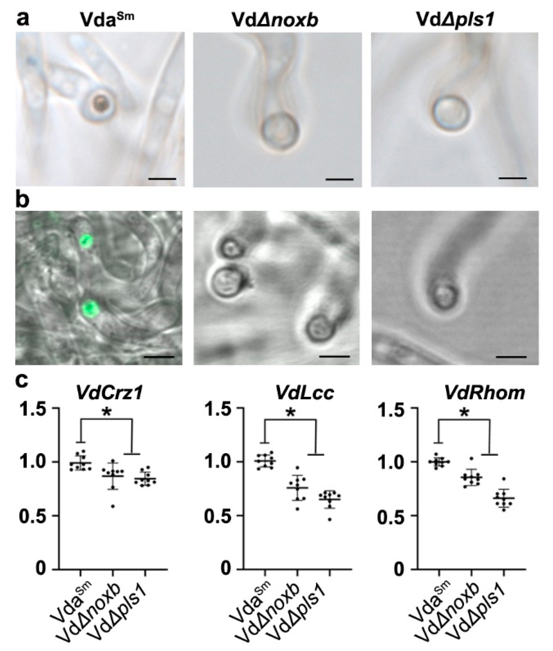
3.3. VdNoxB and VdPls1 Are Required for Penetration Peg Formation and Fungal Pathogenicity

We previously demonstrated that VdNoxB and VdPls1 from V592 are specifically expressed in infection structure and hyphopodium, and are indispensable for penetration peg formation in the colonization of V592 on cotton plants [22]. Therefore, to figure out whether the swelling hyphae (Figure 3e,f) were infection structures with specific molecular feature similar to V592 on cotton plants [22], we amplified and confirmed VdNoxB and VdPls1 homologous sequences from VdaSm. We then generated knockout mutants, VdΔnoxb and VdΔpls1, using the homologous recombination method (Figure S2a–c) [35]. There were no noticeable morphological differences between the VdaSm and the mutant strains (Figure 4a). GFP-fused VdNoxB and VdPls1 under the native promoter were introduced into VdΔnoxb and VdΔpls1 mutants, respectively, to produce the complemented strains VdΔnoxb/VdNoxB and VdΔpls1/VdPls1. Green fluorescence was detected, confirming the stable expression of GFP-fused VdNoxB and VdPls1 in complemented strains (Figure S2d).
We first examined the penetration abilities of the VdsSm, VdΔnoxb and VdΔpls1 mutants and the complemented strains VdΔnoxb/VdNoxB and VdΔpls1/VdPls1 on a cellophane membrane laid on MM, which is used for V592 to induce hyphopodia and penetration pegs [22]. We observed that VdaSm and both complemented strains penetrated from the cellophane membrane and grew on the medium at 3 dpi (Figure 4b). However, fungal hyphae penetration from the cellophane membrane was not observed for either VdΔnoxb or VdΔpls1 mutants (Figure 4b), suggesting that VdNoxB and VdPls1 were also required for VdsSm to induce infection structure. Indeed, hyphopodia with clear penetration pegs to breach the cellophane membrane for VdaSm and complemented strains were observed under microscopy (Figure 4c). Even though hyphopodia was observed, neither the VdΔnoxb nor VdΔpls1 mutants generated penetration pegs (Figure 4c). These data demonstrate that VdNoxB and VdPls1 are indispensable for penetration peg formation in VdaSm to penetrate the cellophane membrane.
We then investigated the roles of VdNoxB and VdPls1 in the pathogenicity of VdaSm on eggplants. The eggplants were inoculated with spores from the wild-type VdaSm, VdΔnoxb and VdΔpls1 mutant strains, as well as with the complemented strains, VdΔnoxb/VdNoxB and VdΔpls1/VdPls1. Compared to VdaSm, both the VdΔnoxb and VdΔpls1 mutant strains displayed significantly reduced disease severity in eggplants (Figure 5a,b). The loss of virulence was restored in both VdΔnoxb/VdNoxB and VdΔpls1/VdPls1 strains (Figure 5a,b). Isolation of diverse VdaSm strains from the infected plants was used to confirm the successful inoculation (Figure 5c). These results indicate that VdNoxB and VdPls1 are essential to the pathogenicity of VdaSm.
Similar to our previous studies on cotton [21,22], we conclude that special infection structures are required for V. dahliae strains colonization of various host plants. A conservative mechanism involving VdNoxB and VdPls1 would be indispensable for penetration peg formation.

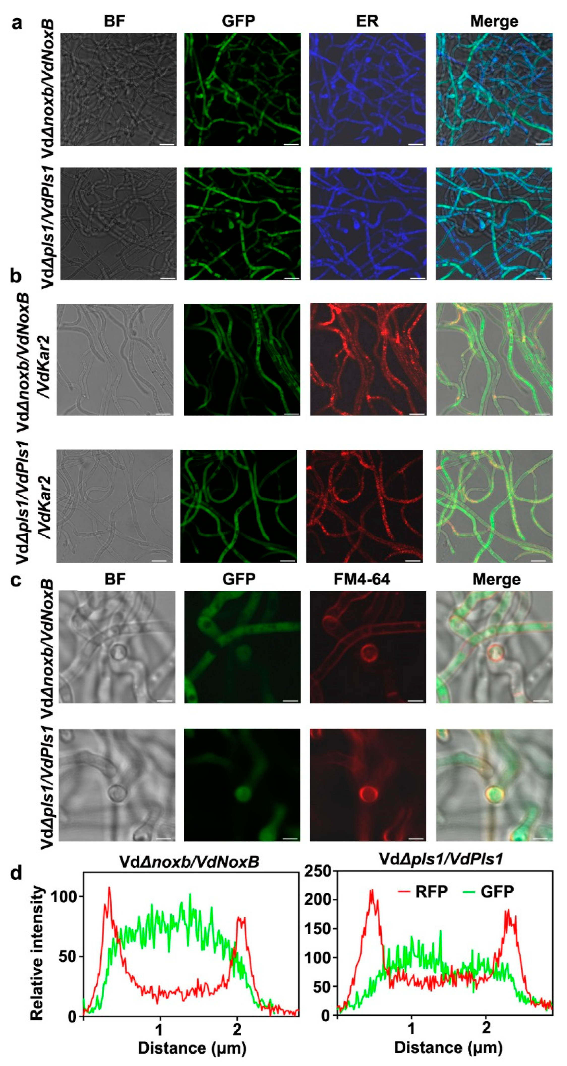
3.4. VdNoxB and VdPls1 Were Highly Expressed in Hyphopodia and Localized at the Position of Penetration Peg Emergence in VdaSm

NoxB and Pls1 were reported to be associated with the ER and specifically accumulate in the infection structures in V. dahliae [22], Magnaporthe oryzae [38] and Botrytis cinerea [39]. VdNoxB and VdPls1 of V592 particularly colocalized at the position of penetration peg emergence and VdPls1 is required for the plasma membrane localization and activation of VdNoxB [22]. To investigate the cellular localization of VdNoxB and VdPls1, the complemented strains VdΔnoxb/VdNoxB and VdΔpls1/VdPls1, in which GFP-fused VdNoxB or VdPls1 was introduced into the corresponding mutant (Figure S2c), were examined under CLSM. The functional activities of GFP-fused proteins were confirmed by complementation of both penetration ability (Figure 4) and pathogenicity (Figure 5). The hyphae grown on the cellophane membrane were stained by ER-Tracker Blue-White DPX (Figure 6a). Either GFP-fused protein colocalized with the ER-tracker signals (Figure 6a). Furthermore, the RFP was fused with the V. dahliae homolog of yeast KAR2, located at the ER and nuclear envelope [40]. RFP-fused VdKar2 was introduced into the VdΔnoxb/VdNoxB and VdΔpls1/VdPls1. Red fluorescence was detected under a microscope, confirming the stable expression of VdKar2-GFP (Figure 6b). Similar to Blue-White DPX staining, overlapping of the RFP signal with the GFP fluorescence in both VdΔnoxb/VdNoxB/VdKar2 and VdΔpls1/VdPls1/VdKar2 was also observed (Figure 6b). Notably, at the base of hyphopodia where penetration pegs developed, the plasma membrane of VdΔnoxb/VdNoxB and VdΔpls1/VdPls1 was clearly stained using FM4-64 (Figure 6c), where linescans also showed the strong GFP signals in a transverse section of individual hyphopodium (Figure 6d). Taken together, these results indicated that both VdNoxB and VdPls1 of VdaSm are associated with the ER, and localized at the position of penetration peg emergence in VdaSm, which is similar to VdNoxB and VdPls1 location in V592 [22].

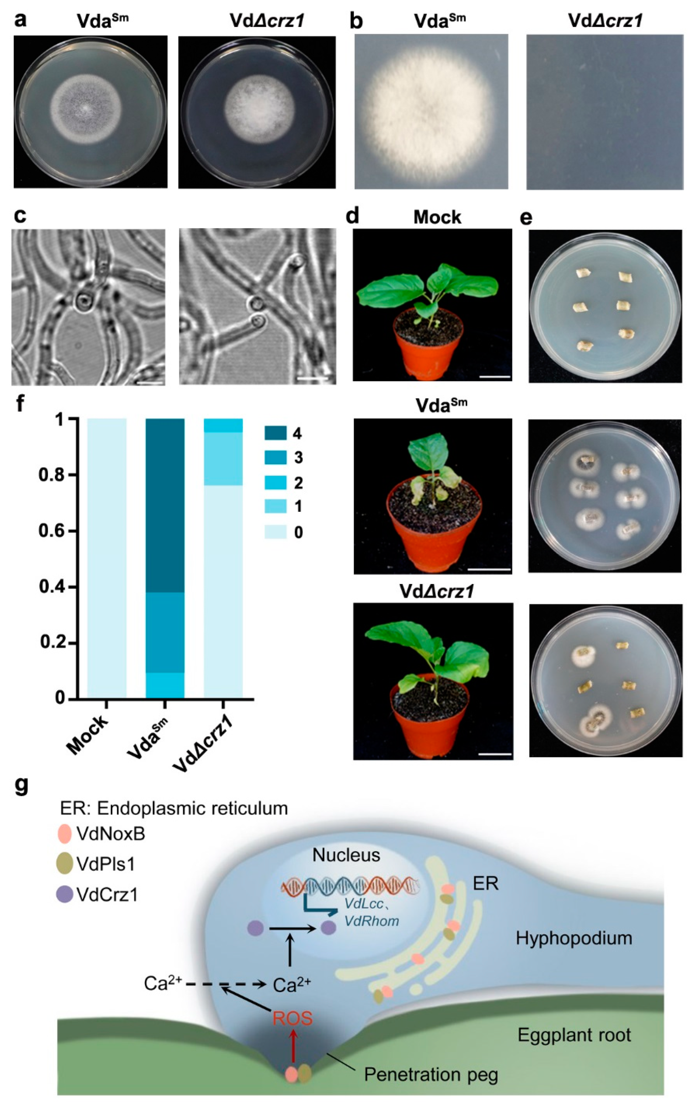
3.5. VdNoxB/VdPls1-Mediated ROS Production Coupled with Ca2+-Activated VdCrz1 Signaling in the Hyphopodium

In V592, VdPls1 regulates ROS burst by influencing the plasma membrane localization of VdNoxB. The VdNoxB/VdPls1-dependent ROS burst is essential for the free Ca2+ elevation in the hyphopodium, which activates VdCrz1 signaling to induce penetration peg development [22].
To confirm whether penetration peg formation in VdaSm shared a molecular mechanism similar to that in V592, we first detected the ROS burst in the hyphopodium. VdaSm hyphae grown on the cellophane membrane were treated with DAB staining for CLSM observation. Intensive ROS signals were detected at the base of hyphopodia in VdaSm, where the penetration pegs were generated (Figure 7a). In contrast, no signal was detected for ROS-specific accumulation in either the VdΔnoxb or VdΔpls1 mutant strains (Figure 7a). Moreover, a tip-high Ca2+ gradient in the hyphopodia of VdaSm was observed with the intracellular calcium indicator Fluo-a AM, whereas Ca2+ was not detectable in the hyphopodia of the VdΔnoxb and VdΔpls1 strains (Figure 7b). Our data indicate that VdNoxB and VdPls1 are required for ROS production and Ca2+ elevation in the hyphopodium [22].
Next, we examined the expression of VdCrz1 and its potential targets VdLcc and VdRhom, encoding M. oryzae orthologs of laccase and rhomboid family membrane protein, respectively [41], to confirm whether Ca2+ elevation affected VdCrz1 signaling in VdaSm. The expression of VdCrz1 was significantly reduced in VdΔnoxb and VdΔpls1 compared to wild-type VdaSm (Figure 7c). As expected, the two target genes of VdCrz1 were also downregulated in both mutant strains (Figure 7c). We then generated the VdCrz1 knockout mutant, VdΔcrz1 (Figure S3a,b), and assayed its ability to induce penetration peg formation and pathogenicity in eggplants. Compared to VdaSm, VdΔcrz1 did not exhibit obvious phenotypic differences on PDA plates (Figure 8a). Similar to VdΔnoxb and VdΔpls1, VdΔcrz1 hyphae penetration from the cellophane membrane at 3 dpi (Figure 8b) and the formation of a penetration peg at 2 dpi was not observed (Figure 8c). Pathogenicity assays showed that VdΔcrz1 displayed greatly reduced virulence in eggplants compared to VdaSm (Figure 8d,f). Isolation of VdaSm and VdΔcrz1 strains from the infected plants was used to confirm the successful inoculation (Figure 8e). Taken together, our results demonstrated that VdNoxB/VdPls1-mediated Ca2+ elevation activates VdCrz1 signaling to regulate the penetration ability and pathogenicity of VdaSm.

4. Discussion

In this study, we demonstrated that the formation of hyphopodia and penetration pegs is required for the initial colonization of VdaSm on eggplant roots. VdNoxB and VdPls1 are indispensable for penetration peg formation and essential to the pathogenicity of VdaSm. Both VdNoxB and VdPls1 localized at the base of hyphopodia, where the penetration pegs were generated. Additionally, VdNoxb and VdPls1 are required for the ROS production. Furthermore, the VdNoxB/VdPls1-dependent ROS burst elevates Ca2+ accumulation in the hyphopodia, which activates VdCrz1 signaling to regulate penetration peg formation (Figure 8g).
To enter the underlying host tissues, many fungi generate elaborate infection structures from emerging penetration hyphae to breach the cuticle and epidermal cell wall [1,42]. Interestingly, studies on host penetration by Rhizoctonia solani showed that the ability to generate infection structures is highly variable even within a given species [42,43]. Similarly, V. dahliae develops infection structures with different characteristics to adapt to host specificity. An appressoria, which is a requirement for leaf infection, was observed during the invasion of V. dahliae on lettuce and fiber flax roots [16,17]. However, we demonstrated that the formation of hyphopodia with penetration peg is essential for the initial colonization of V592 on cotton roots [22]. In this study, we isolated the V. dahliae strain VdaSm from Verticillium wilt eggplants (Figure 1) and observed its infection process with a GFP-labeled isolate (Figure 2). Hyphal swelling at the site of penetration was observed (Figure 3). The penetration ability assay showed that the hyphopodium with penetration pegs are indispensable to breach the cellophane membrane (Figure 4). Furthermore, we demonstrated that the hyphopodia and penetration pegs play essential roles in initial colonization and pathogenicity of VdaSm on eggplant (Figure 5Similar to our previous observation [21,22], we conclude that V. dahliae colonized eggplant, cotton and Arabidopsis with a similar process.
The tissue-adapted infection strategies of M. oryzae have clarified the difference in appressoria and hyphopodia [10,12,13,38]. On the leaf, M. oryzae develops heavily melanized appressoria associated with classical foliar infection, and then the appressoria build up tremendous turgor pressure to penetrate the tough surface [12,44,45]; on the root, hyphal swellings resembling the simple structure hyphopodia has been evident [12]. These results indicated that it is harder for the fungus to breach the leaf than to penetrate the root. Our previous study showed that the average diameter of V. dahliae hyphopodia was smaller than that of M. oryzae appressoria [31], which suggests that more pressure is needed for the fungus to breach the plant leaf compared to the root. Therefore, the previous findings of slight hyphal swelling during V. dahliae infection on oilseed rape and sunflower [19,20] that was thought as non-structural development without molecular characterization would be worth studying further.
Although there is considerable variation in morphology between appressoria and hyphopodia, a study showed that the infection structures share common genetic requirements during M. oryzae colonization [38]. In general, ROS generation is required for the differentiation of a penetration peg from appressoria [23,25,46] or hyphopodia [22]. Nox enzymes function as the major enzymatic producer of ROS [47,48,49]. In several fungal pathogens, the NoxA and NoxB proteins have been shown to be crucial for hyphal tip growth, tissue invasion and virulence [24,26,27,50]. ROS elevation, produced by NoxB, activates Ca2+ channels to facilitate Ca2+ influx for the plant root-hair cell and the fungal hyphae tip polarity [28,29,30]. Pls1, which is expressed during appressoria development, is presumed to be the corresponding integral membrane adaptor for the assembly of the NoxB complex [39,51]. In this present study, VdNoxB and VdPls1 of VdaSm were indispensable for penetration peg formation on the cellophane membrane (Figure 4). We observed that both VdNoxB and VdPls1 localize on the plasma membrane (Figure 6a), particularly at the position of penetration peg emergence (Figure 6b). Similar to our previous results on V592 [22], the ER and plasma membrane location of VdNoxB/VdPls1 was essential for penetration peg formation. In our previous study, yeast two-hybrid and bimolecular fluorescence complementation assays provided evidence of a direct physical interaction between VdNoxB and VdPls1 [22]. Furthermore, we demonstrated that the plasma membrane localization of VdNoxB and VdPls1 is required for the ROS burst in the hyphopodia (Figure 7a), which elevates Ca2+ accumulation at the base of hyphopodia (Figure 7b). Consistent with our previous results on V592 [22], VdNoxB/VdPls1-dependent Ca2+ elevation activated VdCrz1 signaling, and VdCrz1 and its targets were significantly reduced in VdΔnoxb and VdΔpls1 compared to VdaSm (Figure 7). Similar to VdΔnoxb and VdΔpls1, penetration peg formation was not observed in VdΔcrz1 on the cellophane membrane at 2 dpi (Figure 8b,c). VdΔcrz1 displayed significantly reduced virulence in eggplants (Figure 8d).

5. Conclusions

In summary, we observed the infection process of VdaSm on eggplants and identified the infection structure and its molecular features. Similar to our previous study on V592 infection on cotton plants, we conclude that a common genetic pathway regulates host-specific infection-related development in the soil-borne fungus V. dahliae. The finding that Nox/Pls-dependent signaling is required for appressorium formation [23,51], suggests that Nox/Pls are key components for this pathogenic fungal colonization of their hosts. Therefore, Nox/Pls-dependent signaling maybe a desirable target for fungicides. The elaboration of regulatory mechanisms in the upstream of Nox/Pls-dependent signaling, such as cAMP [38,52], is worth studying further. A clear understanding of the process of colonization might help to develop effective fungicides for inhibiting the V. dahliae infection process.

Supplementary Materials

The following supporting information can be downloaded at: https://www.mdpi.com/article/10.3390/jof9040484/s1, Figure S1: The colony morphology and growth rate of VdaSm and VdaSm-GFP on PDA plates; Figure S2: Construction of knockout mutants VdΔnoxb and VdΔpls1, and complemented strains VdΔnoxb/VdNoxB and VdΔpls1/VdPls1; Figure S3: Identification of the VdCrz1 knockout mutant with the primers HPT and VdCrz1 as indicated; Table S1: Primers were used in this study.

Author Contributions

Conceptualization, J.Z. and H.G.; methodology, Q.L., Y.L., H.W. and B.Z.; software, Q.L. and Y.L.; validation, Q.L., Y.L., H.W., B.Z., C.L. and Y.G.; formal analysis, Q.L., Y.L., J.Z. and H.G.; writing—original draft preparation, J.Z.; writing—review and editing, H.G.; visualization, J.Z., Q.L. and Y.L.; supervision, H.G. and J.Z.; project administration, H.G. and J.Z.; funding acquisition, H.G. and J.Z. All authors have read and agreed to the published version of the manuscript.

Funding

This research was funded by National Key R&D Program of China 2022YFD1400800, the National Natural Science Foundation of China (Grant No. 32020103003), strategic Priority Research Program of Chinese Academy of Sciences (Grant No. XDPB16), Xinjiang Production and Construction Corps Science and Technology Plan Project (Grant No. 2022DB014).

Institutional Review Board Statement

Not applicable.

Informed Consent Statement

Not applicable.

Data Availability Statement

Not applicable.

Conflicts of Interest

The authors declare no conflict of interest.

References

  1. Chethana, K.W.T.; Jayawardena, R.S.; Chen, Y.J.; Konta, S.; Tibpromma, S.; Abeywickrama, P.D.; Gomdola, D.; Balasuriya, A.; Xu, J.; Lumyong, S.; et al. Diversity and Function of Appressoria. Pathogens 2021, 10, 746. [Google Scholar] [CrossRef]
  2. Mendgen, K.; Hahn, M.; Deising, H. Morphogenesis and mechanisms of penetration by plant pathogenic fungi. Annu. Rev. Phytopathol. 1996, 34, 367–386. [Google Scholar] [CrossRef]
  3. Lacaze, A.; Joly, D.L. Structural specificity in plant-filamentous pathogen interactions. Mol. Plant Pathol. 2020, 21, 1513–1525. [Google Scholar] [CrossRef] [PubMed]
  4. Genre, A.; Lanfranco, L. Endophytic coming out: The expressorium as a novel fungal structure specialized in outward-directed penetration of the leaf cuticle. New Phytol. 2016, 211, 5–7. [Google Scholar] [CrossRef] [PubMed]
  5. Demoor, A.; Silar, P.; Brun, S. Appressorium: The Breakthrough in Dikarya. J. Fungi 2019, 5, 72. [Google Scholar] [CrossRef] [PubMed]
  6. Deising, H.B.; Werner, S.; Wernitz, M. The role of fungal appressoria in plant infection. Microbes Infect. 2000, 2, 1631–1641. [Google Scholar] [CrossRef] [PubMed]
  7. Kubicek, C.P.; Starr, T.L.; Glass, N.L. Plant cell wall-degrading enzymes and their secretion in plant-pathogenic fungi. Annu. Rev. Phytopathol. 2014, 52, 427–451. [Google Scholar] [CrossRef] [PubMed]
  8. Tucker, S.L.; Talbot, N.J. Surface attachment and pre-penetration stage development by plant pathogenic fungi. Annu. Rev. Phytopathol. 2001, 39, 385–417. [Google Scholar] [CrossRef]
  9. de Jong, T.G.; Hulshof, J.; Prokert, G. Modelling fungal hypha tip growth via viscous sheet approximation. J. Theor. Biol. 2020, 492, 110189. [Google Scholar] [CrossRef]
  10. Marcel, S.; Sawers, R.; Oakeley, E.; Angliker, H.; Paszkowski, U. Tissue-adapted invasion strategies of the rice blast fungus Magnaporthe oryzae. Plant Cell 2010, 22, 3177–3187. [Google Scholar] [CrossRef]
  11. Wilson, R.A.; Talbot, N.J. Under pressure: Investigating the biology of plant infection by Magnaporthe oryzae. Nat. Rev. Microbiol. 2009, 7, 185–195. [Google Scholar] [CrossRef] [PubMed]
  12. Sesma, A.; Osbourn, A.E. The rice leaf blast pathogen undergoes developmental processes typical of root-infecting fungi. Nature 2004, 431, 582–586. [Google Scholar] [CrossRef] [PubMed]
  13. Park, J.Y.; Jin, J.; Lee, Y.W.; Kang, S.; Lee, Y.H. Rice blast fungus (Magnaporthe oryzae) infects Arabidopsis via a mechanism distinct from that required for the infection of rice. Plant Physiol. 2009, 149, 474–486. [Google Scholar] [CrossRef] [PubMed]
  14. Song, R.; Li, J.; Xie, C.; Jian, W.; Yang, X. An Overview of the Molecular Genetics of Plant Resistance to the Verticillium Wilt Pathogen Verticillium dahliae. Int. J. Mol. Sci. 2020, 21, 1120. [Google Scholar] [CrossRef]
  15. Klimes, A.; Dobinson, K.F.; Thomma, B.P.; Klosterman, S.J. Genomics spurs rapid advances in our understanding of the biology of vascular wilt pathogens in the genus Verticillium. Annu. Rev. Phytopathol. 2015, 53, 181–198. [Google Scholar] [CrossRef]
  16. Blum, A.; Bressan, M.; Zahid, A.; Trinsoutrot-Gattin, I.; Driouich, A.; Laval, K. Verticillium Wilt on Fiber Flax: Symptoms and Pathogen Development In Planta. Plant Dis. 2018, 102, 2421–2429. [Google Scholar] [CrossRef]
  17. Vallad, G.E.; Subbarao, K.V. Colonization of resistant and susceptible lettuce cultivars by a green fluorescent protein-tagged isolate of Verticillium dahliae. Phytopathology 2008, 98, 871–885. [Google Scholar] [CrossRef]
  18. Su, X.; Lu, G.; Rehman, L.; Li, X.; Sun, L.; Guo, H.; Cheng, H. mCherry-Labeled Verticillium dahliae Could Be Utilized to Investigate Its Pathogenicity Process in Nicotiana benthamiana. Genes 2018, 9, 508. [Google Scholar] [CrossRef]
  19. Eynck, C.; Koopmann, B.; Grunewaldt-Stoecker, G.; Karlovsky, P.; von Tiedemann, A. Differential interactions of Verticillium longisporum and V. dahliae with Brassica napus detected with molecular and histological techniques. Eur. J. Plant Pathol. 2007, 118, 259–274. [Google Scholar] [CrossRef]
  20. Zhang, Y.; Zhang, J.; Gao, J.; Zhang, G.; Yu, Y.; Zhou, H.; Chen, W.; Zhao, J. The Colonization Process of Sunflower by a Green Fluorescent Protein-Tagged Isolate of Verticillium dahliae and its Seed Transmission. Plant Dis. 2018, 102, 1772–1778. [Google Scholar] [CrossRef]
  21. Zhao, P.; Zhao, Y.L.; Jin, Y.; Zhang, T.; Guo, H.S. Colonization process of Arabidopsis thaliana roots by a green fluorescent protein-tagged isolate of Verticillium dahliae. Protein Cell 2014, 5, 94–98. [Google Scholar] [CrossRef] [PubMed]
  22. Zhao, Y.L.; Zhou, T.T.; Guo, H.S. Hyphopodium-Specific VdNoxB/VdPls1-Dependent ROS-Ca2+ Signaling Is Required for Plant Infection by Verticillium dahliae. PLoS Pathog. 2016, 12, e1005793. [Google Scholar] [CrossRef] [PubMed]
  23. Egan, M.J.; Wang, Z.Y.; Jones, M.A.; Smirnoff, N.; Talbot, N.J. Generation of reactive oxygen species by fungal NADPH oxidases is required for rice blast disease. Proc. Natl. Acad. Sci. USA 2007, 104, 11772–11777. [Google Scholar] [CrossRef] [PubMed]
  24. Heller, J.; Tudzynski, P. Reactive oxygen species in phytopathogenic fungi: Signaling, development, and disease. Annu. Rev. Phytopathol. 2011, 49, 369–390. [Google Scholar] [CrossRef]
  25. Ryder, L.S.; Dagdas, Y.F.; Mentlak, T.A.; Kershaw, M.J.; Thornton, C.R.; Schuster, M.; Chen, J.; Wang, Z.; Talbot, N.J. NADPH oxidases regulate septin-mediated cytoskeletal remodeling during plant infection by the rice blast fungus. Proc. Natl. Acad. Sci. USA 2013, 110, 3179–3184. [Google Scholar] [CrossRef]
  26. Takemoto, D.; Tanaka, A.; Scott, B. NADPH oxidases in fungi: Diverse roles of reactive oxygen species in fungal cellular differentiation. Fungal Genet. Biol. FG B 2007, 44, 1065–1076. [Google Scholar] [CrossRef]
  27. Tudzynski, P.; Heller, J.; Siegmund, U. Reactive oxygen species generation in fungal development and pathogenesis. Curr. Opin. Microbiol. 2012, 15, 653–659. [Google Scholar] [CrossRef]
  28. Cardenas, L. New findings in the mechanisms regulating polar growth in root hair cells. Plant Signal. Behav. 2009, 4, 4–8. [Google Scholar] [CrossRef]
  29. Foreman, J.; Demidchik, V.; Bothwell, J.H.; Mylona, P.; Miedema, H.; Torres, M.A.; Linstead, P.; Costa, S.; Brownlee, C.; Jones, J.D.; et al. Reactive oxygen species produced by NADPH oxidase regulate plant cell growth. Nature 2003, 422, 442–446. [Google Scholar] [CrossRef]
  30. Jackson, S.L.; Heath, I.B. Roles of calcium ions in hyphal tip growth. Microbiol. Rev. 1993, 57, 367–382. [Google Scholar] [CrossRef]
  31. Zhou, T.T.; Zhao, Y.L.; Guo, H.S. Secretory proteins are delivered to the septin-organized penetration interface during root infection by Verticillium dahliae. PLoS Pathog. 2017, 13, e1006275. [Google Scholar] [CrossRef] [PubMed]
  32. Zhang, K.; Su, Y.Y.; Cai, L. An optimized protocol of single spore isolation for fungi. Cryptogam. Mycol. 2013, 34, 349–356. [Google Scholar] [CrossRef]
  33. Gao, F.; Zhou, B.J.; Li, G.Y.; Jia, P.S.; Li, H.; Zhao, Y.L.; Zhao, P.; Xia, G.X.; Guo, H.S. A glutamic acid-rich protein identified in Verticillium dahliae from an insertional mutagenesis affects microsclerotial formation and pathogenicity. PLoS ONE 2010, 5, e15319. [Google Scholar] [CrossRef] [PubMed]
  34. White, T.J. Amplification and direct sequencing of fungal ribosomal RNA genes for phylogenetics. In PCR Protocols: A Guide to Methods and Applications; Academic Press: San Diego, CA, USA, 1990. [Google Scholar]
  35. Wang, S.; Xing, H.; Hua, C.; Guo, H.S.; Zhang, J. An Improved Single-Step Cloning Strategy Simplifies the Agrobacterium tumefaciens-Mediated Transformation (ATMT)-Based Gene-Disruption Method for Verticillium dahliae. Phytopathology 2016, 106, 645–652. [Google Scholar] [CrossRef]
  36. Pathak, R.R.; Lochab, S. A method for rapid isolation of total RNA of high purity and yield from Arthrospira platensis. Can. J. Microbiol. 2010, 56, 578–584. [Google Scholar] [CrossRef]
  37. Inderbitzin, P.; Davis, R.M.; Bostock, R.M.; Subbarao, K.V. Identification and Differentiation of Verticillium Species and V. longisporum Lineages by Simplex and Multiplex PCR Assays. PLoS ONE 2013, 8, e65990. [Google Scholar] [CrossRef]
  38. Tucker, S.L.; Besi, M.I.; Galhano, R.; Franceschetti, M.; Goetz, S.; Lenhert, S.; Osbourn, A.; Sesma, A. Common genetic pathways regulate organ-specific infection-related development in the rice blast fungus. Plant Cell 2010, 22, 953–972. [Google Scholar] [CrossRef]
  39. Siegmund, U.; Heller, J.; van Kan, J.A.; Tudzynski, P. The NADPH oxidase complexes in Botrytis cinerea: Evidence for a close association with the ER and the tetraspanin Pls1. PLoS ONE 2013, 8, e55879. [Google Scholar] [CrossRef]
  40. Rose, M.D.; Misra, L.M.; Vogel, J.P. KAR2, a karyogamy gene, is the yeast homolog of the mammalian BiP/GRP78 gene. Cell 1989, 57, 1211–1221. [Google Scholar] [CrossRef]
  41. Kim, S.; Hu, J.; Oh, Y.; Park, J.; Choi, J.; Lee, Y.H.; Dean, R.A.; Mitchell, T.K. Combining ChIP-chip and expression profiling to model the MoCRZ1 mediated circuit for Ca/calcineurin signaling in the rice blast fungus. PLoS Pathog. 2010, 6, e1000909. [Google Scholar] [CrossRef]
  42. Dean, R.A. Signal pathways and appressorium morphogenesis. Annu. Rev. Phytopathol. 1997, 35, 211–234. [Google Scholar] [CrossRef] [PubMed]
  43. Weinhold, A.R.; Sinclair, J.B. Rhizoctonia solani: Penetration, colonization, and host response. In Rhizoctonia Species: Taxonomy, Molecular Biology, Ecology, Pathology and Disease Control; Springer: Berlin/Heidelberg, Germany, 1996; pp. 163–174. [Google Scholar]
  44. de Jong, J.C.; McCormack, B.J.; Smirnoff, N.; Talbot, N.J. Glycerol generates turgor in rice blast. Nature 1997, 389, 244. [Google Scholar] [CrossRef]
  45. Howard, R.J.; Valent, B. Breaking and entering: Host penetration by the fungal rice blast pathogen Magnaporthe grisea. Annu. Rev. Microbiol. 1996, 50, 491–512. [Google Scholar] [CrossRef]
  46. Gupta, Y.K.; Dagdas, Y.F.; Martinez-Rocha, A.L.; Kershaw, M.J.; Littlejohn, G.R.; Ryder, L.S.; Sklenar, J.; Menke, F.; Talbot, N.J. Septin-Dependent Assembly of the Exocyst Is Essential for Plant Infection by Magnaporthe oryzae. Plant Cell 2015, 27, 3277–3289. [Google Scholar] [CrossRef]
  47. Sumimoto, H. Structure, regulation and evolution of Nox-family NADPH oxidases that produce reactive oxygen species. FEBS J. 2008, 275, 3249–3277. [Google Scholar] [CrossRef]
  48. Scott, B. Conservation of fungal and animal nicotinamide adenine dinucleotide phosphate oxidase complexes. Mol. Microbiol. 2015, 95, 910–913. [Google Scholar] [CrossRef] [PubMed]
  49. Lambeth, J.D. NOX enzymes and the biology of reactive oxygen. Nat. Rev. Immunol. 2004, 4, 181–189. [Google Scholar] [CrossRef] [PubMed]
  50. Aguirre, J.; Rios-Momberg, M.; Hewitt, D.; Hansberg, W. Reactive oxygen species and development in microbial eukaryotes. Trends Microbiol. 2005, 13, 111–118. [Google Scholar] [CrossRef]
  51. Lambou, K.; Malagnac, F.; Barbisan, C.; Tharreau, D.; Lebrun, M.H.; Silar, P. The crucial role of the Pls1 tetraspanin during ascospore germination in Podospora anserina provides an example of the convergent evolution of morphogenetic processes in fungal plant pathogens and saprobes. Eukaryot. Cell 2008, 7, 1809–1818. [Google Scholar] [CrossRef]
  52. Sun, L.; Qin, J.; Rong, W.; Ni, H.; Guo, H.S.; Zhang, J. Cellophane surface-induced gene, VdCSIN1, regulates hyphopodium formation and pathogenesis via cAMP-mediated signalling in Verticillium dahliae. Mol. Plant Pathol. 2019, 20, 323–333. [Google Scholar] [CrossRef]
Figure 1. Eggplant wilt disease symptoms caused by infection with V. dahliae recovered from diseased plants. (a) Hyphae grew from two ends of the cut stems and were transferred to another PDA medium to continue growth; (b) VdaSm infection caused leaf wilting and chlorosis and plant stunting. Similar results were obtained from 20 plants, and representative photographs are shown.
Jof 09 00484 g001
Figure 2. The pathogenicity caused by VdaSm-GFP did not significantly differ from the wild-type isolate. (a) Confocal micrograph of green fluorescence by GFP-labeled VdaSm; (b) Similar wilt symptoms were observed with VdaSm and VdaSm-GFP on 20 eggplants, and representative photographs are shown.
Jof 09 00484 g002
Figure 3. Systemic infection of eggplant roots by VdaSm-GFP. (a) Germinated conidia on the eggplant root surface (12 hpi). c—conidium, gc—germinated conidia, gt—germ tubes; (b) Germ tubes emerging from one end of the conidium (12 hpi); (c) VdaSm-GFP hyphae on the root of eggplant (24 hpi); (d) Hyphae covering the surface with a nonspecific growth pattern (3 dpi); (e) A few hyphae tightly adhered on the root surface penetrated intercellular into the epidermal cells (3 dpi). hp—hyphopodia; (f) Swelling hyphae were observed at the site of penetration to an adjacent cell (4 dpi); (g) A hyphae net within the xylem vessels was observed by 6 dpi; (h) Hyphae filled the root xylem vessels by 8 dpi.
Jof 09 00484 g003
Figure 4. The penetration abilities assay on the cellophane membrane. (a) Colony morphology of wild-type VdaSm, VdΔnoxb and VdΔpls1 mutants, and VdΔnoxb/VdNoxB and VdΔpls1/VdPls1 on PDA plates; (b) Colonies grown on the cellophane membrane (Above) and MM (Below). VdaSm grew on the MM after penetration from the cellophane membrane. Mutant strains lost penetration ability, and complemented strains restored penetration ability; (c) VdaSm and complemented strains developed hyphopodia with penetration pegs. The hyphopodia are indicated by the arrow, and the penetration peg is indicated by asterisks.
Jof 09 00484 g004
Figure 5. Disease symptoms of eggplants infected with VdaSm, mutant strains VdΔnoxb and VdΔpls1, and complemented strains. (a) Photographs were taken at 10 dpi and 20 dpi. Bar = 8 cm. Similar results were obtained from 20 infected plants for each strain, and representative photographs are shown. (b) Disease grade of infected plants by various strains. 21 infected plants for each strain were counted; (c) Isolation of diverse strains from the stems of infected plants.
Jof 09 00484 g005
Figure 6. VdNoxB and VdPls1 were highly expressed in hyphopodium and localized at the base of hyphopodium. (a) Localization of VdNoxB and VdPls1 in the ER. ER was stained with ER-Tracker Blue-White DPX. Bar = 10 μm; (b) Localization of VdNoxB-GFP, VdPls1-GFP and VdKar2RFP. Bar = 10 μm; (c) Localization of VdNoxB and VdPls1 with the membrane of penetration pegs. The plasma membrane was stained using FM4-64, Bar = 2.5 μm. (d) Linescan graphs showing the relative intensity of RFP and GFP at the base of hyphopodium.
Jof 09 00484 g006
Figure 7. VdNoxB/VdPls1-dependent ROS burst affected VdCrz1 signaling by Ca2+ elevation. (a) CLSM observation of ROS accumulation at the base of hyphopodia in VdaSm but not in mutant strains. Hyphae grown on the cellophane membrane were stained with DAB. Bar = 10 μm; (b) Detection of Ca2+ elevation in hyphopodia with Fluo-4 AM. A tip-high gradient Ca2+ was observed in VdaSm but not in the VdΔnoxb and VdΔpls1 strains. Bar = 5 μm; (c) Expression analysis of VdCrz1 and its target genes in VdaSm and mutant strains. The asterisks indicate significant differences (p < 0.05, one-way ANOVA), and error bars show the standard deviations.
Jof 09 00484 g007
Figure 8. VdCrz1 signaling regulated penetration peg formation and pathogenicity of VdaSm on eggplants. (a) Colony morphology of wild-type VdaSm and VdΔcrz1 mutant; (b) The penetration ability detection of VdΔcrz1 on the cellophane membrane at 3 dpi; (c) Penetration peg was not observed in VdΔcrz1. Bar = 5 μm; (d) Disease symptoms of eggplants infected with VdaSm and VdΔcrz1. Similar results were obtained from 20 infected plants for each strain, and representative photographs are shown. Photographs were taken at 15 dpi. Bar = 10 cm; (e) Isolation of VdaSm and VdΔcrz1 mutant strains from the stems of infected plants; (f) Disease grade of infected plants by VdaSm and VdΔcrz1 mutant strains. A total of 21 infected plants for each strain were counted; (g) Schematic overview of VdNoxB/VdPls1-mediated ROS-Ca2+ signaling during penetration pef formation of VdaSm on eggplant roots.
Jof 09 00484 g008
Disclaimer/Publisher’s Note: The statements, opinions and data contained in all publications are solely those of the individual author(s) and contributor(s) and not of MDPI and/or the editor(s). MDPI and/or the editor(s) disclaim responsibility for any injury to people or property resulting from any ideas, methods, instructions or products referred to in the content.

