Article

Arthrobotrys blastospora sp. nov. (Orbiliomycetes): A Living Fossil Displaying Morphological Traits of Mesozoic Carnivorous Fungi

1
Institute of Eastern-Himalaya Biodiversity Research, Dali University, Dali 671003, China
2
Center of Excellence in Fungal Research, Mae Fah Luang University, Chiang Rai 57100, Thailand
3
School of Science, Mae Fah Luang University, Chiang Rai 57100, Thailand
4
Key Laboratory of Yunnan State Education Department on Er’hai Lake Basin Protection and the Sustainable Development Research, Dali University, Dali 671003, China
5
The Provincial Innovation Team of Biodiversity Conservation and Utility of the Three Parallel Rivers from Dali University, Dali University, Dali 671003, China
6
Yunling Black-and-White Snub-Nosed Monkey Observation and Research Station of Yunnan Province, Dali 671003, China
*
Author to whom correspondence should be addressed.
J. Fungi 2023, 9(4), 451; https://doi.org/10.3390/jof9040451
Submission received: 6 March 2023 / Revised: 29 March 2023 / Accepted: 3 April 2023 / Published: 6 April 2023
(This article belongs to the Special Issue New Perspectives on Entomopathogenic and Nematode-Trapping Fungi)

Abstract

:
The evolution of carnivorous fungi in deep time is still poorly understood as their fossil record is scarce. The approximately 100-million-year-old Cretaceous Palaeoanellus dimorphus is the earliest fossil of carnivorous fungi ever discovered. However, its accuracy and ancestral position has been widely questioned because no similar species have been found in modern ecosystems. During a survey of carnivorous fungi in Yunnan, China, two fungal isolates strongly morphologically resembling P. dimorphus were discovered and identified as a new species of Arthrobotrys (Orbiliaceae, Orbiliomycetes), a modern genus of carnivorous fungi. Phylogenetically, Arthrobotrys blastospora sp. nov. forms a sister lineage to A. oligospora. A. blastospora catches nematodes with adhesive networks and produces yeast-like blastospores. This character combination is absent in all other previously known modern carnivorous fungi but is strikingly similar to the Cretaceous P. dimorphus. In this paper, we describe A. blastospora in detail and discuss its relationship to P. dimorphus.

1. Introduction

The origin and evolution of life are the core of biological research [1]. As primary decomposers in nature, fungi play a vital role in the circulation of matter and energy in ecosystems [2,3,4]. Studying fungal evolution is integral to understanding the origin and evolution of life. In nature, most fungi are saprophytic, symbiotic, and parasitic, while a few fungi feed on micro-animals such as nematodes, rhizopods, rotifers, and mites (carnivorous fungi) [5,6,7,8]. This survival strategy of feeding on other microorganisms is generally considered an adaptive evolution of fungi to allow them to adapt to nitrogen deficiency [8,9]. The study of the origin and evolution of carnivorous fungi is crucial for understanding the history of fungal evolution. More than 80% of carnivorous fungi in the whole fungal kingdom belong to Orbiliaceae (Orbiliomycetes, Ascomycota) [9,10]. These fungi capture nematodes with various trapping structures. Modern molecular phylogenetic and morphological studies have divided all Orbiliomycetes carnivorous species into three genera according to type of trapping structure. Drechslerella captures nematodes using constricting rings, Arthrobotrys produces adhesive networks, and Dactylellina captures nematodes with adhesive branches, adhesive knobs, and non-constricting rings [11,12,13]. Although such studies have revealed the phylogenetic relationship among these fungi, the evolutionary hypothesis of Orbiliomycetes carnivorous fungi is still controversial [9,10,11,13,14,15,16,17].
Fossils hold the key that nature provided us to breakthrough in the study of the origin and evolution of life [18,19,20,21]. However, compared with animals and plants, most fungi are tiny and do not have solid tissue structures to form fossils and be discovered by humans. Therefore, understanding the origin and evolution of fungi is very difficult. Excitingly, a few fossils that might be related to carnivorous fungi have been discovered. Jansson and Poinar found several conidia that resembled modern carnivorous fungi and several nematodes with appendages (the morphology of which is similar to adhesive spores or adhesive knobs produced by carnivorous fungi) attached to their bodies and fungal mycelium in their bodies in approximately 26-million-year-old amber [22]. Schmidt et al. [23,24] discovered the oldest relatively complete and clear fossil of a possible carnivorous fungus (Palaeoanellus dimorphus) in approximately 100-million-year-old amber, which caused a stir in the research on carnivorous fungi. Unfortunately, no species similar to this fossil has been found in the modern ecosystem so far. It has therefore been unclear whether this fossil is an ancestor of modern carnivorous fungi [12,23,25] because the character combination found in this fossil was distinct from any extant taxa.
During our large-scale survey of carnivorous fungi in the three parallel rivers region in China, two extraordinary carnivorous fungal isolates were discovered from 8698 carnivorous fungal strains isolated from 3617 soil samples and identified as a new species of the Orbiliomycetes carnivorous fungi. Fascinatingly, the morphological characteristics of this species are different from other modern carnivorous fungi and are strikingly similar to the carnivorous fossil fungus (Palaeoanellus dimorphus) discovered by Schmidt [23,24], according to which we speculate that this new species is a relict descendant of P. dimorphus. The discovery of this new species suggests that P. dimorphus is a possible ancestor of Orbiliomycetes carnivorous fungi and provides more accurate information for the evolutionary study of this group of fungi.

2. Materials and Methods

2.1. Sample Collection

The two carnivorous fungal strains were isolated from two freshwater sediment samples in the Nujiang River Basin, the core area of the three parallel rivers. The sample numbers were EOS-1 (N 27°43′14.60″, E 98°41′30.20″) and EMS-2 (N 27°24′33.20″, E 98°49′34.70″). The freshwater sediment samples were removed from the water with a Peterson bottom sampler (HL-CN, Wuhan Hengling Technology Company, Limited, Wuhan, China). The samples were placed into zip lock bags and stored at 4 °C until processing.

2.2. Fungal Isolation

Three to five g of freshwater sediment sample was sprinkled on the surface of cornmeal agar plates (CMA) with sterile toothpicks. Roughly 5000 nematodes (Panagrellus redivivus Goodey, free-living nematodes) were added as bait to induce the germination of carnivorous fungi [12,26]. The plates were incubated at 26 °C for three weeks and then observed under a stereomicroscope. A sterile needle was used to transfer a single spore of carnivorous fungi to fresh CMA plates. This step was repeated until a pure culture was attained [10,12].

2.3. Morphological Observation

The pure culture was transferred to fresh potato dextrose agar plates (PDA) using a sterile needle and incubated at 26 °C to observe the color and texture of the colony. The pure culture was transferred to the fresh observation well CMA plates (a 2 × 2 cm observation well was created by removing agar from each plate) using a sterile needle. It was incubated at 26 °C until the mycelium overspread the well. Then, approximately 1000 living nematodes (P. redivivus) were added to the well to induce the formation of the trapping structure. The trapping structure in the observation well and the conidiophores extending from the wall of the observation well were photographed with an Olympus BX53 microscope (Olympus Corporation, Tokyo, Japan) and Keyence VHX-6000 super deep scene 3D microscope (Keyence Corporation, Osaka, Japan), respectively. A sterile cover glass was obliquely inserted into the fresh CMA plates. Then, strains were inoculated on the plates at 26 °C. The cover glass was removed after the mycelia covered it and was then placed on the glass slide with 0.3% Melan stain to make a temporary slide [12]. The morphological characteristics of conidia and conidiophores were measured and photographed by an Olympus BX53 microscope (Olympus Corporation, Tokyo, Japan).

2.4. DNA Extraction, PCR Amplification, and Sequencing

The strain was inoculated in PDA plates at 26 °C for ten days. The mycelium was collected using a sterile scalpel. A rapid fungal genomic DNA isolation kit (Sangon Biotech, Limited, Shanghai, China) was used to extract the total genomic DNA. The primer pairs ITS4-ITS5 [27], 526F-1567R [28], and 6F-7R [29] were used to amplify the ITS, TEF, and RPB2 regions. The PCR amplification was performed in a 50 μL reaction system (2 uL DNA template, 3 μL 25 mM MgCl2, 5 μL 10 × PCR buffer, 1 μL 10 μM dNTPs, 2 μL each primer, 1 unit Taq Polymerase, and 34 uL ddH2O) under the following PCR conditions: 4 min of pre-denaturation at 94 °C; followed by 35 cycles of denaturation at 94 °C for 45 s; 1 min of annealing at 52 °C (ITS), 55 ˚C (TEF), or 54 °C (RPB2); and 1.5–2 min of extension at 72 °C, with a final extension of 10 min at 72 °C. A DiaSpin PCR Product Purification Kit (Sangon Biotech, Limited, Shanghai, China) was used to purify the PCR products. The purified PCR products of the ITS and RPB2 regions were sequenced in the forward and reverse directions using PCR primers, and the primer pair 247F-609R was used to sequence the TEF gene (BioSune Biotech, Limited, Shanghai, China). SeqMan v. 7.0 [30] was used to check, edit, and assemble the sequences. The sequences generated in this study were deposited in the GenBank database (NCBI, https://www.ncbi.nlm.nih.gov/ (accessed on 2 December 2022)).

2.5. Phylogenetic Analysis

The sequences generated in this study were deposited in the NCBI Genbank database (Table 1) and compared against the database using BLASTn (https://blast.ncbi.nlm.nih.gov/ (accessed on 1 December 2022)) to determine the attribution of the new isolates. The ITS, TEF, and RPB2 sequences of all reliable taxa of the corresponding genus and partial taxa of the related genus were downloaded (Table 1) according to the BLASTn search results and relevant publications [12,13,31,32]. Three genes were aligned using the online program MAFFT v.7 (http://mafft.cbrc.jp/alignment/server/ (accessed on 3 December 2022)) [33], manually adjusted using BioEdit v7.2.3 [34], and then linked using MEGA6.0 [35]. Vermispora fusarina YXJ02-13-5 was selected as an outgroup. Phylogenetic trees were inferred via maximum likelihood (ML) and Bayesian inference (BI) analyses.
The GTR + I + G, SYM + I + G, and GTR + I + G models were selected as the best-fit optimal substitution models of ITS, TEF, and RPB2, respectively, via jModelTest v2.1.10 [47].
IQ-Tree v1.6.5 [48] was used to implement the maximum likelihood (ML) analysis. The dataset was partitioned, and each gene was analyzed with the corresponding optimal substitution model. The statistical bootstrap support values (BS) were computed using rapid bootstrapping with 1000 replicates [49].
A Bayesian inference (BI) analysis was conducted with MrBayes v. 3.2.6 [50]. Fasta Convert [51] was used to convert the multiple sequence alignment file into a MrBayes-compatible NEXUS file. The dataset was partitioned, and the optimal substitution models of each gene were equivalently replaced to conform to the setting of MrBayes. Six simultaneous Markov chains were run for 10,000,000 generations, and trees were sampled every 100 generations. The first 25% of the trees were discarded, and the remaining trees were used to calculate the posterior probabilities (PP) in the majority rule consensus tree. The above parameters were edited in the MrBayes block in the NEX file.
The trees were visualized with FigTree v1.3.1 [52]. The backbone tree was edited using Microsoft PowerPoint (2013) and Adobe Photoshop CS6 software (Adobe Systems, San Jose, CA, USA).

3. Results

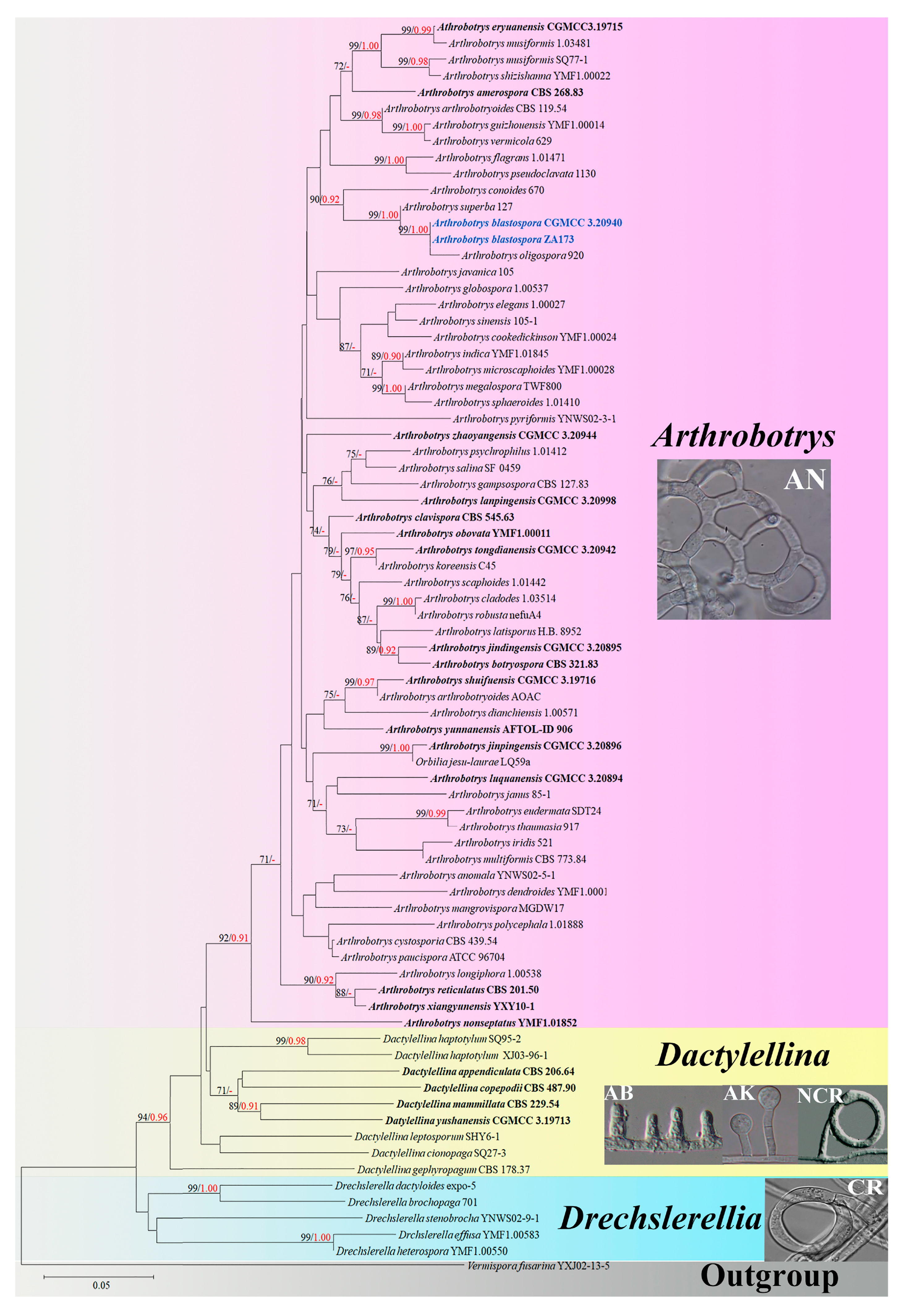
3.1. Phylogenetic Analysis
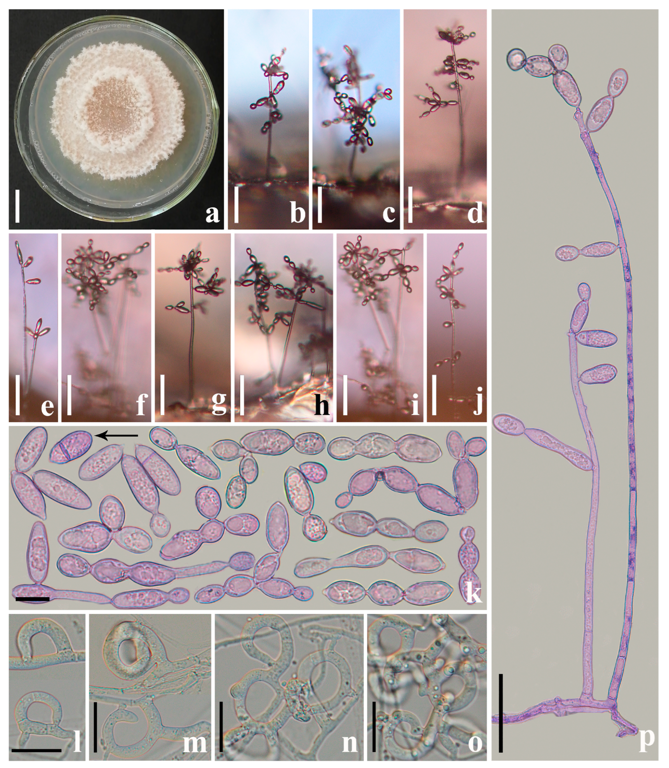
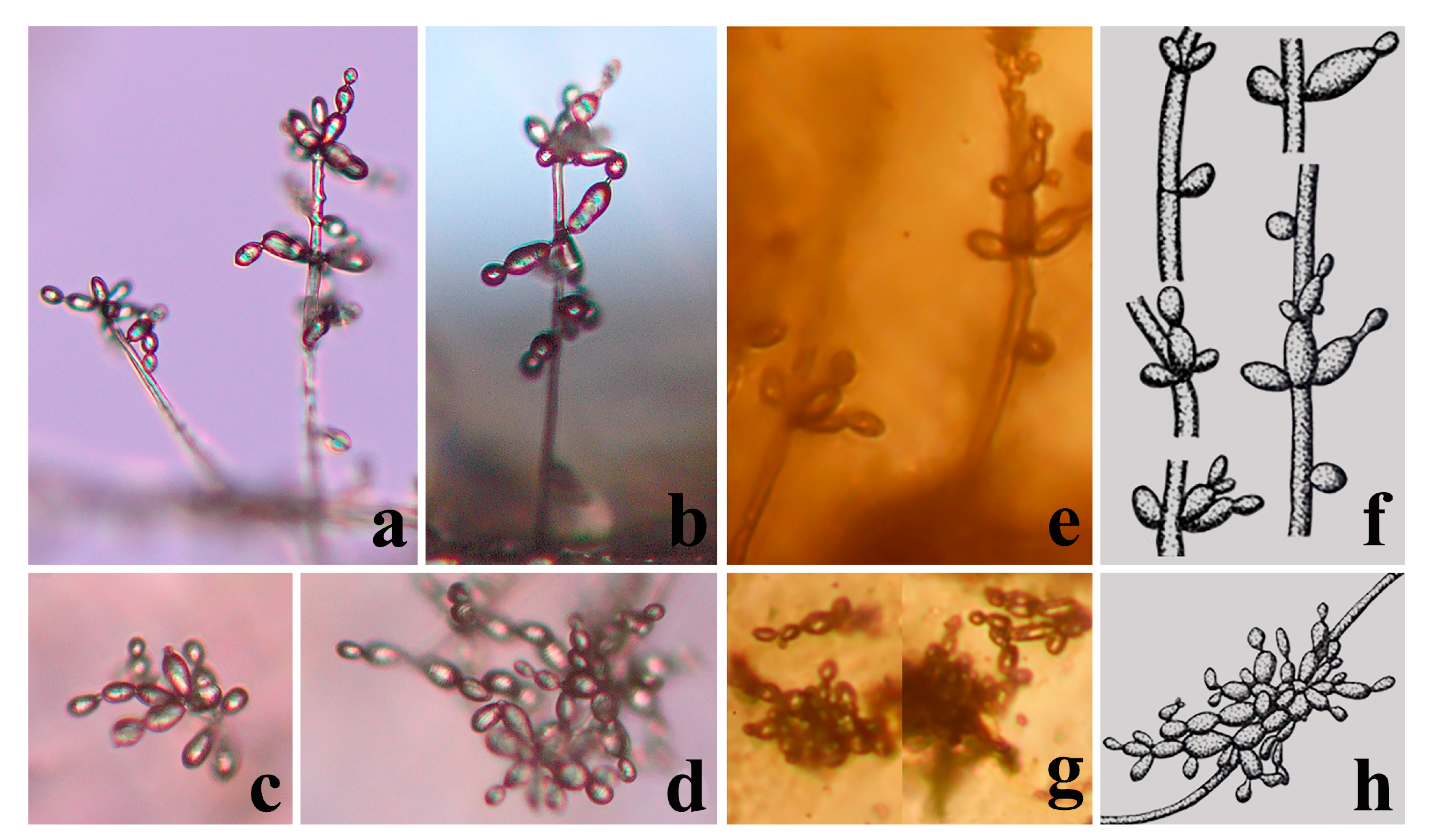
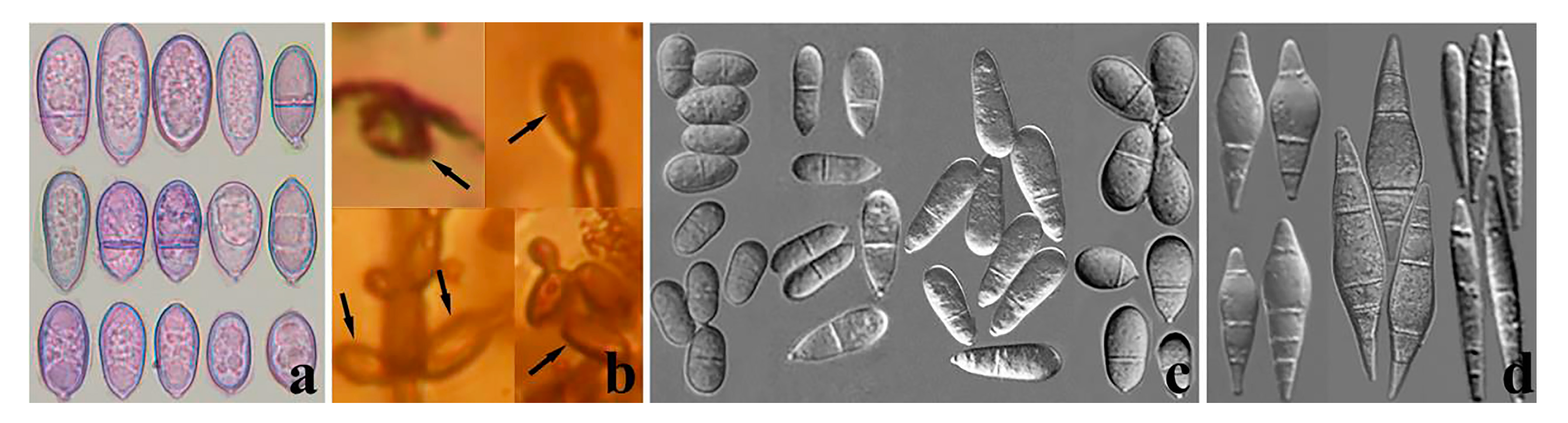
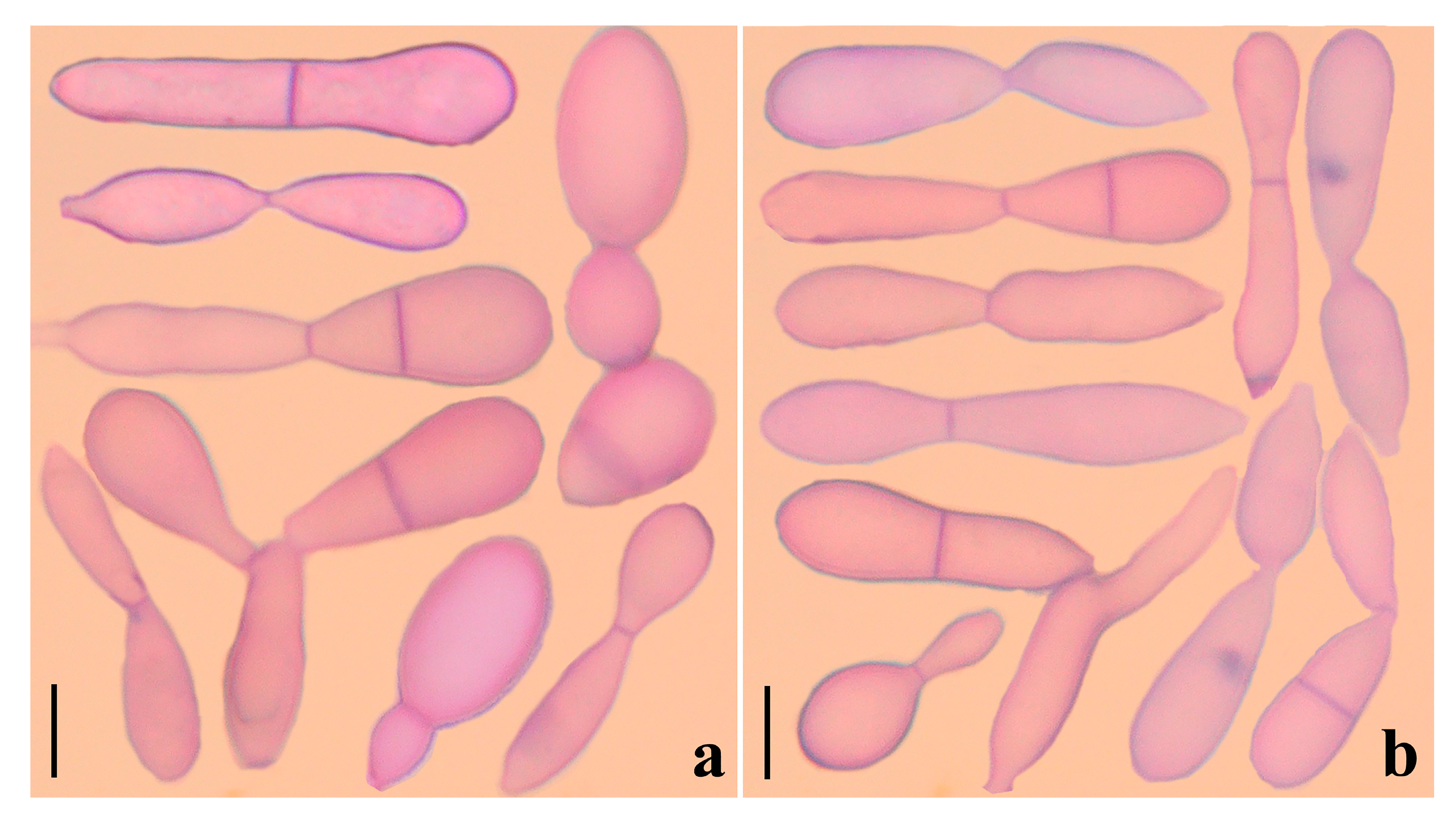
Both new fungal isolates were placed in the Arthrobotrys (Orbiliaceae, Orbiliomycetes) genus according to their type of trapping structure [11,12,13] and the BLASTn search results of ITS, TEF, and RPB2 genes. Therefore, all Arthrobotrys species with valid sequence data (62 isolates representing 59 species) [32] and other related taxa in Orbiliomycetes (9 isolates representing 8 Dactylellina species and 5 isolates representing 5 Drechslerella species) were included in this phylogenetic analysis (Table 1). The final dataset contained 77 ITS, 51 TEF, and 54 RPB2 sequences. The combined DNA dataset comprised 1909 characters (570 for ITS, 531 for TEF, and 708 for RPB2), among which 858 bp are constant, 982 bp are variable, and 770 bp are parsimony informative. After maximum likelihood (ML) analysis, the best-scoring likelihood tree was obtained with a final ML optimization likelihood value of -6867.586931. The Bayesian analysis (BI) evaluated the Bayesian posterior probabilities with a final average standard deviation of the split frequency of 0.009098. The trees generated by maximum likelihood (ML) and Bayesian analysis (BI) showed similar topologies, so the best-scoring ML tree was selected for presentation (Figure 1).
The phylogenetic analysis showed that the tested 72 Orbiliaceae (Orbiliomycetes) carnivorous species were clustered into three clades according to their types of trapping structure. All species catch nematodes with adhesive networks clustered together stably. Both new fungal isolates were placed in Arthrobotrys and formed a basal lineage with A. oligospora and A. superba with 99% MLBS and 1.00 BYPP (Figure 1).

3.2. Taxonomy

Arthrobotrys blastospora F. Zhang and X.Y. Yang sp. nov. (Figure 2, Figure 3a, Figure 4b and Figure 5a).
Index Fungorum number: IF900162; Facesoffungi number: FoF 14034
Etymology: The species name “blastospora” refers to the most prominent feature of this species, i.e., the production of blastospores.
Materials examined: CHINA, Yunnan Province, Nujiang City, Nujiang River, N 27°43′14.60″, E 98°41′30.20″, from freshwater sediment, 18 May 2014, F. Zhang. Holotype CGMC 3.20940, preserved in the China General Microbiological Culture Collection Center. Ex-type culture DLUCC 27-1, preserved in the Dali University Culture Collection.
Colonies white, cottony, and rapidly growing on the PDA medium, reaching 50 mm diam after 7 days at 26 °C. Mycelium 2.5–6 µm wide, hyaline, septate, branched, and smooth. Conidiophores 110–625 µm ( x = 341.5 µm, n = 100) long, 4–6.5 µm ( x = 5 µm, n = 100) wide at the base, gradually tapering upwards to a width of 3–6 µm ( x = 4 µm, n = 100) at the apex, hyaline, erect, septate, unbranched, produced by hyphae or directly by spore germination. This species produces hyaline, yeast-like blastospores, which usually cluster on the tuberculous bulges on the upper half of the conidiophores. A conidium is produced on the conidiophore first; then, a second conidium is formed from the right apex, or occasionally the side apex, of the first conidium, thus continuously producing a chain of blastospores. There is a septum between the two conidia, which tend to separate from each other after maturation. The exfoliated blastospores 13.5 − 62.5 × 7.5 − 16 ( x = 25.6 × 10.5 μm, n = 300) µm, globose, and elliptic to long elliptic, with zero or one septum. Chlamydospores not observed. Capturing nematodes with the adhesive networks in the early stages of its formation usually consists of a single adhesive hypha ring (Figure 2) [12].
Additional specimens examined: CHINA, Yunnan Province, Nujiang City, Nujiang River, N 27°24′33.20″, E 98°49′34.70″, from freshwater sediment, 20 May 2014, F. Zhang. Living culture ZA173.

4. Discussion

4.1. New Species of Arthrobotrys

Arthrobotrys blastospora catches nematodes with adhesive networks, which is consistent with the main characteristics of Arthrobotrys, Orbiliaceae (the largest genus of modern carnivorous fungi) [11,12,13,25]. Our phylogenetic analysis based on ITS, TEF, and RPB2 substantiated that A. blastospora is a member of Arthrobotrys. Phylogenetically, A. blastospora forms the sister lineage to A.oligospora (Figure 1). However, the conidia of all modern carnivorous fungi in Orbiliomycetes are individually born in clusters or singly on the conidiophores, which is significantly different from the catenulate blastospores produced by A. blastospora [12,31]. Therefore, we identified A. blastospora as a new species of Arthrobotrys.

4.2. Palaeoanellus Dimorphus Is an Ancient Ancestor of Modern Orbiliomycetes Carnivorous Fungi

Similar to carnivorous plants, as a highly specialized group in the fungal kingdom, carnivorous fungi are a model for the study of adaptive fungal evolution; their evolution is also a critical node in the study of fungal evolution. Such studies rely heavily on the discovery of fossil fungi. Schmidt et al. [23,24] found the earliest and best-preserved fossil of carnivorous fungi (P. dimorphus) in approximately 100-million-year-old amber from Southwestern France. P. dimorphus produced blastospores which were generated in whorls on small projections of conidiophores and also produced unicellular adhesive hyphal rings to trap nematodes (Figure 3 and Figure 4) [23,24]. However, no structure directly connecting the blastospores to unicellular adhesive hyphal rings was illustrated by Schmidt et al. [23,24]. Therefore, whether the blastospores and unicellular adhesive hyphal rings in this fossil actually represent a single fossil species has been a controversial topic due to their unusual combination [53,54]. In addition, the fossil was not considered an ancestor of modern carnivorous fungi but as belonging to an extinct lineage because no blastospore-producing carnivorous fungi were found in modern ecosystems [12,23,24,25]. A. blastospora reported in this study produced yeast-like blastospores on the small projections of conidiophores and captured nematodes with adhesive networks (a single adhesive hypha ring structure at the initial stage of its formation) (Figure 2) [31]. The discovery of this species confirms the existence of an extant species in nature that produces both specialized nematode-trapping structures and blastospores. Furthermore, A. blastospora and P. dimorphus share strong similarities with regard to the morphological characteristics of conidia, conidiophores, and the manner of catching nematodes with adhesive materials (Figure 3 and Figure 4). Accordingly, we infer that A. blastospora is closely related to P. dimorphus and maintained traits of Mesozoic carnivorous fungi, and P. dimorphus may be an ancient ancestor of modern Orbiliomycetes carnivorous fungi.
Similar to other fungi, convergent evolution has also been observed in carnivorous fungi; for example, some species of Dactylellina (Orbiliaceae, Orbiliomycetes, Ascomycota) and some species of Nematoctonus (Agaricomycetes, Pleurotaceae, Basidiomycota) trap nematodes with stalked adhesive knobs [55]. Therefore, we also cannot rule out the possibility of convergent evolution between A. blastospora and P. dimorphus, which resulted in the sharing of similar characteristics, while having a distant genetic relationship. However, given the scarcity of carnivorous fungi in the fungal kingdom [9,12] and the high similarity of several different structures (conidia, conidiophores, and trapping structures) between A. blastospora and P. dimorphus (Figure 3 and Figure 4) [23,24], we speculate that it is less likely that these two species would have evolved such similar traits with only a distant genetic relationship.

4.3. The Possible Ancestral Position of Palaeoanellus Dimorphus

Among Orbiliomycetes carnivorous fungi, all species are divided into two main groups according to their mechanisms of trapping nematodes. One is the genus Drechslerella, which first diverged from other carnivorous species and produces constricting rings to capture nematodes with the mechanical force generated by the expansion of the cells that make up the rings. Another contains all species in the genera Arthrobotrys and Dactylellina, which catch nematodes with adhesive traps (Figure 1) [11,12,13]. P. dimorphus produced unicellular hyphal rings, which possibly produced a sticky secretion used to capture nematodes [23,24]. This structure is similar to those species in Arthrobotrys and Dactylellina in the manner of trapping nematodes (capture of nematodes performed mainly with adhesive material), but it is quite different from the Drechslerella species, which capture nematodes by mechanical force. Therefore, we speculate that P. dimorphus is more related to Arthrobotrys and Dactylellina, and that P. dimorphus may be the common ancestor of Arthrobotrys, Dactylellina, or Arthrobotrys and Dactylellina.
Generally accepted, modern Orbiliomycetes carnivorous fungi originated from saprophytic fungi without a trapping structure [9,10,11,14,15,16,17]. Their evolution from possessing no trapping structure to complex trapping structures, such as modern adhesive trapping structures, was undeniably a course of gradual complexity. The structural complexity of unicellular adhesive hyphal rings produced by P. dimorphus is lower than that of most modern adhesive trapping structures. Therefore, unicellular adhesive hyphal rings may be considered an intermediate stage in the evolution of structural complexity and the common ancestor of all adhesive trapping structures (Arthrobotrys and Dactylellina). However, based on phylogenetic analysis of multiple genes and molecular clock theory, Yang et al. [9] inferred that the adhesive trapping structures of modern Orbiliomycetes carnivorous fungi originated about 246 million years ago and further evolved around 198–208 million years ago. In contrast, P. dimorphus was found in the amber from 100 million years ago [23,24]. Therefore, it can be inferred that the unicellular adhesive hyphal rings produced by P. dimorphus are probably not the ancestor of all the modern adhesive-trapping structures (Arthrobotrys and Dactylellina).
Phylogenetically, A. blastospora forms a sister lineage to A. oligospora and A. superba (Figure 1). Combined with the morphological similarities between A. blastospora and P. dimorphus, we can infer that P. dimorphus is closely related to the Arthrobotrys species. Concerning morphology, the following aspects can also support the close relationship between P. dimorphus and Arthrobotrys: (1) P. dimorphus produced unicellular adhesive hyphal rings to capture nematodes [23,24]. This structure is morphologically similar to the single-ring stage of adhesive networks produced by Arthrobotry species (Figure 4) [31]. (2) The formation of the unicellular adhesive hyphal rings produced by P. dimorphus initiated with a branch which was first generated on the vegetative mycelia, then the branch was curved and fused with the mycelia to form a ring [23,24]. This process is highly similar to the formation process of adhesive networks produced by Arthrobotrys species [31] (Figure 4). (3) The blastospores produced by A. blastospora are easily separated from each other to form non-septate and 1-septate elliptic conidia. The blastospores produced by P. dimorphus also had this characteristic. The non-septate or 1-septate conidia formed by the separation of blastospores are morphologically similar to those of many species in Arthrobotrys (Figure 5) [12,31]. (4) Among Arthrobotrys species, except A. blastospora, a few conidia of A. oligospora and A. conoides also have a similar morphology to blastospores (Figure 6), which suggests that the formation of blastospores may be an ancestral characteristic of Arthrobotrys, or an inherent feature of other Arthrobotrys species, but it is rarely developed or not developed in culture and thus, it has been overlooked so far. This phenomenon further illustrated the close relationship between P. dimorphus and Arthrobotrys.
By contrast, Dactylellina demonstrates similarities to P. dimorphus only in the aspect of trapping structure: a few species in Dactylellina produce a single ring covered with adhesive material (non-constricting ring) to capture nematodes, which is formed by producing a branch from the vegetative mycelia, and then the branch is curved and fused to form a ring [31]. This structure is similar to the unicellular adhesive hyphal rings produced by P. dimorphus in the morphology and formation process (Figure 4).
In summary, considering that P. dimorphus and Arthrobotrys share a reproductive structure (conidia) and nutritional structure (trapping structure) morphology, we speculate that P. dimorphus is more likely to be the ancestor of Arthrobotrys.

4.4. The Necessity of Strengthening the Research on Carnivorous Fungi in the Three Parallel Rivers

The two A. blastospora strains were isolated from the core area of the three parallel rivers region in China. This region is located in the southwest of the Heng Duan Mountains where mountains alternate with valleys, the terrain is highly diverse, and the region combines tropical, subtropical, temperate, and alpine cold climate types [56,57]. The complex terrain and climate create rich ecosystems and make the region one of the most biodiverse in the world [58,59]. Glaciers did not cover this region during the Quaternary glaciation due to its unique mountain and deep valley landform, particularly its geographical position, formation and evolutionary process. Therefore, this region is a significant refuge for many ancient species, and it is the center of species distribution and the differentiation of many biological groups [60,61,62,63,64]. According to statistics, 34 species of Chinese national protected plants, 600 species of endemic plants, and 20 species of relict plants are distributed in this region [65,66]. This situation renders it possible to find the relict species of carnivorous fungi in this region and suggests that there may be precious living fossils of other groups living in this region. In addition, P. dimorphus was found in the amber from Southwestern France [23,24] and A. blastospora was isolated from Southwestern China, more than 11,000 km from each other. This indicates that Palaeoanellus-type fungi were widely distributed and numerous in the past, giving rise to the extant genera of Oribiliomycetes.

Author Contributions

Methodology, F.Z.; software, F.Z. and Y.-Q.Y.; formal analysis, F.Z., S.B. and Y.-Q.Y.; investigation, F.Z. and F.-P.Z.; data curation, F.Z. and F.-P.Z.; writing—original draft preparation, F.Z.; writing—review and editing, F.Z., S.B., W.X. and X.-Y.Y.; supervision, X.-Y.Y.; project administration, X.-Y.Y.; funding acquisition, W.X. All authors have read and agreed to the published version of the manuscript.

Funding

This research was funded by the Second Tibetan Plateau Scientific Expedition and Research Program (STEP), grant number 2019QZKK0402; and the National Science Foundation Program-Yunnan union fund, grant number U1602262.

Institutional Review Board Statement

Not applicable.

Informed Consent Statement

Not applicable.

Data Availability Statement

The data that support the finding of this study are contained within the article.

Acknowledgments

We sincerely thank Alexander R. Schmidt from the University of Göttingen for their very helpful and supportive comments and suggestions. We thank Liu Xing-Zhong from Nankai University and Yu Ze-Fen from Yunnan University for all their help and suggestions during the writing process.

Conflicts of Interest

The authors declare no conflict of interest.

References

  1. Kennedy, D.; Norman, C. What Don’t We Know? Science 2005, 309, 75. [Google Scholar] [CrossRef] [PubMed] [Green Version]
  2. Gleason, F.H.; Lilje, O. Structure and function of fungal zoospores: Ecological implications. Fungal Ecol. 2009, 2, 53–59. [Google Scholar] [CrossRef]
  3. Grossart, H.-P.; Wyngaert, S.V.D.; Kagami, M.; Wurzbacher, C.; Cunliffe, M.; Rojas-Jimenez, K. Fungi in aquatic ecosystems. Nat. Rev. Genet. 2019, 17, 339–354. [Google Scholar] [CrossRef] [PubMed] [Green Version]
  4. Krauss, G.-J.; Solé, M.; Krauss, G.; Schlosser, D.; Wesenberg, D.; Bärlocher, F. Fungi in freshwaters: Ecology, physiology and biochemical potential. FEMS Microbiol. Rev. 2011, 35, 620–651. [Google Scholar] [CrossRef]
  5. Barron, G.L. Predators and parasites of microscopic animals. In Biology of Conidial Fungi; Cole, G.T., Kendrich, B., Eds.; ACADEMIC Press: New York, NY, USA, 2012; Volume 2, pp. 167–200. [Google Scholar]
  6. Barron, G.L. A new predatory hyphomycete capturing copepods. Can. J. Bot. 1990, 68, 691–696. [Google Scholar] [CrossRef]
  7. Liu, X.-Z.; Gao, R.-H.; Zhang, K.-Q.; Cao, L. Dactylella tenuifusaria sp. nov., a rhizopod-capturing hyphomycete. Mycol. Res. 1996, 100, 236–238. [Google Scholar] [CrossRef]
  8. Liu, X.; Xiang, M.; Che, Y. The living strategy of nematophagous fungi. Mycoscience 2009, 50, 20–25. [Google Scholar] [CrossRef]
  9. Yang, E.; Xu, L.; Yang, Y.; Zhang, X.; Xiang, M.; Wang, C.; An, Z.; Liu, X. Origin and evolution of carnivorism in the Ascomycota (fungi). Proc. Natl. Acad. Sci. USA 2012, 109, 10960–10965. [Google Scholar] [CrossRef] [Green Version]
  10. Li, T.F.; Zhang, K.Q.; Liu, X.Z. Taxonomy of Nematophagous Fungi; Chinese Scientific and Technological Publications: Beijing, China, 2000. [Google Scholar]
  11. Ahrén, D.; Ursing, B.M.; Tunlid, A. Phylogeny of nematode-trapping fungi based on 18S rDNA sequences. FEMS Microbiol. Lett. 1998, 158, 179–184. [Google Scholar] [CrossRef]
  12. Zhang, K.Q.; Hyde, K.D. Nematode-Trapping Fungi; Springer Science & Business: Berlin, Germany, 2014. [Google Scholar]
  13. Yang, Y.; Yang, E.; An, Z.; Liu, X.Z. Evolution of nematode-trapping cells of predatory fungi of the Orbiliaceae based on evidence from rRNA-encoding DNA and multiprotein sequences. Proc. Natl. Acad. Sci. USA 2007, 104, 8379–8384. [Google Scholar] [CrossRef] [Green Version]
  14. Li, Y.; Hyde, K.D.; Jeewon, R.; Cai, L.; Vijaykrishna, D.; Zhang, K. Phylogenetics and evolution of nematode-trapping fungi (Orbiliales) estimated from nuclear and protein coding genes. Mycologia 2005, 97, 1034–1046. [Google Scholar] [CrossRef] [PubMed]
  15. Liou, G.Y.; Tzean, S.S. Phylogeny of the genus Arthrobotrys and allied nematode-trapping fungi based on rDNA sequences. Mycologia 1997, 89, 876–884. [Google Scholar] [CrossRef]
  16. Rubner, A. Revision of predacious hyphomycetes in the Dactylella-Monacrosporium complex. Stud. Mycol. 1996, 39, 1–134. [Google Scholar]
  17. Scholler, M.; Hagedorn, G.; Runner, A. A reevaluation of predatory orbiliaceous fungi. II. A new generic concept. Sydowia-Horn 1999, 51, 89–113. [Google Scholar]
  18. Flynn, L.J. Origin and evolution of the Diatomyidae, with clues to paleoecology from the fossil record. Bull. Carnegie Mus. Nat. Hist. 2007, 39, 173–181. [Google Scholar] [CrossRef]
  19. Gingerich, P.D.; Chaline, J. Origin and evolution of species: Evidence from the fossil record. In Modalités, Rythmes, Mécanismes de L’Évolution Biologique: Gradualisme Phylétique ou Équilibres Ponctués? Chaline, J., Ed.; Colloques Internationaux du Centre National de la Recherche Scientifique: Grand Rapids, MI, USA, 1983; pp. 125–130. [Google Scholar]
  20. Kukalova-Peck, J. Origin and evolution of insect wings and their relation to metamorphosis, as documented by the fossil record. J. Morphol. 1978, 156, 53–125. [Google Scholar] [CrossRef]
  21. Thaler, L. Origin and Evolution of Mice: An Appraisal of Fossil Evidence and Morphological Traits. Poxviruses 1986, 127, 3–11. [Google Scholar] [CrossRef]
  22. Jansson, H.-B.; Poinar, G.O. Some possible fossil nematophagous fungi. Trans. Br. Mycol. Soc. 1986, 87, 471–474. [Google Scholar] [CrossRef]
  23. Schmidt, A.R.; Dorfelt, H.; Perrichot, V. Carnivorous Fungi from Cretaceous Amber. Science 2007, 318, 1743. [Google Scholar] [CrossRef] [Green Version]
  24. Schmidt, A.R.; Dörfelt, H.; Perrichot, V. Palaeoanellus dimorphus gen. et sp. nov. (Deuteromycotina): A Cretaceous predatory fungus. Am. J. Bot. 2008, 95, 1328–1334. [Google Scholar] [CrossRef] [Green Version]
  25. Swe, A.; Li, J.; Zhang, K.Q.; Pointing, S.B.; Jeewon, R.; Hyde, K.D. Nematode-trapping fungi. Curr. Res. Environ. Appl. Mycol. 2011, 1, 1–26. [Google Scholar]
  26. Duddington, C. Notes on the technique of handling predacious fungi. Trans. Br. Mycol. Soc. 1955, 38, 97–103. [Google Scholar] [CrossRef]
  27. White, T.J.; Bruns, T.; Lee, S.; Taylor, J. Amplification and direct sequencing of fungal ribosomal RNA genes for phylogenetics. PCR Protocols: A Guide to Methods and Applications; Academic Press: San Diego, CA, USA, 1990; Volume 18, pp. 315–322. [Google Scholar]
  28. O’Donnell, K.; Kistler, H.C.; Cigelnik, E.; Ploetz, R.C. Multiple evolutionary origins of the fungus causing Panama disease of banana: Concordant evidence from nuclear and mitochondrial gene genealogies. Proc. Natl. Acad. Sci. USA 1998, 95, 2044–2049. [Google Scholar] [CrossRef] [PubMed] [Green Version]
  29. Liu, Y.J.; Whelen, S.; Hall, B.D. Phylogenetic relationships among ascomycetes: Evidence from an RNA polymerse II subunit. Mol. Biol. Evol. 1999, 16, 1799–1808. [Google Scholar] [CrossRef] [Green Version]
  30. Swindell, S.R.; Plasterer, T.N. SEQMAN: Coting assembly. Meth. Mol. Biol. 1997, 70, 75–89. [Google Scholar] [CrossRef]
  31. Zhang, K.Q.; Mo, M.H. Arthrobotrys et gengra cetera cognata. In Flora Fungorum Sinicorum; Science Press: Beijing, China, 2006; Volume 33. [Google Scholar]
  32. Zhang, F.; Boonmee, S.; Bhat, J.D.; Xiao, W.; Yang, X.-Y. New Arthrobotrys Nematode-Trapping Species (Orbiliaceae) from Terrestrial Soils and Freshwater Sediments in China. J. Fungi 2022, 8, 671. [Google Scholar] [CrossRef]
  33. Katoh, K.; Standley, D.M. MAFFT Multiple Sequence Alignment Software Version 7: Improvements in Performance and Usability. Mol. Biol. Evol. 2013, 30, 772–780. Available online: http://mafft.cbrc.jp/alignment/server (accessed on 2 April 2022). [CrossRef] [Green Version]
  34. Hall, T.A. BioEdit: A user-friendly biological sequence alignment editor and analysis program for Windows 95/98/NT. Nucleic Acids Symp. Ser. 1999, 41, 95–98. [Google Scholar]
  35. Tamura, K.; Stecher, G.; Peterson, D.; Filipski, A.; Kumar, S. MEGA6: Molecular Evolutionary Genetics Analysis Version 6.0. Mol. Biol. Evol. 2013, 30, 2725–2729. [Google Scholar] [CrossRef] [Green Version]
  36. Vu, D.; Groenewald, M.; De Vries, M.; Gehrmann, T.; Stielow, B.; Eberhardt, U.; Al-Hatmi, A.; Groenewald, J.Z.; Cardinali, G.; Houbraken, J.; et al. Large-scale generation and analysis of filamentous fungal DNA barcodes boosts coverage for kingdom fungi and reveals thresholds for fungal species and higher taxon delimitation. Stud. Mycol. 2019, 92, 135–154. [Google Scholar] [CrossRef]
  37. Rezakhani, F.; Khodaparast, S.A.; Masigol, H.; Roja-Jimenez, K.; Grossart, H.-P.; Bakhshi, M. A preliminary report of aquatic hyphomycetes isolated from Anzali lagoon (Gilan province, North of Iran). Rostaniha 2019, 20, 123–143. [Google Scholar] [CrossRef]
  38. Li, J.; Liu, Y.; Zhu, H.; Zhang, K.-Q. Phylogenic analysis of adhesion related genes Mad1 revealed a positive selection for the evolution of trapping devices of nematode-trapping fungi. Sci. Rep. 2016, 6, 22609. [Google Scholar] [CrossRef] [PubMed] [Green Version]
  39. Zhang, X.; Zhang, F.; Jiang, L.; Yang, Y.-Q.; Yang, X.-Y.; Xiao, W. Two new nematode-trapping fungi (Arthrobotrys, Orbiliaceae) from Yunnan, China. Phytotaxa 2022, 568, 255–266. [Google Scholar] [CrossRef]
  40. Wu, H.Y.; Kim, D.G.; Ryu, Y.H.; Zhou, X.B. Arthrobotrys koreensis, a new nematode-trapping species from Korea. Sydowia 2012, 64, 129–136. [Google Scholar]
  41. Swe, A.; Jeewon, R.; Pointing, S.B.; Hyde, K.D. Taxonomy and molecular phylogeny of Arthrobotrys mangrovispora, a new marine nematode-trapping fungal species. Bot. Mar. 2008, 51, 331–338. [Google Scholar] [CrossRef]
  42. Li, S.; Yu, Z.; Zhang, Y.; Qiao, M.; Guo, J.; Zhang, K. Arthrobotrys nonseptata, a new anamorph from an Orbilia species. Mycotaxon 2009, 109, 247–254. [Google Scholar] [CrossRef]
  43. Liu, S.; Su, H.; Su, X.; Zhang, F.; Liao, G.; Yang, X. Arthrobotrys xiangyunensis, a novel nematode-trapping taxon from a hot-spring in Yunnan Province, China. Phytotaxa 2014, 174, 89–96. [Google Scholar] [CrossRef]
  44. Mo, M.; Huang, X.; Zhou, W.; Huang, Y.; Hao, Y.E.; Zhang, K. Arthrobotrys yunnanensis sp. nov., the fourth anamorph of Orbilia auricolor. Fungal Divers. 2005, 18, 107–115. [Google Scholar]
  45. Zhang, F.; Zhou, X.-J.; Monkai, J.; Li, F.-T.; Liu, S.-R.; Yang, X.-Y.; Wen, X.; Hyde, K.D. Two new species of nematode-trapping fungi (Dactylellina, Orbiliaceae) from burned forest in Yunnan, China. Phytotaxa 2020, 452, 65–74. [Google Scholar] [CrossRef]
  46. Quijada, L.; Baral, H.-O.; Beltrán-Tejera, E.; Pfister, D.H. Orbilia jesu-laurae (Ascomycota, Orbiliomycetes), a new species of neotropical nematode-trapping fungus from Puerto Rico, supported by morphology and molecular phylogenetics. Willdenowia 2020, 50, 241–251. [Google Scholar] [CrossRef]
  47. Posada, D. jModelTest: Phylogenetic Model Averaging. Mol. Biol. Evol. 2008, 25, 1253–1256. [Google Scholar] [CrossRef] [PubMed]
  48. Nguyen, L.-T.; Schmidt, H.A.; Von Haeseler, A.; Minh, B.Q. IQ-TREE: A Fast and Effective Stochastic Algorithm for Estimating Maximum-Likelihood Phylogenies. Mol. Biol. Evol. 2015, 32, 268–274. [Google Scholar] [CrossRef] [PubMed]
  49. Felsenstein, J. Confidence limits on phylogenies: An approach using the bootstrap. Evolution 1985, 39, 783–791. [Google Scholar] [CrossRef] [PubMed]
  50. Huelsenbeck, J.P.; Ronquist, F. MRBAYES: Bayesian inference of phylogenetic trees. Bioinformatics 2001, 17, 754–755. [Google Scholar] [CrossRef] [Green Version]
  51. Hall, B.G. Phylogenetic Trees Made Easy: A How-to Manual; Sinauer Associates Sunderland: Sunderland, MA, USA, 2007. [Google Scholar]
  52. Rambaut, A. FigTree v1.3.1. 2010. Available online: http://tree.bio.ed.ac.uk/software/figtree/ (accessed on 6 June 2013).
  53. Thorn, R.G.; Scholler, M.; Gams, W. Mycological Research News. Mycol. Res. 2008, 112, 611–612. [Google Scholar] [CrossRef]
  54. Schmidt, A.R.; Dörfelt, H.; Perrichot, V. Palaeomycology: Do we consider extinct lineages in the evaluation of fossil fungi? Mycol. Res. 2009, 113, 275–276. [Google Scholar]
  55. Nordbring-Hertz, B.; Jansson, H.; Tunlid, A. Nematophagous Fungi. eLS 2006, 1, 1–11. [Google Scholar] [CrossRef]
  56. Feng, B.; Zhao, Q.; Xu, J.; Qin, J.; Yang, Z.L. Drainage isolation and climate change-driven population expansion shape the genetic structures of Tuber indicum complex in the Hengduan Mountains region. Sci. Rep. 2016, 6, 21811. [Google Scholar] [CrossRef] [Green Version]
  57. Li, R.S.; Liu, W.; Chen, H. Three Parallel Rivers; China Translation and Publication Corporation: Shenzhen, China, 2001; pp. 44–67. [Google Scholar]
  58. Basnet, D.; Kandel, P.; Chettri, N.; Yang, Y.; Lodhi, M.S.; Htun, N.Z.; Uddin, K.; Sharma, E. Biodiversity Research Trends and Gaps from the Confluence of Three Global Biodiversity Hotspots in the Far-Eastern Himalaya. Int. J. Ecol. 2019, 2019, 1323419. [Google Scholar] [CrossRef] [Green Version]
  59. Lin, S.; Wu, R.; Yang, F.; Wang, J.; Wu, W. Spatial trade-offs and synergies among ecosystem services within a global biodiversity hotspot. Ecol. Indic. 2018, 84, 371–381. [Google Scholar] [CrossRef]
  60. Li, J.W. A review of plant diversity and characteristics on three parallel rivers. Sci. Technol. Inf. 2009, 15, 59–64. [Google Scholar]
  61. Sun, H. Evolution of Arctic-Tertiary fora in Himalayan-Hengduan Mountains. Acta Bot. Yunnanica 2002, 24, 671–688. [Google Scholar]
  62. Sun, H.; Li, Z.M. Qinghai-Tibet Plateau uplift and its impact on Tethys fora. Adv. Earth Sci. 2003, 18, 852–862. [Google Scholar] [CrossRef]
  63. Wang, J.X.; Peng, X.F.; Yang, S.Y. Analysis of tourism resources and environmental vulnerability in three Parallel Rivers World Natural Heritage Site. J. Yunnan Norm. Univ. Nat. Sci. 2005, 25, 59–64. [Google Scholar]
  64. Zhang, D.C.; Zhang, Y.H.; Boufford, D.E.; Sun, H. Elevational patterns of species richness and endemism for some important taxa in the Hengduan Mountains, southwestern China. Biodivers. Conserv. 2009, 18, 699–716. [Google Scholar] [CrossRef]
  65. Fu, G.L. List of Rare and endangered Plants in China. Information System of Chinese Rare and Endangered Plant ISCREP. 1987. Available online: http://www.iplant.cn/rep/news/32 (accessed on 14 November 2022).
  66. Zhou, B. Revising the Yunnan Key Protected Wild Plants List. Acta Bot. Yunnanica 2010, 32, 221–226. [Google Scholar] [CrossRef]
Figure 1. Maximum likelihood tree based on combined ITS, TEF, and RPB2 sequence data from 72 Orbiliaceae (Orbiliomycetes) carnivorous species. Bootstrap support values for maximum likelihoods (black) equal to or greater than 70% and Bayesian posterior probability values (red) equal to or greater than 0.90 are indicated above the nodes. The new isolates are in blue and the type strains are in bold. The genus name and trapping structure corresponding to each clade are indicated on the right. AN, adhesive networks; AB, adhesive branches; AK, adhesive knobs; NCR, non-constricting rings; CR, constricting rings.
Jof 09 00451 g001
Figure 2. Arthrobotrys blastospora (CGMCC 3.20940). (a) Colony; (bj) Conidiophores and blastospores; (k) Blastospores; (lo) Trapping structure: adhesive networks; (p) Conidiophores. Scale bars: (a) = 1 cm; (bj) = 10 μm; (ko) = 20 μm; (p) = 100 μm.
Jof 09 00451 g002
Figure 3. Blastospores and conidiophores of fossil and extant carnivorous fungi. (a,b) Conidiophores of Arthrobotrys blastospora; (c,d) Blastospores of Arthrobotrys blastospora; (e,f) Conidiophores of the fossil Palaeoanellus dimorphus; (g,h) Blastospores of the fossil Palaeoanellus dimorphus, reprinted with permission from Ref. [24] 2023, John Wiley and Sons.
Jof 09 00451 g003
Figure 4. Trapping structure of fossil and extant carnivorous fungi. (a,b) Trapping structure of Palaeoanellus dimorphus: unicellular adhesive hyphal rings, reprinted with permission from Ref. [24]. 2023, John Wiley and Sons; (c,d) The early stages of adhesive networks produced by Arthrobotrys blastospora: single adhesive hyphal rings; (e,f) Trapping structure of some Dactylellina species: non-constricting rings [12].
Jof 09 00451 g004
Figure 5. Conidia of several carnivorous fungi. (a) The single conidia formed by the separation of the blastospore of Arthrobotrys blastospora; (b) The single conidia produced by Palaeoanellus dimorphus, reprinted with permission from Ref. [24]. 2023, John Wiley and Sons; (c) The conidia produced by some Arthrobotrys species [31]; (d) The conidia produced by most of the Dactylellina species [31].
Jof 09 00451 g005
Figure 6. Blastospores of some Arthrobotrys species. (a) The blastospores produced by A. oligospora; (b) The blastospores produced by A. conoides. Scale bars = 10 μm.
Jof 09 00451 g006
Table 1. The GenBank accession numbers of the isolates included in this study. The ex-type strains are in bold. The newly generated sequences are indicated in blue.
Taxon Strain NumberGenBank Accession NumberReference
ITSTEFRPB2
Arthrobotrys amerosporaCBS 268.83NR_159625[36]
Arthrobotrys anomalaYNWS02-5-1AY773451AY773393AY773422[13]
Arthrobotrys arthrobotryoidesCBS 119.54MH857262[36]
Arthrobotrys arthrobotryoidesAOACMF926580Unpublished
Arthrobotrys blastosporaCGMCC 3.20940OQ332405OQ341651OQ341649This study
Arthrobotrys blastosporaZA173OM956088OQ341650OQ341648This study
Arthrobotrys botryosporaCBS 321.83NR_159626[36]
Arthrobotrys cladodes1.03514MH179793MH179616MH179893Unpublished
Arthrobotrys clavisporaCBS 545.63MH858353[36]
Arthrobotrys conoides670AY773455AY773397AY773426[13]
Arthrobotrys cookedickinsonYMF1.00024MF948393MF948550MF948474[12]
Arthrobotrys cystosporiaCBS 439.54MH857384[36]
Arthrobotrys dendroidesYMF1.00010MF948388MF948545MF948469[12]
Arthrobotrys dianchiensis1.00571MH179720MH179826[37]
Arthrobotrys elegans1.00027MH179688MH179797Unpublished
Arthrobotrys eryuanensisCGMCC3.19715MT612105OM850307OM850301[32]
Arthrobotrys eudermataSDT24AY773465AY773407AY773436[13]
Arthrobotrys flagrans1.01471MH179741MH179583MH179845Unpublished
Arthrobotrys gampsosporaCBS 127.83U51960[15]
Arthrobotrys globospora1.00537MH179706MH179562MH179814Unpublished
Arthrobotrys guizhouensisYMF1.00014MF948390MF948547MF948471[12]
Arthrobotrys indicaYMF1.01845 KT932086[38]
Arthrobotrys iridis521AY773452AY773394AY773423[13]
Arthrobotrys janus85-1AY773459AY773401AY773430[13]
Arthrobotrys javanica105EU977514Unpublished
Arthrobotrys jindingensisCGMCC 3.20985OP236810OP272511OP272515[39]
Arthrobotrys jinpingensisCGMCC 3.20896OM855569OM850311OM850305[32]
Arthrobotrys koreensisC45JF304780[40]
Arthrobotrys lanpingensisCGMCC3.20998OM855566OM850308OM850302[32]
Arthrobotrys latisporaH.B. 8952MK493125Unpublished
Arthrobotrys longiphora1.00538MH179707MH179815Unpublished
Arthrobotrys luquanensisCGMCC3.20894OM855567OM850309OM850303[32]
Arthrobotrys mangrovisporaMGDW17EU573354[41]
Arthrobotrys megalosporaTWF800MN013995Unpublished
Arthrobotrys microscaphoidesYMF1.00028MF948395MF948552MF948476[12]
Arthrobotrys multiformisCBS 773.84MH861834[36]
Arthrobotrys musiformisSQ77-1AY773469AY773411AY773440[13]
Arthrobotrys musiformis1.03481MH179783MH179607MH179883Unpublished
Arthrobotrys nonseptataYMF1.01852FJ185261[42]
Arthrobotrys obovataYMF1.00011MF948389MF948546MF948470[12]
Arthrobotrys oligospora920AY773462AY773404AY773433[13]
Arthrobotrys paucisporaATCC 96704EF445991[13]
Arthrobotrys polycephala1.01888MH179760MH179592MH179862Unpublished
Arthrobotrys pseudoclavata1130AY773446AY773388AY773417[13]
Arthrobotrys psychrophila1.01412MH179727MH179578MH179832Unpublished
Arthrobotrys pyriformisYNWS02-3-1AY773450AY773392AY773421[13]
Arthrobotrys reticulataCBS 201.50MH856589.1[36]
Arthrobotrys robustanefuA4MZ326655Unpublished
Arthrobotrys salinaSF 0459KP036623Unpublished
Arthrobotrys scaphoides1.01442MH179732MH179580MH179836Unpublished
Arthrobotrys shizishannaYMF1.00022MF948392MF948549MF948473[12]
Arthrobotrys shuifuensisCGMCC3.19716MT612334OM850306OM850300[32]
Arthrobotrys sinensis105-1AY773445AY773387AY773416[13]
Arthrobotrys sphaeroides1.01410MH179726MH179577MH179831Unpublished
Arthrobotrys superba127EU977558Unpublished
Arthrobotrys thaumasia917AY773461AY773403AY773432[13]
Arthrobotrys tongdianensisCGMCC 3.20942OP236809OP272509OP272513[39]
Arthrobotrys vermicola629AY773454AY773396AY773425[13]
Arthrobotrys xiangyunensisYXY10-1MK537299[43]
Arthrobotrys yunnanensisAFTOL-ID 906DQ491512[44]
Arthrobotrys zhaoyangensisCGMCC3.20944OM855568OM850310OM850304[32]
Dactylellina appendiculataCBS 206.64AF106531DQ358227DQ358229[36]
DatylellinacionopogumSQ27-3AY773467AY773409AY773438[13]
Dactylellina copepodiiCBS 487.90U51964DQ999835DQ999816[13]
DatylellinagephyropagumCBS178.37U51974DQ999847DQ999802[13]
Datylellina haptotylumSQ95-2AY773470AY773412AY773441[13]
Datylellina haptotylumXJ03-96-1DQ999827DQ999849DQ999804[13]
Dactylellina leptosporumSHY6-1AY773466AY773408AY773437[13]
Dactylellina mammillataCBS229.54AY902794DQ999843DQ999817[13]
Dactylellina yushanensisCGMCC3.19713MK372061MN915113MN915112[45]
Drechslerella brochopaga701AY773456AY773398AY773427[13]
Drechslerella dactyloidesexpo-5AY773463AY773405AY773434[13]
Drechslerella effusaYMF1.00583MF948405MF948557MF948484Unpublished
Drechslerella heterosporaYMF1.00550MF948400MF948554MF948480Unpublished
Drechslerella stenobrochaYNWS02-9-1AY773460AY773402AY773431[13]
Orbilia jesu-lauraeLQ59aMN816816[46]
Vermispora fusarinaYXJ02-13-5AY773447AY773389AY773418[13]
Disclaimer/Publisher’s Note: The statements, opinions and data contained in all publications are solely those of the individual author(s) and contributor(s) and not of MDPI and/or the editor(s). MDPI and/or the editor(s) disclaim responsibility for any injury to people or property resulting from any ideas, methods, instructions or products referred to in the content.

