Article

Lignin Promotes Mycelial Growth and Accumulation of Polyphenols and Ergosterol in Lentinula edodes

1
College of Food Science and Nutritional Engineering, China Agricultural University, Beijing 100083, China
2
Academy of National Food and Strategic Reserves Administration, Beijing 100037, China
*
Author to whom correspondence should be addressed.
J. Fungi 2023, 9(2), 237; https://doi.org/10.3390/jof9020237
Submission received: 31 December 2022 / Revised: 31 January 2023 / Accepted: 6 February 2023 / Published: 10 February 2023

Abstract

:
It has been demonstrated that lignin was efficiently degraded by Lentinula edodes (L. edodes). However, the process of lignin degradation and utilization by L. edodes has not been discussed in detail. Therefore, the effects of lignin on L. edodes mycelium growth, chemical compositions, and phenolic profiles were investigated herein. It has been revealed that 0.10% lignin acted as the most effective concentration to accelerate mycelia growth, which yielded the highest biomass of 5.32 ± 0.07 g/L. Furthermore, a 0.10% concentration of lignin promoted the accumulation of phenolic compounds, especially protocatechuic acid, with peak value of 48.5 ± 1.2 μg/g. In contrast, the higher concentration of lignin (0.20%) exerted an inhibitory effect on the growth of L. edodes. Overall, the application of lignin at the optimal concentration of 0.10% could not only enhance the mycelial growth but also accumulate the phenolic acids and raise the nutritional and medical values of L. edodes.

1. Introduction

Lignin is a high-branched polymer primarily composed of three phenyl propane monomers, namely coniferyl alcohol (G), sinapyl alcohol (S), and coumaryl alcohol (H) via both aryl-ether and alkyl linkages [1]. As the most abundant aromatic compound in nature, lignin has been widely recognized as a prospective bioresource for various chemicals as well as biofuels [2,3]. In recent years, the targeted conversion and high-value utilization of lignin by microbiological approaches have attracted widespread attention at home and abroad. Microorganisms such as fungi and bacteria (actinomycetes) are capable of degrading lignin to varying degrees, with white-rot basidiomycetous fungus being most efficient and being able to completely degrade lignin to water and carbon dioxide by its extracellular ligninolytic enzymes [4]. Previously, a wide array of ligninolytic enzymes secreted by white-rot fungi, including laccase, lignin peroxidase (LiP), manganese peroxidase (MnP), versatile peroxidases (VP), catalase, as well as other oxidative enzymes have been characterized [5,6]. The molecular mechanisms of lignin degradation by white-rot fungi have also been studied. In brief, the biodegradation of lignin started from the formation of alkyl propane monomers, followed by the breakdown and ring cleavage of heterogeneous aromatics. The degradation products are generally phenolic compounds, including protocatechuic acid, vanillin, p-coumaric acid, ferulic acid, and gallic acid. Moreover, it was reported that specificity biosynthesis and bioconversion of lignin had been achieved through fungi (Saccharomyces cerevisiae, Phanerochaete chrysosporium, Cutaneotrichosporon yeast) and bacteria (Escherichia coli, Pseudomonas putida, Sphingobium sp. SYK-6, Rhodococcus opacus) [7,8,9,10,11]. These investigations aroused our in-depth exploration of whether the exogenous addition of lignin could promote the accumulation of phenolic compounds in the microbial mycelium.
In recent years, the multi-pharmacological effects of Lentinula edodes (L. edodes), including antioxidative, immunoregulatory, and anticancer properties, have been well documented, which were associated with their bioactive components, such as polysaccharides, phenolic compounds, ergosterols, and terpenoids [12,13]. L. edodes is one of the most common edible mushrooms all over the world and has been widely cultivated and consumed particularly in the Asian countries, owing to its high nutritional value and unique flavor [14,15]. As the typical white-rot fungi, L. edodes commonly grows on sawdust or wood logs by degrading lignocellulose in nature, whose three major components are cellulose, hemicellulose, and lignin. Enzymes involved in lignocellulose degradation as well as their encoding gene clusters have been identified in L. edodes previously [16,17,18].
Currently, lignocellulose derived from different sources is taken as the alternative to sawdust and utilized in L. edodes cultivation for the purpose of forest resource protection and sustainable development [19,20]. The proportion of cellulose, hemicellulose, and lignin varied with the sources and types of lignocellulose [21]. L. edodes presented diverse growth characteristics on different lignocellulose culture substrates. This was attributed to the individual metabolic characteristic of L. edodes on the three components of lignocellulose. It revealed that L. edodes prefers hemicellulose over cellulose as the carbon source [16]. Moreover, our previous study has proved that hemicellulose stimulated mycelia biomass and polysaccharides biosynthesis [22]. Meanwhile, it was found that there was a synergistic relationship between lignin and hemicellulose in lignocellulose degradation in L. edodes [23].
To date, there are many investigations focusing on the biodegradation of lignin as well as the interrelation between lignin, microorganism, and enzymes. Nevertheless, as the precursor of phenolic acids, whether the addition of lignin will affect the metabolism, transformation, and enrichment of phenolic acids in L. edodes mycelia remains to be further explored. Moreover, the impact of lignin on mycelium growth is still unclear during liquid fermentation. To this end, our report aimed to reveal the impacts of lignin on the fugal growth, chemical compositions, as well as accumulation of phenolic compounds in L. edodes during liquid fermentation.

2. Materials and Methods

2.1. Strain and Materials

The L. edodes strain Qiuzai No.7 (Q-7) used in the present study was obtained from Hubei Yuguo Guye Co., Ltd. (Suizhou, China) and was preserved on potato dextrose agar (PDA) medium at 4 °C. Sodium lignosulfonate and xylo-oligosaccharide were purchased from Aladdin Biotechnology Co., Ltd. (Shanghai, China) and Shandong Longlive Bio-Technology Co., Ltd. (Dezhou, China), respectively [22].
Ergosterol as well as phenolic compound standards (gallic acid, protocatechuic acid, caffeic acid, syringic acid, vanillin, p-coumaric acid, and ferulic acid) were purchased from Sigma-Aldrich (St. Louis, MO, USA). Acetonitrile and methanol with a purity of 99.9% as the mobile phase were of high-performance liquid chromatography (HPLC) grade (Merck, Germany). All other chemicals and solvents were of analytical grade and purchased from common suppliers.

2.2. Culture Condition

The basic liquid medium for L. edodes growth was a modified potato dextrose broth (PDB) medium containing 2% (w/v) hemicellulose instead of glucose [22]. Lignin was added to the medium at the beginning of cultivation to provide a final concentration ranging from 0.05%, 0.10%, and 0.20% (w/v), respectively, while the media without lignin served as a control (CK). Prior to autoclaving, the culture media were acidified to pH 6.0 ± 0.1 with 1 M HCl. Experimental mycelia were collected after cultivation for 10 days in a shaking incubator (160 rpm) at 28 °C and 75% relative humidity (RH). RH in the laboratory was maintained at 50–70% constantly. The supernatants were analyzed for enzyme activity. All experiments were conducted in triplicates.

2.3. Determinations of Mycelium Biomass and Compositional Analysis

After 10 days fermentation, experimental mycelia were harvested by centrifugation (5000× g, 20 min) and then washed by deionized water sufficiently, followed by lyophilization. Mycelia yield was measured as follows:
y = M1/V1
in which y is the mycelia or biomass yield (g/L), M1 is the dry weight of mycelia (g), and V1 is the volume of medium (L).
The chemical compositions of cultured mycelia, including crude protein, total polysaccharide, ergosterol, and ash, were determined according to the AOAC methods (AOAC, 1995). Briefly, ash content was measured by carbonization and incineration in a muffle furnace at 550 °C until constant weight. Crude protein content (N × 4.38) was determined according to the micro-Kjeldahl method. Total water-soluble polysaccharides (WSP) were extracted with boiling water and then precipitated with anhydrous ethanol (1:4, v/v) at 4 °C overnight. After centrifugation at 5000× g for 30 min, precipitate was washed three times with five volumes of ethanol, followed by deproteinization with Sevag reagent (1-butanol/chloroform, v/v = 1:4) and lyophilization to obtain the crude polysaccharide. The yields of crude WSP extracts were recorded subsequently. The content of ergosterol was measured according to the method by Niemenmaa, Galkin, and Hatakka (2008), with slight modifications [24]. In short, mycelia were saponified by 10% KOH in methanol at 80 °C for 1 h. The supernatant was mixed with hexane and distilled water and then centrifuged at 6000× g for 15 min. The preceding step was repeated twice, whereafter the n-hexane phase was collected and evaporated under vacuum at 50 °C. Extracted ergosterol was redissolved with 500 μL of methanol (HPLC grade) and filtered through a 0.22 µm membrane. Extracted ergosterol was redissolved with 500 μL of methanol and filtered through a 0.22 µm membrane. Agilent 1260 Infinity HPLC system coupled with a diode array detector (DAD) and a C18 column (4.6 × 250 mm, 5 µm; Agilent, Santa Clara, CA, USA) was employed. A total of 20 μL of sample solution was injected and eluted with methanol at a constant flow rate of 1.0 mL/min at 30 °C.

2.4. Phenolic Compounds Extraction and Determination

The total phenolic was extracted by 80% methanol and measured according to the Folin–Ciocalteu colorimetric method with gallic acid as a standard [25]. The results were expressed as mg of gallic acid equivalents (GAE) per g of dried mycelia. For the investigation of the phenolic compounds profile, phenolic compound extracts were prepared as described by Gbylik-Sikorska et al. (2019) with slight modifications [26]. A total of 1 g lyophilized mycelia were dispersed in 10 mL of acetonitrile and 2 mL of hydrochloric acid. The mixtures were extracted for 30 min and immersed in a water bath with ultrasonic at 30 °C for 2 h. After centrifugation, the supernatant was collected and evaporated to dryness. Residues were redissolved in methanol and filtered through a 0.22 μm PVDF membrane for subsequent analysis.
The phenolic compound analysis was conducted on an Agilent 1260 Infinity HPLC system equipped with a C18 column (4.6 × 250 mm, 5 µm; Agilent, Santa Clara, CA, USA). A diode array detector (DAD) was carried out, using 250 nm, 280 nm, and 320 nm as the preferred wavelengths. A total of 20 μL of the sample was injected and eluted with mobile solvents consisting of 0.1% formic acid in water (A) and acetonitrile (B) at 30 °C. For efficient separation, a gradient elution procedure at a flow rate of 1 mL/min was used as follows:
5 % B   10   min 15 % B   10   min   25 % B   15   min   35 % B   5   min   80 % B   5   min   5 % B

2.5. Enzyme Assays

Mycelia (1.00 g) were homogenized with phosphate buffer solution (PBS) buffer (4 mL, 0.1 M, pH 7.4) in an ice bath. The crude enzyme solution was prepared by collecting the supernatant after centrifugation for 30 min at 10,000× g and 4 °C.
The CAT activity was determined according to the manufacturer’s instruction using a commercial detection kit (Nanjing Jiancheng Bioengineering Institute, China) and expressed as U/mg protein. Total protein content in crude extracts was measured by the Bradford method with bovine serum albumin (BSA) as a standard.
The laccase (Lcc, EC 1.10.3.2) activity was spectrophotometrically determined according to the oxidation rate of 2,2-azino-bis-[3-ethyltiazoline-6-sulfonate] (ABTS) in 20 mM Na-acetate buffer (pH 4.5) in the absorbance at 420 nm. One unit of Lcc activity was defined as the amount of enzyme able to oxidize 1 μmol of ABTS per minute at 20 °C as described by Edae and Alemu (2017) [27].
The Mn-peroxidases (MnP, EC 1.11.1.13) activity was assayed with phenol red as the substrate, and absorbance was recorded at 610 nm as described by Lechner and Papinutti (2006) [28]. Briefly, the reaction solution was mixed with manganese sulfate (0.1 mM), phenol red (0.1 mM), and succinate buffer (50 mM, pH 4.5). The reaction was initiated with the addition of H2O2 (0.1 mM at final concentration). One unit of MnP activity was defined as the amount of enzyme catalyzing 1 μmol of phenol red in one min.

2.6. Statistical Analysis

All experiments were conducted in triplicate, and values were expressed as mean ± standard deviation (SD). Statistical analysis was carried out by one-way ANOVA accompanied by Duncan’s test using SPSS (version 20.0). The significance was set at p < 0.05 and expressed by different letters above the error bars.

3. Results and Discussions

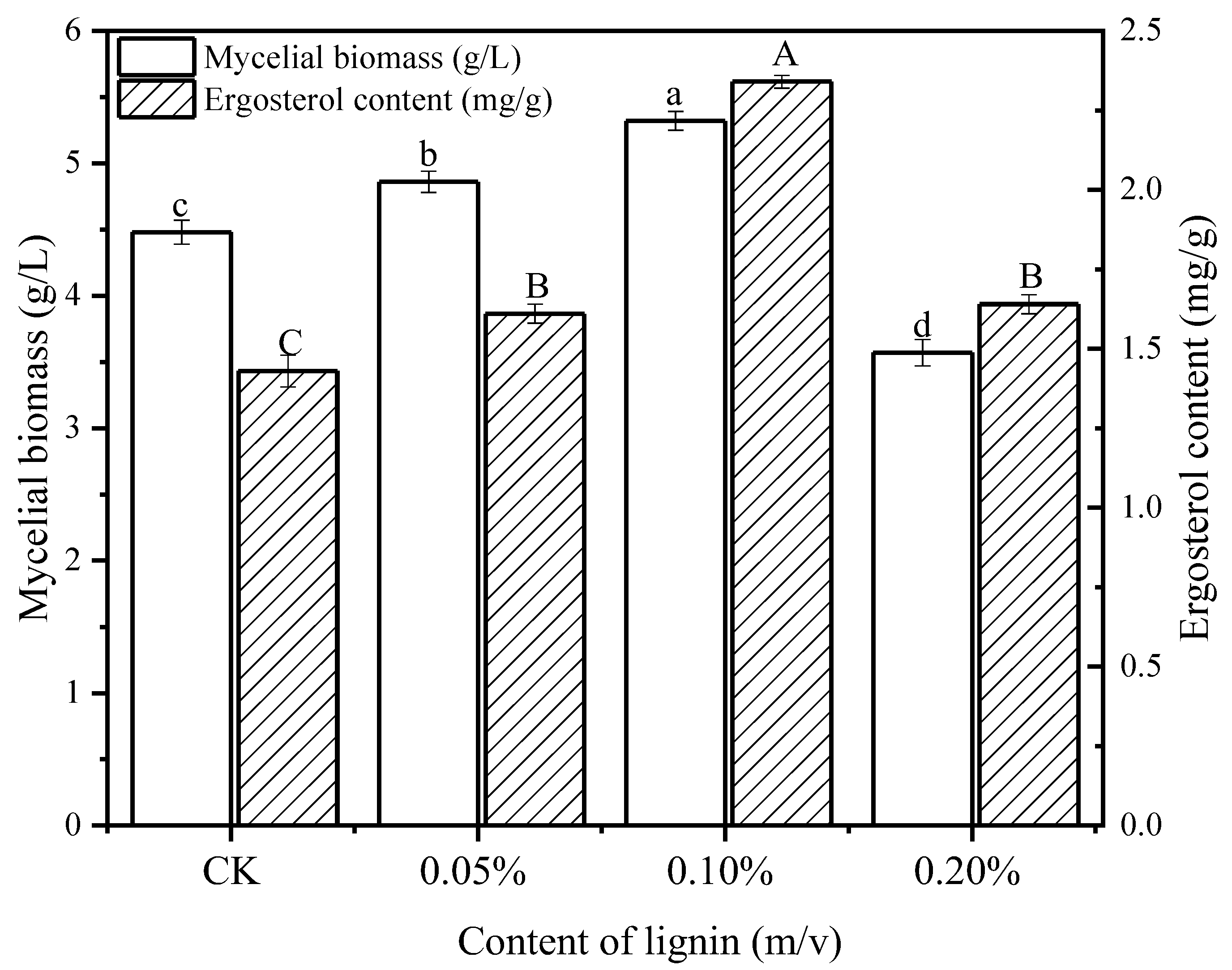
3.1. Effect of Lignin on Mycelia Growth

The effect of lignin on the mycelial growth of L. edodes was evaluated according to the harvested mycelial biomass, as shown in Figure 1. The yield of mycelial biomass refers to the dry weight (g) of mycelia produced per liter of culture medium. It suggested that the addition of lignin has significantly increased the mycelial biomass during 10 d cultivation in a concentration-dependent manner. The highest mycelial biomass (5.32 ± 0.07 g/L) was obtained at the concentration of 0.10%, with an increase of 20% compared with the control group (4.48 ± 0.09 g/L). However, supplementation with lignin at the concentration of 0.20% resulted in a significant decrease in mycelial biomass to 3.57 ± 0.09 g/L, indicating that a higher concentration of lignin might play an inhibitory role in L. edodes growth.
Corresponding with the mycelial biomass increase, the ergosterol content was enhanced due to the addition of lignin. The maximum value achieved was 2.34 ± 0.02 mg/g at the concentration of 0.1% lignin, which was 1.64 times that of the control group (1.43 ± 0.05 mg/g). Similarly, there was a downward trend of ergosterol content at the concentration of 0.2% lignin in comparison with that of 0.10% lignin.
Mycelial biomass is largely affected by culture conditions, including carbon and nitrogen sources, pH, temperature, and additives [29,30]. The addition of lignin was observed to promote fungal growth, in line with previous reports [31,32,33]. On the one side, it is likely that lignin accelerated the consumption rate of carbon source and energy metabolism in L. edodes in response to the elevated abundance and activity of hemicellulases [23,34]. Apart from this, vanillin, the intermediate product during lignin degradation, acted as an activator and stimulated the growth and proliferation of fungal cells [35]. Furthermore, considering that the biodegradation process was a cascade of oxidative reactions, various reactive oxygen species (ROS) would be generated and accumulated, leaving the L. edodes cells in an oxidative stress status. Thereby, an appropriate oxidative stress state (when the concentration of lignin is less than 0.20%) was inferred to promote L. edodes growth, which is consistent with a previous study [36]. Whereas, as reported by IIvashechkin et al. (2014), a culture medium containing lignin stimulated the growth of L. tigrinus simultaneously with a variation in the composition of phospholipids, suggesting that phosphatidic acid acted as a second messenger during the utilization of lignin by L. tigrinus. Therefore, the regulatory mechanism of lignin on the mycelium biomass of L. edodes remains to be further explored. In addition, it was deemed that declined mycelial biomass observed at the concentration of 0.20% resulted from the excess lignin-derived aromatic compounds present in the liquid medium, whose inhibitory effect on fungal growth and biochemical activities was discovered previously [37].
Ergosterol is the primary sterol of the fungal cell membrane and exists as a specific secondary metabolite in fungi proliferation [38]. As an indirect indicator for fungal growth, there was a positive correlation between ergosterol content and fungal biomass (Figure 1) [24]. Similarly, Vikman et al. (2002) discovered that bleached kraft paper that contained 0.2% lignin had an increased concentration of ergosterol in a compost inoculum [32]. As an aerobic process, biosynthesis of ergosterol is closely related to culture conditions and fungal growth status, with the level of dissolved oxygen as a vital parameter [39,40,41]. Noticeably, a higher concentration of oxygen was supplied in the liquid medium owing to the lignin degradation [36].

3.2. Effect of Lignin on Chemical Compositions of L. edodes Mycelium

The chemical composition of L. edodes mycelia cultured with lignin was determined, including total water-soluble polysaccharides (WSP), crude protein, and ash (Table 1). It indicated that these chemical compositions were greatly influenced by the presence of lignin in a concentration-dependent manner. Lignin-cultured L. edodes possessed the highest content of WSP (6.44 ± 0.33 g/100 g) at the concentration of 0.10%, reaching an increase of 2-fold in comparison to control (2.11 ± 0.14 g/100 g). This result was in line with the previous study [42]. Meanwhile, yields of WSP cultured by the supplementary lignin were higher than others reported by previous studies [14,43]. It is concluded that the culture medium consisting of 0.10% lignin was more favorable for the biosynthesis of polysaccharides in L. edodes.
Polysaccharides extracted by water have been proven to exhibit various bio-activities, such as antioxidative and immunoregulatory characteristics [22,44]. Regarding the synthesis of polysaccharides, it is closely correlated with the carbon sources and is enhanced with the increase in carbon source concentration [29]. For further utilization within the cell, hemicellulose was required to be degraded by means of glycosidic bond hydrolysis, which was accelerated by lytic polysaccharide monooxygenases (LPMOs) [45]. It is assumed that more electrons were provided to the LPMO active site owing to the low molecular lignin-derived products, which in turn boosted the hydrolysis of hemicellulose and facilitated the biosynthesis of polysaccharides.
It was also worth noting that crude protein contents in the mycelia of lignin-grown L. edodes were higher than that of the control. Meanwhile, the highest crude protein content (29.24% ± 0.09%) was detected when L. edodes was cultured with 0.10% lignin, with an enhancement of 13.2% versus that of the control group. The protein content was higher than that reported by Carneiro et al. (2013), which was 12.76% ± 0.24% for the marketing L. edodes [46]. Previous proteomic analysis expounded that in the presence of lignin, large amounts of enzymes involved in lignin degradation were secreted, such as manganese peroxidases, laccase, and lignin peroxidases [23]. Ash content gradually accumulated with the increasing lignin concentration, ranging from 3.78% to 6.14%. The variation in ash content might be ascribed to the higher mineral assimilation capacity stimulated by lignin in lignin-cultured mycelia. Uptake of the mineral from a modified PDB culture medium for L. edodes is a bioprocess containing the transport, exchange, complexation, and adsorption of ions [47].

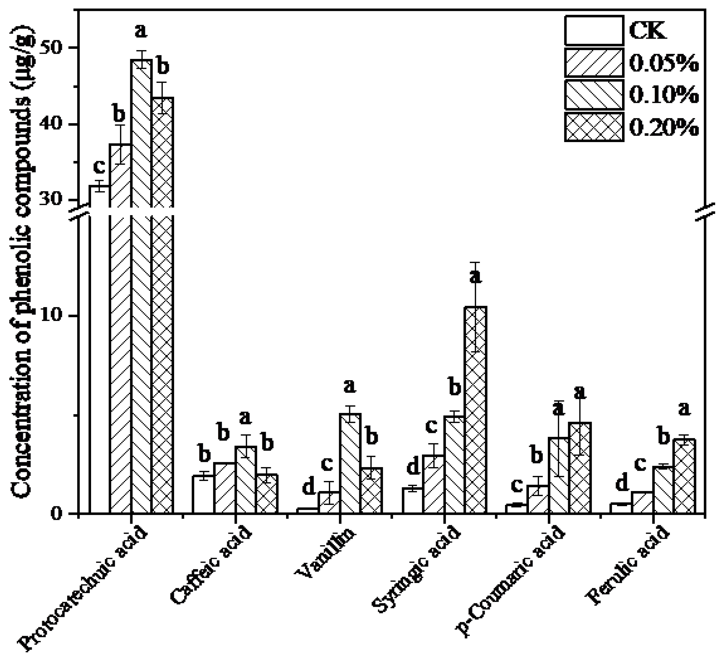
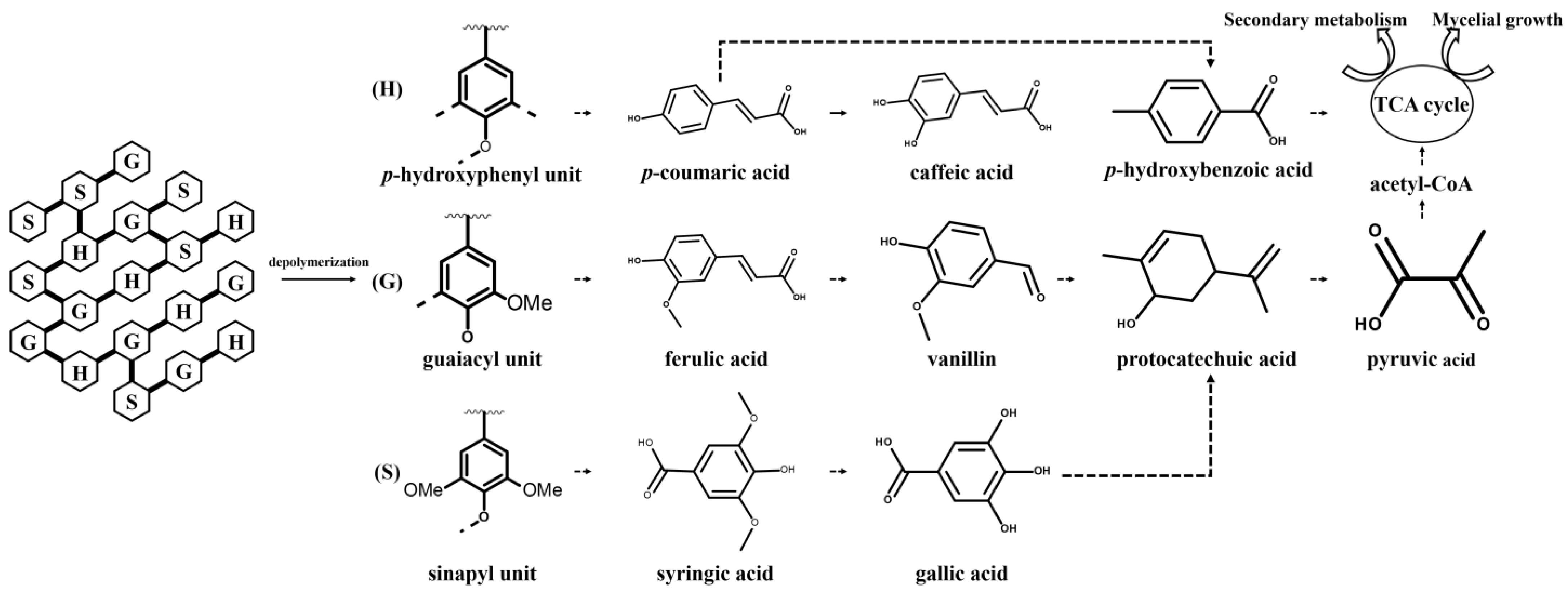
3.3. Effect of Lignin on Phenolic Compounds Metabolism in Mycelia of L. edodes

Considering the complex structure composed of phenylpropanoid units linked by ether bonds and carbon–carbon bonds, depolymerization of lignin potentially released a variety of phenolic intermediates. Phenolic compounds in L. edodes mycelia with known pharmacological activities were detected by HPLC [48]. It was suggested that the addition of lignin had a significant influence on the phenolics in the L. edodes mycelium. Regarding the total phenolic content (Table 1), a gradual increase with increasing lignin concentration was observed, and the total phenolic content had a 2.9-fold increase as compared to that of the control. A total of six phenolic compounds were detected, including protocatechuic acid, caffeic acid, syringic acid, vanillin, p-coumaric acid, and ferulic acid, with contents varying ranged from 0.27 ± 0.04 mg/g to 48.49 ± 1.2 mg/g (Figure 2). It demonstrated that the addition of lignin led to a significant difference in phenolic compounds in different treatment groups (p < 0.05). A total of six phenolic compounds could be divided into two categories. The contents of protocatechuic acid, caffeic acid, and vanillin were increased at the concentration of 0.10%. It is worth noting that protocatechuic acid is the most abundant phenolic acid in the present study, whose level was an order of magnitude higher than others. However, the other three phenolic acids presented a different trend in which p-coumaric acid, ferulic acid, and syringic acid contents are correlated with lignin concentration.
Contents of total phenolics in mycelium varied with the amount of lignin added, which resulted from the depolymerization of lignin and generation of monomer phenols in L. edodes. It was expected that different trends among the phenolic compounds resulted from the degradation pathway of lignin in L. edodes. Based on the obtained results, the putative pathway for the degradation process of lignin and transformation into phenols in L. edodes was outlined, referring to previous literature with slight modifications (Figure 3) [49,50]. Generally, the degradation process often occurred extracellularly, and low-molecular-weight monomers in the lignin polymer denoted as p-hydroxyphenyl (H), guaiacyl (G), and syringyl (S) were released, crossed the cell membrane and entered the cell [49]. The corresponding phenolic acids, referred to as p-coumaric acid, ferulic acid, and syringic acid, were investigated in the present study. As depicted in Figure 2, vanillin and p-coumaric acid are pivotal intermediates during the degradation process, while vanillin is formed from the conversion of ferulic acid and p-coumaric acid. The vanillin content in L. edodes mycelia without lignin was quite low. However, there was a 17.6-time increase after the addition of lignin. Vanillin served as an activator to accelerate the extracellular production of laccase and peroxidase. Moreover, the accumulation of vanillin contributed to the further conversion to downstream protocatechuic acid. Notably, protocatechuic acid is a conserved intermediate product of different monomers in most situations, acting as a “biological funnel” to limit the rate and efficiency of the lignin utilization process [51,52]. Ring cleavage of protocatechuic acid by dioxygenase enzymes occurred, yielding a large quantity of acetyl-CoA and pyruvate for entry into the TCA cycle ultimately. Based on this, it was convinced that the presence of lignin at a concentration of 0.1% raised the central metabolic level of L. edodes, offering more substrates and energies for fungal growth as well as secondary metabolites production. Moreover, the improved content of caffeic acid stemmed from the supplementary lignin. Caffeic acid has been identified to be widely distributed in several edible mushrooms [53]. Structurally, caffeic acid belongs to the hydroxycinnamic acid family, whose biosynthesis commonly takes p-coumaric acid as a precursor and employs p-coumarate-3-hydroxylase (C3H) to transfer a hydroxyl group onto the 3-position of the p-coumaric acid [54]. Hence, biotransformation among diverse phenols occurred in lignin-cultured L. edodes. Based on the experimental data, the addition of lignin improved the production of high-value phenolic compounds, especially lignin-derived phenols in L. edodes.

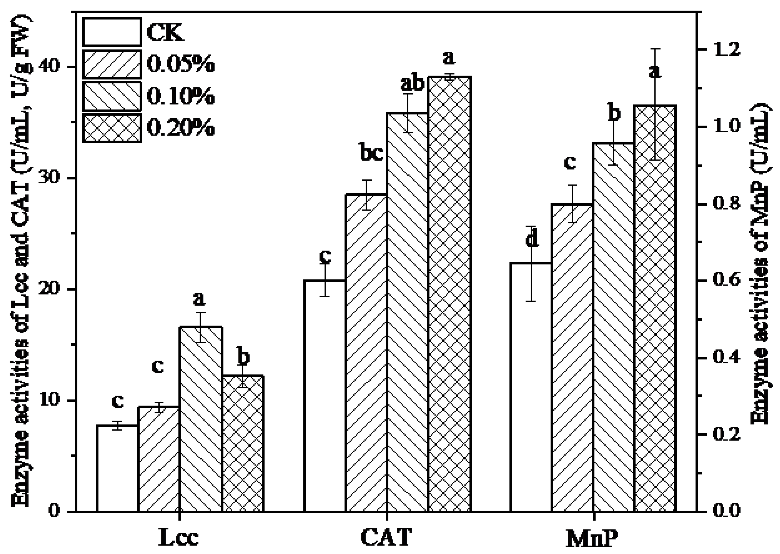
3.4. Effect of Lignin on Ligninolytic Enzymes in L. edodes Mycelia

Prior to further utilization, lignin was depolymerized via the potent extracellular ligninolytic enzymes in L. edodes. Activities of related enzymes, including Lcc, MnP as well as CAT, were evaluated in the presence of lignin (Figure 4). It turned out that 10 days of fermentation with lignin promoted the activities of three enzymes significantly (p < 0.05). Among them, the highest levels of laccase were observed at a concentration of 0.1% lignin, which was 2.1-fold higher compared to the levels recorded in the absence of lignin. In addition, lignin caused up to 1.65 and 1.88-fold increases in MnP and CAT activity, respectively, which reached maximum levels at 0.2% lignin. Meanwhile, a positive correlation was observed between MnP and CAT.
Several types of extracellular oxidative enzymes involved in the degradation of lignin in white-rot fungi have been elucidated thoroughly, including laccase and MnP, the most important ones in lignin depolymerization. Their activities and contents are closely related to the growth status of fungi in response to various culture conditions [55,56]. It has been reported that aromatic compounds, including ferulic acid and vanillin, which were blended in the present medium, stimulated both the Lcc and MnP secretions. In contrast, different strains performed diversely in response to different kinds of aromatic compounds related to their own specific signaling pathways of lignin and phenolic compounds in microorganisms [55]. Additionally, the low activity of MnP was confirmed in L. edodes previously, which might be accounted for by the lack of necessary substrate Mn2+ in the medium [57]. What is more, the production of enzymes in fungi is a dynamic course; thereby, cultivation time plays a vital role in MnP activities. Owing to the generation of large numbers of reactive oxygen species (ROS) during lignin degradation, the increase in the CAT activities mediated by lignin enabled to eliminate of free radicals in the fugal cell and to ease the oxidative stress, thereby ensuring the activation of fungal growth and secondary metabolite synthesis under high-level metabolic conditions.

4. Conclusions

In this work, lignin at a concentration of 0.1% (w/v) was found to promote fungal growth in liquid fermentation, accompanied by an increase in ergosterol, a biomarker of fungal biomass. At the same time, biosynthesis of proteins and WSP with multiple physiological activities were induced, which was possibly attributed to the presence of ROS or lignin derivatives generated during the lignin degradation process. With respect to the profile of phenolic compounds, changes of three monomers of syringic acid, p-coumaric acid, and ferulic acid were lignin-concentration-dependent, while the productions of protocatechuic acid, caffeic acid, and vanillin were the highest in 0.1% lignin-induced growth of L. edodes. Moreover, the activities of lignin-degrading enzymes such as Lcc, MnP, and CAT were increased. Additionally, Lcc and CAT played the primary roles in the degradation of lignin in L. edodes compared to MnP.
Overall, it was recommended that lignin induction was a promising approach for mycelia growth as well as phenolic acid accumulation in L. edodes liquid fermentation. Our present exploration provides insights into the exploitation and application of lignin as well as a foundation for the value-added cultivation of L. edodes.

Author Contributions

Writing—original draft preparation, F.W.; writing—review and editing, H.J., L.Y., and X.Z.; investigation, F.W., H.W., and Q.C.; data curation, X.P. All authors have read and agreed to the published version of the manuscript.

Funding

This research was funded by the National Key R&D Program of China, grant number 2021YFD2100200/2021YFD2100204, and the Guizhou province science and technology plan project (no. 20204Y070).

Institutional Review Board Statement

Not applicable.

Informed Consent Statement

Not applicable.

Data Availability Statement

All data are available in the article.

Conflicts of Interest

The authors declare no conflict of interest.

References

  1. Chio, C.; Sain, M.; Qin, W. Lignin utilization: A review of lignin depolymerization from various aspects. Renew. Sustain. Energy Rev. 2019, 107, 232–249. [Google Scholar] [CrossRef]
  2. Li, C.; Zhao, X.; Wang, A.; Huber, G.W.; Zhang, T. Catalytic transformation of lignin for the production of chemicals and fuels. Chem. Rev. 2015, 115, 11559–11624. [Google Scholar] [CrossRef]
  3. Weng, C.; Peng, X.; Han, Y. Depolymerization and conversion of lignin to value-added bioproducts by microbial and enzymatic catalysis. Biotechnol. Biofuels 2021, 14, 84. [Google Scholar] [CrossRef] [PubMed]
  4. Del Cerro, C.; Erickson, E.; Dong, T.; Wong, A.R.; Eder, E.K.; Purvine, S.O.; Mitchell, H.D.; Weitz, K.K.; Markillie, L.M.; Burnet, M.C.; et al. Intracellular pathways for lignin catabolism in white-rot fungi. Proc. Natl. Acad. Sci. USA 2021, 118, e2017381118. [Google Scholar] [CrossRef] [PubMed]
  5. Janusz, G.; Pawlik, A.; Sulej, J.; Świderska-Burek, U.; Jarosz-Wilkołazka, A.; Paszczyński, A. Lignin degradation: Microorganisms, enzymes involved, genomes analysis and evolution. FEMS Microbiol. Rev. 2017, 41, 941–962. [Google Scholar] [CrossRef] [PubMed]
  6. Bugg, T.D.H.; Ahmad, M.; Hardiman, E.M.; Rahmanpour, R. Pathways for degradation of lignin in bacteria and fungi. Nat. Prod. Rep. 2011, 28, 1883–1896. [Google Scholar] [CrossRef]
  7. Kunjapur, A.M.; Prather, K.L.J. Development of a vanillate biosensor for the vanillin biosynthesis pathway in E. Coli. ACS Synth. Biol. 2019, 8, 1958–1967. [Google Scholar] [CrossRef]
  8. Sonoki, T.; Takahashi, K.; Sugita, H.; Hatamura, M.; Azuma, Y.; Sato, T.; Suzuki, S.; Kamimura, N.; Masai, E. Glucose-Free cis,cis-Muconic Acid Production via New Metabolic Designs Corresponding to the Heterogeneity of Lignin. ACS Sustain. Chem. Eng. 2018, 6, 1256–1264. [Google Scholar] [CrossRef]
  9. Liu, L.; Liu, H.; Zhang, W.; Yao, M.; Li, B.; Liu, D.; Yuan, Y. Engineering the biosynthesis of caffeic acid in saccharomyces cerevisiae with heterologous enzyme combinations. Engineering 2019, 5, 287–295. [Google Scholar] [CrossRef]
  10. Yaguchi, A.; Franaszek, N.; O’Neill, K.; Lee, S.; Blenner, M. Identification of oleaginous yeasts that metabolize aromatic compounds. J. Ind. Microbiol. Biotechnol. 2020, 47, 801–813. [Google Scholar] [CrossRef]
  11. Chatterjee, A.; Delorenzo, D.M.; Carr, R.; Moon, T.S. Bioconversion of renewable feedstocks by Rhodococcus opacus. Curr. Opin. Biotech. 2019, 64, 10–16. [Google Scholar] [CrossRef]
  12. Sheng, K.; Wang, C.; Chen, B.; Kang, M.; Wang, M.; Liu, K.; Wang, M. Recent advances in polysaccharides from Lentinus edodes (Berk.): Isolation, structures and bioactivities. Food Chem. 2021, 358, 129883. [Google Scholar] [CrossRef]
  13. Tang, W.; Liu, C.; Liu, J.; Hu, L.; Huang, Y.; Yuan, L.; Liu, F.; Pan, S.; Chen, S.; Bian, S.; et al. Purification of polysaccharide from Lentinus edodes water extract by membrane separation and its chemical composition and structure characterization. Food Hydrocoll. 2020, 105, 105851. [Google Scholar] [CrossRef]
  14. Thetsrimuang, C.; Khammuang, S.; Chiablaem, K.; Srisomsap, C.; Sarnthima, R. Antioxidant properties and cytotoxicity of crude polysaccharides from Lentinus polychrous Lév. Food Chem. 2011, 128, 634–639. [Google Scholar] [CrossRef]
  15. Liu, Q.; Cui, X.; Song, Z.; Kong, W.; Ng, T.B. Coating shiitake mushrooms (Lentinus edodes) with a polysaccharide from Oudemansiella radicata improves product quality and flavor during postharvest storage. Food Chem. 2021, 352, 129357. [Google Scholar] [CrossRef] [PubMed]
  16. Chen, L.; Gong, Y.; Cai, Y.; Liu, W.; Zhou, Y.; Xiao, Y.; Xu, Z.; Liu, Y.; Lei, X.; Wang, G.; et al. Genome Sequence of the Edible Cultivated Mushroom Lentinula edodes (Shiitake) Reveals Insights into Lignocellulose Degradation. PLoS ONE 2016, 11, e160336. [Google Scholar] [CrossRef] [PubMed]
  17. Sakamoto, Y.; Nakade, K.; Sato, S.; Yoshida, K.; Konno, N. Lentinula edodes Genome Survey and Postharvest Transcriptome Analysis. Appl. Environ. Microbiol. 2017, 83, 2916–2990. [Google Scholar] [CrossRef]
  18. Wong, K.; Cheung, M.; Au, C.; Kwan, H. A novel Lentinula edodes laccase and its comparative enzymology suggest Guaiacol-Based laccase engineering for bioremediation. PLoS ONE 2013, 8, e66426. [Google Scholar] [CrossRef]
  19. De Oliveira Júnior, S.D.; Dos Santos Gouvêa, P.R.; de Aguiar, L.V.B.; Pessoa, V.A.; Dos Santos Cruz Costa, C.L.; Chevreuil, L.R.; Dedo Britonascimento, L.B.; Dos Santos, E.S.; Sales-Campos, C. Production of Lignocellulolytic Enzymes and Phenolic Compounds by Lentinus strigosus from the Amazon Using Solid-State Fermentation (SSF) of Guarana (Paullinia cupana) Residue. Appl. Biochem. Biotech. 2022, 194, 2882–2900. [Google Scholar] [CrossRef]
  20. Gaitan-Hernandez, R.; Zavaleta, M.; Aquino-Bolanos, E.N. Productivity, Physicochemical Changes, and Antioxidant Activity of Shiitake Culinary-Medicinal Mushroom Lentinus edodes (Agaricomycetes) Cultivated on Lignocellulosic Residues. Int. J. Med. Mushrooms 2017, 19, 1041–1052. [Google Scholar] [CrossRef] [PubMed]
  21. Zhou, C.H.; Xia, X.; Lin, C.X.; Tong, D.S.; Beltramini, J. Catalytic conversion of lignocellulosic biomass to fine chemicals and fuels. Chem. Soc. Rev. 2011, 40, 5588. [Google Scholar] [CrossRef] [PubMed]
  22. Wu, F.; Jia, X.; Yin, L.; Cheng, Y.; Miao, Y.; Zhang, X. The Effect of Hemicellulose and Lignin on Properties of Polysaccharides in Lentinus edodes and their Antioxidant Evaluation. Molecules 2019, 24, 1834. [Google Scholar] [CrossRef] [PubMed]
  23. Cai, Y.; Gong, Y.; Liu, W.; Hu, Y.; Chen, L.; Yan, L.; Zhou, Y.; Bian, Y. Comparative secretomic analysis of lignocellulose degradation by Lentinula edodes grown on microcrystalline cellulose, lignosulfonate and glucose. J. Proteom. 2017, 163, 92–101. [Google Scholar] [CrossRef] [PubMed]
  24. Niemenmaa, O.; Galkin, S.; Hatakka, A. Ergosterol contents of some wood-rotting basidiomycete fungi grown in liquid and solid culture conditions. Int. Biodeter. Biodegr. 2008, 62, 125–134. [Google Scholar] [CrossRef]
  25. Feki, F.; Klisurova, D.; Masmoudi, M.A.; Choura, S.; Denev, P.; Trendafilova, A.; Chamkha, M.; Sayadi, S. Optimization of microwave assisted extraction of simmondsins and polyphenols from Jojoba (Simmondsia chinensis) seed cake using Box-Behnken statistical design. Food Chem. 2021, 356, 129670. [Google Scholar] [CrossRef]
  26. Gbylik-Sikorska, M.; Gajda, A.; Nowacka-Kozak, E.; Posyniak, A. Simultaneous determination of 45 antibacterial compounds in mushrooms—Agaricus bisporus by ultra-high performance liquid chromatography-tandem mass spectrometry. J. Chromatogr. A 2019, 1587, 111–118. [Google Scholar] [CrossRef]
  27. Teshome, E.; Melaku, A. Selection and optimization of lignocellulosic substrate for laccase production from Pleurotus species. Int. J. Biotechnol. Mol. Biol. Res. 2017, 8, 38–48. [Google Scholar] [CrossRef]
  28. Lechner, B.E.; Papinutti, V.L. Production of lignocellulosic enzymes during growth and fruiting of the edible fungus Lentinus tigrinus on wheat straw. Process Biochem. 2006, 41, 594–598. [Google Scholar] [CrossRef]
  29. Zhu, Z.; Liu, X.; Tang, Y.; Dong, F.; Sun, H.; Chen, L.; Zhang, Y. Effects of cultural medium on the formation and antitumor activity of polysaccharides by Cordyceps gunnii. J. Biosci. Bioeng. 2016, 122, 494–498. [Google Scholar] [CrossRef]
  30. Kim, H.O.; Lim, J.M.; Joo, J.H.; Kim, S.W.; Hwang, H.J.; Choi, J.W.; Yun, J.W. Optimization of submerged culture condition for the production of mycelial biomass and exopolysaccharides by Agrocybe cylindracea. Bioresour. Technol. 2005, 96, 1175–1182. [Google Scholar] [CrossRef]
  31. Bogdan, V.I.; Sergeeva, Y.E.; Lunin, V.V.; Perminova, I.V.; Konstantinov, A.I.; Zinchenko, G.E.; Bogdan, K.V. Bioconversion of phenolic monomers of lignin and Lignin-Containing substrates by the basidiomycete Lentinus tigrinus. Appl. Biochem. Microbiol. 2018, 54, 198–205. [Google Scholar] [CrossRef]
  32. Vikman, M.; Karjomaa, S.; Kapanen, A.; Wallenius, K.; Itävaara, M. The influence of lignin content and temperature on the biodegradation of lignocellulose in composting conditions. Appl. Microbiol. Biotechnol. 2002, 59, 591–598. [Google Scholar] [PubMed]
  33. Qiu, W.; Liu, J. Fermenting and Lignin Degradability of a White-Rot Fungus Coriolopsis trogii Using Industrial Lignin as Substrate. Appl. Biochem. Biotechnol. 2022, 194, 5220–5235. [Google Scholar] [CrossRef] [PubMed]
  34. Fukushima, Y.; Okada, K.; Kawai, G.; Motai, H. Efficient Production of Mycelium of Lentinus edodes by a Continuous Culture and the Effect of Lignin on Growth. J. Ferment. Bioeng. 1993, 1, 45–48. [Google Scholar] [CrossRef]
  35. Tsujiyama, S.; Muraoka, T.; Takada, N. Biodegradation of 2,4-dichlorophenol by shiitake mushroom (Lentinula edodes) using vanillin as an activator. Biotechnol. Lett. 2013, 35, 1079–1083. [Google Scholar] [CrossRef]
  36. Iivashechkin, A.A.; Sergeeva, I.; Lunin, V.V.; Bogdan, V.I.; Mysiakina, I.S.; Feofilova, E.P. Influence of lignin and oxygen on the growth and the lipid formation of the fungus Lentinus tigrinus. Prikl. Biokhim. Mikrobiol. 2014, 50, 318–323. [Google Scholar] [CrossRef]
  37. Mccue, P.P.; Shetty, K. A model for the involvement of lignin degradation enzymes in phenolic antioxidant mobilization from whole soybean during solid-state bioprocessing by Lentinus edodes. Process Biochem. 2005, 40, 1143–1150. [Google Scholar] [CrossRef]
  38. Koselny, K.; Mutlu, N.; Minard, A.Y.; Kumar, A.; Krysan, D.J.; Wellington, M. A Genome-Wide screen of deletion mutants in the filamentous saccharomyces cerevisiae background identifies ergosterol as a direct trigger of macrophage pyroptosis. mBio 2018, 9, e01204-18. [Google Scholar] [CrossRef]
  39. Liu, J.; Xia, J.; Nie, K.; Wang, F.; Deng, L. Outline of the biosynthesis and regulation of ergosterol in yeast. World J. Microbiol. Biotechnol. 2019, 35, 98. [Google Scholar] [CrossRef]
  40. Blaga, A.C.; Ciobanu, C.; Caşcaval, D.; Galaction, A. Enhancement of ergosterol production by Saccharomyces cerevisiae in batch and fed-batch fermentation processes using n-dodecane as oxygen-vector. Biochem. Eng. J. 2018, 131, 70–76. [Google Scholar] [CrossRef]
  41. Silva, R.R.; Corso, C.R.; Matheus, D.R. Effect of culture conditions on the biomass determination by ergosterol of Lentinus crinitus and Psilocybe castanella. World J. Microbiol. Biotechnol. 2010, 26, 841–846. [Google Scholar] [CrossRef]
  42. Lu, X.; Wang, C.; Li, Y.; Liu, P. Improved production and antioxidant activity of exopolysaccharides by submerged culture of Lentinula edodes by the addition of lignocellulose. J. Biosci. Bioeng. 2022, 134, 162–166. [Google Scholar] [CrossRef]
  43. Kozarski, M.; Klaus, A.; Nikšić, M.; Vrvić, M.M.; Todorović, N.; Jakovljević, D.; Van Griensven, L.J.L.D. Antioxidative activities and chemical characterization of polysaccharide extracts from the widely used mushrooms Ganoderma applanatum, Ganoderma lucidum, Lentinus edodes and Trametes versicolor. J. Food Compos. Anal. 2012, 26, 144–153. [Google Scholar] [CrossRef]
  44. Li, F.; Cui, S.H.; Zha, X.Q.; Bansal, V.; Jiang, Y.L.; Asghar, M.N.; Wang, J.H.; Pan, L.H.; Xu, B.F.; Luo, J.P. Structure and bioactivity of a polysaccharide extracted from protocorm-like bodies of Dendrobium huoshanense. Int. J. Biol. Macromol. Struct. Funct. Interact. 2015, 72, 664–672. [Google Scholar] [CrossRef]
  45. Karnaouri, A.; Muraleedharan, M.N.; Dimarogona, M.; Topakas, E.; Rova, U.; Sandgren, M.; Christakopoulos, P. Recombinant expression of thermostable processive MtEG5 endoglucanase and its synergism with MtLPMO from Myceliophthora thermophila during the hydrolysis of lignocellulosic substrates. Biotechnol. Biofuels 2017, 10, 126. [Google Scholar] [CrossRef]
  46. Carneiro, A.A.J.; Ferreira, I.C.F.R.; Dueñas, M.; Barros, L.; Da Silva, R.; Gomes, E.; Santos-Buelga, C. Chemical composition and antioxidant activity of dried powder formulations of Agaricus blazei and Lentinus edodes. Food Chem. 2013, 138, 2168–2173. [Google Scholar] [CrossRef]
  47. Wu, X.J.; Hansen, C. Effects of Whey Permeate-Based Medium on the Proximate Composition of Lentinus edodes in the Submerged Culture. J. Food Sci. 2006, 71, M174–M179. [Google Scholar] [CrossRef]
  48. Kim, M.; Seguin, P.; Ahn, J.; Kim, J.; Chun, S.; Kim, E.; Seo, S.; Kang, E.; Kim, S.; Park, Y.; et al. Phenolic compound concentration and antioxidant activities of edible and medicinal mushrooms from Korea. J. Agric. Food Chem. 2008, 56, 7265–7270. [Google Scholar] [CrossRef]
  49. Li, X.; Zheng, Y. Biotransformation of lignin: Mechanisms, applications and future work. Biotechnol. Prog. 2019, 36, e2922. [Google Scholar] [CrossRef] [PubMed]
  50. Yaguchi, A.L.; Lee, S.J.; Blenner, M.A. Synthetic Biology towards Engineering Microbial Lignin Biotransformation. Trends Biotechnol. 2021, 39, 1037–1064. [Google Scholar] [CrossRef]
  51. Tomizawa, S.; Chuah, J.; Matsumoto, K.; Doi, Y.; Numata, K. Understanding the limitations in the biosynthesis of polyhydroxyalkanoate (PHA) from lignin derivatives. ACS Sustain. Chem. Eng. 2014, 2, 1106–1113. [Google Scholar] [CrossRef]
  52. Johnson, C.W.; Beckham, G.T. Aromatic catabolic pathway selection for optimal production of pyruvate and lactate from lignin. Metab. Eng. 2015, 28, 240–247. [Google Scholar] [CrossRef] [PubMed]
  53. Lau, B.F.; Abdullah, N. Bioprospecting of Lentinus squarrosulus Mont., An underutilized wild edible mushroom, as a potential source of functional ingredients: A review. Trends Food Sci. Technol. 2017, 61, S0924224416302898. [Google Scholar] [CrossRef]
  54. Xue, Y.; Zhang, Y.; Grace, S.; He, Q. Functional expression of an Arabidopsis p450 enzyme, p-coumarate-3-hydroxylase, in the cyanobacterium Synechocystis PCC 6803 for the biosynthesis of caffeic acid. J. Appl. Phycol. 2014, 26, 219–226. [Google Scholar] [CrossRef]
  55. Elisashvili, V.; Kachlishvili, E.; Khardziani, T.; Agathos, S.N. Effect of aromatic compounds on the production of laccase and manganese peroxidase by white-rot basidiomycetes. J. Ind. Microbiol. Biotechnol. 2010, 37, 1091–1096. [Google Scholar] [CrossRef]
  56. Shutova, V.V.; Revin, V.V.; Makushina, I. The effect of copper ions on the production of laccase by the fungus Lentinus (Panus) tigrinus. Prikl. Biokhim. Mikrobiol. 2008, 44, 683–687. [Google Scholar] [CrossRef]
  57. Elisashvili, V.; Penninckx, M.; Kachlishvili, E.; Tsiklauri, N.; Metreveli, E.; Kharziani, T.; Kvesitadze, G. Lentinus edodes and Pleurotus species lignocellulolytic enzymes activity in submerged and solid-state fermentation of lignocellulosic wastes of different composition. Bioresour. Technol. 2008, 99, 457–462. [Google Scholar] [CrossRef]
Figure 1. Effect of lignin on mycelial biomass and ergosterol content. Experiments were performed in triplicate, and results are presented as mean ± SD. Different letters (a–d, A–C) represented the significant differences in mycelial biomass and ergosterol content among samples (p < 0.05). Statistical analysis was carried out by one-way ANOVA accompanied by Duncan’s test using SPSS (version 20.0).
Jof 09 00237 g001
Figure 2. Contents of phenolic compounds from L. edodes mycelium cultured in lignin-containing medium. Results are presented as mean ± SD, n = 3. Different letters represented the significant differences among sample groups (p < 0.05). Statistical analysis was carried out by one-way ANOVA accompanied by Duncan’s test using SPSS (version 20.0).
Jof 09 00237 g002
Figure 3. Schematic diagram of lignin degradation pathway in L. edodes. The dotted line represents an indirect reaction process. H, G, and S in the figure refer to three phenyl propane monomers of lignin, which named coumaryl alcohol (H), coniferyl alcohol (G), and sinapyl alcohol (S), respectively [49,50].
Jof 09 00237 g003
Figure 4. Enzyme activities of Lcc, CAT, and MnP in the absence or presence of lignin. Results are presented as means ± SD of three replicates. Different letters represented the significant differences among sample groups (p < 0.05). Statistical analysis was carried out by one-way ANOVA accompanied by Duncan’s test using SPSS (version 20.0).
Jof 09 00237 g004
Table 1. Chemical composition of L. edodes cultured in the presence of lignin.
LigninWSP Extracts (g/100 g)Crude Protein (w/w %)Ash (w/w %)Total Phenolics
(mg GAE/g DW)
02.11 ± 0.14 d25.84 ± 0.35 c3.78 ± 0.07 d1.45 ± 0.03 d
0.05% 4.14 ± 0.35 b27.63 ± 0.26 b4.89 ± 0.09 c2.28 ± 0.05 c
0.10%6.44 ± 0.33 a29.24 ± 0.09 a5.21 ± 0.11 b3.87 ± 0.14 b
0.20% 2.94 ± 0.28 c26.17 ± 0.18 c6.14 ± 0.17 a5.62 ± 0.21 a
In each column, different lowercase superscript letters represent significant differences between different treatments (p < 0.05). Statistical analysis was carried out by one-way ANOVA accompanied by Duncan’s test using SPSS (version 20.0). Results are presented as means ± SD of three replicated independent determinations.
Disclaimer/Publisher’s Note: The statements, opinions and data contained in all publications are solely those of the individual author(s) and contributor(s) and not of MDPI and/or the editor(s). MDPI and/or the editor(s) disclaim responsibility for any injury to people or property resulting from any ideas, methods, instructions or products referred to in the content.

