
Sporotrichosis Outbreak Due to Sporothrix brasiliensis in Domestic Cats in Magallanes, Chile: A One-Health-Approach Study

 ,
, 
Abstract
:1. Introduction
2. Materials and Methods
2.1. Geographic Location
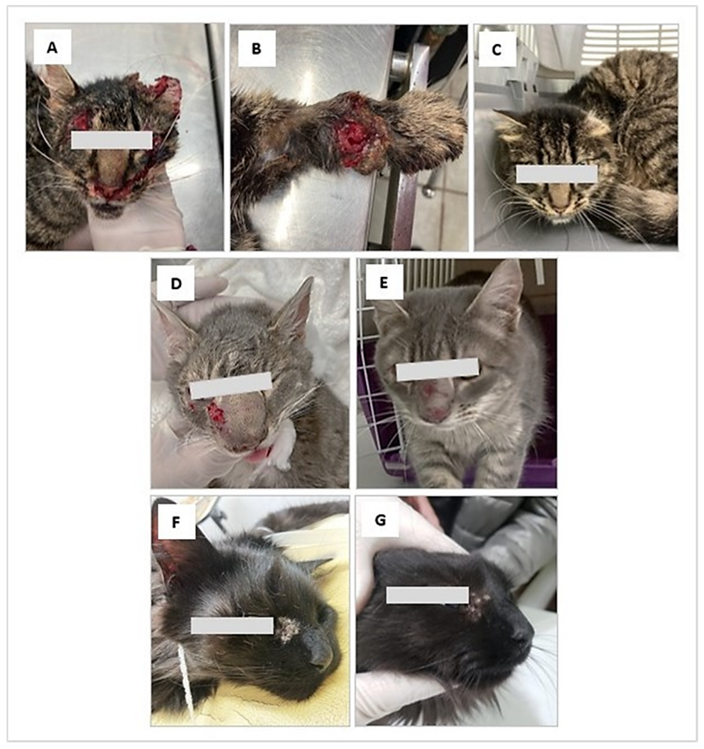
2.2. Case Description
3. Results
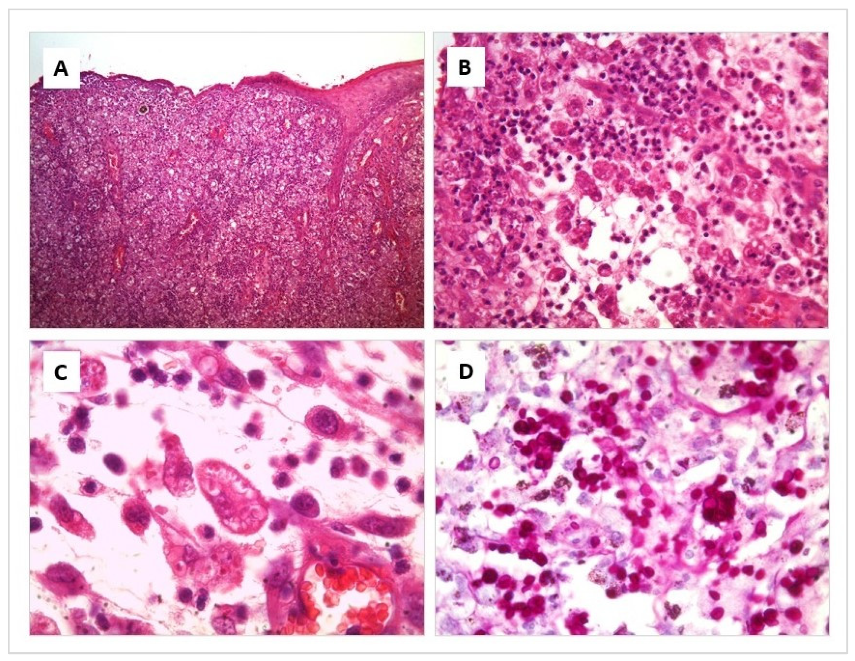
3.1. Histopathological Study
3.2. Mycological Study
3.3. Treatment and Clinical Evolution
4. Discussion
5. Recommendations
Author Contributions
Funding
Institutional Review Board Statement
Informed Consent Statement
Acknowledgments
Conflicts of Interest
References
- Chakrabarti, A.; Bonifaz, A.; Gutierrez-Galhardo, M.C.; Mochizuki, T.; Li, S. Global epidemiology of sporotrichosis. Med. Mycol. 2015, 53, 3–14. [Google Scholar] [CrossRef] [PubMed]
- PAHO. Sporothrix brasiliensis, an Emerging Fungal Pathogen, Notable for Its Zoonotic Transmission and Epidemic Potential for Human and Animal Health in the Americas. Available online: https://www.someve.com.ar/images/noticias/2019/S-brasiliensis_lasAmericas_30082019_ES.pdf (accessed on 15 November 2022).
- Sizar, O.; Talati, R. Sporotrichosis; StatPearls Publishing: Treasure Island, FL, USA, 2022. [Google Scholar]
- Fichman, V.; Freitas, D.F.S.; Valle, A.C.F.D.; de Souza, R.V.; Curi, A.L.L.; Valete-Rosalino, C.M.; de Macedo, P.M.; Varon, A.G.; Figueiredo-Carvalho, M.H.G.; Almeida-Silva, F.; et al. Severe Sporotrichosis Treated with Amphotericin B: A 20-Year Cohort Study in an Endemic Area of Zoonotic Transmission. J. Fungi 2022, 8, 469. [Google Scholar] [CrossRef] [PubMed]
- Criseo, G.; Romeo, O. Ribosomal DNA Sequencing and Phylogenetic Analysis of Environmental Sporothrix schenckii Strains: Comparison with Clinical Isolates. Mycopathologia 2010, 169, 351–358. [Google Scholar] [CrossRef] [PubMed]
- De Oliveira Bento, A.; de Sena Costa, A.S.; Lima, S.L.; do Monte Alves, M.; de Azevedo Melo, A.S.; Rodrigues, A.M.; da Silva-Rocha, W.P.; Milan, E.P.; Chaves, G.M. The spread of cat-transmitted sporotrichosis due to Sporothrix brasiliensis in Brazil towards the Northeast region. PLoS Negl. Trop. Dis. 2021, 15, e0009693. [Google Scholar] [CrossRef]
- Mora-Montes, H.M. Special Issue “Sporothrix and Sporotrichosis 2.0”. J. Fungi 2022, 8, 821. [Google Scholar] [CrossRef]
- Lopes-Bezerra, L.M.; Walker, L.A.; Nino-Vega, G.; Mora-Montes, H.M.; Neves, G.W.P.; Villalobos-Duno, H.; Barreto, L.; Garcia, K.; Franco, B.; Martínez-Álvarez, J.A.; et al. Cell walls of the dimorphic fungal pathogens Sporothrix schenckii and Sporothrix brasiliensis exhibit bilaminate structures and sloughing of extensive and intact layers. PLoS Negl. Trop. Dis. 2018, 12, e0006169. [Google Scholar] [CrossRef]
- Neves, G.W.P.; Wong, S.S.W.; Aimanianda, V.; Simenel, C.; Guijarro, J.I.; Walls, C.; Willment, J.A.; Gow, N.A.R.; Munro, C.A.; Brown, G.D.; et al. Complement-Mediated Differential Immune Response of Human Macrophages to Sporothrix Species Through Interaction with Their Cell Wall Peptidorhamnomannans. Front. Immunol. 2021, 12, 749074. [Google Scholar] [CrossRef]
- Villalobos-Duno, H.L.; Barreto, L.A.; Alvarez-Aular, A.; Mora-Montes, H.M.; Lozoya-Pérez, N.E.; Franco, B.; Lopes-Bezerra, L.M.; Niño-Vega, G.A. Comparison of Cell Wall Polysaccharide Composition and Structure Between Strains of Sporothrix schenckii and Sporothrix brasiliensis. Front. Microbiol. 2021, 12, 726958. [Google Scholar] [CrossRef]
- Gow, N.A.R.; Lenardon, M.D. Architecture of the dynamic fungal cell wall. Nat. Rev. Genet. 2022. Epub ahead of print. [Google Scholar] [CrossRef]
- Rabello, V.B.S.; Almeida-Silva, F.; Scramignon-Costa, B.D.S.; Motta, B.D.S.; de Macedo, P.M.; Teixeira, M.D.M.; Almeida-Paes, R.; Irinyi, L.; Meyer, W.; Zancopé-Oliveira, R.M. Environmental Isolation of Sporothrix brasiliensis in an Area with Recurrent Feline Sporotrichosis Cases. Front. Cell. Infect. Microbiol. 2022, 12, 894297. [Google Scholar] [CrossRef]
- Rodrigues, A.M.; Hagen, F.; de Camargo, Z.P. A Spotlight on Sporothrix and Sporotrichosis. Mycopathologia 2022, 187, 407–411. [Google Scholar] [CrossRef]
- Morris-Jones, R. Sporotrichosis. Clin. Exp. Dermatol. 2002, 27, 427–431. [Google Scholar] [CrossRef] [PubMed]
- Camacho, E.; León-Navarro, I.; Rodríguez-Brito, S.; Mendoza, M.; Niño-Vega, G.A. Molecular epidemiology of human sporotrichosis in Venezuela reveals high frequency of Sporothrix globosa. BMC Infect. Dis. 2015, 15, 94. [Google Scholar] [CrossRef]
- De Oliveira, M.M.; de Almeida-Paes, R.; de Medeiros Muniz, M.; de Lima Barros, M.B.; Galhardo, M.C.; Zancope-Oliveira, R.M. Sporotrichosis caused by Sporothrix globosa in Rio De Janeiro, Brazil: Case report. Mycopathologia 2010, 169, 359–363. [Google Scholar] [CrossRef]
- Brilhante, R.S.N.; de Aguiar, F.R.M.; da Silva, M.L.Q.; de Oliveira, J.S.; de Camargo, Z.P.; Rodrigues, A.M.; Pereira, V.S.; Serpa, R.; Castelo-Branco, D.; Correia, E.E.M.; et al. Antifungal susceptibility of Sporothrix schenckii complex biofilms. Med. Mycol. 2018, 56, 297–306. [Google Scholar] [CrossRef]
- Córdoba, S.; Isla, G.; Szusz, W.; Vivot, W.; Hevia, A.; Davel, G.; Canteros, C.E. Molecular identification and susceptibility profile of Sporothrix schenckii sensu lato isolated in Argentina. Mycoses 2018, 61, 441–448. [Google Scholar] [CrossRef]
- New, D.; Beukers, A.G.; Kidd, S.E.; Merritt, A.J.; Weeks, K.; van Hal, S.J.; Arthur, I. Identification of multiple species and subpopulations among Australian clinical Sporothrix isolates using whole genome sequencing. Med. Mycol. 2019, 57, 905–908. [Google Scholar] [CrossRef]
- Barreto, L.; Velásquez, G.; Mendoza, M.; Camacho, E.; Goncalves, E.; Rodríguez, S.; Niño-Vega, G.A. Geographical distribution and ecological niche modeling of the etiological agents of human sporotrichosis in Venezuela. Braz. J. Microbiol. 2021, 52, 63–71. [Google Scholar] [CrossRef]
- Brilhante, R.S.N.; Fernandes, M.R.; Pereira, V.S.; Costa, A.d.C.; Oliveira, J.S.d.; de Aguiar, L.; Rodrigues, A.M.; de Camargo, Z.P.; Pereira-Neto, W.A.; Sidrim, J.J.C.; et al. Biofilm formation on cat claws by Sporothrix species: An ex vivo model. Microb. Pathog. 2021, 150, 104670. [Google Scholar] [CrossRef]
- García-Carnero, L.C.; Martínez-Álvarez, J.A. Virulence Factors of Sporothrix schenckii. J. Fungi 2022, 8, 318. [Google Scholar] [CrossRef]
- Hernández-Castro, R.; Pinto-Almazán, R.; Arenas, R.; Sánchez-Cárdenas, C.D.; Espinosa-Hernández, V.M.; Sierra-Maeda, K.Y.; Conde-Cuevas, E.; Juárez-Durán, E.R.; Xicohtencatl-Cortes, J.; Carrillo-Casas, E.M.; et al. Epidemiology of clinical sporotrichosis in the Americas in the last ten years. J. Fungi 2022, 8, 588. [Google Scholar] [CrossRef] [PubMed]
- Nava-Pérez, N.; Neri-García, L.G.; Romero-González, O.E.; Terrones-Cruz, J.A.; García-Carnero, L.C.; Mora-Montes, H.M. Biological and clinical attributes of Sporothrix globosa, a causative agent of sporotrichosis. Infect. Drug Resist. 2022, 15, 2067–2090. [Google Scholar] [CrossRef] [PubMed]
- De Meyer, E.M.; de Beer, Z.W.; Summerbell, R.C.; Moharram, A.; de Hoog, G.S.; Vismer, H.F.; Wingfield, M.J. Taxonomy and phylogeny of new wood- and soil-inhabiting Sporothrix species in the Ophiostoma stenoceras-Sporothrix schenckii complex. Mycologia 2008, 100, 647–661. [Google Scholar] [CrossRef]
- Ramírez-Soto, M.C.; Aguilar-Ancori, E.G.; Tirado-Sánchez, A.; Bonifaz, A. Ecological Determinants of Sporotrichosis Etiological Agents. J. Fungi 2018, 4, 95. [Google Scholar] [CrossRef]
- Nesseler, A.; Schauerte, N.; Geiger, C.; Kaerger, K.; Walther, G.; Kurzai, O.; Eisenberg, T. Sporothrix humicola (Ascomycota: Ophiostomatales)—A soil-borne fungus with pathogenic potential in the eastern quoll (Dasyurus viverrinus). Med. Mycol. Case Rep. 2019, 25, 39–44. [Google Scholar] [CrossRef]
- Rodrigues, A.M.; Della Terra, P.P.; Gremião, I.D.; Pereira, S.A.; Orofino-Costa, R.; de Camargo, Z.P. The threat of emerging and re-emerging pathogenic Sporothrix species. Mycopathologia 2020, 185, 813–842. [Google Scholar] [CrossRef]
- Zhao, M.-D.; Zhou, X.; Liu, T.-T.; Yang, Z.-B. Morphological and physiological comparison of taxa comprising the Sporothrix schenckii complex. J. Zhejiang Univ. B 2015, 16, 940–947. [Google Scholar] [CrossRef]
- Fernandes, G.F.; dos Santos, P.O.; Amaral, C.C.; Sasaki, A.A.; Godoy-Martinez, P.; Camargo, Z.P.D. Characteristics of 151 Brazilian Sporothrix schenckii isolates from 5 different geographic regions of Brazil: A forgotten and re-emergent pathogen. Open Mycol. J. 2009, 3, 48–58. [Google Scholar] [CrossRef]
- Sasaki, A.A.; Fernandes, G.F.; Rodrigues, A.M.; Lima, F.M.; Marini, M.M.; dos S. Feitosa, L.; de Melo Teixeira, M.; Felipe, M.S.S.; da Silveira, J.F.; de Camargo, Z.P. Chromosomal polymorphism in the Sporothrix schenckii complex. PLoS ONE 2014, 9, e86819. [Google Scholar] [CrossRef]
- Rodrigues, A.M.; de Melo Teixeira, M.; De Hoog, G.S.; Schubach, T.M.P.; Pereira, S.A.; Fernandes, G.F.; Bezerra, L.M.L.; Felipe, M.S.; De Camargo, Z.P. Phylogenetic Analysis Reveals a High Prevalence of Sporothrix brasiliensis in Feline Sporotrichosis Outbreaks. PLoS Negl. Trop. Dis. 2013, 7, e2281. [Google Scholar] [CrossRef] [Green Version]
- Arrillaga-Moncrieff, I.; Capilla, J.; Mayayo, E.; Marimon, R.; Marine, M.; Genis, J.; Cano-Lira, J.F.; Guarro, J. Different virulence levels of the species of Sporothrix in a murine model. Clin. Microbiol. Infect. 2009, 15, 651–655. [Google Scholar] [CrossRef] [PubMed]
- Fernandes, G.F.; dos Santos, P.O.; Rodrigues, A.M.; Sasaki, A.A.; Burger, E.; de Camargo, Z.P. Characterization of virulence profile, protein secretion and immunogenicity of different Sporothrix schenckii sensu stricto isolates compared with S. globose and S. brasiliensis species. Virulence 2013, 4, 241–249. [Google Scholar] [CrossRef] [PubMed]
- Rossato, L.; Dos Santos, S.S.; Ferreira, L.G.; De Almeida, S.R. The importance of Toll-like receptor 4 during experimental Sporothrix brasiliensis infection. Med. Mycol. 2019, 57, 489–495. [Google Scholar] [CrossRef]
- Corrêa-Moreira, D.; Menezes, R.C.; Romeo, O.; Borba, C.M.; Oliveira, M.M.E. Clinical and Anatomopathological Evaluation of BALB/c Murine Models Infected with Isolates of Seven Pathogenic Sporothrix Species. Pathogens 2021, 10, 1647. [Google Scholar] [CrossRef]
- Schubach, T.M.P.; Schubach, A.; Okamoto, T.; Barros, M.B.L.; Figueiredo, F.B.; Cuzzi, T.; Fialho-Monteiro, P.C.; Reis, R.S.; Perez, M.A.; Wanke, B. Evaluation of an epidemic of sporotrichosis in cats: 347 cases (1998–2001). J. Am. Vet. Med. Assoc. 2004, 224, 1623–1629. [Google Scholar] [CrossRef]
- Etchecopaz, A.; Toscanini, M.; Gisbert, A.; Mas, J.; Scarpa, M.; Iovannitti, C.; Bendezú, K.; Nusblat, A.; Iachini, R.; Cuestas, M. Sporothrix brasiliensis: A Review of an Emerging South American Fungal Pathogen, Its Related Disease, Presentation and Spread in Argentina. J. Fungi 2021, 7, 170. [Google Scholar] [CrossRef]
- Gremião, I.D.F.; Miranda, L.H.M.; Reis, E.G.; Rodrigues, A.M.; Pereira, S.A. Zoonotic Epidemic of Sporotrichosis: Cat to Human Transmission. PLoS Pathog. 2017, 13, e1006077. [Google Scholar] [CrossRef]
- Spanamberg, A.; Araujo, R.; Ravazzolo, A.P.; Driemeier, D.; Driemeier, R.M.S.; Ferreiro, L. Sporothrix brasiliensis on cats with skin ulcers in Southern Brazil. Med. Mycol. 2021, 59, 301–304. [Google Scholar] [CrossRef]
- Gallo, S.; Arias-Rodriguez, C.; Sánchez-Cifuentes, E.A.; Santa-Vélez, C.; Larrañaga-Piñeres, I.; Gaviria-Barrera, M.E.; Vásquez-Ochoa, L.A.; Montoya, D.; Jiménez-Alzate, M.D.P. First three cases of cat-associated zoonotic cutaneous sporotrichosis in Colombia. Int. J. Dermatol. 2022, 61, 1276–1279. [Google Scholar] [CrossRef]
- Dos Santos, G.M.P.; Borba-Santos, L.P.; Vila, T.; Gremião, I.D.F.; Pereira, S.A.; De Souza, W.; Rozental, S. Sporothrix spp. Biofilms Impact in the Zoonotic Transmission Route: Feline Claws Associated Biofilms, Itraconazole Tolerance, and Potential Repurposing for Miltefosine. Pathogens 2022, 11, 206. [Google Scholar] [CrossRef]
- De Andrade Galliano Daros Bastos, F.; Raimundo Cognialli, R.C.; Rodrigues de Farias, M.; Dos Santos Monti, F.; Wu, K.; Queiroz-Telles, F. Spread of Sporothrix spp. through respiratory droplets from infected cats: A potential route of transmission. Med. Mycol. 2022, 60, myac079. [Google Scholar] [CrossRef] [PubMed]
- Rossow, J.A.; Queiroz-Telles, F.; Caceres, D.H.; Beer, K.D.; Jackson, B.R.; Pereira, J.G.; Ferreira Gremião, I.D.; Pereira, S.A. A One Health Approach to Combatting Sporothrix brasiliensis: Narrative Review of an Emerging Zoonotic Fungal Pathogen in South America. J. Fungi 2020, 6, 247. [Google Scholar] [CrossRef] [PubMed]
- Fichman, V.; Freitas, D.F.S.; de Macedo, P.M.; Valle, A.C.F.D.; Almeida-Silva, F.; Zancopé-Oliveira, R.M.; Almeida-Paes, R.; Gutierrez-Galhardo, M.C. Sporotrichosis After Tattooing Caused by Sporothrix brasiliensis. Mycopathologia 2022, 187, 137–139. [Google Scholar] [CrossRef]
- Morgado, D.S.; Castro, R.; Ribeiro-Alves, M.; Corrêa-Moreira, D.; Castro-Alves, J.; Pereira, S.A.; Menezes, R.C.; Oliveira, M.M.E. Global distribution of animal sporotrichosis: A systematic review of Sporothrix sp. identified using molecular tools. Curr. Res. Microb. Sci. 2022, 3, 100140. [Google Scholar] [CrossRef]
- Han, H.S.; Kano, R. Feline sporotrichosis in Asia. Braz. J. Microbiol. 2021, 52, 125–134. [Google Scholar] [CrossRef]
- Veasey, J.V.; Carvalho, G.S.M.; Ruiz, L.R.B.; Neves Neto, M.F.; Zaitz, C. Epidemiological and geographical distribution profile of urban sporotrichosis in the city of São Paulo. An. Bras. Dermatol. 2022, 97, 228–230. [Google Scholar] [CrossRef]
- Almeida-Silva, F.; Rabello, V.B.S.; Scramignon-Costa, B.S.; Zancopé-Oliveira, R.M.; de Macedo, P.M.; Almeida-Paes, R. Beyond Domestic Cats: Environmental Detection of Sporothrix brasiliensis DNA in a Hyperendemic Area of Sporotrichosis in Rio de Janeiro State, Brazil. J. Fungi 2022, 8, 604. [Google Scholar] [CrossRef]
- Boechat, J.S.; Oliveira, M.M.E.; Gremião, I.D.F.; Almeida-Paes, R.; Machado, A.C.S.; Zancopé-Oliveira, R.M.; Oliveira, R.V.C.; Morgado, D.S.; Corrêa, M.L.; Figueiredo, A.B.F.; et al. Sporothrix brasiliensis and Feline Sporotrichosis in the Metropolitan Region of Rio de Janeiro, Brazil (1998–2018). J. Fungi 2022, 8, 749. [Google Scholar] [CrossRef]
- Neto, F.B.D.S.; Gadelha, C.L.; Lopes, I.V.; da Silva, M.B.; de Sousa, B.R.; Dulgheroff, A.C.B.; Guerra, F.Q.S.; Ferraz, C.E.; Magalhães, V.; Oliveira, M.M.E.; et al. Dog-transmitted ocular sporotrichosis. J. Mycol. Med. 2022, 33, 101335. [Google Scholar] [CrossRef]
- De Carvalho, J.A.; Beale, M.A.; Hagen, F.; Fisher, M.C.; Kano, R.; Bonifaz, A.; Toriello, C.; Negroni, R.; Rego, R.S.M.; Gremião, I.D.F.; et al. Trends in the molecular epidemiology and population genetics of emerging Sporothrix species. Stud. Mycol. 2021, 100, 100129. [Google Scholar] [CrossRef]
- De Miranda, L.H.M.; Meli, M.; Conceição-Silva, F.; Novacco, M.; Menezes, R.C.; Pereira, S.A.; Sugiarto, S.; Dos Reis, É.G.; Gremião, I.D.F.; Hofmann-Lehmann, R. Co-infection with feline retrovirus is related to changes in immunological parameters of cats with sporotrichosis. PLoS ONE 2018, 13, e0207644. [Google Scholar] [CrossRef] [PubMed]
- Queiroz-Telles, F.; Buccheri, R.; Benard, G. Sporotrichosis in Immunocompromised Hosts. J. Fungi 2019, 5, 8. [Google Scholar] [CrossRef] [PubMed]
- Gremião, I.D.; Menezes, R.C.; Schubach, T.M.; Figueiredo, A.B.; Cavalcanti, M.C.; Pereira, S.A. Feline sporotrichosis: Epidemiological and clinical aspects. Med. Mycol. 2015, 53, 15–21. [Google Scholar] [CrossRef] [PubMed]
- Boechat, J.S.; Oliveira, M.M.E.; Almeida-Paes, R.; Gremião, I.D.F.; Machado, A.C.S.; Oliveira, R.V.C.; Figueiredo, A.B.F.; Rabello, V.B.S.; Silva, K.B.L.; Zancopé-Oliveira, R.M.; et al. Feline sporotrichosis: Associations between clinical-epidemiological profiles and phenotypic-genotypic characteristics of the etiological agents in the Rio de Janeiro epizootic area. Mem. Inst. Oswaldo Cruz. 2018, 113, 185–196. [Google Scholar] [CrossRef]
- Duangkaew, L.; Yurayart, C.; Limsivilai, O.; Chen, C.; Kasorndorkbua, C. Cutaneous sporotrichosis in a stray cat from Thailand. Med. Mycol. Case Rep. 2018, 23, 46–49. [Google Scholar] [CrossRef]
- Gonsales, F.F.; Fernandes, N.C.C.A.; Mansho, W.; Montenegro, H.; Benites, N.R. Direct PCR of lesions suggestive of sporotrichosis in felines. Arq. Bras. Med. Vet. Zootec. 2020, 72, 2002–2006. [Google Scholar] [CrossRef]
- Lacerda Filho, A.M.; Cavalcante, C.M.; Da Silva, A.B.; Inácio, C.P.; de Lima-Neto, R.G.; de Andrade, M.C.L.; Magalhães, O.M.C.; Dos Santos, F.A.G.; Neves, R.P. High-Virulence Cat-Transmitted Ocular Sporotrichosis. Mycopathologia 2019, 184, 547–549. [Google Scholar] [CrossRef]
- Spinelli, T.P.; Bezerra, L.M.; de Souza, B.O.F.; Rocha, A.; Neto, J.E.; Sá, F.B. Primary conjunctival sporotrichosis in three cats from Northeastern Brazil. Vet. Ophthalmol. 2021, 24, 209–215. [Google Scholar] [CrossRef]
- Freitas, H.M.; da Rocha, R.C.B.; de Farias, M.R.; Moore, B.A.; Montiani-Ferreira, F. Ocular lesions in cats diagnosed with systemic sporotrichosis. Vet. Ophthalmol. 2022. ahead of print. [Google Scholar] [CrossRef]
- Santi, J.P.; Santos, C.R.G.R.; Dos Santos, A.S.; Souza, H.J.M. Intranasal clotrimazole spray 1% associated with oral itraconazole for nasal feline sporotrichosis: A case series. Rev. Bras. Med. Vet. 2022, 44, e004821. [Google Scholar] [CrossRef]
- Rodrigues, A.M.; de Hoog, G.S.; de Camargo, Z.P. Feline Sporotrichosis. In Emerging and Epizootic Fungal Infections in Animals; Seyedmousavi, S., de Hoog, G.S., Guillot, J., Verweij, P.E., Eds.; Springer International Publishing: Cham, Switzerland, 2018; Volume 1, pp. 199–231. ISBN 978-3-319-72093-7. [Google Scholar]
- Moreira, S.M.; Andrade, E.H.P.; Paiva, M.T.; Zibaoui, H.M.; Salvato, L.A.; Azevedo, M.I.; Oliveira, C.S.F.; Soares, D.F.M.; Keller, K.M.; Magalhães, S.L.; et al. Implementation of an Animal Sporotrichosis Surveillance and Control Program, Southeastern Brazil. Emerg. Infect. Dis. 2021, 27, 949–952. [Google Scholar] [CrossRef]
- Ishida, K.; de Castro, R.A.; Borba Dos Santos, L.P.; Quintella, L.P.; Lopes-Bezerra, L.M.; Rozental, S. Amphotericin B, alone or followed by itraconazole therapy, is effective in the control of experimental disseminated sporotrichosis by Sporothrix brasiliensis. Med. Mycol. 2015, 53, 34–41. [Google Scholar] [CrossRef]
- Espinel-Ingroff, A.; Abreu, D.P.B.; Almeida-Paes, R.; Brilhante, R.S.N.; Chakrabarti, A.; Chowdhary, A.; Hagen, F.; Córdoba, S.; Gonzalez, G.M.; Govender, N.P.; et al. Multicenter, International Study of MIC/MEC Distributions for Definition of Epidemiological Cutoff Values for Sporothrix Species Identified by Molecular Methods. Antimicrob. Agents Chemother. 2017, 61, e01057-17. [Google Scholar] [CrossRef]
- De Souza, E.W.; Borba, C.M.; Pereira, S.A.; Gremião, I.D.F.; Langohr, I.M.; Oliveira, M.M.E.; de Oliveira, R.V.C.; da Cunha, C.R.; Zancopé-Oliveira, R.M.; de Miranda, L.H.M.; et al. Clinical features, fungal load, coinfections, histological skin changes, and itraconazole treatment response of cats with sporotrichosis caused by Sporothrix brasiliensis. Sci. Rep. 2018, 8, 9074. [Google Scholar] [CrossRef]
- Maschio-Lima, T.; Marques, M.D.R.; Lemes, T.H.; Brizzotti-Mazuchi, N.S.; Caetano, M.H.; de Almeida, B.G.; Bianco, L.M.; Monteiro, R.C.; Rodrigues, A.M.; de Camargo, Z.P.; et al. Clinical and epidemiological aspects of feline sporotrichosis caused by Sporothrix brasiliensis and in vitro antifungal susceptibility. Vet. Res. Commun. 2021, 45, 171–179. [Google Scholar] [CrossRef]
- Gremião, I.D.F.; de Miranda, L.H.M.; Pereira-Oliveira, G.R.; Menezes, R.C.; Machado, A.C.D.S.; Rodrigues, A.M.; Pereira, S.A. Advances and challenges in the management of feline sporotrichosis. Rev. Iberoam. Micol. 2022, 39, 61–67. [Google Scholar] [CrossRef]
- Rachman, R.; Ligaj, M.; Chinthapalli, S.; Wani, R.S. Zoonotic acquisition of cutaneous Sporothrix braziliensis infection in the UK. BMJ Case Rep. 2022, 15, e248418. [Google Scholar] [CrossRef]
- Arenas, R.; Sánchez-Cardenas, C.D.; Ramirez-Hobak, L.; Arriaga, L.F.R.; Memije, M.E.V. Sporotrichosis: From KOH to Molecular Biology. J. Fungi 2018, 4, 62. [Google Scholar] [CrossRef]
- Marimon, R.; Cano, J.; Gené, J.; Sutton, D.A.; Kawasaki, M.; Guarro, J. Sporothrix brasiliensis, S. globosa, and S. mexicana, Three New Sporothrix Species of Clinical Interest. J. Clin. Microbiol. 2007, 45, 3198–3206. [Google Scholar] [CrossRef]
- Barreira, T.; Corrêa-Moreira, D.; Borba, C.; Moraes, A.; Oliveira, M. Molecular and phenotypic reidentification of Sporothrix schenckii clinical isolates preserved under mineral oil for 34 to 64 years in a culture collection in Brazil. Curr. Res. Microb. Sci. 2022, 3, 100128. [Google Scholar] [CrossRef]
- Liu, T.-T.; Zhang, K.; Zhou, X. Molecular identification of Sporothrix clinical isolates in China. J. Zhejiang Univ. B 2014, 15, 100–108. [Google Scholar] [CrossRef] [PubMed]
- Marimon, R.; Gené, J.; Cano, J.; Trilles, L.; Dos Santos Lazéra, M.; Guarro, J. Molecular Phylogeny of Sporothrix schenckii. J. Clin. Microbiol. 2006, 44, 3251–3256. [Google Scholar] [CrossRef] [PubMed]
- De Beer, Z.; Duong, T.; Wingfield, M. The divorce of Sporothrix and Ophiostoma: Solution to a problematic relationship. Stud. Mycol. 2016, 83, 165–191. [Google Scholar] [CrossRef] [PubMed]
- De Carvalho, J.A.; Monteiro, R.C.; Hagen, F.; de Camargo, Z.P.; Rodrigues, A.M. Trends in Molecular Diagnostics and Genotyping Tools Applied for Emerging Sporothrix Species. J. Fungi 2022, 8, 809. [Google Scholar] [CrossRef]
- CLSI standard M38; CLSI Reference Method for Broth Dilution Antifungal Susceptibility Testing of Filamentous Fungi 2008 Approved Standard—Second Edition. Clinical and Laboratory Standards Institute: Wayne, PA, USA, 2008; 52p.
- Oliveira, D.C.; Lopes, P.G.M.; Spader, T.B.; Mahl, C.D.; Tronco-Alves, G.R.; Lara, V.M.; Santurio, J.M.; Alves, S.H. Antifungal Susceptibilities of Sporothrix albicans, S. brasiliensis, and S. luriei of the S. schenckii Complex Identified in Brazil. J. Clin. Microbiol. 2011, 49, 3047–3049. [Google Scholar] [CrossRef]
- Macêdo-Sales, P.A.; Souza, L.O.P.; Della-Terra, P.P.; Lozoya-Pérez, N.E.; Machado, R.L.D.; Rocha, E.M.D.S.D.; Lopes-Bezerra, L.M.; Guimarães, A.J.; Rodrigues, A.M.; Mora-Montes, H.M.; et al. Coinfection of domestic felines by distinct Sporothrix brasiliensis in the Brazilian sporotrichosis hyperendemic area. Fungal Genet Biol. 2020, 140, 103397. [Google Scholar] [CrossRef]
- Montenegro, H.; Rodrigues, A.M.; Dias, M.A.G.; Da Silva, E.A.; Bernardi, F.; De Camargo, Z.P. Feline sporotrichosis due to Sporothrix brasiliensis: An emerging animal infection in São Paulo, Brazil. BMC Vet. Res. 2014, 10, 269. [Google Scholar] [CrossRef]
- Gremião, I.D.F.; Oliveira, M.M.E.; De Miranda, L.H.M.; Freitas, D.F.S.; Pereira, S.A. Geographic Expansion of Sporotrichosis, Brazil. Emerg. Infect. Dis. 2020, 26, 621–624. [Google Scholar] [CrossRef]
- Pereira, S.A.; Passos, S.R.L.; Silva, J.N.; Gremião, I.D.F.; Figueiredo, F.B.; Teixeira, J.L.; Monteiro, P.C.F.; Schubach, T.M.P. Response to azolic antifungal agents for treating feline sporotrichosis. Vet. Rec. 2010, 166, 290–294. [Google Scholar] [CrossRef]
- Oliveira, M.M.E.; Almeida-Paes, R.; Muniz, M.M.; Gutierrez-Galhardo, M.C.; Zancope-Oliveira, R.M. Phenotypic and Molecular Identification of Sporothrix Isolates from an Epidemic Area of Sporotrichosis in Brazil. Mycopathologia 2011, 172, 257–267. [Google Scholar] [CrossRef]
- Rossi, C.N.; Odaguiri, J.; Larsson, C.E. Retrospective assessment of the treatment of sporotrichosis in cats and dogs using itraconazole. Acta Sci. Vet. 2013, 41, 1–5. [Google Scholar]
- De Miranda, L.H.M.; Silva, J.N.; Gremião, I.D.F.; Menezes, R.C.; Almeida-Paes, R.; Dos Reis, G.; Oliveira, R.D.V.C.D.; De Araujo, D.S.D.A.; Ferreiro, L.; Pereira, S.A. Monitoring Fungal Burden and Viability of Sporothrix spp. in Skin Lesions of Cats for Predicting Antifungal Treatment Response. J. Fungi 2018, 4, 92. [Google Scholar] [CrossRef]
- Almeida-Paes, R.; Brito-Santos, F.; Figueiredo-Carvalho, M.H.G.; Machado, A.C.S.; Oliveira, M.M.E.; Pereira, S.A.; Gutierrez-Galhardo, M.C.; Zancopé-Oliveira, R.M. Minimal inhibitory concentration distributions and epidemiological cutoff values of five antifungal agents against Sporothrix brasiliensis. Mem. Inst. Oswaldo Cruz 2017, 112, 376–381. [Google Scholar] [CrossRef]
- Da Rocha, R.F.D.B.; Schubach, T.M.P.; Pereira, S.A.; dos Reis, G.; Carvalho, B.W.; Gremião, I.D.F. Refractory feline sporotrichosis treated with itraconazole combined with potassium iodide. J. Small Anim. Pract. 2018, 59, 720–721. [Google Scholar] [CrossRef]
- Falcão, E.M.M.; de Lima Filho, J.B.; Campos, D.P.; Valle, A.C.F.D.; Bastos, F.I.; Gutierrez-Galhardo, M.C.; Freitas, D.F.S. Hospitalizações e óbitos relacionados à esporotricose no Brasil (1992–2015) [Hospitalizations and deaths related to sporotrichosis in Brazil (1992–2015)]. Cad. Saude Publica 2019, 35, e00109218. [Google Scholar] [CrossRef]
- Reis, É.G.; Schubach, T.M.; Pereira, S.A.; Silva, J.N.; Carvalho, B.W.; Quintana, M.S.; Gremião, I.D. Association of itraconazole and potassium iodide in the treatment of feline sporotrichosis: A prospective study. Med. Mycol. 2016, 54, 684–690. [Google Scholar] [CrossRef]
- Rodrigues, A.M.; Gonçalves, S.S.; de Carvalho, J.A.; Borba-Santos, L.P.; Rozental, S.; de Camargo, Z.P. Current Progress on Epidemiology, Diagnosis, and Treatment of Sporotrichosis and Their Future Trends. J. Fungi 2022, 8, 776. [Google Scholar] [CrossRef]
- Silva, D.T.; Menezes, R.C.; Gremião, I.D.; Schubach, T.M.; Boechat, J.S.; Pereira, S.A. Zoonotic sporotrichosis: Biosafety procedures. Acta Sci. Vet. 2012, 40, 1067. [Google Scholar]
- Seyedmousavi, S.; Guillot, J.; Tolooe, A.; Verweij, P.E.; de Hoog, G.S. Neglected fungal zoonoses: Hidden threats to man and animals. Clin. Microbiol. Infect. 2015, 21, 416–425. [Google Scholar] [CrossRef] [Green Version]


Disclaimer/Publisher’s Note: The statements, opinions and data contained in all publications are solely those of the individual author(s) and contributor(s) and not of MDPI and/or the editor(s). MDPI and/or the editor(s) disclaim responsibility for any injury to people or property resulting from any ideas, methods, instructions or products referred to in the content. |
© 2023 by the authors. Licensee MDPI, Basel, Switzerland. This article is an open access article distributed under the terms and conditions of the Creative Commons Attribution (CC BY) license (https://creativecommons.org/licenses/by/4.0/).
Share and Cite
Thomson, P.; González, C.; Blank, O.; Ramírez, V.; Río, C.d.; Santibáñez, S.; Pena, P. Sporotrichosis Outbreak Due to Sporothrix brasiliensis in Domestic Cats in Magallanes, Chile: A One-Health-Approach Study. J. Fungi 2023, 9, 226. https://doi.org/10.3390/jof9020226
Thomson P, González C, Blank O, Ramírez V, Río Cd, Santibáñez S, Pena P. Sporotrichosis Outbreak Due to Sporothrix brasiliensis in Domestic Cats in Magallanes, Chile: A One-Health-Approach Study. Journal of Fungi. 2023; 9(2):226. https://doi.org/10.3390/jof9020226
Chicago/Turabian StyleThomson, Pamela, Carlos González, Olivia Blank, Valentina Ramírez, Camila del Río, Sebastián Santibáñez, and Pamela Pena. 2023. "Sporotrichosis Outbreak Due to Sporothrix brasiliensis in Domestic Cats in Magallanes, Chile: A One-Health-Approach Study" Journal of Fungi 9, no. 2: 226. https://doi.org/10.3390/jof9020226