Article

Phenotypic and Genomic Difference among Four Botryosphaeria Pathogens in Chinese Hickory Trunk Canker

Department of Plant Pathology, Zhejiang Agriculture and Forest University, Hangzhou 311300, China
*
Author to whom correspondence should be addressed.
These authors contributed equally to this work.
J. Fungi 2023, 9(2), 204; https://doi.org/10.3390/jof9020204
Submission received: 5 January 2023 / Revised: 25 January 2023 / Accepted: 31 January 2023 / Published: 4 February 2023
(This article belongs to the Special Issue Tree Fungal Disease Problems)

Abstract

:
Botryosphaeria species are amongst the most widespread and important canker and dieback pathogens of trees worldwide, with B. dothidea as one of the most common Botryosphaeria species. However, the information related to the widespread incidence and aggressiveness of B. dothidea among various Botryosphaeria species causing trunk cankers is still poorly investigated. In this study, the metabolic phenotypic diversity and genomic differences of four Chinese hickory canker-related Botryosphaeria pathogens, including B. dothidea, B. qingyuanensis, B. fabicerciana, and B. corticis, were systematically studied to address the competitive fitness of B. dothidea. Large-scale screening of physiologic traits using a phenotypic MicroArray/OmniLog system (PMs) found B. dothidea has a broader spectrum of nitrogen source and greater tolerance toward osmotic pressure (sodium benzoate) and alkali stress among Botryosphaeria species. Moreover, the annotation of B. dothidea species-specific genomic information via a comparative genomics analysis found 143 B. dothidea species-specific genes that not only provides crucial cues in the prediction of B. dothidea species-specific function but also give a basis for the development of a B. dothidea molecular identification method. A species-specific primer set Bd_11F/Bd_11R has been designed based on the sequence of B. dothidea species-specific gene jg11 for the accurate identification of B. dothidea in disease diagnoses. Overall, this study deepens the understanding in the widespread incidence and aggressiveness of B. dothidea among various Botryosphaeria species, providing valuable clues to assist in trunk cankers management.

1. Introduction

Chinese hickory (Carya cathayensis Sarg.), known for the distinctive fragrance and the high nutritional value of its nuts, is an endemic tree species in China [1,2]. Currently, there are about 1,300,000 ha of Chinese hickory cultivated in Zhejiang and Anhui Provinces, which produce an average annual output value of USD 260 million and a processing output value of USD 509 million [3]. However, due to the traditional cultivation methods, monoculturing of single varieties, overfertilization, and excessive application of herbicides, trunk cankers have become the most devastating disease, resulting in severe damage and death to C. cathayensis [4]. In the Zhejiang and Anhui Provinces, nearly 90% of orchard trees have been affected by this disease, and this canker disease continues to expand in range, which seriously threatens the sustainability of the Chinese hickory industry [4,5].
Botryosphaeria Ces. & De Not. species are the most widespread canker and dieback pathogens of a variety of host woody plants worldwide [6,7], including citrus [8,9], apple [10], and pistachio [11]. The fungal species Botryosphaeria dothidea (Moug. ex Fr.) Ces. & De Not. is one of the most common Botryosphaeria species on numerous hosts [4,6,12]. Our previous study found that Botryosphaeria species is also associated with the trunk cankers in Chinese hickory trees, and Lasiodiplodia theobromae (Pat.) Griff. & Maubl., together with four Botryosphaeria species, including B. dothidea; Botryosphaeria qingyuanensis G.Q. Li & S.F. Chen; Botryosphaeria fabicerciana (S.F. Chen, D. Pavlic, M.J. Wingf., & X.D. Zhou) A.J.L. Phillips & A. Alves; and Botryosphaeria cortices (Demaree & Wilcox) Arx & Muller were identified as Chinese hickory canker-causing agents [5]. Interestingly, among four Botryosphaeria species, B. dothidea was also considered to be the dominant species causing Chinese hickory cankers due to its highest isolation frequency and strongest virulence, while the other three species were considered “weak” pathogens [5]. However, the reason accounting for the widespread incidence and aggressiveness of B. dothidea among various Botryosphaeria species causing trunk cankers has not been addressed [5,12,13].
Fungi are physiologically diverse, possessing disparate genomic features and environmental adaptation mechanisms [14]. The information initially encoded in fungal genome is ultimately displayed at the cellular level as functional traits [15,16]. The varieties of functional traits in fungi reflect species-specific life strategies that responsible for their environmental fitness. For instance, regulation of genes involved in nitrogen metabolism plays a role in the ability of fungi to exploit and survive under different environmental situations [17,18,19,20]. Additionally, capability of either osmotic stress tolerance or variant pH adaptation is important for organism overcoming environmental selective pressures [21,22]. Accordingly, large scale analysis from genomic features to physiologic traits tracking microorganism environmental adaptation strategies can be effective to decode the wider prevalence and distribution of dominant species in community.
Traditionally, cellular phenotypes are analyzed one at a time, and efficient measure with adequate scope and sensitivity for full-scale phenotypic analysis was difficult [16,23,24]. Recently, a phenotypic MicroArray/OmniLog system (PMs), with high throughput, has been developed by Biolog Inc. (Hayward, CA, USA). PMs are capable for providing a comprehensive scan of hundreds or thousands physiologic traits and enable the large-scale screening of the metabolic variety that accounts for the phenotypic difference (such as pathogenicity, stress sensitivity) among species [16,23,24]. This system has been applied to analyze the physiological features of many microorganisms, including Bacillus subtilis Cohn [25], Escherichia coli Castellani & Chalmers [15,16], Ralstonia solanacearum (Smith) Yabuuchi [26], Alternaria alternata (Fr.) Keissl. [27], and Botrytis cinerea Persoon [24]. In this study, large-scale screening of physiologic traits of above four Botryosphaeria species that cause Chinese hickory canker were carried out via PM 3 (nitrogen utilization), PM 5 (biosynthetic pathways), PM 9 (osmotic pressure), and PM 10 (ionic strength and pH). B. dothidea was found to have a broader spectrum of nitrogen source, greater tolerance toward osmotic pressure (sodium benzoate) and alkali stress among Botryosphaeria species. Moreover, the comparative genomics analysis found 143 B. dothidea species-specific genes that may responsible for the species-specific functions and not conserved among species. In addition, a species-specific primer set Bd_11F/Bd_11R were designed based on the above comparative genomics analysis for the accurate identification of B. dothidea in diseases diagnosis, prevention, and control. Collectively, this study deepens the understanding for the widespread incidence and aggressiveness of B. dothidea among various Botryosphaeria species will ideally assist in the development of effective measures for trunk cankers management.

2. Materials and Methods

2.1. Origin and Collection of Botryosphaeria Species

The four Botryosphaeria species, including B. dothidea strain BDLA16−7, B. corticis strain BCTK16−35, B. fabicerciana strain BFLG18−2, and B. qingyuanensis BQTK16−30, used in phenotypic characterization and genomic analysis were collected from Chinese hickory tree (cultivar of linan) in Linan, Zhejiang province of China. The four stains have been identified via phylogenetic analyses of ITS, β-TUBLIN, and EF gene sequences combined with morphological analyses in our previous study [5], and their raw sequence data and the whole genome sequence data reported are available at Genome Sequence Archive (GSA, https://ngdc.cncb.ac.cn/gsa/), and the Genome Warehouse (GWH, https://ngdc.cncb.ac.cn/gwh), respectively, in National Genomics Data Center, China National Center for Bioinformation (CNCB-NGDC Members and Partners, 2021) [28], under Bioproject PRJCA005744. The B. dothidea stains used for the accuracy and efficiency detection of species molecular identification method, including one sequenced strain BDLA16−7 [28], 89 identified B. dothidea isolated from diseased Chinese hickory in our previous study [5], and one identified B. dothidea stain associated with citrus branch diseases isolated by Xiao et al. [29]. For each strain used in this study, the single-conidium isolates were stored on potato dextrose agar (PDA; 200 g L−1 potato boiled for half an hour and strained, 20 g L−1 glucose, 12 g L−1 agar) slants at 4 °C.

2.2. Phenotypic Characterization of Botryosphaeria Species

Large-scale screening of physiologic trait of four Botryosphaeria species including B. dothidea strain BDLA16−7, B. corticis strain BCTK16−35, B. fabicerciana strain BFLG18−2, B. qingyuanensis BQTK16−30 was conducted using a phenotypic MicroArray/OmniLog system according to the Biolog standard procedure [15,16]. The concentration of the spore suspension of each tested Botryosphaeria species was adjusted to 1 × 10⁶ conidia/mL before metabolic plate tests. The conidia suspension of each tested Botryosphaeria species was prepared as mentioned in our previous study [5]. Briefly, spores were collected from the 8 to 10 d old colonies grown on PDA plates by pouring 2 to 3 mL of sterile water onto plates, filtering through duplicated gauze, and counting spores with a hemocytometer. Metabolic plates were used to assess the metabolic pathways of nitrogen utilization (PM 3), biosynthetic pathways (PM 5), osmotic pressure (PM 9) and ionic strength and pH (PM 10). To each well of the PM plates, 100 µL of a cell suspension was added with 62% transmittance. The metabolic phenotype test plate was placed in the Biolog system incubator at 28 °C for 96 h. Phenotypic data were recorded every 15 min by the OmniLog 2.4 system capturing digital images of the microarrays and storing turbidity values. Data were then analyzed using Kinetic and Parametric software (Biolog). Phenotypes were estimated according to the area of each well under the kinetic curve of dye formation. The experiment was repeated three times. Heat maps of phenotype analysis was conducted with the software of HemI (Heatmap IIIlustrator, version 1.0).

2.3. Comparison of Genomic Differences among Botryosphaeria Species

Orthologous gene clusters analysis was conducted by OrthoFinder v2.5.4 using all of sequences from the four Botryosphaeria species with defalt parameters. Those genes coding unclusted proteins and proteins in species-specific clusters were selected as candidate B. dothidea species-specific genes. These genes were further search against with genome sequences of other three Botryosphaeria species by NCBI-BLASTn, and genes were absent (0 hits) in all the other three Botryosphaeria species were retained as B. dothidea species-specific genes.

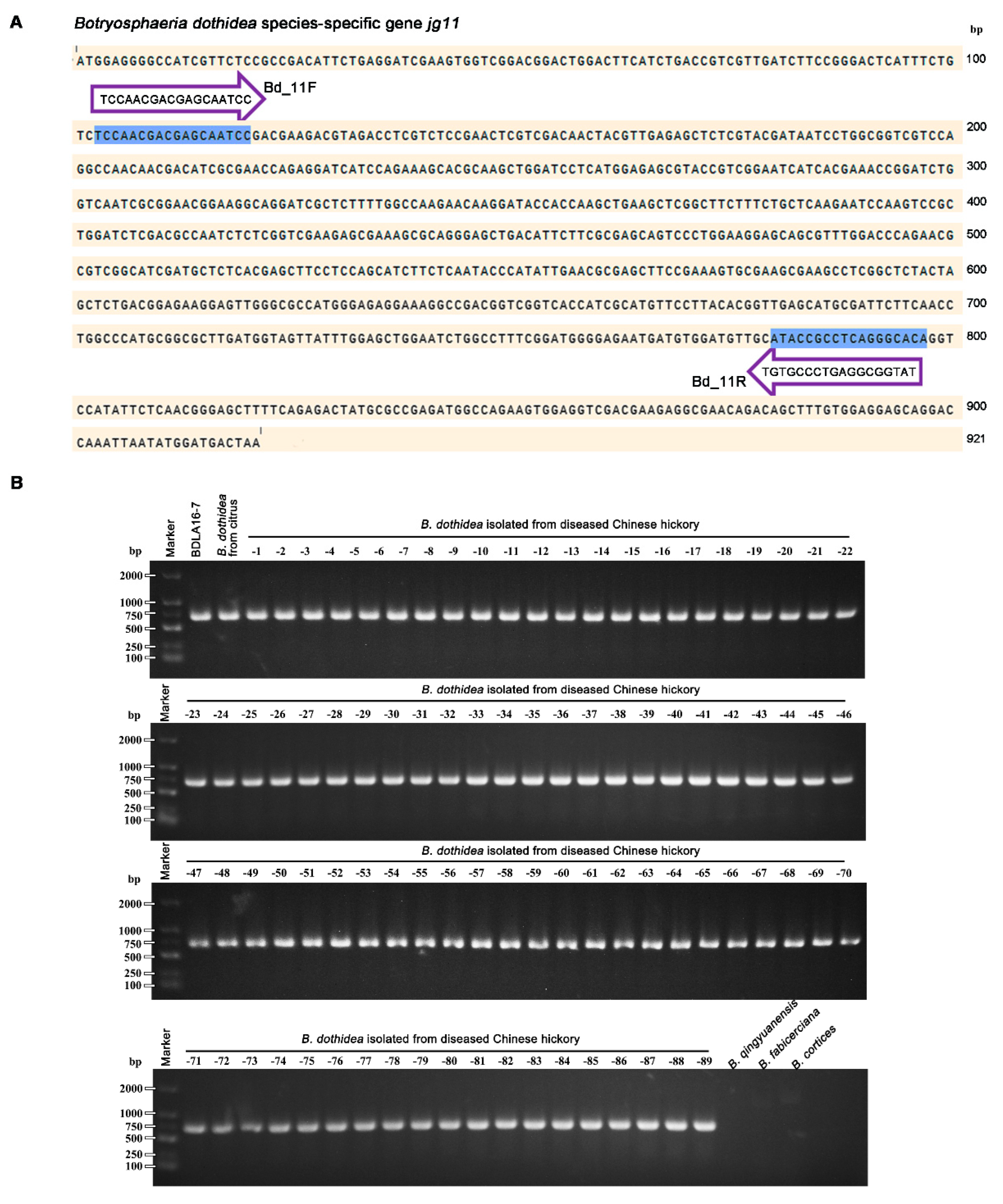
2.4. B. dothidea Rapid Identification Method with Species-Specific Primers

To rapidly identification of B. dothidea, the species-specific genes with length around 1000 bp (listed in the Table S3) were chose for species-specific primer design using Primer3 online web service (https://primer3.org/). For each specific primer the following parameters were checked: primer length, primer melting temperature, GC content, GC clamp, primer secondary structures (hairpins, self-dimer, and cross dimer), repeats, runs, 3′ end stability, avoid template secondary structure, optimum annealing temperature, primer pair tm mismatch calculation. In addition, all species-specific primers were further tested for specificity using web-based Primer-BLAST tool (http://www.ncbi.nlm.nih.gov/tools/primer-blast/index.cgi) to avoid the cross reactivity with the other three Botryosphaeria species. Then primer sets that have no double-ended match with the other three Botryosphaeria species in silico analysis were applied to PCR assays for the screen of the best primers with highest amplification efficiency and specificity.
For the preparation of genomic DNA used in PCR assays, the four Botryosphaeria species were incubated on PDA plates at 25 °C in the dark. After mycelia grew to cover nearly two-thirds of the PDA plate surfaces, the cultures were transferred to a mortar and ground with liquid nitrogen. The resultant powder was placed in a 2 mL centrifuge tube and the mycelial DNA from each isolate was extracted using a Genomic DNA Kit (Sangon Biotech Co., Ltd., Shanghai) according to the manufacturer’s instructions.
The PCR was performed in an Applied Biosystems Veriti Thermal Cycler in 50 μL PCR reaction systems comprising 25 μL of 2 × Rapid Taq master mix (Vazyme, P222-01, Nanjing, China), 1 μL of fungal DNA template, 2 μL of 10 μmol· liter−1 upstream and downstream primers, and 20 μL of ddH2O. PCR reaction conditions included 94 °C for 5 min for denaturation, followed by 35 cycles of 94 °C for 30 s, annealing 57 °C for 30 s, and extension at 72 °C for 45 s, with a final extension at 72 °C for 10 min. Agarose gel electrophoresis (1%) stained with ethidium bromide was then used to verify successful PCR amplification. The experiment was repeated three times.

3. Results

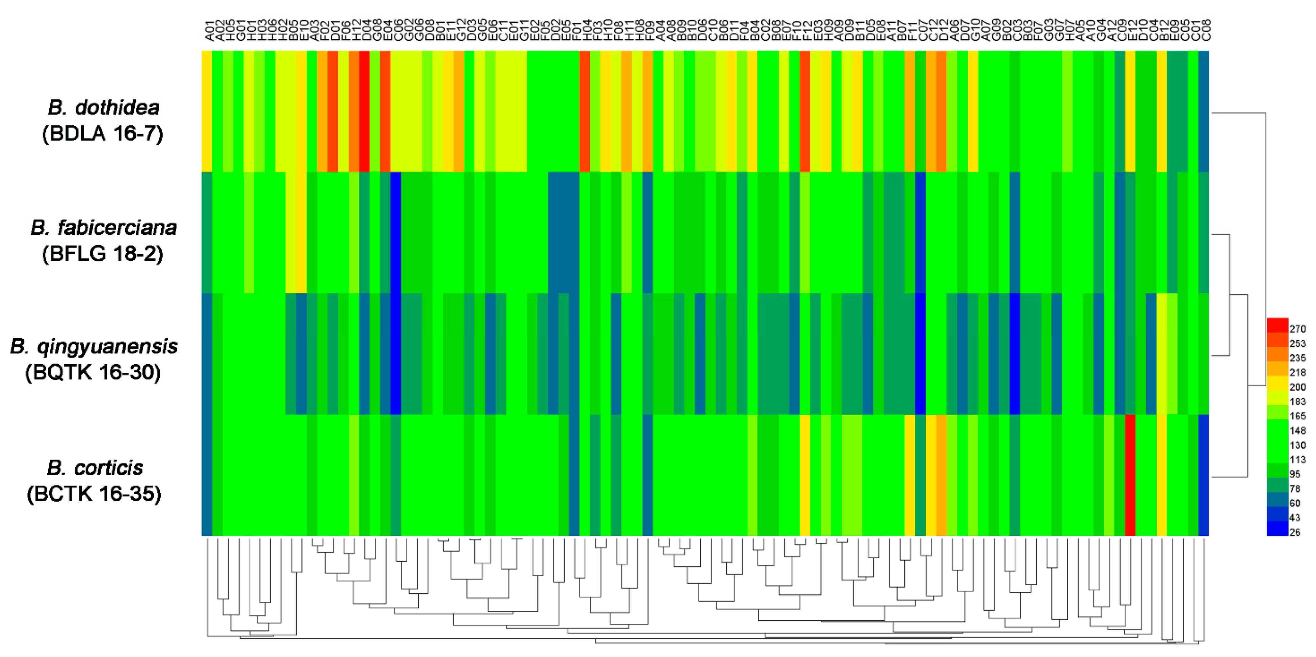
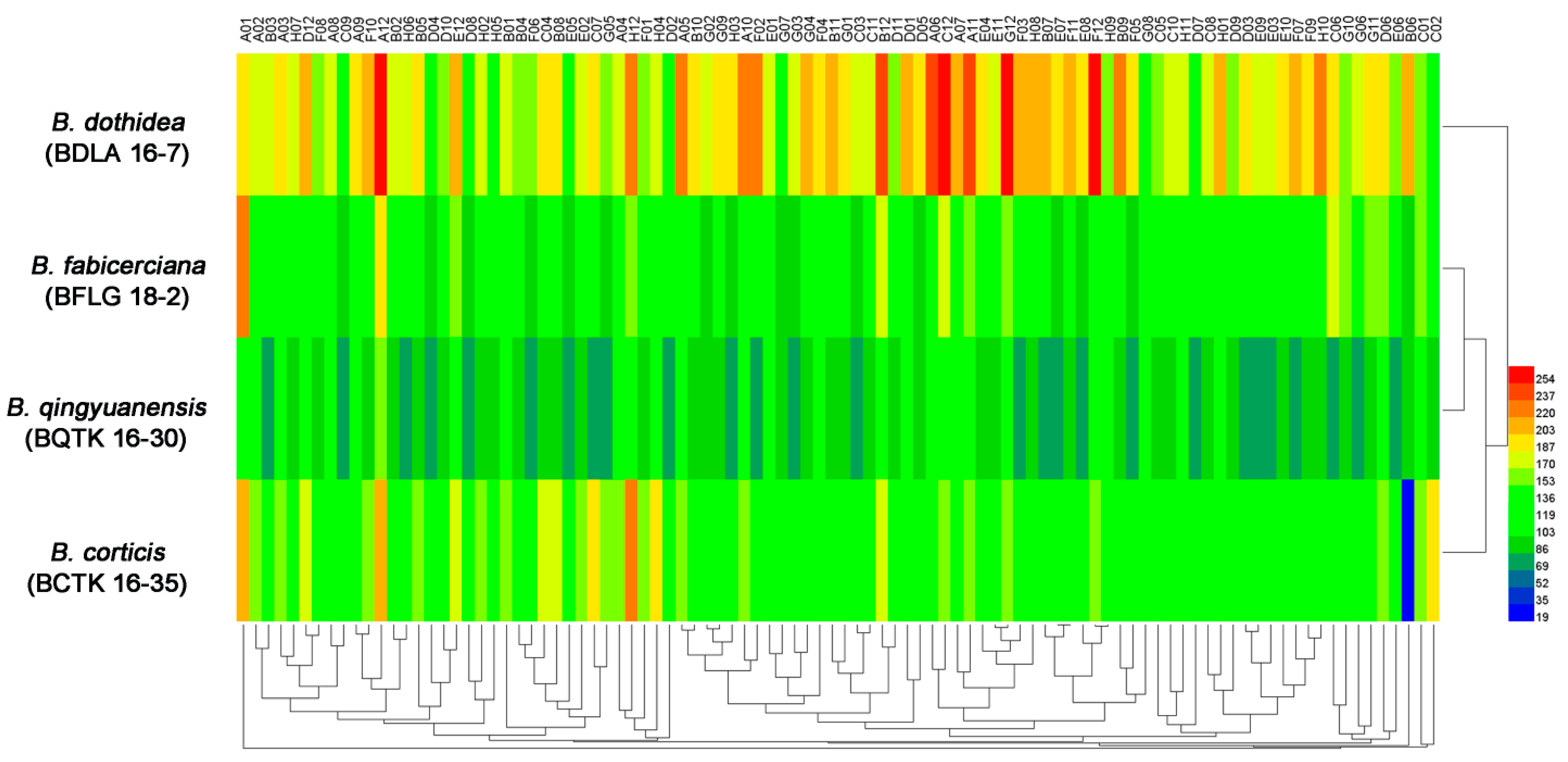
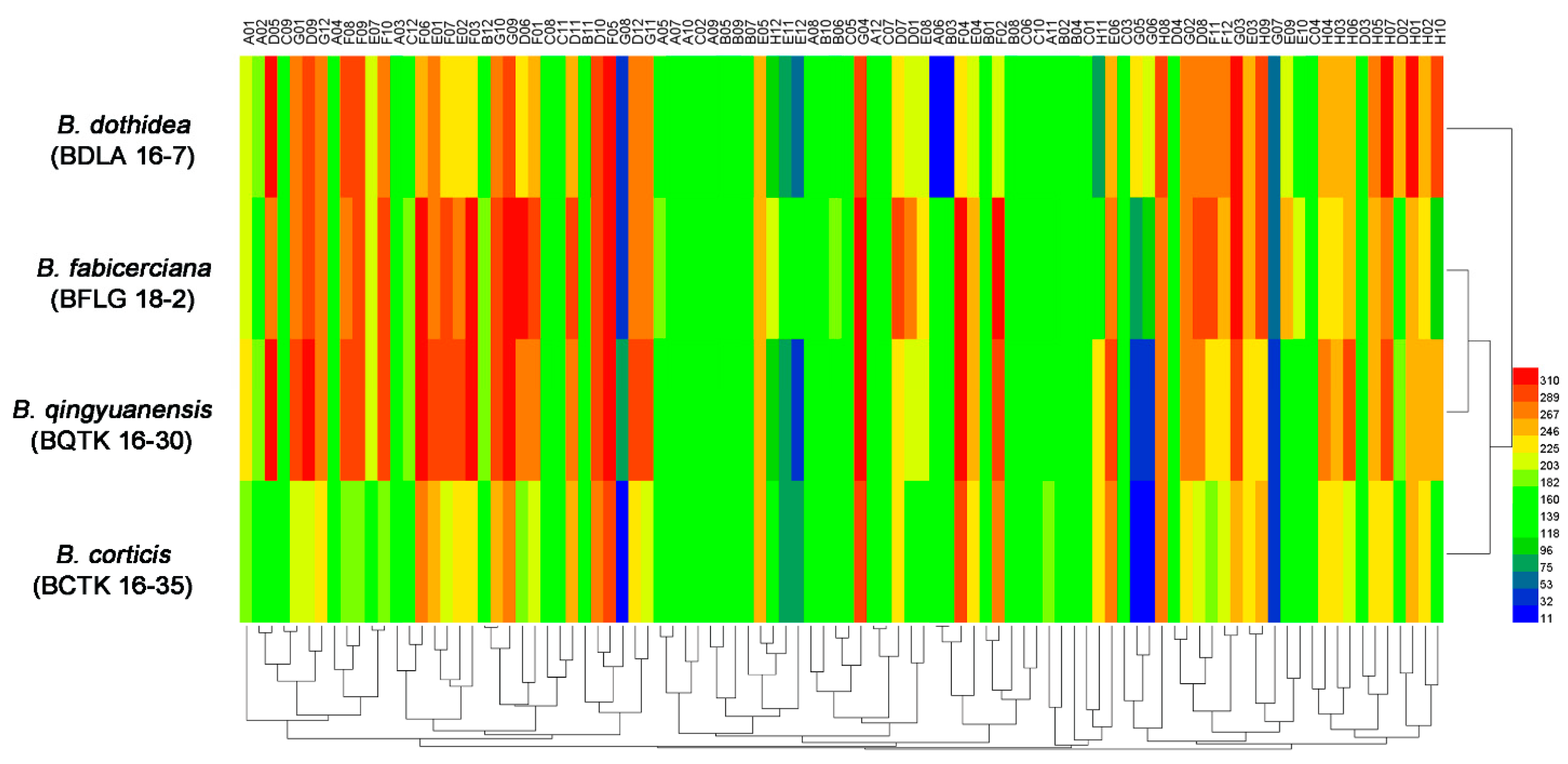
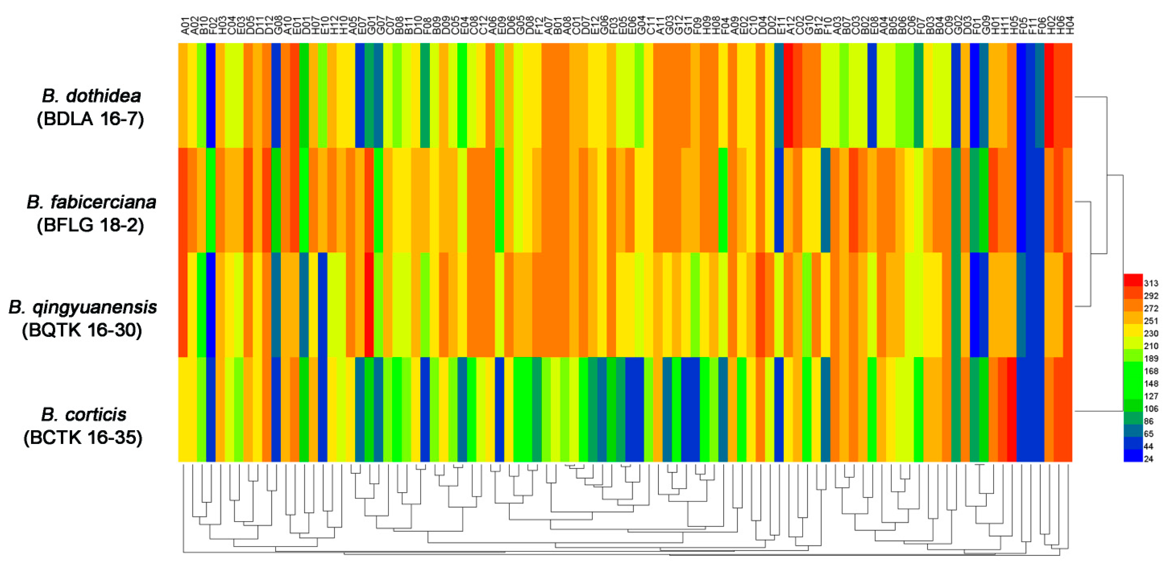
3.1. Phenotypic Characterization of Four Botryosphaeria Species

The PM 3 plate tests characterizing the abilities of four Botryosphaeria species in 95 nitrogen substrates (amino acids) metabolism found B. dothidea presented the highest nitrogen utilization ratio (96.8%), followed by B. fabicerciana (89.5%), B. qingyuanensis (88.4%) and B. corticis (86.3%) (Figure 1, Table 1 and Table S1). In addition, approximately 92 tested compounds were effectively utilized by B. dothidea, except D-serine (plate PM 3, well C8), D-valina (plate PM 3, well C9) and D-galactosamine (plate PM 3, well E9) (Figure 1 and Table S1). In addition, nitrogen sources, including the D-Alanina (plate PM 3, well C3), D-glutamic (plate PM 3, well C6), D-lysine (plate PM 3, well C7), and acetamine (plate PM 3, well E4), were only efficiently metabolized in B. dothidea but not the other three Botryosphaeria species (Figure 1 and Table S1). Surprisingly, for 95 biosynthetic pathways tested via PM 5 plate, all three Botryosphaeria species showed 100% utilization, while B. qingyuanensis demonstrates the inefficiency in L-lysine (plate PM 5, well B6) utilization (Figure 2, Table 1 and Table S1). Taken together, these results revealed the variety among four Botryosphaeria species in the above metabolic and biosynthetic pathways, and B. dothidea demonstrated a broader spectrum of nitrogen source among Botryosphaeria species.
Using the PM 9 plate, phenotypes of the four Botryosphaeria species under various osmotic stresses were tested. As shown in Figure 3, all the four fungi showed active metabolism in utilizing up to 6% potassium chloride (plate PM 9, wells D1 to D4), up to 5% sodium sulfate (plate PM 9, wells D5 to D8), up to 6% sodium, up to 20% ethylene glycol (plate PM 9, wells D9 to D12), up to 6% sodium formate (plate PM 9, wells E1 to E6), up to 12% sodium lactate (plate PM 9, wells F1 to F12), and up to 100 mM sodium nitrite (plate PM 9, wells H1 to H12), while demonstrating significantly attenuated metabolism under the stresses of urea (≥6%, plate PM 9, wells E11 to E12). Interestingly, B. dothidea was found to be capable of utilizing up to 50 mM sodium benzoate (plate PM 9, wells G5 to G6), while the other three could only utilize up to 20 mM sodium benzoate (Figure 3 and Table S1), indicating the possibility that the greater tolerance toward sodium benzoate may account for the widespread incidence and aggressiveness of B. dothidea.
The PM 10 plate assessing the tolerance of pH variation found that though all four Botryosphaeria species were able to survive under pH variation ranged from 3.5 to 10 (plate PM 10, wells A1-A12), and B. dothidea, as well as B. qingyuanensis, was more tolerant to strong alkali (Figure 4 and Table S1). Notably, they were able to maintain active metabolism of multiple nitrogen sources under pH 9.5, for example L-aspartic acid (plate PM 10, well E5), L-glutamic acid (plate PM 10, well E6), L-histidine (plate PM 10, well E9), and L-isoleucine (plate PM 10, well E10), while B. fabicerciana and B. corticis cannot (Figure 4 and Table S1), indicating a stronger pH regulation capability in B. dothidea and B. qingyuanensis.

3.2. Comparison of Genomic Differences among Botryosphaeria Species

According to the NCBI-BLASTn results (gene versus genome, cut at ≥ 95% identity, with 0 hits), we revealed 437 (versus B. corticis), 440 (versus B. fabicerciana) and 185 (versus B. qingyuanensis) B. dothidea specific protein coding genes (Table S2). Combine the above unique gene list, B. dothidea contained 143 species-specific genes absent in the other three Botryosphaeria species (Table S2). Further analysis of the proteins coded by B. dothidea species-specific genes via the KEGG (Kyoto Encyclopedia of Genes and Genomes) pathways (https://www.genome.jp/tools/kofamkoala/) online service found that only 7 proteins were assigned the corresponding KEGG Orthologs (KOs) (Table 2), which may be responsible for the species-specific functions. Additionally, among 7 KEGG annotated proteins, one protein (jg10977) was associated with the calcium permeable stress-gated cation channel, which may have potential roles in the specific osmotic stress tolerance of B. dothidea mentioned above.

3.3. Design and Application of Primers for B. dothidea Molecular Identification

As showed in Figure 5, the screened primer set Bd_11F (5′-TCCAACGACGAGCAATCC-3′) and Bd_11R (5′-TGTGCCCTGAGGCGGTAT-3′) designed based on the sequence of B. dothidea species-specific gene jg11 was found to give an amplicon of 695 bp for B. dothidea strains, including one sequenced strain BDLA16−7 [28], 89 identified B. dothidea isolated from diseased Chinese hickory in our previous study [5], and one identified B. dothidea stain associated with citrus branch diseases [29], but not for B. qingyuanensis, B. fabicerciana, and B. corticis. Taken together, these results showed that sequence of above strain B. dothidea specific genes not only give a predictive information of species-specific function annotation (Table 2) but also provide a basis for the development of B. dothidea molecular identification method. Additionally, the species-specific primer set Bd_11F/Bd_11R was able to carry out the accurate identification of B. dothidea with high efficiency (Figure 5B).

4. Discussion

Botryosphaeria species are amongst the most widespread and important canker and dieback pathogens of trees worldwide [6,7], with B. dothidea as one of the most common Botryosphaeria species [4,5,6,12]. In Chinese hickory trees cankers disease, four Botryosphaeria species, including B. dothidea, B. qingyuanensis, B. fabicerciana, and B. corticis, were identified as Chinese hickory canker-causing agents [5]. Consistently, B. dothidea was also the dominant species due to its highest isolation frequency and strongest virulence [5]. However, the information related to the widespread incidence and aggressiveness of B. dothidea among various Botryosphaeria species causing trunk cankers is poorly understood [4,5,12]. In this study, the metabolic phenotypic diversity of four Chinese hickory canker-causing Botryosphaeria pathogens was systematically studied to address the competitive fitness of B. dothidea via PM 3 (nitrogen utilization), PM 5 (biosynthetic pathways), PM 9 (osmotic pressure), and PM 10 (ionic strength and pH). For nitrogen source metabolization, the highest utilization ratio (96.8%) was found in B. dothidea, followed by B. fabicerciana (89.5%), B. qingyuanensis (88.4%) and B. corticis (86.3%) (Figure 1, Table 1 and Table S1). The efficient utilization of available nitrogen is highly associated with the ability of organism to survive under different environmental situations [30]. For instance, the capability of the methylotrophic yeast Candida boidinii Ramirez to adapt a change in the major available nitrogen source from nitrate to methylamine during the host plant aging was crucial for yeast survival on the leaf environment [18,31]. In addition, the atmospheric N2 fixing endosymbiosis in Ustilago maydis (DC.) Corda, a plant parasitic fungus causing corn smut disease [32], is also a flexible process important for the adaptation of the fungus to survive and grown in media lacking nitrogen [19,33]. Intriguingly, our previous study found that B. dothidea was the most frequently isolated species followed by B. fabicerciana, B. qingyuanensis, and B. cortices [5], which is consistent with the above nitrogen source utilization ratio of the four Botryosphaeria species. These results indicated a possible positive correlation between nitrogen utilization capability and fungal environmental fitness, and the broader spectrum of nitrogen source utilized by B. dothidea may be responsible for its competitive fitness among Botryosphaeria species. However, more researches should be performed to verify this hypothesis in the next study.
Osmotic stress has detrimental effects on microbial survival [22,34,35]. The PM 9 plate testing the phenotypes of the four Botryosphaeria species under various osmotic stresses found that B. dothidea could utilize up to 50 mM sodium benzoate (plate PM 9, wells G5 to G6), while the other three could only utilize up to 20 mM sodium benzoate (Figure 3 and Table S1). Sodium benzoate has been reported to as useful agent with antimicrobial properties that able to inhibit the growth of many microbes, including Pseudomonas aeruginosa (Schroeter) Migula [36], Penicillium commune Thom [37], E. coli, Staphylococcus aureus Rosenbach, Saccharomyces cerevisiae Meyen ex E.C. Hansen [38], and Candida albicans (Robin) Berkhout [39], indicating the possibility that the greater tolerance toward sodium benzoate caused osmotic stress may account for the environmental fitness of B. dothidea. Additionally, the comparative genomics analysis between B. dothidea and the other three Botryosphaeria species found 143 B. dothidea species-specific genes, and further analysis of the above species-specific genes via the KEGG pathways found jg10977 has a predicated role in the calcium permeable stress-gated cation channel. This channel exists in eukaryotic cells from yeasts to animals and plants, and acts as a primary regulator of the initial responses to osmotic pressure [40,41]. Thus, investigating the potential contribution of jg10977 in the specific osmotic stress tolerance of B. dothidea mentioned above could be a valuable degree to further explore the mechanism involved in the wide prevalence and aggressiveness of B. dothidea.
High pH imposes severe stress on the fungal cell, including difficulties in the acquisition of nutrients or reduced availability of essential elements, such as iron or copper [42,43]. Appropriate responses to ambient pH govern fungal virulence during the infection [44]. For example, Sclerotinia sclerotiorum (Libert) de Bary can secrete organic acids acidifying the environment and ensure the proper deployment of cell wall–degrading enzymes (CWDEs) and other pathogenicity factors that function at an optimal pH for their activity during the infection [45]. Additionally, the dedicated Pal/Rim signaling pathway that functions in the response to alkaline pH was shown to be essential for infection in a number of fungal pathogens [42,46], such as C. albicans [47], Fusarium oxysporum Schlechtendahl [48], and Aspergillus fumigatus Fresenius [49]. In this study, PM 10 plate test assessing the tolerance of pH variation found that B. dothidea and B. qingyuanensis were more tolerant to strong alkali among four Botryosphaeria species (Figure 4 and Table S1), indicating a better ability to sense and respond to alkali environment. Significantly, the virulence test in our previous study found the B. dothidea was the most virulent, closely followed by B. qingyuanensis, then B. fabicerciana and B. cortices [5]. Consider the close relation between pH adaptation and fungal virulence [42,44,45], we speculated that the variety in pH regulation capability may be responsible for the virulence differentiation among four Botryosphaeria species.
In the past, the identification of the B. dothidea associated with Chinese hickory trunk cankers mainly relied on morphological identification [5]. In 2019, we developed a quantitative loop-mediated isothermal amplification (q-LAMP) technique to quantitatively monitor B. dothidea in environmental samples based on the different sites of the β-TUBLIN sequence between B. dothidea and other fungi commonly found on Chinese hickory [3]. In 2011, we used the combination of morphological identification and multi-locus phylogenetic analysis of ITS, β-tublin, and EF gene sequences for the accurate distinction of B. dothidea [5]. However, this technique is laborious, expensive, and requires time and knowledge of phylogenetic analysis for identifying the species. Alternatively, the identification of fungal pathogens through a conventional polymerase chain reaction (PCR) using species-specific primers provide an accurate, reproducible, and rapid identification of the species and has been widely used, particularly for economically important plant pathogens [50,51]. In this context, using a comparative genomics analysis, we identified 143 B. dothidea species-specific genes that are not conserved among species (Table S2), and provided a basis for the development of the B. dothidea molecular identification method. The screened primer set Bd_11F (5′-TCCAACGACGAGCAATCC-3′) and Bd_11R (5′-TGTGCCCTGAGGCGGTAT-3′) designed based on the sequence of B. dothidea species-specific gene jg11 was found to be used in the accurate identification of B. dothidea with efficiency and high efficiency (Figure 5B), which is of significant importance for B. dothidea-associated cankers diagnosis, prevention, and control.
Collectively, using the large-scale screening of physiologic traits of four Botryosphaeria species associated with Chinese hickory cankers, we found B. dothidea has a broader spectrum of nitrogen sources and a greater tolerance toward osmotic pressure (sodium benzoate) and alkali stress among Botryosphaeria species. Moreover, the annotation of B. dothidea species-specific genomic information via a comparative genomics analysis not only gives a basis for the development of the B. dothidea molecular identification method but also provides crucial cues in the prediction of B. dothidea species-specific function that account for its high prevalence and wide distribution. However, the speculation of the mechanism involved in the wide prevalence and aggressiveness of B. dothidea in this study still require further study to verify.

Supplementary Materials

The following supporting information can be downloaded at: https://www.mdpi.com/article/10.3390/jof9020204/s1: Table S1: Metabolic profiling of four Botryosphaeria species on Biolog PM plates. ‘+’ and ‘-’ represent the corresponding substrate was metabolized and not metabolized by each Botryosphaeria species examined using the Biolog PM plates. Table S2: Lists of Botryosphaeria dothidea (BDLA16−7) species-specific genes. The comparative genomics analysis of B. dothidea species-specific genes were carried out via the NCBI-BLASTnN (gene versus genome, cut-off at ≥ 95% identity with 0 hits). Table S3: Specific candidate primers for B. dothidea molecular identification based on species-specific genes.

Author Contributions

C.Z. supervised the project; T.M. conceived the study and designed the experiments; T.M., Y.Z. and C.Y. performed the experiments; and T.M., Y.Z., C.Y. and C.Z. analyzed the data and wrote the manuscript. All authors have read and agreed to the published version of the manuscript.

Funding

This research was supported by the Key Research and Development Project of Zhejiang Province, China (2020C02005), and Scientific Research and Development Fund Project of Zhejiang Agriculture and Forestry University (2022LFR052).

Institutional Review Board Statement

Not applicable.

Informed Consent Statement

Not applicable.

Data Availability Statement

The data in this study are available in this article.

Conflicts of Interest

The authors declare no conflict of interest.

References

  1. Zhu, C.G.; Deng, X.Y.; Shi, F. Evaluation of the antioxidant activity of Chinese hickory Carya cathayensis kernel ethanol extraction. Afr. J. Biotechnol. 2008, 7, 2169–2173. [Google Scholar]
  2. Dai, J.D.; Wang, H.D.; Wang, Y.P.; Zhang, C.Q. Management of Chinese hickory (Carya cathayensis) trunk canker through effective fungicide application programs and baseline sensitivity of Botryosphaeria dothidea to trifloxystrobin. Australas Plant Path. 2017, 46, 75–82. [Google Scholar] [CrossRef]
  3. Wang, Q.W.; Zhang, C.Q. q-LAMP assays for the detection of Botryosphaeria dothidea causing Chinese hickory canker in trunk, water, and air samples. Plant Dis. 2019, 103, 3142–3149. [Google Scholar] [CrossRef] [PubMed]
  4. Yao, J.; Mei, L.; Jiang, H.; Hu, G.; Wang, Y. Evaluation of Carya cathayensis resistance to Botryosphaeria trunk canker using grafting on pecan. Sci. Hortic. 2019, 248, 184–188. [Google Scholar] [CrossRef]
  5. Zhuang, C.J.; Wang, Q.W.; Wu, Q.Q.; Qiu, Z.L.; Xu, B.C.; Zhang, C. Diversity of Botryosphaeriaceae species associated with Chinese hickory tree (Carya cathayensis) trunk cankers. Plant Dis. 2021, 105, 3869–3879. [Google Scholar] [CrossRef]
  6. Phillips, A.; Alves, A.; Abdollahzadeh, J.; Slippers, B.; Wingfield, M.J.; Groenewald, J.Z.; Crous, P.W. The Botryosphaeriaceae: Genera and species known from culture. Stud. Mycol. 2013, 76, 51–167. [Google Scholar] [CrossRef]
  7. Xu, S.E.; Yao, J.L.; Wu, F.; Mei, L.; Wang, Y.J. Evaluation of Paenibacillus polymyxa carboxymethylcellulose/poly (vinyl alcohol) formulation for control of Carya cathayensis canker caused by Botryosphaeria dothidea. Forest Pathol. 2018, 48, e12464. [Google Scholar] [CrossRef]
  8. Adesemoye, A.O.; Mayorquin, J.S.; Wang, D.H.; Twizeyimana, M.; Lynch, S.C.; Eskalen, A. Identification of species of Botryosphaeriaceae causing bot gummosis in citrus in California. Plant Dis. 2014, 98, 55–61. [Google Scholar] [CrossRef]
  9. Mayorquin, J.S.; Wang, D.H.; Twizeyimana, M.; Eskalen, A. Identification, distribution, and pathogenicity of Diatrypaceae and Botryosphaeriaceae associated with citrus branch canker in the southern California desert. Plant Dis. 2016, 100, 2402–2413. [Google Scholar] [CrossRef]
  10. Tang, W.; Ding, Z.; Zhou, Z.Q.; Wang, Y.Z.; Guo, L.Y. Phylogenetic and pathogenic analyses show that the causal agent of apple ring rot in China is Botryosphaeria dothidea. Plant Dis. 2012, 96, 486–496. [Google Scholar] [CrossRef]
  11. Ma, Z.; Boehm, E.W.; Luo, Y.; Michailides, T.J. Population structure of Botryosphaeria dothidea from pistachio and other hosts in California. Phytopathology 2001, 91, 665–672. [Google Scholar] [CrossRef] [PubMed] [Green Version]
  12. Mehl, J.; Slippers, B.; Roux, J.; Wingfield, M.J. Cankers and other diseases caused by the Botryosphaeriaceae. Infect. For. Dis. 2013, 298–317. [Google Scholar]
  13. Marsberg, A.; Kemler, M.; Jami, F.; Nagel, J.H.; Postma-Smidt, A.; Naidoo, S.; Wingfield, M.J.; Crous, P.W.; Spatafora, J.W.; Hesse, C.N.; et al. Botryosphaeria dothidea: A latent pathogen of global importance to woody plant health. Mol. Plant Pathol. 2017, 18, 477–488. [Google Scholar] [CrossRef] [PubMed]
  14. Ho, A.; Di Lonardo, D.P.; Bodelier, P.L. Revisiting life strategy concepts in environmental microbial ecology. FEMS Microbiol. Ecol. 2017, 93, fix006. [Google Scholar] [CrossRef]
  15. Bochner, B.R. New technologies to assess genotype-phenotype relationships. Nat. Rev. Genet. 2003, 4, 309–314. [Google Scholar] [CrossRef]
  16. Bochner, B.R.; Gadzinski, P.; Panomitros, E. Phenotype microarrays for high-throughput phenotypic testing and assay of gene function. Genome Res. 2001, 11, 1246–1255. [Google Scholar] [CrossRef]
  17. Barbosa, C.; Mendes-Faia, A.; Mendes-Ferreira, A. The nitrogen source impacts major volatile compounds released by Saccharomyces cerevisiae during alcoholic fermentation. Int. J. Food Microbiol. 2012, 160, 87–93. [Google Scholar] [CrossRef]
  18. Shiraishi, K.; Oku, M.; Uchida, D.; Yurimoto, H.; Sakai, Y. Regulation of nitrate and methylamine metabolism by multiple nitrogen sources in the methylotrophic yeast Candida boidinii. Fems Yeast Res. 2015, 15, fov084. [Google Scholar] [CrossRef]
  19. Sanchez-Arreguin, J.A.; Hernandez-Onate, M.A.; Leon-Ramirez, C.G.; Ruiz-Herrera, J. Transcriptional analysis of the adaptation of Ustilago maydis during growth under nitrogen fixation conditions. J. Basic Microbiol. 2017, 57, 597–604. [Google Scholar] [CrossRef]
  20. Canas, P.; Morales, A.M.; Manso, J.; Romero, E.G.; Vinas, M.; Palomo, E.S. Chemical and sensory characrerization of the aroma of ‘chardonnay’ musts fermented with different nitrogen sources. Cienc Tec Vitivinic 2018, 33, 116–124. [Google Scholar]
  21. Da Silva, L.G.; Martins, M.P.; Sanches, P.R.; Peres, N.; Martinez-Rossi, N.M.; Rossi, A. Saline stress affects the pH-dependent regulation of the transcription factor PacC in the dermatophyte Trichophyton interdigitale. Braz. J. Microbiol. 2020, 51, 1585–1591. [Google Scholar] [CrossRef] [PubMed]
  22. Fedoseeva, E.V.; Tereshina, V.M.; Danilova, O.A.; Ianutsevich, E.A.; Yakimenko, O.S.; Terekhova, V.A. Effect of humic acid on the composition of osmolytes and lipids in a melanin-containing phytopathogenic fungus Alternaria alternata. Environ. Res. 2021, 193, 110395. [Google Scholar] [CrossRef] [PubMed]
  23. Wang, H.C.; Guo, H.; Cai, L.; Cai, L.T.; Guo, Y.S.; Ding, W. Effect of temperature on phenotype characterization of Ralstonia solanacearum from tobacco. Can. J. Plant Pathol. 2020, 42, 164–181. [Google Scholar] [CrossRef]
  24. Wang, H.C.; Li, L.C.; Cai, B.; Cai, L.T.; Chen, X.J.; Yu, Z.H.; Zhang, C.Q. Metabolic phenotype characterization of Botrytis cinerea, the causal agent of gray mold. Front. Microbiol. 2018, 9, 470. [Google Scholar] [CrossRef]
  25. Gusarov, I.; Shatalin, K.; Starodubtseva, M.; Nudler, E. Endogenous nitric oxide protects bacteria against a wide spectrum of antibiotics. Science 2009, 325, 1380–1384. [Google Scholar] [CrossRef]
  26. Chen, X.; Li, L.; Wang, H.; Huang, Y.; Zhang, C. Phenotypic fingerprints of Ralstonia solanacearum under various osmolytes and pH environments. Plant Pathol. J. 2016, 15, 102–107. [Google Scholar] [CrossRef]
  27. Wang, H.; Huang, Y.; Xia, H.; Wang, J.; Wang, M.; Zhang, C.; Lu, H. Phenotypic Analysis of Alternaria alternata, the Causal Agent of Tobacco Brown Spot. Plant Pathol. J. 2015, 14, 79–85. [Google Scholar] [CrossRef]
  28. Bao, J.; Wu, Q.; Huang, J.; Zhang, C.Q. High-quality genome assembly and annotation resource of Botryosphaeria dothidea strain BDLA16−7, causing trunk canker disease on Chinese hickory. Plant Dis. 2022, 106, 1023–1026. [Google Scholar] [CrossRef]
  29. Xiao, X.E.; Wang, W.; Crous, P.W.; Wang, H.K.; Jiao, C.; Huang, F.; Pu, Z.X.; Zhu, Z.R.; Li, H.Y. Species of Botryosphaeriaceae associated with citrus branch diseases in China. Persoonia 2021, 47, 106–135. [Google Scholar] [CrossRef]
  30. Leigh, J.A.; Dodsworth, J.A. Nitrogen regulation in bacteria and archaea. Annu. Rev. Microbiol. 2007, 61, 349–377. [Google Scholar] [CrossRef]
  31. Shiraishi, K.; Oku, M.; Kawaguchi, K.; Uchida, D.; Yurimoto, H.; Sakai, Y. Yeast nitrogen utilization in the phyllosphere during plant lifespan under regulation of autophagy. Sci. Rep. 2015, 5, 9719. [Google Scholar] [CrossRef] [PubMed]
  32. Brefort, T.; Doehlemann, G.; Mendoza-Mendoza, A.; Reissmann, S.; Djamei, A.; Kahmann, R. Ustilago maydis as a pathogen. Annu. Rev. Phytopathol 2009, 47, 423–445. [Google Scholar] [CrossRef] [PubMed]
  33. Ruiz-Herrera, J.; Leon-Ramirez, C.; Vera-Nunez, A.; Sanchez-Arreguin, A.; Ruiz-Medrano, R.; Salgado-Lugo, H.; Sanchez-Segura, L.; Pena-Cabriales, J.J. A novel intracellular nitrogen-fixing symbiosis made by Ustilago maydis and Bacillus spp. New Phytol. 2015, 207, 769–777. [Google Scholar] [CrossRef]
  34. Shabala, S.; Shabala, L. Ion transport and osmotic adjustment in plants and bacteria. Biomol. Concepts 2011, 2, 407–419. [Google Scholar] [CrossRef] [PubMed]
  35. Ma, L.; Li, C.Z. Effects of cobalt and DFMA on polyamine content and membrane-lipid peroxidation in wheat seedling under osmotic stresses. Agric. Res. Arid. Areas 2010, 28, 136. [Google Scholar]
  36. Malla, C.F.; Mireles, N.A.; Ramirez, A.S.; Poveda, J.B.; Tavio, M.M. Aspirin, sodium benzoate and sodium salicylate reverse resistance to colistin in Enterobacteriaceae and Pseudomonas aeruginosa. J. Antimicrob. Chemother. 2020, 75, 3568–3575. [Google Scholar] [CrossRef] [PubMed]
  37. Zhelifonova, V.P.; Antipova, T.V.; Kozlovskii, A.G. Effect of potassium sorbate, sodium benzoate, and sodium nitrite on biosynthesis of cyclopiazonic and mycophenolic acids and citrinin by fungi of the Penicillium genus. Appl. Biochem. Micro. 2017, 53, 711–714. [Google Scholar] [CrossRef]
  38. Yardimci, B.K.; Sahin, S.C.; Sever, N.I.; Ozek, N.S. Biochemical effects of sodium benzoate, potassium sorbate and sodium nitrite on food spoilage yeast Saccharomyces cerevisiae. Biologia 2022, 77, 547–557. [Google Scholar] [CrossRef]
  39. Dong, Y.L.; Ding, Z.F.; Song, L.X.; Zhang, D.S.; Xie, C.J.; Zhang, S.J.; Feng, L.; Liu, H.L.; Pang, Q.X. Sodium benzoate delays the development of Drosophila melanogaster larvae and alters commensal microbiota in adult Flies. Front. Microbiol. 2022, 13, 911928. [Google Scholar] [CrossRef]
  40. Jiang, L.; Pan, H. Functions of CaPhm7 in the regulation of ion homeostasis, drug tolerance, filamentation and virulence in Candida albicans. BMC Microbiol. 2018, 18, 49. [Google Scholar] [CrossRef]
  41. Medrano-Soto, A.; Moreno-Hagelsieb, G.; McLaughlin, D.; Ye, Z.S.; Hendargo, K.J.; Saier, M.J. Bioinformatic characterization of the anoctamin superfamily of Ca2+-activated ion channels and lipid scramblases. PLoS ONE 2018, 13, e0192851. [Google Scholar] [CrossRef] [PubMed]
  42. Fernandes, T.R.; Segorbe, D.; Prusky, D.; Di Pietro, A. How alkalinization drives fungal pathogenicity. PLoS Pathog. 2017, 13, e1006621. [Google Scholar] [CrossRef] [PubMed]
  43. Cyert, M.S.; Philpott, C.C. Regulation of cation balance in Saccharomyces cerevisiae. Genetics 2013, 193, 677–713. [Google Scholar] [CrossRef]
  44. Tardi-Ovadia, R.; Linker, R.; Tsror, L.L. Direct estimation of local pH change at infection sites of fungi in potato tubers. Phytopathology 2017, 107, 132–137. [Google Scholar] [CrossRef] [Green Version]
  45. Xu, L.; Li, G.; Jiang, D.; Chen, W. Sclerotinia sclerotiorum: An evaluation of virulence theories. Annu. Rev. Phytopathol 2018, 56, 311–338. [Google Scholar] [CrossRef]
  46. Davis, D.A. How human pathogenic fungi sense and adapt to pH: The link to virulence. Curr. Opin. Microbiol. 2009, 12, 365–370. [Google Scholar] [CrossRef]
  47. Davis, D.; Edwards, J.J.; Mitchell, A.P.; Ibrahim, A.S. Candida albicans RIM101 pH response pathway is required for host-pathogen interactions. Infect Immun. 2000, 68, 5953–5959. [Google Scholar] [CrossRef] [PubMed]
  48. Ortoneda, M.; Guarro, J.; Madrid, M.P.; Caracuel, Z.; Roncero, M.I.; Mayayo, E.; Di Pietro, A. Fusarium oxysporum as a multihost model for the genetic dissection of fungal virulence in plants and mammals. Infect Immun. 2004, 72, 1760–1766. [Google Scholar] [CrossRef]
  49. Bertuzzi, M.; Schrettl, M.; Alcazar-Fuoli, L.; Cairns, T.C.; Munoz, A.; Walker, L.A.; Herbst, S.; Safari, M.; Cheverton, A.M.; Chen, D.; et al. Correction: The pH-Responsive PacC transcription factor of Aspergillus fumigatus governs epithelial entry and tissue invasion during pulmonary aspergillosis. PLoS Pathog. 2015, 11, e1004943. [Google Scholar] [CrossRef]
  50. Fan, X.; Zhang, J.; Yang, L.; Wu, M.D.; Chen, W.D.; Li, G.Q. Development of PCR-based assays for detecting and differentiating three species of Botrytis infecting broad bean. Plant Dis. 2015, 99, 691–698. [Google Scholar] [CrossRef]
  51. Ni, H.F.; Yang, H.R.; Chen, R.S.; Hung, T.H.; Liou, R.F. A nested multiplex PCR for species-specific identification and detection of Botryosphaeriaceae species on mango. Eur. J. Plant Pathol. 2012, 133, 819–828. [Google Scholar] [CrossRef]
Figure 1. | Overview of metabolic phenotypes of four Botryosphaeria species, including B. dothidea strain BDLA16−7, B. corticis strain BCTK16−35, B. fabicerciana strain BFLG18−2, and B. qingyuanensis strain BQTK16−30, on 95 nitrogen sources tested using the PM 3 plate. Heatmap of maximum area values of 95 nitrogen sources expressed as maximum curve area monitored during 96 h of incubation. The legend of color code from blue to green, and red shades represent low, moderate, and high utilization rate of nitrogen sources, respectively.
Jof 09 00204 g001
Figure 2. | Overview of phenotypes of four Botryosphaeria species, including B. dothidea strain BDLA16−7, B. corticis strain BCTK16−35, B. fabicerciana strain BFLG18−2, and B. qingyuanensis strain BQTK16-30, on 95 biosynthetic pathways tested using the PM 5 plate. Heatmap of maximum area values of 95 biosynthetic pathways expressed as maximum curve area monitored during 96 h of incubation. The legend of color code from blue to green, and red shades represent low, moderate, and high metabolic rate during the corresponding biosynthetic processes, respectively.
Jof 09 00204 g002
Figure 3. | Overview of osmotic stress tolerance of four Botryosphaeria species, including B. dothidea strain BDLA16−7, B. corticis strain BCTK16−35, B. fabicerciana strain BFLG18−2, and B. qingyuanensis strain BQTK16−30, under 96 osmotic conditions tested using the PM 9 plate. Heatmap of maximum area values of 96 ionic conditions expressed as maximum curve area monitored during 96 h of incubation. The legend of color code from blue to green, and red shades represent low, moderate, and high metabolic rate under the corresponding osmotic stresses, respectively.
Jof 09 00204 g003
Figure 4. | Overview of pH regulation capability of four Botryosphaeria species, including B. dothidea strain BDLA16−7, B. corticis strain BCTK16−35, B. fabicerciana strain BFLG18−2, and B. qingyuanensis strain BQTK16−30, under 96 pH conditions tested using the PM 10 plate. Heatmap of maximum area values of 96 pH conditions expressed as maximum curve area monitored during 96 h of incubation. The legend of color code from blue to green, and red shades represent low, moderate, and high metabolic rate under the corresponding pH stresses, respectively.
Jof 09 00204 g004
Figure 5. Species−specific primer set Bd_11F/R was able to carry out the accurate identification of B. dothidea. (A) The species-specific primers Bd_11F and Bd_11R designed based on the species-specific gene jg11 for molecular identification of B. dothidea. The forward and reverse primer sequences were highlighted with shade and arrow for orientation. (B) Agarose gel electrophoresis showed the species-specific primer set Bd_11F/R was able to carry out the accurate molecular identification of B. dothidea. The primers could give an amplicon of 695 bp for B. dothidea strains, including one sequenced strain BDLA16−7, one identified B. dothidea stain associated with citrus branch diseases, and 89 identified B. dothidea isolated from diseased Chinese hickory, but not for B. qingyuanensis (BQTK16−30), B. fabicerciana (BFLG18−2), and B. corticis (BCTK16−35).
Jof 09 00204 g005
Table 1. Metabolic ratio of four Botryosphaeria spp. on the corresponding Biolog MicroPlates.
StrainAmino Acid Nitrogen Substrates (%)Biosynthetic Pathways (%) Osmotic and Ionic Conditions (%)pH Condition (%)
B. dothidea
(BDLA 16−7)
96.810093.889.6
B. fabicerciana
(BFLG 18−2)
89.510093.868.8
B. qingyuanensis
(BQTK 16−30)
88.498.993.889.6
B. corticis
(BCTK 16−35)
86.310093.868.8
Percentages listed in each column is the metabolic ratio of four Botryosphaeria spp. on the corresponding Biolog MicroPlates. Metabolic ratio (%) = the number of substrates metabolized by the corresponding Botryosphaeria spp. in each kind of Biolog microplate/the total number of the substrates in each kind of Biolog microplate × 100%.
Table 2. The functional Kyoto Encyclopedia of Genes and Genomes (KEGG) analysis of B. dothidea species-specific genes.
Gene NameKEGG Ortholog(KO)ThrshldScoreE-ValueKO Definition
jg9527K2332248.0374.26.8 × 10−23aspirochlorine biosynthesis cytochrome P450 monooxygenase
jg3332K22993508.27561.66.3 × 10−171bifunctional Delta-12/omega-3 fatty acid desaturase
jg10959K2350491.30266.62.9 × 10−81protein SERAC1
jg10977K21989150.87173.25.8 × 10−53calcium permeable stress-gated cation channel
g8479K01183118.77159.11.4 × 10−48chitinase
jg3335K01687579.77924.54 × 10−280dihydroxy-acid dehydratase
jg3334K0686798.20108.34.4 × 10−33uncharacterized protein
Disclaimer/Publisher’s Note: The statements, opinions and data contained in all publications are solely those of the individual author(s) and contributor(s) and not of MDPI and/or the editor(s). MDPI and/or the editor(s) disclaim responsibility for any injury to people or property resulting from any ideas, methods, instructions or products referred to in the content.

