Article

Functional Analysis of the P-Type ATPases Apt2-4 from Cryptococcus neoformans by Heterologous Expression in Saccharomyces cerevisiae

by
Sarina Veit
1,
Sabine Laerbusch
1,
Rosa L. López-Marqués
2,* and
Thomas Günther Pomorski
1,2,*
1
Department of Molecular Biochemistry, Faculty of Chemistry and Biochemistry, Ruhr University Bochum, 44801 Bochum, Germany
2
Department of Plant and Environmental Sciences, University of Copenhagen, Thorvaldsensvej 40, DK-1871 Frederiksberg C, Denmark
*
Authors to whom correspondence should be addressed.
J. Fungi 2023, 9(2), 202; https://doi.org/10.3390/jof9020202
Submission received: 27 December 2022 / Revised: 1 February 2023 / Accepted: 2 February 2023 / Published: 4 February 2023
(This article belongs to the Section Fungal Cell Biology, Metabolism and Physiology)

Abstract

:
Lipid flippases of the P4-ATPase family actively transport phospholipids across cell membranes, an activity essential for key cellular processes such as vesicle budding and membrane trafficking. Members of this transporter family have also been implicated in the development of drug resistance in fungi. The encapsulated fungal pathogen Cryptococcus neoformans contains four P4-ATPases, among which Apt2-4p are poorly characterized. Using heterologous expression in the flippase-deficient S. cerevisiae strain dnf1Δdnf2Δdrs2Δ, we tested their lipid flippase activity in comparison to Apt1p using complementation tests and fluorescent lipid uptake assays. Apt2p and Apt3p required the co-expression of the C. neoformans Cdc50 protein for activity. Apt2p/Cdc50p displayed a narrow substrate specificity, limited to phosphatidylethanolamine and –choline. Despite its inability to transport fluorescent lipids, the Apt3p/Cdc50p complex still rescued the cold-sensitive phenotype of dnf1Δdnf2Δdrs2Δ, suggesting a functional role for the flippase in the secretory pathway. Apt4p, the closest homolog to Saccharomyces Neo1p, which does not require a Cdc50 protein, was unable to complement several flippase-deficient mutant phenotypes, neither in the presence nor absence of a β-subunit. These results identify C. neoformans Cdc50 as an essential subunit for Apt1-3p and provide a first insight into the molecular mechanisms underlying their physiological functions.

1. Introduction

Cryptococcosis is a severe infection of the central nervous system, especially the brain tissue, that leads to meningoencephalitis and is the second most common cause of death in immunocompromised HIV patients [1]. It is predominantly caused by the encapsulated yeast Cryptococcus neoformans, which enters the blood stream through the lung, thereby occasionally resulting in additional pneumonia [2]. In 2022, the World Helath Organization (WHO) declared this as a critical pathogen for which research and development efforts should be prioritized [3]. The currently available drugs against C. neoformans are limited by their ability to cross the blood-brain barrier and their toxicity causes severe side effects. In addition, the requirement for prolonged treatment (6–12 months) favors the development of fungal drug resistance [4,5]. Novel therapeutic strategies could target the capsule morphology and polysaccharide secretion, which are crucial for the virulence of C. neoformans, highlighting the role that the secretory pathway plays in its pathogenicity [6].
Throughout secretion and especially for vesicular transport, cellular membranes undergo dynamic morphological changes and re-organization in terms of lipid composition and distribution over the two leaflets of the bilayer. A key player in these processes are P4-ATPases that catalyze the translocation of phospholipids from the exoplasmic to the cytosolic leaflet of biological membranes, a process termed “lipid flipping” [7]. Members of this transporter family have also been implicated in the development of drug resistance in fungi, which includes resistance to alkyllysophospholipids [8,9,10]. Due to their importance in fungal infection, the functional characterization of these so-called flippases is essential to understanding the infection mechanism and subsequently using them as new fungal drug targets, as recently shown for the malaria parasite [11].
In fungi, the family of P4-ATPases is best studied for S. cerevisiae. This non-pathogenic yeast expresses five P4-ATPases, including Dnf1p and Dnf2p at the plasma membrane (PM), Drs2p and Dnf3p mostly in the trans-Golgi network, and Neo1p in the endosomal membranes and Golgi [12,13]. In C. neoformans, only four P4-ATPases, Apt1-4p, were identified by sequence alignment [14,15]. Deletion studies in vivo revealed that Apt1p plays an important role in polysaccharide export, capsule size, and virulence [14,16]. In contrast, separate APT2-4 deletions did not show any detectable effect except for the APT3 deletion, which caused a minor increase in sensitivity to the trafficking inhibitor BFA, as well as fluconazole, an inhibitor for ergosterol biosynthesis [17]. However, functional redundancy among flippases has been observed before, emphasizing the importance of heterologous expression strategies for proper analysis [18]. In fact, Apt1p was recently functionally characterized based on heterologous expression in S. cerevisiae as a lipid transporter with broad substrate specificity. Besides the glycerophospholipids phosphatidylserine (PS), -choline (PC), -ethanolamine (PE), and -glycerol (PG), the transport of galactosyl ceramide (GalCer), glucosyl ceramide (GlcCer), and sphingomyelin (SM) was detected [19]. Substrate specificity and key features of Apt2-4p remain to be assessed.
Most P4-ATPases are functional only as a heterodimer with their β-subunit, which is involved in the localization to the destination membrane, as well as in the lipid transport activity itself [8,20,21,22]. While the S. cerevisiae genome contains three genes coding for β-subunits (CDC50, LEM3, and CRF1) [23], only one gene of this family, previously named CDC50, was identified in the genome of C. neoformans. Studies in vivo demonstrated that Cdc50p localizes throughout the whole secretory pathway and its deletion leads to reduced virulence, higher drug sensitivity, and exposure of phosphatidylserine on the outer leaflet of the plasma membrane [9,17]. Further studies showed impaired lipid uptake at the plasma membrane via Apt1p, when expressed without Cdc50p, presumably due to impaired trafficking and therefore the retention of Apt1p in the endoplasmic reticulum [19]. Moreover, recent studies showed a strong interaction between subunits via the extracellular loop of Cdc50p, which motivated the design of antifungal peptides [24]. The subclass of P4B-ATPases seems to work without a β-subunit and comprises among others Neo1p from S. cerevisiae, which shares high sequence similarities with Apt4p [25].
In this study, we performed a functional characterization of Apt2p, Apt3p, and Apt4p in comparison to Apt1p through heterologous expression. We employed a well-established system based on the dnf1Δdnf2Δdrs2Δ background in S. cerevisiae [26]. This mutant strain is deleted in three out of five endogenous P4-ATPases, consequently displaying a low background of phospholipid uptake at the plasma membrane and altered membrane lipid asymmetry. Since previous studies did not show clear phenotypes for single APT2-4 deletion mutants due to redundancy, we used the heterologous expression system to provide first insight into the subcellular localization and substrate specificity of Apt2-4p, and to help unravel their physiological functions. Furthermore, we addressed the dependency on Cdc50p co-expression for proper Apt protein localization and flippase activity.

2. Materials and Methods

2.1. Materials

Miltefosine (#850337) and most NBD-lipids were purchased from Avanti Polar Lipids (Alabaster, AL, USA), including 1-palmitoyl-2-{6-[(7-nitro-2-1,3-benzoxadiazol-4-yl)amino]hexanoyl}-sn-glycero-3-phosphocholine (NBD-PC; #810130), 1-palmitoyl-2-{6-[(7-nitro-2-1,3-benzoxadiazol-4-yl)amino]hexanoyl}-sn-glycero-3-phosphoethanol-amine (NBD-PE; #810153), 1-palmitoyl-2-{6-[(7-nitro-2-1,3-benzoxadiazol-4-yl)amino]hexanoyl}-sn-glycero-3-phosphoserine (ammonium salt) (NBD-PS; #810192), 1-palmitoyl-2-{6-[(7-nitro-2-1,3-benzoxadiazol-4-yl)amino]hexanoyl}-sn-glycero-3-[phospho-rac-(1-glycerol)] (ammonium salt) (NBD-PG; #810163), N-[6-[(7-nitro-2-1,3-benzoxadiazol-4-yl)amino]hexanoyl]-sphingosine-1-phosphocholine (NBD-SM; #810218), N-[6-[(7-nitro-2-1,3-benzoxadiazol-4-yl)amino]hexanoyl]-D-galactosyl-β1-1′-sphingosine (NBD-GalCer; #810220), and N-[6-[(7-nitro-2-1,3-benzoxadiazol-4-yl)amino]hexanoyl]-D-glucosyl-β1-1′-sphingosine (NBD-GlcCer; #810222). N-[(1S,2R,3E)-1-[[(4-O-beta-D-galactopyranosyl-beta-D-glucopyranosyl)oxy]methyl]-2-hydroxy-3-heptadecen-1-yl]-hexadecanamide-d3 (NBD-LacCer, #Cay24625-1) was purchased from Biomol (Hamburg, Germany). The detergent n-dodecyl-β-D-maltopyranoside (DDM) was purchased from GlyconBiochemicals GmbH (Luckenwalde, Germany). Unless otherwise indicated, chemicals were obtained from Sigma-Aldrich (München, Germany). Protease inhibitor cocktail contained aprotinin (1 mg/mL), leupeptin (1 mg/mL), pepstatin A (1 mg/mL; Roth), antipain (5 mg/mL), and benzamidine (0.157 mg/mL) in dimethylsulfoxide and was used at a 1:1000 dilution.

2.2. Plasmid Construction

Primers and plasmids used in this study are listed in Supplementary Tables S1 and S2, respectively. StellarTM Competent Cells included in the InFusion cloning kit (Takara Clontech, Mountain View, CA, USA) were used for this work following manufacturer’s instructions. Transformed cells were grown in LB medium (0.5% (w/v) yeast extract, 1% (w/v) peptone, 1% (w/v) NaCl, 1.5% (w/v) agar for plates) supplemented with 100 µg/mL ampicillin. Standard PCR reactions were performed with Q5 High-Fidelity DNA Polymerase (NEB, Ipswich, MA, USA). The pESC-URA vector (Agilent Technologies, Santa Clara, CA, USA) digested with BamHI was used as the backbone for APT-containing constructs without CDC50, while the pESC-URA-APT1-GFP-CDC50-FLAG plasmid [19] digested with the same enzyme was utilized for CDC50-containing versions. The APT2, APT3, and APT4 sequences were obtained as synthetic cDNA clones from Twist Bioscience (San Francisco, CA, USA). APT3 and APT4 were purchased as full length sequences (APT3_J9VGP8; APT4_J9VM87) and APT2 as two fragments (APT2_J9VQH2_Nt, APT2_J9VQH2_Ct). APT2 C- and N-terminal fragments were amplified with primer sets #2-Nt and #2-Ct, respectively. As a template for the myc-tagged fragment with a GSGSGSG linker, the pESC-URA-APT1-myc-CDC50-FLAG [19] was used and amplified with the respective primer set (#2-myc). The three overlapping fragments were introduced into the corresponding vectors by InFusion cloning (Takara Clontech) yielding pESC-URA-APT2-myc-CDC50-FLAG and pESC-URA-APT2-myc. Likewise, APT3 and APT4 were amplified with primer sets #3 and #4, and their corresponding myc fragment using primer sets #3-myc and #4-myc. After InFusion cloning into both vectors, pESC-URA-APT3-myc-CDC50-FLAG, pESC-URA-APT3-myc, pESC-URA-APT4-myc-CDC50-FLAG, and pESC-URA-APT4-myc were obtained. Similarly, pESC-URA-APT1-GFP-CDC50-FLAG was used as a template for GFP-versions with a GSGSGSG linker, using primer sets #2-GFP, #3-GFP and #4-GFP. Infusion cloning with the corresponding APT2, APT3 and APT4 fragments into both vectors yielded pESC-URA-APT2-GFP-CDC50-FLAG, pESC-URA-APT2-GFP, pESC-URA-APT3-GFP-CDC50-FLAG, pESC-URA-APT3-GFP, pESC-URA-APT4-GFP-CDC50-FLAG, and pESC-URA-APT4-GFP. All constructs were verified by sequencing.

2.3. Yeast Strains and Media

The flippase-deficient strain dnf1Δdnf2Δdrs2Δ (ZHY709; MATα his3 leu2 ura3 met15 dnf1∆ dnf2∆ drs2::LEU2; [26]) and corresponding wild-type strain BY4741 (MATa his3 leu2 ura3 met15; EUROSCARF), and MTY628-15BL (MATa his3_1 leu2_0 ura3_0 lys2_0 neo1_::KanMX pRS315-neo1-1; [25]) with MTY219RRL (MATa his3_1 leu2_0 ura3_0 lys2_0 neo1_::KanMX pRS315-NEO1; [25]) as wild-type strain were transformed fresh for every experiment using the lithium acetate method [27] and cultured at 28 °C in selective standard synthetic glucose (SD) or galactose (SG) medium containing 0.7% (w/v) Yeast Nitrogen base without amino acids (Formedium, Hunstanton, UK), 0.192% (w/v) drop-out mix without uracil and 2% (w/v) glucose or galactose, respectively. For solid medium, 2% (w/v) agar was added.

2.4. Functional Complementation Assays

For growth tests, cells were cultivated overnight in liquid SD media at 28 °C with 100 rpm shaking and diluted with ddH2O to an OD600 of 0.1 before drops of 3 µL were spotted on SG plates and incubated at 28 °C for 4 days. For gradient plates containing cytotoxic drugs, a maximum of 0.75 µM duramycin and 144 µg/mL miltefosine was used. For cold-sensitivity tests, serial 1:5 dilutions with starting OD600 of 0.2 were spotted on SG and SD plates and incubated at 20 °C for ten days. In the case of NEO1 complementation tests, cells were spotted as serial 1:5 dilutions with starting OD600 0.3 on Yeast Peptone Galactose (YPG) plates containing 2% (w/v) peptone, 1% (w/v) yeast extract, 2% (w/v) galactose and 2% (w/v) agar and were incubated either at 30 °C (control) or above the restrictive temperature of the neo1-1ts strain at 35 °C [13,25] for 48 h.

2.5. Membrane Preparation and Gradient Fractionation

Fresh transformants (3 to 10 colonies) were used to inoculate 100 mL of selective SD medium followed by incubation (16 h, 28 °C, 100 rpm). Cells were washed and 500 mL of selective SG medium was inoculated with a start OD600 of 0.5. After expression at 25 °C and 100 rpm for 26 h, cells were harvested, washed, and lysed mechanically with the use of 0.5 mm acid-washed glass beads in ice-cold lysis buffer I (0.8 M sorbitol, 10 mM EDTA, 50 mM HEPES-KOH, pH 7.2) supplemented with protease inhibitor cocktail and 0.25 mM phenylmethylsulfonyl fluoride (PMSF). The lysate was cleared by centrifugation (10 min, 500× g, 4 °C), and membranes were collected from the supernatant by further centrifugation (130,000× g, 1 h, 4 °C). For fractionation studies, membranes corresponding to an OD600 of 1 in 1 L of culture were loaded on top of a sucrose gradient (from bottom to top, 1.25 mL of 53, 43, 33.5 and 29.2% (w/w) sucrose prepared in lysis buffer I) and centrifuged in a swinging buckets rotor (SW32 Ti) at 130,000× g for 17 h at 4 °C. Fractions (500 µL) were collected from the top, diluted with lysis buffer I, and membranes were collected from each fraction by centrifugation (130,000× g, 1 h, 4 °C) followed by immunoblotting. Protein blots were probed with anti-Pma1p antibodies (Invitrogen, Waltham, MA, USA; #MA1-91567) to detect the plasma membrane and anti-Dpm1p antibodies (Invitrogen; #A6429) for the endoplasmic reticulum, respectively. Apt-myc proteins were detected via anti-myc antibodies (Invitrogen; #13-2500) and Cdc50-FLAG was detected via anti-FLAG M2 antibody (Sigma-Aldrich; #F3165).

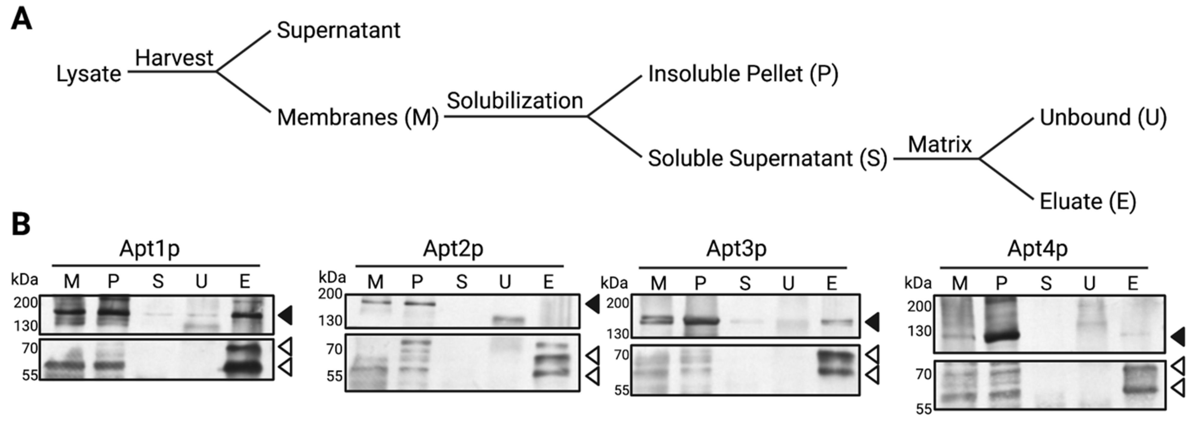
2.6. Co-Immunoprecipitation

For co-immunoprecipitation, lysis buffer II (150 mM NaCl, 20 mM HEPES-NaOH, pH 7.4) was used and the procedure was performed as described previously [19]. Briefly, membranes were solubilized with 0.6% (w/v) n-dodecyl-β-D-maltopiranoside (DDM) at a protein concentration of 1 mg/mL. The solubilized fraction was loaded on an anti-FLAG M2 matrix and incubated for 16 h with end-over-end rotation, followed by elution with FLAG-peptide. During the purification steps, samples were collected and analyzed by immunoblotting.

2.7. NBD-lipid Uptake and Flow Cytometry

Fresh transformants (3 to 10 colonies) were inoculated in 50 mL of selective SG medium. Cultures were incubated in Erlenmeyer flasks at 25 °C and 110 rpm for 16 h to induce expression. After dilution to an OD600 of 0.2 in a total volume of 50 mL, cultures were incubated for another 4 h under the same conditions to ensure similar growth. Afterwards, the NBD-lipid uptake was performed as described in [28]. Briefly, cells were washed and resuspended in selective SG media to an OD600 of 8. Subsequently, 250 µL of cells were transferred to a conical glass tube and incubated in a 30 °C water bath for 10 min. Short-chain NBD-lipids were dissolved in dimethylsulfoxide (DMSO) to a concentration of 10 mM and 1.5 µL of the solution were added to the cells. After incubation at 30 °C for 30 min, the transport was terminated by placing the cells on ice and addition of sodium azide. Subsequently, excess NBD-lipids were extracted with bovine serum albumin (BSA) and, finally, cells were resuspended in 250 µL phosphate buffered saline (PBS; 150 mM NaCl, 8 mM Na2HPO4, 2 mM KH2PO4, pH 7.0). For flow cytometric analysis, 50 µL of cell suspension were labeled with 1 µg propidium iodide, diluted with 1 mL PBS and analyzed with a CyFlow® SL cytometer (Sysmex Partec, Goerlitz, Germany). Using a flow rate of 600 cells/s, a total of 20,000 cells were measured. Viable cells were selected based on forward/side-scatter gating and propidium iodide exclusion. Data were analyzed with FlowJoTM (BD Life Sciences-FlowJo, Ashland, OR, USA).

2.8. Sequence Alignment and Similarity Analysis
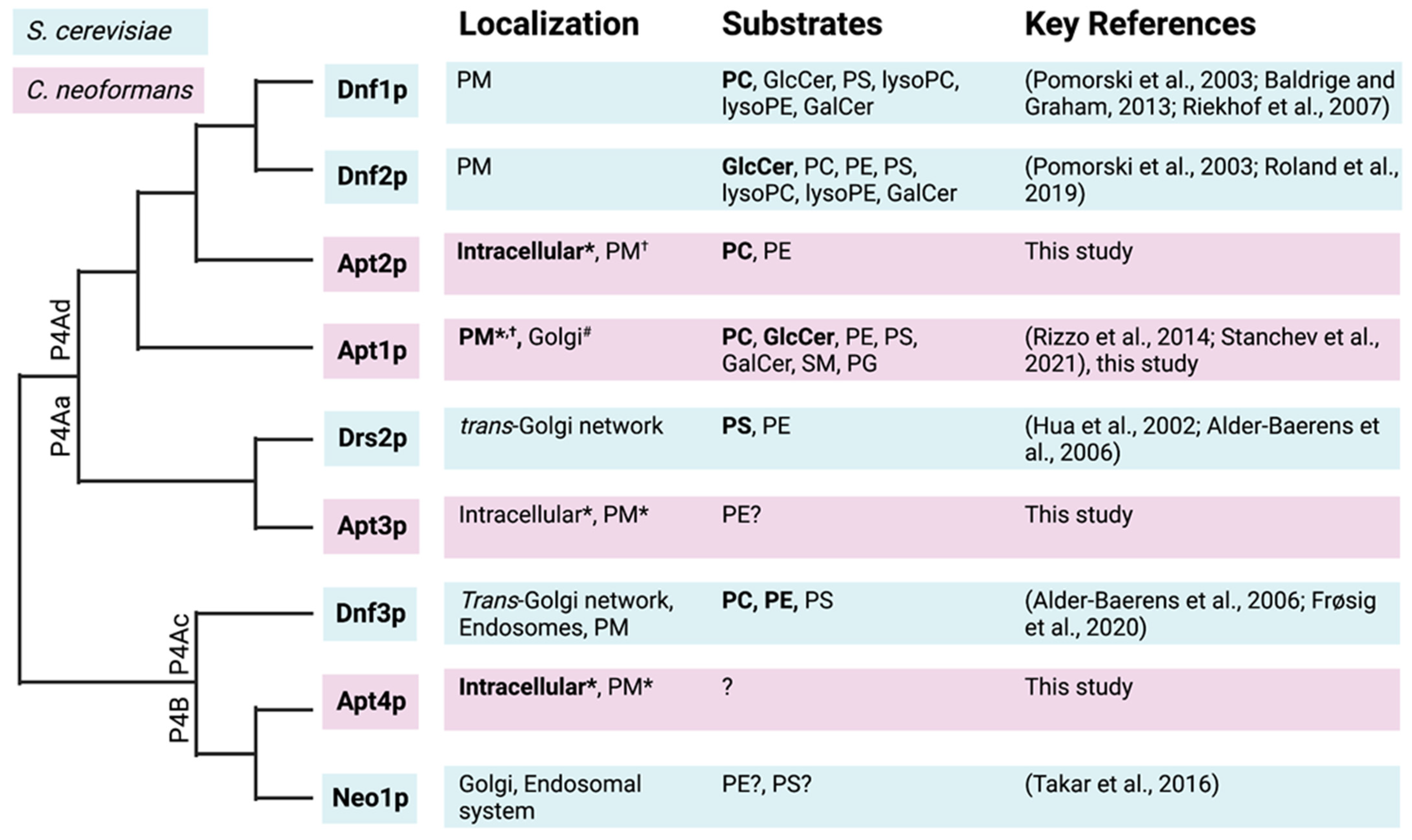
Protein sequences and structures were obtained from Uniprot and PDB, respectively. From C. neoformans: Apt1p (J9VZ19), Apt2p (J9VQH2), Apt3p (J9VGP8), Apt4p (J9VM87), Cdc50p (J9VW44); from S. cerevisiae: Dnf1p (P32660; 7kyc), Dnf2p (Q12675; 7kya), Dnf3p (Q12674), Drs2p (P39524; 6roh), Neo1p (P40527; 7rd7). Sequence alignment for phylogenetic analysis was performed in MEGAX [29] with MUSCLE [30]. Identity and similarity values are based on EMBOSS Needle online tool for pairwise sequence alignment [31]. The alignment was further used, together with PDB structures for corresponding S. cerevisiae homologs, as target-template to predict the transmembrane domains of Apt1-4p using SWISS-MODEL [32]. Pfam [33] was used to identify protein family domains and topology was visualized with Protter [34]. Glycosylation sites in Cdc50p were predicted using the NetGlyc server [35].

3. Results

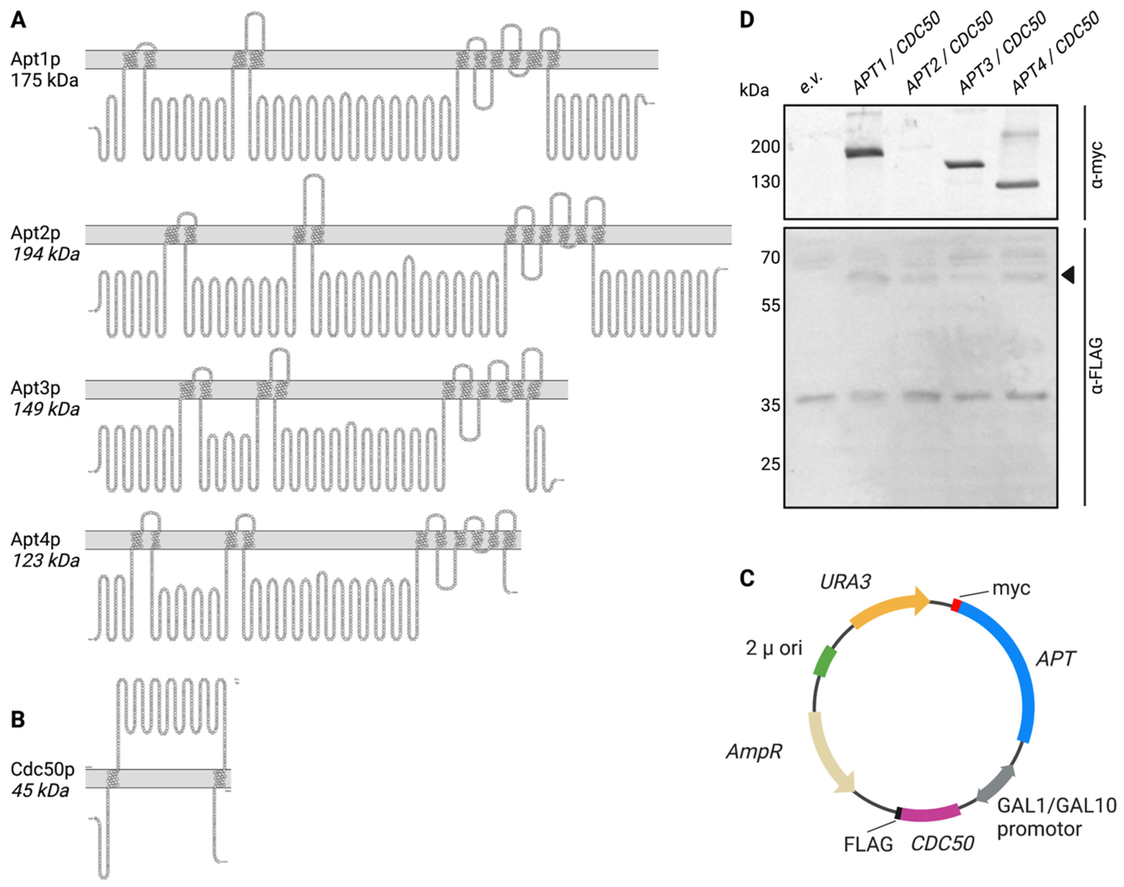
3.1. Expression of Heterologous APT1-4 and CDC50 in S. cerevisiae

Sequence homology searches revealed that, in addition to the previously studied Apt1p [19] three putative P4-ATPases Apt2p, Apt3p, and Apt4p are present in C. neoformans [19,36]. Apt2p, Apt3p, and Apt4p contain all the features of functional P-type ATPases, including the presence of 10 hydrophobic membrane-spanning helices (Figure 1A) and a characteristic pattern of conserved residues (Supplementary Figure S1). Common motifs found in all P4-ATPases [37] include: (i) the DKTGT motif, containing an aspartate that undergoes phosphorylation–dephosphorylation cycles during ATP hydrolysis and is typical for all P-type ATPases; (ii) a DGET motif that acts as an in-built phosphatase and enables dephosphorylation of the aspartate residue in the DKTGT motif; (iii) Conserved motifs TGD(K/R) and GDG(A/G)ND in the big cytosolic loop between transmembrane helix (TM)4 and TM5 necessary for Mg2+ binding; (iv) a conserved PISL motif in TM4 that is essential for lipid flipping. In agreement to its similarity to P4B-ATPase, Apt4p contains a IHRG motif in TM5 (FYNK in P4A-ATPases) and a small deletion of 6-8 amino acids after TM4 with respect to Apt1-3p [38,39].
To facilitate functional studies of Apt1-4p, the corresponding genes were cloned in a pESC-URA vector containing a bidirectional galactose-inducible GAL1/GAL10 promoter for expression in the flippase-deficient yeast strain dnf1Δdnf2Δdrs2Δ. Apt1-4p were C-terminally tagged with a myc-tag while an N-terminal FLAG-tag was added to Cdc50p (Figure 1C). After heterologous expression at 25 °C to facilitate proper folding, total membrane extracts were analyzed by immunoblotting. As shown in Figure 1D, all APTs are expressed, as demonstrated by the detection of bands of the predicted size (Figure 1A), albeit Apt2p shows a significantly lower expression level than the other Apt proteins. Cdc50p is expressed to comparable levels in all strains and detected as a band of higher molecular weight than expected based on the amino acid sequence, due to glycosylation [19].

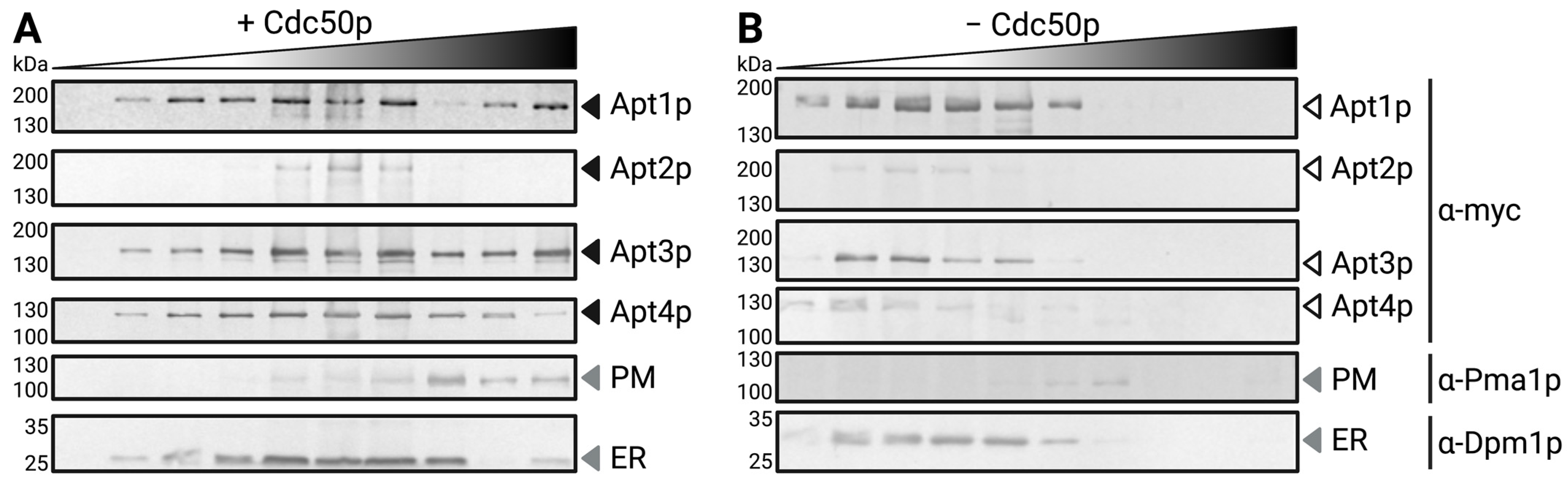
3.2. Subcellular Localization of Heterologous Apt1-4p and Interaction with Cdc50p

For determination of Apt1-4p subcellular localization and its dependency on the presence of Cdc50p, total membranes were fractionated by a discontinuous sucrose gradient (Figure 2). In co-expression with Cdc50p, Apt1p and Apt3p clearly reached the plasma membrane. In contrast, only a small amount of Apt4p was detectable in the plasma membrane fraction, while Apt2p localized to intracellular membranes. In the absence of Cdc50p, all four P4-ATPases displayed a prominent intracellular localization. Results were confirmed by heterologous expression of GFP-tagged Apt variants followed by fluorescence microscopy (Supplementary Figure S2).
To test for a physical interaction between Apt1-4p and Cdc50p, co-immunoprecipitation by FLAG-purification was performed (Figure 3A). Total membranes from cells co-expressing Cdc50p-FLAG with the respective Apt proteins were solubilized with detergent and subjected to anti-FLAG-affinity purification. Unbound non-interacting proteins were then discarded and bound proteins were eluted using a buffer containing FLAG-peptide. The fractions collected during affinity purification were subsequently analyzed via immunoblot (Figure 3B). Apt1p and Apt3p were detectable in the eluates, indicating a strong interaction with Cdc50p-FLAG. However, we were unable to solubilize Apt2p and Apt4p, preventing any conclusions about their interaction with the β-subunit by this approach.

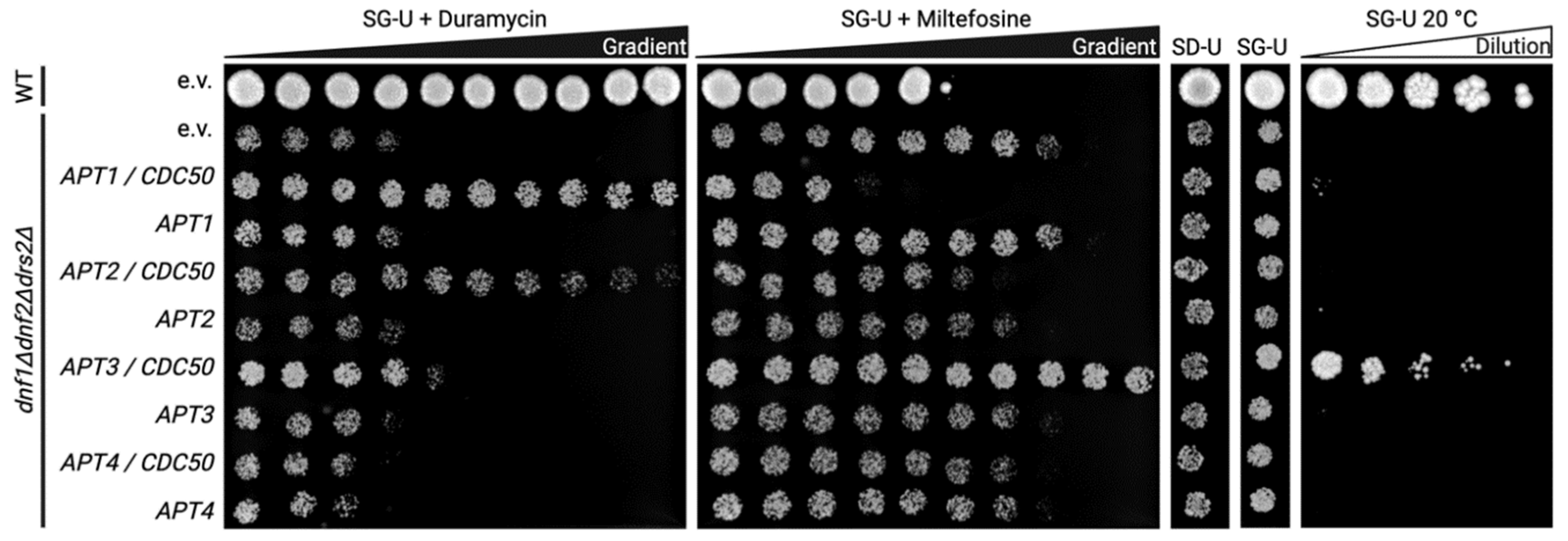
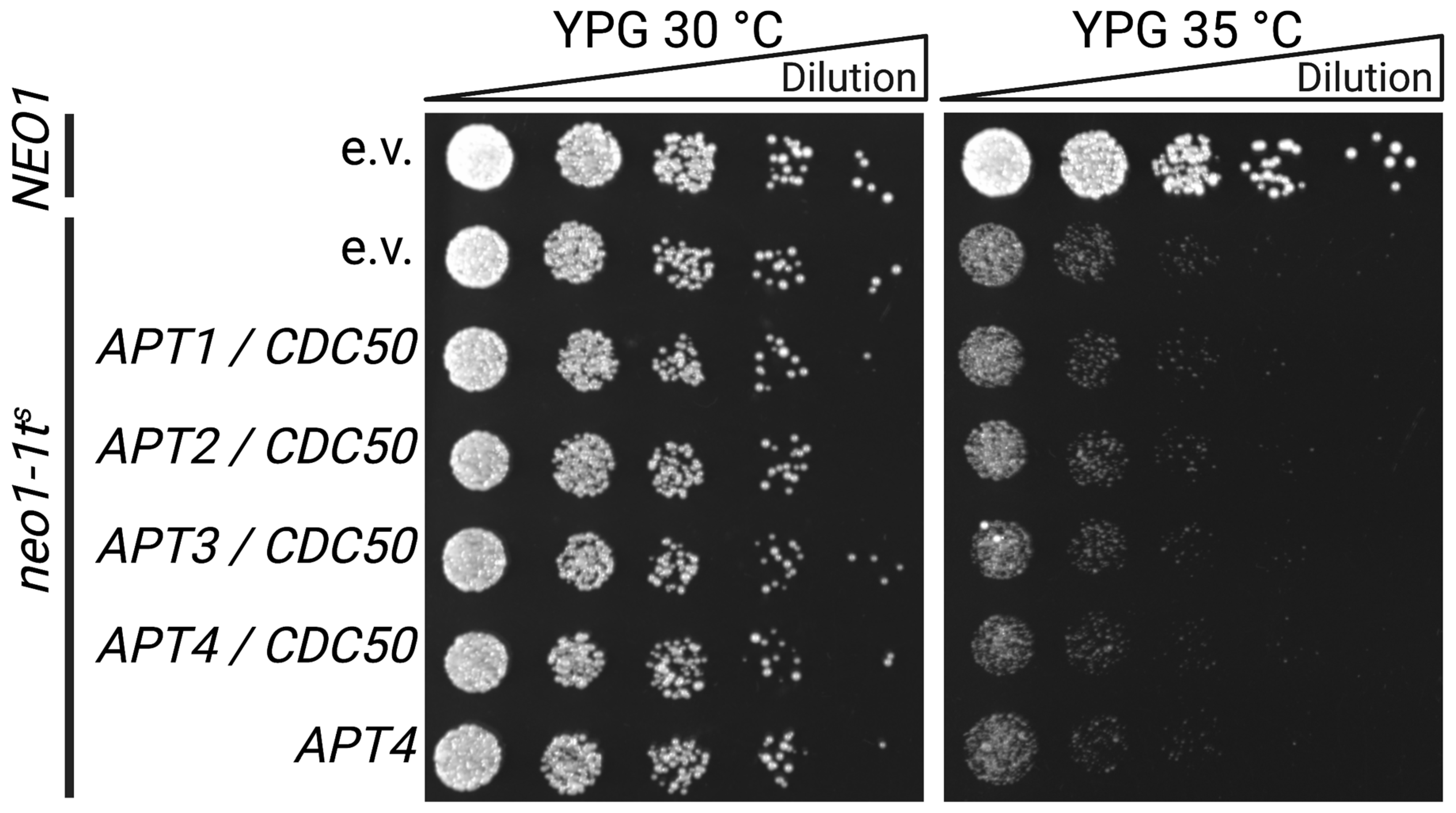
3.3. Functional Status of Heterologously Expressed Apt1-4p in S. cerevisiae

For functional analysis, flippase-deficient dnf1Δdnf2Δdrs2Δ cells expressing APT1-4 genes either alone or in combination with CDC50 were spotted on different selective plates (Figure 4). The flippase-deficient strain dnf1Δdnf2Δdrs2Δ or the corresponding wild-type strain (BY4741) transformed with the empty vector were used as negative and positive controls, respectively. Cytotoxic drugs were added to the plates to probe for lipid translocation. Duramycin is a small pore-forming tetracyclic peptide that binds to exposed PE at the cell surface and causes cell death [40,41]. In the presence of increasing concentrations of duramycin, dnf1Δdnf2Δdrs2Δ cells failed to grow due to their flippase deficiency. However, growth was recovered upon co-expression of CDC50 with either APT1, APT2 or APT3, albeit to different extents, implying a PE flippase activity of the P4-ATPases strictly in complex with the β-subunit. APT4 failed to complement the dnf1Δdnf2Δdrs2Δ phenotype. Miltefosine is a choline-containing lyso-PC analog that is toxic only after uptake across the plasma membrane [5]. Wild-type yeasts are sensitive to miltefosine, whereas dnf1Δdnf2Δdrs2Δ is not due to the lack of plasma-membrane-localized flippases (Figure 4). Upon co-expression of CDC50 with either APT1 or APT2, cells became sensitized to miltefosine, whereas cells co-expressing CDC50 and APT4 remained tolerant. Interestingly, the APT3 transformants showed an even lower sensitivity for miltefosine than the negative empty vector control. This CDC50-dependent increase in cell viability is also reflected in the cell density per drop throughout all galactose plates. Furthermore, the dnf1Δdnf2Δdrs2Δ cells display a cold-sensitive phenotype [26] that was rescued by co-expression of APT3 and CDC50, but not by any other APT/CDC50 combination, indicating an intracellular (flippase) activity.
S. cerevisiae strains lacking NEO1 are not viable, but deletion of the gene is possible when the yeast is simultaneously complemented with a plasmid-borne temperature-sensitive allele of NEO1, as is the case for the neo1-1ts strain [25]. Above the restrictive temperature of 34 °C, the mutant neo1-1ts protein is inactive, and cell growth can only be recovered upon expression of an additional active, non-temperature-sensitive version of Neo1p. The neo1-1ts strain was transformed with the empty vector or with APT1-4 together with CDC50 and dilutions were spotted on YPG plates followed by incubation at 30 °C or 35 °C (Figure 5). As a positive control, a Δneo1 strain complemented with a plasmid bearing wild-type NEO1 was used. Apt1-4p/Cdc50p complexes were unable to complement the NEO1 deficiency of neo1-1ts cells, which grew similarly to the empty vector control. As Apt4p is the closest homolog of Neo1p, which does not require a β-subunit, we also tested Apt4p in the absence of Cdc50p co-expression and observed no functional complementation.

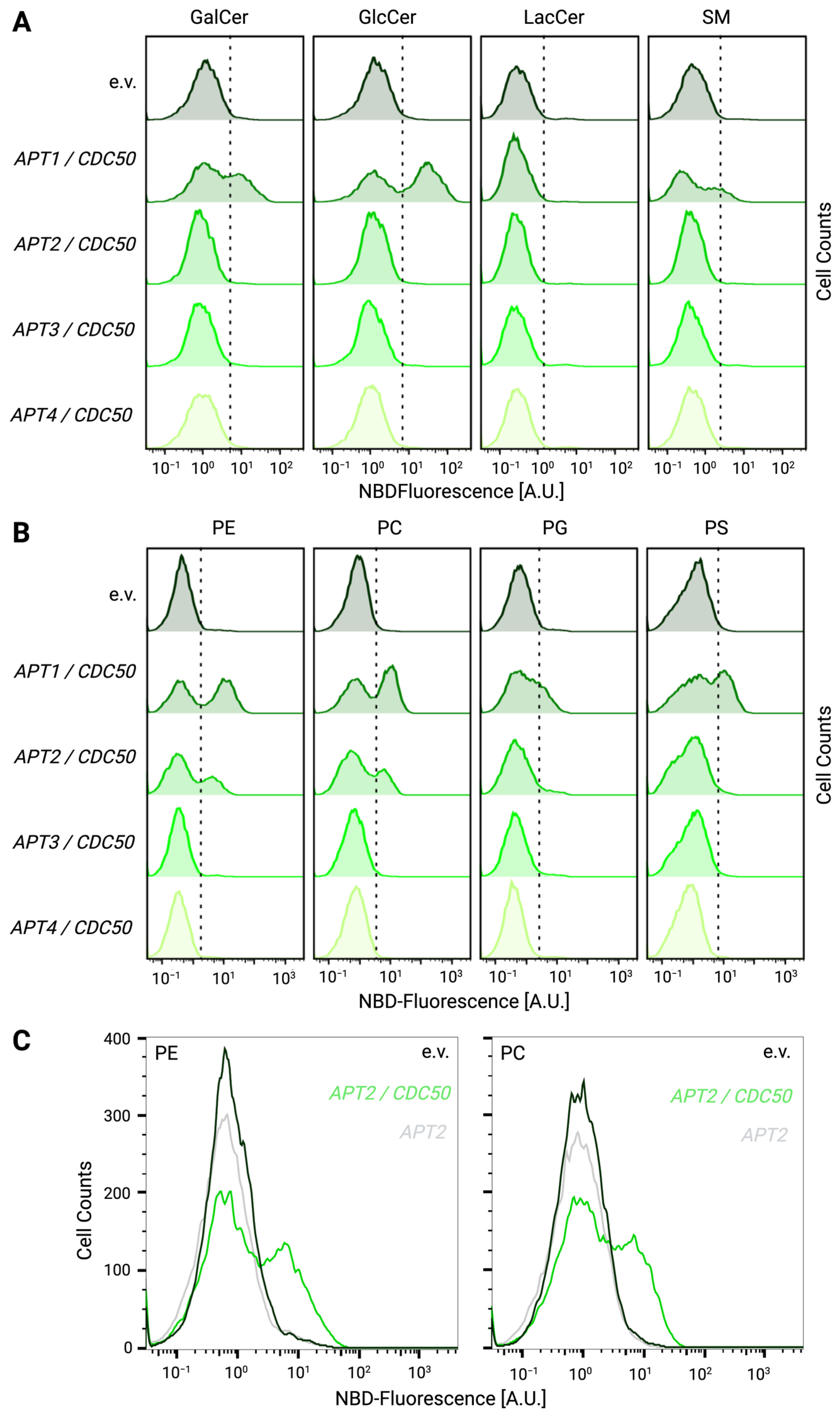
3.4. Heterologously Expressed Apt1p and Apt2p Support Lipid Flippase Activity
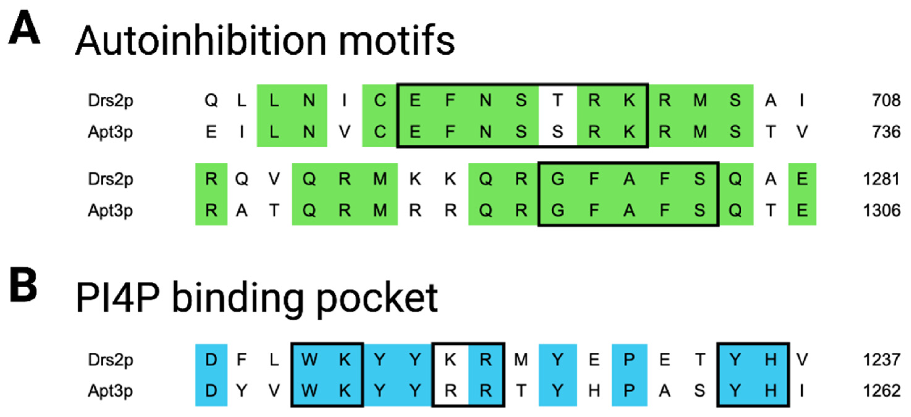
For further functional analysis of lipid uptake at the plasma membrane, the flippase-deficient dnf1Δdnf2Δdrs2Δ strain expressing APT1-4 constructs with or without CDC50 or carrying an empty vector as negative control were incubated with fluorescent NBD-labeled lipids. Following the removal of surface-exposed probe, the remaining fluorescence was assayed by flow cytometry and the NBD-fluorescence of each transformant was plotted relative to the empty vector (Figure 6). For each given NBD-lipid, increased uptake would result in a higher fluorescence signal and a right shift of the cell population compared to the empty vector control. Based on this, we were able to reproduce the flippase activity of Apt1p with its broad substrate specificity for all tested glycerophospholipids (PS, PE, PC, PG) and all ceramides except lactosyl ceramide (GlcCer, GalCer, SM) (Figure 6A,B) [19]. In contrast, Apt2p shows specific, Cdc50p-dependent transport of PE and PC (Figure 6B,C). No flippase activity at the plasma membrane could be detected for Apt3p or Apt4p for the tested lipids (Figure 6A,B).

4. Discussion

This study provides the first biochemical analysis regarding substrate preference and subunit requirements among the C. neoformans P4-ATPases Apt2-4p based on heterologous expression in the flippase-deficient S. cerevisiae strain dnf1Δdnf2Δdrs2Δ. This expression system was used previously in our laboratory to characterize Apt1p as a lipid flippase of broad substrate specificity [19]. Here, we studied Apt2-4p in comparison to Apt1p. Western blotting confirmed the successful expression of Apt1-4 proteins together with the Cdc50 protein, with Apt2p showing lower relative expression levels. Sucrose density gradient fractionation showed that Apt1p and Apt3p localize to the plasma membrane, when co-expressed with Cdc50p. A minor fraction of Apt4p localizes to the plasma membrane independently of Cdc50p, while Apt2p appears to be restricted to inner membranes in both cases. However, due to low expression levels, it cannot be ruled out that Apt2p reaches the plasma membrane in amounts below the detection limit. In fact, since cells expressing APT2 displayed uptake of exogenously applied NBD-lipids, at least a portion of the protein must reach the plasma membrane. Given that heterologous overexpression can lead to protein mislocalization [42], the localization of Apt2-4p in Cryptococcus remains to be confirmed.
Direct interaction with Cdc50p was confirmed for Apt1p and Apt3p after detergent solubilization and co-immunoprecipitation, but these assays were unsuccessful for Apt2p and Apt4p due to failed solubilization, which is probably a consequence of the heterologous expression system employed. However, recent structural studies on yeast and mammalian P4A-ATPases demonstrated that a tight interaction between the transporter and its β-subunit is necessary for lipid coordination and translocation [43,44,45,46,47,48]. In our work, Apt2p was found to require Cdc50p for flippase activity, both in functional complementation and lipid uptake assays (Figure 4 and Figure 6), which indicates that the proteins are directly interacting to form an active complex. Phylogenetic analysis revealed that Apt2p and Apt3p both belong to the P4A-ATPase subclade, which is in agreement with our finding that they both require a β-subunit for functionality (Figure 7; [19]). The requirement of Apt1-3p for Cdc50p explains the complex phenotypes observed for C. neoformans cells deleted in CDC50, which include PS exposure to the outer membrane leaflet, reduced virulence, and higher drug sensitivity [9,17].
Apt2p shares a high sequence similarity with Dnf1/2p but displays a narrower substrate specificity, limited to PC and PE (Figure 7). In contrast to mammals, where PC is evenly distributed among both leaflets of the plasma membrane or accumulated in the outer leaflet, yeast appears to maintain PC almost completely in the cytosolic leaflet [49,50]. Thus, Apt2p in complex with Cdc50p might help restrict both PE and PC to the cytoplasmic leaflet of the Cryptococcus plasma membrane, thus contributing to transbilayer lipid asymmetry. While the main localization of Apt2p, at least in Saccharomyces, seemed to be in endomembranes, Neo1p has been shown to contribute to the PS and PE asymmetry of the plasma membrane from an internal location corresponding to Golgi/endosomal compartments [25]. Apt-dependent clearance of PC and other phospholipids from the cell surface would lead to an enrichment of sphingolipids in the exoplasmic leaflet. As sphingolipids have saturated acyl chains and therefore pack at a higher density than glycerophospholipids, their enrichment in the exoplasmic leaflet of the plasma membrane would support its barrier function.
Apt3p and Drs2p share 53.9% sequence identity and our results show that both proteins also share functional similarity. First, the cold-sensitivity phenotype of the dnf1Δdnf2Δdrs2Δ strain, related to the drs2 deletion [26], was rescued by APT3/CDC50 co-expression. Second, the APT3/CDC50-complemented cells displayed higher fitness on galactose and miltefosine media as compared to the parental strain. Third, in line with the reported PE-transport activity for Drs2p [58], a low but significant resistance to duramycin was observed for APT3/CDC50-complemented cells. Given that Drs2p serves a critical role in membrane trafficking events from the trans-Golgi, Apt3p could serve a similar function in Cryptococcus. In agreement with this notion, deletion of APT3 in C. neoformans renders these cells sensitive to the secretory pathway inhibitor BFA [17]. Previous work identified amino acid motifs at the C-terminal end of Drs2p that are required for its regulation [12,13,46,59,60,61]. Among these, a GFAFS sequence at the C-terminus interacts with an EFNSTRK motif located in the cytosolic loop between TM4 and TM5, overlapping the nucleotide-binding domain and inhibiting ATPase activity. This autoinhibition is partially released by binding of phosphatidylinositol-4 phosphate (PI4P) to a highly positively charged pocket upstream of the GFAFS domain in the interface with the membrane. All motifs required for Drs2p autoinhibition, and especially the core residues, are extremely well-conserved in Apt3p (Figure 8), suggesting that both proteins are not only functional homologs, but also similarly regulated.
Apt4p belongs to the subclass of P4B-ATPases, which also includes S. cerevisiae Neo1p (Figure 7) and ATP9A and ATP9B in mammals [62]. This subclass does not appear to use a β-subunit, suggesting that Apt4p might be able to function independently of Cdc50p [25,63]. Whether this holds true remains to be investigated. We observed a shift in the subcellular localization upon co-expression of Apt4p with Cdc50p but did not observe complementation of the neo1-1ts mutant, neither in the presence or in absence of Cdc50p. Likewise, the physiological role of Apt4p remains to be elucidated. In yeast and mammals, members of the P4B-ATPase subclass localize primarily to the Golgi and the endosomal system [25,62] and serve a role in the recycling of endosomes [64].
Taken together, our results provide a first insight into the biochemical characteristics of the P4-ATPases Apt2p and Apt3p from C. neoformans. The Saccharomyces expression strategy presented here provides a platform for future purification and reconstitution to enable further detailed molecular analysis. The recent design of an antifungal peptide that binds the Cdc50p extracellular loop demonstrates that P4-ATPases are a viable pharmacological target to bypass drug resistance in Cryptococcus [24]. In this context, the heterologous expression system presented here can be a powerful tool for more comprehensive inhibitor screenings.

Supplementary Materials

The following supporting information can be downloaded at: https://www.mdpi.com/article/10.3390/jof9020202/s1, Figure S1: Topology prediction of Apt1-4p and Cdc50p from Cryptococcus neoformans; Figure S2: Intracellular localization of GFP-tagged Apt proteins upon heterologous expression in the P4-ATPase-deficient S. cerevisiae strain dnf1Δdnf2Δdrs2Δ. Table S1: Primer sets used for cloning; Table S2: Yeast expression plasmids used in this study.

Author Contributions

T.G.P. and R.L.L.-M. conceived the project and supervised the research. S.V. characterized the yeast strains with the assistance of S.L. S.L. generated plasmids. S.V. and R.L.L.-M. carried out phylogenetic and sequence comparison analyses. S.V. wrote the manuscript and revised it with the help of all authors. All authors commented on the final version. All authors have read and agreed to the published version of the manuscript.

Funding

This work was funded in part by the German Research Foundation (GU 1133/13-1). Work in the López-Marqués lab is financed by the Novo Nordisk Foundation (Project number NNF19OC0056580/NovoCrops), the Carlsberg Foundation (project number CF21-0389) and the Independent Research Fund Denmark | Nature and Universe (FNU, project number 1026-00024B). S.V. acknowledges funding from the Studienstiftung des deutschen Volkes.

Institutional Review Board Statement

Not applicable.

Data Availability Statement

All the data are within the article and Supplementary Materials. All the data are to be shared upon request (Thomas Günther Pomorski, thomas.guenther-pomorski@rub.de).

Acknowledgments

We are grateful to Todd Graham (Vanderbilt University, USA) for providing yeast strains, Marcus Peters (Ruhr University Bochum, Germany) for granting us access to the flow cytometer, and Lyubomir Dimitrov Stanchev (Novo Nordisk Foundation Center for Biosustainability, Denmark) for continuous support. Figures were created with biorender.com, accessed on 1 February 2023.

Conflicts of Interest

The authors declare no conflict of interest. The funders had no role in the design of the study; in the collection, analyses, or interpretation of data; in the writing of the manuscript; or in the decision to publish the results.

References

  1. Rajasingham, R.; Smith, R.M.; Park, B.J.; Jarvis, J.N.; Govender, N.P.; Chiller, T.M.; Denning, D.W.; Loyse, A.; Boulware, D.R. Global Burden of Disease of HIV-Associated Cryptococcal Meningitis: An Updated Analysis. Lancet Infect. Dis. 2017, 17, 873–881. [Google Scholar] [CrossRef]
  2. Maziarz, E.K.; Perfect, J.R. Cryptococcosis. Infect. Dis. Clin. North Am. 2016, 30, 179–206. [Google Scholar] [CrossRef]
  3. World Health Organisation. WHO Fungal Priority Pathogens List to Guide Research, Development and Public Health Action; Licence: CC BY-NC-SA 3.0 IGO; World Health Organisation: Geneva, Switzerland, 2022. [Google Scholar]
  4. Bongomin, F.; Oladele, R.O.; Gago, S.; Moore, C.B.; Richardson, M.D. A Systematic Review of Fluconazole Resistance in Clinical Isolates of Cryptococcus Species. Mycoses 2018, 61, 290–297. [Google Scholar] [CrossRef]
  5. Spadari, C.d.C.; Wirth, F.; Lopes, L.B.; Ishida, K. New Approaches for Cryptococcosis Treatment. Microorganisms 2020, 8, 613. [Google Scholar] [CrossRef]
  6. McClelland, E.E.; Bernhardt, P.; Casadevall, A. Estimating the Relative Contributions of Virulence Factors for Pathogenic Microbes. Infect Immun. 2006, 74, 1500–1504. [Google Scholar] [CrossRef] [PubMed]
  7. Andersen, J.P.; Vestergaard, A.L.; Mikkelsen, S.A.; Mogensen, L.S.; Chalat, M.; Molday, R.S. P4-ATPases as Phospholipid Flippases—Structure, Function, and Enigmas. Front. Physiol. 2016, 7, 275. [Google Scholar] [CrossRef]
  8. Hanson, P.K.; Malone, L.; Birchmore, J.L.; Nichols, J.W. Lem3p Is Essential for the Uptake and Potency of Alkylphosphocholine Drugs, Edelfosine and Miltefosine*. J. Biol. Chem. 2003, 278, 36041–36050. [Google Scholar] [CrossRef] [PubMed]
  9. Huang, W.; Liao, G.; Baker, G.M.; Wang, Y.; Lau, R.; Paderu, P.; Perlin, D.S.; Xue, C. Lipid Flippase Subunit Cdc50 Mediates Drug Resistance and Virulence in Cryptococcus neoformans. mBio 2016, 7, e00478-16. [Google Scholar] [CrossRef]
  10. Xu, D.; Zhang, X.; Zhang, B.; Zeng, X.; Mao, H.; Xu, H.; Jiang, L.; Li, F. The Lipid Flippase Subunit Cdc50 Is Required for Antifungal Drug Resistance, Endocytosis, Hyphal Development and Virulence in Candida Albicans. FEMS Yeast Res. 2019, 19, foz033. [Google Scholar] [CrossRef] [PubMed]
  11. Cowell, A.N.; Istvan, E.S.; Lukens, A.K.; Gomez-Lorenzo, M.G.; Vanaerschot, M.; Sakata-Kato, T.; Flannery, E.L.; Magistrado, P.; Owen, E.; Abraham, M.; et al. Mapping the Malaria Parasite Druggable Genome by Using in Vitro Evolution and Chemogenomics. Science 2018, 359, 191–199. [Google Scholar] [CrossRef] [Green Version]
  12. Azouaoui, H.; Montigny, C.; Dieudonné, T.; Champeil, P.; Jacquot, A.; Vázquez-Ibar, J.L.; Maréchal, P.L.; Ulstrup, J.; Ash, M.-R.; Lyons, J.A.; et al. High Phosphatidylinositol 4-Phosphate (PI4P)-Dependent ATPase Activity for the Drs2p-Cdc50p Flippase after Removal of Its N- and C-Terminal Extensions. J. Biol. Chem. 2017, 292, 7954–7970. [Google Scholar] [CrossRef]
  13. Timcenko, M.; Lyons, J.A.; Januliene, D.; Ulstrup, J.J.; Dieudonné, T.; Montigny, C.; Ash, M.-R.; Karlsen, J.L.; Boesen, T.; Kühlbrandt, W.; et al. Structure and Autoregulation of a P4-ATPase Lipid Flippase. Nature 2019, 571, 366–370. [Google Scholar] [CrossRef]
  14. Hu, G.; Kronstad, J.W. A Putative P-Type ATPase, Apt1, Is Involved in Stress Tolerance and Virulence in Cryptococcus neoformans. Eukaryot Cell 2010, 9, 74–83. [Google Scholar] [CrossRef]
  15. Rizzo, J.; Oliveira, D.L.; Joffe, L.S.; Hu, G.; Gazos-Lopes, F.; Fonseca, F.L.; Almeida, I.C.; Frases, S.; Kronstad, J.W.; Rodrigues, M.L. Role of the Apt1 Protein in Polysaccharide Secretion by Cryptococcus neoformans. Eukaryot Cell 2014, 13, 715–726. [Google Scholar] [CrossRef] [PubMed]
  16. Rizzo, J.; Colombo, A.C.; Zamith-Miranda, D.; Silva, V.K.A.; Allegood, J.C.; Casadevall, A.; Del Poeta, M.; Nosanchuk, J.D.; Kronstad, J.W.; Rodrigues, M.L. The Putative Flippase Apt1 Is Required for Intracellular Membrane Architecture and Biosynthesis of Polysaccharide and Lipids in Cryptococcus neoformans. Biochim. Biophys. Acta Mol. Cell Res. 2018, 1865, 532–541. [Google Scholar] [CrossRef] [PubMed]
  17. Hu, G.; Caza, M.; Bakkeren, E.; Kretschmer, M.; Bairwa, G.; Reiner, E.; Kronstad, J. A P4-ATPase Subunit of the Cdc50 Family Plays a Role in Iron Acquisition and Virulence in Cryptococcus neoformans. Cell Microbiol 2017, 19, e12718. [Google Scholar] [CrossRef] [PubMed]
  18. Graham, T.R. Flippases and Vesicle-Mediated Protein Transport. Trends Cell Biol. 2004, 14, 670–677. [Google Scholar] [CrossRef] [PubMed]
  19. Stanchev, L.D.; Rizzo, J.; Peschel, R.; Pazurek, L.A.; Bredegaard, L.; Veit, S.; Laerbusch, S.; Rodrigues, M.L.; López-Marqués, R.L.; Günther Pomorski, T. P-Type ATPase Apt1 of the Fungal Pathogen Cryptococcus neoformans Is a Lipid Flippase of Broad Substrate Specificity. J. Fungi 2021, 7, 843. [Google Scholar] [CrossRef] [PubMed]
  20. Bryde, S.; Hennrich, H.; Verhulst, P.M.; Devaux, P.F.; Lenoir, G.; Holthuis, J.C.M. CDC50 Proteins Are Critical Components of the Human Class-1 P4-ATPase Transport Machinery*. J. Biol. Chem. 2010, 285, 40562–40572. [Google Scholar] [CrossRef] [PubMed]
  21. Lenoir, G.; Williamson, P.; Puts, C.F.; Holthuis, J.C.M. Cdc50p Plays a Vital Role in the ATPase Reaction Cycle of the Putative Aminophospholipid Transporter Drs2p*. J. Biol. Chem. 2009, 284, 17956–17967. [Google Scholar] [CrossRef] [Green Version]
  22. Segawa, K.; Kurata, S.; Nagata, S. The CDC50A Extracellular Domain Is Required for Forming a Functional Complex with and Chaperoning Phospholipid Flippases to the Plasma Membrane. J. Biol. Chem. 2018, 293, 2172–2182. [Google Scholar] [CrossRef]
  23. Sebastian, T.T.; Baldridge, R.D.; Xu, P.; Graham, T.R. Phospholipid Flippases: Building Asymmetric Membranes and Transport Vesicles. Biochim. Et Biophys. Acta (BBA)-Mol. Cell Biol. Lipids 2012, 1821, 1068–1077. [Google Scholar] [CrossRef]
  24. Tancer, R.J.; Wang, Y.; Pawar, S.; Xue, C.; Wiedman, G.R. Development of Antifungal Peptides against Cryptococcus neoformans; Leveraging Knowledge about the Cdc50Δ Mutant Susceptibility for Lead Compound Development. Microbiol. Spectr. 2022, 10, e0043922. [Google Scholar] [CrossRef]
  25. Takar, M.; Wu, Y.; Graham, T.R. The Essential Neo1 Protein from Budding Yeast Plays a Role in Establishing Aminophospholipid Asymmetry of the Plasma Membrane. J. Biol. Chem. 2016, 291, 15727–15739. [Google Scholar] [CrossRef]
  26. Hua, Z.; Fatheddin, P.; Graham, T.R. An Essential Subfamily of Drs2p-Related P-Type ATPases Is Required for Protein Trafficking between Golgi Complex and Endosomal/Vacuolar System. MBoC 2002, 13, 3162–3177. [Google Scholar] [CrossRef]
  27. Gietz, R.D.; Schiestl, R.H. High-Efficiency Yeast Transformation Using the LiAc/SS Carrier DNA/PEG Method. Nat. Protoc. 2007, 2, 31–34. [Google Scholar] [CrossRef] [PubMed]
  28. Jensen, M.S.; Costa, S.; Günther-Pomorski, T.; López-Marqués, R.L. Cell-Based Lipid Flippase Assay Employing Fluorescent Lipid Derivatives. In P-Type ATPases: Methods and Protocols; Bublitz, M., Ed.; Methods in Molecular Biology; Springer: New York, NY, USA, 2016; pp. 371–382. ISBN 978-1-4939-3179-8. [Google Scholar]
  29. Kumar, S.; Stecher, G.; Li, M.; Knyaz, C.; Tamura, K. MEGA X: Molecular Evolutionary Genetics Analysis across Computing Platforms. Mol. Biol. Evol. 2018, 35, 1547–1549. [Google Scholar] [CrossRef] [PubMed]
  30. Edgar, R.C. MUSCLE: Multiple Sequence Alignment with High Accuracy and High Throughput. Nucleic Acids Res. 2004, 32, 1792–1797. [Google Scholar] [CrossRef]
  31. Madeira, F.; Pearce, M.; Tivey, A.R.N.; Basutkar, P.; Lee, J.; Edbali, O.; Madhusoodanan, N.; Kolesnikov, A.; Lopez, R. Search and Sequence Analysis Tools Services from EMBL-EBI in 2022. Nucleic Acids Res. 2022, 50, W276–W279. [Google Scholar] [CrossRef] [PubMed]
  32. Waterhouse, A.; Bertoni, M.; Bienert, S.; Studer, G.; Tauriello, G.; Gumienny, R.; Heer, F.T.; de Beer, T.A.P.; Rempfer, C.; Bordoli, L.; et al. SWISS-MODEL: Homology Modelling of Protein Structures and Complexes. Nucleic Acids Res. 2018, 46, W296–W303. [Google Scholar] [CrossRef] [PubMed] [Green Version]
  33. Sonnhammer, E.L.L.; Eddy, S.R.; Birney, E.; Bateman, A.; Durbin, R. Pfam: Multiple Sequence Alignments and HMM-Profiles of Protein Domains. Nucleic Acids Res. 1998, 26, 320–322. [Google Scholar] [CrossRef]
  34. Omasits, U.; Ahrens, C.H.; Müller, S.; Wollscheid, B. Protter: Interactive Protein Feature Visualization and Integration with Experimental Proteomic Data. Bioinformatics 2014, 30, 884–886. [Google Scholar] [CrossRef]
  35. Gupta, R.; Brunak, S. Prediction of Glycosylation across the Human Proteome and the Correlation to Protein Function. Pac. Symp. Biocomput. 2002, 7, 310–322. [Google Scholar]
  36. Jain, B.K.; Wagner, A.S.; Reynolds, T.B.; Graham, T.R. Lipid Transport by Candida albicans Dnf2 Is Required for Hyphal Growth and Virulence. Infect Immun. 2022, 90, e0041622. [Google Scholar] [CrossRef]
  37. Palmgren, M.G.; Nissen, P. P-Type ATPases. Annu. Rev. Biophys. 2011, 40, 243–266. [Google Scholar] [CrossRef]
  38. Bai, L.; Jain, B.K.; You, Q.; Duan, H.D.; Takar, M.; Graham, T.R.; Li, H. Structural Basis of the P4B ATPase Lipid Flippase Activity. Nat. Commun. 2021, 12, 5963. [Google Scholar] [CrossRef]
  39. Coleman, J.A.; Vestergaard, A.L.; Molday, R.S.; Vilsen, B.; Peter Andersen, J. Critical Role of a Transmembrane Lysine in Aminophospholipid Transport by Mammalian Photoreceptor P4-ATPase ATP8A2. Proc. Natl. Acad. Sci. USA 2012, 109, 1449–1454. [Google Scholar] [CrossRef] [PubMed]
  40. Iwamoto, K.; Hayakawa, T.; Murate, M.; Makino, A.; Ito, K.; Fujisawa, T.; Kobayashi, T. Curvature-Dependent Recognition of Ethanolamine Phospholipids by Duramycin and Cinnamycin. Biophys. J. 2007, 93, 1608–1619. [Google Scholar] [CrossRef]
  41. Rzeźnicka, I.I.; Sovago, M.; Backus, E.H.G.; Bonn, M.; Yamada, T.; Kobayashi, T.; Kawai, M. Duramycin-Induced Destabilization of a Phosphatidylethanolamine Monolayer at the Air−Water Interface Observed by Vibrational Sum-Frequency Generation Spectroscopy. Langmuir 2010, 26, 16055–16062. [Google Scholar] [CrossRef] [PubMed]
  42. Romanos, M.A.; Scorer, C.A.; Clare, J.J. Foreign Gene Expression in Yeast: A Review. Yeast 1992, 8, 423–488. [Google Scholar] [CrossRef]
  43. Bai, L.; You, Q.; Jain, B.K.; Duan, H.D.; Kovach, A.; Graham, T.R.; Li, H. Transport Mechanism of P4 ATPase Phosphatidylcholine Flippases. Elife 2020, 9, e62163. [Google Scholar] [CrossRef]
  44. Bai, L.; Kovach, A.; You, Q.; Hsu, H.-C.; Zhao, G.; Li, H. Autoinhibition and Activation Mechanisms of the Eukaryotic Lipid Flippase Drs2p-Cdc50p. Nat. Commun. 2019, 10, 4142. [Google Scholar] [CrossRef]
  45. Cheng, M.-T.; Chen, Y.; Chen, Z.-P.; Liu, X.; Zhang, Z.; Chen, Y.; Hou, W.-T.; Zhou, C.-Z. Structural Insights into the Activation of Autoinhibited Human Lipid Flippase ATP8B1 upon Substrate Binding. Proc. Natl. Acad. Sci. USA 2022, 119, e2118656119. [Google Scholar] [CrossRef] [PubMed]
  46. Dieudonné, T.; Herrera, S.A.; Laursen, M.J.; Lejeune, M.; Stock, C.; Slimani, K.; Jaxel, C.; Lyons, J.A.; Montigny, C.; Pomorski, T.G.; et al. Autoinhibition and Regulation by Phosphoinositides of ATP8B1, a Human Lipid Flippase Associated with Intrahepatic Cholestatic Disorders. Elife 2022, 11, e75272. [Google Scholar] [CrossRef]
  47. He, Y.; Xu, J.; Wu, X.; Li, L. Structures of a P4-ATPase Lipid Flippase in Lipid Bilayers. Protein Cell 2020, 11, 458–463. [Google Scholar] [CrossRef]
  48. Xu, J.; He, Y.; Wu, X.; Li, L. Conformational Changes of a Phosphatidylcholine Flippase in Lipid Membranes. Cell Rep. 2022, 38, 110518. [Google Scholar] [CrossRef] [PubMed]
  49. Iyoshi, S.; Cheng, J.; Tatematsu, T.; Takatori, S.; Taki, M.; Yamamoto, Y.; Salic, A.; Fujimoto, T. Asymmetrical Distribution of Choline Phospholipids Revealed by Click Chemistry and Freeze-Fracture Electron Microscopy. ACS Chem. Biol. 2014, 9, 2217–2222. [Google Scholar] [CrossRef]
  50. Rawyler, A.; Van der Schaft, P.H.; Roelofsen, B.; Op den Kamp, J.A.F. Phospholipid Localization in the Plasma Membrane of Friend Erythroleukemic Cells and Mouse Erythrocytes. Biochemistry 1985, 24, 1777–1783. [Google Scholar] [CrossRef] [PubMed]
  51. Palmgren, M.; Østerberg, J.T.; Nintemann, S.J.; Poulsen, L.R.; López-Marqués, R.L. Evolution and a Revised Nomenclature of P4 ATPases, a Eukaryotic Family of Lipid Flippases. Biochim. Et Biophys. Acta (BBA)-Biomembr. 2019, 1861, 1135–1151. [Google Scholar] [CrossRef]
  52. Pomorski, T.; Lombardi, R.; Riezman, H.; Devaux, P.F.; van Meer, G.; Holthuis, J.C.M. Drs2p-Related P-Type ATPases Dnf1p and Dnf2p Are Required for Phospholipid Translocation across the Yeast Plasma Membrane and Serve a Role in Endocytosis. Mol. Biol. Cell 2003, 14, 1240–1254. [Google Scholar] [CrossRef]
  53. Baldridge, R.D.; Graham, T.R. Two-Gate Mechanism for Phospholipid Selection and Transport by Type IV P-Type ATPases. Proc. Natl. Acad. Sci. USA 2013, 110, E358–E367. [Google Scholar] [CrossRef] [Green Version]
  54. Riekhof, W.R.; Wu, J.; Gijón, M.A.; Zarini, S.; Murphy, R.C.; Voelker, D.R. Lysophosphatidylcholine Metabolism in Saccharomyces cerevisiae: The Role of P-Type ATPases in Transport and a Broad Specificity Acyltransferase in Acylation. J. Biol. Chem. 2007, 282, 36853–36861. [Google Scholar] [CrossRef]
  55. Roland, B.P.; Naito, T.; Best, J.T.; Arnaiz-Yépez, C.; Takatsu, H.; Yu, R.J.; Shin, H.-W.; Graham, T.R. Yeast and Human P4-ATPases Transport Glycosphingolipids Using Conserved Structural Motifs. J. Biol. Chem. 2019, 294, 1794–1806. [Google Scholar] [CrossRef]
  56. Alder-Baerens, N.; Lisman, Q.; Luong, L.; Pomorski, T.; Holthuis, J.C.M. Loss of P4 ATPases Drs2p and Dnf3p Disrupts Aminophospholipid Transport and Asymmetry in Yeast Post-Golgi Secretory Vesicles. Mol. Biol. Cell 2006, 17, 1632–1642. [Google Scholar] [CrossRef]
  57. Frøsig, M.M.; Costa, S.R.; Liesche, J.; Østerberg, J.T.; Hanisch, S.; Nintemann, S.; Sørensen, H.; Palmgren, M.; Pomorski, T.G.; López-Marqués, R.L. Pseudohyphal Growth in Saccharomyces Cerevisiae Involves Protein Kinase-Regulated Lipid Flippases. J. Cell Sci. 2020, 133, jcs235994. [Google Scholar] [CrossRef] [PubMed]
  58. Baldridge, R.D.; Xu, P.; Graham, T.R. Type IV P-Type ATPases Distinguish Mono- versus Diacyl Phosphatidylserine Using a Cytofacial Exit Gate in the Membrane Domain. J. Biol. Chem. 2013, 288, 19516–19527. [Google Scholar] [CrossRef] [PubMed]
  59. Lamy, A.; Macarini-Bruzaferro, E.; Dieudonné, T.; Perálvarez-Marín, A.; Lenoir, G.; Montigny, C.; Le Maire, M.; Vázquez-Ibar, J.L. ATP2, The Essential P4-ATPase of Malaria Parasites, Catalyzes Lipid-Stimulated ATP Hydrolysis in Complex with a Cdc50 β-Subunit. Emerg. Microbes Infect. 2021, 10, 132–147. [Google Scholar] [CrossRef]
  60. Natarajan, P.; Liu, K.; Patil, D.V.; Sciorra, V.A.; Jackson, C.L.; Graham, T.R. Regulation of a Golgi Flippase by Phosphoinositides and an ArfGEF. Nat. Cell Biol. 2009, 11, 1421–1426. [Google Scholar] [CrossRef]
  61. Zhou, X.; Sebastian, T.T.; Graham, T.R. Auto-Inhibition of Drs2p, a Yeast Phospholipid Flippase, by Its Carboxyl-Terminal Tail*. J. Biol. Chem. 2013, 288, 31807–31815. [Google Scholar] [CrossRef] [PubMed]
  62. Takatsu, H.; Baba, K.; Shima, T.; Umino, H.; Kato, U.; Umeda, M.; Nakayama, K.; Shin, H.-W. ATP9B, a P4-ATPase (a Putative Aminophospholipid Translocase), Localizes to the Trans-Golgi Network in a CDC50 Protein-Independent Manner. J. Biol. Chem. 2011, 286, 38159–38167. [Google Scholar] [CrossRef] [PubMed]
  63. Saito, K.; Fujimura-Kamada, K.; Furuta, N.; Kato, U.; Umeda, M.; Tanaka, K. Cdc50p, a Protein Required for Polarized Growth, Associates with the Drs2p P-Type ATPase Implicated in Phospholipid Translocation in Saccharomyces cerevisiae. Mol. Biol. Cell 2004, 15, 3418–3432. [Google Scholar] [CrossRef] [Green Version]
  64. Wicky, S.; Schwarz, H.; Singer-Krüger, B. Molecular Interactions of Yeast Neo1p, an Essential Member of the Drs2 Family of Aminophospholipid Translocases, and Its Role in Membrane Trafficking within the Endomembrane System. Mol. Cell. Biol. 2004, 24, 7402–7418. [Google Scholar] [CrossRef] [PubMed] [Green Version]
Figure 1. Topology and heterologous expression of APT1-4/CDC50 in the P4-ATPase-deficient S. cerevisiae strain dnf1Δdnf2Δdrs2Δ. Topology prediction of Apt1-4p (A) Cdc50p (B) based on sequence alignment with corresponding S. cerevisiae homologs was visualized with Protter. (C): architecture of pESC-URA-based plasmids used to express myc-tagged APT1-4 together with FLAG-tagged CDC50 under a bidirectional GAL1/GAL10-promoter. 2µ ori: origin of replication; AmpR: ampicillin resistance gene, URA3: uracil selection marker. (D): Immunoblot showing the expression of myc-tagged Apt1-4p and FLAG-tagged Cdc50p (black arrowhead) in the P4-ATPase-deficient S. cerevisiae strain dnf1Δdnf2Δdrs2Δ in comparison to the empty vector (e.v.). The mobilities of marker proteins of known mass (kDa) are indicated on the left.
Jof 09 00202 g001
Figure 2. Intracellular localization of Apt proteins upon heterologous expression in the P4-ATPase-deficient S. cerevisiae strain dnf1Δdnf2Δdrs2Δ. (A,B): dnf1Δdnf2Δdrs2Δ yeast cells expressing myc-tagged Apt1-4p with ((A), black arrows) or without ((B), white arrows) FLAG-tagged Cdc50p were lysed, and cellular membranes were fractionated on a sucrose step gradient. Gradient fractions were immunoblotted using antibodies against the myc-epitope or several organellar markers. Fractionation profiles of protein markers correspond to cells expressing myc-tagged APT3 but were determined for each gradient individually with similar results. PM, plasma membrane; ER, endoplasmic reticulum.
Jof 09 00202 g002
Figure 3. Co-immunoprecipitation of Apt proteins with Cdc50p-FLAG. (A): FLAG-affinity purification scheme. After cell lysis, membranes were harvested by centrifugation and solubilized with 0.6% (w/v) DDM. Insoluble material was pelleted by ultracentrifugation and the soluble supernatant was incubated with FLAG-affinity matrix to bind Cdc50p-FLAG. The supernatant of this incubation containing unbound proteins was discarded and bound proteins were eluted by FLAG-peptide addition. (B): Apt1-4p-myc were expressed with Cdc50p-FLAG and purified according to (A). Equal volumes of each fraction were loaded onto SDS-PAGE gels, except for the eluate fraction, for which a 10× volume was loaded. Apt1-4p and Cdc50p were detected via anti-myc (black arrows) and anti-FLAG antibodies (white arrows), respectively. Glycosylation variants are indicated for Cdc50p.
Jof 09 00202 g003
Figure 4. Complementation analysis for APT1-4/CDC50 combinations expressed in the P4-ATPase-deficient S. cerevisiae strain dnf1Δdnf2Δdrs2Δ. Functional complementation of the P4-ATPase-deficient yeast strain dnf1Δdnf2Δdrs2Δ expressing myc-tagged Apt1-4p with and without FLAG-tagged Cdc50p. Wild-type (BY4741; WT) and triple mutant cells transformed with empty plasmids were used as positive and negative controls, respectively. Cells were spotted at a constant 0.1 OD600 on glucose- (SD, control) or galactose-containing (SG, induced gene expression) plates supplemented with a concentration gradient of the indicated toxins (the direction of the gradient is indicated by a filled triangle) and incubated at 28 °C for three days. For cold-sensitivity testing, serial 1:5 dilutions starting at 0.2 OD600 were spotted on SG plates and incubated at 20 °C for ten days. The experiments were repeated three times with identical results.
Jof 09 00202 g004
Figure 5. neo1-1ts complementation assay. A neo1 deletion strain expressing a temperature-sensitive allele of NEO1 (neo1-1ts) was transformed with an empty vector (e.v., negative control) or with plasmids bearing APT-myc genes and CDC50-FLAG. A neo1 deletion strain expressing NEO1 from a plasmid was transformed with an e.v. as positive control. Serial 1:5 dilutions starting at 0.3 OD600 (indicated by triangles) were spotted on rich galactose plates to induce expression and incubated at 30 °C (control) or above the neo1-1ts restrictive temperature at 35 °C for 48 h. The experiment was repeated three times with identical results.
Jof 09 00202 g005
Figure 6. Lipid flippase activity of Apt1-4p/Cdc50p upon heterologous expression in the P4-ATPase-deficient S. cerevisiae dnf1Δdnf2Δdrs2Δ strain. dnf1Δdnf2Δdrs2Δ yeast cells expressing APT1-4/CDC50 were labeled with the indicated NBD-lipids and analyzed by flow cytometry. At least 10,000 cells are represented by each histogram. Cells that present higher fluorescence than the threshold (dotted vertical line) were considered as cells positive for flippase activity. (A): C6-NBD-sphingolipids tested included Galactosyl ceramide (GalCer), Glucosyl ceramide GlcCer), Lactosyl ceramide (LacCer), and Sphingomyelin (SM). (B): C6-NBD-glycerophospholipids tested included Phosphatidylcholine (PC), Phosphatidylethanolamine (PE), Phosphatidylglycerol (PG), Phosphatidylserine (PS), (C): APT2 was analyzed with and without CDC50 co-expression for PE and PC uptake in comparison to an empty vector (e.v.) control. All experiments were repeated at least two times with identical results.
Jof 09 00202 g006
Figure 7. Phylogenetic comparisons of P4-ATPases from S. cerevisiae and C. neoformans. The phylogenic tree was generated using MUSCLE and sequences from Uniprot. Localization and substrate specificities are indicated based on key references. Subclade divisions within the P4A clade are as described in [51]. For accession numbers, see Materials and Methods. References: [15,19,25,26,52,53,54,55,56,57]. PM, plasma membrane; PE, phosphatidylethanolamine; PC, phosphatidylcholine; PS, phosphatidylserine; PG, phosphatidylglycerol; GlcCer, glucosyl ceramide; GalCer, galactosyl ceramide; SM, sphingomyelin. * based on subcellular fractionation; based on fluorescence microscopy and lipid uptake studies; # putative.
Jof 09 00202 g007
Figure 8. Conserved motifs from Drs2p in Apt3p. Sequence alignment of S. cerevisiae Drs2p and C. neoformans Apt3p via MUSCLE revealed the conservation of the previously identified Drs2p C-terminal autoinhibition site together with its binding site within the large cytosolic loop (A), as well as the PI4P binding site (B). Conserved amino acids are shown in green/blue, and the amino acids identified as important for the motif are highlighted in a box.
Jof 09 00202 g008
Disclaimer/Publisher’s Note: The statements, opinions and data contained in all publications are solely those of the individual author(s) and contributor(s) and not of MDPI and/or the editor(s). MDPI and/or the editor(s) disclaim responsibility for any injury to people or property resulting from any ideas, methods, instructions or products referred to in the content.

