Article

Antifungal Activity of Chitosan against Histoplasma capsulatum in Planktonic and Biofilm Forms: A Therapeutic Strategy in the Future?

by
Raimunda Sâmia Nogueira Brilhante
1,*,
Anderson da Cunha Costa
1,
Jacó Ricarte Lima de Mesquita
2,
Gessica dos Santos Araújo
3,
Rosemeyre Souza Freire
4,
João Victor Serra Nunes
4,
Augusto Feynman Dias Nobre
1,
Mirele Rodrigues Fernandes
1,
Marcos Fábio Gadelha Rocha
3,
Waldemiro de Aquino Pereira Neto
1,
Thomas Crouzier
5,
Ulrike Schimpf
5 and
Rodrigo Silveira Viera
6
1
Department of Pathology and Legal Medicine, School of Medicine, Specialized Medical Mycology Center, Postgraduate Program in Medical Sciences, Federal University of Ceará, Rua Barão de Canindé, 210, Montese, Fortaleza 60425-540, CE, Brazil
2
São José Hospital, Fortaleza 60455-610, CE, Brazil
3
Postgraduate in Veterinary Sciences, Faculty of Veterinary, State University of Ceará, Dr. Silas Munguba Avenue, 1700, Itaperi Campus, Fortaleza 60714-903, CE, Brazil
4
Analytical Center, Department of Physics, Federal University of Ceará, Fortaleza 60020-181, CE, Brazil
5
KTH Royal Institute of Technology, School of Engineering Sciences in Chemistry, Biotechnology and Health, Department of Chemistry, Division of Glycoscience, AlbaNova University Center, 106 91 Stockholm, Sweden
6
Department of Chemical Engineering, Federal University of Ceará, Fortaleza 60440-900, CE, Brazil
*
Author to whom correspondence should be addressed.
J. Fungi 2023, 9(12), 1201; https://doi.org/10.3390/jof9121201
Submission received: 30 October 2023 / Revised: 9 December 2023 / Accepted: 10 December 2023 / Published: 15 December 2023
(This article belongs to the Special Issue New Strategies to Combat Human Fungal Infections)

Abstract

:
Histoplasmosis is a respiratory disease caused by Histoplasma capsulatum, a dimorphic fungus, with high mortality and morbidity rates, especially in immunocompromised patients. Considering the small existing therapeutic arsenal, new treatment approaches are still required. Chitosan, a linear polysaccharide obtained from partial chitin deacetylation, has anti-inflammatory, antimicrobial, biocompatibility, biodegradability, and non-toxicity properties. Chitosan with different deacetylation degrees and molecular weights has been explored as a potential agent against fungal pathogens. In this study, the chitosan antifungal activity against H. capsulatum was evaluated using the broth microdilution assay, obtaining minimum inhibitory concentrations (MIC) ranging from 32 to 128 µg/mL in the filamentous phase and 8 to 64 µg/mL in the yeast phase. Chitosan combined with classical antifungal drugs showed a synergic effect, reducing chitosan’s MICs by 32 times, demonstrating that there were no antagonistic interactions relating to any of the strains tested. A synergism between chitosan and amphotericin B or itraconazole was detected in the yeast-like form for all strains tested. For H. capsulatum biofilms, chitosan reduced biomass and metabolic activity by about 40% at 512 µg/mL. In conclusion, studying chitosan as a therapeutic strategy against Histoplasma capsulatum is promising, mainly considering its numerous possible applications, including its combination with other compounds.

1. Introduction

Histoplasma capsulatum is a dimorphic fungus whose saprophytic filamentous form is found in soils, especially those containing bird and bat feces, and whose parasitic yeast form is found in humans and animals [1,2]. Inhalation of conidia or mycelial fragments of H. capsulatum in contaminated environments is the primary mode of infection of both humans and animals, with these fungal elements readily carried through the air. Furthermore, on rare occasions, direct transmission of the parasitic form can occur through tissue transplantation or laboratory accidents [3,4]. Currently, histoplasmosis is one of the most prevalent systemic mycoses in the Americas [5]. In recent decades, systemic and invasive fungal infections have become more common, especially among immunocompromised patients. Annually, there are an estimated 300 million cases of severe fungal infection and 1.6 million deaths worldwide [6,7]. This situation primarily arises from the rising number of immunocompromised patients, such as those with AIDS, cancer, and transplant recipients, in addition to the increase in drug resistance, and the emergence and re-emergence of pathogens like Histoplasma capsulatum [7,8].
H. capsulatum displays a variety of virulence attributes that affect its interaction with host immune cells, allowing it to evade the immune response and replicate in a new environment, including features such as thermal dimorphism and biofilm formation [9,10]. The in vitro biofilm-forming ability of this fungal species has been reported for the yeast form [10,11] and has also been reported for other dimorphic fungi such as Sporothrix spp. and Paracoccidaides spp. [12,13].
Biofilms are microbial communities that firmly adhere to biotic or abiotic surfaces and are protected by an extracellular polymeric matrix composed of polysaccharides, proteins, nucleic acids, and lipids. These cells present more phenotypic, genetic, and structural heterogeneity in comparison with planktonic cells [14,15]. In the host, biofilms play a critical role in the development of the infection, making their formation an important virulence factor, since it confers greater resistance to specific mediators of immune response and increased resistance to antimicrobial agents. In the environment, this form of growth prevails over planktonic growth and provides protection for the fungal structures against environmental aggressors, such as Ultraviolet (UV) radiation, dehydration, extreme temperatures, and chemical agents [14,16]. The in vitro studies conducted with the goal of characterizing these fungal biofilms have employed various techniques, including the use of XTT salt (2,3-bis(2-methoxy-4-nitro-5-sulfophenyl)-5-[carbamoylphenylamino]-2-tetrazolium hydroxide) to assess metabolic activity during biofilm development and antifungal sensitivity testing. Additionally, crystal violet staining is utilized to quantify biomass [17,18]. Biofilm evaluations can also be conducted using specialized microscopic techniques, including scanning electron microscopy, to examine fungal biofilms’ morphology [19].
At present, the first-line histoplasmosis treatment is the administration of itraconazole and amphotericin B. However, relapses and refractory infections have been reported, predominantly due to biofilm formation [20,21]. In this context, developing target drugs is challenging [22].
Biopolymers like chitosan, a linear polysaccharide originating from partially deacetylated chitin, have been employed to counteract both planktonic and biofilm cells of diverse microorganisms, including other fungi such as Candida spp. [23], Aspergillus fumigatus. [24], and Sporothrix brasiliensis [23]. This biopolymer has gained extensive usage as an antimicrobial agent due to its biocompatibility, biodegradability, low toxicity, and significant functional potential, attributable to its amino and hydroxyl groups [25].
In a previous study conducted by our research group, chitosan with three different molecular weights (high, medium, and low) was investigated against the planktonic and biofilm forms of Sporothrix brasiliensis [23]. The results demonstrated that low molecular weight chitosan (LWC) had the most potent inhibitory effect against both the planktonic and biofilm forms of the fungus since smaller quantities of chitosan were needed to inhibit the growth of S. brasiliensis. In the present study, we evaluated the antifungal activity of low molecular weight chitosan, its interactions with antifungal drugs against planktonic cells, and its effect on biofilm formation in both filamentous and yeast forms of Histoplasma capsulatum.

2. Materials and Methods

2.1. Low Molecular Weight Chitosan Samples

The low molecular weight (LMW 448869) chitosan used in this study was obtained from Sigma-Aldrich (Saint Louis, MO, USA). The LWC was characterized regarding its molecular weight (MW) and deacetylation degree (DD) by viscometry and potentiometric degree, respectively, in a previous study described by us (Garcia et al., 2020 [23]). The MW was 206.4 kg.mol−1 and the DD was 79%.

2.2. Fungal Culture

A total of 20 isolates of H. capsulatum were included in this study. All isolates were obtained from the culture collection of the Specialized Center for Medical Mycology (CEMM) of Federal University of Ceará, Brazil. The isolates were stored in physiological saline at 4 °C or on Potato Dextrose Agar supplemented with 10% DMSO (Dimethyl Sulfoxide, Sigma, St. Louis, MO, USA) at −20 °C. Before storage, these isolates underwent identification through mycological, immunological, and molecular techniques. For the assessment of purity and viability of the isolates taken from the stock, micromorphological analysis was conducted after inoculation on Potato Agar and incubation for 7–15 days at room temperature (−28 °C). The cultivation of H. capsulatum in the yeast phase was achieved from cultures in the filamentous phase subjected to successive subcultures on Sabouraud Agar and Brain Heart Infusion (BHI) Agar supplemented with 10% sheep blood, followed by incubation at 35 °C. The procedures were performed in a class II biological safety cabinet in a biosafety level 3 laboratory.

2.3. Preparation of Chitosan and Control Drugs

Chitosan (1 g) was dissolved in 100 mL of 1% (v/v) glacial acetic acid solution (Panreac, Barcelona, Spain) and stored under refrigeration. Amphotericin B (AMB) and itraconazole (ITC) (Sigma, St. Louis, MO, USA) were used as control drugs and were dissolved in dimethyl sulfoxide (DMSO) (Sigma-Aldrich), according to the Clinical and Laboratory Standards Institute [26]. Subsequently, AMB and ITC were prepared in Roswell Park Memorial Institute (RPMI) 1640 medium (Sigma, St. Louis, MO, USA) buffered to pH 7.0 with 0.165 M MOPS (3-[N-Morpholino] propane sulfonic acid) (Sigma-Aldrich).

2.4. Antifungal Susceptibility Test in Planktonic Cells of H. capsulatum

The sensitivity tests were conducted with the 20 strains of H. capsulatum using the broth microdilution technique, based on the reference methods of document M38-3rd edition [26] for the filamentous form and M60-2nd edition [27]. for the yeast form, with adaptations. The compounds were tested at final concentrations of 1–1024 µg/mL for chitosan and 0.03–16 µg/mL for amphotericin B and itraconazole. The inoculants were obtained from growth of H. capsulatum cultures on potato dextrose agar (PDA). The cells were suspended in sterile saline, after each fungal suspension was adjusted to 1 on the McFarland for the filamentous form and 0.5 on the McFarland scale for the yeast form. The 1:10 dilutions were made in RPMI 1640 buffered to pH 7.0 with MOPS 0.165 M to obtain inoculants with a final concentration from 1 × 10⁵ to 5 × 10⁵ CFU/mL for the filamentous form and 1 × 10³ to 5 × 10³ CFU/mL for the yeast form. The tests were performed in duplicate, and the results were read visually after the incubation period of 72 h at 35 °C for the filamentous form and 96 h at 37 °C for the yeast form. [11] We considered the MIC of chitosan, itraconazole, and amphotericin B as the lowest concentration capable of inhibiting the visual growth of fungi compared to growth control by 50, 80, and 100%, respectively. Candida parapsilosis ATCC 22019 and Candida krusei ATCC 6258 were used as quality controls for the experiments, which were carried out in duplicate at two different time points.

2.5. Susceptibility Test of Biofilm Formation

Flat-bottomed 96-well plates were employed for the formation of filamentous and yeast forms of biofilms. A 200 µL aliquot of fungal suspension at 1 × 10⁶ conidia/mL (filamentous form) or 2 × 10⁵ cells/mL (yeast form) prepared in RPMI 1640 medium was added to 96-well microplates and incubated in an oven at 35 °C (filamentous form) or 37 °C (yeast-form) for 24 h to allow for cellular adhesion. Chitosan, amphotericin B, and itraconazole were tested against planktonic strains at concentrations of MIC, 10 × MIC, and 50 × MIC. After the 24-h incubation period for microorganisms to adhere to the wells, the supernatant in each well was removed and the biofilms were washed with sterile PBS once. The wells then received 200 µL of RPMI medium (controls) or drugs at the concentrations mentioned earlier. The plates were incubated for 72 h at 35 °C (filamentous form) or 37 °C (yeast form). The susceptibility of sessile cells to chitosan and antifungals was analyzed by quantifying biomass and metabolic activity using violet crystal staining and the MTT reduction assay along with morphological visualization using scanning electron microscopy, respectively [28].

2.6. Biomass Quantification Assay

The biomass of the biofilms was quantified using the crystal violet staining technique. The wells containing biofilms were washed twice with PBS and fixed with 100% methanol for 5 min (GQ—Grupo Química, São Paulo, Brazil). Then, the methanol was removed, and the wells were air-dried at room temperature. Subsequently, 200 μL of a 0.3% crystal violet solution was added to each well for 20 min. After this period, the wells were washed twice with distilled water. The wells were then destained by adding a 33% acetic acid solution. The remaining content in each well was transferred to the well of the new plate, and the absorbance was measured at 540 nm using a spectrophotometer [17]. All tests were performed in duplicate at two different time points.

2.7. Metabolic Activity Quantification Assay

The metabolic activity of the biofilms was quantified using the 2,3-bis-(2-methoxy-4-nitro-5-sulfophenyl)-5-[(phenylamino) carbonyl]-2H-tetrazolium (MTT; Sigma, Darmstadt, Germany) reduction assay [17]. This assay was performed using a stock solution of MTT (1 µg/mL in PBS) and menadione (1 mM in ethanol). To all wells containing biofilm, 125 μL of MTT solution was added, and after 3 h of incubation at 35 °C, protected from light, the color change was measured with a spectrophotometer at 492 nm. The minimum inhibitory concentration in biofilm (MICB) was determined as the lowest concentration capable of inhibiting 50% (MICB50) or 80% (MICB80) of the biofilm’s metabolic activity in comparison with the drug-free control’s activity [29]. All tests were conducted in duplicate at two different time points.

2.8. Evaluation of the Morphology and Structure of H. capsulatum Biofilms

The LWC effect on the morphology and structure of mature H. capsulatum biofilms was investigated using scanning electron microscopy (SEM). Mature biofilms were formed as described in the previous section in 24-well plates and treated with MICB80 of chitosan, as previously described.
For SEM analyses, the biofilms were fixed with 500 μL of 2.5% glutaraldehyde solution (Sigma-Aldrich, St Louis, MO, USA) in cacodylate buffer (0.15 M) (Electron Microscopy Sciences, Hatfield, PA, USA) and 0.1% Alcian blue (Sigma-Aldrich, São Paulo, Brazil), at 4 °C, overnight, to preserve fungal structures. Subsequently, biofilms were washed with cacodylate buffer twice, followed by dehydration in baths containing ascending ethanol concentrations (50, 70, 80, 95, and 100%) for 10 min at each dehydration level, repeating the dehydration with 100% ethanol once more. After drying, biofilms were dehydrated with hexamethyldisilazane (DMSO) (Sigma, St. Louis, MO, USA) for 30 min and stored at 35 °C overnight. Then, coverslips were coated with 10 nm of gold (Emitech Q150T, Shanghai, China) and observed with a Quanta FEG 450 scanning electron microscope (Thermo fisher, São Paulo, Brazil) under a high vacuum mode at 20 kV using a secondary electron detector (Thermo fisher, São Paulo, Brazil) [17].

2.9. Pharmacological Interaction—Checkerboard

To evaluate interactions between chitosan and antifungals (AMB, ITC), strains of H. capsulatum were selected randomly in both filamentous and yeast forms, and were submitted to the checkerboard broth microdilution method with a 7-row by 11-column configuration [30]. The checkerboard assay was performed as described by Brilhante et al. (2020) [17], with adaptations. The combinations of chitosan (8 to 512 µg/mL) with amphotericin or itraconazole (0.01 to 16 µg/mL) were evaluated. In the assays with planktonic cells, combinations of variable concentrations of chitosan ranging from 4 to 256 µg/mL for the filamentous form and 0.5 to 32 µg/mL for the yeast form of H. capsulatum were used, combined with 0.007 to 8 µg/mL of AMB or ITC. Inoculums were prepared to a final concentration of 0.4–5 × 10⁴ cfu/mL (filamentous form) or 0.5–2.5 × 10³ cfu/mL (yeast form) in RPMI 1640 medium as previously described. The microplates were incubated at 35 °C (filamentous form) or at 37 °C for 96 h (yeast form) or 72 h, and the MICs were defined following the antifungal reading points defined in the CLSI [11]. MIC values were defined as the lowest concentration capable of inhibiting 100% (amphotericin) or 80% (itraconazole) of the growth of fungi by the combination between chitosan and amphotericin B or itraconazole. The MIC values obtained were used to determine the fractional inhibitory concentration index (FICI). The interactions were defined as synergistic when FICI ≤ 0.5, indifferent when 0.5 < FICI ≤ 4, and antagonistic when FICI > 4 [31]. All tests were conducted in duplicate at two different time points.

2.10. Statistical Analyses

Experimental results are expressed as means ± standard deviations (SD). Student’s t-test and one-way analysis of variance (ANOVA) were applied for comparisons where the data exhibited asymmetry, while the nonparametric Wilcoxon or Friedman test was used otherwise, followed by Dunn’s post-test. Differences were considered statistically significant at p < 0.05. The statistical analyses were conducted using GraphPad Prism 7.0 (GraphPad Software, San Diego, CA, USA).

3. Results

3.1. Antifungal Susceptibility

Chitosan showed inhibitory activity against the H. capsulatum in its filamentous and yeast forms (Table 1 and Table 2, respectively). The MIC values of chitosan in the filamentous form of H. capsulatum ranged from 32 to 128 µg/mL. For the antifungal drugs, the MIC values obtained ranged from 0.25 to 2 µg/mL for amphotericin B and from 0.25 to 1 µg/mL for itraconazole (Table 1).
The MIC values of chitosan for the yeast form of H. capsulatum ranged from 8 to 64 µg/mL. The MIC values observed for antifungal drugs ranged from 0.03 to 0.5 µg/mL for amphotericin B and 0.25 to 0.5 µg/mL for itraconazole, as shown in Table 2.

3.2. Pharmacological Interactions

The MIC values of the drugs alone and combined with chitosan and the FICI values obtained for the filamentous and yeast forms of H. capsulatum are reported in Table 3 and Table 4, respectively. For the combinations of chitosan with antifungals against the filamentous form of H. capsulatum, synergy was observed with ITC involving two of the tested strains. In these combinations, the MICs of the antifungals were 4 to 16 times lower than those observed individually. On the other hand, the combinations with AMB were all indifferent, and no antagonistic interaction was observed in the combination with either drug.
Pharmacological interactions between LWC and the antifungals AMB and ITC were also tested. MIC results for drugs, individually and in combination, against H. capsulatum in its filamentous form are presented in Table 3. LWC did not exhibit any antagonism and displayed synergy against some strains. LWC combined with AMB and ITC reduced the MIC by up to 32 times in some situations. The decreases in MIC are proportional to the MICs of the isolated drugs, with more modest reductions in the MICs of antifungals compared to the decreases observed in chitosan. Thus, demonstrating the advantageous combined effects.
The MIC values for antifungal drugs, either alone or combined with LWC, against yeast forms of H. capsulatum are presented in Table 4. Synergistic interaction between LWC and AMB or ITC was observed, resulting in a reduction of up to 16-fold in LWC MIC (Table 4). Similar to the filamentous form, no antagonism was observed, and all combinations exhibited synergistic results against all tested strains.

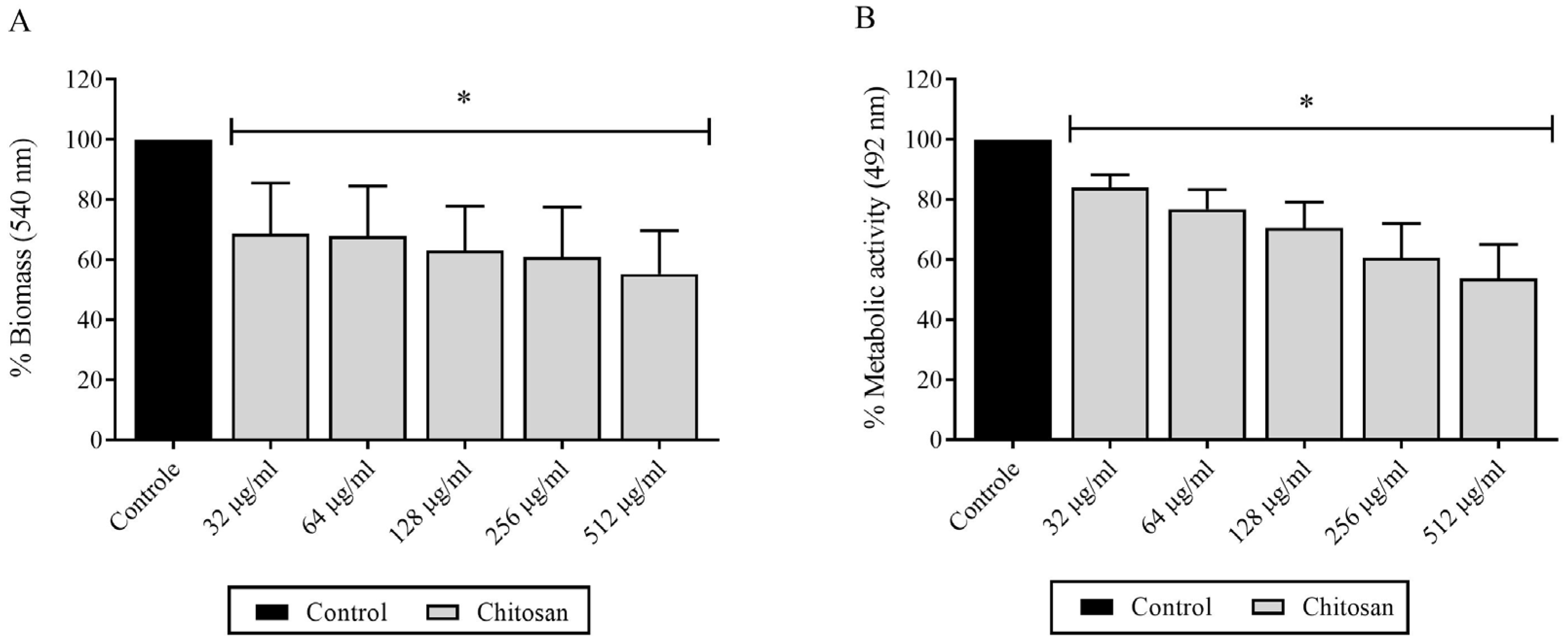
3.3. Evaluation of Chitosan Effects on H. capsulatum Biofilm Formation

Chitosan caused significant reductions in the biomass and metabolic activity of H. capsulatum biofilms in the filamentous form (Figure 1A). Biomass reductions were observed when exposing H. capsulatum biofilms to chitosan, demonstrating a dose-dependent effect [128 µg/mL causing 47% reduction] (p < 0.05). In the assessment of metabolic activity, a significant reduction of 30% was obtained from 32 to 512 µg/mL of chitosan (p < 0.05) (Figure 1B). There were no significant reductions among the different strains tested.
Chitosan also caused significant reductions in the biomass and metabolic activity of H. capsulatum biofilms in the yeast form (Figure 2). Reductions in biomass occurred when H. capsulatum biofilms were exposed to chitosan, with a dose-dependent relationship [32 µg/mL, resulting in 40% reduction] (p < 0.05). In the evaluation of metabolic activity, a notable reduction of 40% was achieved with a chitosan concentration range of 32 to 512 µg/mL (p < 0.05) (Figure 2B). No significant reductions were observed among the various tested strains.

3.4. Structural Analysis of Biofilms

SEM was performed to depict structural differences between the biofilms of H. capsulatum treated, or not, with chitosan (Figure 3A–C). In the absence of LMW chitosan, biofilms of H. capsulatum showed fungal cells that were organized into dense structures and composed of multilayers of associated cells besides an extracellular matrix (Figure 3A). Biofilms treated with chitosan at 4 × MIC100% and 8 × MIC100% exhibited less dense structures with less cells and less extracellular matrix (Figure 3B,C). After exposure to chitosan (128 µg/mL), a decrease in cell mass was observed (Figure 3B,C) compared to the control without chitosan exposure (Figure 3A). The cluster structure of blastoconidia was no longer evident after treatment (Figure 3B,C), and the cells that remained on the Thermanox slides displayed reduced viability, as can be observed in Figure 3B,C.

4. Discussion

Chitosan had an antifungal effect on the planktonic growth of H. capsulatum. It was able to inhibit the growth of the fungal pathogen in the filamentous and yeast forms. Previous studies have reported chitosan inhibition on the growth of other fungal pathogens, with results similar to those found in this study, such as Aspergillus fumigatus [32] and Sporothrix brasiliensis [23,33]. The results obtained for the antifungal drugs amphotericin B and itraconazole are in the same range of concentrations described by other studies [34,35,36].
Chitosan has attracted attention as a versatile biopolymer because of its cationic characteristics, biocompatibility, biodegradability, low toxicity, and good adsorption capabilities [37]. Numerous studies have examined the mechanism of chitosan [25,38,39]. It primarily functions through electrostatic interactions between its positively charged protonated amino groups and the negatively charged components of cell surfaces [40]. This interaction can be manifested through various pathways, and the broad molecular weight of chitosan (spanning from oligo-chitosan to low, medium, and high molecular weight) is one of the inherent factors that can affect its mechanism of action [41]. In a recent study, the antifungal properties of chitosan with different molecular weights were analyzed against three fungal species, C. albicans, F. solani, and A. niger. In that study, the authors found varying inhibition results depending on the fungal species and chitosan molecular weight, demonstrating that molecular weight can influence the observed antifungal activity [42]. According to the authors, low molecular weight chitosan may have a stronger impact on intracellular processes because it can more easily enter cells due to its small size and low zeta potential. Low molecular weight (LMW) chitosan can permeate the cell wall, interact with DNA, and impede mRNA synthesis and transcription. It has also been proposed that LMW chitosan can induce cell membrane rupture, representing a dual mechanism of action [23]. One hypothesis is that chitosan acts by permeabilizing the plasma membrane, leading to intracellular overproduction of reactive oxygen species (ROS), potentially inducing cell death [43].
In general, the yeast form of H. capsulatum exhibited MICs approximately four times lower than those obtained for the filamentous form in combination with the compounds tested in this study. As a dimorphic fungus, in nature, it exists in the filamentous (infectious) form, and when it enters the host, a temperature change triggers the fungal transition to the yeast form. A series of changes are necessary to become a yeast, from activating genes such (as the Ryp1 gene) to alterations in the fungal cell wall composition [44]. Therefore, this difference in susceptibility may be related to differences in the proportions of fungal cell wall constituents between filamentous and yeast forms, such as glucosamine and β-1,3-glucan [45]. The cell wall is essential for fungal growth since it provides stability and protection against osmotic stress. Furthermore, this structure is responsible for maintaining cellular shape. Changes in the cell wall structure have previously been linked to reduced susceptibility to amphotericin B in Candida spp. [46]. Thus, we hypothesize that differences in the cell wall composition between the filamentous and yeast forms of H. capsulatum may influence susceptibility to antifungal agents and chitosan.
The interaction effect of LWC and amphotericin B or itraconazole on the growth of H. capsulatum revealed that these combined drugs have synergistic effects in different combinations for H. capsulatum in the filamentous form, without any antagonistic effect. In these cases, the reduction in the MIC of the antifungal agent reached up to 16 times. In assays involving the yeast form, the results were more promising, and synergism between LWC and amphotericin B or itraconazole was detected for all strains tested. Synergism between chitosan and antifungal agents has previously been reported in experiments involving two Candida species [47].
Based on amphotericin B’s action in creating pores in fungal cell membranes and itraconazole’s effect of inhibiting ergosterol biosynthesis, leading to impaired fungal plasma membrane integrity, there may be an increased penetration of LWC into the cells [48]. The combined action on different biological processes or molecular targets could explain the synergism.
Chitosan caused reductions of approximately 50% in the rates of biomass and metabolic activity of biofilms. Micrographs obtained by SEM of H. capsulatum biofilms in the yeast form (the infective form) showed that untreated chitosan-free biofilms were robust, with higher cell numbers. In contrast, chitosan-treated biofilms had a decrease in the number of cells, demonstrating that chitosan acts on biofilms by killing cells. Other studies have demonstrated a reduction in biofilm formation in the presence of low molecular weight chitosan in Candida albicans [49] and Sporothrix brasiliensis [23], with reductions in biomass production and metabolic activity. Based on a clinical trial, another study investigated the effect of low molecular weight chitosan on Candida albicans biofilms in denture stomatitis. The authors concluded that chitosan has antifungal efficacy on biofilm formation in patients along with inherent biocompatibility, making it a promising candidate for use as an antifungal mouthwash [50].
It is known that during infection, the fungus H. capsulatum needs to maintain the stability of the cell membrane for its survival [51,52]. LMW chitosan is capable of causing cell membrane rupture, promoting an effect incompatible with the metabolic activity of the fungus [23]. It is also known that one of the identified effects of chitosan in general is the production of reactive oxygen species in cells. Previous studies have shown that oxidative stress is crucial in forming and maintaining biofilms [53,54]. Increased production of reactive oxygen species can lead to less biofilm with less extracellular matrix production [53]. Consequently, fungal pathogens, like C. albicans, have evolved various tactics to counteract the oxidative stress produced as a byproduct of aerobic respiration, thereby preserving redox homeostasis within cells [54,55]. In this sense, some pathogens use enzymatic mechanisms such as peroxidases to try to survive oxidative stress in the host [56,57]. Such effects may explain how polymers, whose action is the production of reactive oxygen species, cause reductions in the formation and maintenance of fungal biofilms, which could also explain the reductions found in the tests carried out in this study.

5. Conclusions

Based on our in vitro results, the low molecular weight chitosan can inhibit strains of H. capsulatum in the filamentous and yeast forms, having pharmacological synergism with traditional antifungals. Moreover, chitosan inhibited the biomass and metabolic activity rates of H. capsulatum biofilms significantly, influencing the biofilm formation. In addition to our findings, low molecular wave chitosan has been extensively studied against various pathogens, both in vitro and in vivo, in recent years. Thus, the application of chitosan can be an innovative therapeutic strategy in the future.

Author Contributions

Conceptualization, R.S.N.B. and M.F.G.R.; methodology, A.d.C.C., A.F.D.N., M.R.F. and G.d.S.A.; validation, R.S.F. and J.V.S.N.; investigation, J.R.L.d.M.; resources, T.C. and U.S.; data curation, W.d.A.P.N.; writing—original draft preparation, A.d.C.C.; writing—review and editing, R.S.V.; supervision, T.C. and U.S. All authors have read and agreed to the published version of the manuscript.

Funding

This research was funded by the Brazilian agencies National Council for Scientific and Techno-logical Development (CNPq; grant numbers 305890/2021-2; 200068/2022-9, 200077/2022-8, 408671/2023-8); and the Office for the Improvement of Higher Education Personnel (CAPES; grant number 88881.168799/2018-00).

Institutional Review Board Statement

Not applicable.

Informed Consent Statement

Not applicable.

Data Availability Statement

Data are contained within the article.

Conflicts of Interest

The authors declare no conflict of interest.

References

  1. Sil, A. Molecular Regulation of Histoplasma Dimorphism. Curr. Opin. Microbiol. 2019, 52, 151–157. [Google Scholar] [CrossRef]
  2. Gugnani, H.C.; Denning, D.W. Infection of Bats with Histoplasma Species. Med. Mycol. 2023, 61, myad080. [Google Scholar] [CrossRef] [PubMed]
  3. Adenis, A.A.; Aznar, C.; Couppié, P. Histoplasmosis in HIV-Infected Patients: A Review of New Developments and Remaining Gaps. Curr. Trop. Med. Rep. 2014, 1, 119–128. [Google Scholar] [CrossRef] [PubMed]
  4. Limaye, A.P.; Connolly, P.A.; Sagar, M.; Fritsche, T.R.; Cookson, B.T.; Wheat, L.J.; Stamm, W.E. Transmission of Histoplasma Capsulatum by Organ Transplantation. N. Engl. J. Med. 2000, 343, 1163–1166. [Google Scholar] [CrossRef]
  5. Falci, D.R.; Dalla Lana, D.F.; Pasqualotto, A.C. The Era of Histoplasmosis in Brazilian Endemic Mycoses. Lancet Reg. Health Am. 2021, 3, 100037. [Google Scholar] [CrossRef]
  6. Kainz, K.; Bauer, M.A.; Madeo, F.; Carmona-Gutierrez, D. Fungal Infections in Humans: The Silent Crisis. Microb. Cell 2020, 7, 143–145. [Google Scholar] [CrossRef]
  7. Rodrigues, M.L.; Nosanchuk, J.D. Fungal Diseases as Neglected Pathogens: A Wake-up Call to Public Health Officials. PLoS Negl. Trop. Dis. 2020, 14, e0007964. [Google Scholar] [CrossRef]
  8. Berman, J.; Krysan, D.J. Drug Resistance and Tolerance in Fungi. Nat. Rev. Microbiol. 2020, 18, 319–331. [Google Scholar] [CrossRef]
  9. Garfoot, A.L.; Rappleye, C.A. Histoplasma Capsulatum Surmounts Obstacles to Intracellular Pathogenesis. FEBS J. 2016, 283, 619–633. [Google Scholar] [CrossRef] [PubMed]
  10. Pitangui, N.S.; Sardi, J.C.O.; Silva, J.F.; Benaducci, T.; Moraes da Silva, R.A.; Rodríguez-Arellanes, G.; Taylor, M.L.; Mendes-Giannini, M.J.S.; Fusco-Almeida, A.M. Adhesion of Histoplasma capsulatum to Pneumocytes and Biofilm Formation on an Abiotic Surface. Biofouling 2012, 28, 711–718. [Google Scholar] [CrossRef]
  11. Brilhante, R.S.N.; de Lima, R.A.C.; de Farias Marques, F.J.; Silva, N.F.; Caetano, É.P.; de Souza Collares Maia Castelo-Branco, D.; de Jesus Pinheiro Gomes Bandeira, T.; Moreira, J.L.B.; de Aguiar Cordeiro, R.; Monteiro, A.J. Histoplasma capsulatum in Planktonic and Biofilm Forms: In Vitro Susceptibility to Amphotericin B, Itraconazole and Farnesol. J. Med. Microbiol. 2015, 64, 394–399. [Google Scholar] [CrossRef]
  12. Brilhante, R.S.N.; Sales, J.A.; da Silva, M.L.Q.; de Oliveira, J.S.; de Alencar Pereira, L.; Pereira-Neto, W.A.; de Aguiar Cordeiro, R.; Sidrim, J.J.C.; de Souza Collares Maia Castelo-Branco, D.; Rocha, M.F.G. Antifungal Susceptibility and Virulence of Candida Parapsilosis Species Complex: An Overview of Their Pathogenic Potential. J. Med. Microbiol. 2018, 67, 903–914. [Google Scholar] [CrossRef] [PubMed]
  13. de Cássia Orlandi Sardi, J.; de Souza Pitangui, N.; Voltan, A.R.; Braz, J.D.; Machado, M.P.; Fusco Almeida, A.M.; Mendes Giannini, M.J.S. In Vitro Paracoccidioides Brasiliensis Biofilm and Gene Expression of Adhesins and Hydrolytic Enzymes. Virulence 2015, 6, 642–651. [Google Scholar] [CrossRef]
  14. Costa-Orlandi, C.B.; Sardi, J.C.O.; Pitangui, N.S.; de Oliveira, H.C.; Scorzoni, L.; Galeane, M.C.; Medina-Alarcón, K.P.; Melo, W.C.M.A.; Marcelino, M.Y.; Braz, J.D. Fungal Biofilms and Polymicrobial Diseases. J. Fungi 2017, 3, 22. [Google Scholar] [CrossRef] [PubMed]
  15. Kumar, P.; Mishra, S.; Singh, S. Advanced Acuity in Microbial Biofilm Genesis, Development, Associated Clinical Infections and Control. J. Anti-Infect. 2017, 19, 20–31. [Google Scholar] [CrossRef]
  16. Sardi, J.D.C.O.; Pitangui, N.D.S.; Rodriguez-Arellanes, G.; Taylor, M.L.; Fusco-Almeida, A.M.; Mendes-Giannini, M.J.S. Highlights in Pathogenic Fungal Biofilms. Rev. Iberoam. Micol. 2014, 31, 22–29. [Google Scholar] [CrossRef]
  17. Brilhante, R.S.N.; Aguiar, F.R.M.; Silva, M.L.Q.; Oliveira, J.S.; Camargo, Z.P.; Rodrigues, A.M.; Pereira, V.S.; Serpa, R.; de Souza Collares Maia Castelo-Branco, D.; Correia, E.E.M.; et al. Antifungal Susceptibility of Sporothrix schenckii Complex Biofilms. Med. Mycol. 2018, 56, 297–306. [Google Scholar] [CrossRef]
  18. de Aguiar Cordeiro, R.; Serpa, R.; Alexandre, C.F.U.; de Farias Marques, F.J.; de Melo, C.V.S.; da Silva Franco, J.; de Jesus Evangelista, A.J.; de Camargo, Z.P.; Brilhante, R.S.N.; Rocha, M.F.G. Trichosporon Inkin Biofilms Produce Extracellular Proteases and Exhibit Resistance to Antifungals. J. Med. Microbiol. 2015, 64, 1277–1286. [Google Scholar] [CrossRef]
  19. Brilhante, R.S.N.; Correia, E.E.M.; de Melo Guedes, G.M.; Pereira, V.S.; de Oliveira, J.S.; Bandeira, S.P.; de Alencar, L.P.; de Andrade, A.R.C.; de Souza Collares Maia Castelo-Branco, D.; de Aguiar Cordeiro, R.; et al. Quantitative and Structural Analyses of the in Vitro and Ex Vivo Biofilm-Forming Ability of Dermatophytes. J. Med. Microbiol. 2017, 66, 1045–1052. [Google Scholar] [CrossRef]
  20. Roudbary, M.; Vahedi-Shahandashti, R.; dos Santos, A.L.S.; Roudbar Mohammadi, S.; Aslani, P.; Lass-Flörl, C.; Rodrigues, C.F. Biofilm Formation in Clinically Relevant Filamentous Fungi: A Therapeutic Challenge. Crit. Rev. Microbiol. 2022, 48, 197–221. [Google Scholar] [CrossRef]
  21. Azar, M.M.; Loyd, J.L.; Relich, R.F.; Wheat, L.J.; Hage, C.A. Current Concepts in the Epidemiology, Diagnosis, and Management of Histoplasmosis Syndromes. Semin. Respir. Crit. Care Med. 2020, 41, 13–30. [Google Scholar] [CrossRef] [PubMed]
  22. Yu, J.-L.; Dai, Q.-Q.; Li, G.-B. Deep Learning in Target Prediction and Drug Repositioning: Recent Advances and Challenges. Drug Discov. Today 2021, 27, 1796–1814. [Google Scholar] [CrossRef] [PubMed]
  23. Garcia, L.G.S.; de Melo Guedes, G.M.; Fonseca, X.M.Q.C.; Pereira-Neto, W.A.; Castelo-Branco, D.S.C.M.; Sidrim, J.J.C.; de Aguiar Cordeiro, R.; Rocha, M.F.G.; Vieira, R.S.; Brilhante, R.S.N. Antifungal Activity of Different Molecular Weight Chitosans against Planktonic Cells and Biofilm of Sporothrix brasiliensis. Int. J. Biol. Macromol. 2020, 143, 341–348. [Google Scholar] [CrossRef] [PubMed]
  24. Kvasničková, E.; Paulíček, V.; Paldrychová, M.; Ježdík, R.; Maťátková, O.; Masák, J. Aspergillus fumigatus DBM 4057 Biofilm Formation Is Inhibited by Chitosan, in Contrast to Baicalein and Rhamnolipid. World J. Microbiol. Biotechnol. 2016, 32, 187. [Google Scholar] [CrossRef] [PubMed]
  25. Lopez-Moya, F.; Suarez-Fernandez, M.; Lopez-Llorca, L.V. Molecular Mechanisms of Chitosan Interactions with Fungi and Plants. Int. J. Mol. Sci. 2019, 20, 332. [Google Scholar] [CrossRef] [PubMed]
  26. CLSI. Reference Method for Broth Dilution Antifungal Susceptibility Testing of Filamentous Fungi, 3rd ed.; Clinical and Laboratory Standards Institute: Wayne, PA, USA, 2017; ISBN 1562388304. [Google Scholar]
  27. CLSI. Reference Method for Broth Dilution Antifungal Susceptibility Testing of Yeasts, 2nd ed.; Clinical and Laboratory Standards Institute: Wayne, PA, USA, 2020; ISBN 978-1-68440-083-6. [Google Scholar]
  28. Brilhante, R.S.N.; Lopes, R.G.P.; de Aguiar, L.; de Oliveira, J.S.; dos Santos Araújo, G.; Paixão, G.C.; de Aquino Pereira-Neto, W.; Freire, R.S.; Nunes, J.V.S.; de Lima, R.P.; et al. Inhibitory Effect of Proteinase K against Dermatophyte Biofilms: An Alternative for Increasing the Antifungal Effects of Terbinafine and Griseofulvin. Biofouling 2022, 38, 286–297. [Google Scholar] [CrossRef]
  29. Pierce, C.G.; Uppuluri, P.; Tristan, A.R.; Wormley, F.L., Jr.; Mowat, E.; Ramage, G.; Lopez-Ribot, J.L. A Simple and Reproducible 96-Well Plate-Based Method for the Formation of Fungal Biofilms and Its Application to Antifungal Susceptibility Testing. Nat. Protoc. 2008, 3, 1494. [Google Scholar] [CrossRef]
  30. Almeida-Paes, R.; Figueiredo-Carvalho, M.H.G.; Brito-Santos, F.; Almeida-Silva, F.; Oliveira, M.M.E.; Zancope-Oliveira, R.M. Melanins Protect Sporothrix brasiliensis and Sporothrix schenckii from the Antifungal Effects of Terbinafine. PLoS ONE 2016, 11, 1–13. [Google Scholar] [CrossRef]
  31. Odds, F.C. Synergy, Antagonism, and What the Chequerboard Puts between Them. J. Antimicrob. Chemother. 2003, 52, 1. [Google Scholar] [CrossRef]
  32. Madu, U.L.; Ogundeji, A.O.; Folorunso, O.S.; Albertyn, J.; Pohl, C.H.; Sebolai, O.M. The Repurposing of the Antimalaria Drug, Primaquine, as a Photosensitizer to Inactivate Cryptococcal Cells. Photochem 2021, 1, 275–286. [Google Scholar] [CrossRef]
  33. Pearce, A.N.; Chen, D.; Edmeades, L.R.; Cadelis, M.M.; Troudi, A.; Brunel, J.M.; Bourguet-Kondracki, M.-L.; Copp, B.R. Repurposing Primaquine as a Polyamine Conjugate to Become an Antibiotic Adjuvant. Bioorg. Med. Chem. 2021, 38, 116110. [Google Scholar] [CrossRef]
  34. Brilhante, R.S.N.; de Melo Guedes, G.M.; Riello, G.B.; Ribeiro, J.F.; Alencar, L.P.; Bandeira, S.P.; Castelo-Branco, D.S.C.M.; Oliveira, J.S.; Freire, J.M.M.; de Mesquita, J.R.L. RYP1 Gene as a Target for Molecular Diagnosis of Histoplasmosis. J. Microbiol. Methods 2016, 130, 112–114. [Google Scholar] [CrossRef] [PubMed]
  35. Bongomin, F.; Kwizera, R.; Baluku, J.B.; Asio, L.G.; Otu, A.A. Treatment of Histoplasmosis. In Histoplasma and Histoplasmosis; IntechOpen: London, UK, 2020; p. 69. [Google Scholar]
  36. Brilhante, R.S.N.; de Melo Guedes, G.M.; da Silva, M.L.Q.; Castelo-Branco, D.S.C.M.; de Aguiar Cordeiro, R.; Sidrim, J.J.C.; Rocha, M.F.G. A Proposal for Antifungal Epidemiological Cut-off Values against Histoplasma capsulatum Var. Capsulatum Based on the Susceptibility of Isolates from HIV-Infected Patients with Disseminated Histoplasmosis in Northeast Brazil. Int. J. Antimicrob. Agents 2018, 52, 272–277. [Google Scholar] [CrossRef] [PubMed]
  37. Gonçalves, C.; Ferreira, N.; Lourenço, L. Production of Low Molecular Weight Chitosan and Chitooligosaccharides (COS): A Review. Polymers 2021, 13, 2466. [Google Scholar] [CrossRef] [PubMed]
  38. Adhikari, H.S.; Yadav, P.N. Anticancer Activity of Chitosan, Chitosan Derivatives, and Their Mechanism of Action. Int. J. Biomater. 2018, 2018, 2952085. [Google Scholar] [CrossRef] [PubMed]
  39. Grande-Tovar, C.D.; Chaves-López, C.; Serio, A.; Rossi, C.; Paparella, A. Chitosan Coatings Enriched with Essential Oils: Effects on Fungi Involved in Fruit Decay and Mechanisms of Action. Trends Food Sci. Technol. 2018, 78, 61–71. [Google Scholar] [CrossRef]
  40. Kulikov, S.N.; Lisovskaya, S.A.; Zelenikhin, P.V.; Bezrodnykh, E.A.; Shakirova, D.R.; Blagodatskikh, I.V.; Tikhonov, V.E. Antifungal Activity of Oligochitosans (Short Chain Chitosans) against Some Candida Species and Clinical Isolates of Candida albicans: Molecular Weight–Activity Relationship. Eur. J. Med. Chem. 2014, 74, 169–178. [Google Scholar] [CrossRef] [PubMed]
  41. Ma, Z.; Garrido-Maestu, A.; Jeong, K.C. Application, Mode of Action, and in vivo Activity of Chitosan and Its Micro-and Nanoparticles as Antimicrobial Agents: A Review. Carbohydr. Polym. 2017, 176, 257–265. [Google Scholar] [CrossRef]
  42. Poznanski, P.; Hameed, A.; Orczyk, W. Chitosan and Chitosan Nanoparticles: Parameters Enhancing Antifungal Activity. Molecules 2023, 28, 2996. [Google Scholar] [CrossRef]
  43. Lopez-Moya, F.; Colom-Valiente, M.F.; Martinez-Peinado, P.; Martinez-Lopez, J.E.; Puelles, E.; Sempere-Ortells, J.M.; Lopez-Llorca, L.V. Carbon and Nitrogen Limitation Increase Chitosan Antifungal Activity in Neurospora crassa and Fungal Human Pathogens. Fungal Biol. 2015, 119, 154–169. [Google Scholar] [CrossRef]
  44. Valdez, A.F.; Miranda, D.Z.; Guimarães, A.J.; Nimrichter, L.; Nosanchuk, J.D. Pathogenicity & Virulence of Histoplasma capsulatum-A Multifaceted Organism Adapted to Intracellular Environments. Virulence 2022, 13, 2137987. [Google Scholar]
  45. Martínez-Álvarez, J.A.; Pérez-García, L.A.; Mellado-Mojica, E.; López, M.G.; Martínez-Duncker, I.; Lópes-Bezerra, L.M.; Mora-Montes, H.M. Sporothrix schenckii Sensu stricto and Sporothrix brasiliensis Are Differentially Recognized by Human Peripheral Blood Mononuclear Cells. Front. Microbiol. 2017, 8, 843. [Google Scholar] [CrossRef] [PubMed]
  46. Mesa-Arango, A.C.; Rueda, C.; Román, E.; Quintin, J.; Terrón, M.C.; Luque, D.; Netea, M.G.; Pla, J.; Zaragoza, O. Cell Wall Changes in Amphotericin B-Resistant Strains from Candida tropicalis and Relationship with the Immune Responses Elicited by the Host. Antimicrob. Agents Chemother. 2016, 60, 2326–2335. [Google Scholar] [CrossRef] [PubMed]
  47. Lo, W.-H.; Deng, F.-S.; Chang, C.-J.; Lin, C.-H. Synergistic Antifungal Activity of Chitosan with Fluconazole against Candida albicans, Candida tropicalis, and Fluconazole-Resistant Strains. Molecules 2020, 25, 5114. [Google Scholar] [CrossRef] [PubMed]
  48. Johnson, M.D.; MacDougall, C.; Ostrosky-Zeichner, L.; Perfect, J.R.; Rex, J.H. Combination Antifungal Therapy. Antimicrob. Agents Chemother. 2004, 48, 693–715. [Google Scholar] [CrossRef] [PubMed]
  49. Srimaneepong, V.; Thanamee, T.; Wattanasirmkit, K.; Muangsawat, S.; Matangkasombut, O. Efficacy of Low-molecular Weight Chitosan against Candida albicans Biofilm on Polymethyl Methacrylate Resin. Aust. Dent. J. 2021, 66, 262–269. [Google Scholar] [CrossRef]
  50. Atai, Z.; Atai, M.; Amini, J. In Vivo Study of Antifungal Effects of Low-Molecular-Weight Chitosan against Candida albicans. J. Oral Sci. 2017, 59, 425–430. [Google Scholar] [CrossRef]
  51. Riddell IV, J.; Wheat, L.J. Central Nervous System Infection with Histoplasma capsulatum. J. Fungi 2019, 5, 70. [Google Scholar] [CrossRef]
  52. Horwath, M.C.; Fecher, R.A.; Deepe, G.S., Jr. Histoplasma capsulatum, Lung Infection and Immunity. Future Microbiol. 2015, 10, 967–975. [Google Scholar] [CrossRef]
  53. Arce Miranda, J.E.; Sotomayor, C.E.; Albesa, I.; Paraje, M.G. Oxidative and Nitrosative Stress in Staphylococcus Aureus Biofilm. FEMS Microbiol. Lett. 2011, 315, 23–29. [Google Scholar] [CrossRef]
  54. Arce Miranda, J.E.; Baronetti, J.L.; Sotomayor, C.E.; Paraje, M.G. Oxidative and Nitrosative Stress Responses during Macrophage–Candida Albicans Biofilm Interaction. Med. Mycol. 2019, 57, 101–113. [Google Scholar] [CrossRef]
  55. Pais, P.; Vagueiro, S.; Mil-Homens, D.; Pimenta, A.I.; Viana, R.; Okamoto, M.; Chibana, H.; Fialho, A.M.; Teixeira, M.C. A New Regulator in the Crossroads of Oxidative Stress Resistance and Virulence in Candida Glabrata: The Transcription Factor CgTog1. Virulence 2020, 11, 1522–1538. [Google Scholar] [CrossRef] [PubMed]
  56. Lee, S.-Y.; Chen, H.-F.; Yeh, Y.-C.; Xue, Y.-P.; Lan, C.-Y. The Transcription Factor Sfp1 Regulates the Oxidative Stress Response in Candida Albicans. Microorganisms 2019, 7, 131. [Google Scholar] [CrossRef] [PubMed]
  57. Caceres, I.; El Khoury, R.; Bailly, S.; Oswald, I.P.; Puel, O.; Bailly, J.-D. Piperine Inhibits Aflatoxin B1 Production in Aspergillus Flavus by Modulating Fungal Oxidative Stress Response. Fungal Genet. Biol. 2017, 107, 77–85. [Google Scholar] [CrossRef] [PubMed]
Figure 1. Metabolic activity and biomass of H. capsulatum biofilms in filamentous form. exposed to different concentrations of chitosan: (A) Biofilm biomass exposed to drug; (B) Metabolic activity of drug-exposed biofilm. * Significant difference compared to control (p < 0.05).
Jof 09 01201 g001
Figure 2. Metabolic activity and biomass of H. capsulatum biofilms in yest form exposed to different concentrations of chitosan: (A) Biofilm biomass exposed to drug; (B) Metabolic activity of drug-exposed biofilm. * Significant difference compared to control (p < 0.05).
Jof 09 01201 g002
Figure 3. Structure and morphology of the H. capsulatum biofilm. SEM images of biofilms with untreated control, with an arrow pointing to the presence of extracellular polymeric substances (EPS): (A), and with treatment of 512 μg/mL chitosan (B) and 128 μg/mL chitosan (C). There is a reduction in the biofilm caused by the rupture of EPS in the biofilm, with only a few cells adhering to the surface after treatment with chitosan.
Jof 09 01201 g003
Table 1. Minimum inhibitory concentration of amphotericin B, itraconazole, and chitosan against strains of H. capsulatum in the filamentous form.
StrainsMinimum Inhibitory Concentration (µg/mL)
(CEMM Code)AMBITCCHI
CEMM 03-2-0882164
CEMM 03-2-0900.50.2532
CEMM 03-3-0030.25164
CEMM 03-3-0262164
CEMM 03-3-03320.2564
CEMM 03-3-0340.50.2532
CEMM 03-3-0362164
CEMM 03-3-0372164
CEMM 03-3-03821128
CEMM 03-3-0390.50.0364
CEMM 03-3-01510.2532
CEMM 03-3-0722164
CEMM 03-3-1000.50.0364
CEMM 03-3-0240.50.03128
CEMM 03-3-0490.35132
CEMM 03-3-06620.2532
CEMM 03-3-05210.2532
CEMM 03-3-0700.5164
CEMM 03-3-0401164
CEMM 03-3-0530.50.0332
CEMM: Center for Medical Mycology, AMB: amphotericin B, ITC: itraconazole, CHI: chitosan.
Table 2. Minimum inhibitory concentration of amphotericin B, itraconazole, and chitosan against yeast form of H. capsulatum.
StrainsMinimum Inhibitory Concentration (µg/mL)
(CEMM Code)AMBITCCHI
CEMM 03-2-0880.250.58
CEMM 03-2-0900.030.2532
CEMM 03-3-0030.50.564
CEMM 03-3-0260.030.564
CEMM 03-3-0370.030.58
CEMM 03-3-0380.250.2564
CEMM 03-3-0390.50.532
CEMM 03-3-0150.030.2516
CEMM 03-3-0720.50.516
CEMM 03-3-1000.030.532
CEMM 03-3-0330.250.2516
CEMM 03-3-0340.250.2532
CEMM 03-3-0360.250.2532
CEMM: Center for Medical Mycology, AMB: amphotericin B, ITC: itraconazole, CHI: chitosan.
Table 3. Minimum inhibitory concentration of chitosan in combination with antifungals against planktonic cells of H. capsulatum, in filamentous form, and fractional inhibitory concentration index.
CEMM CodeIsolated Drugs (µg/mL)Combined Drugs (µg/mL)FICI
CHIAMBITCCHI/AMBCHI/ITCCHI/AMBCHI/ITC
CEMM 03-2-088642116/0.516/0.50,75 I1.25 I
CEMM 03-2-0901280.50.2564/232/11.5 I0.75 I
CEMM 03-3-003640.25116/0.2516/0.250.5 I0.75 I
CEMM 03-3-026642132/0.012532/0.1250.625 I1.5 I
CEMM 03-3-033642432/116/0.251.5 I0.75 I
CEMM 03-3-034640.50.2516/216/0.1251.25 I0.03 S
CEMM 03-3-036642116/216/0.251.25 I0.03 S
CEMM 03-3-037322132/132/0.252.0 I1.25 I
CEMM: Centre for Medical Mycology, AMB: Amphotericin B, ITC: Itraconazole, CHI: chitosan, FICI: fractional inhibitory concentration index, the interaction was classified as follows: synergistic (S; FICI ≤ 0.5), indifferent (I; 0.5 < FICI ≤ 4.0) or antagonistic (A; FICI > 4.0).
Table 4. Minimum inhibitory concentration of chitosan in combination with antifungals against planktonic cells of H. capsulatum in the yeast form, and fractional inhibitory concentration index.
CEMM CodeIsolated Drugs (µg/mL)Combined Drugs (µg/mL)FICI
CHIAMBCHI/AMBCHI/ITCCHI/AMBCHI/ITC
CEMM 03-2-088320.254/0.031252/0.00781250.125 S0.3125 S
CEMM 03-2-090320.1254/0.016252/0.00781250.125 S0.3125 S
CEMM 03-3-003320.52/0.031252/0.00781250.125 S0.3125 S
CEMM 03-3-026640.52/0.031252/0.00781250.125 S0.3125 S
CEMM 03-3-033640.52/0.031252/0.00781250.125 S0.3125 S
CEMM 03-3-034640.52/0.031252/0.00781250.125 S0.3125 S
CEMM 03-3-036320.52/0.031252/0.00781250.125 S0.3125 S
CEMM: Center for Medical Mycology, AMB: Amphotericin B, ITC: Itraconazole, CHI: chitosan, FICI: fractional inhibitory concentration index, classified as synergistic (S; FICI ≤ 0.5), indifferent (I; 0.5 < FICI ≤ 4.0) or antagonistic (A; FICI > 4.0).
Disclaimer/Publisher’s Note: The statements, opinions and data contained in all publications are solely those of the individual author(s) and contributor(s) and not of MDPI and/or the editor(s). MDPI and/or the editor(s) disclaim responsibility for any injury to people or property resulting from any ideas, methods, instructions or products referred to in the content.

