Article

Regulatory Ability of Lactiplantibacillus plantarum on Human Skin Health by Counteracting In Vitro Malassezia furfur Effects

by
Alessandra Fusco
1,*,†,
Brunella Perfetto
1,†,
Vittoria Savio
1,
Adriana Chiaromonte
1,
Giovanna Torelli
1,
Giovanna Donnarumma
1,* and
Adone Baroni
2
1
Department of Experimental Medicine, Section of Microbiology and Clinical Microbiology, University of Campania “Luigi Vanvitelli”, 80138 Naples, Italy
2
Department of Mental and Physical Health and Preventive Medicine, University of Campania “Luigi Vanvitelli”, 80138 Naples, Italy
*
Authors to whom correspondence should be addressed.
These authors contributed equally to this work.
J. Fungi 2023, 9(12), 1153; https://doi.org/10.3390/jof9121153
Submission received: 3 October 2023 / Revised: 23 November 2023 / Accepted: 28 November 2023 / Published: 29 November 2023
(This article belongs to the Special Issue New Strategies to Combat Human Fungal Infections)

Abstract

:
The skin serves as the first barrier against pathogen attacks, thanks to its multifunctional microbial community. Malassezia furfur is a commensal organism of normal cutaneous microflora but is also a cause of skin diseases. It acts on different cell pattern recognition receptors (TLRs, AhR, NLRP3 inflammasome) leading to cellular damage, barrier impairment, and inflammatory cytokines production. Lactobacillus spp. Is an endogenous inhabitant of healthy skin, and studies have proven its beneficial role in wound healing, skin inflammation, and protection against pathogen infections. The aim of our study is to demonstrate the ability of live Lactiplantibacillus plantarum to interfere with the harmful effects of the yeast on human keratinocytes (HaCat) in vitro. To enable this, the cells were treated with M. furfur, either alone or in the presence of L. plantarum. To study the inflammasome activation, cells require a stimulus triggering inflammation (LPS) before M. furfur infection, with or without L. plantarum. L. plantarum effectively counteracts all the harmful strategies of yeast, reducing the phospholipase activity, accelerating wound repair, restoring barrier integrity, reducing AhR and NLRP3 inflammasome activation, and, consequently, releasing inflammatory cytokines. Although lactobacilli have a long history of use in fermented foods, it can be speculated that they can also have health-promoting activities when topically applied.

1. Introduction

To maintain healthy skin function, it is important to have a balanced skin microbiota, as alterations in its integrity are linked to several cutaneous diseases [1,2]. The microbes present on the skin form a barrier capable of counteracting environmental colonization through various mechanisms, including resource exclusion, direct inhibition, and/or interference with the colonization. The cutaneous microbiota improves the physical skin barrier, contributing to the differentiation and epithelialization, stimulating the innate and adaptive immunity, releasing antimicrobial peptides, and influencing the development of protective immunity [1,2]. Recent research has emphasized the role of functional probiotics in intestinal function and the maintenance of healthy skin [3,4,5,6]. However, the mechanism of the interactions between the maintenance of the skin barrier and probiotics has not been elucidated yet.
The yeasts of the genus Malassezia colonize human skin after birth. They are saprophytes and, as such, are generally well-tolerated by the human immune system. However, under appropriate conditions, they can invade the stratum corneum and interact with the host immune system, developing their pathogenic potential. Within the genus Malassezia, Malassezia furfur is involved in skin diseases of varying severity, such as pityriasis versicolor, seborrheic dermatitis, atopic eczema, dermatitis, and dandruff [7,8,9]. It is very probable that the inflammatory effects exerted on the host cells by Malassezia and the impairment of the epithelial barrier result from a complicated balance involving different molecules. The expression of these molecules is mediated not by one, but by different pathways involving different receptors, cofactors, and transcription factors.
It has been shown that the triggering of some skin inflammatory processes is linked to the response of mast cells and other cellular types to stimulation by Malassezia spp. through the canonical Toll-like receptor 2 (TLR2)/MyD88 pathway. This stimulation leads to either the up- or downregulation of cytokine, chemokine, and adhesion molecules in host effector cells. Following the triggering of inflammation, metalloproteinases and tight junctions can be modulated, too [10,11].
For the control of membrane permeability and integrity, crucial structures are the tight junctions, including occludin, claudins (integral membrane proteins), and the junction adhesion molecules that interact with the zonula occludens (ZO-1, ZO-2, and ZO-3). Additionally, growth factors, extracellular matrix components (ECM), and the metalloproteinase family (MMPs) play an important role in the repair of damaged skin. Among the MMPs, type IV collagenases such as MMP2 and 9, released by keratinocytes, have catalytic effects on laminin, collagens, and gelatin, thereby playing an important role in remodeling the normal architecture of the damaged tissue [12,13,14].
The virulence factors of various pathogens, including secretory hydrolytic enzymes, proteinases, lipolytic enzymes, lipases, and phospholipases, can destabilize the host cell membrane. The phospholipase of Malassezia spp. is one of the different factors responsible for skin barrier impairment during the complex interaction between yeasts and the host [15,16].
Malassezia yeasts can also synthesize several indolic compounds that act as potent ligands for a nuclear receptor the aryl hydrocarbon (AhR), which has pluripotent biological functions in the skin and other organs. After activation, AhR in the nucleus dimerizes with the aryl hydrocarbon nuclear translocator (ARNT). This complex Ahr/ARNT is able to link to xenobiotic responsive elements (XRES), increasing the transcription of several genes and the expression of the associated proteins such as CYP1A1, OVOL1, filaggrin (FLG), loricrin (LOR), and involucrin (INV) [17,18,19]. Mutagen metabolites and reactive oxygen species (ROS), by activating AhR, lead to the expression of CYP1A1, a functional biomarker present in mammal cells, responsible for the activation of the AhR-mediated signaling pathway [20].
The innate immune response serves as the first line of defense against different pathogens. Pathogen-associated molecular patterns (PAMPS) bind with the pattern recognition receptors (PRR) on the cell surface, with some PRRs capable of binding specific fungal cell wall components of Malassezia. Among these PRRs are the inflammasomes, which include: (i) sensor proteins, such as Nod-like receptors (NLRP1, NLRP3, NLRC4, and AIM2); (ii) an adaptor protein, such as an apoptosis-associated speck-like protein containing C-terminal caspase recruitment domain (CARD) (ASC); (iii) an effector protease protein, such as caspase-1. The assembly of inflammasomes and the activation of caspase-1 are responsible for cleaving and activating inflammatory cytokines pre-forms, including pro-IL-1β and pro-IL-18, into mature forms. Their subsequent release induces an inflammatory response [21,22,23].
Over the past few decades, live microorganisms defined as probiotics, including Bifidobacterium, Saccharomyces, Enterococcus, Bacillus, and Lactobacillus, have been successfully used to improve human health by regulating the immune response. Additionally, different products of bacterial derivation, particularly those released during the fermentation processes of food matrices or in culture, known as post-biotics, have been studied for their anti-inflammatory properties. Recently, Lactic bacilli have been extensively studied, evaluating their beneficial effects on the skin. It has been demonstrated how Lactiplantibacillus plantarun can act to alleviate atopic dermatitis symptoms in adults, due to its immunomodulatory effects, or can act against UVB damage in dermal fibroblasts and hairless mice [24,25,26,27]. Karczewski J. et al. showed the protective effect of L. plantarum on epithelial barriers in vivo [12]. Jeong JH et al. demonstrated the beneficial effects of L. plantarum on the intestine and also the effects of L. plantarum LTA as anti-photoaging on the skin by regulating the expression of MMP1 through the intake of “beneficial food” [28]. Brandi et al. showed that Lactobacilli lysates are able to induce the dysregulation of proteins such as interleukins in keratinocytes and activate some transcription factors such as NFKB [5]. In addition, soluble fractions of some lactobacilli strains have also been correlated with the modulation of nitric oxide synthase 2 (NOS2) in HaCat cells by Lombardi et al. [6].
The aim of our study is to demonstrate that the presence of L. plantarum is able to interfere with the harmful effects of M. furfur in an in vitro experimental model using human keratinocyte HaCat cells.

2. Materials and Methods

2.1. Cell Cultures

The immortalized human keratinocyte HaCat cell line (Elabsciences, Houston, TX, USA) was cultured in Dulbecco’s Modified Eagle Medium (DMEM–Gibco, Waltham, MA, USA), supplemented with 1% Penstrep, 1% glutamine, and 10% fetal calf serum (Invitrogen, Carlsbad, CA, USA) at 37 °C in air and 5% CO2. Before performing the experiments, the cells were seeded in 6-well plates until reaching 80% confluence.

2.2. Microorganisms and Culture Media

M. furfur ATCC® 14521, obtained from the American Type Culture Collection (Rockville, MD, USA), was grown for four days at 30 °C in Sabouraud dextrose agar (SDA-Oxoid, Milan, Italy) containing peptone (1%), glucose (4%), olive oil (2%), and Tween 80 (0.2%). The cells were collected in PBS, separated from the medium by centrifugation at 2800× g for 5 min, washed twice with PBS, gently vortexed to avoid yeast aggregation, and re-suspended in DMEM. For each experiment, a ratio of 30:1 yeasts/cell was used. L. plantarum (ATCC® 8014) was cultured in Man, Rogosa, and Sharpe medium (MRS-Oxoid, Milan, Italy) at 37 °C under microaerophilic conditions.

2.3. Phospholipase Assay

To evaluate the probable effects of L. plantarum on M. furfur phospholipase activity, we carried out a co-culture of L. plantarum and M. furfur, both at 0.3 O.D. (approximatively 106 CFUs/mL) in MRS broth at 30 °C for 48 h. As a positive test control, we also inoculated M. furfur alone in MRS for 48 h to ensure the viability of M. furfur in this culture medium. At the end of this time, M. furfur was placed on SDA containing peptone (1%), glucose (4%), olive oil (2%), and Tween 80 (0.2%), with the addition of 10 mM of β-endorphin (Merck, Darmstadt, Germany), and was incubated at 30 °C for 10 days. Subsequently, four individual colonies of the yeast were picked, transferred to egg-yolk agar (SDA containing 1 M sodium chloride, 0.005 M calcium chloride, and 10% sterile egg yolk), and incubated for an additional 10 days at 30 °C [29,30,31].

2.4. Cell Treatments

For the evaluation of barrier integrity and AhR pathway, semi-confluent cell monolayers were treated with M. furfur at a ratio of 30:1 yeasts/cell for 24 and 48 h. Alternatively, before M. furfur addition, HaCat cells were pre-treated for 2 h with L. plantarum (~108 CFUs/mL) at a multiplicity of infection (MOI) of 100, at 37 °C at 5% CO2 in DMEM without antibiotics. For inflammasome activation, cells were pre-treated with LPS of Salmonella enterica subsp. enterica serovar Typhimurium (Merck, Darmstadt, Germany) at a concentration of 100 ng/mL for 4 h [23]. Subsequently, they were infected with M. furfur (30:1 yeasts/cell) alone or in the presence of L. plantarum (~108 CFUs/mL) for 4 h at 37 °C at 5% CO2.

2.5. Real-Time PCR

At the end of the experiments, the total mRNA was extracted from the HaCat cells. Further, 500 ng of the mRNA was reverse-transcribed (Expand Reverse Transcriptase- Roche, Monza, Italy) into complementary DNA (cDNA) using random hexamer primers (Random hexamers-Roche) at 42 °C for 45 min, according to the manufacturer’s instructions. Real-time PCR for AhR, CyP1A1, Occludin, Zonulin-1, Claudin-1, Filaggrin, Nrf-2, MMP-2, Caspase-1, IL-1 β, IL-18, and NLRP-3 was carried out with the LC Fast Start DNA Master SYBR Green kit using 2 μL of cDNA corresponding to 40 ng of the total RNA for a final volume of 20 μL, 3 μM MgCl2, and 0.5 μM sense and antisense primers (Table 1). At the end of each run, the melting curve profiles were obtained by cooling the sample to 65 °C for 15 s and then slowly heating it at 0.20 °C/s up to 95 °C with continuous measurements of the fluorescence to confirm the amplification of specific transcripts. Cycle-to-cycle fluorescence emission readings were monitored and analyzed using LightCycler® 2.0 Software (Roche Diagnostics, Monza, Italy). Melting curves were generated after each run to confirm the amplification of specific transcripts. The β-actin coding gene, one of the most commonly used housekeeping genes, was used as the internal control gene. All reactions were carried out in triplicate, and the relative expression of a specific mRNA was determined by calculating the fold change relative to the β-actin control. The fold change of the tested gene mRNA was obtained using the Roche 2.0 Software for the amplification efficiency of each primer, as calculated by the dilution curve. The specificities of the amplification products were verified by subjecting the amplification products to electrophoresis on 1.5% agarose gel and visualization with ethidium bromide staining [32].

2.6. ELISA Assay

The presence of AhR, MMP-2, Caspase-1, IL-1β, IL-18 (Elabsciences Biotechnology Inc., Houston, TX, USA), and Nrf-2 (ThermoFisher Scientific, Waltham, MA, USA) was detected in cell supernatants, while Zonulin-1, Occludin, Claudin-1, (Elabscience Biotechnology Inc.), and Filaggrin (Cusabio, Houston, TX, USA) were detected in lysates of HaCat cells infected with M. furfur and L. plantarum, with or without LPS, using enzyme-linked immuno-sorbent assay (ELISA), according to the manufacturer’s instructions.

2.7. Scratch Wound Healing Assay

For scratch wound healing assay, HaCat cells were seeded in 12-well tissue culture plates in DMEM medium supplemented with 1% glutamine and 10% fetal calf serum without antibiotics at 37 °C in 5% CO2. When the cells reached a confluence of ~80%, L. plantarum (~108 CFUs/mL), at a multiplicity of infection (MOI) of 100:1 cell, was added. After 6 h of incubation, a scratch was made on the monolayer using a sterile p200 pipette tip. Cells were then slightly washed with PBS to remove debris, the medium was replaced with fresh DMEM medium, and the cells were infected with M. furfur at a ratio of 30:1 yeasts/cell. The scratch was monitored until 48 h were completed, comparing the area of the scratch of the control cells with those treated with Malassezia alone or with the pretreatment of L. plantarum.

2.8. Statistical Analysis

Significant differences among groups were assessed through two-way ANOVA by using GraphPad Prism 8.0, and the comparison between the means was calculated using student’s t-test. The data are expressed as means ± standard deviation (SD) of three independent experiments.

3. Results

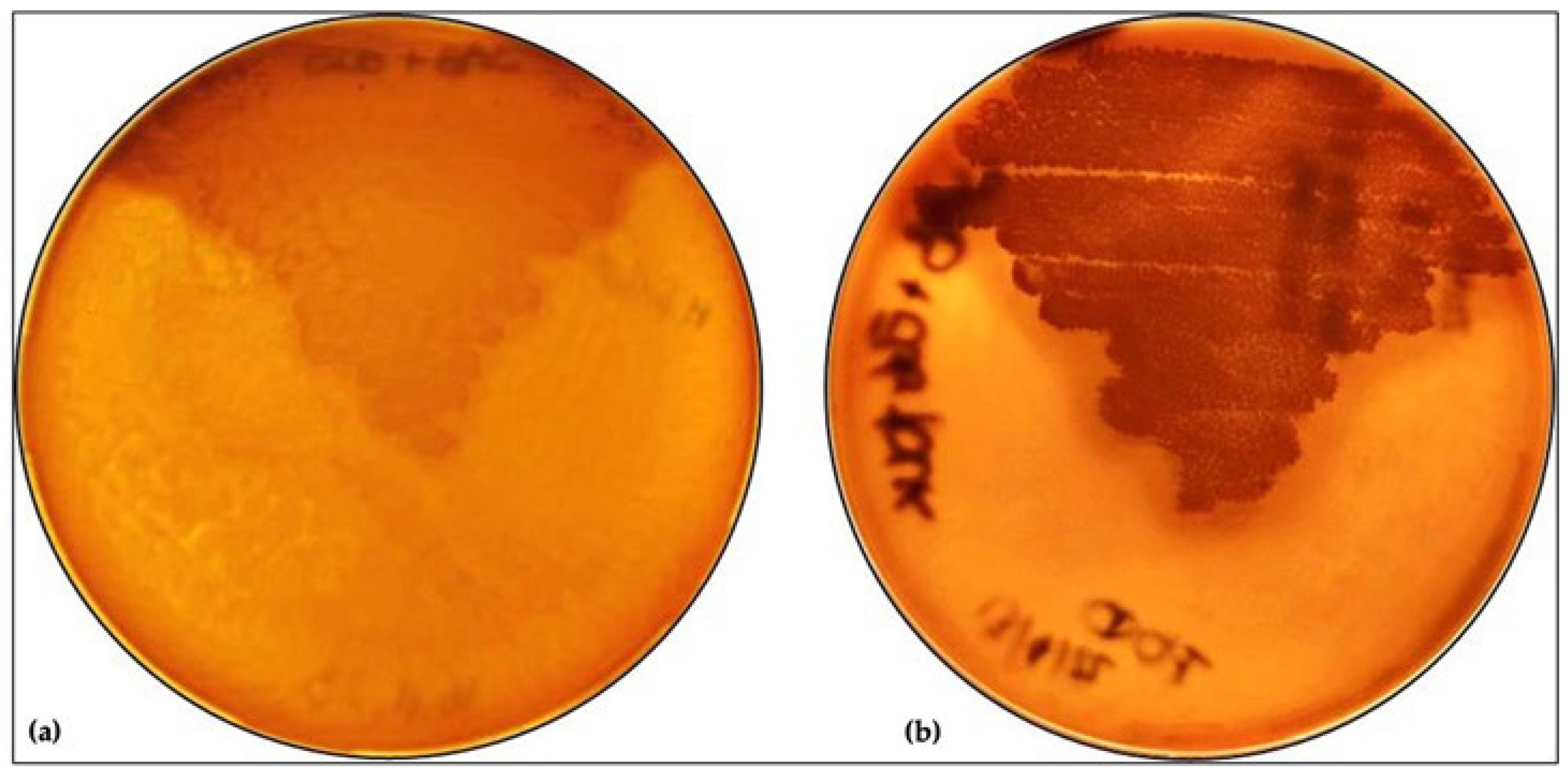
3.1. Effect of L. plantarum on M. furfur Phospholipase Activity

To assess the influence of the L. plantarum on the expression of M. furfur’s virulence factors, the phospholipase activity assay was carried out by incubating M. furfur for 48 h with L. plantarum. Subsequently, M. furfur was incubated first on the medium with β-endorphin for 10 days and then on egg-yolk agar for an additional 10 days. The phospholipase activity was revealed by the appearance of a precipitation halo around the colonies, resulting from the release of calcium and fatty acids due to the degradation of the phospholipids in the egg yolk. As shown in Figure 1, the phospholipase activity is somewhat reduced after incubation with L. plantarum.

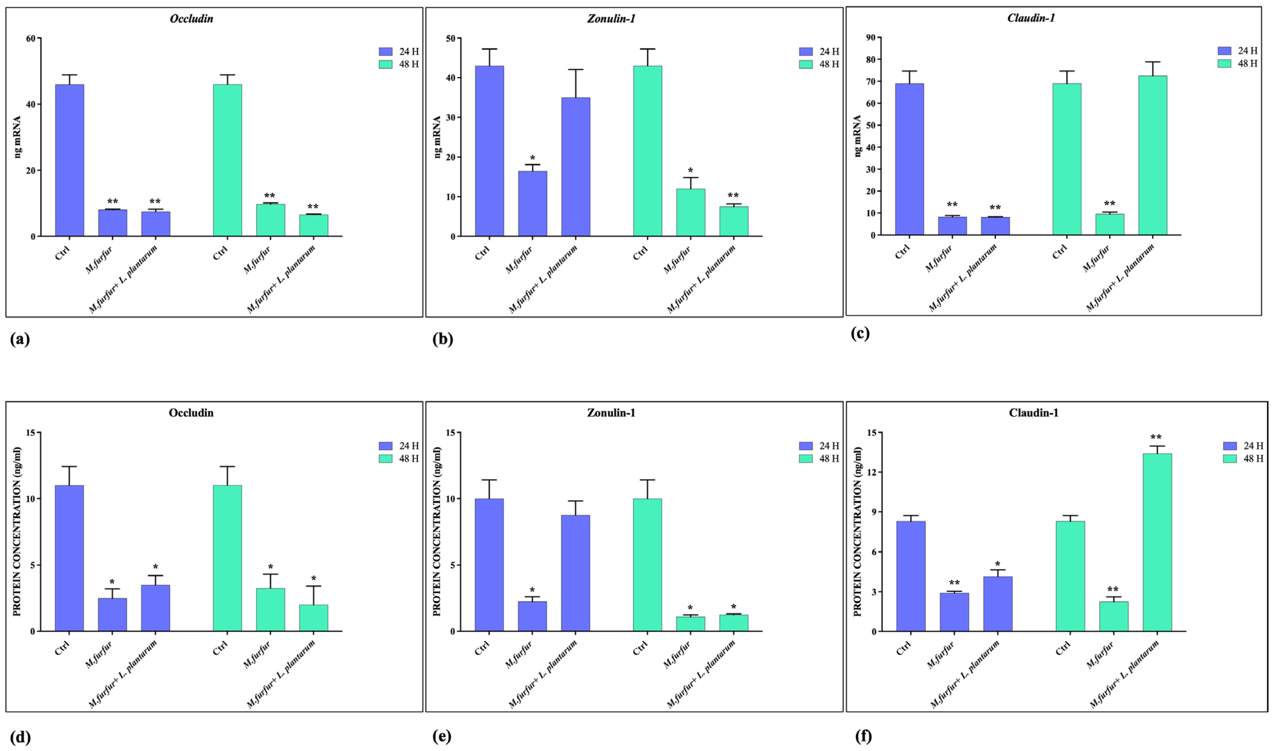
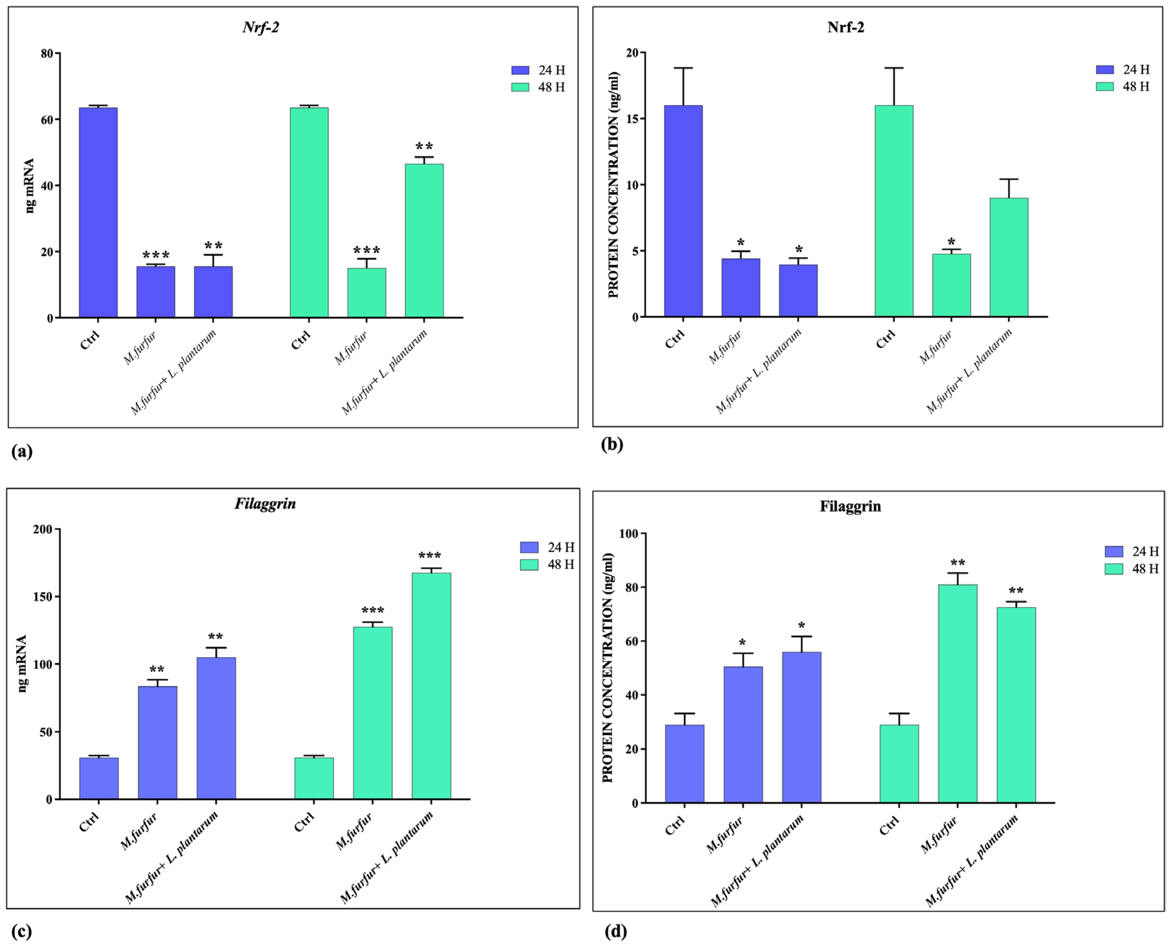
3.2. Effect of L. plantarum on Barrier Integrity

The ability of L. plantarum to strengthen the epithelial barrier was evaluated by analyzing the expression of the genes encoding the TJs: Occludin, Zonulin, and Claudin-1, by using real-time PCR (Figure 2a–c) and measuring the production of the corresponding proteins by ELISA assay (Figure 2d–f). The data obtained indicate that M. furfur alone damages the epithelial barrier, leading to the consequent reduction of TJs expression. However, pretreatment with L. plantarum before infection improves the barrier conditions, maintaining levels of Claudin-1 and Zonulin (up to 24 h of infection), similar to the uninfected control. Meanwhile, it has no effect on the expression of Occludin.

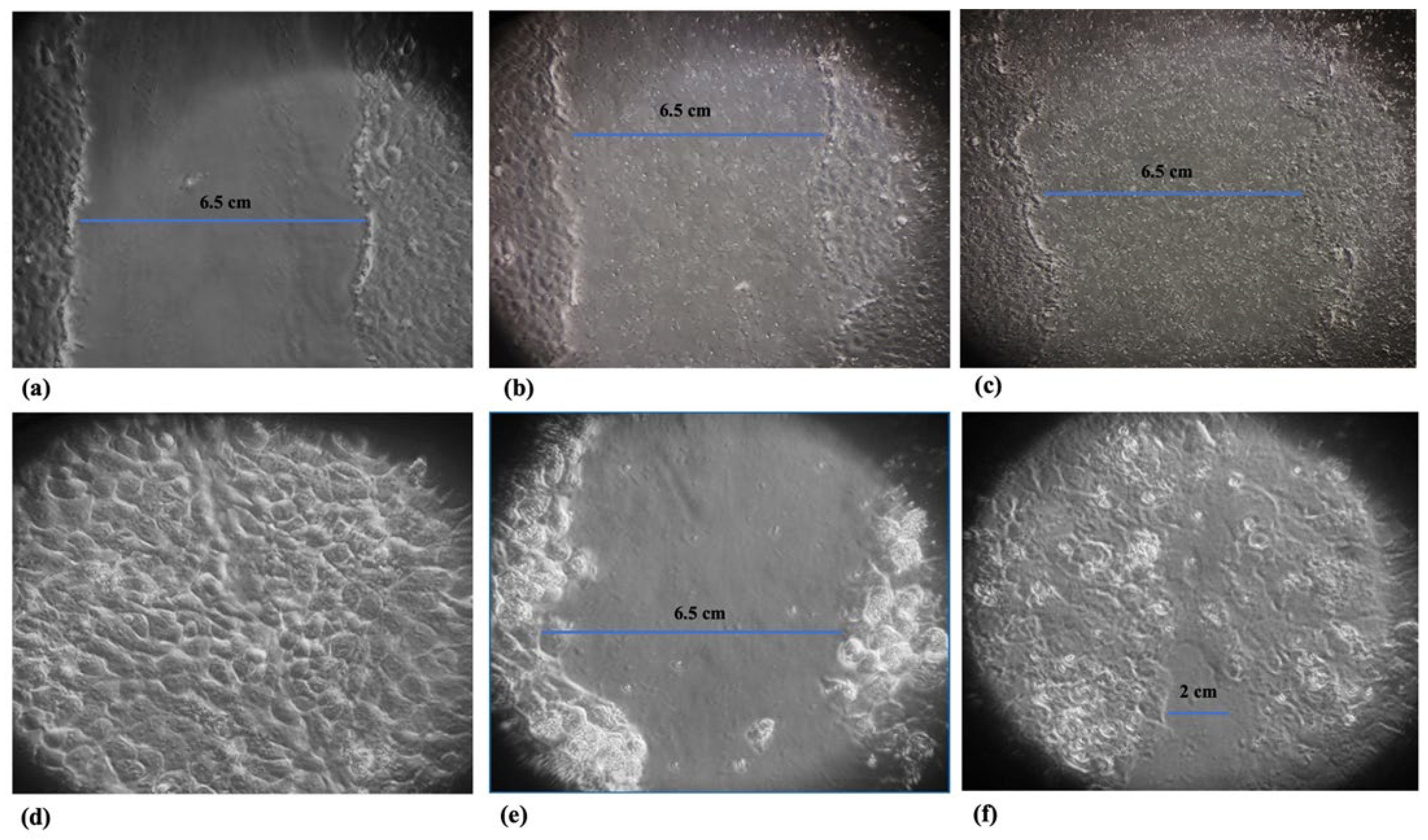
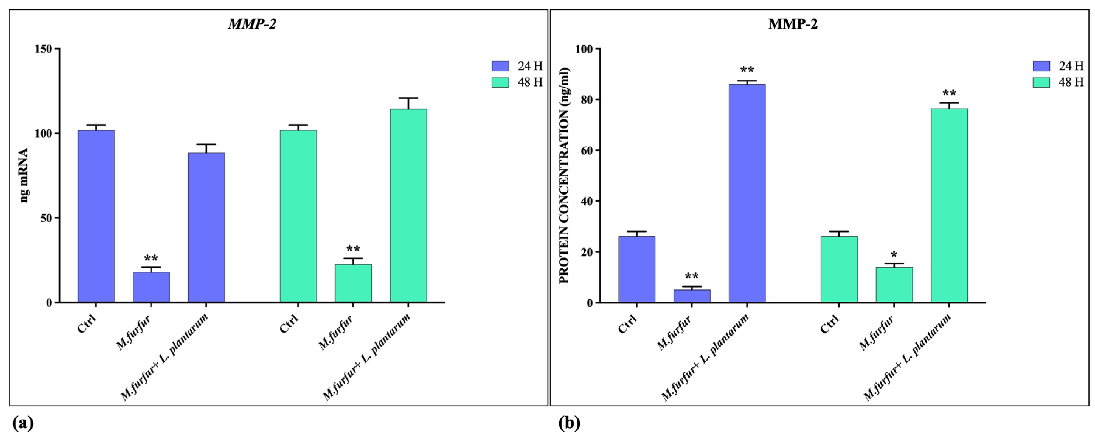
3.3. Tissue Repair Activity of L. plantarum

The ability of L. plantarum to promote tissue repair was evaluated by wound healing assay and by analyzing the induction of MMP-2 expression. As shown in Figure 3, the presence of L. plantarum promotes wound healing, which is inhibited in the presence of M. furfur alone. Furthermore (Figure 4), the presence of L. plantarum stimulates the production of MMP-2, reduced by M. furfur.

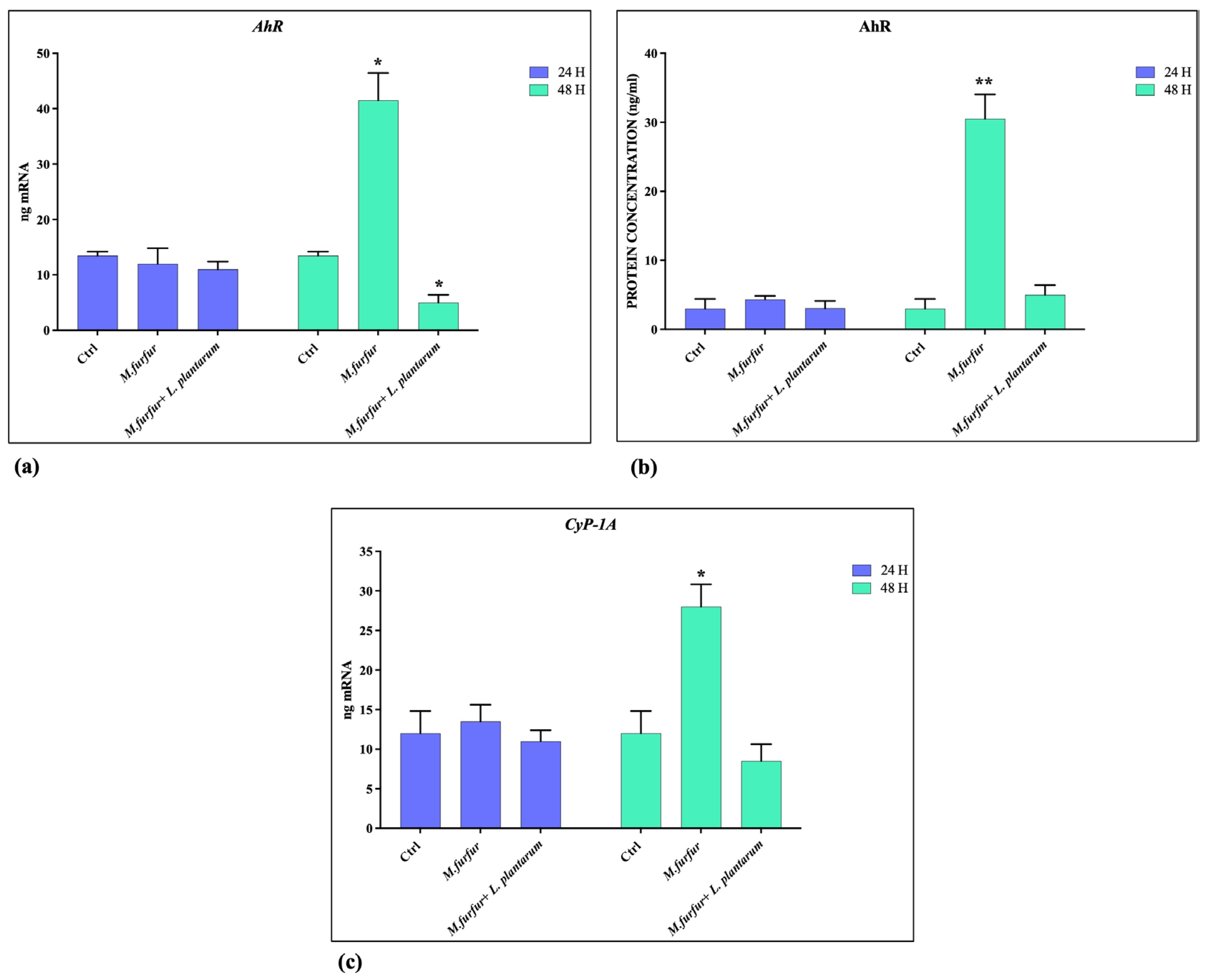
3.4. Activation of AhR Pathways

The analysis of AhR pathways activation demonstrated that M. furfur is able to activate this receptor (Figure 5a,b) and Cyp1A1 (Figure 5c) after 48 h of treatment. In addition; the downregulation of Nrf-2 expression caused by M. furfur infection is counteracted by the activity of L. plantarum after 48 h (Figure 6a,b), while the Filaggrin-1 expression was enhanced (Figure 6c,d), both at 24 and 48 h. Following pretreatment, L. plantarum inhibits the expression of AhR and CyP1A1 induced by M.furfur, keeping high Filaggrin expression.

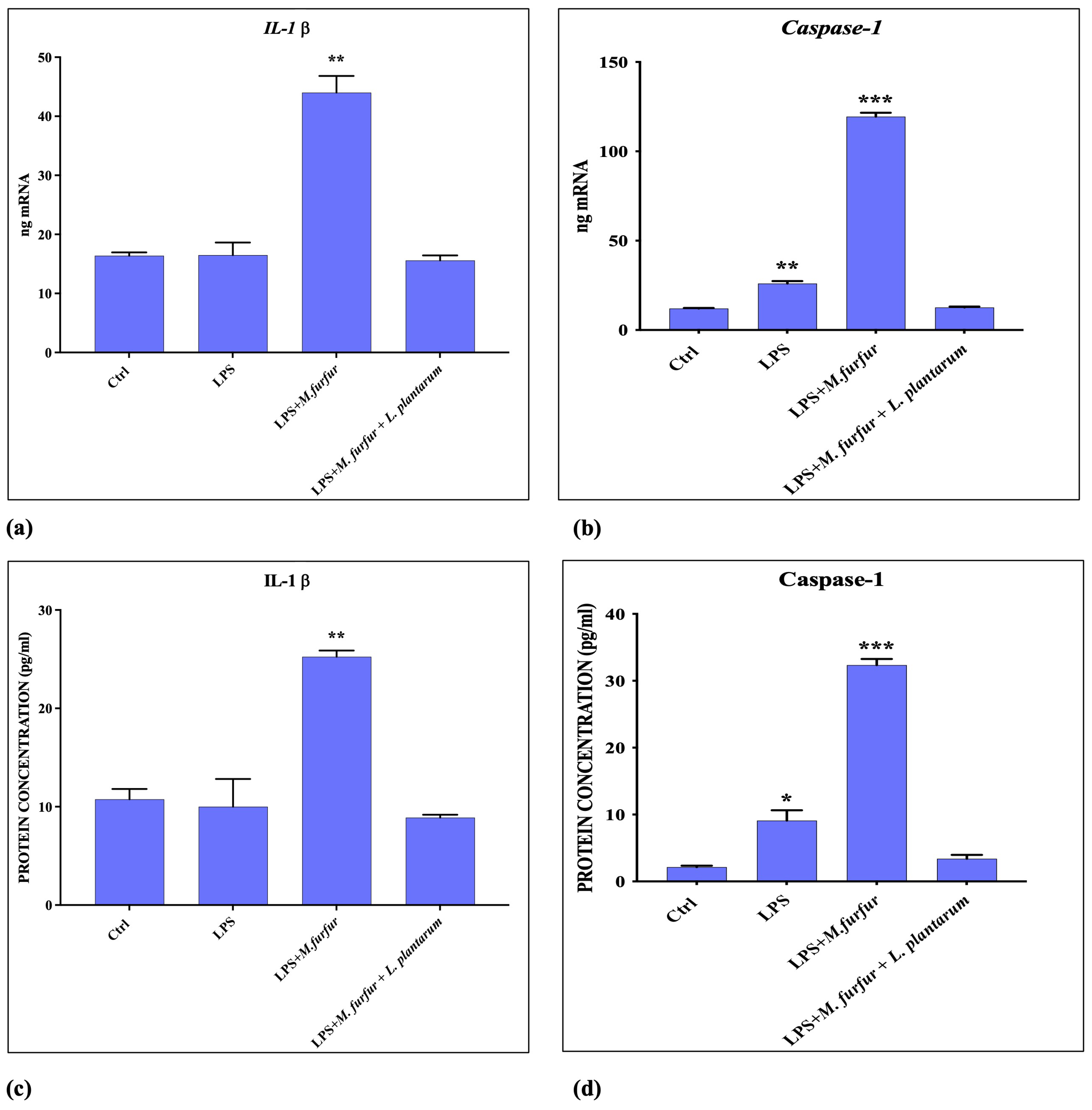
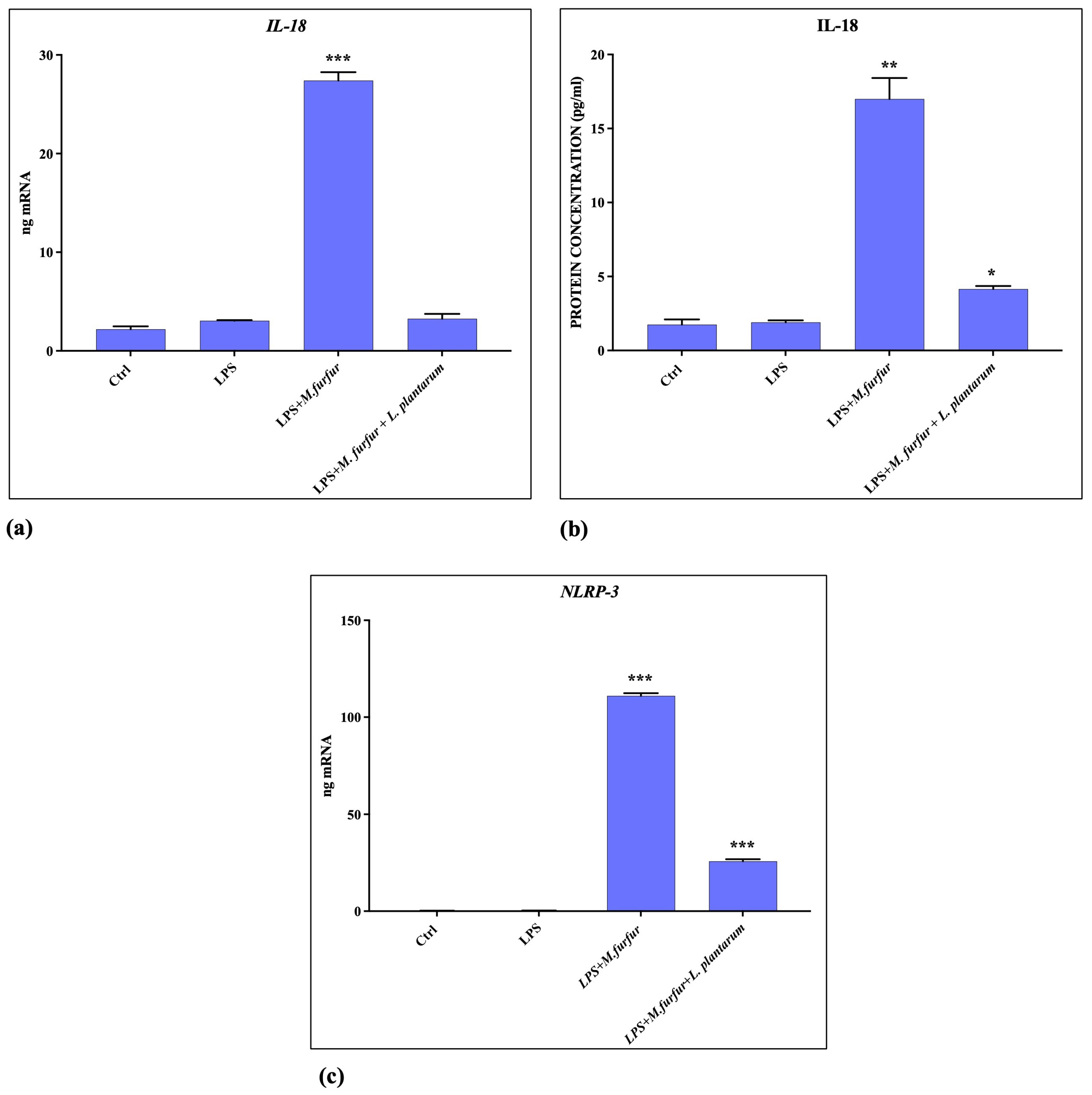
3.5. Inflammasome Activation

The analysis of inflammasome activation was carried out by cell pretreatment with S. Typhimurium LPS and then adding M. furfur, either alone or in presence of L. plantarum. The data obtained (Figure 7 and Figure 8) show that M. furfur activates the inflammasome by inducing the expression of Il-1β, Caspase-1, IL-18, and NLRP-3 after 4 h of infection, while the simultaneous addition of L. plantarum totally inhibits this activation, bringing them back to the control values.

4. Discussion

Commensal bacteria are beneficial for skin health, acting on the host immune system and counteracting the attack of pathogenic strains. Lactobacilli have often been used in fermented foods due to their ability to colonize various body districts, such as the gastrointestinal tract, urogenital tract, and nasal cavity. It is not well-known whether lactobacilli could also colonize and promote skin health. Sarah Lebeer et al. [33] demonstrated that taxa of Lactobacillus, typically associated with the human vagina, were also present on the skin as the most prevalent species. Additionally, nomadic or free-living lactobacilli, including L. plantarum, are frequently detected, suggesting that some lactobacilli on the skin could also result from fermented food sources and thus be transient passengers.
Malassezia spp. also exist as commensals in normal cutaneous microflora, but can cause dandruff, seborrheic dermatitis, pityriasis versicolor, and onycomycosis, and can exacerbate atopic dermatitis and psoriasis. At the basis of the double-faced commensal vs pathogens, there are probably changes in both the fungus (e.g., variable secretion of AhR agonist) and the host conditions [34]. In addition, M. furfur might be an excellent model for studying how the activation of multiple pattern recognition receptors (TLRs, AhR pathway, NLRP3 inflammasome) and the induction of several cells’ regulatory molecules coordinate the different responses of cells in contact with yeast [19,35,36,37,38]. Here, we demonstrate how L. plantarum can counteract the effects of M. furfur induced in human keratinocytes (HaCat) through different pathways of activation. Initially, L. plantarum can be beneficial for skin colonized by M. furfur by reducing the production of phospholipase, considered a Malassezia virulence factor, and promoting healing of wounds inhibited in the presence of lipophilic yeast. Additionally, it acts to limit barrier damage. Chronic skin inflammation is often associated with changes in epithelial permeability, as confirmed by studies on the expression of several claudins, measurements of transepithelial electrical resistance, and experiments with RNA interference in polarized gut epithelial cell lines. These permeability disfunctions are consistently associated with alterations of TJs [12]. We demonstrated that M. furfur reduces the levels of occludin, zonulin, and claudin in HaCat cells in vitro. L. plantarum pretreatment is able to restore levels of zonulin and claudin back to, and beyond, those of the control at 24 and 48 h, respectively. However, it does not act on the reduction of occludin levels induced by M. furfur. L. plantarum also increases the level of MMP2, both at 24 and 48 h, which was downregulated by Malassezia, probably related to the increased level of claudin. Recently, Oku et al. demonstrated that, during the granulation phase, there are high levels of MMP-2, which remain elevated throughout the entire remodeling phase of ECM. This evidence may be responsible for keratinocytes’ migration and has been linked to an increased expression of Claudin-1 [14]. Keratinocyte differentiation is regulated by several factors of transcription, such as AHR, OVOL1/2, MYC, NOTCH1, CEBP, and PPAR. Inflammatory cytokines, phytochemicals, and UV-mediated oxidative stress are able to activate or modulate these transcription factors. For instance, IL-4 and IL-13, pathogenic for atopic dermatitis, and IL-17A, pathogenic for psoriasis, lead to a downregulation of filaggrin. This contributes to the dry, barrier-impaired skin lesions in these pathologies. In some cases, simultaneously with the activation of AhR, antioxidant ligands activate the phosphorylation of Nrf2, which translocates to the nucleus, binds small MAf proteins, recognizes antioxidant-responsive elements, and induces the expression of target genes [17,18,25,39]. Here, we report that M. furfur treatment induces an activation of AhR, CyP1A1, and a consequent increase in filaggrin expression at 48 h. The presence of L. plantarum reduces AhR and CyP1A1 activation at 48 h, enhances the NrF-2 levels downregulated by Malassezia, and maintains high levels of filaggrin.
Previous reports have shown that different fungi, including some strains of Malassezia spp., are able to activate the NLRP3 inflammasome, leading to robust secretion of the pro-inflammatory cytokine IL-1β and IL18 in human antigen-presenting cells or in other cell types, favoring cell colonization and chronic skin inflammation. In contrast, M. furfur seems unable to activate the pathway NLRP3 inflammasome in human keratinocytes but is necessary a triggering stimulus such as a PAMPS, the LPS [21,23,40]. In this paper, we demonstrated that M. furfur activation of the NRLP inflammasome in HaCat cells caused an increase of NLRP3, Caspase-1, and subsequently increased levels of IL-1β and IL-18. However, the presence of L. plantarum reduces the expression of the inflammatory signals triggered by LPS and carried forward by M. furfur infection.
In fungal infections, the most common therapeutic options include topical and systemic agents. The poor effects of topical antimycotics, the need for long treatments with consequent damage to the liver and kidneys, and the emergence of resistant variants limit the therapeutic efficacy of current antifungal drugs. The need to resort to alternative therapies has stimulated both basic research and clinical studies, such as the use of lactobacilli in topical formulations [33]. Studies have been conducted on acne, atopic dermatitis, and rosacea [41,42].
With the limitation of in vitro experimental models, our results strongly support the hypothesis that L. plantarum, as a probiotic, can improve skin health. Particularly in our study, L. plantarum, used as live bacteria in cell pretreatment before Malassezia infection, reduces yeast growth in culture, reduces its phospholipase activity, and promotes the repair of tissue damage slowed down by Malassezia. In addition, it blocks the membrane alterations in human keratinocytes infected with M. furfur, modulating the TJs and the AhR-Nrf2-mediated responses, and reduces the inflammatory process mediated by NLR3-inflammasome, IL-1β, induced in HaCat cells by M. furfur infection. Our experimental model of pretreatment tries to mimic a condition that predicts, in vivo, the effect of Malassezia spp. on the skin of a host who uses a pharmacobiotic as a protective agent (such as cosmetics, drugs, or “beneficial food”), speculating that this pharmacobiotic can also be used post-infection in combination with traditional antifungal therapies, thanks to its ability to manipulate the cutaneous microbiome and promote skin health. This expands the spectrum of available treatment options for topical applications in both aesthetic and regenerative medicine.

Author Contributions

Conceptualization, B.P. and A.F.; methodology, V.S.; formal analysis, V.S. and A.C.; investigation, A.C. and G.T.; resources, G.D.; data curation, A.F.; writing—original draft preparation, B.P.; writing—review and editing, A.F.; supervision, G.D. and A.B. All authors have read and agreed to the published version of the manuscript.

Funding

This research received no external funding.

Institutional Review Board Statement

Not applicable.

Informed Consent Statement

Not applicable.

Data Availability Statement

The authors confirm that the data supporting the findings of this study are available within the article.

Conflicts of Interest

The authors declare no conflict of interest.

References

  1. Braun, C.; Patra, V.; Lina, G.; Nicolas, J.F.; Vocanson, M.; Nosbaum, A. The role of skin dysbiosis in atopic dermatitis. Eur. J. Dermatol. 2022, 32, 439–444. [Google Scholar]
  2. Carmona-Cruz, S.; Orozco-Covarrubias, L.; Sáez-de-Ocariz, M. The Human Skin Microbiome in Selected Cutaneous Diseases. Front. Cell. Infect. Microbiol. 2022, 12, 834135. [Google Scholar] [CrossRef]
  3. Yan, F.; Polk, D.B. Probiotics and Probiotic-Derived Functional Factors-Mechanistic Insights into Applications for Intestinal Homeostasis. Front. Immunol. 2020, 11, 428. [Google Scholar] [CrossRef]
  4. Zheng, Y.; Zhang, Z.; Tang, P.; Wu, Y.; Zhang, A.; Li, D.; Wang, C.Z.; Wan, J.Y.; Yao, H.; Yuan, C.S. Probiotics fortify intestinal barrier function: A systematic review and meta-analysis of randomized trials. Front. Immunol. 2023, 14, 1143548. [Google Scholar] [CrossRef]
  5. Brandi, J.; Cheri, S.; Manfredi, M.; Di Carlo, C.; Vita Vanella, V.; Federici, F.; Bombiero, E.; Bazaj, A.; Rizzi, E.; Manna, L.; et al. Exploring the wound healing, anti-inflammatory, anti-pathogenic and proteomic effects of lactic acid bacteria on keratinocytes. Sci. Rep. 2020, 10, 11572. [Google Scholar] [CrossRef]
  6. Lombardi, F.; Palumbo, P.; Mattei, A.; Augello, F.R.; Cifone, M.G.; Giuliani, M.; Cinque, B. Soluble Fraction from Lysates of Selected Probiotic Strains Differently Influences Re-Epithelialization of HaCaT Scratched Monolayer through a Mechanism Involving Nitric Oxide Synthase 2. Biomolecules 2019, 9, 756. [Google Scholar] [CrossRef]
  7. Li, W.; Zhang, Z.W.; Luo, Y.; Liang, N.; Pi, X.X.; Fan, Y.M. Molecular epidemiology, in vitro susceptibility and exoenzyme screening of Malassezia clinical isolates. J. Med. Microbiol. 2020, 69, 436–442. [Google Scholar] [CrossRef]
  8. Hamdino, M.; Saudy, A.A.; El-Shahed, L.H.; Taha, M. Identification of Malassezia species isolated from some Malassezia associated skin diseases. J. Mycol. Med. 2022, 32, 101301. [Google Scholar] [CrossRef]
  9. Ashbee, H.R. Update on the genus Malassezia. Med. Mycol. 2007, 45, 287–303. [Google Scholar] [CrossRef]
  10. Baroni, A.; Orlando, M.; Donnarumma, G.; Farro, P.; Iovene, M.R.; Tufano, M.A.; Buommino, E. Toll-like receptor 2 (TLR2) mediates intracellular signaling in human keratinocytes in response to Malassezia furfur. Arch. Dermatol. Res. 2006, 297, 280–288. [Google Scholar] [CrossRef]
  11. Brasch, J.; Morig, A.; Neumann, B.; Proksch, E. Expression of antimicrobial peptides and toll-like receptors is increased in tinea and pityriasis versicolor. Mycoses 2014, 57, 147–152. [Google Scholar] [CrossRef]
  12. Karczewski, J.; Troost, F.J.; Konings, I.; Dekker, J.; Kleerebezem, M.; Brummer, R.J.; Wells, J.M. Regulation of human epithelial tight junction proteins by Lactobacillus plantarum in vivo and protective effects on the epithelial barrier. Am. J. Physiol. Gastrointest. Liver Physiol. 2010, 298, G851–G859. [Google Scholar] [CrossRef]
  13. Vera, L.; Caley, M.; O’Toole, E.A. Matrix metalloproteinases and epidermal wound repair. Cell Tissue Res. 2013, 351, 255–268. [Google Scholar]
  14. Oku, N.; Sasabe, E.; Ueta, E.; Yamamoto, T.; Osaki, T. Tight junction protein claudin-1 enhances the invasive activity of oral squamous cell carcinoma cells by promoting cleavage of laminin-5 gamma2 chain via matrix metalloproteinase (MMP)-2 and membrane-type MMP-1. Cancer Res. 2006, 66, 5251–5257. [Google Scholar] [CrossRef]
  15. Fusco, A.; Savio, V.; Cammarota, M.; Donnarumma, G.; Baroni, A. Decreased expression of Malassezia furfur virulence factors after Q-switched Nd:YAG laser irradiation. Eur. J. Dermatol. 2021, 17, 470–472. [Google Scholar] [CrossRef]
  16. Tee, C.B.; Sei, Y.; Kajiwara, S. Secreted Hydrolytic and Haemolytic Activities of Malassezia Clinical Strains. Mycopathologia 2019, 184, 227–238. [Google Scholar] [CrossRef]
  17. Sato, Y.; Fujimura, T.; Tanita, K.; Chunbing, L.; Matsushita, S.; Fujisawa, Y.; Otsuka, A.; Yamamoto, Y.; Hidaka, T.; Aiba, S. Malassezia-derived aryl hydrocarbon receptor ligands enhance the CCL20/Th17/soluble CD163 pathogenic axis in extra-mammary Paget’s disease. Exp. Dermatol. 2019, 28, 933–939. [Google Scholar] [CrossRef]
  18. Furue, M. Regulation of Filaggrin, Loricrin, and Involucrin by IL-4, IL-13, IL-17A, IL-22, AHR, and NRF2: Pathogenic Implications in Atopic Dermatitis. Int. J. Mol. Sci. 2020, 21, 5382. [Google Scholar] [CrossRef]
  19. Buommino, E.; Baroni, A.; Papulino, C.; Nocera, F.P.; Coretti, L.; Donnarumma, G.; De Filippis, A.; De Martino, L. Malassezia pachydermatis up-regulates AhR related CYP1A1 gene and epidermal barrier markers in human keratinocytes. Med. Mycol. 2018, 56, 987–993. [Google Scholar] [CrossRef]
  20. Zhang, J.; Zhang, X.; Xia, P.; Zhang, R.; Wu, Y.; Xia, J.; Su, G.; Zhang, J.; Giesy, J.P.; Wang, Z.; et al. Activation of AhR-mediated toxicity pathway by emerging pollutants polychlorinated diphenyl sulfides. Chemosphere 2016, 144, 1754–1762. [Google Scholar] [CrossRef]
  21. Park, H.R.; Oh, J.H.; Lee, Y.J.; Park, S.H.; Lee, Y.W.; Lee, S.; Kang, H.; Kim, J.E. Inflammasome-mediated Inflammation by Malassezia in human keratinocytes: A comparative analysis with different strains. Mycoses 2021, 64, 292–299. [Google Scholar] [CrossRef] [PubMed]
  22. Kistowska, M.; Fenini, G.; Jankovic, D.; Feldmeyer, L.; Kerl, K.; Bosshard, P.; Contassot, E.; French, L.E. Malassezia yeasts activate the NLRP3 inflammasome in antigen- presenting cells via Syk-kinase signalling. Exp. Dermatol. 2014, 23, 884–889. [Google Scholar] [CrossRef] [PubMed]
  23. Wolf, A.J.; Limon, J.J.; Nguyen, C.; Prince, A.; Castro, A.; Underhill, D.M. Malassezia spp. induce inflammatory cytokines and activate NLRP3 inflammasomes in phagocytes. J. Leukoc. Biol. 2021, 109, 161–172. [Google Scholar] [CrossRef] [PubMed]
  24. Tsai, W.H.; Chou, C.H.; Chiang, Y.J.; Lin, C.G.; Lee, C.H. Regulatory effects of Lactobacillus plantarum GMNL6 on human skin health by improving skin microbiome. Int. J. Med. Sci. 2021, 18, 1114–1120. [Google Scholar] [CrossRef] [PubMed]
  25. Yan, X.; Tsuji, G.; Hashimoto-Hachiya, A.; Furue, M. Galactomyces Ferment Filtrate Potentiates an Anti-Inflammaging System in Keratinocytes. J. Clin. Med. 2022, 11, 6338. [Google Scholar] [CrossRef] [PubMed]
  26. Ong, J.S.; Taylor, T.D.; Yong, C.C.; Khoo, B.Y.; Sasidharan, S.; Choi, S.B.; Ohno, H.; Liong, M.T. Lactobacillus plantarum USM8613 Aids in Wound Healing and Suppresses Staphylococcus aureus Infection at Wound Sites. Probiotics Antimicrob. Proteins 2020, 12, 125–137. [Google Scholar] [CrossRef]
  27. Lolou, V.; Panayiotidis, M.I. Functional Role of Probiotics and Prebiotics on Skin Health and Disease. Fermentation 2019, 5, 41. [Google Scholar] [CrossRef]
  28. Jeong, J.H.; Lee, C.Y.; Chung, D.K. Probiotic Lactic Acid Bacteria and Skin Health. Crit. Rev. Food. Sci. Nutr. 2016, 56, 2331–2337. [Google Scholar] [CrossRef]
  29. Jain, C.; Das, S.; Saha, R.; Ramachandran, V.G.; Bhattacharya, S.N.; Dar, S. Detection of phospholipase production by egg yolk-agar in Malassezia isolates from diseased and healthy human host. Int. J. Med. Sci. Public Health 2017, 6, 1–5. [Google Scholar] [CrossRef]
  30. Vlachos, C.; Gaitanis, G.; Alexopoulos, E.C.; Papadopoulou, C.; Bassukas, I.D. Phospholipase activity after β-endorphin exposure discriminates Malassezia strains isolated from healthy and seborrhoeic dermatitis skin. J. Eur. Acad. Dermatol. Venereol. 2013, 27, 1575–1578. [Google Scholar] [CrossRef]
  31. Cafarchia, C.; Gasser, R.B.; Latrofa, M.S.; Parisi, A.; Campbell, B.E.; Otranto, D. Genetic variants of Malassezia pachydermatis from canine skin: Body distribution and phospholipase activity. FEMS Yeast Res. 2008, 8, 451–459. [Google Scholar] [CrossRef] [PubMed]
  32. Fusco, A.; Savio, V.; Cimini, D.; D’Ambrosio, S.; Chiaromonte, A.; Schiraldi, C.; Donnarumma, G. In Vitro Evaluation of the Most Active Probiotic Strains Able to Improve the Intestinal Barrier Functions and to Prevent Inflammatory Diseases of the Gastrointestinal System. Biomedicines 2023, 11, 865. [Google Scholar] [CrossRef] [PubMed]
  33. Lebeer, S.; Oerlemans, E.F.M.; Claes, I.; Henkens, T.; Delanghe, L.; Wuyts, S.; Spacova, I.; van den Broek, M.F.L.; Tuyaerts, I.; Wittouck, S.; et al. Selective targeting of skin pathobionts and inflammation with topically applied lactobacilli. Cell Rep. Med. 2022, 15, 100521. [Google Scholar] [CrossRef] [PubMed]
  34. Vijaya Chandra, S.H.; Srinivas, R.; Dawson, T.L., Jr.; Common, J.E. Cutaneous Malassezia: Commensal, Pathogen, or Protector? Front. Cell. Infect. Microbiol. 2021, 10, 614446. [Google Scholar] [CrossRef] [PubMed]
  35. Angrisano, T.; Pero, R.; Paoletti, I.; Keller, S.; Lembo, L.; Baroni, A.; Chiariotti, L.; Lembo, F.; Donnarumma, G. Epigenetic regulation of IL-8 and β-defensin genes in human keratinocytes in response to Malassezia furfur. J. Investig. Dermatol. 2013, 133, 2101–2104. [Google Scholar] [CrossRef]
  36. Sparber, F.; Ruchti, F.; LeibundGut-Landmann, S. Host Immunity to Malassezia in Health and Disease. Front. Cell. Infect. Microbiol. 2020, 10, 198. [Google Scholar] [CrossRef]
  37. Sparber, F.; LeibundGut-Landmann, S. Host Responses to Malassezia spp. in the Mammalian Skin. Front. Immunol. 2017, 8, 1614. [Google Scholar] [CrossRef]
  38. Buommino, E.; Nocera, F.P.; Parisi, A.; Rizzo, A.; Donnarumma, G.; Mallardo, K.; Fiorito, F.; Baroni, A.; De Martino, L. Correlation between genetic variability and virulence factors in clinical strains of Malassezia pachydermatis of animal origin. New Microbiol. 2016, 39, 216–223. [Google Scholar]
  39. Fernández-Gallego, N.; Sánchez-Madrid, F.; Cibrian, D. Role of AHR Ligands in Skin Homeostasis and Cutaneous Inflammation. Cells 2021, 10, 3176. [Google Scholar] [CrossRef]
  40. Antushevich, H. Interplays between inflammasomes and viruses, bacteria (pathogenic and probiotic), yeasts and parasites. Immunol. Lett. 2020, 228, 1–14. [Google Scholar] [CrossRef]
  41. Lee, Y.B.; Byun, E.J.; Kim, H.S. Potential Role of the Microbiome in Acne: A Comprehensive Review. J. Clin. Med. 2019, 8, 987. [Google Scholar] [CrossRef] [PubMed]
  42. França, K. Topical Probiotics in Dermatological Therapy and Skincare: A Concise Review. Dermatol. Ther. 2021, 11, 71–77. [Google Scholar] [CrossRef] [PubMed]
Figure 1. Egg yolk agar with M. furfur pretreated for 48 h, either with L. plantarum (a) or alone (b). (scale 1:0.8).
Jof 09 01153 g001
Figure 2. Tight junction expression analysis. (ac) mRNA expression of Occludin, Zonulin, and Claudin-1 in HaCat cells treated with M. furfur (30:1 MOI), either alone or pretreated for 2 h with L. plantarum (100:1 MOI) after 24 and 48 h; (df) Protein expression of Occludin, Zonulin, and Claudin-1 in the same experimental conditions and for the same time periods. The data are representative of three different experiments ± SD. Significant differences are indicated by * p < 0.05, ** p < 0.01.
Jof 09 01153 g002
Figure 3. Scratch wound healing assay: (ac) Time 0, magnification 10×; Control cell, HaCat treated with M. furfur (30:1 yeasts/cell), and HaCat infected with M.furfur (30:1 yeasts/cell) with pretreatment for 6 h with L. plantarum (100:1 bacteria/cell), respectively; (df) The same samples after 48 h of incubation, magnification 20×.
Jof 09 01153 g003
Figure 4. Metalloproteinase expression evaluation. mRNA (a) and protein (b) expression of MMP2 in HaCat cell treated with M. furfur (30:1 MOI), either alone or pretreated for 2 h with L. plantarum (100:1 MOI) after 24 and 48 h. The data are representative of three different experiments ± SD. Significant differences are indicated by * p < 0.05, ** p < 0.01.
Jof 09 01153 g004
Figure 5. AhR pathway activation analysis. mRNA (a,b) and protein (c) expression of AhR and Cyp-1A in HaCat cell treated with M. furfur (30:1 MOI), either alone or pretreated for 2 h with L. plantarum (100:1 MOI) after 24 and 48 h. The data are representative of three different experiments ± SD. Significant differences are indicated by * p < 0.05, ** p < 0.01.
Jof 09 01153 g005
Figure 6. Nrf-2 and Filaggrin activation analysis. mRNA (a,c) and protein (b,d) expression of Nrf-2 and Filaggrin in HaCat cell treated with M. furfur (30:1 MOI), either alone or pretreated for 2 h with L. plantarum (100:1 MOI) after 24 and 48 h; The data are representative of three different experiments ± SD. Significant differences are indicated by * p < 0.05, ** p < 0.01, *** p < 0.001.
Jof 09 01153 g006
Figure 7. Inflammasome activation evaluation. mRNA (a,b) and protein (c,d) expression of IL-1β and Caspase-1 in HaCat cell pre-treated for 4 h with LPS of S. Typhimurium and infected with M. furfur alone or in presence of L. plantarum after 24 and 48 h. The data are representative of three different experiments ± SD. Significant differences are indicated by * p < 0.05, ** p < 0.01, *** p < 0.001.
Jof 09 01153 g007
Figure 8. Inflammasome activation evaluation. mRNA (a,c) and protein (b) expression of IL-18 and NLRP-3 in HaCat cell pre-treated for 4 h with LPS of S. Typhimurium and infected with M. furfur alone or in the presence of L. plantarum after 24 and 48 h. The data are representative of three different experiments ± SD. Significant differences are indicated by * p < 0.05, ** p < 0.01, *** p < 0.001.
Jof 09 01153 g008
Table 1. Primer’s sequences and amplification programs used in this study.
GenePrimer’s SequenceConditionsProduct (bp)
AhR5′-ACCTACGCCAGTCGCAA-3′
5′-CTGACGCTGAGCCTAAGAAC-3′
30″ at 95 °C, 30″ at 60 °C, 30″ at 72 °C for 40 cycles200
CyP1A15′-TCCAGAGACAACAGGTAAAACA-3′
5′-AGGAAGGGCAGAGGAATGTGAT-3′
15″ at 95 °C, 35″ at 60 °C, 35″ at 72 °C for 45 cycles371
Occludin5′-TCAGGGAATATCCACCTATCACTTCAG-3′
5′-CATCAGCAGCAGCCATGTACTCTTCAC-3′
10″ at 95 °C, 45″ at 60 °C for 40 cycles188
Zonulin-15′-AGGGGCAGTGGTGGTTTTCTGTTCTTTC-3′
5′-GCAGAGGTCAAAGTTCAAGGCTCAAGAGG-3′
10″ at 95 °C, 45″ at 60 °C for 40 cycles217
Claudin-15′-CTGGGAGGTGCCCTACTTTG-3′
5′-ACACGTAGTCTTTCCCGCTG-3′
1″ at 95 °C, 30″ at 60 °C, 20″ at 72 °C for 40 cycles128
NrF25′-ACCACCCACAACTTACTGCC-3′
5′-GCCATAGGAGTATGGGGGAT-3′
5″ at 95 °C, 2″ at 60 °C, 5″ at 72 °C for 40 cycles121
MMP-25′-TGACGGTAAGGACGGACTC-3′
5′-TGGAAGCGGATTGGAAACT-3′
5″ at 94 °C, 7″ at 57 °C, 14″ at 72 °C for 40 cycles342
Caspase-15′-GCCTGTTCCTGTGATGTGGAG-3′
5′-TGCCCACAGACATTCATACAGTTTC-3′
15″ at 95 °C, 1′ at 60 °C for 40 cycles165
IL-1 β5′-GCATCCAGCTACGAATCTCC-3′
5′-CCACATTCAGCACAGGACTC-3′
5″ at 95 °C, 9″ at 58 °C, 19″ at 72 °C for 40 cycles708
IL-185′-GATTACTTTGGCAAGCTTGAA-3′
5′-GCTTTCGTTTTGAACAGTGAA-3′
5″ at 95 °C, 6″ at 53 °C, 12″ at 72 °C for 40 cycles470
NLRP-35′-GATCTTCGCTGCGATCAACA-3′
5′-GGGATTCGAAACACGTGCATTA-3′
5″ at 95 °C, 34″ at 60 °C for 40 cycles93
β-ACTIN5′-CGTGGGCCGCCCTAGGCACCA-3′
5′-TTGGCCTTAGGGTTCAGGGGGG-3′
243
Disclaimer/Publisher’s Note: The statements, opinions and data contained in all publications are solely those of the individual author(s) and contributor(s) and not of MDPI and/or the editor(s). MDPI and/or the editor(s) disclaim responsibility for any injury to people or property resulting from any ideas, methods, instructions or products referred to in the content.

