Article

Colletotrichum Species Associated with Anthracnose Disease of Watermelon (Citrullus lanatus) in China

1
Key Laboratory of Fruit and Cucurbit Biology, Zhengzhou Fruit Research Institute, Chinese Academy of Agricultural Sciences, Zhengzhou 450009, China
2
Key Laboratory of Horticultural Plant Biology, Ministry of Education, College of Plant Science and Technology, Huazhong Agricultural University, Wuhan 430070, China
3
Department of Plant Pathology, Henan Agricultural University, Zhengzhou 450002, China
*
Author to whom correspondence should be addressed.
J. Fungi 2022, 8(8), 790; https://doi.org/10.3390/jof8080790
Submission received: 8 July 2022 / Revised: 25 July 2022 / Accepted: 25 July 2022 / Published: 28 July 2022

Abstract

:
Colletotrichum species are important plant pathogens, causing anthracnose in virtually every crop grown throughout the world. However, little is known about the species that infect watermelon. A total of 526 strains were isolated from diseased watermelon samples of eight major watermelon growing provinces in China. Phylogenetic analyses using seven loci (ITS, gadph, chs-1, his3, act, tub2, and gs) coupled with morphology of 146 representative isolates showed that they belonged to 12 known species of Colletotrichum, including C. aenigma, C. chlorophyti, C. fructicola, C. jiangxiense, C. karstii, C. magnum, C. nymphaeae, C. nigrum, C. orbiculare, C. plurivorum, C. sojae, and C. truncatum and three new species, here described as C. citrulli, C. kaifengense, and C. qilinense. Colletotrichum orbiculare was the dominant species. Pathogenicity tests revealed that all isolates of the species described above were pathogenic, with C. magnum and C. kaifengense being the most aggressive to leaves and fruits, respectively. This is the first report of C. aenigma, C. chlorophyti, C. fructicola, C. jiangxiense, C. nymphaeae, C. nigrum, C. plurivorum, and C. sojae on watermelon. These findings shed light on the Colletotrichum spp. involved in watermelon anthracnose and provide useful information for implementing effective control of watermelon anthracnose in China.

1. Introduction

Colletotrichum is the most common and important genus of plant pathogenic fungi, saprobes, and endophytes [1,2,3]. Species of Colletotrichum spp. infect numerous plant crops worldwide, e.g., apple (Malus pumila), chili (Capsicum spp.), coffee (Coffea spp.), grape (Vitis vinifera), longan (Dimocarpus longan), mango (Mangifera indica), olive (Canarium album), orange (Citrus spp.), pear (Pyrus spp.), peach (Prunus persica), strawberry (Fragaria ananassa), and tea (Camellia spp.) [4,5,6,7,8,9,10,11,12,13,14,15,16,17,18,19,20,21].
During the first half of the 20th century, many species of the plant-pathogenic fungal genus Colletotrichum were defined, relying on the hosts from which they were originally isolated [22]. Based on his revision of the genus primarily on morphological characteristics in culture, Von Arx reduced approximately 750 species to only 11 [23]. However, identifying Colletotrichum species is challenging due to the instability of their morphological characteristics, which are affected by experimental methods and conditions [1,24,25]. Along with the increased availability of DNA sequencing technology, a very large volume of DNA sequence data has been generated, allowing fungal taxonomy through phylogenetics, including genealogical concordance [26,27]. Following the adoption of multi-locus phylogenetic analysis together with morphological characteristics, the classification and species concepts in Colletotrichum taxa worldwide have changed significantly [1,28,29,30,31,32,33]. Nearly all acknowledged species studied were grouped into 16 Colletotrichum species complexes, and more than 16 singleton species have been identified [9,30,31,32,33,34,35,36,37,38,39,40,41,42,43].
Watermelon (Citrullus lanatus (Thunb.) Matsum. et Nakai), belongs to the xerophytic genus Citrullus Schrad. ex Eckl. et Zeyh of the botanical family Cucurbitaceae and is one of the most important commercial crops worldwide [44]. It is the third-most widespread fruit crop after apple and orange in China (China Agriculture Research System Statistical Data 2018). In 2019, the total area harvested in China was 1,471,581 ha, with a yield of 60,861,241 t, accounting for 60.6% of the world’s production of watermelon [45]. However, anthracnose diseases are one of the important factors, limiting the commercial production of watermelon [26]. Anthracnose displays spots and blights on the aerial parts of plant in the fields. Owing to anthracnose, watermelon production can be reduced by 5–20%, and even causes no harvest [46]. Moreover, it is an important post-harvest pathogen and becomes active after the fruit has been stored or appears on the market shelf. Owing to Colletotrichum disease, up to 100% of the fruit can be lost during storage and transportation [2].
Cucurbitaceae crops, especially watermelon (Citrullus lanatus), melon (Cucumis melo), and cucumber (Cucumis sativus), are important plant hosts of Colletotrichum spp. [47]. Seven Colletotrichum species have been reported in watermelon, belonging to C. gloeosporioides, C. gloeosporioides f.sp. cucurbitae, C. karstii, C. magnum, C. scovillei, C. orbiculare, and C. truncatum [32,37,38,48,49,50,51]. First described from cucumber, the species C. gloeosporioides f.sp. cucurbitae is widely regarded as a synonym for C. orbiculare [52]. However, Colletotrichum species associated with watermelon remain largely unresolved, with only six individual species identified. Moreover, these studies used a limited number of samples and areas, therefore species diversity might have been underestimated.
The objectives of the present study were as follows: (i) to identify the Colletotrichum species causing anthracnose in watermelon in the major production provinces in China based on multi-locus phylogenetic analyses combined with morphological characterization; (ii) to evaluate the pathogenicity of the different Colletotrichum species.

2. Materials and Methods

2.1. Collection and Isolates

The survey was conducted from 2018 to 2020 in watermelon fields in various geographical areas of China. Samples were collected from eight provinces (Henan, Jiangsu, Zhejiang, Jilin, Liaoning, Hebei, Jiangxi, and Hainan) (Table 1). Anthracnose symptoms on watermelon leaves were small circular or irregular spots, pale brown in the center with medium to dark brown margins, while rows of fusiform brown sunken spots were formed on stems (Figure 1a–g). Three symptom types were observed on fruits: (1) small round sunken spots (Figure 1k,l) and (2) large brown or black sunken rot lesions, forming orange conidia under humid conditions (Figure 1h–j,m,n).
Samples were surface-sterilized by dipping in 75% ethanol for 10 s and rinsing three times with sterile distilled water. Tissue pieces (5 mm) were excised from areas neighboring the diseased tissue. Excised tissue was placed onto potato dextrose agar (PDA, 20% diced potato, 2% dextrose, and 1.5% agar, and distilled water) after surface sterilization (70% ethanol for 15 s, 5% NaOCl for 5 min, washed three times in sterile water and dried on sterilized filter paper) [53]. The plates were incubated at 27 °C in the dark for 5 d. Six single colonies of each strain were isolated using the single-spore isolation method [54]. All isolates were stored in 25% glycerol at −80 °C. Type specimens of the new species from this study were deposited in the Mycological Herbarium, Institute of Microbiology, Chinese Academy of Sciences, Beijing, China (HMAS), and ex-type living cultures were deposited in the China General Microbiological Culture Collection Center (CGMCC), Beijing, China.

2.2. DNA Extraction, PCR Amplification, and Sequencing

Isolates were transferred to fresh PDA plates and incubated at 27 °C for 7–19 days. Genomic DNA of 146 representative isolates was extracted using the Ezup Column Fungi Genomic DNA Purification Kit (Sangon Biotech, Shanghai, China). Seven loci, including the internal transcribed spacer regions and intervening 5.8S nrRNA gene (ITS) and partial sequences of the glyceraldehyde-3-phosphate dehydrogenase (gadph), chitin synthase 1 (chs-1), actin (act), histone3 (his3), beta-tubulin (tub2), and glutamine synthetase (gs) genes were amplified and sequenced using the primer pairs ITS-1F [55] + ITS-4 [56], GDF1 + GDR1 [57], CHS-354R + CHS-79F [58], ACT-512 F + ACT-783R [58], CYLH3F + CYLH3R [59], T1 [60] +Bt2b [61] and GSF1 + GSR1 [57], respectively. PCR was performed in a total volume of 25 μL. The PCR mixture contained 1 μL 20× diluted genomic DNA, 12.5 μL 2 × Rapid Taq Master Mix (Vazyme, Nanjing, China), and 0.2 μM of each primer. The PCR reactions were mostly as described by Woudenberg et al. (2009) [62], but were modified by using an annealing temperature of 58 °C for act, gadph, and chs-1, 56 °C for tub2, his3, and gs. PCR amplifications were performed in Mastercycler X50 (Eppendorf, Hamburg, Germany). PCR amplicons were purified and sequenced at the Sangon Biotech (Shanghai, China) Company, Ltd. Forward and reverse primers were assembled to obtain consensus sequences using DNAMAN (v. 9.0; Lynnon Biosoft, San Ramon, USA). Sequences generated in this study were deposited in GenBank (Supplementary Table S1).

2.3. Phylogenetic Analyses

The DNA sequences were aligned using MAFFT v. 7 [63]. and manually adjusted using MEGA (version 11.0.10, Mega Limited, Auckland, New Zealand) where necessary. Multiple sequences of ITS, gadph, act, tub2, chs-1, and gs were concatenated using SequenceMatrix 1.8 [64]. A Markov Chain Monte Carlo (MCMC) algorithm was used to generate phylogenetic trees with Bayesian inference (BI) using MrBayes v. 3.2.6 [65] for the combined sequence datasets. Best-fit models of nucleotide substitution for each gene partition were determined by Akaike information criterion (AIC) of MrModeltest v. 2.3 [66] (Table 2). Two analyses of four MCMC chains were conducted from random trees with 6 × 10⁶ generations for C. orbiculare and C. gloeosporioides species complexes, and 2 × 10⁶ generations for the C. boninense, C. orchidearum, C. magnum species complexes, and C. acutatum, C. truncatum species complexes and the related reference species involved in the same phylogenetic tree. The analyses were sampled every 1000 generations.
Alignment gaps were treated as missing and data all characters were unordered and of equal weight. The first 25% of trees were discarded as the burn-in phase of each analysis and posterior probabilities were determined from the remaining trees. Convergence of all parameters (i.e., effective sample sizes >200) was visually confirmed using Tracer 1.7.1 [67]. The maximum parsimony analyses (MP) were performed on the multi-locus alignment using PAUP (Phylogenetic Analysis Using Parsimony) v. 4.0a169 [68]. Phylogenetic trees were generated using the heuristic search option with 1000 random sequence additions and tree bisection and reconstruction (TBR) as the branch-swapping algorithm. Maxtrees were set up to 5000, branches of zero length were collapsed, and all multiple parsimonious trees were saved. Clade stability was assessed by bootstrap analysis with 1000 replicates. Afterward, tree length (TL), consistency index (CI), retention index (RI), rescaled consistency index (RC), and homoplasy index (HI) were calculated for the resulting tree. Furthermore, maximum likelihood (ML) analyses were implemented on the multi-locus alignments using the IQ-TREE 2 [69]. Clade stability was assessed using a bootstrap analysis with 1000 replicates. Phylogenetic trees were visualized in FigTree v. 1.4.4 [70]. The alignments and phylogenetic trees were deposited in TreeBASE (submission number: 29157).
The phylogenetically related ambiguous species were analyzed using the Genealogical Concordance Phylogenetic Species Recognition (GCPSR) model with a pairwise homoplasy index (PHI) test as described by Quaedvlieg et al. (2014) [26]. The PHI test was performed in SplitsTree4 [71,72,73] to determine the recombination level within phylogenetically closely related species using a six-locus concatenated dataset (ITS, gadph, chs-1, his3, act, and tub2). Pairwise homoplasy index results below 0.05 indicated significant recombination in the dataset. The relationship between closely related species was visualized by constructing a splits graph.

2.4. Morphological Analysis

Mycelial plugs (5-mm diam) from the margin of actively growing cultures were transferred on PDA, oatmeal agar (OA) [53], and synthetic nutrient-poor agar medium (SNA) [74] and incubated at 27 °C in the dark. Colony characteristics were noted after 9 d, and colony diameters were measured daily for 3 d, 5 d, and 7 d to calculate their mycelial average growth rates (mm/d). Additionally, the shape, color, and size of acervali, conidia, conidiophores, asci, ascospores, and seta were observed using light microscopy (Nikon SMZ-1500 and Olympus BX51, Japan or Leica TCS SP5, Germany). Moreover, to determine their sizes, 30 conidia, seta, or ascospores were measured. The formation of conidial appressoria was induced by dropping a conidial suspension in 1% glucose (107 conidia/mL; 50 μL) on a concavity slide, placed inside plates containing moistened filter papers with sterile distilled water, and then incubated at 27 °C in darkness. After 48 h, the sizes of 30 conidial appressoria were measured.

2.5. Prevalence

To determine the prevalence of Colletotrichum species in the sampled provinces, Colletotrichum species were isolated from infected watermelon organs (leafs, stems, or fruits). The isolation rate (IR) for each species was calculated using the formula, IR% = (Ns/Nt) × 100, where Ns is the number of isolates from the same species and Nt is the total number of isolates from each sample-collected province [11,14].

2.6. Pathogenicity Tests

Sixteen representative isolates of each Colletotrichum species were used in the pathogenicity tests on detached leaves of Citrullus lanatus cv. Hongheping (5–6 true leaves). Healthy leaves were collected from plants growing in pots in a greenhouse. The leaves were washed with tap water, then submerged in 75% ethanol for 30 s, washed three times with sterile water, and finally air-dried on sterilized filter paper. The leaves were inoculated using the wound/drop and non-wound/drop inoculation methods [75,76]. A drop of 10 µL spore suspension (106 conidia/mL) was individually placed onto the left side of the leaf after wounding once by pin-pricking the upper surface with a sterilized needle (insect pin, 0.5 mm diam). A 10 µL drop of sterile water was placed on the right side of the same leaf as a control. The process was repeated with unwounded leaves. The leaves were incubated at 27 °C and 100% humidity in the dark. The infection rate was calculated 4 d post-inoculation (dpi) using the formula PI (%) = Nf/Np × 100, where Nf is the number of infected points and Np is the total number of inoculated points [19]. All isolates were tested in triplicate.
Pathogenicity tests were also performed on detached mature watermelon fruits of Citrullus lanatus cv. Motong in triplicate. Healthy-looking fruits were collected from plants growing in the greenhouse. The fruits were washed with tap water, then submerged in 75% ethanol for 30 s and washed three times with sterile water, finally air-dried on sterilized filter paper. Wound/drop and non-wound/drop inoculation methods were used [28,75,76]. For the wound inoculation, a drop of 10 µL spore suspension (106 conidia/mL) was placed directly onto fruit surfaces after wounding once by pin-pricking with a sterilized needle (5 mm deep). In the non-wound inoculation, the same spore suspension was placed directly on the unwounded watermelon fruit skin. Sterile water was used as the control. The fruits were incubated at 27 °C and at 100% humidity in the dark. The infection rate was calculated at 6 dpi using the formula PI (%) = Nf/Np × 100, where Nf is the number of infected points and Np is the total number of inoculated points [19].

3. Results

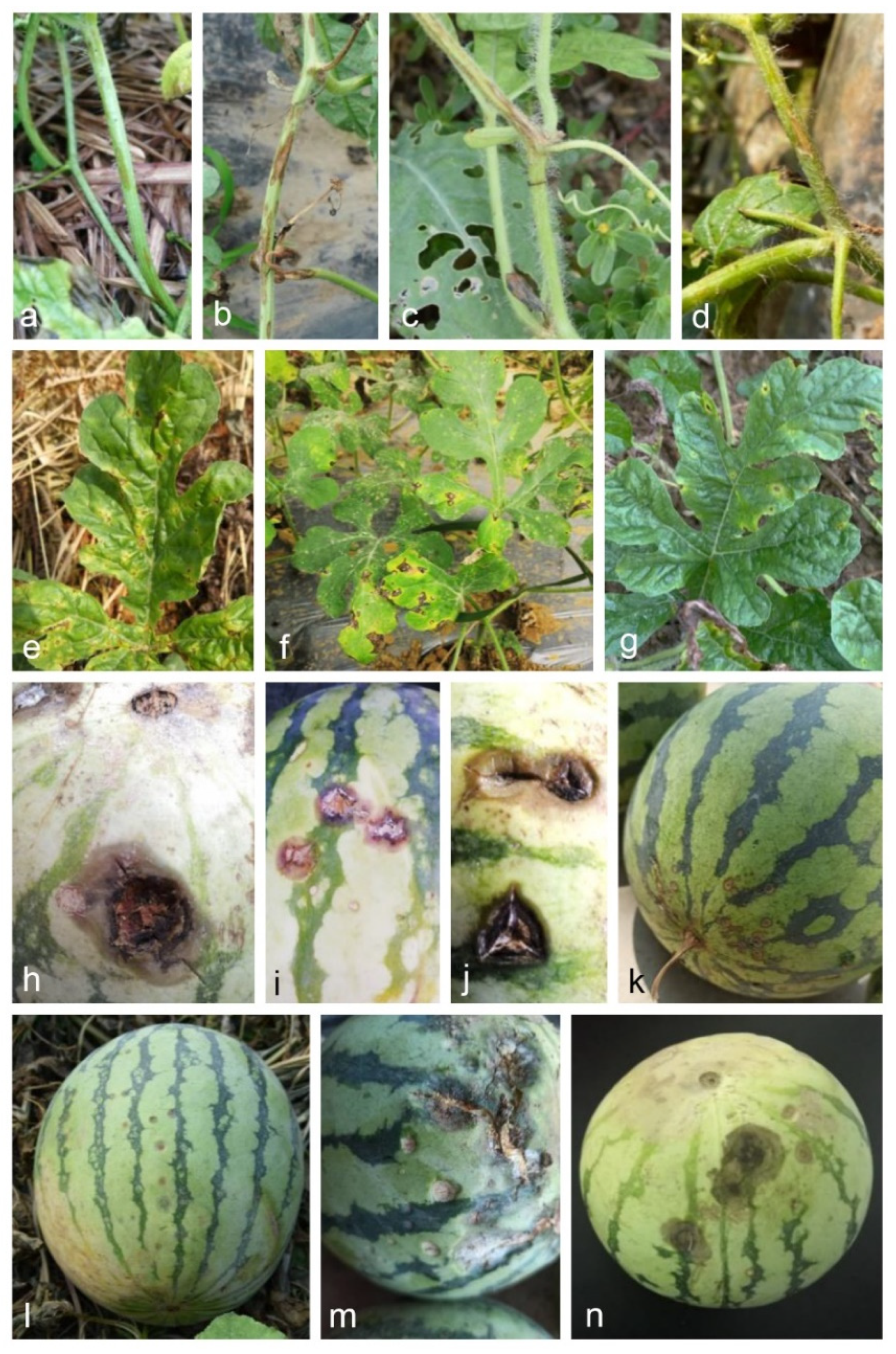
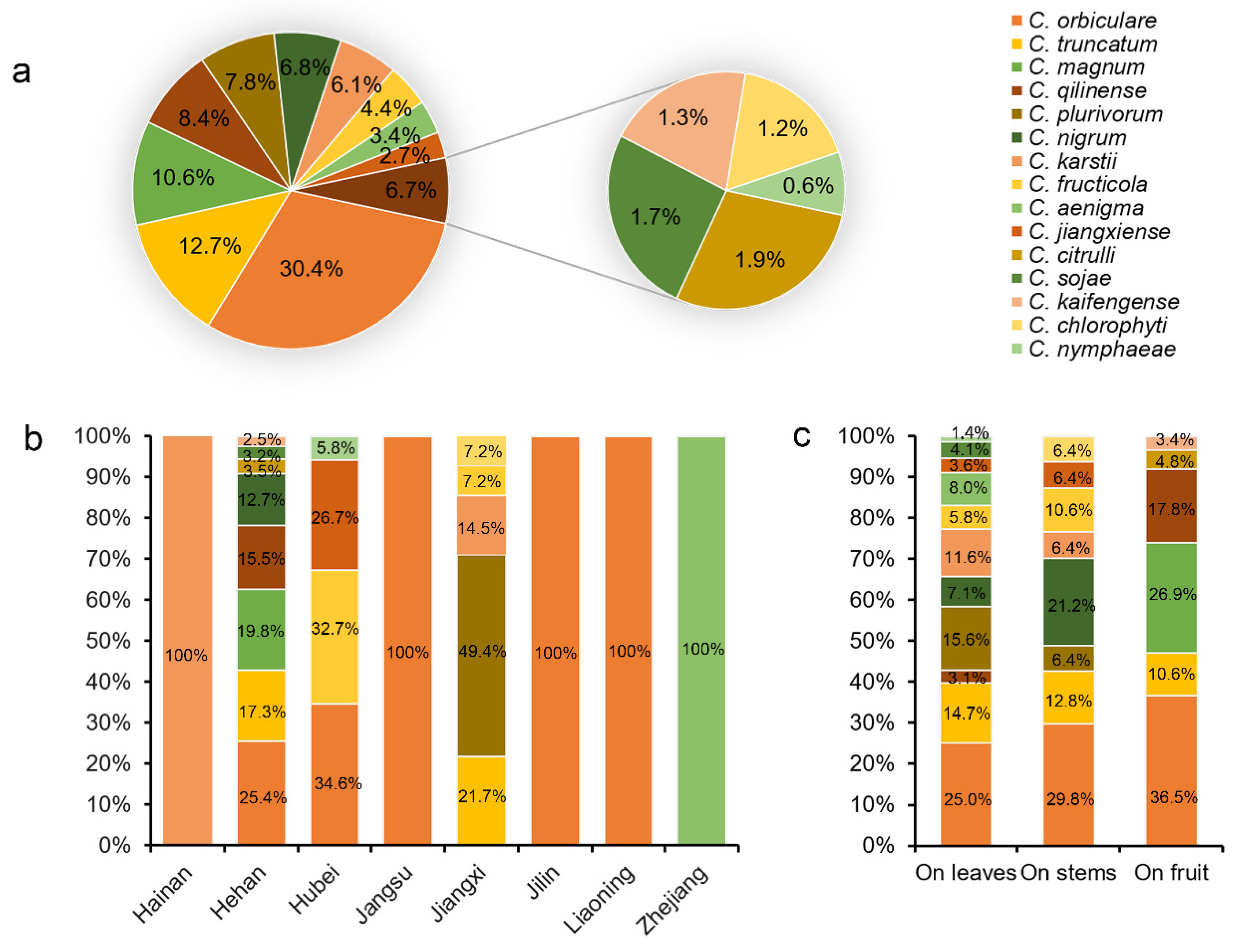
3.1. Collection of Watermelon Anthracnose Samples and Strain Isolation

Watermelon anthracnose was common in all the fields and provinces surveyed. A total of 224 diseased watermelon samples (146 leaves, 57 stems, and 21 fruits) were collected from eight provinces in China. From them, 526 Colletotrichum strains were isolated based on the colony morphologies on PDA and ITS sequence data (Table 1). Based on megablast searches in GenBank using ITS sequences, the colony morphological characteristics on PDA, and morphological characteristics conidia isolates were primarily divided into eight groups. Of those, 146 representative isolates were selected for further analysis.

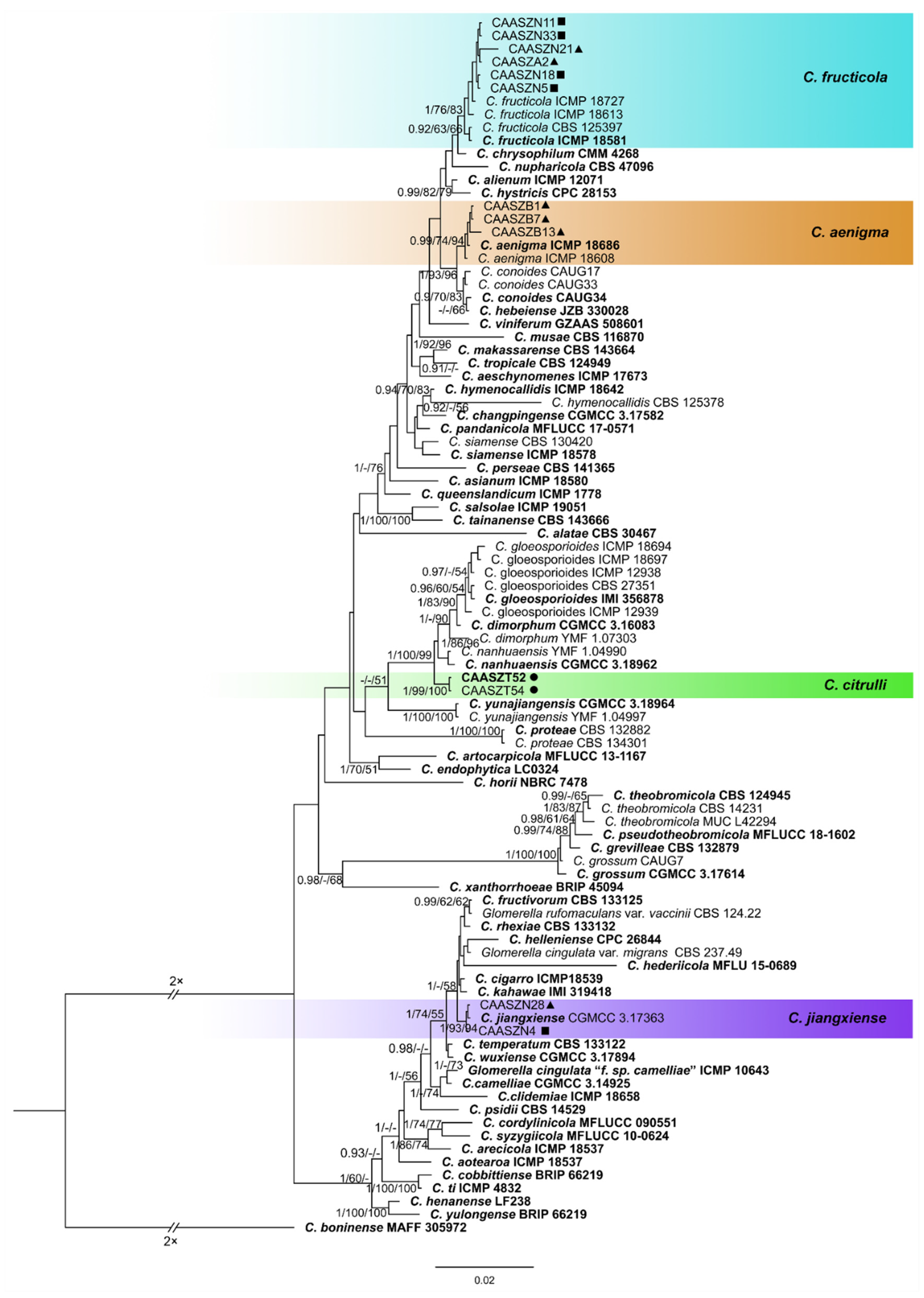
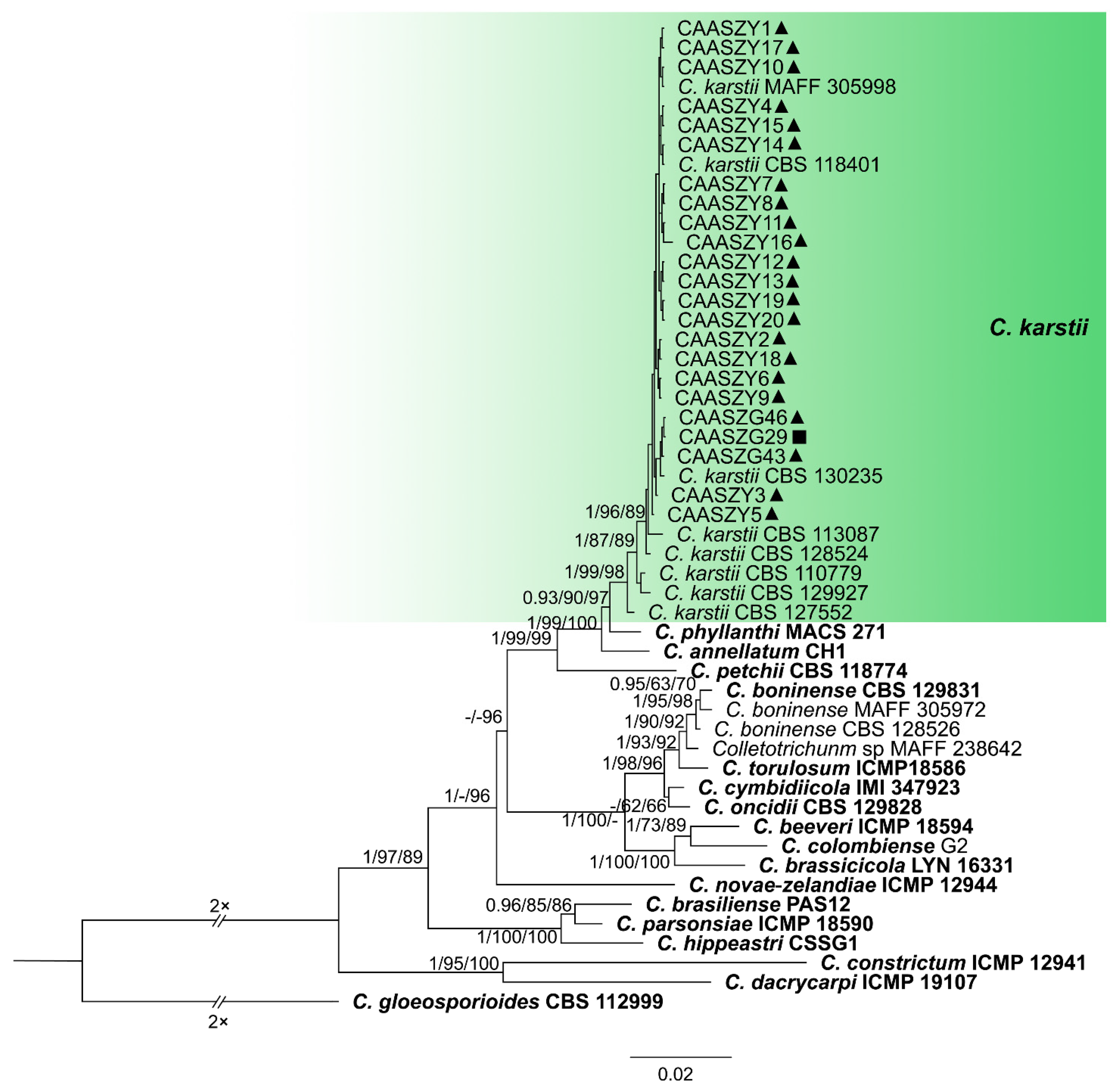
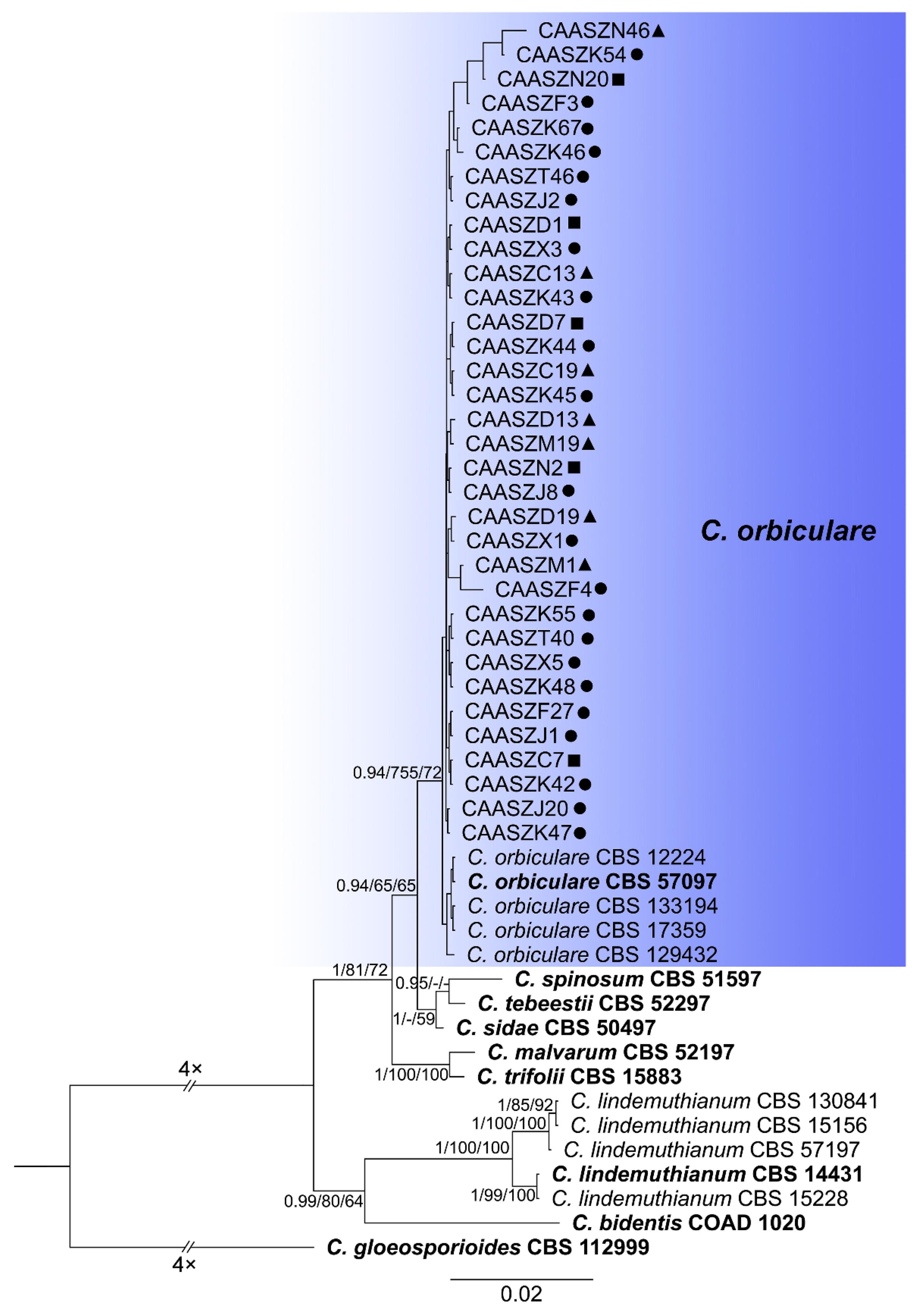
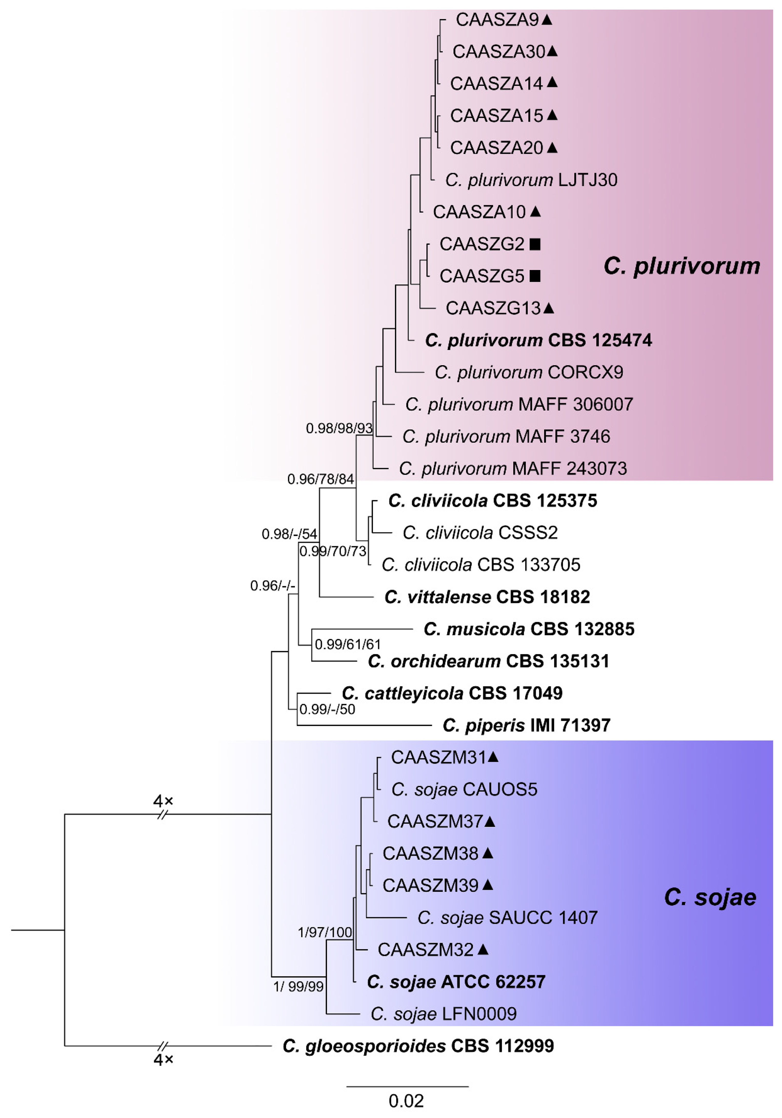
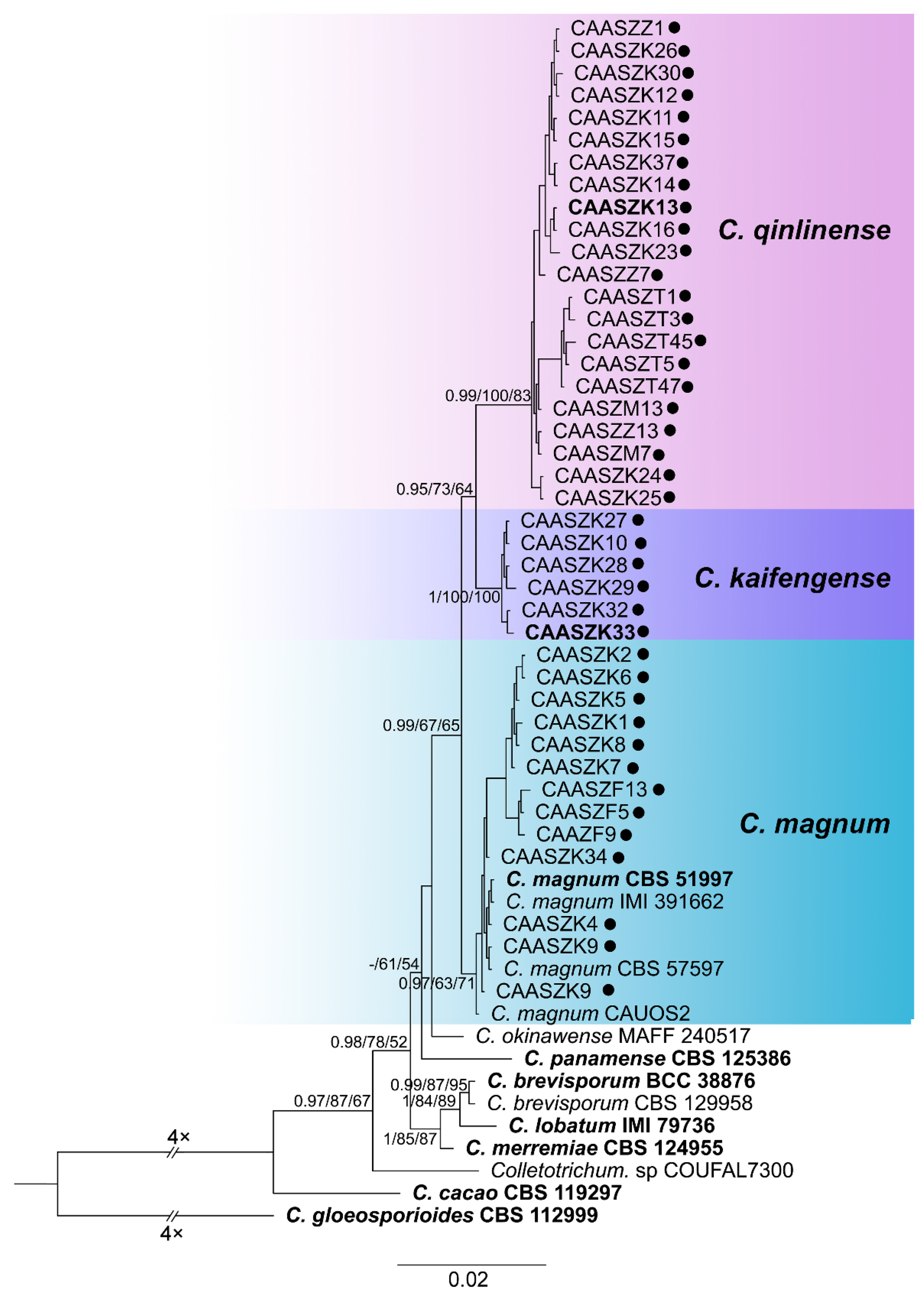
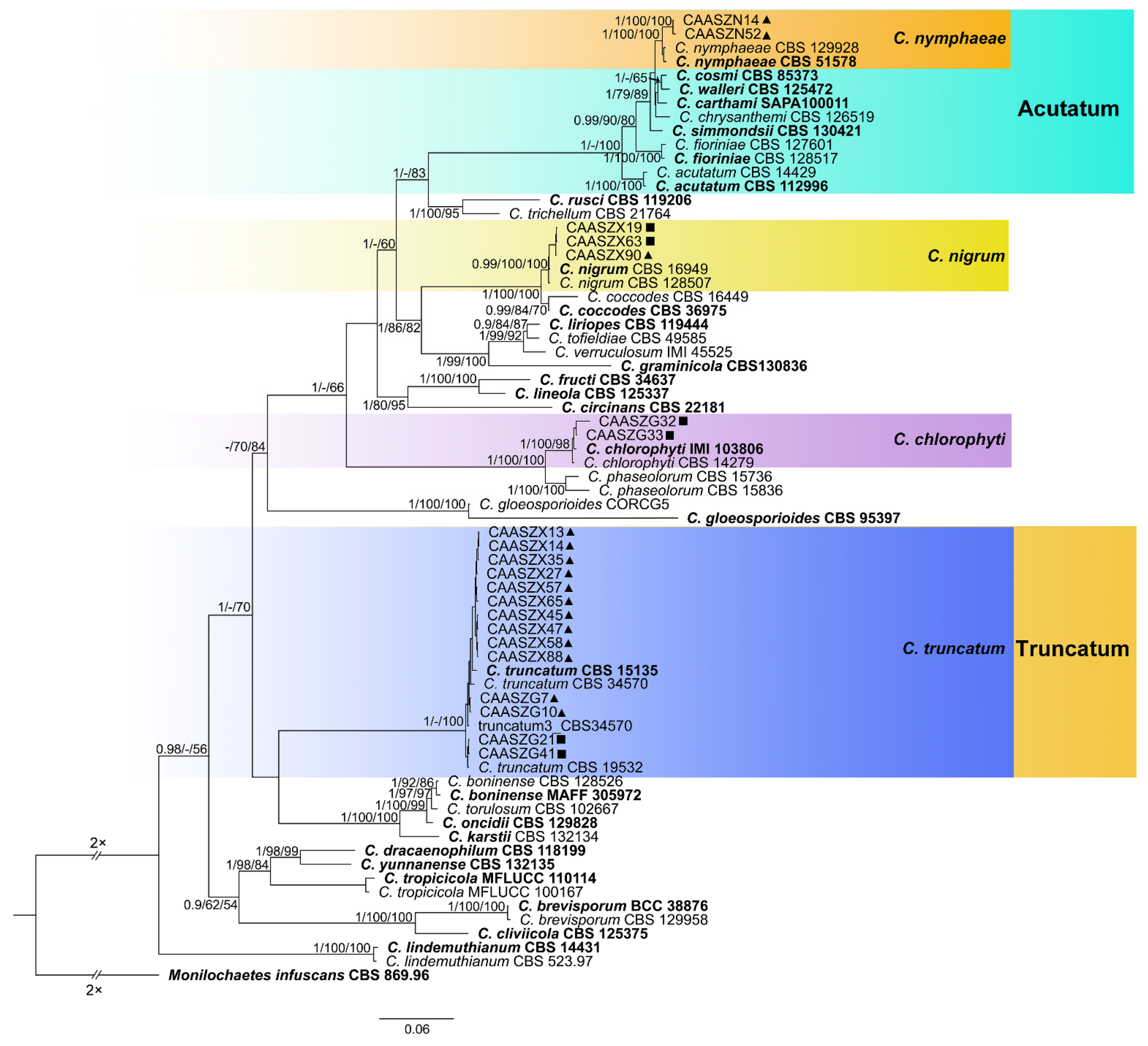
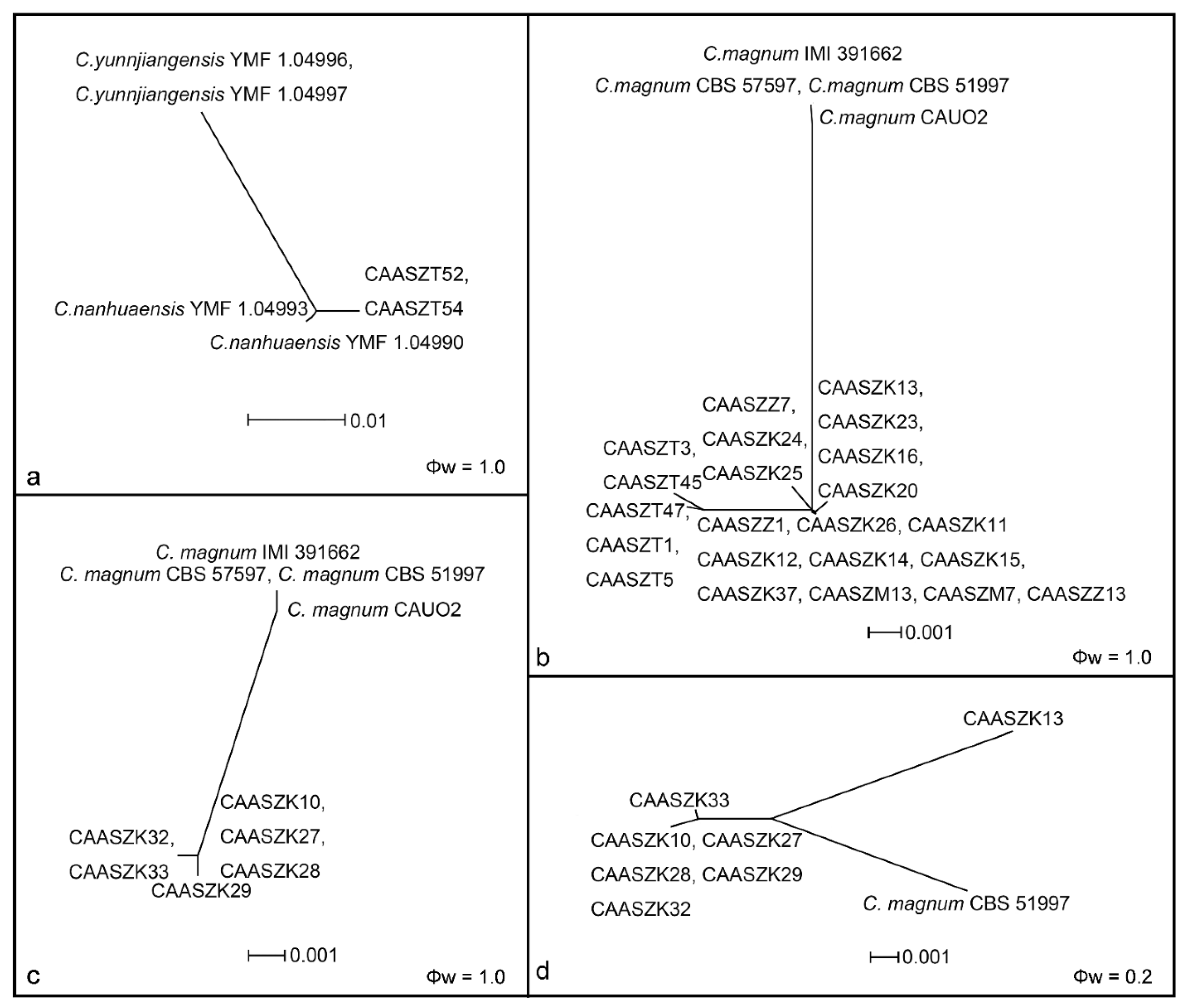
3.2. Multi-Locus Phylogenetic Analyses

The 146 representative isolates and related Colletotrichum species including the outgroup were subjected to multi-locus phylogenetic analyses (Supplementary Tables S1 and S2) with 7-locus phylogenetic analyses (ITS, gadph, chs-1, his3, act, tub2 and gs) for C. orbiculare species complexes, 6-locus phylogenetic analyses (ITS, gadph, chs-1, his3, act, and tub2) for those belonging to the C. gloeosporioides, C. boninense, C. magnum, and C. orchidearum species complexes, or with 5-locus phylogenetic analyses (ITS, gadph, chs-1, his3, act, and tub2) for other species complexes of without available his3 sequences. The consensus tree obtained from maximum parsimony and maximum likelihood analyses confirmed the tree topology obtained with Bayesian inference (Figure 2, Figure 3, Figure 4, Figure 5, Figure 6 and Figure 7).
The phylogenetic tree was constructed for the isolates of the C. gloeosporioides species complex, in which six isolates were clustered with C. fructicola, six with C. aenigma, and two with C. jiangxiense. In addition, the two isolates formed a distinct clade (Bayesian posterior probabilities value 1/PAUP bootstrap support value 99/iqtree bootstrap support value 100) as a sister group to C. nanhuaensis, which clustered distantly from any known species in the complex (Figure 2). For isolates in the C. boninense species complex, 23 isolates were clustered with C. karstii (Figure 3). For the isolates in the C. orbiculare species complex, 34 isolates were clustered with C. orbiculare (Figure 4). For isolates of C. orchidearum species complex, 14 isolates were clustered in two clades, 9 with C. plurivorum, and 5 with C. sojae (Figure 5). In the C. magnum species complex, 14 isolates were clustered with C. magnum. The remaining 29 isolates were clustered in two distinct clades (1/99/100, 1/99/96) as sisters to C. magnum, and distanted from any known species in the complex (Figure 6). The remaining 21 isolates were clustered in four clades corresponding to C. nymphaeae (2 isolates), C. nigrum (3 isolates), C. chlorophyti (2 isolates), and C. truncatum (14 isolates) (Figure 7). The PHI test indicated no significant recombination events between C. citrulli and C.nanhuaensis (Figure 8a). In addition, the results showed that there were no significant recombination events between C. qilinense and C. magnum (Figure 8b), or between C. kaifengense and C. magnum (Figure 8c). The results revealed no significant recombination events between C. kaifengense, a sister group of C. qilinense, and C. magnum (Figure 8b,c).

3.3. Multi-Locus Phylogenetic Analyses

Analyses of the prevalence of the Colletotrichum species associated with watermelon in China showed that C. orbiculare isolates (160 isolates, 28.9% of the total isolates) were predominantly isolated from five provinces (Henan, Hubei, Liaoning, Jilin, and Jiangsu), followed by C. truncatum (67 isolates, 8.8%, isolated from Henan and Jiangxi), C. qilinense (44 isolates, 8.4%, isolated from Henan), C. plurivorum (41 isolates, 8.4%, isolated from Jiangxi), C. magnum (56 isolates, 8.4%, isolated from Henan), C. nigrum (37 isolates, 6.8%, isolated from Henan), C. karstii (32 isolates, 6.1%, isolated from Jiangxi and Hainan), C. fructicola (23 isolates, 4.4%, isolated from Jiangxi and Hubei), and C. aenigma (18 isolates, 3.4%, isolated from Zhejiang). The remaining six species account for 9.3% of total isolates (Figure 9a). These results suggested that C. orbiculare is the most prevalent species of Colletotrichum sp. on watermelon in China (Figure 9a).
Analyses of the isolation rate of 15 Colletotrichum species in each sampled province revealed that C. orbiculare was commonly isolated in Henan and Hubei provinces, accounting for 25.4% and 34.6% of the obtained isolates, respectively (Figure 9b). In the Jiangxi province, C. plurivorum strains were dominant, accounting for 49.4% of total isolates from this province (Figure 9b). Moreover, all the C. orbiculare isolates were from Liaoning, Jilin, and Jiangsu provinces, and C. aenigma isolates were isolated from Zhejiang province (Figure 9b). Analyses of the Colletotrichum species isolated from three organs of watermelon revealed that 11 Colletotrichum species (C. orbiculare, C. truncatum, C. qilinense, C. plurivorum, C. nigrum, C. karstii, C. fructicola, C. aenigma, C. jiangxiense, C. sojae, and C. nymphaeae) were isolated from leaves, 8 (C. orbiculare, C. karstii, C. truncatum, C. plurivorum, C. fructicola, C. nigrum, C. jiangxiense, and C. chlorophyti) were isolated from stems, and 5 (C. orbiculare, C. citrulli, C. magnum, C. qilinnense, and C. kaifengense) were isolated from the fruits. Of those, C. orbiculare strains were dominant and accounted for 25.0%, 29.8%, and 36.5% of total isolates from leaves, stems, and fruits, respectively (Figure 9c).

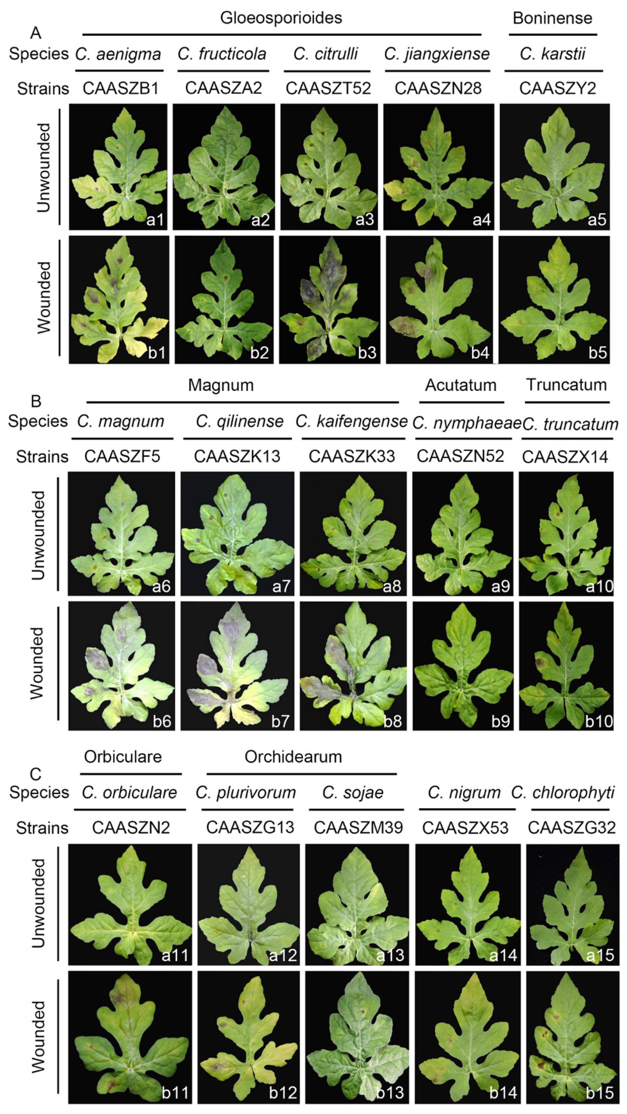
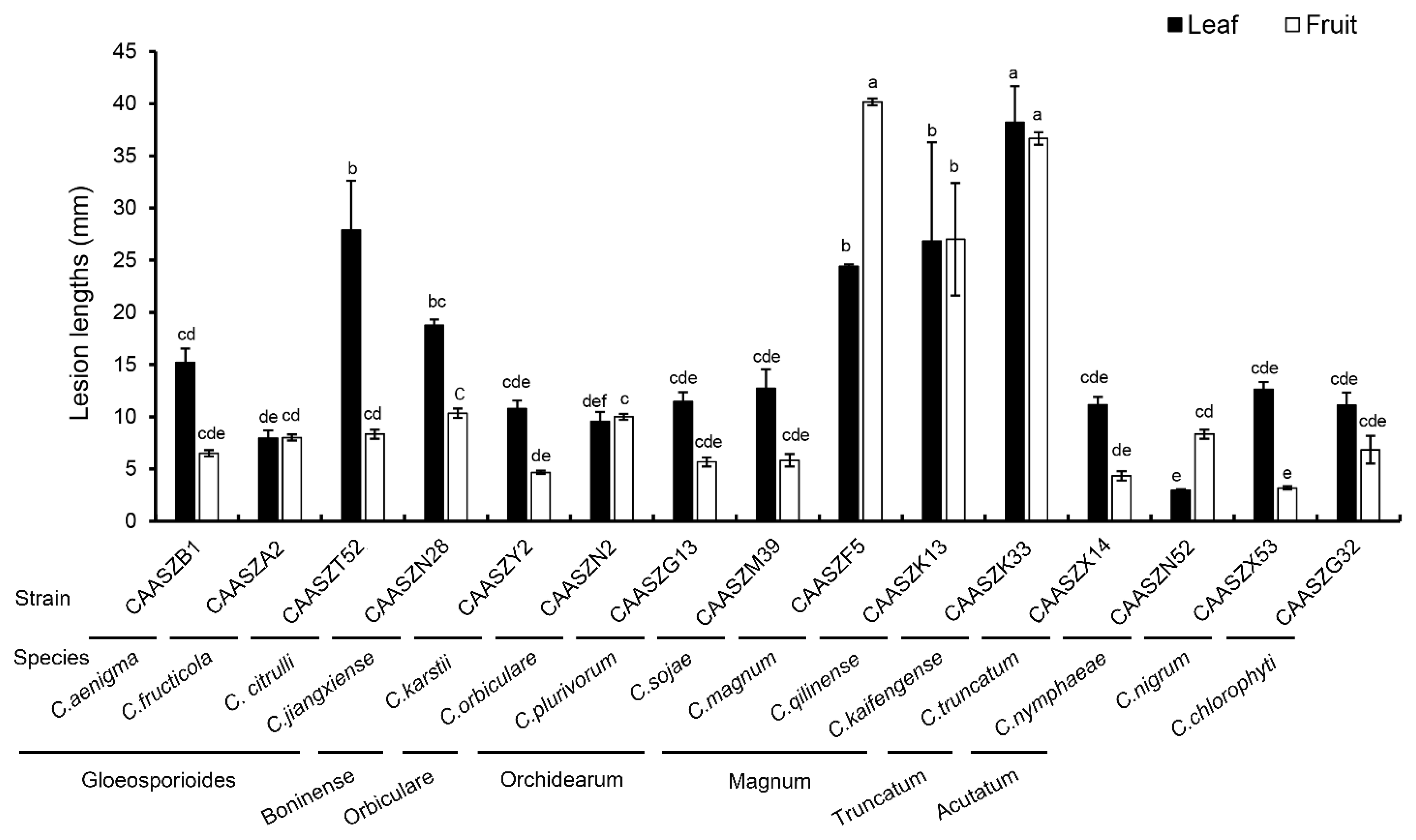
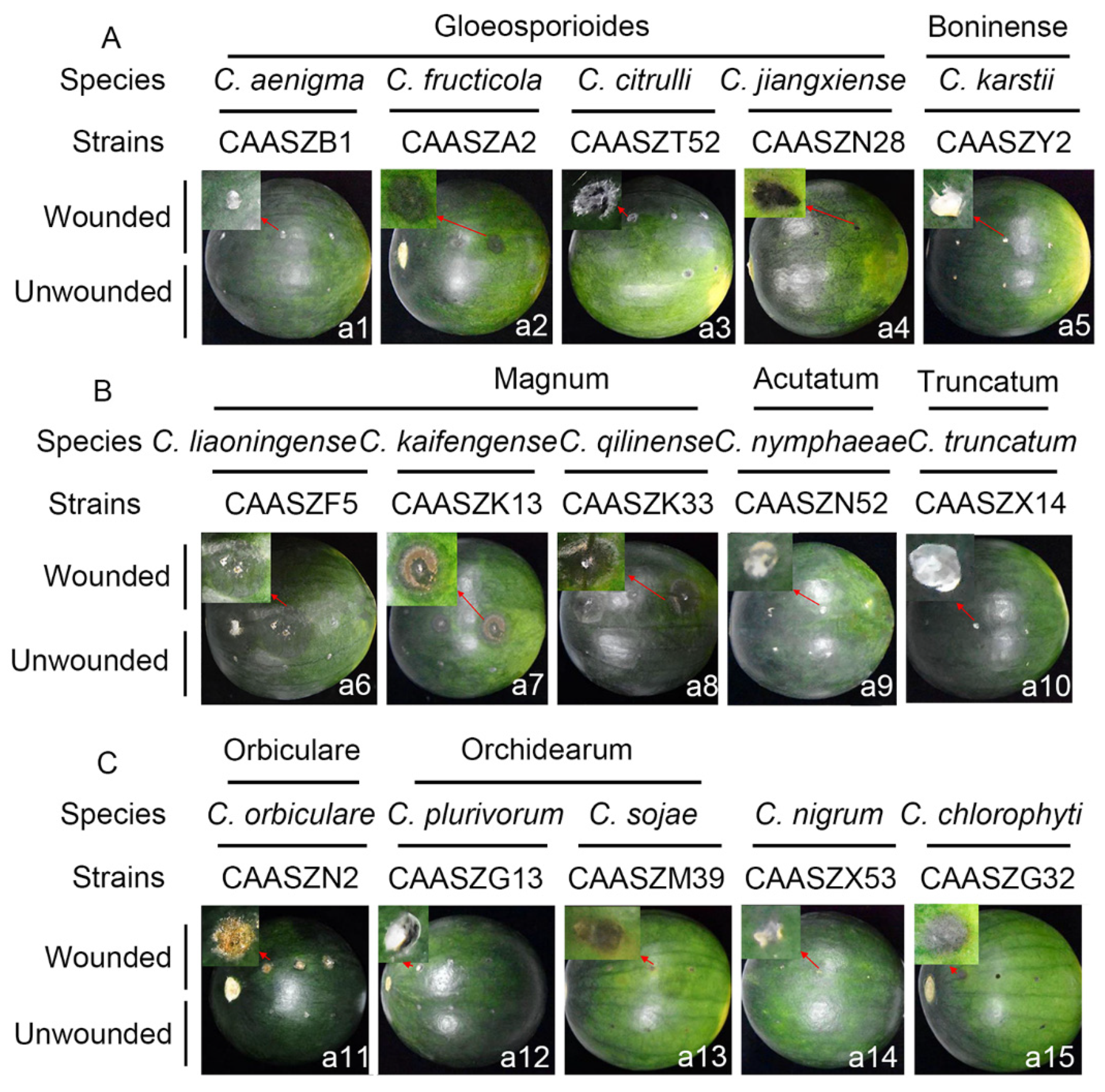
3.4. Pathogenicity Assay
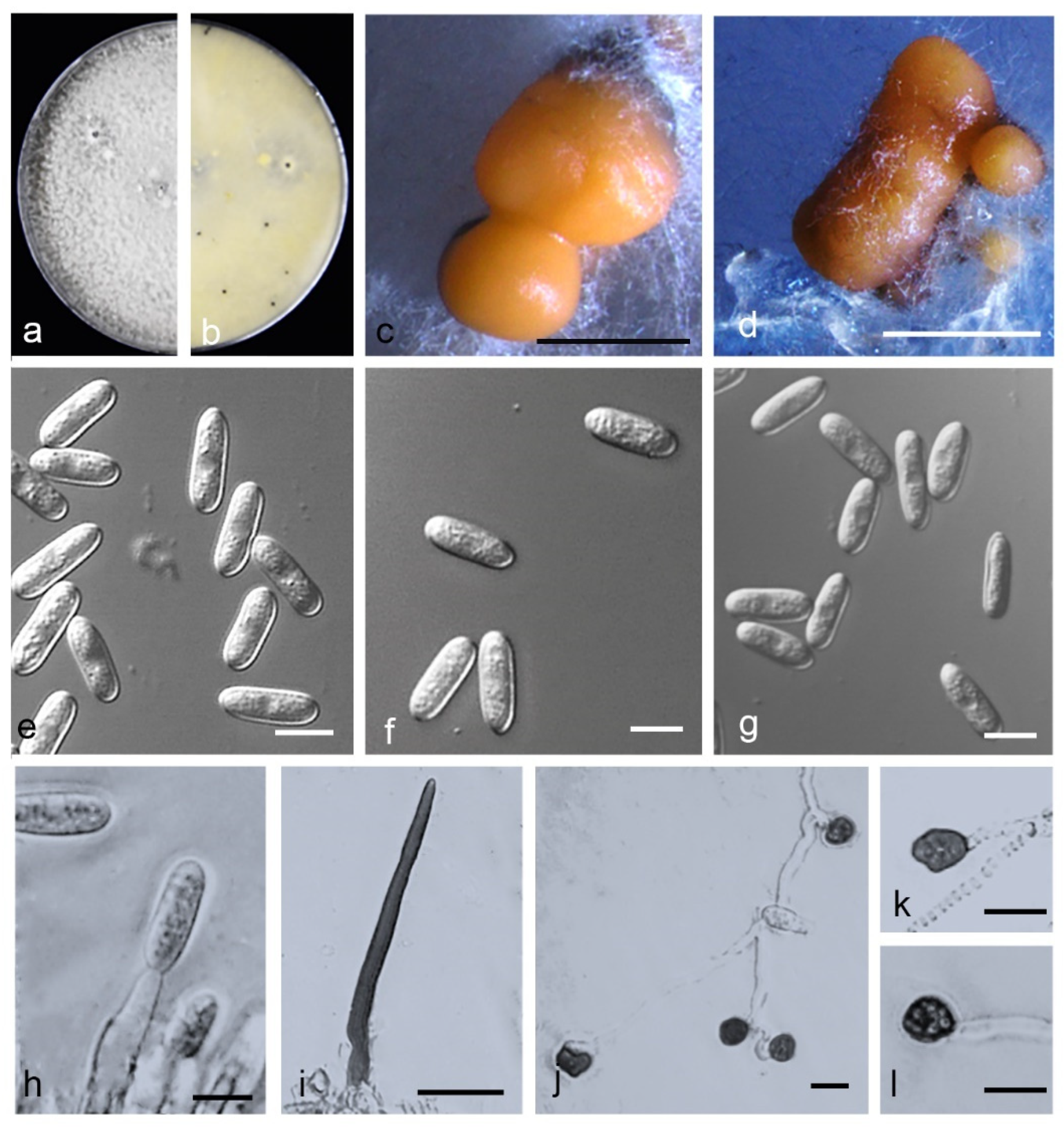
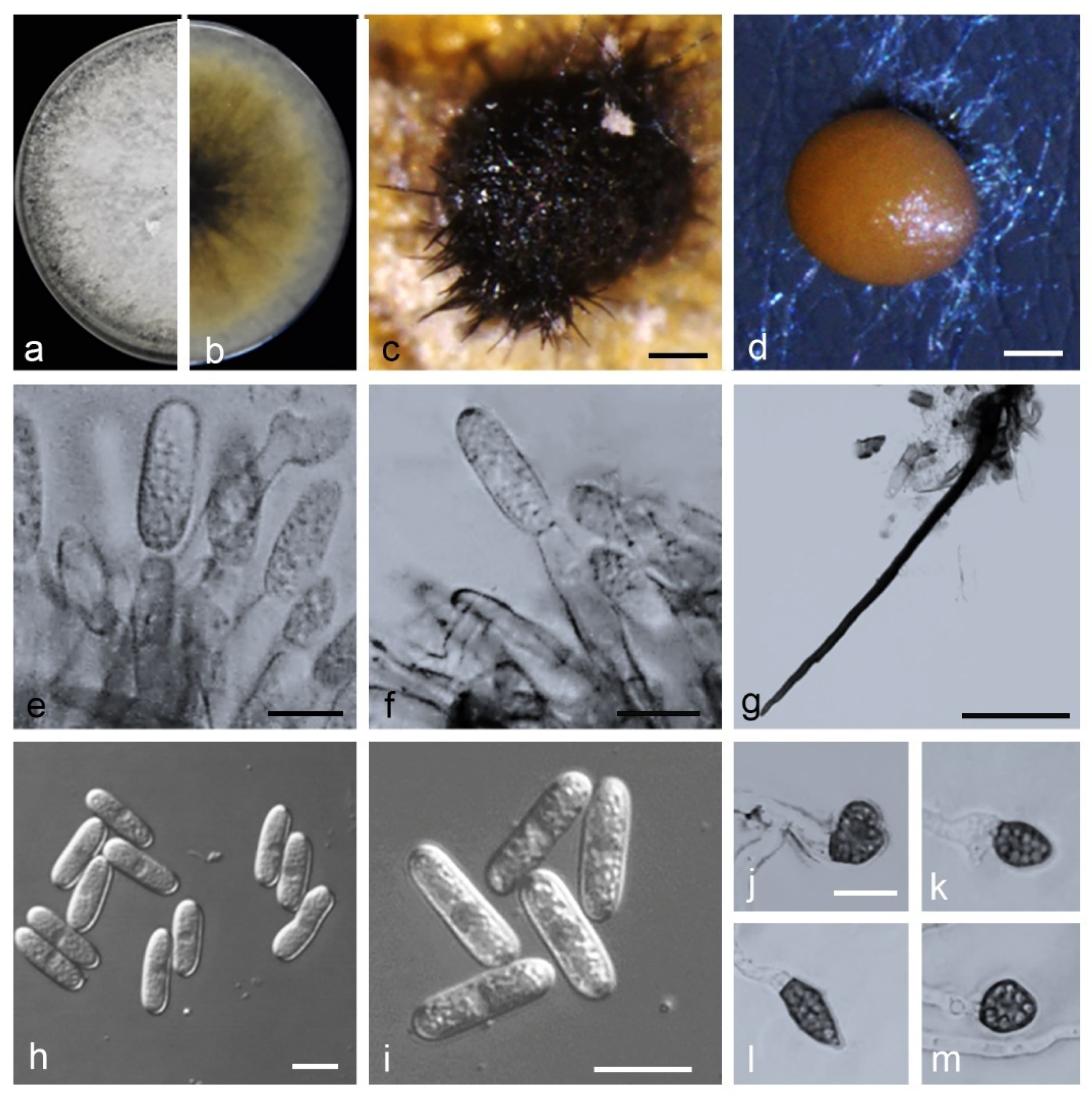
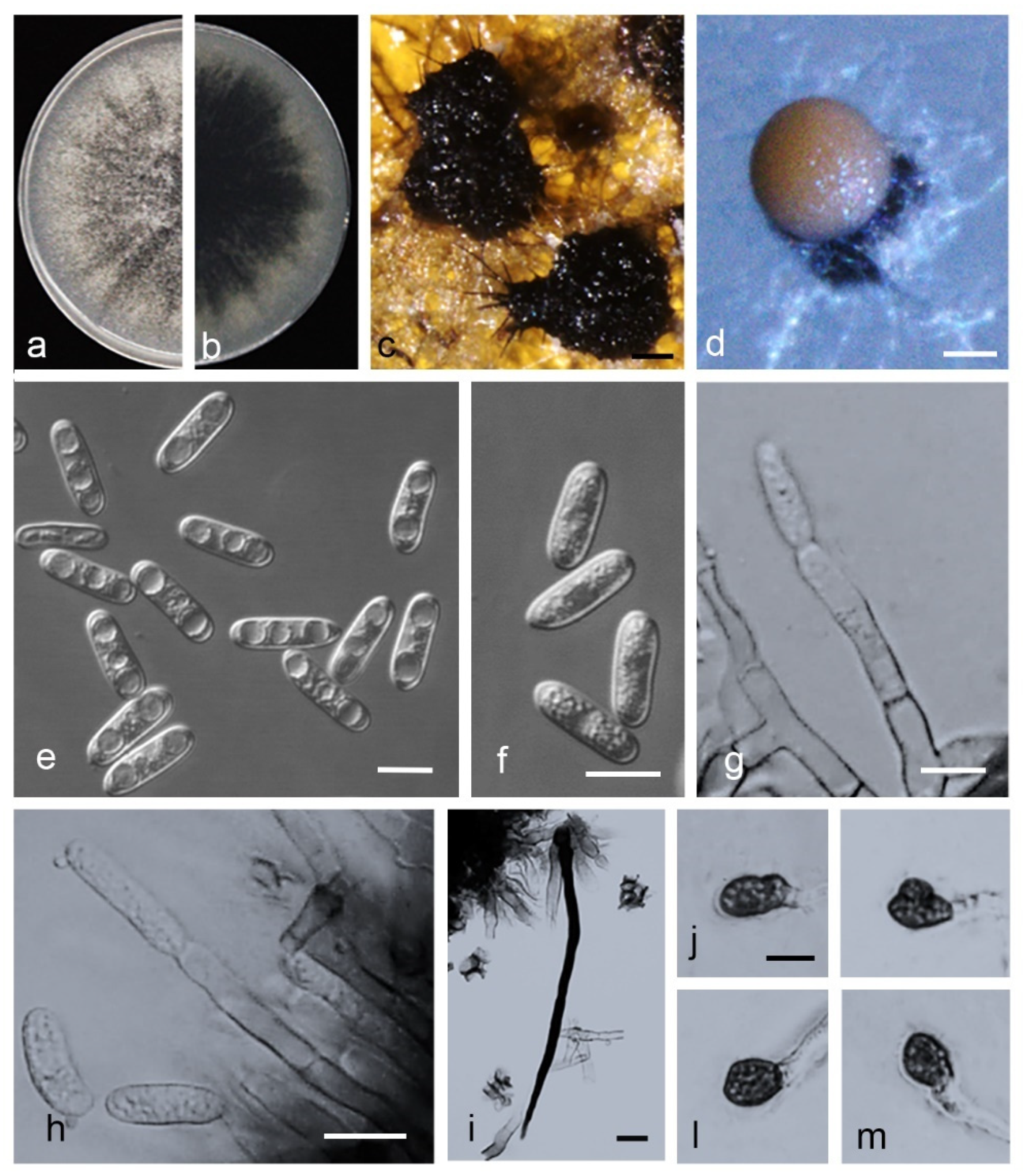
Fifteen representative Colletotrichum isolates were selected to prove Koch’s postulates. At 4 dpi, all the Colletotrichum isolates developed brown to black lesions symptoms of anthracnose on detached wounded leaves of Citrullus lanatus cv. Hongheping inoculated by a spore suspension, and C. fructicola, C. chlorophyte, and C. nymphaeae were unable to infect non-wounded leaves (Figure 10(a2,a10,a15), Table 3). Under wounded conditions, pathogenicity tests demonstrated that C. kaifengense (mean ± SD = 32.33 ± 6.35 mm), C. citrulli (mean ± SD = 27.89 ± 4.71 mm), C. qilinense (mean ± SD = 26.84 ± 9.45 mm), and C. magnum (mean ± SD = 24.42 ± 0.17 mm) were highly aggressive on watermelon leaves (Figure 11). However, C. fructicola, C. karstii, and C. nymphaeae showed only weak moderate aggression on watermelon leaves (Figure 12). Under unwounded conditions, C. qilinense (isolate CAASZK13) had the highest infection rate at 77.8% (Table 3). No lesions were induced in the control leaves inoculated with sterile water.
Pathogenicity was also accessed on detached watermelon fruits of Citrullus lanatus cv. Motong. Under unwounded conditions, C. aenigma, C. fructicola, C. nigrum, and C. truncatum did not infect the non-wounded fruits (Table 3). C. karstii, C. orbiculare, C. magnum, and C. sojae had the highest infection incidence at 100% (Table 3). Under wounded conditions, there was clear variation in aggression among species. Importantly, C. qilinense induced large brown or dark brown lesions and formed concentric rings of conidia at 6 dpi (Figure 12). The isolates of the C. magnum species complex, including C. magnum (mean ± SD = 40.2 ± 0.3 mm), C. qilinense (mean ± SD = 36.7 ± 0.6 mm), and C. kaifengense (mean ± SD = 27.0 ± 5.4 mm) induced significantly longer lesions than others (Figure 12).

3.5. Taxonomy

Based on DNA sequence data and the morphological characteristics, the 146 isolates were assigned to 15 species, including 8 species reported from watermelon for the first time, and 3 species proved to represent new taxa. The three new species in culture are characterized below.
Colletotrichum citrulli—Z. Guo & Q.S. Gu, sp. nov. (Figure 13).
MycoBank Number: MB842245.
Etymology: The species epithet is derived from the host plant, Citrullus lanatus.
Holotype: China, Henan Province, Zhoukou City, Taikang County, on fruits of Citrullus lanatus, June 2020, Z. Guo. Holotype HMAS 351572, Ex-type culture CGMCC 3.20769 = CAASZT52.
Sexual morph not observed. Asexual morph developed on OA. Chlamydospores not observed. Conidiomata acervular formed on a cushion of angular cells. Conidiophores hyaline to pale brown, aseptate, unbranched. Conidiogenous cells hyaline, cylindrical, 10.5–17 × 3–6 µm, opening 1–2 µm. Conidia hyaline, smooth-walled, aseptate, apex subacute or obtuse, contents with 1–2 guttules, 15–18.5 × 5–7 µm, mean ± SD = 16.2 ± 0.9 × 5.6 ± 0.5 μm, L/W ratio = 2.9; Appressoria single or in groups, medium to dark brown, variable in shape, often navicular to bullet-shaped, circular, clavate, smooth-walled to undulate, 8–12 × 6–10 µm, mean ± SD = 9.9 ± 0.2 × 7.3 ± 0.8 μm, L/W ratio = 1.4.
Asexual morph developed on SNA. Setae medium to dark brown, smooth to finely verruculose close to the tip, the tip rounded, 1–3- aseptate, 42–79 μm long, base inflated, usually paler, 3–6.5 μm diam. Conidia hyaline, smooth-walled, aseptate, apex subacute or obtuse, contents with 1–2 guttules, 14–17.5 × 4–6 µm, mean ± SD = 15.9 ± 1.0 × 5.3 ± 0.4 μm, L/W ratio = 3.0.
Culture characteristics: Colonies on PDA flat with entire margin, aerial mycelium dense, cottony, surface pale cinnamon, covered with irregular grey to black conidiomata, reverse same colors. Colony diam 69.0–70.0 mm in 5 d. Colonies on OA flat with entire margin, aerial mycelium lacking, partly covered with numerous orange conidia, surface white, reverse pale grey, 73.0–73.5 mm in 5 d. Colonies on SNA flat with entire margin, aerial mycelium sparse, surface pale grey, reverse same colors, 66.5–68.0 mm in 5 d.
Notes: isolates of C. citrulli are phylogenetically most closely related to C. nanhuaensis (Figure 2). They were distinguished by ITS (with 99.15% sequence identity) and gadph (97.63%). Furthermore, the PHI test (Φw = 1) did not detect recombination events between C. citrulli and C. nanhuaensis (Figure 8a). In morphology, C. citrulli differs from C. nanhuaensis by having longer conidia (14.65–18.4 5 × 4.75–6.6 5 µm vs. 10.5–16 × 4.5–6 µm) [33].
Colletotrichum kaifengense Z. Guo & Q. S. Gu, sp. nov. (Figure 14).
MycoBank Number: MB842244.
Etymology: Named after Kaifeng, the city in Henan Province, China, where the species was collected.
Holotype: China, Henan Province, Kaifeng City, Jinming County, on fruits of Citrullus lanatus, August 2019, Z. Guo. Holotype HMAS 351574, Ex-type culture CGMCC 3.20768 = CAASZK33.
Sexual morph not observed. Asexual morph developed on PDA. Chlamydospores not observed. Conidiomata acervular, conidiophores, and setae formed on a cushion of angular cells. Conidiophores hyaline, pale-to medium brown, smooth-walled, aseptate, unbranched. Conidiogenous cells hyaline, cylindrical, 11–21 × 4–5 µm. Setae medium to dark brown, smooth-walled, the tip acuted, aseptate, 70–153 μm. Conidia hyaline, smooth-walled, aseptate, cylindrical, both ends bluntly rounded, 13.5–20 × 4–6.5 µm, mean ± SD = 17.4 ± 1.7× 4.9 ± 0.5 μm, L/W ratio = 3.5. Appressoria single or in loose groups, pale to medium brown, bullet-shaped, circular, smooth-walled, spathulate or irregular, 8–14.5 × 5.5–11 µm, mean ± SD = 11.1 ± 1.9× 7.9 ± 1.4μm, L/W ratio = 1.5.
Asexual morph developed on OA. Setae medium to dark brown, smooth-walled, aseptate, 130.0–200 μm, the tip acute. Conidia hyaline, smooth-walled, aseptate, cylindrical, both ends bluntly rounded, 16–20.5 × 4–6.5 µm, mean ± SD = 18.2 ± 1.1× 5.3 ± 0.5 μm, L/W ratio = 3.5.
Asexual morph developed on SNA. Conidiophores, and Setae formed on hyphae. Setae medium to dark brown, smooth-walled, aseptate, 82.5–177.5 μm, the tip acute. Conidia hyaline, smooth-walled, aseptate, cylindrical, both ends bluntly rounded, 16–22.5 × 4–5.5 µm, mean ± SD = 20.0 ± 1.2× 4.8 ± 0.4 μm, L/W ratio = 4.2.
Culture characteristics: Colonies on PDA flat with entire margin, aerial mycelium dense, floccose, surface pale grey to white, reverse grey in the center with cinnamon margin, 75.0–76.0 mm in 7 d. Colonies on OA flat with entire margin, aerial mycelium lacking, surface white, covered with small black conidiomata with orange conidial masses, reverse white, covered with irregular black conidiomata, 50.0–52.0 mm in 7 d. Colonies on SNA flat with entire margin, short felty whitish aerial mycelium, surface white, reverse same colors, 50.5–51.5 mm in 7 d.
Notes: isolates of C. kaifengense are phylogenetically closely related to C. magnum (Figure 6). They were distinguished by gadph (93.24%) and his3 (98.55%). Furthermore, the PHI test (Φw = 1) did not detect recombination events between C. kaifengense and C. magnum (Figure 8c). They were distinguished by gadph (88.14%) and chs-1 (98.65%). Furthermore, the PHI test (Φw = 1) did not detect recombination events between C. qilinense and C. kaifengense (Figure 8d).
Colletotrichum qilinense Z. Guo & Q.S. Gu, sp. nov. (Figure 15).
MycoBank Number: MB842247;
Etymology. Referring to the host variety (Citrullus lanatus cv. qilinwang) from which the fungus was collected.
Holotype: China, Henan Province, Kaifeng City, Weishi County, on fruits of Citrullus lanatus, 20 August 2019, Z. Guo. Holotype HMAS 351573, Ex-type culture CGMCC 3.20767 = CAASZK13).
Sexual morph not observed. Asexual morph developed on PDA. Chlamydospores not observed. Conidiomata acervular, conidiophores, and setae formed on a cushion of angular cells. Conidiophores hyaline, medium to dark brown, smooth-walled, aseptate, unbranched. Conidiogenous cells hyaline, cylindrical, 9–23 × 4–6 µm. Setae medium to dark brown, smooth-walled, the tip acuted, 75.5–188 μm. Conidia hyaline, smooth-walled, cylindrical, the apex rounded, the base rounded to truncate, 17.5–23 × 5–6 µm, mean ± SD = 19.7 ± 1.4 × 5.4 ± 0.4 μm, L/W ratio = 3.7. Appressoria single or in loose groups, medium to dark brown, bullet-shaped, smooth-walled, aseptate, mostly ovoid or ellipsoidal to irregular in outline, 8–16 × 5–11 µm, mean ± SD = 10.6 ± 2.1 × 7.7 ± 1.5 μm, L/W ratio = 1.4.
Asexual morph developed on OA. Setae medium to dark brown, smooth-walled, the tip acuted, base inflated or not, 96.7–178.4 μm. Conidia hyaline, smooth-walled, aseptate or aseptate, cylindrical, the apex rounded, the base rounded to truncate, 14–20 × 5.5–6.5 µm, mean ± SD = 17.6 ± 1.5 × 5.3 ± 0.5 μm, L/W ratio = 3.3.
Asexual morph developed on SNA. Conidiophores are directly formed on hyphae. Setae medium to dark brown, smooth-walled, the tip acuted, aseptate, 83–203 μm. Conidia hyaline, smooth-walled, aseptate, cylindrical, both ends bluntly rounded, 16–20 × 4.5–6 µm, mean ± SD = 17.8 ± 1.0 × 5.1 ± 0.4μm, L/W ratio = 3.5.
Culture characteristics: colonies (CAASZK13) on PDA flat with entire margin, aerial mycelium lacking, floccose, surface dark grey to black, reverse black in center with white margin, 75.50–76.0 mm in 7 d. Colonies on OA flat with entire margin, surface white, partly covered with short felty whitish aerial mycelium, reverse white, covered with irregular black conidiomata, 53.0–54.0 mm in 7 d. Colonies on SNA flat with entire margin, short felty whitish aerial mycelium, surface white, reverse same colors, 51.0–52.0 mm in 7 d.
Notes: isolates of C. qilinense are phylogenetically closely related to C. magnum (Figure 6). They were distinguished by gadph (87.08%), chs-1 (99.71%), and act (96.57%). Furthermore, the PHI test (Φw = 1) did not detect recombination events between C. qilinense and C. magnum (Figure 8d).

4. Discussion

The main objective of this study was to identify the Colletotrichum species associated with watermelon in China. We obtained 526 single spore strains from watermelon stems, leaves, and fruits displaying anthracnose symptoms. Based on multi-locus data combined with morphological characteristics we revealed 15 species allocated in 7 species complexes, including gloeosporioides (C. aenigma, C. fructicola, C. jiangxiense, and C. citrulli), boninense (C. karstii), orbiculare (C. orbiculare), orchidearum (C. plurivorum and C. sojae), magnum (C. liaoningense, C. magnum, C. qilinnense, and C. kaifengense), Truncatum (C. truncatum), and acutatum (C. nymphaeae) and two singleton species (C. nigrum, and C. chlorophyti). It is the first report of C. aenigma, C. fructicola, C. jiangxiense, C. plurivorum, C. sojae, C. magnum, C. nymphaeae, C. nigrum, and C. chlorophyti causing anthracnose of watermelon. Importantly, this study differentiated three new species, namely C. citrulli, C. qilinnense, and C. kaifengense.
The species complexes of the Colletotrichum include several taxa [30,31,32,36,37,38,39,43]. Thus, the complex species related to the 15 Colletotrichum species recovered in this study were selected and included in the analyses. In previous studies, although morphological characters delimited some Colletotrichum species, some species were difficult to describe due to their morphological variability [25,77]. Thus, in recent studies, the multi-locus phylogenetic analyses combined with morphological characteristics including measurements of setae, conidia, and appressoria, colony characters, and growth rates were able to resolve the complexes and species within Colletotrichum. spp [12,16,17,18,78,79]. In this study, most of the Colletotrichum species were determined by multi-locus DNA sequence analyses, and some species also exhibited specific morphological characteristics. For example, the growth rate of C. orbiculare was significantly lower than that of the other 14 Colletotrichum species (Supplementary Figure S1). It is worth noting that the Colletotrichum species associated with watermelon differ in their ability to form sexual morphs and setae. Importantly, only four species, C. aenigma, C. fructicola, C. karstii, and C. plurivorum, produced ascospores on OA plates (Supplementary Table S3). Additionally, setae of C. jiangxiense, C. nigrum, and C. nymphaeae were not observed on PDA, OA, and SNA plates (Supplementary Table S3).
The prevalence of a Colletotrichum species associated with watermelon anthracnose is closely related to the sampling area and plant tissue. For example, some of Colletotrichum species, including C. jiangxiense, C. citrulli, C. sojae, C. magnum, C. qilinnense, C. kaifengense, C. nymphaeae, C. nigrum, and C. chlorophyti associated with watermelon showed restricted distribution. Notably, C. orbiculare was isolated from five provinces (Liaoning, Jilin, Henan, Hubei, and Jiangsu), and was dominant in our investigation of the major watermelon-growing regions in China (Figure 9a). It is worth noting that geographical preference was found for C. aenigma, in agreement with recent results in China [18].
Some Colletotrichum species are host-organ-specific and, for example, predominantly infect leaves of apple and pear [80]. This study revealed that more Colletotrichum species infect watermelon leaves than stems or fruits. Three Colletotrichum species including C. aenigma, C. sojae, and C. nymphaeae were only isolated from the leaves; C. chlorophyti was only isolated from the stems; and five Colletotrichum species, including C. citrulli, C. magnum, C. qilinnense, and C. kaifengense, were only isolated from fruits (Figure 9c).
Pathogenicity of 15 Colletotrichum species isolated from watermelon samples in China was tested on watermelon fruits and leaves. It is worth noting that all Colletotrichum species could infect wounded fruits and leaves. However, five Colletotrichum species, including C. aenigma, C. fructicola, C. magnum, C. nigrum, and C. truncatum, did not cause disease symptoms on the non-wounded watermelon fruits. Three Colletotrichum species (C. fructicola, C. nymphaeae, and C. chlorophyti) did not cause lesions on the non-wounded leaves (Table 3). This phenomenon is related to inoculation methods and conditions, quiescent infection, and structural defense of the host tissue. In this study, pathogenicity experiments were conducted on wounded or unwounded tissues under extreme conditions of artificial inoculation. It remains to be seen how the symptoms and lesion development are displayed under natural conditions. The quiescent infection is an important feature of Colletotrichum spp., and it mainly occurs prior to harvest and eventually leads to fruit rot [81,82,83]. A previous study indicated that the cuticle and epidermis were important as resistance barriers to the initial infection. Wounding can break the quiescent infection and enhance the infectiousness [84].
Different Colletotrichum species have varying aggressiveness on the host [17,18,85]. In this study, most isolates belonging to the C. magnum species complex showed higher aggressiveness than those of the C. gloeosporioides, C. boninense, C. orbiculare, C. orchidearum, C. truncatum, and C. acutatum species complexes (Figure 11). Studies suggest that C. magnum caused watermelon anthracnose in USA and chili anthracnose in China [16,32,43]. However, it was never reported on watermelon in China. Thus, this study here represents the first report of C. magnum on watermelon in China. Colletotrichum qilinense and C. kaifengense were found to cause anthracnose on watermelon fruits and were described as new species in this study. Noteworthy, among the 15 representative Colletotrichum species in inoculated wounded watermelon leaves, C. kaifengense was the most aggressive one (Figure 11).
Previous studies revealed that 15 Colletotrichum species were identified from chili fruits with anthracnose symptoms in China, including C. fioriniae, C. fructicola, C. gloeosporioides, C. scovillei, C. truncatum, C. conoides, C. grossum, C. magnum, C. aenigma, C. cliviae, C. endophytica, C. hymenocallidis, C. incanum, C. karstii, and C. viniferum [16,43]. Furthermore, it is interesting that five Colletotrichum species, including C. fructicola, C. truncatum, C. magnum, C. aenigma, and C. karstii, were also isolated from watermelon in this study. Colletotrichum fructicola was reported for the first time, associated with anthracnose symptoms from strawberry in USA and Canada [30]. This is the first report of C. fructicola to induce anthracnose of watermelon (Figure 2). Colletotrichum truncatum was the dominant species on chili in China [16], and it was also found to cause anthracnose on watermelon leaves, stems, and fruits. Colletotrichum aenigma was first reported on chili in China, where it was found to cause anthracnose on fruits [16], but this is the first report of C. aenigma on Citrullus lanatus, where it was found to cause anthracnose on watermelon leaves. Colletotrichum karstii was reported causing fruit spots on chili in China [16] and bitter rot on strawberry in the Australia [86]. This is the first report of C. karstii in China, which caused watermelon leaf spot.
This study provides useful information about ecology and pathogenicity of 15 Colletotrichum spp. involved in watermelon anthracnose. This is of great significance for the control of anthracnose disease in different regions in China.

5. Conclusions

This study provides the first summary of 15 Colletotrichum spp. associated with watermelon in China. Furthermore, this study presents the first reports of C. aenigma, C. fructicola, C. jiangxiense, C. plurivorum, C. sojae, C. nymphaeae, C. nigrum, and C. chlorophyti, and three new species (C. citrulli, C. qilinnense, and C. kaifengense), causing anthracnose of watermelon in China.

Supplementary Materials

The following supporting information can be downloaded at: https://www.mdpi.com/article/10.3390/jof8080790/s1, Table S1. List of 148 representative isolates of 15 Colletotrichum spp. collected from watermelon in China, with details host and location, and Genbank No; Table S2. Strains of the Colletotrichum species in this paper with details about host and location, and Genbank No; Table S3. The sizes of seta, conidia, appresoria, and ascospores of the representative isolates of Colletotrichum spp. obtained in this study; Figure S1. The growth rate of the representative isolates of Colletotrichum spp. obtained in this study. Data were analyzed with SPSS Statistics 19.0 (WinWrap Basic; http://www.winwrap.com (accessed on 18 December 2021)) by one-way analysis of variance, and means were compared using Duncan’s test at a significance level of p = 0.05. Letters over the error bars indicate the significant difference at the p = 0.05 level.

Author Contributions

Conceptualization, Z.G. and Q.-S.G.; methodology, Z.G.; software, Z.G. and H.-J.W.; validation, Z.G. and Q.-S.G.; formal analysis, Z.G.; investigation, Z.G., B.P. and Q.-S.G.; data curation, Z.G. and Q.-S.G.; writing—original draft preparation, Z.G. and Q.-S.G.; writing—review and editing, H.-J.W., M.Z., C.-X.L. and Q.-S.G.; visualization, Z.G.; supervision, B.-S.K., L.-M.L. and Q.-S.G.; project administration and funding acquisition, Q.-S.G. All authors have read and agreed to the published version of the manuscript.

Funding

This work was financially supported Agricultural Science and Technology Innovation Program (CAAS-ASTIP-2022-ZFRI-09), Central Public-interest Scientific Institution Basal Research Fund (No. 1610192022105), and National Natural Science Foundation of China (No. U21A20229).

Institutional Review Board Statement

Not applicable.

Informed Consent Statement

Not applicable.

Data Availability Statement

Alignments generated during the current study are available from TreeBASE (study 29157). All sequence data are available in the NCBI GenBank, following the accession numbers in the manuscript.

Acknowledgments

We sincerely thank the reviewers for their contributions during the revision process.

Conflicts of Interest

The authors declare no conflict of interest. The funders had no role in the design of the study; in the collection, analyses, or interpretation of data; in the writing of the manuscript; or in the decision to publish the results.

References

  1. Cannon, P.F.; Damm, U.; Johnston, P.R.; Weir, B.S. Colletotrichum-current status and future directions. Stud. Mycol. 2012, 73, 181–213. [Google Scholar] [CrossRef] [PubMed] [Green Version]
  2. Dean, R.; Van Kan, J.A.; Pretorius, Z.A.; Hammond-Kosack, K.E.; Di Pietro, A.; Spanu, P.D.; Rudd, J.J.; Dickman, M.; Kahmann, R.; Ellis, J.; et al. The Top 10 fungal pathogens in molecular plant pathology. Mol. Plant Pathol. 2012, 13, 414–430. [Google Scholar] [CrossRef] [PubMed] [Green Version]
  3. Zheng, H.; Yu, Z.; Jiang, X.; Fang, L.; Qiao, M. Endophytic Colletotrichum Species from Aquatic Plants in Southwest China. J. Fungi 2022, 8, 87. [Google Scholar] [CrossRef]
  4. Denoyes-Rothan, B.; Guerin, G.; Delye, C.; Smith, B.; Minz, D.; Maymon, M.; Freeman, S. Genetic diversity and pathogenic variability among isolates of Colletotrichum species from strawberry. Phytopathology 2003, 93, 219–228. [Google Scholar] [CrossRef] [Green Version]
  5. Prihastuti, H.; McKenzie, E.; Hyde, K.; Cai, L.; Mckenzie, H.; Hyde, E. Characterization of Colletotrichum species associated with coffee berries in northern Thailand. Fungal Divers. 2009, 39, 89–109. [Google Scholar]
  6. Phoulivong, S.; McKenzie, E.H.C.; Hyde, K.D. Cross infection of Colletotrichum species; a case study with tropical fruits. Curr. Res. Environ. Appl. Mycol. 2012, 2, 99–111. [Google Scholar] [CrossRef] [Green Version]
  7. Huang, F.; Chen, G.Q.; Hou, X.; Fu, Y.S.; Cai, L.; Hyde, K.D.; Li, H.Y. Colletotrichum species associated with cultivated citrus in China. Fungal Divers. 2013, 61, 61–74. [Google Scholar] [CrossRef]
  8. Lima, N.B.; Batista, M.V.D.A.; De Morais, M.A.; Barbosa, M.A.G.; Michereff, S.J.; Hyde, K.D.; Câmara, M.P.S. Five Colletotrichum species are responsible for mango anthracnose in northeastern Brazil. Fungal Divers. 2013, 61, 75–88. [Google Scholar] [CrossRef]
  9. Liu, F.; Damm, U.; Cai, L.; Crous, P.W. Species of the Colletotrichum gloeosporioides complex associated with anthracnose diseases of Proteaceae. Fungal Divers. 2013, 61, 89–105. [Google Scholar] [CrossRef]
  10. Schena, L.; Mosca, S.; Cacciola, S.O.; Faedda, R.; Sanzani, S.M.; Agosteo, G.E.; Sergeeva, V.; di San Lio, G.M. Species of the Colletotrichum gloeosporioides and C. boninense complexes associated with olive anthracnose. Plant Pathol. 2014, 63, 437–446. [Google Scholar] [CrossRef]
  11. Vieira, W.A.S.; Michereff, S.J.; de Morais, M.A.; Hyde, K.D.; Câmara, M.P.S. Endophytic species of Colletotrichum associated with mango in northeastern Brazil. Fungal Divers. 2014, 67, 181–202. [Google Scholar] [CrossRef]
  12. Yan, J.-Y.; Jayawardena, M.M.R.S.; Goonasekara, I.D.; Wang, Y.; Zhang, W.; Liu, M.; Huang, J.B.; Wang, Z.Y.; Shang, J.J.; Peng, Y.L.; et al. Diverse species of Colletotrichum associated with grapevine anthracnose in China. Fungal Divers. 2014, 71, 233–246. [Google Scholar] [CrossRef]
  13. La Hoz, C.J.P.-D.; Calderón, C.; Rincón, A.M.; Cárdenas, M.; Danies, G.; López-Kleine, L.; Restrepo, S.; Jiménez, P. Species from the Colletotrichum acutatum, Colletotrichum boninense and Colletotrichum gloeosporioidesspecies complexes associated with tree tomato and mango crops in Colombia. Plant Pathol. 2016, 65, 227–237. [Google Scholar] [CrossRef]
  14. Wang, Y.C.; Hao, X.Y.; Wang, L.; Bin, X.; Wang, X.C.; Yang, Y.J. Diverse Colletotrichum species cause anthracnose of tea plants (Camellia sinensis (L.) O. Kuntze) in China. Sci. Rep. 2016, 6, 35287. [Google Scholar] [CrossRef] [Green Version]
  15. De Silva, D.D.; Ades, P.K.; Crous, P.W.; Taylor, P.W.J. Colletotrichum species associated with chili anthracnose in Australia. Plant Pathol. 2017, 66, 254–267. [Google Scholar] [CrossRef]
  16. Diao, Y.Z.; Zhang, C.; Liu, F.; Wang, W.Z.; Liu, L.; Cai, L.; Liu, X.L. Colletotrichum species causing anthracnose disease of chili in China. Persoonia 2017, 38, 20–37. [Google Scholar] [CrossRef] [PubMed] [Green Version]
  17. Guarnaccia, V.; Groenewald, J.Z.; Polizzi, G.; Crous, P.W. High species diversity in Colletotrichum associated with citrus diseases in Europe. Persoonia 2017, 39, 32–50. [Google Scholar] [CrossRef]
  18. Fu, M.; Crous, P.W.; Bai, Q.; Zhang, P.F.; Xiang, J.; Guo, Y.S.; Zhao, F.F.; Yang, M.M.; Hong, N.; Xu, W.X.; et al. Colletotrichum species associated with anthracnose of Pyrus spp. in China. Persoonia 2019, 42, 1–35. [Google Scholar] [CrossRef] [Green Version]
  19. Wang, Y.; Chen, J.Y.; Xu, X.; Cheng, J.; Zheng, L.; Huang, J.; Li, D.W. Identification and Characterization of Colletotrichum Species Associated with Anthracnose Disease of Camellia oleifera in China. Plant Dis. 2020, 104, 474–482. [Google Scholar] [CrossRef]
  20. Wang, W.; de Silva, D.D.; Moslemi, A.; Edwards, J.; Ades, P.K.; Crous, P.W.; Taylor, P.W.J. Colletotrichum Species Causing Anthracnose of Citrus in Australia. J. Fungi 2021, 7, 47. [Google Scholar] [CrossRef]
  21. Tan, Q.; Schnabel, G.; Chaisiri, C.; Yin, L.F.; Yin, W.X.; Luo, C.X. Colletotrichum Species Associated with Peaches in China. J. Fungi 2022, 8, 313. [Google Scholar] [CrossRef] [PubMed]
  22. Sutton, B.C. The Coelomycetes: Fungi Imperfecti with Pycnidia Acervuli and Stromata; Commonwelth Mycological Institute: Kew, UK, 1980. [Google Scholar]
  23. Von Arx, J.A. Die Arten der Gattung Colletotrichum Cda. Phytopathol. Z. 1957, 29, 413–468. [Google Scholar]
  24. Hyde, K.D.; Cai, L.; McKenzie, E.; Yang, Y.L.; Zhang, J.Z.; Prihastuti, H. Colletotrichum: A catalogue of confusion. Fungal Divers. 2009, 39, 1–17. [Google Scholar]
  25. Hyde, K.D.; Chomnunti, P.; Crous, P.W.; Groenewald, J.Z.; Damm, U.; Ko, T.W.K.; Shivas, R.G.; Summerell, B.A.; Tan, Y.P. A case for re-inventory of Australia’s plant pathogens. Persoonia 2010, 25, 50–60. [Google Scholar] [CrossRef] [PubMed] [Green Version]
  26. Quaedvlieg, W.; Binder, M.; Groenewald, J.Z.; Summerell, B.A.; Carnegie, A.J.; Burgess, T.I.; Crous, P.W. Introducing the Consolidated Species Concept to resolve species in the Teratosphaeriaceae. Persoonia 2014, 33, 1–40. [Google Scholar] [CrossRef] [PubMed] [Green Version]
  27. Tsang, C.C.; Tang, J.Y.M.; Lau, S.K.P.; Woo, P.C.Y. Taxonomy and evolution of Aspergillus, Penicillium and Talaromyces in the omics era-Past, present and future. Comput. Struct. Biotechnol. J. 2018, 16, 197–210. [Google Scholar] [CrossRef]
  28. Cai, L.; Hyde, K.; Taylor, P.; Weir, B.; Waller, J.; Abang, M.; Zhang, J.Z.; Yang, Y.L.; Phoulivong, S.; Liu, Z.Y. A polyphasic approach for studying Colletotrichum. Fungal Divers. 2009, 39, 183–204. [Google Scholar]
  29. Yang, Y.L.; Liu, Z.Y.; Cai, L.; Hyde, K.; Yu, Z.N.; McKenzie, E. Colletotrichum anthracnose of Amaryllidaceae. Fungal Divers. 2009, 39, 123–146. [Google Scholar]
  30. Weir, B.S.; Johnston, P.R.; Damm, U. The Colletotrichum gloeosporioides species complex. Stud. Mycol. 2012, 73, 115–180. [Google Scholar] [CrossRef] [Green Version]
  31. Liu, F.; Cai, L.; Crous, P.W.; Damm, U. The Colletotrichum gigasporum species complex. Persoonia 2014, 33, 83–97. [Google Scholar] [CrossRef] [Green Version]
  32. Damm, U.; Sato, T.; Alizadeh, A.; Groenewald, J.Z.; Crous, P.W. The Colletotrichum dracaenophilum, C. magnum and C. orchidearum species complexes. Stud. Mycol. 2019, 92, 1–46. [Google Scholar] [CrossRef] [PubMed]
  33. Yu, Z.; Jiang, X.; Zheng, H.; Zhang, H.; Qiao, M. Fourteen New Species of Foliar Colletotrichum Associated with the Invasive Plant Ageratina adenophora and Surrounding Crops. J. Fungi 2022, 8, 185. [Google Scholar] [CrossRef] [PubMed]
  34. Damm, U.; Woudenberg, J.H.C.; Cannon, P.; Crous, P.W. Colletotrichum species with curved conidia from herbaceous hosts. Fungal Divers. 2009, 39, 45. [Google Scholar]
  35. Shivas, R.; Tan, Y.P. A taxonomic re-assessment of Colletotrichum acutatum, introducing C. fioriniae comb. et stat. nov. and C. simmondsii sp. nov. Fungal Divers. 2009, 39, 111–122. [Google Scholar]
  36. Damm, U.; Cannon, P.F.; Woudenberg, J.H.; Crous, P.W. The Colletotrichum acutatum species complex. Stud. Mycol. 2012, 73, 37–113. [Google Scholar] [CrossRef] [PubMed] [Green Version]
  37. Damm, U.; Cannon, P.F.; Woudenberg, J.H.; Johnston, P.R.; Weir, B.S.; Tan, Y.P.; Shivas, R.G.; Crous, P.W. The Colletotrichum boninense species complex. Stud. Mycol. 2012, 73, 1–36. [Google Scholar] [CrossRef] [PubMed] [Green Version]
  38. Damm, U.; Cannon, P.F.; Liu, F.; Barreto, R.W.; Guatimosim, E.; Crous, P.W. The Colletotrichum orbiculare species complex: Important pathogens of field crops and weeds. Fungal Divers. 2013, 61, 29–59. [Google Scholar] [CrossRef]
  39. Damm, U.; O’Connell, R.J.; Groenewald, J.Z.; Crous, P.W. The Colletotrichum destructivum species complex-hemibiotrophic pathogens of forage and field crops. Stud. Mycol. 2014, 79, 49–84. [Google Scholar] [CrossRef] [PubMed]
  40. Marin-Felix, Y.; Groenewald, J.Z.; Cai, L.; Chen, Q.; Marincowitz, S.; Barnes, I.; Bensch, K.; Braun, U.; Camporesi, E.; Damm, U.; et al. Genera of phytopathogenic fungi: GOPHY 1. Stud. Mycol. 2017, 86, 99–216. [Google Scholar] [CrossRef] [Green Version]
  41. Jayawardena, R.S.; Hyde, K.D.; Chen, Y.J.; Papp, V.; Palla, B.; Papp, D.; Bhunjun, C.S.; Hurdeal, V.G.; Senwanna, C.; Manawasinghe, I.S.; et al. One stop shop IV: Taxonomic update with molecular phylogeny for important phytopathogenic genera: 76–100 (2020). Fungal Divers. 2020, 103, 87–218. [Google Scholar] [CrossRef]
  42. Bhunjun, C.S.; Phukhamsakda, C.; Jayawardena, R.S.; Jeewon, R.; Promputtha, I.; Hyde, K.D. Investigating species boundaries in Colletotrichum. Fungal Divers. 2021, 107, 107–127. [Google Scholar] [CrossRef]
  43. Liu, F.; Ma, Z.Y.; Hou, L.W.; Diao, Y.Z.; Wu, W.P.; Damm, U.; Song, S.; Cai, L. Updating species diversity of Colletotrichum, with a phylogenomic overview. Stud. Mycol. 2022, 101, 1–56. [Google Scholar] [CrossRef]
  44. Guo, S.; Zhang, J.; Sun, H.; Salse, J.; Lucas, W.J.; Zhang, H.; Zheng, Y.; Mao, L.; Ren, Y.; Wang, Z.; et al. The draft genome of watermelon (Citrullus lanatus) and resequencing of 20 diverse accessions. Nat. Genet. 2013, 45, 51–58. [Google Scholar] [CrossRef] [Green Version]
  45. FAO. Food and Agricultural Organization of the United Nations (FAOSTAT) Website. 2020. Available online: https://www.fao.org/faostat/zh/#data/QCL (accessed on 7 January 2022).
  46. Prusky, D. Pathogen quiescence in postharvest diseases. Annul. Rev. Phytopathol. 1996, 34, 413–434. [Google Scholar] [CrossRef] [PubMed] [Green Version]
  47. Farr, D.F.; Aime, M.C.; Rossman, A.Y.; Palm, M.E. Species of Colletotrichum on agavaceae. Mycol. Res. 2006, 110, 1395–1408. [Google Scholar] [CrossRef] [PubMed]
  48. Du, M.Z.; Schardl, C.L.; Nuckles, E.M.; Vaillancourt, L.J. Using mating-type gene sequences for improved phylogenetic resolution of Colletotrichum species complexes. Mycologia 2005, 97, 641–658. [Google Scholar] [CrossRef]
  49. Zivkovic, S.T.; Stosic, S.S.; Stevanovic, M.L.; Gasic, K.M.; Aleksic, G.A.; Vucurovic, I.B.; Ristic, D.T. Colletotrichum orbiculare on watermelon: Identification and in vitro inhibition by antagonistic fungi. Zb. Matice Srp. Za Prir. Nauk. 2017, 331–343. [Google Scholar] [CrossRef]
  50. UASD (U.S. Department of Agriculture). U.S. National Fungus Collections Statistics. 2022. Available online: https://www.fao.org/faostat/zh/#data/QCL (accessed on 20 April 2022).
  51. Goh, K.S.; Balasubramaniam, J.; Sani, S.F.; Alam, M.W.; Ismail, N.A.; Gleason, M.L.; Rosli, H. First Report of Colletotrichum scovillei Causing Anthracnose on Watermelon (Citrullus lanatus) in Malaysia. Plant Dis. 2022; Advance online publication. [Google Scholar] [CrossRef]
  52. Da Silva, V.N.; Guzzo, S.D.; Lucon, C.M.M.; Harakava, R. Promoção de crescimento e indução de resistencia a antracnose por Trichoderma spp. em pepineiro. Pesqui. Agropecu. Bras. 2011, 46, 1609–1618. [Google Scholar] [CrossRef] [Green Version]
  53. Fang, Z. Plant Disease Research Methods, 3rd ed.; China Agriculture Press: Beijing, China, 1998; pp. 122–145. [Google Scholar]
  54. Chomnunti, P.; Hongsanan, S.; Aguirre-Hudson, B.; Tian, Q.; Peršoh, D.; Dhami, M.K.; Alias, A.S.; Xu, J.; Liu, X.; Stadler, M.; et al. The sooty moulds. Fungal Divers. 2014, 66, 1–36. [Google Scholar] [CrossRef]
  55. Gardes, M.; Bruns, T.D. ITS primers with enhanced specificity for basidiomycetes-application to the identification of mycorrhizae and rusts. Mol. Ecol. 1993, 2, 113–118. [Google Scholar] [CrossRef]
  56. White, T.; Bruns, T.; Lee, S.; Taylor, J.; Innis, M.; Gelfand, D.; Sninsky, J. Amplification and Direct Sequencing of Fungal Ribosomal RNA Genes for Phylogenetics. In PCR Protocols; Academic Press: San Diego, CA, USA, 1990; Volume 31, pp. 315–322. [Google Scholar]
  57. Guerber, J.C.; Liu, B.; Correll, J.C.; Johnston, P.R. Characterization of Diversity in Colletotrichum acutatum sensu lato by Sequence Analysis of Two Gene Introns, mtDNA and Intron RFLPs, and Mating Compatibility. Mycologia 2003, 95, 872–895. [Google Scholar] [CrossRef] [PubMed]
  58. Carbone, I.; Kohn, L.M. A method for designing primer sets for speciation studies in filamentous ascomycetes. Mycologia 1999, 91, 553–556. [Google Scholar] [CrossRef]
  59. Crous, P.W.; Groenewald, J.Z.; Risede, J.-M.; Simoneau, P.; Hywel-Jones, N. Calonectria species and their Cylindrocladium anamorphs: Species with sphaeropedunculate vesicles. Stud. Mycol. 2004, 50, 415–430. [Google Scholar]
  60. O’Donnell, K.; Cigelnik, E. Two Divergent Intragenomic rDNA ITS2 Types within a Monophyletic Lineage of the Fungus Fusarium Are Nonorthologous. Mol. Phyl. Evol. 1997, 7, 103–116. [Google Scholar] [CrossRef]
  61. Glass, L.N.; Donaldson, G.C. Development of primer sets designed for use with the PCR to amplify conserved genes from Filamentous Ascomycetes. Am. Soc. Microbiol. 1995, 61, 1320–1330. [Google Scholar] [CrossRef] [Green Version]
  62. Woudenberg, J.H.; Aveskamp, M.M.; de Gruyter, J.; Spiers, A.G.; Crous, P.W. Multiple Didymella teleomorphs are linked to the Phoma clematidina morphotype. Persoonia 2009, 22, 56–62. [Google Scholar] [CrossRef] [PubMed] [Green Version]
  63. Katoh, K.; Standley, D.M. MAFFT multiple sequence alignment software version 7: Improvements in performance and usability. Mol. Biol. Evol. 2013, 30, 772–780. [Google Scholar] [CrossRef] [PubMed] [Green Version]
  64. Vaidya, G.; Lohman, D.J.; Meier, R. SequenceMatrix: Concatenation software for the fast assembly of multi-gene datasets with character set and codon information. Cladistics 2011, 27, 171–180. [Google Scholar] [CrossRef]
  65. Ronquist, F.; Huelsenbeck, J.P. MrBayes 3: Bayesian phylogenetic inference under mixed models. Bioinformatics 2003, 19, 1572–1574. [Google Scholar] [CrossRef] [Green Version]
  66. Nylander, J. MrModeltest V2. Program Distributed by the Author. Bioinformatics 2004, 24, 581–583. [Google Scholar] [CrossRef] [Green Version]
  67. Rambaut, A.; Drummond, A.J.; Xie, D.; Baele, G.; Suchard, M.A. Posterior Summarization in Bayesian Phylogenetics Using Tracer 1.7. Syst. Biol. 2018, 67, 901–904. [Google Scholar] [CrossRef] [PubMed] [Green Version]
  68. Swofford, L.D.; Bell, D.C. PAUP 4.0 b10. In Phylogenetic Analysis Using Parsimony (* and Other Methods); Sinauer Associates: Sunderland, MA, USA, 2002. [Google Scholar]
  69. Minh, B.Q.; Schmidt, H.A.; Chernomor, O.; Schrempf, D.; Woodhams, M.D.; von Haeseler, A.; Lanfear, R. IQ-TREE 2: New Models and Efficient Methods for Phylogenetic Inference in the Genomic Era. Mol. Biol. Evol. 2020, 37, 1530–1534. [Google Scholar] [CrossRef] [PubMed] [Green Version]
  70. Rambaut, A. FigTree v. 1.4.4. Institute of Evolutionary Biology, University of Edinburgh. 2018. Available online: https://tree.bio.ed.ac.uk/software/figtree/ (accessed on 1 March 2022).
  71. Huson, D.H. SplitsTree: Analyzing and visualizing evolutionary data. Bioinformatics 1998, 14, 68–73. [Google Scholar] [CrossRef] [PubMed]
  72. Huson, D.H.; Kloepper, T.H. Computing recombination networks from binary sequences. Bioinformatics 2005, 21 (Suppl. S2), ii159–ii165. [Google Scholar] [CrossRef] [PubMed] [Green Version]
  73. Huson, D.H.; Bryant, D. Application of phylogenetic networks in evolutionary studies. Mol. Biol. Evol. 2006, 23, 254–267. [Google Scholar] [CrossRef] [PubMed]
  74. Nirenberg, H. Untersuchungen über die Morphologische und Biologische Differenzierung in der Fusarium-Sektion Liseola. Mitteilungen aus der Biologischen Bundesanstalt für Landund Forstwirtschaft (Berlin-Dahlem). 1976. No. 169. Available online: http://pascal-francis.inist.fr/vibad/index.php?action=getRecordDetail&idt=PASCAL7710018408 (accessed on 1 March 2022).
  75. Lin, Q.; Kanchana-udomkarn, C.; Jaunet, T.; Mongkolporn, O. Genetic analysis of resistance to pepper anthracnose caused by Colletotrichum capsici. Thail. J. Agric. Sci. 2004, 35, 259–264. [Google Scholar]
  76. Than, P.P.; Jeewon, R.; Hyde, K.D.; Pongsupasamit, S.; Mongkolporn, O.; Taylor, P.W.J. Characterization and pathogenicity of Colletotrichum species associated with anthracnose on chilli (Capsicum spp.) in Thailand. Plant Pathol. 2008, 57, 562–572. [Google Scholar] [CrossRef]
  77. Crouch, J.A.; Clarke, B.; Hillman, B. Unraveling Evolutionary Relationships Among the Divergent Lineages of Colletotrichum Causing Anthracnose Disease in Turfgrass and Corn. Phytopathology 2006, 96, 46–60. [Google Scholar] [CrossRef] [Green Version]
  78. Liu, F.; Weir, B.S.; Damm, U.; Crous, P.W.; Wang, Y.; Liu, B.; Wang, M.; Zhang, M.; Cai, L. Unravelling Colletotrichum species associated with Camellia: Employing ApMat and GS loci to resolve species in the C. gloeosporioides complex. Persoonia 2015, 35, 63–86. [Google Scholar] [CrossRef] [Green Version]
  79. Zhao, Q.; Chen, X.; Ren, G.; Wang, J.; Liu, L.; Qian, W.; Wang, J. First report of Colletotrichum chlorophyti causing peanut anthracnose in China. Plant Dis. 2020, 105, 226. [Google Scholar] [CrossRef]
  80. Tashiro, N.; Manabe, K.; Ide, Y. Emergence and frequency of highly benzimidazole-resistant Colletotrichum gloeosporioides, pathogen of Japanese pear anthracnose, after discontinued use of benzimidazole. J. Gen. Plant Pathol. 2012, 78, 221–226. [Google Scholar] [CrossRef]
  81. Peres, N.A.; Timmer, L.W.; Adaskaveg, J.E.; Correll, J.C. Lifestyles of Colletotrichum acutatum. Plant Dis. 2005, 89, 784–796. [Google Scholar] [CrossRef] [PubMed] [Green Version]
  82. Alkan, N.; Friedlander, G.; Ment, D.; Prusky, D.; Fluhr, R. Simultaneous transcriptome analysis of Colletotrichum gloeosporioides and tomato fruit pathosystem reveals novel fungal pathogenicity and fruit defense strategies. New Phytol. 2015, 205, 801–815. [Google Scholar] [CrossRef] [PubMed]
  83. De Silva, D.D.; Crous, P.W.; Ades, P.K.; Hyde, K.D.; Taylor, P.W.J. Life styles of Colletotrichum species and implications for plant biosecurity. Fungal Biol. Rev. 2017, 31, 155–168. [Google Scholar] [CrossRef]
  84. Jiang, J.; Zhai, H.; Li, H.; Wang, Z.; Chen, Y.; Hong, N.; Wang, G.; Chofong, G.N.; Xu, W. Identification and characterization of Colletotrichum fructicola causing black spots on young fruits related to bitter rot of pear (Pyrus bretschneideri Rehd.) in China. Crop Prot. 2014, 58, 41–48. [Google Scholar] [CrossRef]
  85. De Silva, D.D.; Groenewald, J.Z.; Crous, P.W.; Ades, P.K.; Nasruddin, A.; Mongkolporn, O.; Taylor, P.W.J. Identification, prevalence and pathogenicity of Colletotrichum species causing anthracnose of Capsicum annuum in Asia. IMA Fungus 2019, 10, 8. [Google Scholar] [CrossRef]
  86. Shivas, R.G.; Tan, Y.P.; Edwards, J.; Dinh, Q.; Maxwell, A.; Andjic, V.; Liberato, J.R.; Anderson, C.; Beasley, D.R.; Bransgrove, K.; et al. Colletotrichum species in Australia. Australas. Plant Pathol. 2016, 45, 447–464. [Google Scholar] [CrossRef]
Figure 1. Symptoms of watermelon anthracnose on leaves, stems, and fruits in the field. (ad) Symptoms on leaves of Citrullus lanatus; (eg) symptoms on stems of Citrullus lanatus; (hn) symptoms on fruits of Citrullus lanatus.
Jof 08 00790 g001
Figure 2. A Bayesian inference phylogenetic tree of 92 isolates in the C. gloeosporioides species complex. The species C. boninense (MAFF 305972) was selected as an outgroup. The tree was built using concatenated sequences of the ITS, gadph, chs-1, his3, act, and tub2 genes. Bayesian posterior probability (PP ≥ 0.90), MP bootstrap support values (MP ≥ 60%), and iqtree bootstrap support values (ML ≥ 50%) were shown at the nodes (PP/MP/ML). Ex-type isolates are in bold. Colored blocks indicate clades containing isolates from Citrullus lanatus in this study; triangles indicate strains isolated from leaves, rectangle indicate strains isolated from stems, circles indicate strains isolated from fruits. The scale bar indicates 0.02 expected changes per site.
Jof 08 00790 g002
Figure 3. A Bayesian inference phylogenetic tree of 50 isolates in the C. boninense species complex. The species C. gloeosporioides (CBS 112999) was selected as an outgroup. The tree was built using concatenated sequences of the ITS, gadph, chs-1, his3, act, and tub2 genes. Bayesian posterior probability (PP ≥ 0.90), MP bootstrap support values (MP ≥ 60%), and iqtree bootstrap support values (ML ≥ 50%) were shown at the nodes (PP/MP/ML). Ex-type isolates are in bold. Colored blocks indicate clades containing isolates from Citrullus lanatus in this study; triangles indicate strains isolated from leaves, rectangle indicate strains isolated from stems. The scale bar indicates 0.02 expected changes per site.
Jof 08 00790 g003
Figure 4. A Bayesian inference phylogenetic tree of 50 isolates in the C. orbiculare species complex. The species C. gloeosporioides (CBS 112999) was selected as an outgroup. The tree was built using concatenated sequences of the ITS, gadph, chs-1, his3, act, tub2, and GS genes. Bayesian posterior probability (PP ≥ 0.90), MP bootstrap support values (MP ≥ 60%), and iqtree bootstrap support values (ML ≥ 50%) were shown at the nodes (PP/MP/ML). Ex-type isolates are in bold. Colored blocks indicate clades containing isolates from Citrullus lanatus in this study; triangles indicate strains isolated from leaves, rectangle indicate strains isolated from stems, circles indicate strains isolated from fruits. The scale bar indicates 0.02 expected changes per site.
Jof 08 00790 g004
Figure 5. A Bayesian inference phylogenetic tree of 32 isolates in the C. orchidearum species complex. The species C. gloeosporioides (CBS 112999) was selected as an outgroup. The tree was built using concatenated sequences of the ITS, gadph, chs-1, his3, act, and tub2 genes. Bayesian posterior probability (PP ≥ 0.90), MP bootstrap support values (MP ≥ 60%), and iqtree bootstrap support values (ML ≥ 50%) were shown at the nodes (PP/MP/ML). Ex-type isolates are in bold. Colored blocks indicate clades containing isolates from Citrullus lanatus in this study; triangles indicate strains isolated from leaves, rectangle indicate strains isolated from stems. The scale bar indicates 0.02 expected changes per site.
Jof 08 00790 g005
Figure 6. A Bayesian inference phylogenetic tree of 52 isolates in the C. magnum species complex. The species C. gloeosporioides (CBS 112999) was selected as an outgroup. The tree was built using concatenated sequences of the ITS, gadph, chs-1, his3, act, and tub2 genes. Bayesian posterior probability (PP ≥ 0.90), MP bootstrap support values (MP ≥ 60%), and iqtree bootstrap support values (ML ≥ 50%) were shown at the nodes (PP/MP/ML). Ex-type isolates are in bold. Colored blocks indicate clades containing isolates from Citrullus lanatus in this study; circles indicate strains isolated from fruits. The scale bar indicates 0.02 expected changes per site.
Jof 08 00790 g006
Figure 7. Phylogenetic tree generated by Bayesian inference based on concatenated sequences of the ITS, gadph, chs-1, his3, act and tub2 genes. Monilochaetes infuscans (CBS 869.96) was selected as an outgroup. Bayesian posterior probability (PP ≥ 0.90), MP bootstrap support values (MP ≥ 60%), and iqtree bootstrap support values (ML ≥ 50%) were shown at the nodes (PP/MP/ML). Ex-type isolates are in bold. Colored blocks indicate clades containing isolates from Citrullus lanatus in this study; triangles indicate strains isolated from leaves, rectangle indicate strains isolated from stems. The scale bar indicates 0.06 expected changes per site.
Jof 08 00790 g007
Figure 8. The result of the pairwise homoplasy index (PHI) tests of closely related species using both LogDet transformation and Splits decomposition. The PHI of C. citrulli (a) or C. kaifengense (b) or C. qilinense (c) or C. kaifengense and C. qilinense (d) and their phylogenetically related isolates or species, respectively. PHI test value (Φw) < 0.05 indicate significant recombination within the dataset.
Jof 08 00790 g008
Figure 9. The prevalence of Colletotrichum species isolated from watermelon. (a) Overall isolation rate (%) of Colletotrichum species; (b,c) isolation rate (%) of Colletotrichum species from part of sampled province (b) and watermelon organs (c), respectively.
Jof 08 00790 g009
Figure 10. Symptoms of watermelon leaves (Citrullus lanatus cv. Hongheping) induced by inoculation of spore suspensions of 15 Colletotrichum spp. under unwounded and wounded conditions. The symptoms caused by these species were photographed at 4 dpi. (a1a15) Under wounded condi-tions, symptoms induced by the isolates/species belonging to the C. gloeosporioides, C. boninense, C. magnum, C. acutatum, C. truncatum, C. orbiculare, and C. orchidearum complexe species or singleton species, respectively. (b1b15) Under unwounded conditions, symptoms induced by the iso-lates/species belonging to the C. gloeosporioides, C. boninense, C. magnum, C. acutatum, C. truncatum, C. orbiculare, C. orchidearum complexe species or singleton species, respectively. The symptoms induced by the isolates/species belonging to the C. gloeosporioides complex and the C. boninense complex (A), the C. magnum complex (B), and other complexes or singleton species (C), respectively. The inoculation was conducted by dropping 1 × 10⁷ spores (conidia or ascospores) per mL on about 6–8 detached true leaves of Citrullus lanatus cv. Hongheping in three replicates after wounded by pin-pricking each leaf for one time with a sterilized needle (wounded) or kept unwounded (unwounded).
Jof 08 00790 g010
Figure 11. Lesion lengths on wounded watermelon leaves (Citrullus. lanatus cv. Hongheping) and fruits (Citrullus. lanatus cv. Motong) at 4 dpi and 6 dpi, respectively, induced by conidial suspensions of 16 representative isolates of 16 Colletotrichum spp. The involved isolates and their belonging are indicated at the bottom of the bars. Data were analyzed with SPSS Statistics 19.0 (WinWrap Basic; http://www.winwrap.com (accessed on 18 December 2021)) by one-way analysis of variance, and means were compared using Duncan’s test at a significance level of p = 0.05. Letters over the error bars indicate the significant difference at the p = 0.05 level.
Jof 08 00790 g011
Figure 12. Symptoms of watermelon fruits (Citrullus. lanatus cv. Motong) induced by inoculation of spore suspensions of 15 Colletotrichum spp. under unwounded and wounded conditions. The symptoms caused by these species were photographed at 6 dpi. (a1a15) Under wounded and unwounded conditions, s symptoms induced by the isolates/species belonging to the C. gloeosporioides, C. boninense, C. magnum, C. acutatum, C. truncatum, C. orbiculare, and C. orchidearum complexe species or singleton species, respectively. The symptoms induced by the isolates/species belonging to the C. gloeosporioides complex and the C. boninense complex (A), the C. magnum complex (B), and other complexes or singleton species (C), respectively. The inoculation was conducted by dropping 1 × 10⁷ spores (conidia or ascospores) per mL on detached fruits of Citrullus. lanatus cv. Motong in three replicates after wounded by pin-pricking each leaf for one time with a sterilized needle (wounded) or kept unwounded (unwounded).
Jof 08 00790 g012
Figure 13. Colletotrichum citrulli. (a,b) Front and back view, respectively, of 9-d-old PDA culture; (c,d) conidiomata; (eg) conidia; (h) conidiophores; (i) seta; (jl) appressoria; (aq) isolate CAASZT52; (a,b) produced on PDA agar medium; (c,eh) produced on OA agar medium; (d,il) produced on SNA agar medium. Scale bars: (c,d) = 1000 μm; (i) = 20 μm; (h,jl) = 10 μm.
Jof 08 00790 g013
Figure 14. Colletotrichum kaifengense. (a,b) Front and back view, respectively, of 9-d-old PDA culture; (c,d) conidiomata; (e,f) conidiophores; (g) seta; (h,i) conidia; (jm) appressoria; (am) isolate CAASZK33; (ac,g) produced on PDA agar medium; (df,hm) produced on SNA agar medium). Scale bars: (c,d) = 100 μm; (g) = 50 μm; (e,f,hm) = 10 μm.
Jof 08 00790 g014
Figure 15. Colletotrichum qilinense. (a,b) Front and back view, respectively, of 9-d-old PDA culture; (c,d) conidiomata; (e,f) conidia; (g,h) conidiophores; (i) seta; (jm) appressoria; (ae,hm) isolate CAASZK13; (f,g) isolate CAASZZ7; (ac) produced on PDA agar medium; (dm) produced on OA agar medium). Scale bars: (c,d) = 100 μm; (i) = 20 μm; (eh,im) = 10 μm; j applies to (jm).
Jof 08 00790 g015
Table 1. A list of Colletotrichum isolates collected from Citrullus lanatus in China.
LocationHost TissueYearLatitude and LongitudeNumber of Isolates
Jiyang, Hainanleaf and stem2018109.58° E, 18.28° N20
Tongxu, Henanfruit2019114.47° E, 34.48° N8
Weishi, Henanfruit2019114.29° E, 34.35° N16
Xiangfu, Henanfruit2019114.35° E, 34.77° N6
Jinming, Henanfruit2019114.31° E, 34.87° N19
fruit2020 26
Zhongmou, Henanleaf and stem2019113.98° E, 34.71° N25
leaf and stem2020 9
Xinzheng, Henanfruit2019113.74° E, 34.39° N8
Fengcheng, Liaoningleaf and stem2019124.06° E, 40.45° N24
Ningbo, Zhejiangleaf and stem2019121.54° E, 29.87° N18
Taikang, Hennanfruit2019114.62° E, 34.11° N44
Ganzhou, Jingxileaf and stem2020115.78° E, 25.60° N48
Xinxiang, Henanleaf, stem and fruit2020113.85° E, 35.30° N91
Enshi, Hubeileaf and stem2020109.47° E, 30.29° N52
Fugou, Henanfruit2020114.39° E, 34.05° N31
Yancheng, Jiangsufruit2020120.15° E, 33.34° N22
Gaoan, Jiangxileaf and stem2020115.37° E, 28.41° N35
Tongyu, Jilinleaf2020123.08° E, 44.80° N24
Total 526
Table 2. Nucleotide substitution models used in the phylogenetic analyses.
GeneITSgadphchs-1his3acttub2gs
Gloeosporioides cladeGTR + IHKY + IK80 + GGTR + GGTR + ISYM + G
Boninense cladeSYM + I + GHKY + GK80 + GGTR + I+GGTR + GK80 + I
Orbiculare cladeGTRSYMSYM + IGTR + GHKY + GHKYGTR + G
Orchidearum cladeGTR + IHKYGTR + GHKY + GHKYHKY + I
Magnum cladeGTR + GHKY + GGTR + IHKY + IHKY + GGTR + G
Acutatum clade and other taxaGTR + I + GHKY + I + GGTR + I+GHKY + I + GHKY + I + G
Table 3. Incidence of infection on leaves of Citrullus lanatus cv. Hongheping and fruit of Citrullus lanatus cv. Motong by Colletotrichum species.
SpeciesStrainInfection Incidence %
Leaf BioassayFruit Bioassay
C.aenigmaCAASZB144.40.0
C.fructicolaCAASZA20.00.0
C. citrullusCAASZT5266.766.7
C.jiangxienseCAASZN2844.433.3
C.karstiiCAASZY244.4100.0
C.orbiculareCAASZN244.4100.0
C.plurivorumCAASZG1355.666.7
C.sojaeCAASZM3933.3100.0
C.magnumCAASZF566.7100.0
C.qilinenseCAASZK1377.866.7
C.kaifengenseCAASZK3366.766.7
C.nymphaeaeCAASZN520.066.7
C.nigrumCAASZX5344.40.0
C.chlorophytiCAASZG320.033.3
C.truncatumCAASZX1433.30.0
Publisher’s Note: MDPI stays neutral with regard to jurisdictional claims in published maps and institutional affiliations.

