Article

Species Identification and In Vitro Antifungal Susceptibility of Paecilomyces/Purpureocillium Species Isolated from Clinical Respiratory Samples: A Multicenter Study

1
Unité de Parasitologie-Mycologie, Département Prévention, Diagnostic, Traitement des Infections, CHU Henri Mondor, Assistance Publique des Hôpitaux de Paris (AP-HP), 94000 Créteil, France
2
DYNAMYC 7380, Faculté de Santé, Université Paris-Est Créteil (UPEC), 94010 Créteil, France
3
Laboratoire de Parasitologie-Mycologie, CHU La Pitié Salpetrière, AP-HP, 75013 Paris, France
4
Sorbonne Université, Inserm, Centre de Recherche Saint-Antoine, CRSA, AP-HP, Hôpital Saint-Antoine, Service de Parasitologie-Mycologie, 75012 Paris, France
5
Division of Laboratory Medicine, Laboratory of Bacteriology, University Hospital of Geneva, 1205 Geneva, Switzerland
6
Laboratoire de Parasitologie-Mycologie, Pôle de Biopathologie, CHU de Reims, Université de Reims Champagne Ardenne, ESCAPE EA7510, 51100 Reims, France
7
Laboratoire de Parasitologie-Mycologie, CHU Bichat Claude Bernard, AP-HP, 75018 Paris, France
8
Laboratoire de Parasitologie-Mycologie, CHU Lille, Inserm U1285-CNRS 8576, 59000 Lille, France
9
Laboratoire de Parasitologie-Mycologie, CHU Nice, 06000 Nice, France
10
Laboratoire de Parasitologie-Mycologie, CHU Saint Etienne, 42270 Saint Etienne, France
11
Laboratoire de Parasitologie-Mycologie, CHU de Rouen, 76000 Rouen, France
12
Aix Marseille University, IRD, AP-HM, SSA, VITROME, IHU Méditerranée Infection, 13005 Marseille, France
13
Unité de Parasitologie-Mycologie, Service de Microbiologie, Hôpital Européen Georges Pompidou, AP-HP, 75015 Paris, France
14
Faculté de Médecine, Université Paris-Sorbonne, 75006 Paris, France
*
Author to whom correspondence should be addressed.
J. Fungi 2022, 8(7), 684; https://doi.org/10.3390/jof8070684
Submission received: 28 March 2022 / Revised: 1 June 2022 / Accepted: 23 June 2022 / Published: 29 June 2022
(This article belongs to the Special Issue Women in Mycology)

Abstract

:
Paecilomyces spp. are emerging fungal pathogens, where Paecilomyces lilacinus and Paecilomyces variotii are the most reported species. Taxonomic and phylogenetic revisions in this genus have shown that P. variotii represents a species complex, whereas P. lilacinus is related to another genus called Purpureocillium. The aims of this study were to identify clinical isolates of Paecilomyces spp. at the species level, and to determine their antifungal susceptibility profiles. 70 clinical Paecilomyces spp. isolates were identified by MALDI-TOF Mass Spectrometry (MS) and by multilocus rDNA genes sequencing including ITS and the D1/D2 genes. Among the 70 Paecilomyces spp. isolates, 28 were identified as P. lilacinum, 26 as P. variotii stricto sensu, and 16 as P. maximus. For antifungal susceptibility testing, Minimal Inhibitory Concentrations (MICs) or Minimal Effective Concentrations (MECs) were determined for 8 antifungals. All P. lilacinum isolates had high MICs and MECs of amphotericin B and echinocandins, respectively, unlike P. variotii and P. maximus. For azole drugs, MICs were molecule- and species- dependent. The differences in in vitro susceptibility to antifungals underline the importance of accurate species identification. The MALDI–TOF MS can be a good alternative in routine laboratory to ensure fast identification of Paecilomyces spp. and P. lilacinum.

1. Introduction

Paecilomyces spp. are hyaline moulds ubiquitously present in air, soil, decaying plants, and food products [1]. A small number of these fungi are opportunistic pathogens that can cause various diseases in humans and other mammals [2]. Paecilomyces fungi often infect immunocompromised patients, however immunocompetent patients are not spared and can become infected by direct inoculation of the fungus following trauma. Clinical presentations of Paecilomyces spp., such as cutaneous or catheter-related infections, ocular infections, peritonitis, sinusitis, pneumonia, osteomyelitis or fungemia have been reported in the medical literature [3,4,5,6,7].
Until the 2000s, Paecilomyces variotii and Paecilomyces lilacinus were the most commonly described species in Paecilomyces infections and colonisations affecting humans [3,6,7,8]. The two species show morphological similarities but can be differentiated based on conidial colour and growth rates. Over the last decade, molecular analyses showed that these two species are not related and ultimately belong to two different genera [9]. P. variotii belongs to the order Eurotiales, and P. lilacinus to the order Hypocreales, under a new family called Ophiocordycipitaceae. P. variotti currently represents a species complex including P. variotii sensu stricto, Paecilomyces maximus, Paecilomyces divaricatus, Paecilomyces brunneolus, and Paecilomyces dactylethromorphus [10]. Similarly, the nomenclature of Paecilomyces lilacinus was changed to Purpureocillium lilacinum, based on detailed phylogenetic analyses and partial gene sequencing of 18S rRNA [11].
Antifungal susceptibility data and treatment options for Paecilomyces and Purpureocillium infections are not well codified [2,12]. Available in vitro antifungal susceptibility data suggest significant differences in Minimal Inhibitory Concentration (MIC) ranges between species [13,14,15]. Therefore, accurate and early identification of Paecilomyces seems important to specify the culprit species, to predict its intrinsic resistance to antifungal agents, and to provide appropriate treatment, especially in high-risk patients.
Herein, we studied 70 respiratory isolates identified in a first instance as Paecilomyces spp. or Purpureocilium lilacinum based on their morphological characters and MALDI-TOF MS analysis. Species identification was further confirmed by sequence analysis of the intergenic transcribed spacer (ITS) regions, including the 5.8S rDNA, and the D1/D2 regions of 28S rDNA. Furthermore, the antifungal susceptibility profiles of Paecilomyces/Purureocillium species were also evaluated using EUCAST method.

2. Materials and Methods

2.1. Fungal Isolates

A total of 70 respiratory isolates of Paecilomyces spp. from patients were retrospectively collected between January 2002 and December 2018 in one Swiss and ten French university hospitals. Fungi were identified according to their morphological characters and/or MALDI-TOF MS in each center. All isolates were then sent to Creteil center and stored at −20 °C until use. The study was conducted in compliance with the ethical and legal requirements of the French law (15 April 2019) and the Declaration of Helsinki. Written or verbal informed consent from all participants was waived since isolates were collected as part of routine clinical work and patients’ identifiable information had already been anonymized prior to analysis.

2.2. Matrix-Assisted Laser Desorption Ionization-Time of Flight (MALDI-TOF) Mass Spectrometry Identification

All isolates were identified by MALDI-TOF MS using the Mass Spectrometry Identification 2 (MSI 2) platform database [16]. Fungal proteins were extracted from a mature subculture on Malt extract-agar medium (VWR, Rosny-Sous-Bois, France) using a previously described extraction protocol with minor modifications [17]. Briefly, a loop-full of mycelial colonies was transferred into a 1.5 mL microtube containing 300 μL of pure water and 900 μL of pure ethanol. After two centrifugations at 13,000× g for 2 min, the pellet was suspended in 25 μL of 70% formic acid and 25 μL of 100% acetonitrile. A sample of 1.0 μL of the fungal extract supernatant was spotted on a 96-spot polished steel plate in duplicate (Bruker Daltonics, Billerica, MA, USA) and allowed to completely dry at room temperature. Then, 1 μL of the IVD matrix HCCA portioned solution (Bruker Daltonik, Ref: 8290200, Billerica, MA, USA) was added. Protein spectra were analyzed using FlexControlTM software with and the MBT compass software (Bruker, Billerica, MA, USA) and compared with Mass Spectrometry Identification (MSI) platform database 2. Identification was retained when the MSI score was above or equal to 20%. If several identification results were proposed for the same specimen, only the result with the best score was retained.

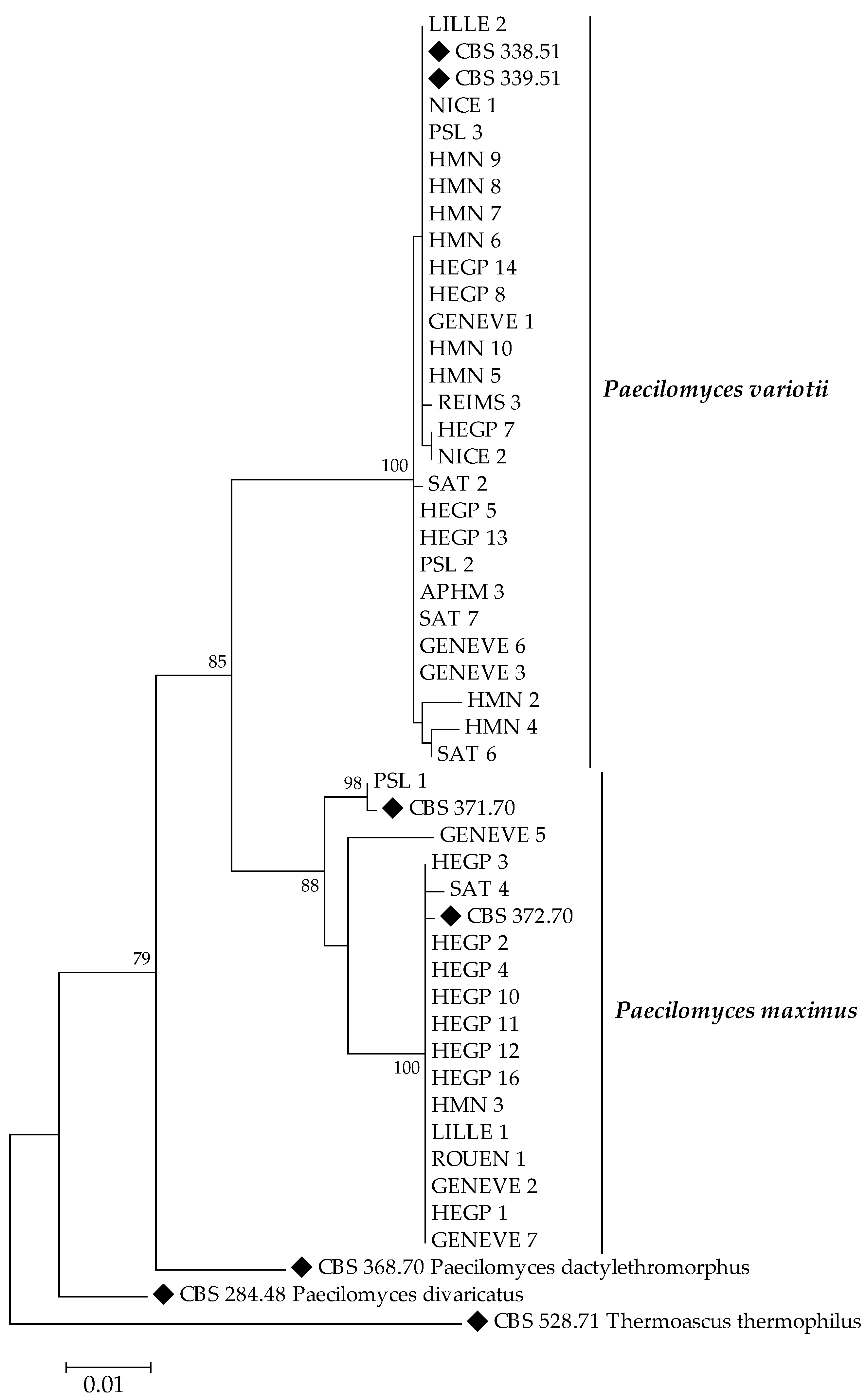
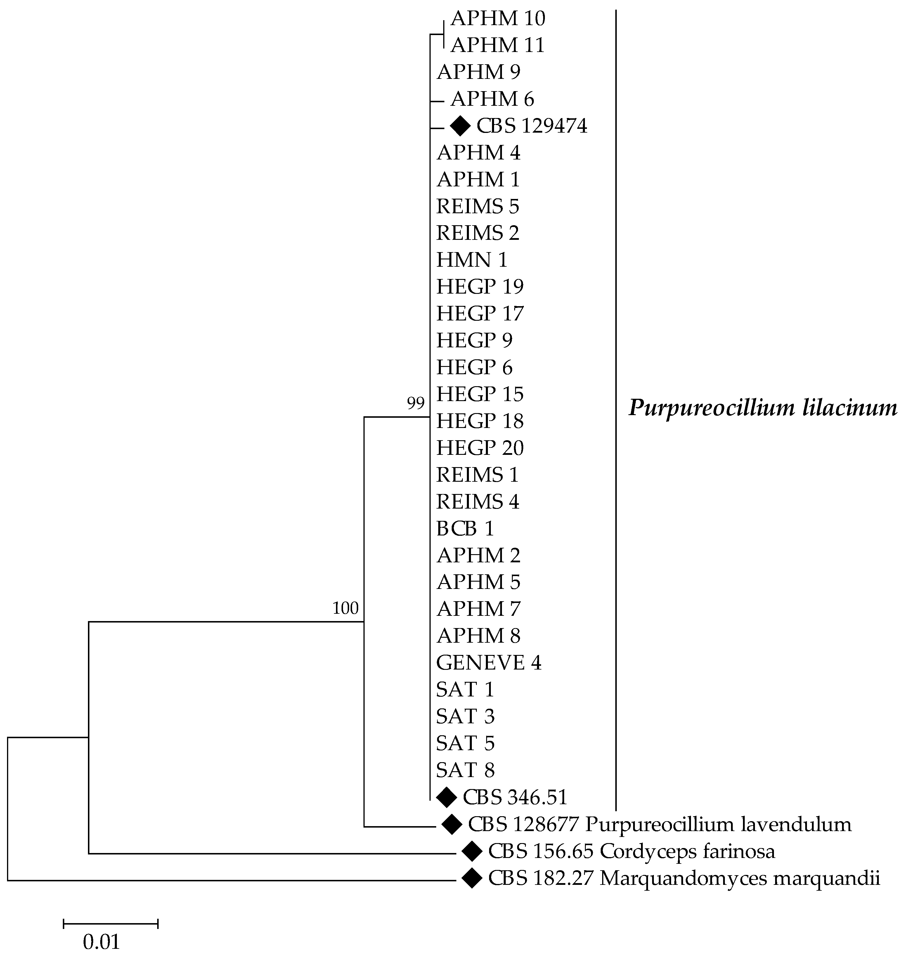
2.3. Molecular Identification and Phylogenetic Analysis

Molecular identification was performed by ITS DNA gene sequencing, including the 5.8S rDNA, and the D1/D2 gene regions, as recommended in previous studies for the identification of Paecylomyces spp. [18]. Whole genomic DNA was extracted from a mature subculture using a QIAamp DNA Mini Kit (Qiagen Sciences Ing., Courtaboeuf, France) after a step of beading in a MagNA Lyser instrument (Roche Diagnostics, Meylan, France). PCRs were performed in a 25 µL-final volume containing 1X HF buffer (ThermoFisher, Les Ulis, France), 100 µM of each deoxynucleosidetriphosphates (dNTPs), 1µM of each primer (i.e., ITS1 forward/ITS4 reverse [19] or D1/D2 NL-1 forward/NL-4 reverse [18]), 3% of DMSO, 1 unit of Phusion™High-Fidelity DNA Polymerase (ThermoFisher), and 50 ng of genomic DNA in a GeneAmp® PCR system 9700 (Applied Biosystems™, Waltham, MA, USA). Sanger sequencing with BigDye ™ Terminator (ThermoFisher scientific) was performed at the Genomic platform of H. Mondor Biomedical Research Institute using the same primer pairs. The obtained sequences were analyzed using Chromas 2.6.6 software (Technelysium Pty Ltd., South Brisbane, Australia) and were compared with reference sequences retrieved from Westerdijk Fungal Biodiversity Institute database (https://wi.knaw.nl/ (accessed on 3 November 2021)) [20]. Genus and species level identifications were attributed using an identity score of ≥99% with respect to a reference entry. Multiple-sequence alignments were performed for each gene in MEGA 7.0.26 software (Auckland, New Zealand) using the CLUSTALW algorithm with manual adjustment. The phylogenetic tree was constructed using Maximum likelihood method and tested with 1000 rapid bootstrap inferences; it included sequences of the reference strain of each species and outgroup species sequences (Table 1).

2.4. Antifungal Susceptibility Testing

In vitro antifungal susceptibility testing was performed following the European Committee for Antimicrobial Susceptibility Testing (EUCAST) microdilution broth reference for filamentous fungi [21]. Eight antifungal agents were tested: amphotericin B (AMB), voriconazole (VRC), itraconazole (ITC), caspofungin (CAS) (Sigma-Aldrich, Saint-Quentin Fallavier, France), posaconazole (PCZ; MSD, Kenilworth, NJ, USA), isavuconazole (ISA; Basilea Pharmaceutica International Ltd., Basel, Switzerland), anidulafungin (AND; Pfizer Pharma New York, NY, USA), and micafungin (MCF; Astellas Pharma Inc., Tokyo, Japan). Each antifungal drug was tested in concentrations ranging from 0.016 to 8 mg/L. Candida parapsilosis ATCC 22019 and Candida krusei ATCC 6258 were included as quality controls. Results were read after 48 h of incubation at 37 °C. For polyenes and azoles AMB, VRC, ITC, PCZ, and ISA, the Minimal Inhibitory Concentrations (MICs) were determined both visually, as the lowest drug concentration leading to a complete inhibition of fungal growth (100% inhibition), and spectrophotometrically by optical density at 550 nm, using a 90% growth inhibition endpoint. On the other hand, for echinocandins CAS, MCF, and AND, the Minimal Effective Concentrations (MECs) were defined as the lowest concentration of the antifungal drug resulting in rounded and compact hyphal growth, compared with the unchanged fungal growth in the control without the antifungal drug.
For calculations, if the upper-limit drug concentration, i.e., 8 mg/L, did not inhibit fungal growth, a higher concentration of 16 mg/L was used.

3. Results

3.1. Isolates and Samples

Seventy clinical isolates of Paecilomyces spp. or Purpureocillium lilacinum taken from cultures of respiratory specimens of patients were retrospectively collected in many French regions and in a Swiss center. Of these isolates, 60% (n = 42) came from five Parisian (APHP) hospitals [Bichat (BCB), Henri Mondor (HMN), Pitie-Salpetriere (PSL), Saint Antoine (SAT), and European Georges Pompidou Hospitals (HEGP)]. The remaining 40% (n = 28) came from hospitals in the North (Lille, Rouen, Reims), and South-East (Saint-Etienne, Nice, Marseille (APHM)) of France, and from Switzerland (Geneva). The respiratory samples from which isolates were extracted included bronchoalveolar lavage fluids (30%, n = 21), bronchial aspirates (34%, n = 24), and sputa (36%, n = 25). None of these isolates were responsible for proven or probable invasive fungal infections. Therefore, they were considered as colonizers.

3.2. Species Identification

The MALDI-TOF MS identified 97% (n = 68/70) of the isolates at species level (identification scores ≥ 20). Species were P. lilacinum (n = 28/68, 41%), P. variotii (n = 25/68, 37%), and P. maximus (n = 15/68, 22%). The remaining two isolates could not be identified correctly at the species level by the MSI 2 database. Indeed, ITS and D1/D2 rDNA genes sequencing identified all isolates to the species level with the same identification. ITS and D1/D2 rDNA genes sequencing identified all 28 isolates of P. lilacinum, as did MALDI-TOF MS. For P. variotii species, one isolate was misidentified by MALDI-TOF MS and confused with P. maximus. For P. maximus species, one isolate was also not identified to species level by MALDI-TOF MS.
Phylogenetic relationships of the isolates and type strains species are illustrated in Figure 1 and Figure 2. Maximum likelihood trees, based on ITS and D1D2 sequences of isolates showed three distinct clades in Paecilomyces maximus.

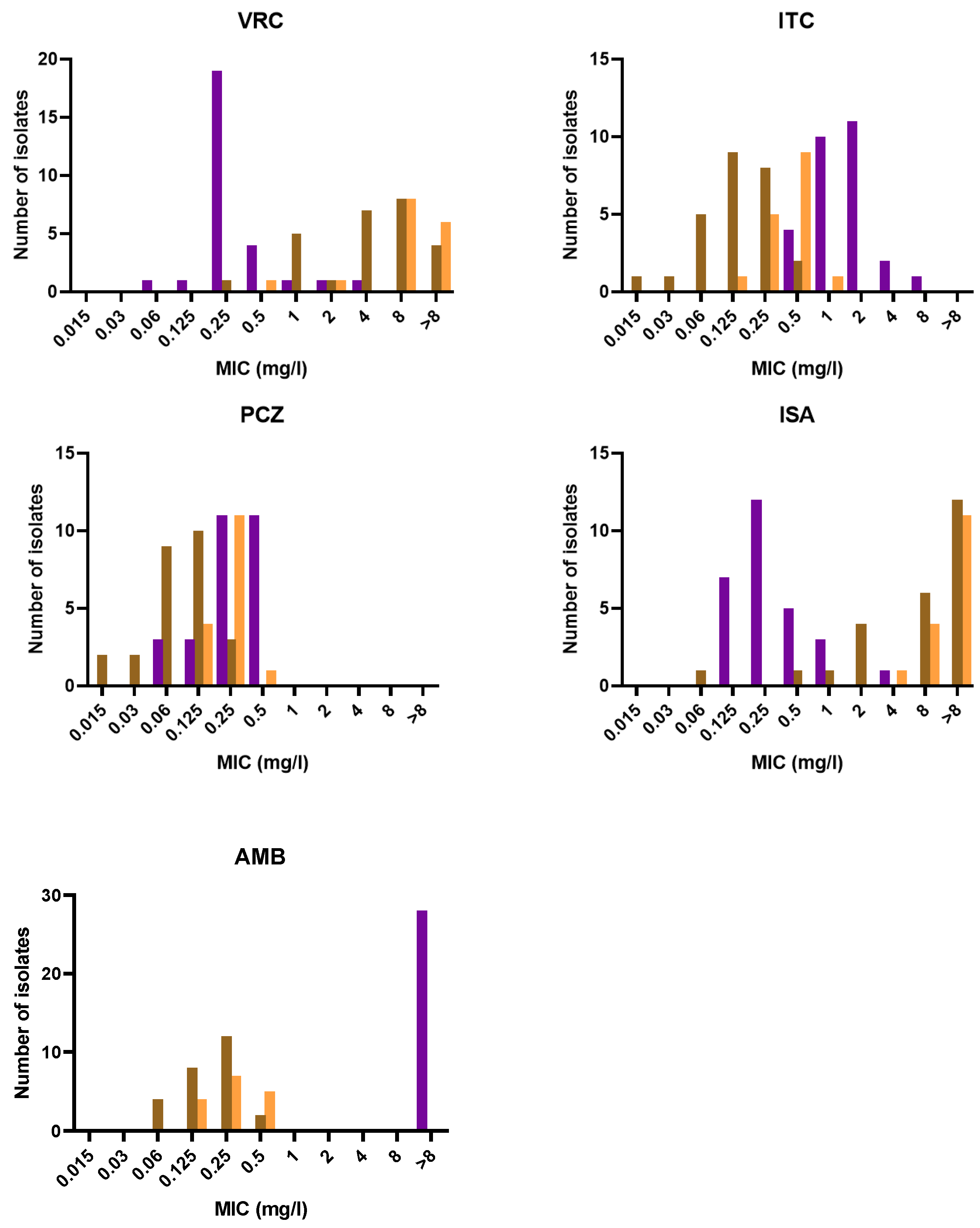
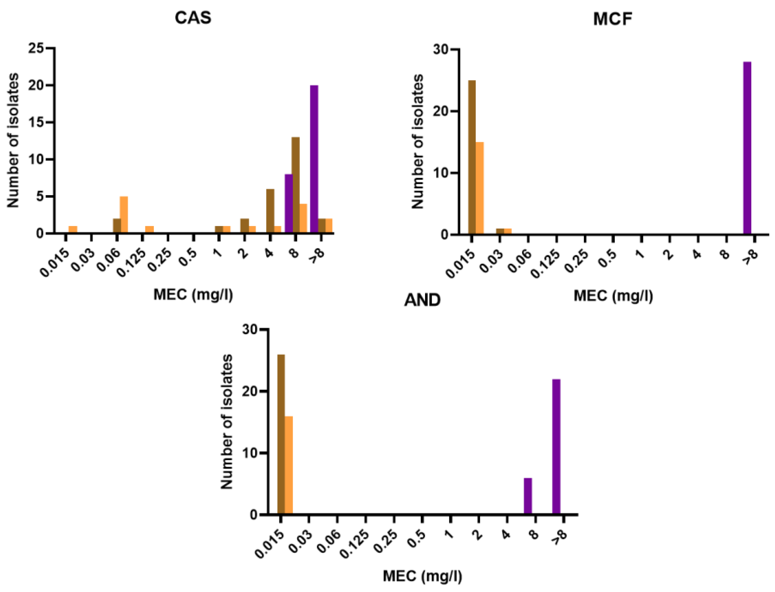
3.3. Antifungal Susceptibility

The distributions of MICs/MECs of the different antifungals for each species are shown in Figure 3 and Figure 4. The results of the antifungal susceptibility testing (range, geometric mean MIC (GMIC), MIC50/MEC50, and MIC90/MEC90) on the 70 isolates are presented in Table 2. All P. lilacinum isolates showed growth inhibition at high MICs of amphotericin B (>8 mg/L) and high MECs of echinocandins (caspofungin, micafungin, and anidulafungin; >8 mg/L). In contrast, P. variotii and P. maximus were inhibited at low MICs of amphotericin B (≤0.5 mg/L) but their response to echinocandins was drug-dependant (Figure S1). For instance, MCF and AND were enough to inhibit both species due to their very low MECs (≤0.06 mg/L), whereas with caspofungin the MECs ranged from 0.01 to 16 mg/L. For azole drugs, the MICs were drug- and species- dependent. Overall, PCZ showed the best in vitro activity against P. lilacinum and Paecilomyces species, with MICs ≤ 0.5 mg/L. VRC and ISA were active in vitro against P. lilacinum (GMICs < 0.5 mg/L) but showed poor in vitro activity against P. variotii and P. maximus with GMICs of >4 mg/L. ITC showed intermediate antifungal activity against P. lilacinum with a GMIC of 1.414 mg/L and lower GMICs for Paecilomyces species.

4. Discussion

In recent years, we have observed the emergence of fungal infections in humans and animals caused by environmental moulds [2,12,22]. Among these moulds, Paecilomyces spp. and Purpureocillium lilacinum stand as ubiquitous, widely present in the environment, and are increasingly detected in respiratory samples of hospitalised patients. Such moulds are listed among the emerging agents that can cause localized fungal colonization, sometimes with other filamentous fungi, as well as infections in immunocompetent patients. Additionally, they can induce invasive infections especially in immunocompromised patients.
As for many filamentous fungi, the taxonomy of Paecilomyces spp. has evolved in recent years thanks to application of molecular methods. Currently, the genus Paecilomyces counts 10 species going from Paecilomyces variotii sensu lato complex (including P. variotii stricto sensu, Paecilomyces brunneolus, P. maximus, Paecilomyces dactylethromorphus, and Paecilomyces divaricatus), Paecilomyces fulvus, Paecilomyces niveus, Paecilomyces tabacinus, Paecilomyces lagunculariae, to Paecilomyces zollerniae [23]. Of the P. variotii sensu lato complex, P. maximus, previously called P. formosus, might represent in itself a complex of at least three genotypes [23]. However, to date, the three cryptic species have not been named yet. Paecilomyces lilacinus is now part of the new genus Purpureocillium which was first introduced by Luanga-Ard et al., in 2011 [11]. At present, the new genus includes five species named P. lilacinum, Purpureocillium lavendulum, Purpureocillium takamizusanense, Purpureocillium roseum, and Purpureocillium atypicola [24,25,26]. The latest taxonomic changes considerably complicate the identification of Paecilomyces spp. or Purpureocillium spp. in routine work of clinical microbiology laboratory. To our knowledge, few studies conducted in the field of clinical mycology have focused on the diversity of Paecilomyces species isolated from human samples, especially in the context of respiratory colonization [13,15,18]. The extremotolerant nature is thought to contribute to the pathogenic potential of these fungi whose presence in clinical samples might be explained by their omnipresence in food and indoor environments. Additionally, being immunodepressed may prompt respiratory colonization to become infection as it is the case with other moulds. In this context, we studied the distribution of Paecilomyces species and determined their in vitro susceptibility to the most common antifungal agents. In our study, P. variotii sensu lato was the most common species (n = 42; 60%) followed by P. lilacinum (n = 28; 40%).
Of the P. variotii sensu lato complex, only P. variotii stricto sensu and P. maximus were identified by both ITS and D1/D2 rDNA genes sequencing. The choice of molecular targets for the identification of Paecilomyces spp. and Purpureocillium spp. was based on the recommendations proposed by the ECMM in cooperation with ISHAM and ASM in 2021 [2].
Our results suggest using MALDI-TOF MS as a good tool to identify Paecilomyces spp. and Purpureocillium lilacinum in routine laboratory work. The concordance between molecular and MALDI-TOF MS scores reached 100% for P. lilacinum, whereas it was 96% for P. variotii stricto sensu and 94% for P. maximus on MSI 2 database platform as compared with the reference sequencing method (Table S1). However, the performance of MALDI-TOF MS for mould identification is database-dependent [18,27]. In 2014, Barker et al. [18] evaluated the performance of MALDI-TOF MS in the identification of 77 genetically confirmed isolates of Paecilomyces species. He showed the interest of using MALDI-TOF reference library for identifications. In their studies, the agreement between the molecular and proteomic methods was 92.2% only after supplementation of MALDI-TOF MS database with type strains.
In our study, MALDI-TOF MS misidentified two isolates, one P. variotii and one P. maximus. With the former, the MSI 2 database was not able to distinguish whether it was P. variotii or P. maximus because the two scores were related, whereas the latter was identified as P. maximus with an MSI 2 score lower than 20%. These results suggest the need for continuous updating of MALDI-TOF MS databases to obtain better performance, i.e., accurate identification of Paecilomyces spp.
Significant differences in in vitro antifungal susceptibility were observed between P. variotii sensu lato and P. lilacinum, which is consistent with the findings of other studies [4,5,6,8,13,14,15]. In general, the antifungal susceptibility profiles of P. variotii stricto sensu, P. maximus, P. dactylethromorphus, and P. divaricatus appeared to be similar to what other studies had already shown, although a limited number of isolates were tested in those studies [13,14,15]. Previous data demonstrated that P. lilacinum had its growth inhibited at higher in vitro MICs/MECs of amphotericin B, itraconazole, and echinocandins as compared with P. variotii sensu lato, which is generally more susceptible. On the contrary, high MICs of voriconazole were needed for P. variotii but not for P. lilacinum [4,5]. In our study, all azoles were active on P. lilacinum, even ITC which showed a wider range of MICs. On the other hand, all P. lilacinum isolates had elevated high MICs of AMB (>8 mg/L), suggesting an intrinsic resistance. These results are particularly interesting since few filamentous fungi are naturally resistant to AMB, a broad-spectrum antifungal agent with fungicidal activity. According to the literature, only a few fungal species are resistant to AMB, such as Aspergillus terreus, Aspergillus tanneri, Fusarium spp. or Lomentospora prolificans, whose molecular resistance mechanism is mostly unknown [28]. However, it is now known that the resistance mechanism of A. terreus to AMB is complex and multifaceted [29,30,31].
P. variotii and P. maximus showed a different susceptibility profile with a GM of MICs/MECs of <0.5 mg/L of AMB, ITC, PCZ, MCF, and AND. The MECs of CAS were variable as we used GMECs of 4.095 mg/L for P. variotii and GMECs of 0.740 mg/L for P. maximus. In contrast, VRC and ISA were not active against P. variotii and P. maximus (GMICs > 4 mg/L). According to our results, Paecilomyces spp. showed resistance to both VRC and ISA. Recently, some studies have shown a resistance to voriconazole in moulds such as Rasamsonia complex or Acremonium spp. [32], and in both, the resistance mechanism is still unknown. These data suggest that azole resistance concern not only Aspergillus spp. but also other environmental moulds. To our knowledge, our study is the first to have investigated the in vitro sensitivity of isavuconazole in P. variotii strains [4]. The mechanism of cross-resistance to voriconazole and isavuconazole in P. variotii and P. maximus remains to be explored.
In conclusion, in the respiratory samples we used in our study, only P. lilacinum, P. variotii stricto sensu, and P. maximus were found. The variability in the observed in vitro susceptibilities to antifungal drugs underlines the importance of precise and correct fungal identification at the species level in order to optimize treatment of Paecilomyces spp. or Purpureocillium lilacinum–related infections. The therapeutic difficulties originate from the in vitro resistance of P. lilacinum to AMB and echinocandins, and from the resistance of P. variotii and P. maximus to azoles. Eventually, MALDI–TOF MS proved to be a rapid and reliable alternative to use in routine identification of Paecilomyces spp. and P. lilacinum in clinical mycology laboratories.

Supplementary Materials

The following supporting information can be downloaded at: https://www.mdpi.com/article/10.3390/jof8070684/s1, Table S1: Concordance between MADLI-TOF and DNA gene sequencing identification; Figure S1: Fungal growth used for MECs determination.

Author Contributions

Conceptualization, F.B. and E.D.; methodology, F.B. and E.D.; software, I.V. and A.-C.N.; data analysis and investigation, L.M., N.A.-A., I.V. and A.-C.N.; resources, J.G., A.R., A.H., C.B., B.S., L.H., H.R., M.D., S.R., C.H., R.P., E.D. and F.B.; writing—original draft preparation, L.M., I.V., N.A.-A. and F.B.; writing—review and editing, L.M., I.V., N.A.-A., E.D. and F.B. All authors have read and agreed to the published version of the manuscript.

Funding

This research received no external funding.

Institutional Review Board Statement

Ethical review and approval were waived for this study as it was retrospectively conducted on isolates collected through routine clinical work.

Informed Consent Statement

Patients’ identifiable information had already been anonymized. No written or verbal informed consent was necessary for patients to participate in this study.

Data Availability Statement

The obtained sequences were submitted to GenBank under accession numbers ON853835 to ON853904 and ON853920 to ON853989 for ITS and D1/D2, respectively.

Acknowledgments

We gratefully acknowledge Suhad Assad for her critical linguistic review, and the genomic platform of Henri Mondor Biomedical Research Institute (Créteil, France), for the Sanger sequencing.

Conflicts of Interest

Over the past five years, E.D. has received research grants from MSD and Gilead; travel grants from Gilead, MSD, Pfizer, and Astellas; and speaker’s fees from Gilead, MSD, and Astellas. F.B. received travel grants from Gilead and Pfizer, and payment for lectures from Gilead.

References

  1. Samson, R. Paecilomyces and some allied hyphomycetes. Stud. Mycol. 1974, 6, 1–119. [Google Scholar]
  2. Hoenigl, M.; Salmanton-García, J.; Walsh, T.J.; Nucci, M.; Neoh, C.F.; Jenks, J.D.; Lackner, M.; Sprute, R.; Al-Hatmi, A.M.S.; Bassetti, M.; et al. Global guideline for the diagnosis and management of rare mould infections: An initiative of the European Confederation of Medical Mycology in cooperation with the Tnternational Society for Human and Animal Mycology and the American Society for Microbiology. Lancet Infect. Dis. 2021, 21, e246–e257. [Google Scholar] [CrossRef] [PubMed]
  3. Eren, D.; Eroglu, E.; Ulu Kilic, A.; Atalay, M.A.; Mumcu, N.; Sipahioglu, M.H.; Canoz, O.; Koc, A.N.; Oymak, O. Cutaneous clcerations caused by Paecilomyces variotii in a renal transplant recipient. Transpl. Infect. Dis. 2018, 20, e12871. [Google Scholar] [CrossRef] [PubMed]
  4. Sprute, R.; Salmanton-García, J.; Sal, E.; Malaj, X.; Falces-Romero, I.; Hatvani, L.; Heinemann, M.; Klimko, N.; López-Soria, L.; Meletiadis, J.; et al. Characterization and outcome of invasive infections due to Paecilomyces variotii: Analysis of patients from the FungiScope® registry and literature reports. J. Antimicrob. Chemother. 2021, 76, 765–774. [Google Scholar] [CrossRef] [PubMed]
  5. Sprute, R.; Salmanton-García, J.; Sal, E.; Malaj, X.; Ráčil, Z.; Ruiz de Alegría Puig, C.; Falces-Romero, I.; Barać, A.; Desoubeaux, G.; Kindo, A.J.; et al. Invasive infections with Purpureocillium lilacinum: Clinical characteristics and outcome of 101 cases from FungiScope® and the literature. J. Antimicrob. Chemother. 2021, 76, 1593–1603. [Google Scholar] [CrossRef]
  6. Pastor, F.J.; Guarro, J. Clinical manifestations, treatment and outcome of Paecilomyces lilacinus infections. Clin. Microbiol. Infect. 2006, 12, 948–960. [Google Scholar] [CrossRef] [Green Version]
  7. Chen, Y.-T.; Yeh, L.-K.; Ma, D.H.K.; Lin, H.-C.; Sun, C.-C.; Tan, H.-Y.; Chen, H.-C.; Chen, S.-Y.; Sun, P.-L.; Hsiao, C.-H. Paecilomyces/Purpureocillium keratitis: A consecutive study with a case series and literature review. Med. Mycol. 2020, 58, 293–299. [Google Scholar] [CrossRef]
  8. Feldman, R.; Cockerham, L.; Buchan, B.W.; Lu, Z.; Huang, A.M. Treatment of Paecilomyces variotii pneumonia with posaconazole: Case report and literature review. Mycoses 2016, 59, 746–750. [Google Scholar] [CrossRef]
  9. Luangsa-ard, J.J.; Hywel-Jones, N.L.; Samson, R.A. The polyphyletic nature of Paecilomyces sensu lato based on 18S-Generated rDNA phylogeny. Mycologia 2004, 96, 773–780. [Google Scholar] [CrossRef]
  10. Samson, R.A.; Houbraken, J.; Varga, J.; Frisvad, J.C. Polyphasic taxonomy of the heat resistant ascomycete genus Byssochlamys and its Paecilomyces anamorphs. Persoonia 2009, 22, 14–27. [Google Scholar] [CrossRef] [Green Version]
  11. Luangsa-Ard, J.; Houbraken, J.; van Doorn, T.; Hong, S.-B.; Borman, A.M.; Hywel-Jones, N.L.; Samson, R.A. Purpureocillium, a new genus for the medically important Paecilomyces lilacinus. FEMS Microbiol. Lett. 2011, 321, 141–149. [Google Scholar] [CrossRef] [PubMed]
  12. Tortorano, A.M.; Richardson, M.; Roilides, E.; van Diepeningen, A.; Caira, M.; Munoz, P.; Johnson, E.; Meletiadis, J.; Pana, Z.-D.; Lackner, M.; et al. ESCMID and ECMM joint guidelines on diagnosis and management of hyalohyphomycosis: Fusarium spp., Scedosporium spp. and others. Clin. Microbiol. Infect. 2014, 20, 27–46. [Google Scholar] [CrossRef] [PubMed] [Green Version]
  13. Castelli, M.V.; Alastruey-Izquierdo, A.; Cuesta, I.; Monzon, A.; Mellado, E.; Rodriguez-Tudela, J.L.; Cuenca-Estrella, M. Susceptibility testing and molecular classification of Paecilomyces spp. Antimicrob. Agents Chemother. 2008, 52, 2926–2928. [Google Scholar] [CrossRef] [PubMed] [Green Version]
  14. Aguilar, C.; Pujol, I.; Sala, J.; Guarro, J. Antifungal susceptibilities of Paecilomyces species. Antimicrob. Agents Chemother. 1998, 42, 1601–1604. [Google Scholar] [CrossRef] [PubMed] [Green Version]
  15. Houbraken, J.; Verweij, P.E.; Rijs, A.J.M.M.; Borman, A.M.; Samson, R.A. Identification of Paecilomyces variotii in clinical samples and settings. J. Clin. Microbiol. 2010, 48, 2754–2761. [Google Scholar] [CrossRef] [Green Version]
  16. MSI 2 Platform Database. Available online: https://msi.happy-dev.fr/ (accessed on 18 March 2022).
  17. Imbert, S.; Normand, A.C.; Gabriel, F.; Cassaing, S.; Bonnal, C.; Costa, D.; Lachaud, L.; Hasseine, L.; Kristensen, L.; Schuttler, C.; et al. Multi-centric evaluation of the online MSI Platform for the identification of cryptic and rare species of Aspergillus by MALDI-TOF. Med. Mycol. 2019, 57, 962–968. [Google Scholar] [CrossRef]
  18. Barker, A.P.; Horan, J.L.; Slechta, E.S.; Alexander, B.D.; Hanson, K.E. Complexities associated with the molecular and proteomic identification of Paecilomyces species in the clinical mycology laboratory. Med. Mycol. 2014, 52, 537–545. [Google Scholar] [CrossRef] [Green Version]
  19. White, T. Amplification and direct sequencing of fungal ribosomal RNA genes for phylogenetics. PCR Protoc. A Guide Methods Appl. 1990, 315–322. [Google Scholar] [CrossRef]
  20. Vu, D.; Groenewald, M.; de Vries, M.; Gehrmann, T.; Stielow, B.; Eberhardt, U.; Al-Hatmi, A.; Groenewald, J.Z.; Cardinali, G.; Houbraken, J.; et al. Large-scale generation and analysis of filamentous fungal DNA barcodes boosts coverage for kingdom fungi and reveals thresholds for fungal species and higher taxon delimitation. Stud. Mycol. 2019, 92, 135–154. [Google Scholar] [CrossRef]
  21. EUCAST Method for Susceptibility Testing of Moulds. Available online: https://www.eucast.org/astoffungi/methodsinantifungalsusceptibilitytesting/ast_of_moulds/ (accessed on 10 October 2021).
  22. Jacobs, S.E.; Wengenack, N.L.; Walsh, T.J. Non-Aspergillus Hyaline Molds: Emerging causes of sino-pulmonary fungal infections and other invasive mycoses. Semin. Respir. Crit. Care Med. 2020, 41, 115–130. [Google Scholar] [CrossRef]
  23. Houbraken, J.; Kocsubé, S.; Visagie, C.M.; Yilmaz, N.; Wang, X.-C.; Meijer, M.; Kraak, B.; Hubka, V.; Bensch, K.; Samson, R.A.; et al. Classification of Aspergillus, Penicillium, Talaromyces and related genera (Eurotiales): An overview of families, genera, subgenera, sections, series and species. Stud. Mycol. 2020, 95, 5–169. [Google Scholar] [CrossRef] [PubMed]
  24. Ban, S.; Azuma, Y.; Sato, H.; Suzuki, K.-I.; Nakagiri, A. Isaria takamizusanensis is the anamorph of Cordyceps ryogamimontana, warranting a new combination, Purpureocillium takamizusanense Comb. Nov. Int. J. Syst. Evol. Microbiol. 2015, 65, 2459–2465. [Google Scholar] [CrossRef] [PubMed]
  25. Perdomo, H.; Cano, J.; Gené, J.; García, D.; Hernández, M.; Guarro, J. Polyphasic analysis of Purpureocillium lilacinum isolates from different origins and proposal of the new species Purpureocillium lavendulum. Mycologia 2013, 105, 151–161. [Google Scholar] [CrossRef] [PubMed]
  26. Calvillo-Medina, R.P.; Ponce-Angulo, D.G.; Raymundo, T.; Müller-Morales, C.A.; Escudero-Leyva, E.; Campos Guillén, J.; Bautista-de Lucio, V.M. Purpureocillium roseum sp. Nov. A new ocular pathogen for Humans and mice resistant to antifungals. Mycoses 2021, 64, 162–173. [Google Scholar] [CrossRef]
  27. Chen, Y.-S.; Liu, Y.-H.; Teng, S.-H.; Liao, C.-H.; Hung, C.-C.; Sheng, W.-H.; Teng, L.-J.; Hsueh, P.-R. Evaluation of the Matrix-Assisted Laser Desorption/Ionization Time-of-Flight mass spectrometry Bruker Biotyper for identification of Penicillium marneffei, Paecilomyces species, Fusarium solani, Rhizopus species, and Pseudallescheria boydii. Front. Microbiol. 2015, 6, 679. [Google Scholar] [CrossRef] [Green Version]
  28. Carolus, H.; Pierson, S.; Lagrou, K.; Van Dijck, P. Amphotericin B and other polyenes-discovery, clinical use, mode of action and drug resistance. J. Fungi 2020, 6, 321. [Google Scholar] [CrossRef] [PubMed]
  29. Blum, G.; Hörtnagl, C.; Jukic, E.; Erbeznik, T.; Pümpel, T.; Dietrich, H.; Nagl, M.; Speth, C.; Rambach, G.; Lass-Flörl, C. New insight into amphotericin B resistance in Aspergillus terreus. Antimicrob. Agents Chemother. 2013, 57, 1583–1588. [Google Scholar] [CrossRef] [PubMed] [Green Version]
  30. Jukic, E.; Blatzer, M.; Posch, W.; Steger, M.; Binder, U.; Lass-Flörl, C.; Wilflingseder, D. Oxidative stress response tips the balance in Aspergillus terreus amphotericin B resistance. Antimicrob. Agents Chemother. 2017, 61, e00670-17. [Google Scholar] [CrossRef] [Green Version]
  31. Posch, W.; Blatzer, M.; Wilflingseder, D.; Lass-Flörl, C. Aspergillus terreus: Novel lessons learned on amphotericin B resistance. Med. Mycol. 2018, 56, S73–S82. [Google Scholar] [CrossRef] [Green Version]
  32. Sharma, C.; Chowdhary, A. Molecular bases of antifungal resistance in filamentous fungi. Int. J. Antimicrob. Agents 2017, 50, 607–616. [Google Scholar] [CrossRef]
Figure 1. Maximum Likelihood tree of Paecilomyces spp. based on combined data set of ITS and D1/D2. Numbers above the nodes represent bootstrapping values generated from 1000 replicates, using a Kimura 2-parameter model. Only values above 70% are indicated. Thermoascus thermophilus CBS 528.71 has been used as the outgroup.
Jof 08 00684 g001
Figure 2. Maximum Likelihood tree of Purpureocillium spp. based on combined data set of ITS and D1/D2. Numbers above the nodes represent bootstrapping values generated from 1000 replicates, using a Kimura 2-parameter model. Only values above 70% are indicated. Cordyceps farinosa CBS 156.65 and Marquandomyces marquandii CBS 182.27 have been used as outgroups.
Jof 08 00684 g002
Figure 3. Distributions of azoles and amphotericin B MICs for P. lilacinum (purple), P. variotii (brown) and P. maximus (orange). VRC: voriconazole; PCZ: posaconazole; ITC: Itraconazole; ISA: Isavuconazole, AMB: amphotericin B.
Jof 08 00684 g003
Figure 4. Distributions of echinocandins MECs for P. lilacinum (purple), P. variotii (brown) and P. maximus (orange) CAS: caspofungin. MCF: micafungin; AND: anidulafungin.
Jof 08 00684 g004
Table 1. Sequences reference used in this study [20].
CBS No Gene Bank Accession N°
SpeciesITS [20]D1D2 [20]
CBS 372.70 T Paecilomyces maximusMH859719.1MH871470.1
CBS 371.70 T Paecilomyces maximusMH859718.1MH871469.1
CBS 339.51 Paecilomyces variotiiMH856887.1MH868409.1
CBS 338.51 Paecilomyces variotiiMH856886.1MH868408.1
CBS 284.48 T Paecilomyces divaricatusMH856344.1MH867896.1
CBS 368.70 Paecilomyces dactylethromorphusMH859715.1MH871467.1
CBS 129474 Purpureocillium lilacinumMH865347.1MH876802.1
CBS 346.51 Purpureocillium lilacinumMH856891.1MH868413.1
CBS 528.71 TThermoascus thermophilusMH860254.1MH872018.1
CBS 128677 TPurpureocillium lavendulumMH864976.1MH876429.1
CBS 182.27 TMarquandomyces marquandiiMH854923.1MH866418.1
CBS 156.65Cordyceps farinosaMH858528.1MH870163.1
T: Type species.
Table 2. Results of in vitro antifungal susceptibility testing on the 70 isolates using the EUCAST method.
Species and DrugRangeGMMIC50/MEC50MIC90/MEC90
(mg/L)(mg/L)(mg/L)(mg/L)
Purpureocillium lilacinum (n = 28)
VRC0.06–40.3200.250.5
ITC0.5–81.41412
PCZ0.06–0.50.2620.250.5
ISA0.125–40.3050.251
CAS8–1613.1251616
MCF16–16161616
AND8–1613.7921616
AMB16–16161616
Paecilomyces variotii stricto sensu (n = 26)
VRC0.25–164.108416
ITC0.015–0.50.1300.1250.25
PCZ0.015–0.250.0800.060.125
ISA0.06–165.499816
CAS0.06–164.09588
MCF0.015–0.030.0150.0150.015
AND0.015–0.0150.0150.0150.015
AMB0.06–0.50.1710.250.25
Paecilomyces maximus (n = 16)
VRC0.5–168.775816
ITC0.125–10.3790.50.5
PCZ0.125–0.50.2280.250.25
ISA4–1612.6991616
CAS0.015–160.74618
MCF0.015–0.060.0160.0150.015
AND0.015–0.0150.0150.0150.015
AMB0.125–0.50.2620.250.5
GM: Geometric Mean; MIC: Minimum Inhibitory Concentration; MEC: Minimum Effective Concentration; VRC: Voriconazole; ITC: Itraconazole; PCZ: Posaconazole; ISA: Isavuconazole; CAS: Caspofungin, MCF: Micafungin, AND: Anidulafungin; AMB: Amphotericin B.
Publisher’s Note: MDPI stays neutral with regard to jurisdictional claims in published maps and institutional affiliations.

