Article

Phylogeny and Diversity of the Genus Pseudohydnum (Auriculariales, Basidiomycota)

1
Institute of Microbiology, School of Ecology and Nature Conservation, Beijing Forestry University, Beijing 100083, China
2
School of Agronomy and Life Sciences, Zhaotong University, Zhaotong 657000, China
3
Tasmanian Institute of Agriculture, Private Bag 98, Hobart, TAS 7001, Australia
4
Manaaki Whenua, Landcare Research, P.O. Box 69040, Lincoln 7608, New Zealand
*
Authors to whom correspondence should be addressed.
J. Fungi 2022, 8(7), 658; https://doi.org/10.3390/jof8070658
Submission received: 18 May 2022 / Revised: 20 June 2022 / Accepted: 21 June 2022 / Published: 23 June 2022
(This article belongs to the Special Issue Women in Mycology)

Abstract

:
The toothed jelly fungus Pseudohydnum gelatinosum was originally described from Europe. The name has a broad sense and the species has been widely reported almost all over the world. We have studied samples of so-called P. gelatinosum from Asia and Oceania. Based on morphology, hosts, geography, and phylogenetic analysis using the internal transcribed spacer regions (ITSs) and the large subunit of nuclear ribosomal RNA gene (nLSU), four new species, P. himalayanum, P. orbiculare, P. sinogelatinosum, and P. tasmanicum, from China, New Zealand, and Australia are described and illustrated, and a new combination, Pseudohydnum totarae, is proposed. The five new taxa can be differentiated by the shape of their basidiomata, pileal surface color when fresh, spine size, basidiospore dimensions, shape of hyphidia, hosts, and biogeography. Phylogenetically, most of these taxa are distantly related, and different base pairs among these taxa mostly account for >2% nucleotides in the ITS regions.

1. Introduction

Pseudohydnum P. Karst. is an easily recognizable genus in the Auriculariales, characterized macroscopically by flabellate or conchate and gelatinous basidiomata with a hydnoid hymenophore, and microscopically by longitudinally cruciate-septate basidia [1,2]. The genus was classified in the Tremellales because of its longitudinally septate basidia [2,3]. Bandoni considered Pseudohydnum a member of the Auriculariales based on micromorphological, ultrastructural, ecological, and developmental data, and this has been confirmed with phylogenetic analysis [4,5,6].
Currently, three species of Pseudohydnum, P. brunneiceps Y.L. Chen et al., P. gelatinosum (Scop.) P. Karst., and P. translucens Lloyd, are accepted [1,7,8]. In addition, two forms and two varieties of P. gelatinosum are recognized: P. gelatinosum f. album (Bres.) Kobayasi and P. gelatinosum f. fuscum (Bres.) Kobayasi from Europe and Japan [9,10], P. gelatinosum var. bisporum Lowy & Courtec. from French Guiana [11], and P. gelatinosum var. paucidentatum Lowy from Bolivia [12,13].
Pseudohydnum guepinioides Rick and P. thelephorum (Lév.) Lloyd were described from Brazil [14] and French Guiana [15], respectively, but both of them are considered synonyms of Trechispora thelephora (Lév.) Ryvarden [16].
The ‘toothed jelly’ fungus Pseudohydnum gelatinosum is the type species of the genus and was originally described from Croatia in Europe as Hydnum gelatinosum Scop. [17]. The name is applied broadly and the species has been widely reported from Europe [3,18,19], North America [20,21,22], Central and South America [2,11,13,23], Asia [24,25,26], and Oceania [27,28,29,30,31]. The fungus is considered to be edible as well as medicinal, with antitumor and antioxidant properties [21,22,32], and a recent study has shown the extract from P. gelatinosum to have antimicrobial activity [33].
Recently, more samples of so-called Pseudohydnum gelatinosum were collected from Asia and Oceania, and the aim of the present study is to clarify the species diversity of Pseudohydnum in the two land masses and outline a phylogeny of Pseudohydnum based on the known data from over the world.

2. Materials and Methods

2.1. Morphological Studies

The studied specimens are deposited in the herbaria of the Institute of Microbiology, Beijing Forestry University (BJFC) and Landcare Research (PDD), New Zealand. Morphological descriptions are based on field notes and voucher specimens. Color terms are from Anonymous [34], Petersen [35], and Kornerup and Wanscher [36]. Microscopic structures refer to Pippola and Kotiranta [37], Malysheva et al. [38], and Fan et al. [39]. Handmade sections of voucher basidiomata were examined using a Nikon Eclipse 80i microscope (magnification ×1000) after being mounted in 5% KOH for five minutes and treated with 1% Phloxine B (C20H4Br4Cl2K2O5). Microscopic structures were photographed using a Nikon Digital Sight DS-L3 or Leica ICC50 HD camera. At least 20 basidia and basidiospores from each specimen were measured. Spore measurements are quoted as means and standard deviations (±1.5 σ) or with 5% of measurements excluded from each end of the range given in parentheses. Stalks were excluded for basidia measurements, and the hilar appendages were excluded for basidiospore measurements. The following abbreviations are used in the descriptions: IKI = Melzer’s reagent, IKI– = neither amyloid nor dextrinoid, CB = Cotton Blue, CB– = acyanophilous in Cotton Blue, L = arithmetic average of all spore lengths, W = arithmetic average of all spore widths, Q = L/W ratios, n (a/b) = number of spores (a) measured from a given number (b) of specimens, and σ = standard deviation from the mean.

2.2. DNA Extraction, Amplification, and Sequencing

A CTAB rapid plant genome extraction kit-DN14 (Aidlab Biotechnologies Co., Ltd., Beijing, China) or DNA easy Plant Mini Kit (Aidlab Biotechnologies, Beijing, China) was used to obtain DNA from dried specimens and perform a polymerase chain reaction (PCR) according to the manufacturer’s instructions, with some modifications [40,41]. Two DNA gene fragments, i.e., the internal transcribed spacer (ITS) and large subunit nuclear ribosomal RNA gene (nLSU), were amplified using the primer pairs ITS5/ITS4 [42] and LR0R/LR7 (https://sites.duke.edu/vilgalyslab/rdna_primers_for_fungi/, accessed on 22 April 2022) [43]. The PCR procedure for ITS was as follows: initial denaturation at 95 °C for 3 min, followed by 35 cycles at 94 °C for 40 s, 54 °C for 45 s, and 72 °C for 1 min and a final extension of 72 °C for 10 min. The PCR procedure for nLSU was as follows: initial denaturation at 94 °C for 1 min, followed by 35 cycles at 94 °C for 30 s, 50 °C for 1 min, and 72 °C for 1.5 min and a final extension of 72 °C for 10 min. DNA sequencing was performed at the Beijing Genomics Institute (BGI) and Manaaki Whenua, Landcare Research, Auckland. All newly generated sequences have been submitted to GenBank and are listed in Table 1.

2.3. Molecular Phylogenetics Analysis

The concatenated ITS + nLSU dataset included 34 ITS sequences and 22 nLSU sequences from 34 samples representing twelve taxa. The dataset had an aligned length of 1942 characters, of which 1521 were constant, 107 were variable and parsimony-uninformative, and 294 were parsimony-informative. MP analysis yield a tree (TL = 690, CI = 0.799, RI = 0.915, RC = 0.721, HI = 0.201). The best model for the concatenated ITS + nLSU dataset estimated and applied in the BI analysis was ‘GTR + F + I + G4’, lset nst = 6, rates = invgamma; prset statefreqpr = dirichlet (1,1,1,1).
The sequences generated for this study were aligned with additional sequences downloaded from GenBank. Both ITS and nLSU sequences were aligned using MAFFT v. 7 online (https://mafft.cbrc.jp/alignment/server/, accessed on 22 April 2022), adjusting the direction of nucleotide sequences according to the first sequence (accurate enough for most cases) and selecting the G-INS-i iterative refinement method [44]. Alignments were manually adjusted to maximum alignment and minimize gaps by BioEdit v. 7.0.9 [45]. A dataset composed of concatenated ITS + nLSU sequences was used to determine the phylogenetic position of the new species. Protomerulius subreflexus (Lloyd) O. Miettinen & Ryvarden and Protomerulius substuppeus (Berk. & Cooke) Ryvarden were selected as the outgroups, following Chen et al. [8].
Maximum Parsimony (MP) analysis was used for the dataset in PAUP* v. 4.0b10 [46]. All characters were equally weighted, and gaps were treated as missing data. A heuristic search option was used to infer the trees with TBR branch swapping and 1000 random sequence additions. All parsimonious trees were saved. The max-trees were set to 5000. Branches were collapsed if the minimum branch length was zero. Clade robustness was assessed using bootstrap analysis with 1000 replicates [47]. Descriptive Tree Statistics Tree Length (TL), Consistency Index (CI), Retention Index (RI), Rescaled Consistency Index (RC), and Homoplasy Index (HI) were calculated for each maximum parsimonious tree.
The CIPRES Science Gateway was used for Maximum Likelihood (ML) analysis based on the dataset in [48]. All parameters in the ML analysis used default settings, and statistical support values were obtained using nonparametric bootstrapping with 1000 replicates.
Bayesian Inference (BI) analysis based on the dataset was carried out using MrBayes v3.2.6 [49]. The best substitution model for the datasets was selected by ModelFinder [50], and trees were sampled every 100 generations. Four Markov chains were run from random starting trees for 2 million generations. Trees were sampled every 1000th generation. The first 25% of sampled trees were discarded as burn-in, whereas other trees were used to construct a 50% majority consensus tree and for calculating Bayesian posterior probabilities (BPPs).

3. Results

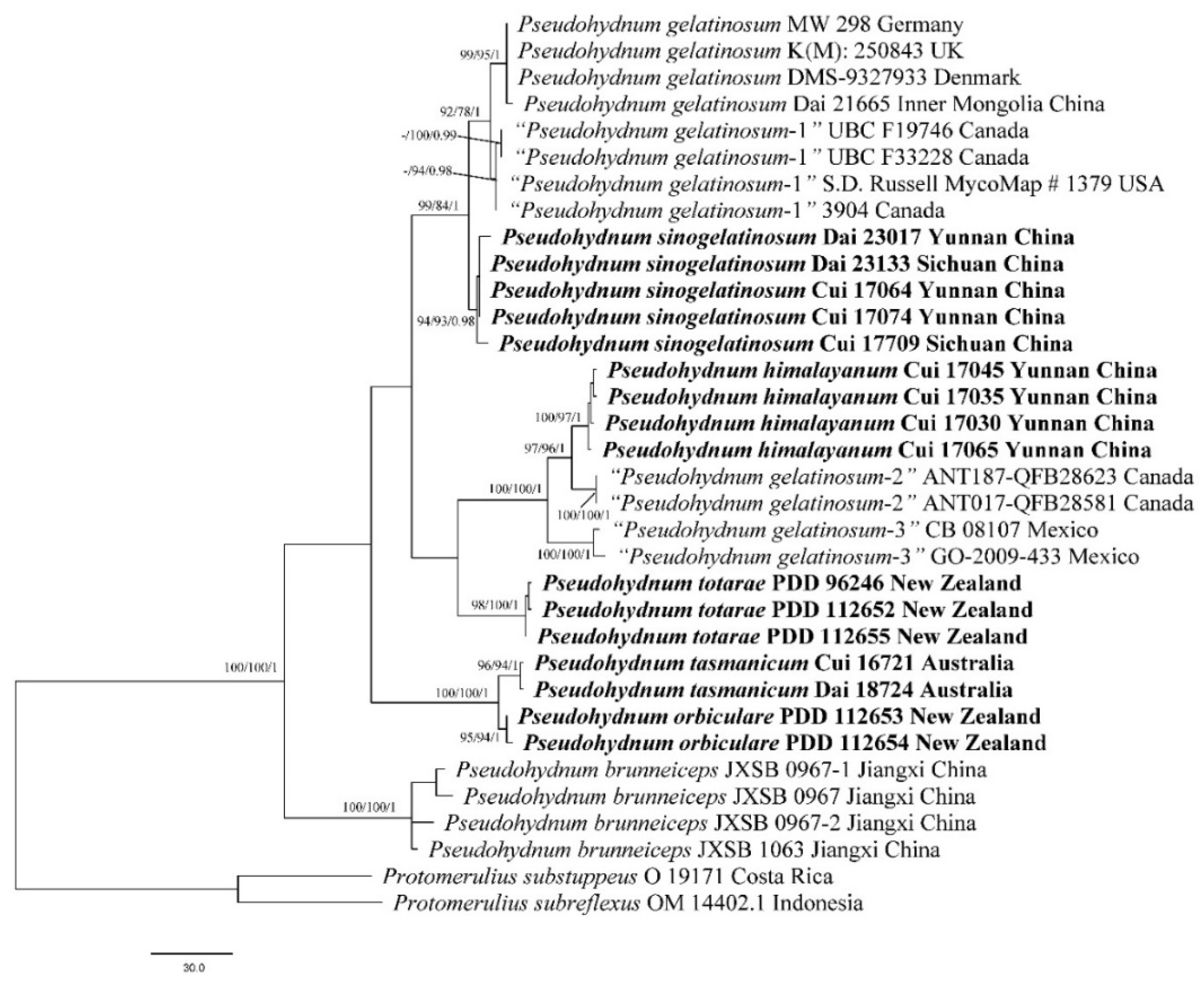
3.1. Molecular Phylogeny
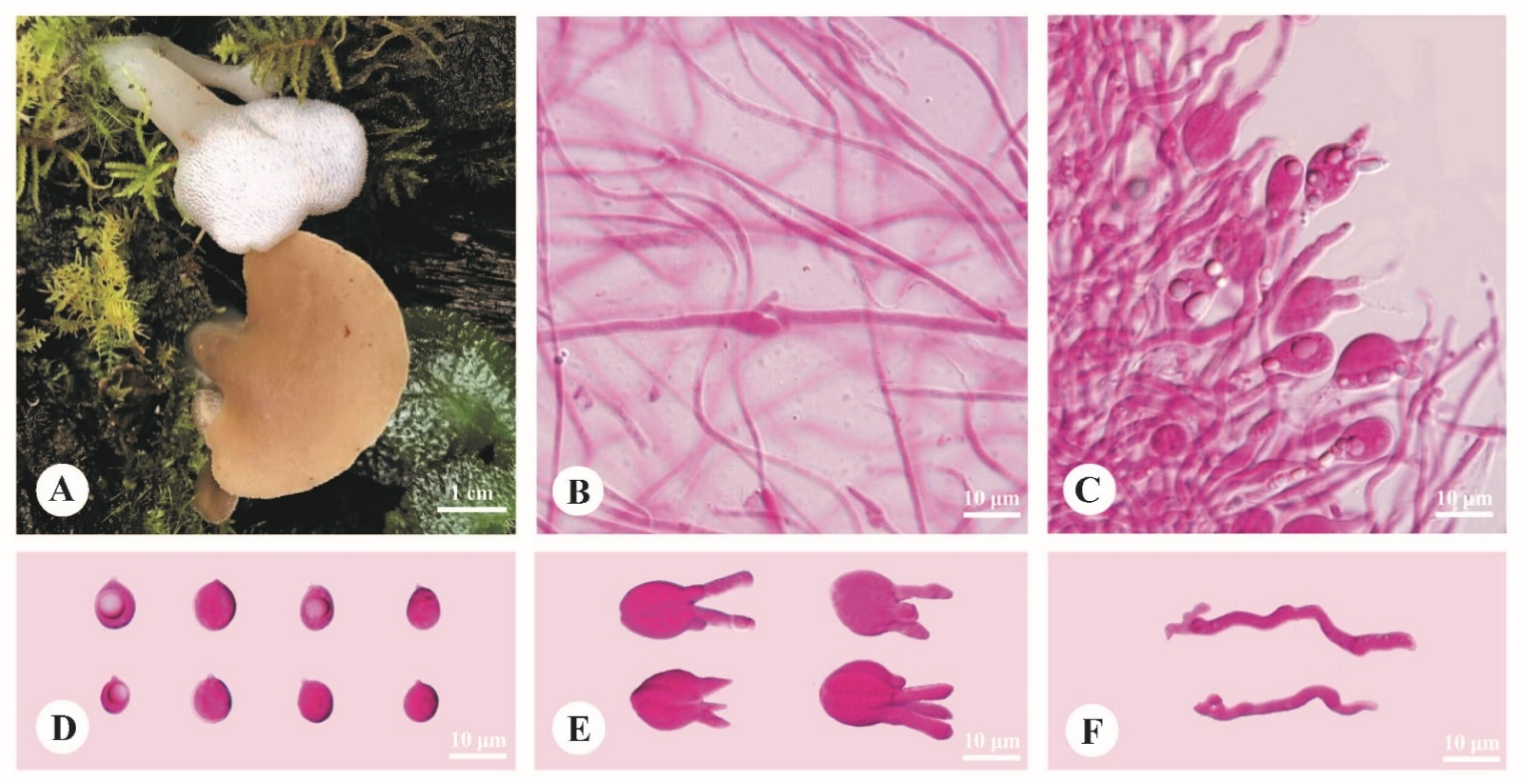
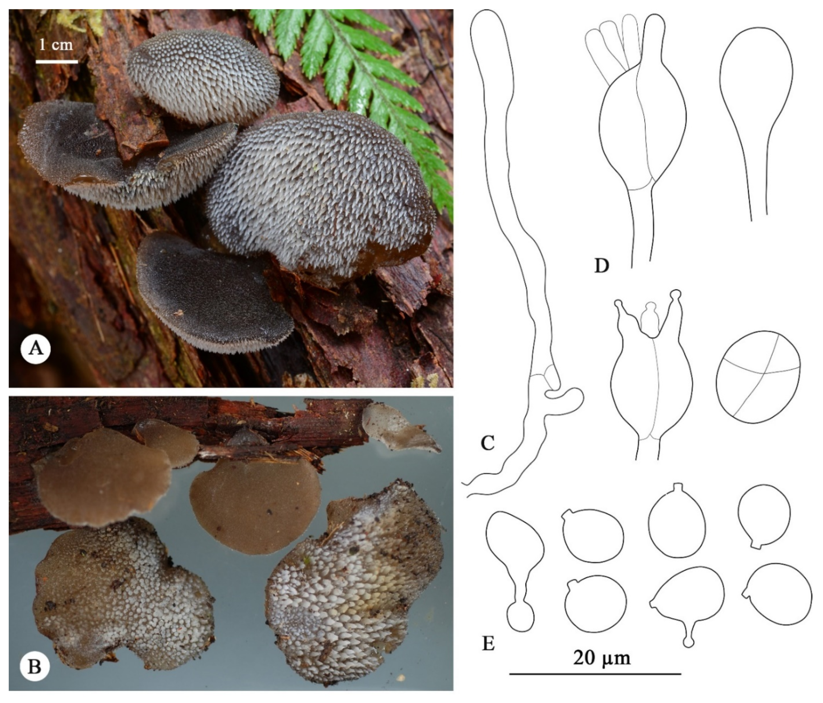
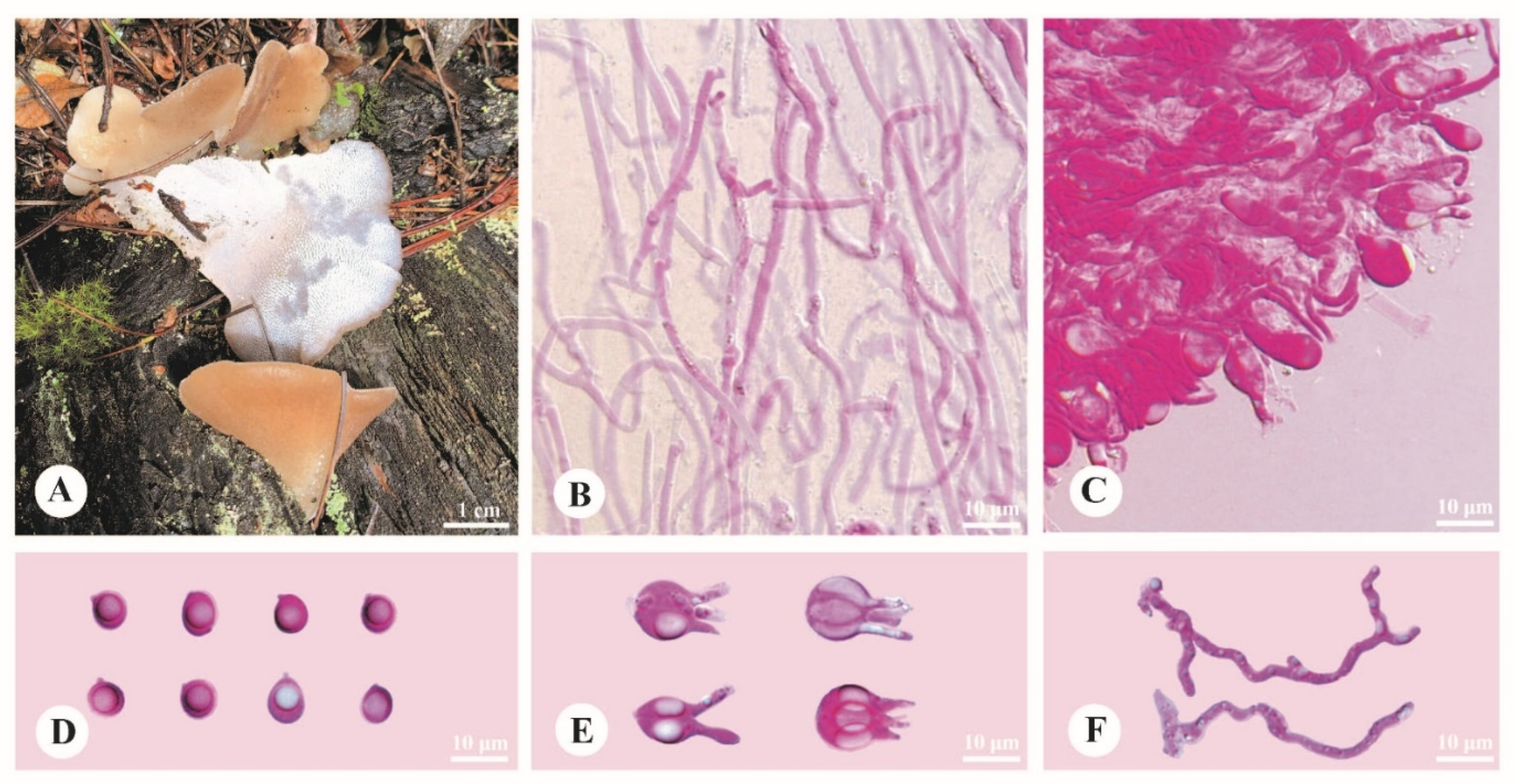
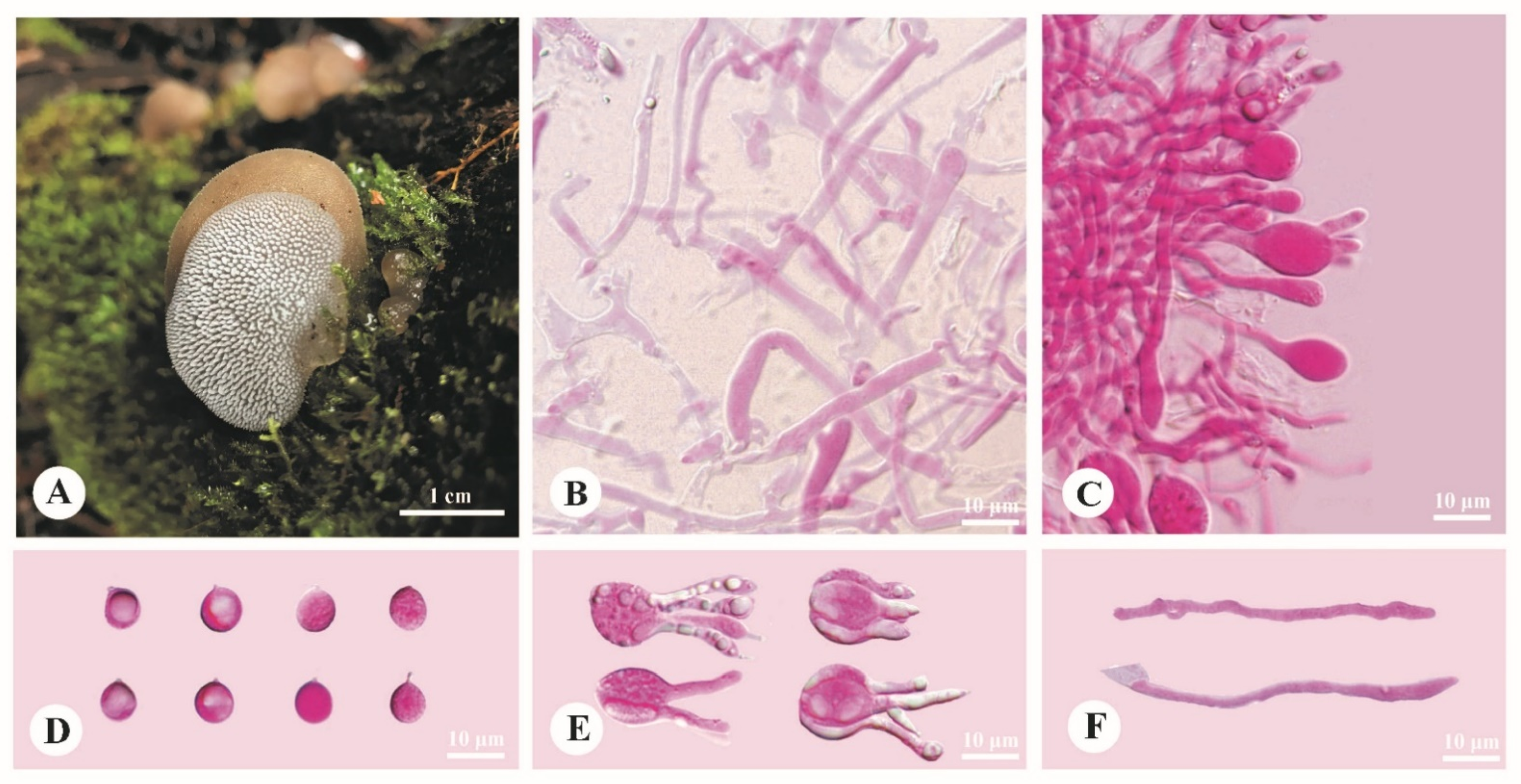
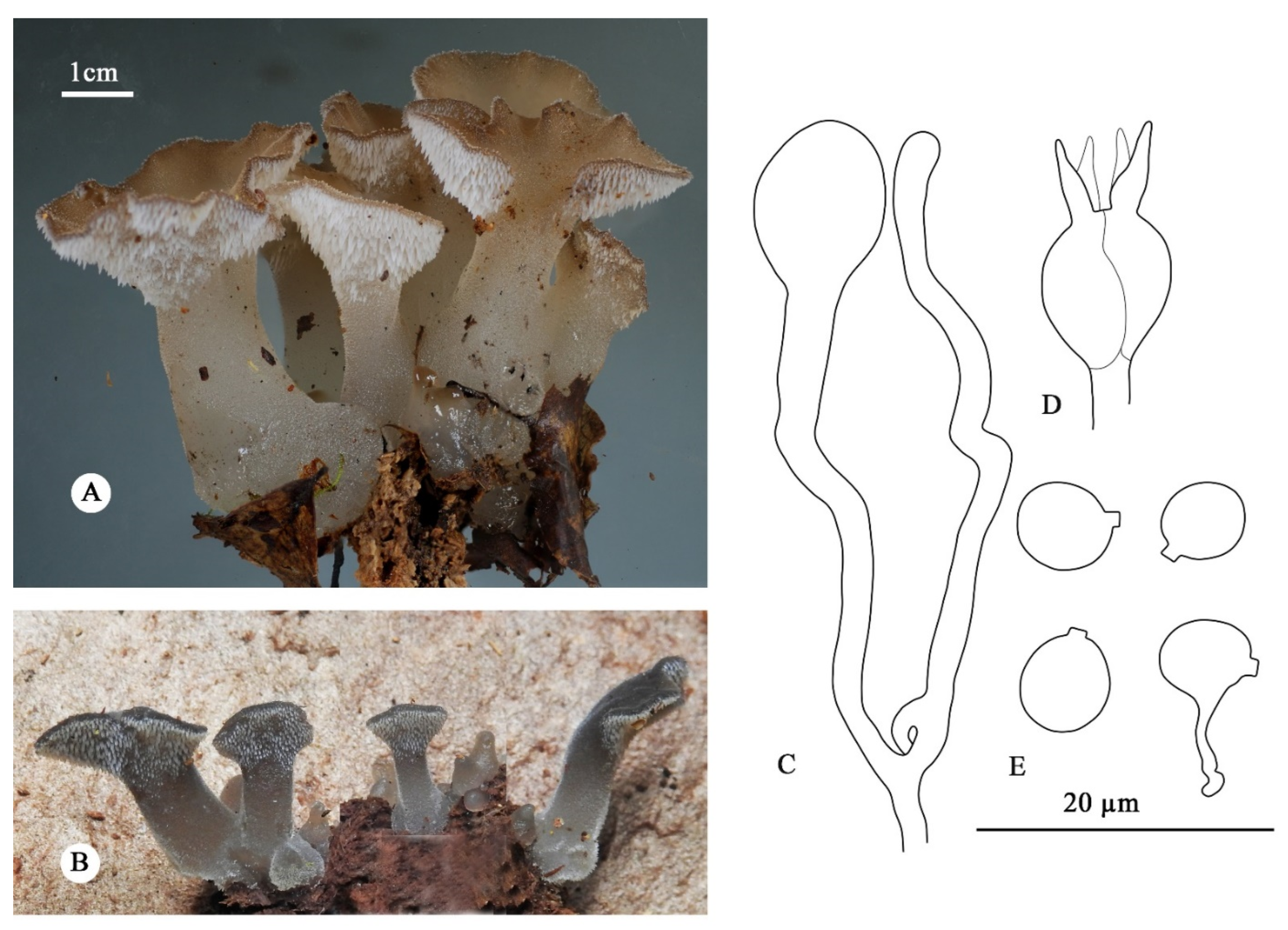
BI analysis yielded a similar topology to MP and ML analysis, with an average standard deviation of split frequencies = 0.004227. Only the MP tree is provided here (Figure 1). Branches that received bootstrap support for MP (MP-BS), ML (ML-BS), and BI (BPP) greater than or equal to 70% (MP-BS and ML-BS) and 0.90 (BPP) were considered as significantly supported, respectively.
The current phylogeny placed all samples of Pseudohydnum in a fully supported clade (Figure 1). Two previously accepted species, P. brunneiceps and P. gelatinosum, received strong support. The samples from North America are treated as “Pseudohydnum gelatinosum-1”, “Pseudohydnum gelatinosum-2”, and “Pseudohydnum gelatinosum-3”. Nine Chinese specimens formed two distinct lineages, and seven Oceanian samples formed three distinct linages nested in Pseudohydnum.

3.2. Taxonomy

Pseudohydnum himalayanum Y.C. Dai, F. Wu & H.M. Zhou, sp. nov. Figure 2.
Mycobank number: 844487.
Type—China. Yunnan Province, Lijiang, Yulong County, Yulong Snow Mountain, on rotten wood of Abies, 27.253333°, 100.174444°, elev. 3200 m, 16 September 2018, Cui 17045 (holotype, BJFC030344).
EtymologyHimalayanum (Lat.): refers to the species being found in the east Himalayan area.
Diagnosis—Differs from other Pseudohydnum species by stipitate and confluent basidiomata, clay-pink to cinnamon pileal surface when fresh, dense spines 5–6 per mm at base, growth on Abies in high altitude (>3000 m), and occurrence in Southwest China.
Basidiomata—Annual, gelatinous when fresh, brittle when dry, with a lateral stipe, occasionally confluent sharing a stipe base. Pilei flabelliform, projecting up to 2.5 cm, 3.8 cm wide and 0.3 mm thick when dry. Pileal surface clay-pink (6D4) to cinnamon (6D6) when fresh, becoming purplish/date-brown when dry. Spines white and conical when fresh, become grayish brown when dry, 5–6 per mm at base, up to 0.2 mm long. Context translucent when fresh. Stipe concolorous with pileal surface, translucent when fresh, up to 2.5 cm long and 3 mm in diam. when dry.
Hyphal structure—Monomitic; generative hyphae with clamp connections. Contextual hyphae hyaline, thin- to slightly thick-walled, frequently branched, interwoven, 5–12 μm in diam. Tramal hyphae hyaline, thin-walled, frequently branched, interwoven, 1.5–2.5 μm in diam. Hyphidia simple. Basidia four-celled, barrel-shaped, ovoid to subglobose or globose, bearing guttules, 12–17.5 × 6–13.5 μm; sterigmata up to 12 μm long and 2–3.5 μm in diam. Probasidia fusiform to lageniform, 18–23 × 8–9 μm. Basidiospores broadly ellipsoid to subglobose, hyaline, thin-walled, occasionally with a guttule, IKI–, CB–, (6.5–)7–8.5 (–9) × (5.5–)6–7.2 (–8) μm, L = 7.75 μm, W = 6.55 μm, Q = 1.14–1.22 (n = 90/3).
Habitat—The species is rather common on rotten wood of Abies at altitudes higher than 3000 m.
Distribution—Distributed in mountain areas with coniferous forests of Southwest China.
Additional specimens examined (paratypes)—China. Tibet, Bomi County, the first tunnel from Bomi to Motuo, on fallen trunk of Abies, 29.783889°, 95.697222°, elev. 3500 m, Dai 23554 (BJFC038126); Yunnan Province, Lijiang, Yulong County, Ninety-nine Longtan, on rotten wood of Abies, elev. 3200 m, 15 September 2018, Cui 17030 (BJFC030329); Yulong Snow Mountain, on rotten wood of Abies, 27.253333°, 100.174444°, elev. 3200 m, 15 September 2018, Cui 17035 (BJFC030334); on fallen trunk of Abies, 16 September 2018, Cui 17065 (BJFC030364).
Pseudohydnum orbiculare J.A. Cooper, sp. nov. Figure 3.
Mycobank number: 844488.
Type—New Zealand. South Island, West Coast Region, Ship Creek, Haast, on an unidentified rotten branch, −43.759431°, 169.149383°, 5 May 2018, NS2608 (holotype, PDD 112653).
EtymologyOrbiculare (Lat.): refers to the species having orbicular basidiomata.
Diagnosis—Differs from other Pseudohydnum species by pileate basidiomata without a well-formed pseudostipe, orbicular pilei, dark-gray to black pileal surface when fresh, hymenophore with sparse spines (0.5–1 per mm at base), growth mostly on angiosperm, and distribution so far restricted to New Zealand.
Basidiomata—Annual, gelatinous when fresh, pileate, not confluent, in grouped tiers. Pilei laterally attached, with a narrow point of attachment, orbicular to dimidiate, up to 50 mm diam., pileal surface white to grayish brown (7E3–9E3) to reddish brown (11E5), smooth to minutely velutinate, with scattered, bluntly conical spines up to 0.2 mm long. Hymenophore white to pale-brown (8B2). Hymenophore spines white, conical, gelatinous, 2–4 mm long, and 1–2 mm wide at the base, 0.5–1 per mm at base. Context 0.5–1.5 mm thick, concolorous with pileal surface.
Hyphal structure—Monomitic; generative hyphae with clamp connections. Contextual and spine tramal hyphae hyaline, thin- to slightly thick-walled, frequently branched, agglutinate,1.5–9 µm in diam. Basidia longitudinally cruciate-septate, 10–14 × 10 µm, with four sterigmata up to 10 × 2 µm. Probasidia lageniform to clavate with stalks 1.5–2 µm in diam. Hyphidia irregular, mostly unbranched, slightly swollen towards apex, clamped at the base. Basidiospores (excluding apiculus) broadly ellipsoid to subglobose, hyaline, thin-walled, occasionally with a guttule, germinating by repetition, IKI–, CB–, 6.5–7.9 × 5.6–6.8 µm, L = 7.2 µm (σ = 0.44), W = 6.2 µm (σ = 0.39), Q = 1.17 (σ = 0.08) (n = 43/2).
Habitat—The species is commonly associated with angiosperm hosts, including southern beech (Nothofagaceae), but several collections were obtained from rotten trunks of Pinus radiata.
Distribution—Distributed throughout New Zealand but perhaps more common in the south.
Additional specimen examined (paratype)—New Zealand. South Island, West Coast Region, Lake Brunner, on an unidentified rotten branch, −42.620192°, 171.501389°,11 May 2018, J. Davies (PDD 112654).
Pseudohydnum sinogelatinosum Y.C. Dai, F. Wu & H.M. Zhou, sp. nov. Figure 4.
Mycobank number: 844489.
Type—China. Yunnan Province, Lijiang, Ninglang County, Luguhu Nature Reserve, on stump of Pinus, 27.522500°, 100.738889°, elev. 3180 m, 9 September 2021, Dai 23017 (holotype, BJFC037590).
EtymologySinogelatinosum (Lat.): refers to the species being similar to Pseudohydnum gelatinosum but occurring in China.
Diagnosis—Differs from other Pseudohydnum species by lateral stipitate basidiomata, pinkish buff to cinnamon-buff pileal surface when fresh, hymenophore with spines 3–4 per mm at base, strongly flexuous and branched hyphidia, growth on gymnosperm wood at high altitudes (>3000 m), and occurrence in Southwest China.
Basidiomata—Annual, gelatinous when fresh, brittle when dry, usually solitary, with a lateral stipe. Pilei applanate, occasionally lobed with an undulating margin, projecting up to 4.7 cm, 2.9 cm wide, 1.2 mm thick when dry. Pileal surface pinkish buff (5A3) to cinnamon-buff (4/5B4) when fresh, becoming milk-coffee to cigar-brown when dry. Spines white and conical when fresh, cream when dry, 3–4 per mm at base, up to 1 mm long when fresh. Context translucent when fresh. Stipe concolorous with pileal surface, shrinking to the base, translucent when fresh, up to 3.2 cm long and 4 mm in diam. when fresh.
Hyphal structure—Monomitic; generative hyphae with clamp connections. Contextual hyphae hyaline, thin- to slightly thick-walled, frequently branched, interwoven, 3–15 μm in diam. Tramal hyphae hyaline, thin-walled, frequently branched, interwoven, 2–3 μm in diam. Hyphidia strongly flexuous, occasionally branched. Basidia occasionally four-celled, barrel-shaped, globose to subglobose or ovoid, bearing guttules, 12–15 × 10–12 μm; sterigmata up to 8.5 μm long and 2.5–3 μm in diam. Probasidia fusiform to lageniform, 16.5–20 × 6.5–9 μm. Basidiospores broadly ellipsoid, ovoid to subglobose, hyaline, thin-walled, with a big guttule, IKI–, CB–, 7–9 (–9.2) × (5.2–)6–7.2 (–8) μm, L = 7.74 μm, W = 6.4 μm, Q = 1.16–1.24 (n = 90/3).
Habitat—The species is rather common on rotten gymnosperm wood at altitudes higher than 3000 m.
Distribution—Distributed in mountain areas with coniferous forests of Southwest China.
Additional specimens examined (paratypes)—China. Sichuan Province, Luding County, Hailuogou Forest Park, on dead tree of Abies, 29.574167°, 101.985556°, elev. 3000 m, 8 October 2021, Dai 23133 (BJFC037704); Jiulong County, Wuxuhai Forest Park, on fallen trunk of Picea, elev. 3200 m, 13 September 2019, Cui 17709 (BJFC034568); Yunnan Province, Lijiang, Yulong Snow Mountain, on fallen trunk of Abies, 27.253889°, 100.174722°, elev. 3200 m, 16 September 2018, Cui 17064 (BJFC030363); on fallen trunk of Picea, 16 September 2018, Cui 17074 (BJFC030373).
Pseudohydnum tasmanicum Y.C. Dai & G.M. Gates, sp. nov. Figure 5.
Mycobank number: 844490.
Type—Australia. Tasmania, the Arve Loop Forest Reserve, on dead tree of Eucalyptus, −43.166667°, 146.808333°, 15 May 2018, Cui 16721 (holotype, BJFC030020).
EtymologyTasmanicum (Lat.): refers to the species being found in Tasmania, Australia.
Diagnosis—Differs from other Pseudohydnum species by pileate basidiomata with a rudimentary stipe base, a vinaceous gray to smoke-gray pileal surface when fresh, hymenophore with spines 2–3 per mm at base, broadly ovoid to subglobose basidiospores, growth on Eucalyptus and Nothofagus, and occurrence in Tasmania, Australia.
Basidiomata—Annual, pileate, with a rudimentary stipe base, a few imbricate, gelatinous when fresh, brittle when dry. Pilei dimidiate, shell-shaped, projecting up to 1.1 cm, 1.8 cm wide and 1.2 mm thick when dry. Pileal surface light vinaceous gray (13B2/3) to smoke-gray (F34) when fresh, become mouse-gray when dry. Spines white and conical when fresh, become buff when dry, 2–3 per mm at base, up to 1 mm long when dry. Context translucent, vinaceous buff when dry.
Hyphal structure—Monomitic; generative hyphae with clamp connections. Contextual hyphae hyaline, thin- to slightly thick-walled, occasionally branched, interwoven, 3–8 μm in diam. Tramal hyphae hyaline, thin-walled, usually branched, interwoven, 2–3 μm in diam. Hyphidia simple, hyaline. Cystidia absent. Basidia frequently four-celled, occasionally two-celled, barrel-shaped, ovoid to subglobose, bearing guttules, 12–15 × 10–11 μm; sterigmata up to 14 μm long and 2–3 μm in diam. Basidiospores broadly ovoid to subglobose, hyaline, thin-walled, occasionally with a guttule, IKI–, CB–, 7.2–9 (–9.2) × (5.7–)6–7.2 (–8) μm, L = 8.19 μm, W = 6.49 μm, Q = 1.23–1.3 (n = 60/2).
Habitat—The species was found on a fallen angiosperm trunk and dead standing angiosperm trees, e.g., Eucalyptus and Nothofagus.
Distribution—So far known from Tasmania, Australia.
Additional specimen examined (paratype)—Australia. Tasmania, Mount Field Forest, close to Mount National Park, on fallen trunk of Nothofagus, −42.683333°, 146.700000°, 14 May 2018, Dai 18724 (BJFC027192).
Pseudohydnum totarae (Lloyd) J.A. Cooper, comb. nov. Figure 6.
Mycobank number: 844491.
BasionymAuricula totarae Lloyd, Mycol. Writ. (Cincinnati) 6 (Letter 62): 935 (1920).
Type—New Zealand. North Island, Wellington Region, Weraroa, on rotting wood of Podocarpus totara, 12 July 1919, G.H. Cunningham (holotype BPI 701378, isotype PDD 225).
EtymologyTotarae (Lat.): refers to the substrate, Podocarpus totara, of the type collection.
Diagnosis—Differs from other Pseudohydnum species by stipitate and confluent basidiomata, spathulate pilei, grayish brown to reddish brown upper surface when fresh, hymenophore with sparse spines (0.8–1.2 per mm at base), growth on gymnosperm wood, and known distribution restricted to New Zealand.
Basidiomata—Annual, gelatinous when fresh, stipitate, solitary to confluent at the base. Pilei spathulate 10–40 mm diam., pileal surface white to grayish brown (7E3–9E3) to reddish brown (11E5), smooth to minutely velutinate, with scattered, bluntly conical spines up to 0.2 mm long. Hymenophore white to pale-brown (8B2). Hymenophore spines white, conical, gelatinous, 1–3 mm long, around 1 mm diam. at base. Context translucent when fresh, 1–2 mm thick, concolorous with pileal surface. Pseudostipe lateral to eccentric, 20–50 mm long, and 8–14 mm in diam., cylindrical, sometimes laterally compressed, sometimes expanding towards the base and apex, concolorous with pileus, surface velutinate with blunt short spines similar to the pileal surface.
Hyphal structure—Monomitic; generative hyphae with clamp connections. Contextual and spine tramal hyphae hyaline, thin- to slightly thick-walled, frequently branched, agglutinate, 2–9 µm in diam. Basidia longitudinally cruciate-septate, 9–13 × 8 µm with four sterigmata up to 12 × 2 µm. Probasidia lageniform to clavate, with stalks 2–3 µm in diam. Hyphidia irregular, mostly unbranched, slightly swollen towards apex, clamped at the base. Basidiospores (excluding apiculus) broadly ellipsoid to subglobose, hyaline, thin-walled, occasionally with a guttule, germinating by repetition, IKI–, CB–, 5.5–6.5 × 4.8–5.7 µm, L = 6 µm, σ = 0.31; W = 5.2 µm, σ = 0.30; Q = 1.17, σ = 0.08 (n = 40/2).
Habitat—Originally described on rotten wood of Totara (Podocarpus totara). The species appears to be commonly associated with decaying gymnosperm wood; other recorded substrates include Kauri (Agathis australis) and Rimu (Dacrydium cupressinum).
Distribution—Throughout New Zealand but more common in the north and west in podocarp forests.
Additional specimens examined—New Zealand. North Island, Northland region, Trounson Kauri Park, on rotten wood of Agathis autralis, −35.72129000°, 173.64801000°, 11 May 2017, J.A. Cooper, JAC14517 (PDD 106891). Waikato Region, Pureora Forest, Rimu Track, on unidentified decorticate branch, −38.58341571°, 175.6337815°, 19 May 2011, J.A. Cooper, JAC11991 (PDD 96246). South Island, West Coast region, Moana, on unidentified rotten wood, −42.319907°, 171.478214°, 9 May 2018, N. Siegel, NS2672 (PDD 112652); Haupiri, on unidentified rotten wood, −42.572551°, 171.886597°, 11 May 2018, S. Carlson (PDD 112655).
Notes—The species was described as Auricula totarae by C.G. Lloyd (USA) in 1920 from material sent to him by C.G. Cunningham in New Zealand. The original description is sparse but refers to the protuberances on the upper surface of the fruiting bodies and the presence of microscopic globose bodies that Lloyd assumed to be basidia. The isotype consists of several small fruiting bodies and fragments. The material appears to be immature, but some fragments do bear the characteristic hymenophore spines of Pseudohydnum as well as the upper surface protuberances mentioned by Lloyd. The contemporary writings of Lloyd indicate that he was familiar with Pseudohydnum gelatinosum and we must assume that his placement of the species in Auricula was due to his having failed to observe the hymenophore spines. No mature basidia or spores were found in the isotype (PDD 225), although the immature globose basidia mentioned by Lloyd were abundant. Several of the fruiting bodies show a well-developed stipe. There is no doubt that this taxon is the same as the one re-described here based on modern taxonomy.

4. Discussion

There is unrecognized cryptic diversity within the genus Pseudohydnum, including three taxa detected from North America in our phylogenetic analysis (Figure 1). The different base pairs among these taxa account for >2% nucleotides in the ITS regions, and these taxa are treated as “Pseudohydnum gelatinosum-1”, “Pseudohydnum gelatinosum-2”, and “Pseudohydnum gelatinosum-3” in our study because we did not study the representative collections.
Pseudohydnum gelatinosum seems to be distributed in Eurasia (Table 1, Figure 1), and it has smaller basidiospores (5–6 × 4.5–5.5 μm) than those in P. himalayanum (7–8.5 × 6–7.2 μm), P. orbiculare (6.5–7.9 × 5.6–6.8 µm), P. sinogelatinosum (7–9 × 6–7.2 μm), and P. tasmanicum (7.2–9 × 6–7.2 μm) [3]. Pseudohydnum gelatinosum resembles P. totarae, sharing similar spore dimensions (5–6 × 4.5–5.5 μm vs. 5.5–6.5 × 4.8–5.7 µm), but the former has dense spines (5–7 per mm at base vs. 0.8–1.2 per mm at base).
Pseudohydnum himalayanum, P. sinogelatinosum, P. orbiculare, and P. tasmanicum have similar basidiospore dimensions. The former two species have dense spines (3–6 per mm at base) and are boreal species found at altitudes higher than 3000 m in Southwest China. The latter two species have sparse spines (0.5–3 per mm at base) and are temperate species found in Oceania.
Pseudohydnum himalayanum and P. sinogelatinosum have an overlapping distribution in Southwest China, but the former has 5–6 spines per mm at base and unbranched hyphidia, while the latter has 3–4 spines per mm at base and branched hyphidia.
Pseudohydnum tasmanicum and P. orbiculare are closely related phylogenetically (Figure 1) and share pileate basidiomata with circular pilei and no stipe, but P. tasmanicum has a distribution in Tasmania, an island in the far south of Australia, while the distribution of P. orbiculare is restricted to New Zealand. In addition, P. tasmanicum has a vinaceous gray to smoke-gray pileal surface, a hymenophore with 2–3 spines per mm at base, while P. orbiculare has a dark-gray, brown to black pileal surface when fresh and a hymenophore with sparse spines (0.5–1 per mm at base).
The two New Zealand species of Pseudohydnum have a similar size and identical range of coloration, from translucent white to gray or brown to reddish brown. They are distinguished by different basidiomata morphology; being spathulate with a pseudostipe in P. totarae, and orbicular to fan-shaped and without a pseudostipe in P. orbiculare. In addition, the former species prefers gymnosperm hosts, especially podocarp wood, and the latter is more common on angiosperm hosts, although the distinction is not absolute because of confirmed records of P. orbiculare on rotten wood of Pinus radiata, which is an exotic species in New Zealand. Pseudohydnum orbiculare is closely related to P. tasmanicum from Australia. ITS sequences show a consistent difference of 1.8%. This pair of species exhibit few or no morphological or ecological differences, which is probably a consequence of a relatively recent dispersal event with subsequent genetic isolation and divergence or maybe when Gondwana split Tasmania off from New Zealand. They are nevertheless good independent phylogenetic species.
Pseudohydnum gelatinosum var. bisporum differs from our new taxa by producing two basidiospores [11,12]. Pseudohydnum gelatinosum var. paucidentatum is different from our new taxa by producing only a few spines [13].
Pseudohydnum brunneiceps differs from our new taxa by its vinaceous brown to fuscous pileal surface when fresh, distinctly confluent basidiomata, and growth on Cryptomeria in a subtropical area of China [8].
Pseudohydnum translucens has pure white basidiomata when soaked and dry and subglobose basidiospores measuring 4–5 μm [7]. Lloyd thought that P. translucens and P. gelatinosum belonged to different genera since P. gelatinosum has a homogeneous texture both in spine and context, while P. translucens has a heterogeneous texture and the hymenial layer is different from the context, although both are white [7]. Anyhow, Pseudohydnum translucens is easily distinguished from our new taxa by its shorter basidiospores (4–5 μm vs. 5.5–9 μm long in our new taxa).
Phylogenetically, Pseudohydnum sinogelatinosum is related to P. gelatinosum and “P. gelatinosum-1 from North America, but P. sinogelatinosum is readily distinguished from P. gelatinosum by bigger basidiospores (7–9 × 6–7.2 μm vs. 5–6 × 4.5–5.5 μm), sparser spines (3–4 per mm at base vs. 5–7 per mm at base), and branched hyphidia (unbranched hyphidia in P. gelatinosum) [3]. We did not study the samples of “P. gelatinosum-1” and cannot comment on them.

Author Contributions

Design of the research: H.-M.Z., Y.-C.D. and J.A.C.; performance of the research: H.-M.Z. and J.A.C.; data analysis and interpretation: H.-M.Z., Y.-C.D., F.W. and J.A.C.; collection of the materials: H.-G.L., H.-M.Z., Y.-C.D., J.A.C. and G.M.G.; writing and revision of the manuscript: Y.-C.D. and F.W. All authors have read and agreed to the published version of the manuscript.

Funding

Hong-Min Zhou, Hong-Gao Li, Fang Wu, and Yu-Cheng Dai are supported by the National Natural Science Foundation of China (project nos. U1802231, 32161143013, and 32070006). Jerry Cooper is supported through the Manaaki Whenua Biota Portfolio, with funding from the Science and Innovation Group of the New Zealand Ministry of Business, Innovation and Employment.

Institutional Review Board Statement

Not applicable.

Informed Consent Statement

Not applicable.

Data Availability Statement

Publicly available datasets were analyzed in this study. The data can be found here: https://www.ncbi.nlm.nih.gov/; https://www.mycobank.org/page/Simple%20names%20search [51,52] (accessed on 22 April 2022).

Conflicts of Interest

The authors declare no conflict of interest.

References

  1. Karsten, P.A. Auriculariei, Clavariei et Tremellini, in paroecia Tammela crescentes. Not. Sällskapets Fauna Flora Fenn. Förhandlingar 1868, 9, 365–374. [Google Scholar]
  2. Niveiro, N.; Popoff, O.F. Pseudohydnum gelatinosum (Tremellales, Basidiomycota) in the Argentinean Yungas. Bol. Soc. Argent. Bot. 2011, 46, 223–226. [Google Scholar]
  3. Breitenbach, J.; Kränzlin, F. Fungi of Switzerland 2. Non-Gilled Fungi, Heterobasidiomycetes, Aphyllophorales, Gasteromycetes; Verlag Mykologia Lucerne Switzerland: Luzern, Switzerland, 1986. [Google Scholar]
  4. Bandoni, R.J. The Tremellales and Auriculariales: An alternative classification. Trans. Mycol. Soc. Jpn. 1984, 25, 489–530. [Google Scholar]
  5. Weiß, M.; Oberwinkler, F. Phylogenetic relationships in Auriculariales and related groups—Hypotheses derived from nuclear ribosomal DNA sequences. Mycol. Res. 2001, 105, 103–415. [Google Scholar] [CrossRef]
  6. Malysheva, V.; Spirin, V. Taxonomy and phylogeny of the Auriculariales (Agaricomycetes, Basidiomycota) with stereoid basidiocarps. Fungal Biol. 2017, 121, 689–715. [Google Scholar] [CrossRef] [Green Version]
  7. Lloyd, C.G. Mycological notes 75. Mycol. Writ. 1925, 7, 1349–1364. [Google Scholar]
  8. Chen, Y.L.; Su, M.S.; Zhang, L.P.; Zou, Q.; Wu, F.; Zeng, N.K.; Liu, M. Pseudohydnum brunneiceps (Auriculariales, Basidiomycota), a new species from Central China. Phytotaxa 2020, 441, 87–94. [Google Scholar] [CrossRef]
  9. Bresadola, G. Iconographia Mycologica; Gruppo Micologico “G. Bresadola”: Trento, Italy, 1932; Volume 23, pp. 1100–1150. [Google Scholar]
  10. Kobayasi, Y. Monographic studies of Japanese Tremellaceous fungi 6. Nagaoa 1954, 4, 36–47. [Google Scholar]
  11. Courtecuisse, R.; Lowy, B. Elements for a mycological inventory of the vicinity of ‘Saut Pararé’ (Arataye River) and ‘Nouragues Inselberg’ (French Guiana) 3. Heterobasidiomycetideae. Studies on the flora of the Guianas 52. Mycotaxon 1990, 39, 329–344. [Google Scholar]
  12. Lowy, B. New or noteworthy Tremellales from Bolivia. Mycologia 1959, 51, 840–850. [Google Scholar] [CrossRef]
  13. Lowy, B. Tremellales. Flora Neotrop. 1971, 6, 1–153. [Google Scholar]
  14. Rick, J.E. Fungi austro-americani exs. Fasc 1. Ann. Mycol. 1904, 2, 406–410. [Google Scholar]
  15. Léveillé, J.H. Champignons exotiques. Ann. Sci. Nat. Bot. 1844, 2, 167–221. [Google Scholar]
  16. Ryvarden, L. A note on the genus Hydnodon Banker. Synop. Fungorum 2002, 15, 31–33. [Google Scholar]
  17. Scopoli, J.A. Flora Carniolica; Impensis Ioannis Pauli Krauss: Vienna, Austria, 1772. [Google Scholar]
  18. Pemberton, R.T. Agglutinins (lectins) from some British higher fungi. Mycol. Res. 1994, 98, 277–290. [Google Scholar] [CrossRef]
  19. Salo, P.; Niemela, T.; Salo, U. Suomen Sieniopas; WSOY: Helsinik, Finland, 2005. [Google Scholar]
  20. Volk, T.J.; Burdsall, H.H.; Reynolds, K. Checklist and host index of wood-inhabiting fungi of Alaska. Mycotaxon 1994, 52, 1–46. [Google Scholar]
  21. Boulet, B. Les Champignons des Arbres de L’est de I’Amérique de Nord; Les Publications du Québec: Québec, QC, Canada, 2003. [Google Scholar]
  22. Binion, D.E.; Stephenson, S.L.; Roody, W.C.; Burdsall, H.H.; Vasilyeva, L.N.; Miller, O.K. Macrofungi Associated with Oaks of Eastern North America; West Virginia University Press: Morgantown, WV, USA, 2008. [Google Scholar]
  23. Kisimova-Horovitz, L.; Oberwinkler, F.; Gómez, L.D. Basidiomicetos resupinados de Costa Rica. Myxariaceaes. Jülich, Sebacinaceae Wells & Oberwy, Tremellodendropsidaceae Jülich. Rev. Biol. Trop. 2000, 48, 519–538. [Google Scholar]
  24. Hongo, T.; Izawa, M. Yama-Kei Field Books No.7; Yama-Kei Publishers Co., Ltd.: Tokyo, Japan, 2006. [Google Scholar]
  25. Dai, Y.C. A revised checklist of corticioid and hydnoid fungi in China for 2010. Mycoscience 2011, 52, 69–79. [Google Scholar] [CrossRef]
  26. Li, Y.; Li, T.H.; Yang, Z.L.; Tolgor, B.; Dai, Y.C. Atlas of Chinese Macrofungal Resources; Center China Farmer Press: Zhengzhou, China, 2015. [Google Scholar]
  27. McNabb, R.F.R. New Zealand Tremellales 1. N. Z. J. Bot. 1964, 2, 403–414. [Google Scholar] [CrossRef]
  28. Fuhrer, B.; Robinson, R. Rainforest Fungi of Tasmania and South-East Australia; CSIRO: Tasmania, Australia, 1992. [Google Scholar]
  29. Hood, I.A. An Illustrated Guide to Fungi on Wood in New Zealand; Auckland University Press: Auckland, New Zealand, 1992. [Google Scholar]
  30. Ridley, G.S.; Horne, D. A Photographic Guide to Mushrooms and Other Fungi of New Zealand; New Holland: Auckland, New Zealand, 2006. [Google Scholar]
  31. Gates, G.; Ratkowsky, D. A Field Guide to Tasmanian Fungi; Tasmanian Field Naturalists Club: Hobart, Australia, 2016. [Google Scholar]
  32. Wu, F.; Zhou, L.W.; Yang, Z.L.; Bau, T.; Li, T.H.; Dai, Y.C. Resource Diversity of Chinese macrofungi: Edible, medicinal and poisonous species. Fungal Divers. 2019, 98, 1–76. [Google Scholar] [CrossRef]
  33. Sternisa, M.; Sabotic, J.; Klancnik, A. A novel approach using growth curve analysis to distinguish between antimicrobial and anti-biofilm activities against Salmonella. Int. J. Food Microbiol. 2022, 364, 109520. [Google Scholar] [CrossRef]
  34. The Royal Botanic Garden Edinburgh. Flora of British Fungi; Colour Identification Chart; Her Majesty’s Stationery Office: London, UK, 1969. [Google Scholar]
  35. Petersen, J.H. Farvekort. The Danish Mycological Society’s Colour-Chart; Foreningen til Svampekundskabens Fremme: Greve, Italy, 1996; pp. 1–6. [Google Scholar]
  36. Kornerup, A.; Wanscher, J.H. Methuen Handbook of Colour, 3rd ed; Eyre Methuen: London, UK, 1978. [Google Scholar]
  37. Pippola, E.; Kotiranta, H. The genus Tremella (Basidiomycota, Tremellales) in Finland. Ann. Bot. Fenn. 2008, 45, 401–434. [Google Scholar] [CrossRef]
  38. Malysheva, V.; Malysheva, E.F.; Bulakh, E.M. The genus Tremella (Tremellales, Basidiomycota) in Russia with description of two new species and proposal of one nomenclatural combination. Phytotaxa 2015, 238, 40–70. [Google Scholar] [CrossRef]
  39. Fan, L.F.; Alvarenga, R.L.M.; Gibertoni, T.B.; Wu, F.; Dai, Y.C. Four new species in the Tremella fibulifera complex (Tremellales, Basidiomycota). MycoKeys 2021, 82, 33–56. [Google Scholar] [CrossRef]
  40. Sun, Y.-F.; Costa-Rezende, D.; Xing, J.-H.; Zhou, J.-L.; Zhang, B.; Gibertoni, T.; Gates, G.; Glen, M.; Dai, Y.-C.; Cui, B.-K. Multi-gene phylogeny and taxonomy of Amauroderma s.lat. (Ganodermataceae). Persoonia 2020, 44, 206–239. [Google Scholar] [CrossRef]
  41. Chen, J.J.; Dai, Y.C. Two new species of Physisporinus (Polyporales, Basidiomycota) from Yunnan, Southwest China. Mycol. Prog. 2021, 20, 1–10. [Google Scholar] [CrossRef]
  42. White, T.J.; Bruns, T.; Lee, S.; Taylor, J. Amplification and Direct Sequencing of Fungal Ribosomal RNA Genes for Phylogenetics. In PCR Protocols: A Guide to Methods and Applications; Innis, M.A., Gelfand, D.H., Sninsky, J.J., White, M.J.T., Eds.; Academic Press: San Diego, FL, USA, 1990; pp. 315–322. [Google Scholar] [CrossRef]
  43. Vilgalys, R.; Hester, M. Rapid genetic identification and mapping of enzymatically amplified ribosomal DNA from several Cryptococcus species. J. Bacteriol. 1990, 172, 4238–4246. [Google Scholar] [CrossRef] [Green Version]
  44. Katoh, K.; Rozewicki, J.; Yamada, K.D. MAFFT online service: Multiple sequence alignment, interactive sequence choice and visualization. Brief. Bioinform. 2017, 20, 1160–1166. [Google Scholar] [CrossRef] [Green Version]
  45. Hall, T.A. Bioedit: A User-Friendly Biological Sequence Alignment Editor and Analysis Program for Windows 95/98/NT. In Nucleic Acids Symposium Series; Information Retrieval Ltd.: London, UK, 1999; Volume 41, pp. 95–98. [Google Scholar] [CrossRef]
  46. Swofford, D.L. PAUP*: Phylogenetic Analysis Using Parsimony (*and Other Methods), version 4.0 Beta; Sinauer Associates: Sunderland, MA, USA, 2002. [Google Scholar]
  47. Felsenstein, J. Confidence intervals on phylogenetics: An approach using the bootstrap. Evolution 1985, 39, 783–791. [Google Scholar] [CrossRef]
  48. Miller, M.A.; Holder, M.T.; Vos, R.; Midford, P.E.; Liebowitz, T.; Chan, L.; Hoover, P.; Warnow, T. The CIPRES Portals. CIPRES. 2009. Available online: http://www.phylo.org/sub_sections/portal (accessed on 4 April 2022).
  49. Ronquist, F.; Teslenko, M.; van der Mark, P.; Ayres, D.L.; Darling, A.; Höhna, S.; Larget, B.; Liu, L.; Suchard, M.A.; Huelsenbeck, J.P. MrBayes 3.2: Efficient Bayesian Phylogenetic Inference and Model Choice across a Large Model Space. Syst. Biol. 2012, 61, 539–542. [Google Scholar] [CrossRef] [Green Version]
  50. Kalyaanamoorthy, S.; Minh, B.Q.; Wong, T.K.F.; Von Haeseler, A.; Jermiin, L.S. ModelFinder: Fast model selection for accurate phylogenetic estimates. Nat. Methods 2017, 14, 587–589. [Google Scholar] [CrossRef] [PubMed] [Green Version]
  51. Sayers, E.W.; Cavanaugh, M.; Clark, K.; Pruitt, K.D.; Schoch, C.L.; Sherry, S.T.; Karsch-Mizrachi, I. GenBank. Nucleic Acids Res. 2021, 49, D92–D96. [Google Scholar] [CrossRef] [PubMed]
  52. Crous, P.W.; Gams, W.; Stalpers, J.A.; Robert, V.; Stegehuis, G. MycoBank: An online initiative to launch mycology into the 21st century. Stud. Mycol. 2004, 50, 19–22. [Google Scholar]
Figure 1. MP strict consensus tree illustrating the phylogeny of Pseudohydnum based on the concatenated ITS + nLSU dataset. Branches are labeled with bootstrap support for MP, ML, and BI results greater than or equal to 70% (MP-BS and ML-BS) and 0.90 (BPP). New taxa are in bold.
Jof 08 00658 g001
Figure 2. Basidiomata and microscopic structures of Pseudohydnum himalayanum (holotype, Cui 17045). (A) Basidiomata. (B) Tramal hyphae. (C) A section of hymenium. (D) Basidiospores. (E) Basidia. (F) Hyphidia.
Jof 08 00658 g002
Figure 3. Basidiomata and microscopic structures of Pseudohydnum orbiculare. (A) Basidiomata (holotype PDD 112653). (B) Basidiomata (PDD 112654). (C) A hyphidium. (D) Basidia. (E) Basidiospores.
Jof 08 00658 g003
Figure 4. Basidiomata and microscopic structures of Pseudohydnum sinogelatinosum (holotype, Dai 23017). (A) Basidiomata. (B) Tramal hyphae. (C) A section of hymenium. (D) Basidiospores. (E) Basidia. (F) Hyphidia.
Jof 08 00658 g004
Figure 5. Basidiomata and microscopic structures of Pseudohydnum tasmanicum (holotype, Cui 16721). (A) Basidiomata. (B) Tramal hyphae. (C) A section of hymenium. (D) Basidiospores. (E) Basidia. (F) Hyphidia.
Jof 08 00658 g005
Figure 6. Basidiomata and microscopic structures of Pseudohydnum totarae. (A) Basidiomata (PDD 112655). (B) Basidiomata (PDD 106891). (C) A probasidium and a hyphidium. (D) A basidum. (E) Basidiospores.
Jof 08 00658 g006
Table 1. Taxa information and GenBank accession numbers of the sequences used in this study.
SpeciesLocationSampleITSnLSU
Pseudohydnum brunneicepsJiangxi, ChinaJXSB 0967MN497254MN497259
Pseudohydnum brunneicepsJiangxi, ChinaJXSB 0967-1MN497255MN497260
Pseudohydnum brunneicepsJiangxi, ChinaJXSB 0967-2MN497256MN497261
Pseudohydnum brunneicepsJiangxi, ChinaJXSB 1063MN497257MN497258
Pseudohydnum gelatinosumInner Mongolia, ChinaDai 21665ON243826ON243924
Pseudohydnum gelatinosumDenmarkDMS-9327933MT644890MT644890
Pseudohydnum gelatinosumGermanyMW 298DQ520094DQ520094
Pseudohydnum gelatinosumUKK(M): 250843MZ159722-
Pseudohydnum gelatinosum-1”Canada3904KM406980-
Pseudohydnum gelatinosum-1”CanadaUBC F19746HQ604801HQ604801
Pseudohydnum gelatinosum-1”CanadaUBC F33228MG953967-
Pseudohydnum gelatinosum-1”USAS.D. Russell MycoMap # 1379MK575262-
Pseudohydnum gelatinosum-2”CanadaANT017-QFB28581MN992496-
Pseudohydnum gelatinosum-2”CanadaANT187-QFB28623MN992495-
Pseudohydnum gelatinosum-3”MexicoCB 08107KT875091-
Pseudohydnum gelatinosum-3”MexicoGO-2009-433KC152166-
Pseudohydnum himalayanumYunnan, ChinaCui 17030ON243827ON243925
Pseudohydnum himalayanumYunnan, ChinaCui 17035ON243828ON243926
Pseudohydnum himalayanumYunnan, ChinaCui 17045ON243829ON243927
Pseudohydnum himalayanumYunnan, ChinaCui 17065ON243830ON243928
Pseudohydnum orbiculareNew ZealandPDD 112653ON243831ON243929
Pseudohydnum orbiculareNew ZealandPDD 112654ON243832-
Pseudohydnum sinogelatinosumYunnan, ChinaCui 17064ON243833-
Pseudohydnum sinogelatinosumYunnan, ChinaCui 17074ON243834ON243930
Pseudohydnum sinogelatinosumSichuan, ChinaCui 17709ON243835ON243931
Pseudohydnum sinogelatinosumYunnan, ChinaDai 23017ON243836ON243932
Pseudohydnum sinogelatinosumSichuan, ChinaDai 23133ON243837ON243933
Pseudohydnum tasmanicumAustraliaCui 16721ON243838ON243934
Pseudohydnum tasmanicumAustraliaDai 18724ON243839ON243935
Pseudohydnum totaraeNew ZealandPDD 96246ON243840-
Pseudohydnum totaraeNew ZealandPDD 112652ON243841-
Pseudohydnum totaraeNew ZealandPDD 112655ON243842ON243936
Protomerulius subreflexusIndonesiaOM 14402.1MG757508MG757508
Protomerulius substuppeusCosta RicaO 19171JX134482JQ764649
New sequences are in bold.
Publisher’s Note: MDPI stays neutral with regard to jurisdictional claims in published maps and institutional affiliations.

