Article

Extracellular Enzymatic Activities of Oceanic Pelagic Fungal Strains and the Influence of Temperature

by
Katherine Salazar Alekseyeva
1,*,
Gerhard J. Herndl
1,2 and
Federico Baltar
1,*
1
Department of Functional and Evolutionary Ecology, University of Vienna, 1030 Vienna, Austria
2
Department of Marine Microbiology and Biogeochemistry, Royal Netherlands Institute for Sea Research (NIOZ), University of Utrecht, 1790 Texel, The Netherlands
*
Authors to whom correspondence should be addressed.
J. Fungi 2022, 8(6), 571; https://doi.org/10.3390/jof8060571
Submission received: 27 April 2022 / Revised: 23 May 2022 / Accepted: 23 May 2022 / Published: 26 May 2022
(This article belongs to the Special Issue Marine Fungus)

Abstract

:
Although terrestrial and aquatic fungi are well-known decomposers of organic matter, the role of marine fungi remains largely unknown. Recent studies based on omics suggest that marine fungi potentially play a major role in elemental cycles. However, there is very limited information on the diversity of extracellular enzymatic activities performed by pelagic fungi in the ocean and how these might be affected by community composition and/or critical environmental parameters such as temperature. In order to obtain information on the potential metabolic activity of marine fungi, extracellular enzymatic activities (EEA) were investigated. Five marine fungal species belonging to the most abundant pelagic phyla (Ascomycota and Basidiomycota) were grown at 5 °C and 20 °C, and fluorogenic enzymatic assays were performed using six substrate analogues for the hydrolysis of carbohydrates (β-glucosidase, β-xylosidase, and N-acetyl-β-D-glucosaminidase), amino acids (leucine aminopeptidase), and of organic phosphorus (alkaline phosphatase) and sulfur compounds (sulfatase). Remarkably, all fungal strains were capable of hydrolyzing all the offered substrates. However, the hydrolysis rate (Vmax) and half-saturation constant (Km) varied among the fungal strains depending on the enzyme type. Temperature had a strong impact on the EEAs, resulting in Q10 values of up to 6.1 and was species and substrate dependent. The observed impact of temperature on fungal EEA suggests that warming of the global ocean might alter the contribution of pelagic fungi in marine biogeochemical cycles.

1. Introduction

Fungi are eukaryotic and osmoheterotrophic organisms depending on organic matter to grow and obtain energy [1]. Osmotrophy involves the secretion of different enzymes to break down complex biological polymers into smaller monomers that can then be taken up through the cell wall [2]. Due to this conversion of organic matter, osmoheterotrophs such as fungi should be major players in the recycling of organic matter. In the marine environment, most of the research on extracellular enzymatic activity (EEA) has been focused on prokaryotes [3,4]. Only a few studies have reported on fungal EEA related to phytoplankton blooms [5] or the degradation of plant-derived matter [6]. As a result, the ecological role of fungi in the biogeochemistry of the oceans, which represents the largest habitat in the Earth’s biosphere, remains poorly known [7]. This contrasts with recent evidence of fungal biomass dominating the bathypelagic marine snow [8]. Additionally, recent studies based on omics suggest that pelagic fungi harbor genes indicative of an active role in marine biogeochemical cycles [9]. It has also been shown that a large variety of carbohydrate active enzymes (CAZymes) are expressed in marine pelagic fungi [10]. Still, there is very limited information on the diversity of EEA in pelagic fungi in the ocean, and how it might be affected by community composition and/or environmental parameters such as temperature.
Approximately 70% of the Earth’s biosphere is composed of persistently cold environments, from the deep sea to polar regions [11,12]. Depending on the optimal growth temperature, organisms living there can be psychrophilic or psychrotrophic [13]. These organisms need to be well adapted to low temperatures, low nutrient availability, and light seasonality [14,15]. Moreover, as low temperatures influence the biochemical reaction rates, organisms must be prepared to overcome those challenges [11].
Here, we investigated the kinetic parameters (Vmax and Km) of the EEA of five oceanic fungal isolates using six different fluorogenic substrate analogues. β-glucosidase and β-xylosidase were used as a proxy for the degradation of plant-derived matter; N-acetyl-β-D-glucosaminidase were used for the utilization of animal and fungal chitinous compounds; alkaline phosphatase and leucine aminopeptidase were used for the cleavage of phosphate moieties and peptides, respectively; and sulfatase was used for the degradation of sulfate esters in macromolecules. We used five fungal strains isolated from the oceanic water column, with two strains belonging to the phylum Ascomycota and three to Basidiomycota. We selected these strains because Ascomycota and Basidiomycota are the most abundant pelagic fungal phyla [9,16,17]. Furthermore, the influence of temperature on fungal EEAs was determined.

2. Materials and Methods

2.1. Culture of Fungi Species

The fungal species Blastobotrys parvus (HA 1620), Metschnikowia australis (HA 635), Rhodotorula sphaerocarpa (HB 738), and Sakaguchia dacryoidea (HB 877) were obtained from the Austrian Center of Biological Resources (ACBR). All these species were isolated from Antarctic Ocean waters at temperatures ranging from -1.24 °C to 5.60 °C [18,19,20,21]. M. australis and R. sphaerocarpa were isolated close to the South Shetland Islands and Marguerite Bay, respectively. The fungus Rhodotorula mucilaginosa was isolated from the Atlantic Ocean at 21.03 °C during the Poseidon cruise on board of RV Sarmiento de Gamboa in March 2019. The maximum temperature growth reported is 25 °C for B. parvus [18] and M. australis [19], and 30 °C for R. mucilaginosa [22], R. sphaerocarpa [21], and S. dacryoidea [20]. In order to have fresh cultures, the pure isolates were cultured on yeast malt extract agar [23,24] for one week. Afterwards, an initial amount of each fungus was diluted in artificial seawater (30 g/L sea salts S9883 Sigma-Aldrich, Vienna, Austria) to obtain an OD660 ≈ 1 [25]. The optical density (OD) was measured with a UV-1800 Shimadzu spectrophotometer. Then, 10 mL of this fungal culture was inoculated into an autoclaved growth medium containing 2 g/L of glucose, malt extract, peptone, and yeast extract; 35 g/L of artificial sea salts (S9883 Sigma-Aldrich); and 0.50 g/L of chloramphenicol. Afterwards, 150 mL of this medium containing fungi was filled in Schott bottles and to compare the effect of temperature, all strains were grown in triplicate at 5 °C and 20 °C on a rotary shaker (Jeio Tech ISS-7100 Incubated Shaker, Daejeon, South Chungcheong, Republic of Korea). The culture growth was tracked daily by OD. Once the exponential phase was reached, bottles with similar OD values were chosen in triplicate for further analysis (EEA and biomass).

2.2. Determining Extracellular Enzymatic Activity and Fungal Biomass

Fluorogenic substrate analogues such as 4-methylumbelliferyl β-D-glucopyranoside (M3633 Sigma-Aldrich), 4-methylumbelliferyl β-D-xylopyranoside (M7008 Sigma-Aldrich), and 4-methylumbelliferyl N-acetyl-β-D-glucosaminide (M2133 Sigma-Aldrich) were used to estimate the potential activity of the enzymes β-glucosidase (BGL), β-xylosidase (BXY), and N-acetyl-β-D-glucosaminidase (NAG), respectively (Table 1). These enzymes can hydrolyze cellulose [26,27], chitin, and xylan [28,29], respectively, thus mediating carbohydrate degradation by marine fungi. The hydrolysis of 4-methylumbelliferyl phosphate (M8883 Sigma-Aldrich) and N-succinyl-Ala-Ala-Pro-Phe-7-amido-4-methylcoumarin (L2145 Sigma-Aldrich) was used to estimate the potential enzymatic activity of alkaline phosphatase (ALP) and leucine aminopeptidase (LAP), respectively. ALP is indicative of the capability of microbes to acquire inorganic phosphorus from organic molecules, and LAP is involved in the hydrolysis of proteins and peptides [30]. Finally, 4-methylumbelliferyl sulfate potassium salt (M7133 Sigma-Aldrich) was used to determine the activity of sulfatase (SUL) degrading sulfate esters in macromolecules. The hydrolysis of these fluorogenic substrates analogues were standardized to the corresponding fluorophores. The fluorophores methylcoumaryl amide (MCA) (A9891 Sigma-Aldrich) and methylumbelliferone (MUF) (M1381 Sigma-Aldrich) were dissolved in 2-methoxyethanol to obtain a final concentration of 100, 50, 10, and 1 μM, and 2000, 1000, 100, and 50 μM.
Sterile microplates of 96 wells with an F bottom and low protein binding (XT64.1, Carl Roth, Karlsruhe, Baden-Wurtemberg, Germany) were used. The standards were distributed to each biological triplicate to establish a standard calibration curve, and each biological triplicate without any addition was used as a blank to determine the background fluorescence of the medium. Serial dilutions of the fluorogenic substrate were established resulting in 12 final concentrations ranging from 100 to 0.05 μM. The fluorescence was measured with a Tecan Infinite 200 PRO at an excitation wavelength of 365 nm and an emission wavelength of 445 nm. An initial measurement was performed, and then, every hour, a measurement was made over a total period of 3 h. Between measurements, the microplates were incubated in the dark at their respective temperature.
For fungal biomass determination, combusted (450 °C; for 6 h) Whatman GF/F filters (WHA1825047 Sigma-Aldrich, 47 mm filter diameter) were individually wrapped in aluminum foil and weighed. Then, 40 mL of each fungal culture triplicate was gently filtered onto a combusted and weighed filter, and was dried at 80 °C for 3 days. Thereafter, the sample was weighed again to determine the fungal biomass as dry weight.

2.3. Determination of the Kinetic Parameters of the Extracellular Enzymatic Activity (EEA)

The increase in fluorescence over time (3 h) was transformed into hydrolysis rate [μmol L−1 h−1] using the equation obtained from the standard calibration lines of MCA and MUF. The resulting hydrolysis rates were fitted directly with the Michaelis–Menten equation using nonlinear least-squares regression analysis with R software [31]. The enzymatic kinetic parameters maximum velocity (Vmax) and half-saturation constant (Km) were calculated. To obtain the biomass-specific activity, the Vmax was normalized to the fungal biomass obtained from the dry weight. Additionally, the Q10 value was calculated to identify the dependence of enzyme activity on temperature [32].

2.4. Statistical Analyses

For the kinetic parameters Vmax and Km, a one-way analysis of variance (ANOVA) was performed to determine differences among species and temperature. Tukey’s Honestly Significant Difference (Tukey’s HSD) was used for a multiple and simultaneous comparison between species and to identify significance at the species level. A Student’s T-test was used to test the normal distribution of the data. A principal component analysis (PCA) was performed to analyze the fungal enzymatic activity by species and substrate. For this purpose, the prcomp command of the R software was used. To maximize the sum of the variance of the squared loadings, Varimax rotation was performed.

3. Results

Remarkably, all the fungal strains were hydrolyzing all the fluorogenic substrates offered (Figure 1 and Figure 2). The kinetic parameters Vmax and Km of the EEA varied, however, among the different fungal strains and substrates (Figure 3 and Figure 4). Additionally, the response of EEA to temperature was species and substrate dependent (Figure 5 and Figure 6).

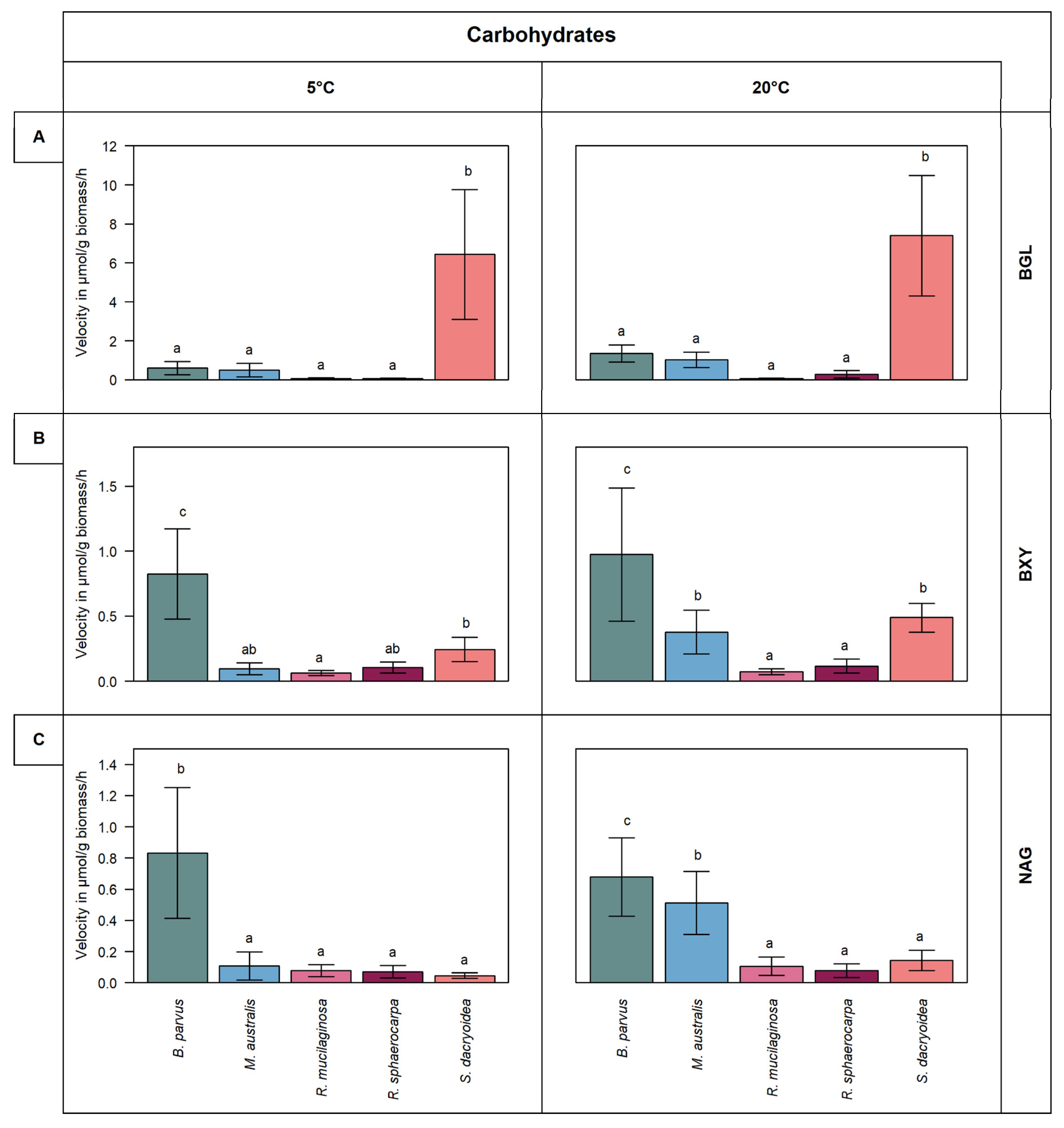
3.1. Carbohydrate Active Enzymes

3.1.1. β-Glucosidase (BGL)

S. dacryoidea exhibited a significantly higher Vmax for BGL than the other fungal strains (t-test; p < 0.001) at 5 °C (6.4 ± 3.2 μmol/g biomass/h) and 20 °C (7.4 ± 3.0 μmol/g biomass/h) (Figure 1A). The other fungal strains exhibited generally low BGL activity, particularly at 5 °C. At 20 °C, the BGL activity was slightly higher than at 5 °C, except for R. mucilaginosa, which maintained a low BGL activity at both temperatures. Consequently, Vmax was a species- and temperature-specific value, with Q10 values varying between 1.4 and 2.4, except for R. mucilaginosa (Figure 5).
The Km was significantly higher in S. dacryoidea than in the other fungal strains (t-test; p < 0.001) and ranged between 139.6 ± 19.1 μM and 78.6 ± 11.9 μM (Figure 3A). Moreover, the Km of S. dacryoidea was significantly higher at 20 °C than at 5 °C (t-test; p < 0.001). Nonetheless, the other two species of the phylum Basidiomycota, R. sphaerocarpa and R. mucilaginosa, showed low Km values, especially the latter. Finally, for the Ascomycota species, the Km was highest at 5 °C (t-test; p = 0.01) with 12.9 ± 3.4 μM for B. parvus and 38.7 ± 14.6 for M. australis.

3.1.2. β-Xylosidase (BXY)

All fungal strains tested were capable to cleave xylose, however, at low rates (Figure 1B). B. parvus exhibited a significantly higher Vmax for BXY than the other strains (t-test; p < 0.001). In B. parvus, the Vmax was 0.8 ± 0.3 μmol/g biomass/h at 5 °C and 1.0 ± 0.5 μmol/g biomass/h at 20 °C. In the other strains, the hydrolysis rates were higher at 20 °C than at 5 °C, amounting to 0.5 and 0.1 μmol/g biomass/h, respectively. When all the fungal species were compared, the temperature had a greater effect in M. australis and S. dacryoidea with Q10 values of 2.1 and 1.8, respectively, than in the other fungal strains (Figure 5). For B. parvus and both species of the genus Rhodotorula, Q10 values close to 1 suggested that Vmax was independent of the temperature.
The Km varied significantly between both temperatures (t-test; p = 0.90) (Figure 3B). At 5 °C, S. dacryoidea showed a high Km (49.7 ± 27.6 μM) followed by B. parvus with a Km value of 37.2 ± 8.5 μM. In contrast, at 20 °C, the Km decreased in both B. parvus and S. dacryoidea but increased in M. australis. The other two species, R. sphaerocarpa and R. mucilaginosa, showed low Km values corresponding to their low Vmax.

3.1.3. N-acetyl-β-D-glucosaminidase (NAG)

B. parvus exhibited similar NAG hydrolysis rates at 5 °C and 20 °C (t-test; p = 0.39) (Figure 1C). At 5 °C, the Vmax was 0.8 ± 0.4 μmol/g biomass/h and at 20 °C 0.7 ± 0.2 μmol/g biomass/h. Although the other strains exhibited low NAG enzymatic activity, it increased from 5 °C to 20 °C, particularly in M. australis and S. dacryoidea. In M. australis the Vmax increased from 0.1 ± 0.08 μmol/g biomass/h to 0.5 ± 0.2 μmol/g biomass/h and in S. dacryoidea from 0.04 ± 0.02 μmol/g biomass/h to 0.1 ± 0.06 μmol/g biomass/h. The Q10 values for NAG in M. australis and S. dacryoidea were 2.1 and 1.1, respectively (Figure 5).
The high NAG enzymatic activity detected in B. parvus coincided with a high Km (Figure 3C). At 5 °C, the Km was 2.9 ± 1.8 μM, while at 20 °C it was 12.1 ± 5.6 μM. At 20 °C, the Km of M. australis increased to 8.7 ± 4.6 μM. Thus, the Km values of these two Ascomycota species increased with temperature, but they remained low in the three Basidiomycota species.

3.2. Extracellular Enzymes Targeting Proteins, Phosphorus, and Sulfur Compounds

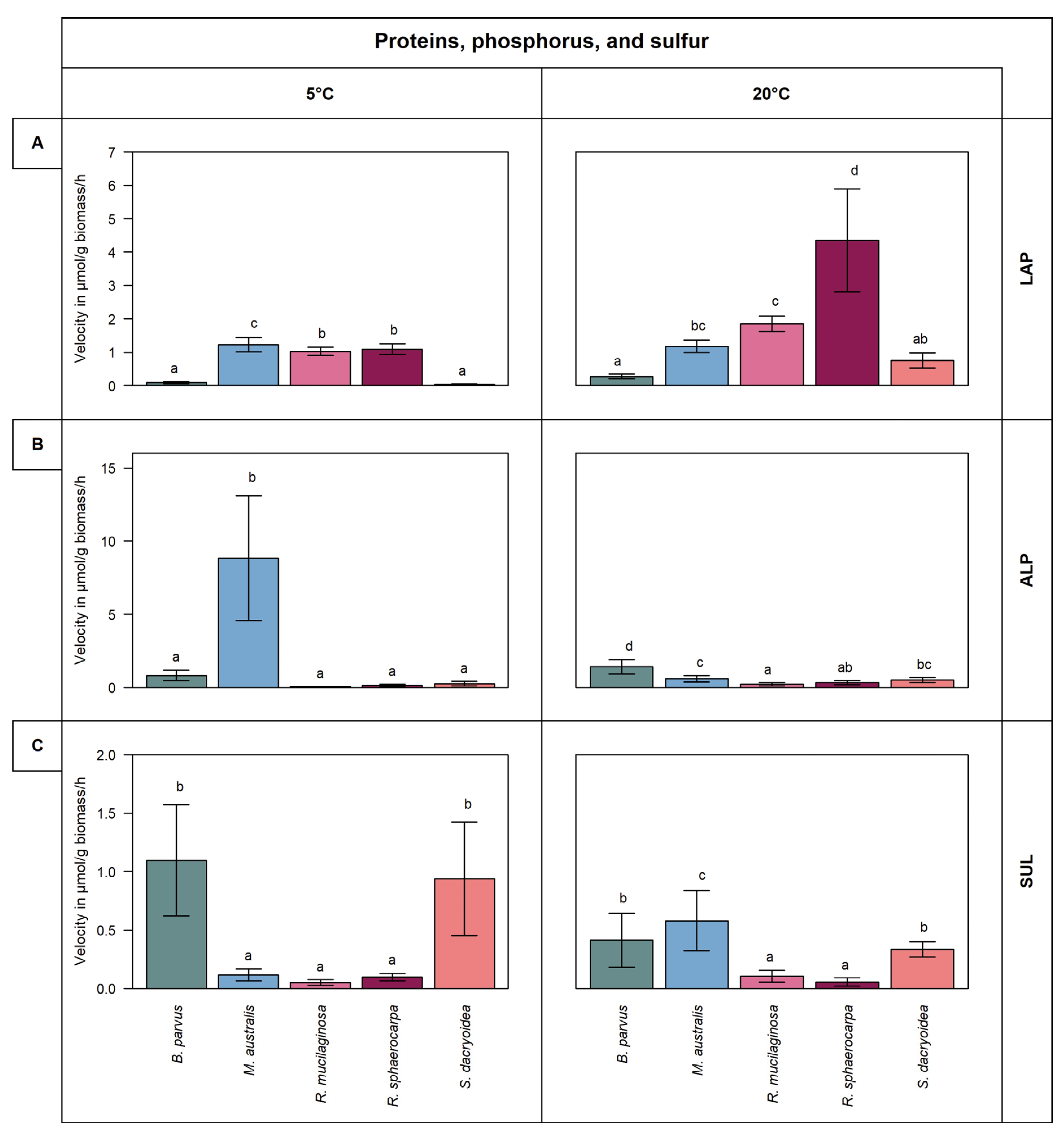
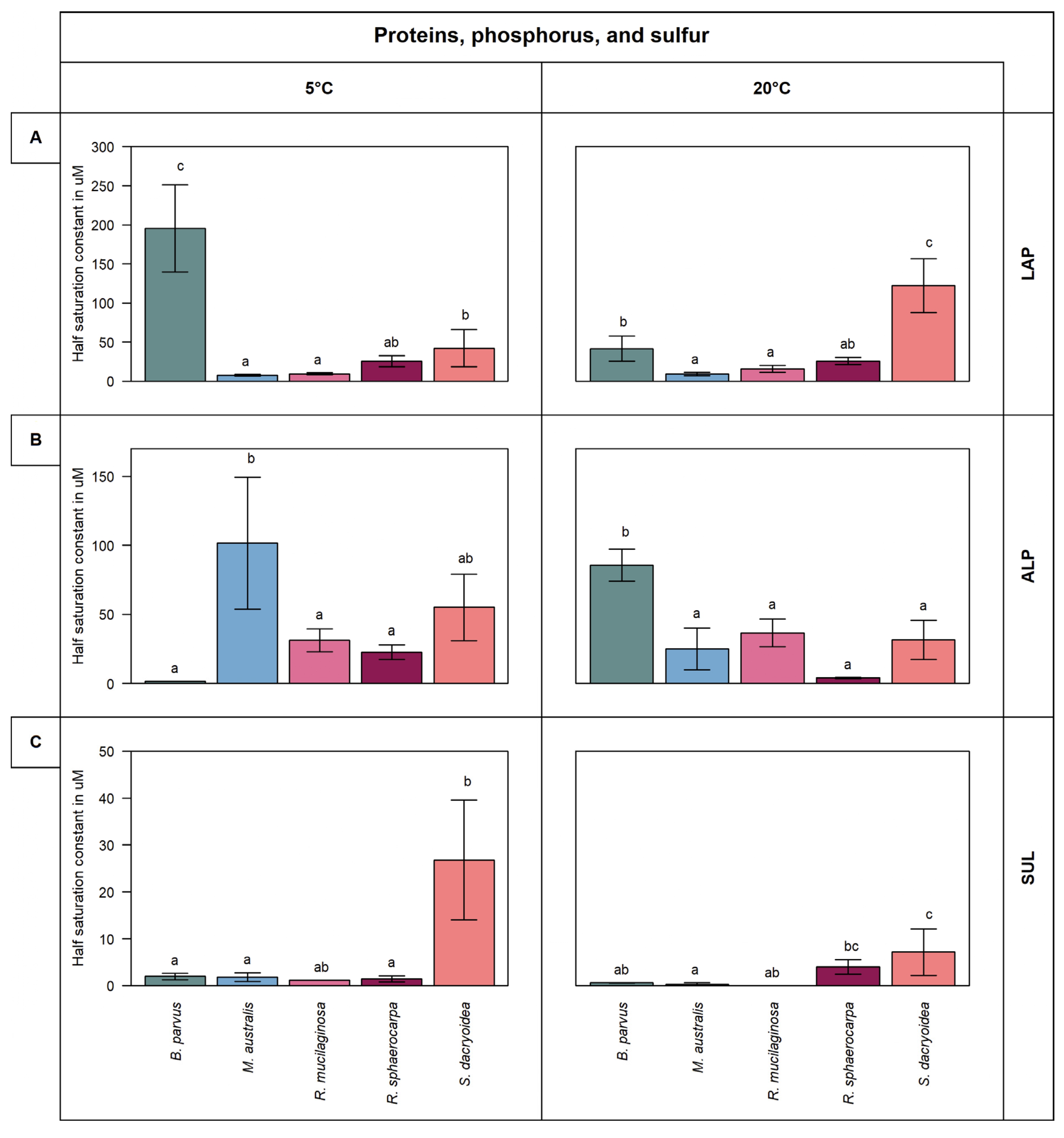
3.2.1. Leucine Aminopeptidase (LAP)

Generally, the LAP activity was higher at 20 °C than at 5 °C in all the fungal strains (t-test; p < 0.001), except in M. australis (t-test; p = 0.46) (Figure 2A). The LAP activity in R. sphaerocarpa at 20 °C was 4.3 ± 1.5 μmol/g biomass/h; however, it was only 1.1 ± 0.2 μmol/g biomass/h (t-test; p < 0.001) at 5 °C resulting in a Q10 of 6.1 (Figure 5). All the other fungal strains had Q10 values ranging from 1.9 to 1.1. Thus, the LAP activity was the only extracellular enzymatic activity tested that showed a temperature dependency in all the fungal strains (Figure 5).
B. parvus and S. dacryoidea exhibited significantly higher Km values than the other fungal strains (t-test; p < 0.001) (Figure 4A). The Km values varied between 195.4 μM and 41.6 μM in B. parvus and between 122.3 μM and 42.2 μM in S. dacryoidea. While in B. parvus, the Km decreased with increasing temperature, and in S. dacryoidea, the Km value incremented with increasing temperature.

3.2.2. Alkaline Phosphatase (ALP)

At 5 °C, M. australis exhibited a significantly higher Vmax (8.8 ± 3.7 μmol/g biomass/h) than the other fungal isolates (t-test; p = 0.03) (Figure 2B). At 20 °C, however, the Vmax was about 10-fold lower than at 5 °C (0.6 ± 0.2 μmol/g biomass/h). In contrast, in all the other fungal strains, Vmax slightly increased with temperature (Figure 2B). The Q10 values ranging from 2.6 to 1.0 indicated a moderate temperature dependency of ALP in all the fungal strains examined, except in M. australis (Figure 5).
The Km was significantly higher in the Ascomycota than in the Basidiomycota strains (t-test; p = 0.01) (Figure 4B). M. australis exhibited a higher Km at 5 °C (101.5 ± 43.5 μM) than at 20 °C, whereas the Km in B. parvus was higher at 20 °C (85.7 ± 10.5 μM) than at 5 °C. The other fungal species belonging to the Basidiomycota phylum exhibited Km values ranging from 55.1 μM to 4.1 μM (Figure 4B).

3.2.3. Sulfatase (SUL)

Sulfatase (SUL) activity was generally low compared to the other extracellular enzymatic activities (Figure 2C). Higher Vmax values were determined for B. parvus and S. dacryoidea at 5 °C (1.1 and 0.9 μmol/g biomass/h, respectively) (t-test; p = 0.01) than at 20 °C (0.4 and 0.3 μmol/g biomass/h, respectively). In contrast, for M. australis, R. mucilaginosa, and R. sphaerocarpa, the SUL activity increased with temperature. The Q10 value in M. australis was 1.9 while all the other fungal strains were <1.0 (Figure 5). With the exception of R. sphaerocarpa, all the fungal strains exhibited higher Km values at 5 °C than at 20 °C (t-test; p ≤ 0.002) (Figure 4C).

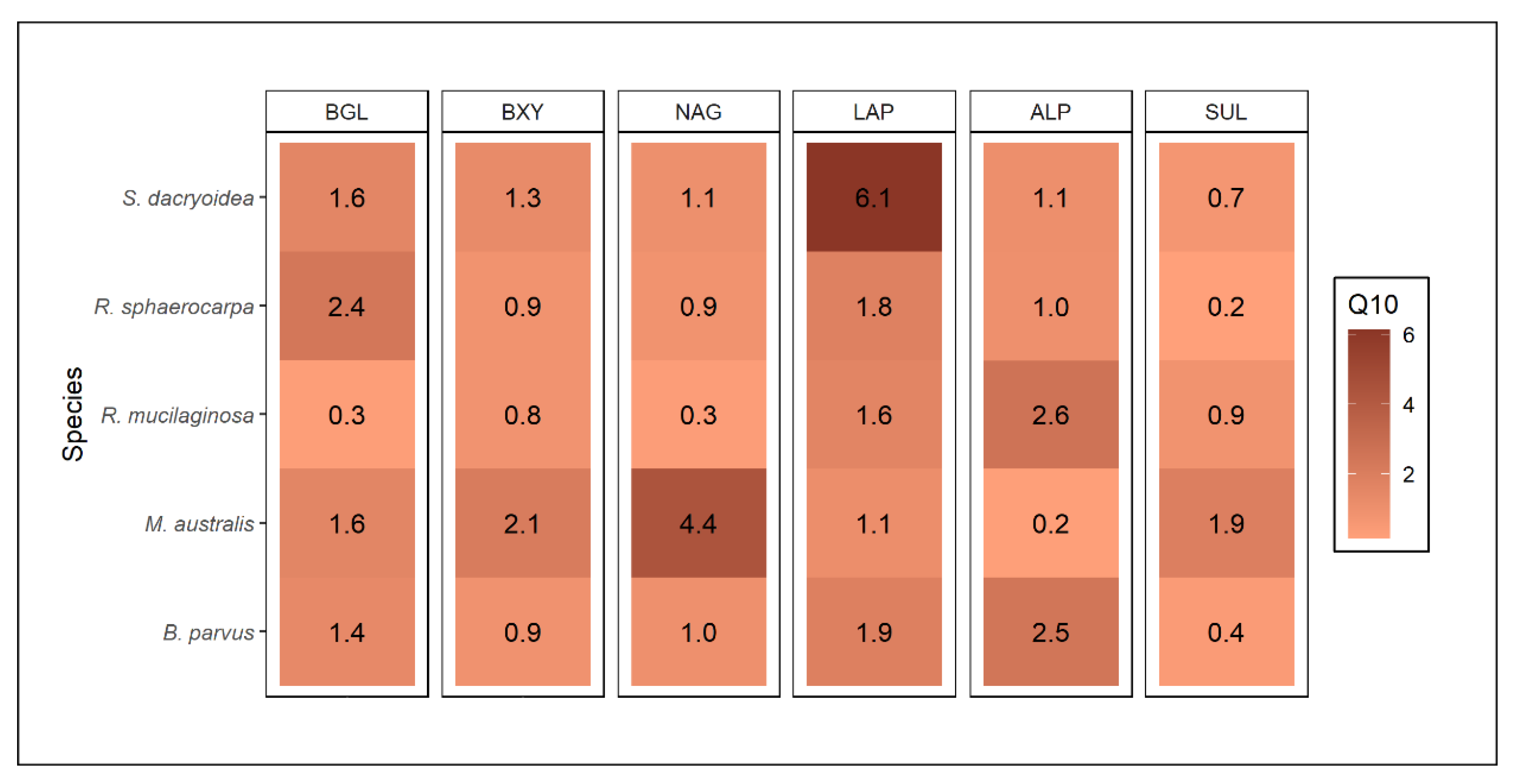
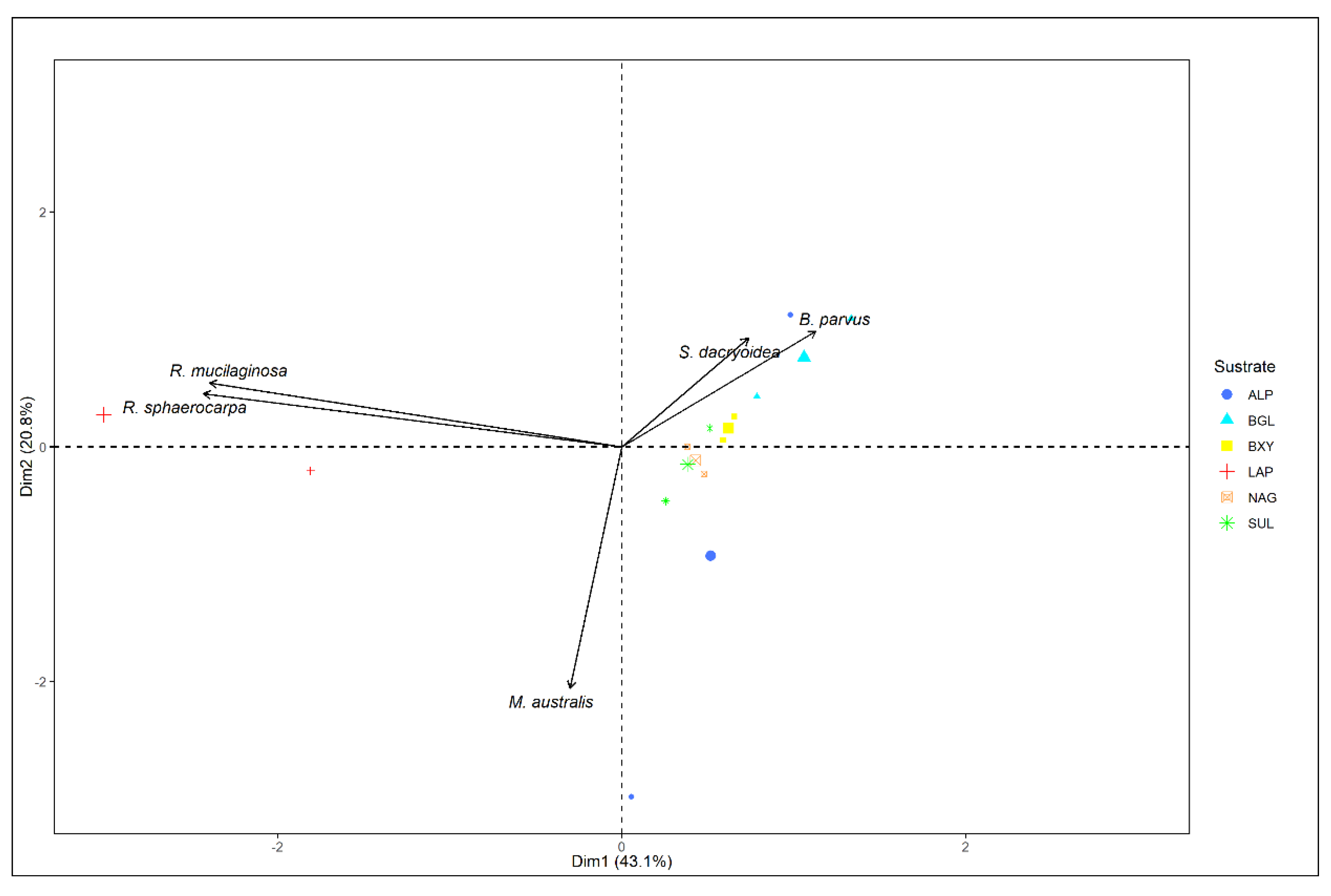
3.3. Relation between Enzyme Kinetic Parameters, Enzyme Types, and Phylogeny

PCA analysis allowed for the comparison of the five studied marine fungal species and the different extracellular enzymatic activities determined. The explained variance of the dataset was 63.9% (Figure 6). The minor angle between R. mucilaginosa and R. sphaerocarpa belonging to the same genera (Rhodotorula) indicated very similar activity levels and extracellular enzyme characteristics. In contrast, the other fungal strains substantially differed in their extracellular enzymatic activity and enzyme characteristics, independent of their taxonomic affiliation.

4. Discussion

All the studied marine pelagic fungi species, B. parvus, M. australis, R. mucilaginosa, R. sphaerocarpa, and S. dacryoidea, produced extracellular enzymes to degrade substrate analogues of carbohydrates such as cellulose, chitin, and xylan. Additionally, they all produced enzymes to cleave off amino acids, phosphate, and sulfate esters from organic compounds.

4.1. Extracellular Enzymatic Activities of Pelagic Fungal Isolates

4.1.1. Influence of Taxonomy/Diversity on the Different EEAs

Since each organism has specific enzymatic capabilities and substrate preferences [33], extracellular enzymatic activities (EEA) can be used as functional traits to investigate functional diversity [34]. Hence, the different EEAs detected in the studied marine fungi can be used to infer their influence on marine ecological processes. Interestingly, we found that the two species belonging to the genus Rhodotorula (R. mucilaginosa and R. sphaerocarpa) exhibited similar kinetic parameters (Vmax and Km) for the majority of extracellular enzymes (Figure 1, Figure 2, Figure 3, Figure 4 and Figure 6). This might indicate some degree of trait conservation among organisms on the genus level, although more species of this genus need to be investigated before a firm conclusion can be drawn. Differences were observed, however, in the EEAs of all the other fungal strains.
Polysaccharides, as the most abundant organic compound class, but also the most complex one, require a wide range of enzymes to degrade them [35,36]. We measured three EEAs responsible for the cleavage of cellulose, xylan, and chitin (Figure 1). In terrestrial environments, the plant cell wall is composed mainly of cellulose and protected by lignin [37]. In marine ecosystems, cellulose is present in the algae cell wall and covered by distinct polymers [38]. Nonetheless, as marine cellulose is more accessible, but less frequent than other substrates, only specific microorganisms are capable of degrading cellulose [39]. Some studies have reported cellulose degradation by marine fungi like Arthrinium saccharicola [40] and Lulworthia floridana [41]. Other studies have also described cellulose hydrolysis by wider distributed fungal species, for instance, Aspergillus niger [42] and Trichoderma virens [43]. Vaz, et al. [44] showed that 76% of the studied marine fungi exhibit cellulolytic activity. In this case, even though all the marine fungal species used were able to cleave cellulose, S. dacryoidea dominated this EEA (Figure 1A). Hudson [45] stated that each fungi species have different capacities to decompose cellulose due to diverse enzymatic machinery. S. dacryoidea exhibited a high Km indicating a low affinity to the substrate [31]. This also suggests that, even though the overall enzymatic activity is high, the substrate dissociates easily from the enzyme [46].
Xylan is a polysaccharide formed by residual monosaccharides called xylose [29]. Similar to cellulose, in terrestrial environments, xylan can be found in plants [47], whereas in the ocean, xylan can be present in algae [48,49]. Fungi can degrade xylose via the oxidoreductase pathway [50] as it is a primary carbon source [28]. Even though the general EEA was low (Figure 1B), we can deduce that the studied marine fungi are capable of releasing enzymes related to the hydrolysis of xylose. Raghukumar, et al. [51] identified low xylanase activity rates of fungal coastal strains. Duarte, et al. [52] also reported xylanase activity of Antarctic fungal strains but highlighted a higher activity of Basidiomycota over Ascomycota strains. In this study, we could not identify a clear difference between these two phyla. Nonetheless, the low Km suggests a high affinity of the enzyme to the substrate at low concentrations (Figure 3B). Thus, it seems likely that pelagic marine fungi might use xylose as a carbon source even when present at low concentrations.
Chitin is one of the most abundant naturally occurring polysaccharides, and it is an essential component of the cell wall of fungi, the exoskeleton of arthropods, the radula of mollusks, and the beak of cephalopods [53,54]. Although the overall chitinase activity was low, we can infer that the studied marine fungi are capable of degrading chitin (Figure 1C). The chitin degradation by marine fungi has been reported in species such as Lecanicillium muscarium [55] and Verticillium lecanii [56]. Interestingly, the number of fungal enzymes involved in the degradation of chitin is related to the fungal chitin content, which varies strongly between fungal species and is dependent on the growth mode [57]. For instance, the hyphae-like fungal cell wall consists of 10 to 20% of chitin [54], whereas yeast-like fungi have a rather low chitin content of 0.5 to 5% [58]. The filamentous fungus B. parvus exhibited high chitinase activity (Figure 1C). Moreover, the low Km obtained for this species suggests that only a low substrate concentration is needed to saturate its chitinase. Thus, pelagic marine fungi likely use chitin as a carbon source even when present at only low concentrations.
Leucine aminopeptidase is a critical biological enzyme due to its key role in the degradation of proteins [59]. In the present study, this enzymatic activity was different for each fungal species. Despite the majority of microbial LAP being intracellular, extracellular enzymes have been reported in filamentous fungi [60]. All the species we tested showed LAP activity with R. sphaerocarpa exhibiting the highest Vmax. The substrate concentration needed to achieve half Vmax varied among species (Figure 4A). Although B. parvus had one of the lowest Vmax, its Km was the highest (Figure 2A and Figure 4A). In contrast, R. sphaerocarpa exhibited high substrate affinity and a low Km (Figure 4A) Thus, as the kinetics for LAP varied among the fungal species, the protein hydrolysis in the ocean might be species dependent.
Inorganic phosphate (Pi) is the preferred phosphorus source for microbial uptake. In surface waters, however, Pi frequently limits phytoplankton productivity [61]. To overcome this P-limitation, microorganisms use dissolved organic phosphorus (DOP) [62]. For prokaryotic microorganisms, Baltar et al. [63] reported that irrespective of the phosphate bioavailability, the activation of alkaline phosphatase was related to sporadic pulses of organic matter. Thus, a high Km might be beneficial to allow for high cleavage rates when organic substrate availability is high. The species R. sphaerocarpa, R. mucilaginosa, and S. dacryoidea, belonging to the phylum Basidiomycota, exhibited a low enzymatic activity but a high Km (Figure 2B and Figure 4B). In contrast, the species of the phylum Ascomycota (B. parvus and M. australis) seem to be more suitable to overcome this P-limitation, but a higher amount of substrate, and hence of organic matter, might be needed for this purpose.
In living organisms, sulfur is the sixth most abundant element as it can be found in amino acids, such as cysteine and methionine, but also in polysaccharides and proteoglycans [35]. In contrast to terrestrial polysaccharides, several marine polysaccharides, especially in the cell wall of macroalgae, are highly sulfated [35]. The terrestrial fungus Fusarium proliferatum was reported to produce sulfatase for the assimilation of sulfated fucoidans of brown algae [64]. In the present study, the species of the genus Rhodotorula maintained a low sulfatase activity, whereas it varied among the other species. The Vmax and Km were high in S. dacryoidea, indicating fast hydrolysis and low substrate affinity. As all the fungal species showed different sulfatase activity, we can deduce that these marine fungi might be capable to use sulfated amino acids as well as carbohydrates.
In this study, even though we analyzed just a few hydrolysis possibilities, the functional diversity seems to be broad in marine fungi. As suggested by Berlemont [65] and Baltar et al. [10], marine fungi are potentially involved in carbohydrates’ degradation. Based on our results, we can infer that marine pelagic fungi are actively utilizing carbohydrates such as cellulose, chitin, and xylose, as well as some carbohydrates with sulfur content, potentially of algal origin [47].
The ocean is a complex and diverse ecosystem composed of microorganisms such as archaea, bacteria, fungi, protists, and viruses. As osmoheterotrophic organisms, fungi might provide intermediate decomposition products needed for other microorganisms. These also might lead to the proliferation or inhibition of other microbes as well as enzymatic activities. As marine fungi can utilize a wide range of organic substrates [66], nutrient availability can impact the magnitude and distribution of extracellular enzymatic activities [33].

4.1.2. Temperature Influence

Temperature is considered one of the most important abiotic factors because it influences essentially all biochemical reactions [67]. Enzymes are sensitive to temperature [46] influencing the kinetics along with the substrate binding property and stability [33,68]. According to the Van’t Hoff rule, a temperature increase of 10 °C can double a reaction rate. Q10 values lower than 1.0 would indicate a reaction rate completely independent of temperature, whereas values above 1 indicate thermodependency [69].
The results obtained in this study indicate that the majority of enzymatic activities were lower at 5 °C than at 20 °C (Figure 1 and Figure 2). In general terms, at 5 °C the species R. mucilaginosa and R. sphaerocarpa maintained Vmax values as low as 0.1 μmol/g biomass/h, whereas the other species exhibited only half of the activity at 5 °C, particularly for BGL and ALP (Figure 1 and Figure 2). This reduced enzymatic synthesis at a low temperature might be due to limited transcriptional and translational activity, limited protein folding, and DNA and RNA secondary structures’ stabilization [11]. B. parvus, for all the substrates except ALP, had a higher Km at 5 °C, which suggests that the affinity of this species for some substrates increases with increasing temperature. Taken together, the effect of temperature on the characteristics of extracellular enzymes depends on the fungal species and the type of enzyme.
Psychrophiles microorganisms have evolved a complex range of adaptation strategies, such as production of antifreeze proteins [70] and exopolysaccharides (EPS) [71], high levels of unsaturated fatty acids to maintain the membrane fluidity [72], and certain enzymes adapted to those temperatures [11]. Gerday et al. [73] described a peculiar type of extracellular enzymes known as “cold-adapted enzymes” produced by microorganisms living at low temperatures. For these enzymes, the reaction rate is dependent on the encounter rate of the enzyme and substrate, so it is controlled mainly by diffusion, and it is temperature independent [14,74].
Aghajari et al. [75] suggested that the main structural feature of these “cold-adapted enzymes” is flexibility or plasticity. The structures involved in the catalytic cycle are more flexible, whereas other structures that do not participate in the catalytic cycle might be more rigid [67,76]. For instance, the chitinase of Glaciozyma antarctica presented fewer salt bridges and hydrogen bonds, which increased its flexibility [12,77]. Another key structural feature of these enzymes is stability [73,74], with for example, amino acids modifications in key regions of the protein [77,78,79,80]. Nonetheless, there is not a single strategy, as each cold-adapted enzyme can perform different ways to enhance its activity at low temperatures [12,74].
Cold-adapted enzymes have been reported from a wide variety of marine fungi [12,44,52,55,56,77,81,82]. M. australis and R. sphaerocarpa were one of the few species that showed a noticeable enzymatic activity at 5 °C for ALP and SUL, respectively (Figure 2B,C). M. australis is an endemic species of Antarctic waters [19,83], whereas R. sphaerocarpa was originally isolated close to Marguerite Bay on the west side of the Antarctic Peninsula but has a wider distribution including the Caribbean Sea [84] and the Andaman Sea [85], among others. Low temperatures exert high selective pressure on endemic organisms [86], such as alkalinity phosphatase in M. australis. For this species, the substrate-binding affinity was lower at 5 °C, whereas for R. sphaerocarpa, Km was lower at this temperature (Figure 4). This suggested that the enzyme–substrate complex of M. australis ALP is more effective at higher substrate concentration typical for Antarctic waters known as a major high-nutrient low-chlorophyll region in the global ocean [87].
Fungal cold-adapted chitinases have been previously reported [77,88,89]. Ramli et al. [77] found that the chitinase sequence of G. antarctica had a low sequence identity with other chitinases. Moreover, they found that the enzyme flexibility was due to certain amino acids substitutions in the surface and loop regions. In this study, at 5 °C, we could only identify a higher chitinase activity for B. parvus. For the rest of the species, there was a positive enzymatic activity, but higher at 20 °C.
Microorganisms isolated from cold environments can also display kinetic parameters similar to those of their mesophilic counterparts [69,90]. Ito et al. [91] deduced that a high Q10 value (>2) is due to a conformational change in proteins and indicates the need for high activation energy. We found that only for LAP, all the examined fungal species expressed Q10 values higher than 1. Generally, the temperature where an enzyme can achieve its highest activity does not match the optimal growth temperature of the microorganism that is producing it [78]. Apparently, cold-adapted species, such as B. parvus, M. australis, R. sphaerocarpa, and S. dacryoidea, can respond to a temperature rise by increasing enzymatic activity. According to the Arrhenius equation, temperature can influence the activation energy needed to initiate a chemical reaction, and hence, its rate. At a higher temperature, the molecules gain energy to move faster, which also increases the collisions between enzymes and substrates. As a result, elevated extracellular enzymatic activity at increasing temperatures in the surface waters might lead to changes in cleavage and uptake rate of organic matter in oceanic fungi.

5. Conclusions

In the present study, we have shown that different marine fungal strains exhibit varying extracellular enzyme characteristics with Vmax and Km values varying over a range of one order of magnitude. Although the fungal species were isolated from coastal Antarctic waters (B. parvus, M. australis, R. sphaerocarpa, and S. dacryoidea), and hence are potentially adapted to low temperatures, they exhibited higher extracellular enzymatic activity at 20 °C than at 5 °C, with some exceptions. While in some fungal strains the Km values for specific extracellular enzymes were higher at low temperatures, for other enzymes they were lower. Additionally, there was considerable species-specific variability in the extracellular enzymatic activity. Taken together, our study indicates that temperature might be one of most important physical factors controlling marine fungal extracellular enzymatic activity. Thus, species composition and temperature determine the role of marine fungi in organic matter cleavage in the global ocean.

Author Contributions

Conceptualization, K.S.A. and F.B.; data curation, K.S.A.; formal analysis, K.S.A.; funding acquisition, G.J.H. and F.B.; investigation, K.S.A.; methodology, K.S.A. and F.B.; project administration, F.B.; software, K.S.A.; supervision, F.B.; visualization, K.S.A.; writing—original draft, K.S.A.; writing—review and editing, K.S.A., G.J.H., and F.B. All authors have read and agreed to the published version of the manuscript.

Funding

F.B. was supported by the Austrian Science Fund (FWF) projects OCEANIDES (P34304-B), ENIGMA (TAI534), and EXEBIO (P35248). G.J.H. was supported by the Austrian Science Fund (FWF) project ARTEMIS (P28781-B21), the projects I486-B09 and P23234-B11 by the European Research Council (ERC) under the European Community’s Seventh Framework Programme (FP7/2007–2013)/ERC grant agreement 268595 (MEDEA project). Open Access Funding by the Austrian Science Fund (FWF).

Institutional Review Board Statement

Not applicable.

Informed Consent Statement

Not applicable.

Data Availability Statement

The raw data supporting the conclusions of this article will be made available by the authors, without undue reservation to any qualified researcher.

Acknowledgments

We would like to thank Charlotte Doebke, Marilena Heitger, Marina Montserrat Díez, and Katarína Tamášová for their valuable help in the laboratory work.

Conflicts of Interest

The authors declare no conflict of interest.

References

  1. Nevalainen, H.; Kautto, L.; Te’o, J. Methods for isolation and cultivation of filamentous fungi. Methods Mol. Biol. 2014, 1096, 3–16. [Google Scholar]
  2. Richards, T.A.; Jones, M.D.; Leonard, G.; Bass, D. Marine fungi: Their ecology and molecular diversity. Annu. Rev. Mar. Sci. 2012, 4, 495–522. [Google Scholar] [CrossRef] [PubMed]
  3. Arrieta, J.M.; Herndl, G.J. Changes in bacterial β-glucosidase diversity during a coastal phytoplankton bloom. Limnol. Oceanogr. 2002, 47, 594–599. [Google Scholar] [CrossRef]
  4. Baltar, F.; Arístegui, J.; Sintes, E.; Van Aken, H.M.; Gasol, J.M.; Herndl, G.J. Prokaryotic extracellular enzymatic activity in relation to biomass production and respiration in the meso-and bathypelagic waters of the (sub) tropical Atlantic. Environ. Microbiol. 2009, 11, 1998–2014. [Google Scholar] [CrossRef]
  5. Gutiérrez, M.H.; Pantoja, S.; Tejos, E.; Quiñones, R.A. The role of fungi in processing marine organic matter in the upwelling ecosystem off Chile. Mar. Biol. 2011, 158, 205–219. [Google Scholar] [CrossRef]
  6. Cunliffe, M.; Hollingsworth, A.; Bain, C.; Sharma, V.; Taylor, J.D. Algal polysaccharide utilisation by saprotrophic planktonic marine fungi. Fungal Ecol. 2017, 30, 135–138. [Google Scholar] [CrossRef]
  7. Gao, Z.; Johnson, Z.I.; Wang, G. Molecular characterization of the spatial diversity and novel lineages of mycoplankton in Hawaiian coastal waters. ISME J. 2010, 4, 111–120. [Google Scholar] [CrossRef]
  8. Bochdansky, A.B.; Clouse, M.A.; Herndl, G.J. Eukaryotic microbes, principally fungi and labyrinthulomycetes, dominate biomass on bathypelagic marine snow. ISME J. 2017, 11, 362–373. [Google Scholar] [CrossRef]
  9. Morales, S.E.; Biswas, A.; Herndl, G.J.; Baltar, F. Global Structuring of Phylogenetic and Functional Diversity of Pelagic Fungi by Depth and Temperature. Front. Mar. Sci. 2019, 6, 131. [Google Scholar] [CrossRef] [Green Version]
  10. Baltar, F.; Zhao, Z.; Herndl, G.J. Potential and expression of carbohydrate utilization by marine fungi in the global ocean. Microbiome 2021, 9, 106. [Google Scholar] [CrossRef]
  11. D’Amico, S.; Collins, T.; Marx, J.C.; Feller, G.; Gerday, C.; Gerday, C. Psychrophilic microorganisms: Challenges for life. EMBO Rep. 2006, 7, 385–389. [Google Scholar] [CrossRef] [PubMed]
  12. Yusof, N.A.; Hashim, N.H.F.; Bharudin, I. Cold adaptation strategies and the potential of psychrophilic enzymes from the antarctic yeast, Glaciozyma antarctica PI12. J. Fungi 2021, 7, 528. [Google Scholar] [CrossRef] [PubMed]
  13. Morita, R.Y. Psychrophilic bacteria. Bacteriol. Rev. 1975, 39, 144–167. [Google Scholar] [CrossRef] [PubMed]
  14. Feller, G.; Gerday, C. Psychrophilic enzymes: Hot topics in cold adaptation. Nat. Rev. Microbiol. 2003, 1, 200–208. [Google Scholar] [CrossRef]
  15. Vonnahme, T.R.; Dietrich, U.; Hassett, B.T. Progress in microbial ecology in ice-covered seas. In YOUMARES 9—The Oceans: Our Research, Our Future; Springer: Cham, Switzerland, 2020; p. 261. [Google Scholar]
  16. Amend, A.; Burgaud, G.; Cunliffe, M.; Edgcomb, V.P.; Ettinger, C.L.; Gutiérrez, M.H.; Heitman, J.; Hom, E.F.Y.; Ianiri, G.; Jones, A.C.; et al. Fungi in the Marine Environment: Open Questions and Unsolved Problems. mBio 2019, 10, e01189-18. [Google Scholar] [CrossRef] [Green Version]
  17. Taylor, J.D.; Cunliffe, M. Multi-year assessment of coastal planktonic fungi reveals environmental drivers of diversity and abundance. ISME J. 2016, 10, 2118–2128. [Google Scholar] [CrossRef] [Green Version]
  18. Fell, J.; Statzell, A.C. Sympodiomyces gen. n., a yeast-like organism from southern marine waters. Antonie Van Leeuwenhoek 1971, 37, 359–367. [Google Scholar] [CrossRef]
  19. Fell, J.W.; Hunter, I.L. Isolation of heterothallic yeast strains of Metschnikowia Kamienski and their mating reactions with Chlamydozyma wickerham spp. Antonie Van Leeuwenhoek 1968, 34, 365–376. [Google Scholar] [CrossRef]
  20. Fell, J.W.; Hunter, I.L.; Tallman, A.S. Marine basidiomycetous yeasts (Rhodosporidium spp. n.) with tetrapolar and multiple allelic bipolar mating systems. Can. J. Microbiol. 1973, 19, 643–657. [Google Scholar] [CrossRef]
  21. Newell, S.Y.; Fell, J. The perfect form of a marine-occurring yeast of the genus Rhodotorula. Mycologia 1970, 62, 272–281. [Google Scholar] [CrossRef]
  22. Sampaio, J.P. Rhodotorula Harrison (1928). In The Yeasts; Elsevier: Burlington, VT, USA, 2011; pp. 1873–1927. [Google Scholar]
  23. Wickerham, L.J. A taxonomic study of Monilia albicans with special emphasis on morphology and morphological variation. J. Tropical Med. Hyg. 1939, 42, 174–179. [Google Scholar]
  24. Wickerham, L.J. Taxonomy of Yeasts; US Department of Agriculture: Washington, DC, USA, 1951.
  25. Salazar Alekseyeva, K.; Mähnert, B.; Berthiller, F.; Breyer, E.; Herndl, G.J.; Baltar, F. Adapting an Ergosterol Extraction Method with Marine Yeasts for the Quantification of Oceanic Fungal Biomass. J. Fungi 2021, 7, 690. [Google Scholar] [CrossRef]
  26. Norkrans, B. Degradation of cellulose. Annu. Rev. Phytopathol. 1963, 1, 325–350. [Google Scholar] [CrossRef]
  27. Pointing, S.B.; Vrijmoed, L.L.P.; Jones, E.B.G. A Qualitative Assessment of Lignocellulose Degrading Enzyme Activity in Marine Fungi. Bot. Mar. 1998, 41, 293–298. [Google Scholar] [CrossRef]
  28. Collins, T.; Gerday, C.; Feller, G. Xylanases, xylanase families and extremophilic xylanases. FEMS Microbiol. Rev. 2005, 29, 3–23. [Google Scholar] [CrossRef] [PubMed] [Green Version]
  29. dos Santos, J.A.; Vieira, J.M.F.; Videira, A.; Meirelles, L.A.; Rodrigues, A.; Taniwaki, M.H.; Sette, L.D. Marine-derived fungus Aspergillus cf. tubingensis LAMAI 31: A new genetic resource for xylanase production. AMB Express 2016, 6, 25. [Google Scholar]
  30. Hoppe, H.-G. Significance of exoenzymatic activities in the ecology of brackish water: Measurements by means of methylumbelliferyl-substrates. Mar. Ecol. Prog. Ser. 1983, 11, 299–308. [Google Scholar] [CrossRef]
  31. Huitema, C.; Horsman, G. Analyzing Enzyme Kinetic Data Using the Powerful Statistical Capabilities of R. bioRxiv 2018, 1, 316588. [Google Scholar]
  32. Sterratt, D.C. Q10: The Effect of Temperature on Ion Channel Kinetics, in Encyclopedia of Computational Neuroscience; Jaeger, D., Jung, R., Eds.; Springer: New York, NY, USA, 2015; pp. 2551–2552. [Google Scholar]
  33. Arnosti, C.; Bell, C.; Moorhead, D.L.; Sinsabaugh, R.L.; Steen, A.D.; Stromberger, M.; Wallenstein, M.; Weintraub, M.N. Extracellular enzymes in terrestrial, freshwater, and marine environments: Perspectives on system variability and common research needs. Biogeochemistry 2014, 117, 5–21. [Google Scholar] [CrossRef]
  34. Cullings, K.; Courty, P.-E. Saprotrophic capabilities as functional traits to study functional diversity and resilience of ectomycorrhizal community. Oecologia 2009, 161, 661–664. [Google Scholar] [CrossRef]
  35. Helbert, W. Marine Polysaccharide Sulfatases. Front. Mar. Sci. 2017, 4, 6. [Google Scholar] [CrossRef] [Green Version]
  36. Metreveli, E.; Khardziani, T.; Elisashvili, V. The Carbon Source Controls the Secretion and Yield of Polysaccharide-Hydrolyzing Enzymes of Basidiomycetes. Biomolecules 2021, 11, 1341. [Google Scholar] [CrossRef]
  37. Houtman, C.J.; Atalla, R.H. Cellulose-Lignin Interactions (A Computational Study). Plant Physiol. 1995, 107, 977–984. [Google Scholar] [CrossRef] [PubMed] [Green Version]
  38. Domozych, D.; Ciancia, M.; Fangel, J.; Mikkelsen, M.; Ulvskov, P.; Willats, W. The Cell Walls of Green Algae: A Journey through Evolution and Diversity. Front. Plant Sci. 2012, 3, 82. [Google Scholar] [CrossRef] [Green Version]
  39. Arora, D.K. Handbook of Applied Mycology: Volume 1: Soil and Plants; CRC Press: Boca Raton, FL, USA, 1991. [Google Scholar]
  40. Hong, J.-H.; Jang, S.; Heo, Y.M.; Min, M.; Lee, H.; Lee, Y.M.; Lee, H.; Kim, J.-J. Investigation of Marine-Derived Fungal Diversity and Their Exploitable Biological Activities. Mar. Drugs 2015, 13, 4137–4155. [Google Scholar] [CrossRef] [PubMed]
  41. Meyers, S.; Scott, E. Cellulose degradation by Lulworthia floridana and other lignicolous marine fungi. Mar. Biol. 1968, 2, 41–46. [Google Scholar] [CrossRef]
  42. Baraldo Junior, A.; Borges, D.G.; Tardioli, P.W.; Farinas, C.S. Characterization of β-glucosidase produced by Aspergillus niger under solid-state fermentation and partially purified using MANAE-Agarose. Biotechnol. Res. Int. 2014, 2014, 317092. [Google Scholar] [CrossRef] [Green Version]
  43. Ab Wahab, A.F.F.; Karim, N.A.A.; Ling, J.G.; Hasan, N.S.; Yong, H.Y.; Bharudin, I.; Kamaruddin, S.; Bakar, F.D.A.; Murad, A.M. Functional characterisation of cellobiohydrolase I (Cbh1) from Trichoderma virens UKM1 expressed in Aspergillus niger. Protein Expr. Purif. 2019, 154, 52–61. [Google Scholar] [CrossRef]
  44. Vaz, A.B.M.; Rosa, L.H.; Vieira, M.L.A.; de Garcia, V.; Brandão, L.R.; Teixeira, L.C.R.S.; Moliné, M.; Libkind, D.; van Broock, M.; Rosa, C. The diversity, extracellular enzymatic activities and photoprotective compounds of yeasts isolated in Antarctica. Braz. J. Microbiol. 2011, 42, 937–947. [Google Scholar] [CrossRef]
  45. Hudson, H.J. Fungal Saprophytism; Edward Arnold: London, UK, 1972. [Google Scholar]
  46. Roussel, M.R. A Life Scientist’s Guide to Physical Chemistry; Cambridge University Press: Cambridge, UK, 2012. [Google Scholar]
  47. Balabanova, L.; Slepchenko, L.; Son, O.; Tekutyeva, L. Biotechnology Potential of Marine Fungi Degrading Plant and Algae Polymeric Substrates. Front. Microbiol. 2018, 9, 1527. [Google Scholar] [CrossRef] [Green Version]
  48. Goddard-Borger, E.D.; Sakaguchi, K.; Reitinger, S.; Watanabe, N.; Ito, M.; Withers, S.G. Mechanistic insights into the 1,3-xylanases: Useful enzymes for manipulation of algal biomass. J. Am. Chem. Soc. 2012, 134, 3895–3902. [Google Scholar] [CrossRef] [PubMed]
  49. Synytsya, A.; Čopíková, J.; Kim, W.J.; Park, Y.I. Cell Wall Polysaccharides of Marine Algae. In Springer Handbook of Marine Biotechnology; Kim, S.-K., Ed.; Springer: Berlin/Heidelberg, Germany, 2015; pp. 543–590. [Google Scholar]
  50. Domingues, R.; Bondar, M.; Palolo, I.; Queirós, O.; de Almeida, C.D.; Cesário, M.T. Xylose Metabolism in Bacteria—Opportunities and Challenges towards Efficient Lignocellulosic Biomass-Based Biorefineries. Appl. Sci. 2021, 11, 8112. [Google Scholar] [CrossRef]
  51. Raghukumar, C.; Raghukumar, S.; Chinnaraj, A.; Chandramohan, D.; D’souza, T.; Reddy, C. Laccase and Other Lignocellulose Modifying Enzymes of Marine Fungi Isolated from the Coast of India. Botanica Marina. 1994, 37, 515–523. [Google Scholar] [CrossRef]
  52. Duarte, A.W.F.; Dayo-Owoyemi, I.; Nobre, F.; Pagnocca, F.C.; Chaud, L.C.S.; Pessoa, A.; Felipe, M.G.A.; Sette, L.D. Taxonomic assessment and enzymes production by yeasts isolated from marine and terrestrial Antarctic samples. Extremophiles 2013, 17, 1023–1035. [Google Scholar] [CrossRef] [PubMed]
  53. Numata, K.; Kaplan, D.L. 20—Biologically derived scaffolds. In Advanced Wound Repair Therapies; Farrar, D., Ed.; Woodhead Publishing: Sawston, UK, 2011; pp. 524–551. [Google Scholar]
  54. Ruiz-Herrera, J. Fungal Cell Wall: Structure, Synthesis, and Assembly; CRC Press: Boca Raton, FL, USA, 1991. [Google Scholar]
  55. Fenice, M. The psychrotolerant Antarctic fungus Lecanicillium muscarium CCFEE 5003: A powerful producer of cold-tolerant chitinolytic enzymes. Molecules 2016, 21, 447. [Google Scholar] [CrossRef] [Green Version]
  56. Fenice, M.; Selbmann, L.; Di Giambattista, R.; Federici, F. Chitinolytic activity at low temperature of an Antarctic strain (A3) of Verticillium lecanii. Res. Microbiol. 1998, 149, 289–300. [Google Scholar] [CrossRef]
  57. Hartl, L.; Zach, S.; Seidl-Seiboth, V. Fungal chitinases: Diversity, mechanistic properties and biotechnological potential. Appl. Microbiol. Biotechnol. 2012, 93, 533–543. [Google Scholar] [CrossRef] [Green Version]
  58. Garcia-Rubio, R.; de Oliveira, H.C.; Rivera, J.; Trevijano-Contador, N. The Fungal Cell Wall: Candida, Cryptococcus, and Aspergillus Species. Front. Microbiol. 2020, 10, 2993. [Google Scholar] [CrossRef]
  59. Burley, S.K.; David, P.R.; Taylor, A.; Lipscomb, W.N. Molecular structure of leucine aminopeptidase at 2.7—A resolution. Proc. Natl. Acad. Sci. USA 1990, 87, 6878–6882. [Google Scholar] [CrossRef] [Green Version]
  60. Nampoothiri, K.M.; Nagy, V.; Kovacs, K.; Szakacs, G.; Pandey, A. L-leucine aminopeptidase production by filamentous Aspergillus fungi. Lett. Appl. Microbiol. 2005, 41, 498–504. [Google Scholar] [CrossRef]
  61. Karl, D.M. Phosphorus, the staff of life. Nature 2000, 406, 31–33. [Google Scholar] [CrossRef] [PubMed]
  62. Srivastava, A.; Saavedra, D.E.M.; Thomson, B.; García, J.A.L.; Zhao, Z.; Patrick, W.M.; Herndl, G.J.; Baltar, F. Enzyme promiscuity in natural environments: Alkaline phosphatase in the ocean. ISME J. 2021, 15, 3375–3383. [Google Scholar] [CrossRef] [PubMed]
  63. Baltar, F.; Lundin, D.; Palovaara, J.; Lekunberri, I.; Reinthaler, T.; Herndl, G.J.; Pinhassi, J. Prokaryotic Responses to Ammonium and Organic Carbon Reveal Alternative CO2 Fixation Pathways and Importance of Alkaline Phosphatase in the Mesopelagic North Atlantic. Front. Microbiol. 2016, 7, 1670. [Google Scholar] [CrossRef] [PubMed] [Green Version]
  64. Shvetsova, S.V.; Zhurishkina, E.V.; Bobrov, K.S.; Ronzhina, N.L.; Lapina, I.M.; Ivanen, D.R.; Gagkaeva, T.Y.; Kulminskaya, A.A. The novel strain Fusarium proliferatum LE1 (RCAM02409) produces α-l-fucosidase and arylsulfatase during the growth on fucoidan. J. Basic Microbiol. 2015, 55, 471–479. [Google Scholar] [CrossRef] [PubMed]
  65. Berlemont, R. Distribution and diversity of enzymes for polysaccharide degradation in fungi. Sci. Rep. 2017, 7, 222. [Google Scholar] [CrossRef] [PubMed] [Green Version]
  66. Loque, C.P.; Medeiros, A.O.; Pellizzari, F.M.; Oliveira, E.C.; Rosa, C.A.; Rosa, L.H. Fungal community associated with marine macroalgae from Antarctica. Polar Biol. 2010, 33, 641–648. [Google Scholar] [CrossRef]
  67. Georlette, D.; Blaise, V.; Collins, T.; D’Amico, S.; Gratia, E.; Hoyoux, A.; Marx, J.-C.; Sonan, G.; Feller, G.; Gerday, C. Some like it cold: Biocatalysis at low temperatures. FEMS Microbiol. Rev. 2004, 28, 25–42. [Google Scholar] [CrossRef] [Green Version]
  68. John, A.B.; Diana, E.V.; Paul, J.H. Effects of temperature on growth rate, cell composition and nitrogen metabolism in the marine diatom Thalassiosira pseudonana (Bacillariophyceae). Mar. Ecol. Prog. Ser. 2002, 225, 139–146. [Google Scholar]
  69. Collins, T.; D’Amico, S.; Marx, J.C.; Feller, G.; Gerday, C. Cold-Adapted Enzymes. Physiol. Biochem. Extrem. 2007, 1, 165–179. [Google Scholar]
  70. Kawahara, H.; Iwanaka, Y.; Higa, S.; Muryoi, N.; Sato, M.; Honda, M.; Omura, H.; Obata, H. A novel, intracellular antifreeze protein in an antarctic bacterium, Flavobacterium xanthum. Cryoletters 2007, 28, 39–49. [Google Scholar]
  71. Krembs, C.; Eicken, H.; Junge, K.; Deming, J. High concentrations of exopolymeric substances in Arctic winter sea ice: Implications for the polar ocean carbon cycle and cryoprotection of diatoms. Deep. Sea Res. Part I Oceanogr. Res. Pap. 2002, 49, 2163–2181. [Google Scholar] [CrossRef]
  72. Los, D.A.; Murata, N. Membrane fluidity and its roles in the perception of environmental signals. Biochim. Et Biophys. Acta 2004, 1666, 142–157. [Google Scholar] [CrossRef] [PubMed] [Green Version]
  73. Gerday, C.; Aittaleb, M.; Bentahir, M.; Chessa, J.-P.; Claverie, P.; Collins, T.; D’Amico, S.; Dumont, J.; Garsoux, G.; Georlette, D. Cold-adapted enzymes: From fundamentals to biotechnology. Trends Biotechnol. 2000, 18, 103–107. [Google Scholar] [CrossRef]
  74. Feller, G.; Gerday, C. Psychrophilic enzymes: Molecular basis of cold adaptation. Cell. Mol. Life Sci. CMLS 1997, 53, 830–841. [Google Scholar] [CrossRef] [PubMed] [Green Version]
  75. Aghajari, N.; Haser, R.; Feller, G.; Gerday, C. Crystallization and preliminary X-ray diffraction studies of α-amylase from the antarctic psychrophile Alteromonas haloplanctis A23. Protein Sci. 1996, 5, 2128–2129. [Google Scholar] [CrossRef] [Green Version]
  76. Truongvan, N.; Jang, S.-H.; Lee, C. Flexibility and stability trade-off in active site of cold-adapted Pseudomonas mandelii esterase EstK. Biochemistry 2016, 55, 3542–3549. [Google Scholar] [CrossRef]
  77. Ramli, A.N.M.; Mahadi, N.M.; Shamsir, M.S.; Rabu, A.; Joyce-Tan, K.H.; Murad, A.M.A.; Illias, R. Structural prediction of a novel chitinase from the psychrophilic Glaciozyma antarctica PI12 and an analysis of its structural properties and function. J. Comput. Aided Mol. Des. 2012, 26, 947–961. [Google Scholar] [CrossRef]
  78. Baeza, M.; Zúñiga, S.; Peragallo, V.; Barahona, S.; Alcaino, J.; Cifuentes, V. Identification of stress-related genes and a comparative analysis of the amino acid compositions of translated coding sequences based on draft genome sequences of Antarctic yeasts. Front. Microbiol. 2021, 12, 133. [Google Scholar] [CrossRef]
  79. DasSarma, S.; Capes, M.D.; Karan, R.; DasSarma, P. Amino acid substitutions in cold-adapted proteins from Halorubrum lacusprofundi, an extremely halophilic microbe from Antarctica. PLoS ONE 2013, 8, e58587. [Google Scholar] [CrossRef] [Green Version]
  80. Michetti, D.; Brandsdal, B.O.; Bon, D.; Isaksen, G.V.; Tiberti, M.; Papaleo, E. A comparative study of cold-and warm-adapted Endonucleases A using sequence analyses and molecular dynamics simulations. PLoS ONE 2017, 12, e0169586. [Google Scholar] [CrossRef]
  81. Duarte, A.W.F.; Barato, M.B.; Nobre, F.S.; Polezel, D.A.; de Oliveira, T.B.; dos Santos, J.A.; Rodrigues, A.; Sette, L.D. Production of cold-adapted enzymes by filamentous fungi from King George Island, Antarctica. Polar Biol. 2018, 41, 2511–2521. [Google Scholar] [CrossRef] [Green Version]
  82. Baharudin, I.; Zaki, N.; Bakar, F.A.; Mahadi, N.; NAJMUDIN, N.; Illias, R.; Murad, A. Comparison of RNA extraction methods for transcript analysis from the psychrophilic yeast, Glaciozyma antarctica. Malays. Appl. Biol. 2014, 43, 71–79. [Google Scholar]
  83. Fell, J.W.; Pitt, J.I. Taxonomy of the yeast genus Metschnikowia: A correction and a new variety. J. Bacteriol. 1969, 98, 853–854. [Google Scholar] [CrossRef] [PubMed] [Green Version]
  84. Van Uden, N.; Fell, J. Marine yeasts. Adv. Microbiol. Sea. 1968, 1, 167–201. [Google Scholar]
  85. Hoondee, P.; Wattanagonniyom, T.; Weeraphan, T.; Tanasupawat, S.; Savarajara, A. Occurrence of oleaginous yeast from mangrove forest in Thailand. World J. Microbiol. Biotechnol. 2019, 35, 1–17. [Google Scholar] [CrossRef]
  86. Gerday, C.; Aittaleb, M.; Arpigny, J.L.; Baise, E.; Chessa, J.-P.; Garsoux, G.; Petrescu, I.; Feller, G. Psychrophilic enzymes: A thermodynamic challenge. Biochim. Et Biophys. Acta 1997, 1342, 119–131. [Google Scholar] [CrossRef]
  87. Dugdale, R.C.; Wilkerson, F.P. Low specific nitrate uptake rate: A common feature of high-nutrient, low-chlorophyll marine ecosystems. Limnol. Oceanogr. 1991, 36, 1678–1688. [Google Scholar] [CrossRef]
  88. Plíhal, O.; Sklenář, J.; Hofbauerová, K.; Novák, P.; Man, P.; Pompach, P.; Kavan, D.; Ryšlavá, H.; Weignerová, L.; Charvátová-Pišvejcová, A. Large propeptides of fungal β-N-acetylhexosaminidases are novel enzyme regulators that must be intracellularly processed to control activity, dimerization, and secretion into the extracellular environment. Biochemistry 2007, 46, 2719–2734. [Google Scholar] [CrossRef]
  89. Reyes, F.; Calatayud, J.; Vazquez, C.; Martínez, M.J. β-N-Acetylglucosaminidase from Aspergillus nidulans which degrades chitin oligomers during autolysis. FEMS Microbiol. Lett. 1989, 65, 83–87. [Google Scholar] [CrossRef]
  90. Santiago, M.; Ramírez-Sarmiento, C.A.; Zamora, R.A.; Parra, L.P. Discovery, molecular mechanisms, and industrial applications of cold-active enzymes. Front. Microbiol. 2016, 7, 1408. [Google Scholar] [CrossRef]
  91. Ito, E.; Ikemoto, Y.; Yoshioka, T. Thermodynamic implications of high Q 10 of thermo-TRP channels in living cells. Biophysics 2015, 11, 33–38. [Google Scholar] [CrossRef] [PubMed] [Green Version]
Figure 1. Vmax in µmol/g biomass/h obtained from the total enzymatic activity for the substrates representing carbohydrates such as (A) β-glucosidase (BGL), (B) β-xylosidase (BXY), and (C) N-acetyl-β-D-glucosaminidase (NAG) of five marine fungal isolates B. parvus, M. australis, R. mucilaginosa, R. sphaerocarpa, and S. dacryoidea at 5 °C and 20 °C. According to Tukey’s HSD, bars denoted by a different letter (a, b, and c) are significantly different (p < 0.05), whereas bars denoted by a common letter (ab) are not significantly different.
Jof 08 00571 g001
Figure 2. Vmax in µmol/g biomass/h obtained from the total enzymatic activity for the substrates representing proteins, phosphorus, and sulfur, such as (A) leucine aminopeptidase (LAP), (B) alkaline phosphatase (ALP), and (C) sulfatase (SUL) of the five marine fungal isolates B. parvus, M. australis, R. mucilaginosa, R. sphaerocarpa, and S. dacryoidea at 5 °C and 20 °C. According to Tukey’s HSD, bars denoted by a different letter (a, b, c, and d) are significantly different (p < 0.05), whereas bars denoted by a common letter (ab and bc) are not significantly different.
Jof 08 00571 g002
Figure 3. Km in µM obtained from the total enzymatic activity for the substrates representing carbohydrates, such as (A) β-glucosidase (BGL), (B) β-xylosidase (BXY), and (C) N-acetyl-β-D-glucosaminidase (NAG) of the five marine fungal isolates (B. parvus, M. australis, R. mucilaginosa, R. sphaerocarpa, and S. dacryoidea). Measurements were performed at 5 °C and 20 °C in the exponential phase. According to Tukey’s HSD, bars denoted by a different letter (a, b, and c) are significantly different (p < 0.05), whereas bars denoted by a common letter (ab) are not significantly different.
Jof 08 00571 g003
Figure 4. Km in µM obtained from the total enzymatic activity for the substrates representing proteins, phosphorus, and sulfur, such as (A) leucine aminopeptidase (LAP), (B) alkaline phosphatase (ALP), and (C) sulfatase (SUL) of the five marine fungal isolates (B. parvus, M. australis, R. mucilaginosa, R. sphaerocarpa, and S. dacryoidea). Measurements were performed at 5 °C and 20 °C in the exponential phase. According to Tukey’s HSD, bars denoted by a different letter (a, b, and c) are significantly different (p < 0.05), whereas bars denoted by a common letter (ab and bc) are not significantly different.
Jof 08 00571 g004
Figure 5. Q10 of the normalized total enzymatic activity (Vmax) with the biomass (dry weight) for the substrates β-glucosidase (BGL), β-xylosidase (BXY), N-acetyl-β-D-glucosaminidase (NAG), leucine aminopeptidase (LAP), alkaline phosphatase (ALP), and sulfatase (SUL) and the species B. parvus, M. australis, R. mucilaginosa, R. sphaerocarpa, and S. dacryoidea.
Jof 08 00571 g005
Figure 6. PCA plot from the normalization of the total enzymatic activity (Vmax) with the biomass (dry weight) at 5 °C and 20 °C for all the species. This was compared for each substrate, which corresponds to alkaline phosphatase (ALP), β-glucosidase (BGL), β-xylosidase (BXY), leucine aminopeptidase (LAP), N-acetyl-β-D-glucosaminidase (NAG), and sulfatase (SUL).
Jof 08 00571 g006
Table 1. Targeted enzymes with an analogue fluorogenic substrate and their respective standards, methylumbelliferyl (MUF) and methylcoumaryl (MCA) amide.
TargetCodeNameStandard
CarbohydratesBGLβ-glucosidaseMUF
BXYβ-xylosidaseMUF
NAGN-acetyl-β-D-glucosaminidaseMUF
Proteins, peptidesLAPLeucine aminopeptidaseMCA
PhosphorusALPAlkaline phosphataseMUF
SulfurSULSulfataseMUF
Publisher’s Note: MDPI stays neutral with regard to jurisdictional claims in published maps and institutional affiliations.

