Article

A Phylogenetic and Taxonomic Study on Phellodon (Bankeraceae, Thelephorales) from China

1
Institute of Microbiology, School of Ecology and Nature Conservation, Beijing Forestry University, Beijing 100083, China
2
College of Forestry, Henan Agricultural University, Zhengzhou 450002, China
3
Sichuan Institute of Edible Fungi, Sichuan Academy of Agricultural Sciences, Chengdu 610066, China
*
Author to whom correspondence should be addressed.
J. Fungi 2022, 8(5), 429; https://doi.org/10.3390/jof8050429
Submission received: 23 March 2022 / Revised: 16 April 2022 / Accepted: 20 April 2022 / Published: 22 April 2022

Abstract

:
In this study, phylogenetic analyses of Phellodon from China were carried out based on sequences from the internal transcribed spacer (ITS) regions, the large subunit of nuclear ribosomal RNA gene (nLSU), the small subunit of nuclear ribosomal RNA gene (nSSU), the largest subunit of RNA polymerase II (RPB1), and the second largest subunit of RNA polymerase II (RPB2), combined with morphological characters of the collected specimens in China. The fruiting bodies of the specimens were used to observe their characteristics, and three new species of Phellodon are discovered. Phellodon crassipileatus is characterized by its pale brown to dark brown pileal surface, tomentose pileal margin, white spines, and the presence of clamp connections in generative hyphae of pileal surface, context, and stipe. Phellodon griseofuscus is characterized by its dark brown to black pileal surface, white to pale brown pileal margin, the presence of both simple septa and clamp connections in generative hyphae of spines, and moderately long basidia. Phellodon perchocolatus is characterized by its woody and broad pileus, brown to greyish brown pileal surface when fresh, tomentose pileal margin when young, which becomes glabrous with age, and the presence of both simple septa and clamp connections in the generative hyphae of the spines. This is the first time both single and multi-genes analysis is used in such a phylogenetic and taxonomic study on Phellodon, which can provide the basis for the phylogenetic study of the genus.

1. Introduction

Phellodon P. Karst. was established by Petter Adolf Karsten and typified by P. niger (Fr.) P. Karst [1]. The genus, together with Hydnellum P. Karst. and Sarcodon Quél. ex P. Karst. were stipitate hydnoids, and they were affiliated to Bankeraceae of Thelephorales. All of the three genera belong to ectomycorrhizal fungi, which are associated with broad-leaved or coniferous trees in forest ecological systems [2,3,4]. Ectomycorrhizal fungi are symbionts of trees in forests, which can reflect the conservation state of forest ecosystems [5]. They can connect plant roots to soil by promoting the decomposition of organic matter in soil and the absorption of organic and inorganic elements by host plants [6]. Therefore, they are of great significance to the growth of plants and the material circulation of ecosystems. During the second half of the 20th century, the numbers of most species of stipitate hydnoid fungi have declined [7], and many species have been included in national red lists [8]. This is most likely ascribed to habitat loss due to forestry operations, such as massive logging, the disappearance of old Picea forests on calcareous soils, deciduous forest transformed into coniferous forest, direct effects of air pollutants, and forest soil acidification [7,9]. In addition, sulfur and nitrogen depositions and soil acidification also contribute to the decline of stipitate hydnoid fungi [7,9,10]. In recent decades, the number of stipitate hydnoid fungi has dropped significantly, which reflects that we need to pay more attention to protecting them [4]. Meanwhile, discovering new species of stipites hydnoid fungi is also of great significance for helping us to further recognize and protect them.
Macro-morphologically, species of Phellodon, Hydnellum, and Sarcodon are relatively similar in having single to concrescent basidiomata and spines. However, the three genera can be distinguished by the color of their basidiospores. Traditionally, species in Hydnellum and Sarcodon have brown basidiospores, while species in Phellodon have white basidiospores [4]. While in a recent comprehensive study, Larsson et al. [11] suggested that basidiospore size can distinguish the Hydnellum and Sarcodon, species in Hydnellum have basidiospore lengths in the range 4.45−6.95 µm while the corresponding range for Sarcodon is 7.4−9 µm. Species in Phellodon are characterized by solitary to gregarious or concrescent, stipitate basidiomata, hydnoid hymenophore, and echinulate basidiospores [12], and often occur in forests of Fagaceae and Pinaceae [2,6].
In 1881, Karsten divided the genus Hydnellum into two parts: the white toothed and the dark toothed, and the former was named Phellodon [13]. Banker [13] revised all of the Hydnaceae found in the continent of North America and its adjacent areas, which included Hydnellum, Phellodon, and Sarcodon, and 10 species of Phellodon were described based on morphological features. Species of Phellodon were described only based on morphological characteristics in the past few decades, which resulted in the lack of molecular basis for taxonomic studies of the genus [11,13,14,15,16,17,18,19,20,21,22,23,24,25]. Morphological and phylogenetic studies were used to identify the genus in recent years. Parfitt et al. [4] carried out a systematic study of Hydnellum and Phellodon based on molecular and morphological analyses, which identified the taxonomic status of the known Phellodon species from Britain. Ainsworth et al. [26] revealed the cryptic taxa of the genera Hydnellum and Phellodon based on the combination of molecular and morphological analysis. Moreover, Baird et al. [27] reevaluated the species of stipitate hydnums from the southern United States, and 41 distinct taxa of Hydnellum, Phellodon, and Sarcodon were determined. At the same time, they described 10 species of Phellodon. They provided phylogenetic analyses on Phellodon based on ITS sequences, which provided a morphological and molecular basis for taxonomic and phylogenetic studies of the genus. Furthermore, Bankera fuligineoalba (J.C. Schmidt) Pouzar, the typified species of Bankera Coker and Beers, was recombined in Phellodon in their study, which suggests that the genus Bankera has already been combined into Phellodon. In recent years, the genus has been studied in China. Mu et al. [12] described Phellodon subconfluens H.S. Yuan and F. Wu in Liaoning Province based on morphological characters and molecular data. Later, Song et al. [28] described four species of Phellodon, P. atroardesiacus B.K. Cui and C.G. Song, P. cinereofuscus B.K. Cui, and C.G. Song, P. stramineus B.K. Cui, and C.G. Song and P. yunnanensis B.K. Cui, and C.G. Song, based on morphological characters and ITS sequences data from southwestern China [28].
During the investigations of stipitate hydnoid fungi from China, abundant fruiting bodies were obtained, and three undescribed species of Phellodon were discovered. To confirm the affinity of the undescribed species corresponding to Phellodon, phylogenetic analyses were carried out based on ITS and ITS + nLSU + nSSU + RPB1 + RPB2 sequences. The new species were described based on the combination of morphological and phylogenetic analysis.

2. Materials and Methods

2.1. Morphological Studies

Methods of specimen collection and preservation followed the methods of Wang [29]. The specimens used in this study were collected during the annual growing season of macrofungi. At the same time, the specimen information, host trees, ecological habits, location, altitude, collector, date were recorded, and the photos of the fruiting bodies and growth environment were taken. Then, the specimens were dried and bagged in time for preservation. After that, the specimens were registered and deposited at the herbarium of the Institute of Microbiology, Beijing Forestry University (BJFC). Macromorphological descriptions were based on the field notes and measurements of herbarium specimens. Microscopic characteristics, measurements, and drawings were made from slide preparations stained with Cotton Blue and Melzer’s reagent and observed at magnifications up to ×1000 under a light microscope (Nikon Eclipse E 80i microscope, Nikon, Tokyo, Japan) following Liu et al. [30]. Basidiospores were measured from sections cut from the spines. The following abbreviations are used: IKI, Melzer’s reagent; IKI–, neither amyloid nor dextrinoid; KOH, 5% potassium hydroxide; CB, Cotton Blue; CB–, acyanophilous; L, mean spore length (arithmetic average of all spores); W, mean spore width (arithmetic average of all spores); Q, variation in the L/W ratios between the specimens studied; n (a/b), and number of spores (a) measured from given number (b) of specimens. A field Emission Scanning Electron Microscope (FESEM) Hitachi SU-8010 (Hitachi, Ltd., Tokyo, Japan) was used to film the spore’s morphology. Sections were studied at up to 2200 times magnification, according to the method by Sun et al. [31].

2.2. Molecular Study and Phylogenetic Analysis

DNA extraction, amplification, and sequencing: the CTAB rapid plant genome extraction kit (Aidlab Bio technologies Co., Ltd., Beijing, China) was used to obtain PCR products from dry specimens, and for polymerase chain reaction (PCR), according to the manufacturer’s instructions with some modifications [32]. The primer pairs ITS5/ITS4, LR0R/LR7, NS1/NS4, AF/Cr, and 5F/7Cr were used to amplify ITS, nLSU, nSSU, RPB1, and RPB2 sequences [28]. The PCR process for ITS was as follows: initial denaturation at 95 °C for 3 min, followed by 35 cycles at 94 °C for 40 s, 56 °C for 45 s and 72 °C for 1 min and a final extension of 72 °C for 10 min. The PCR process for nLSU and nSSU was as follows: initial denaturation at 94 °C for 1 min, followed by 35 cycles at 94 °C for 30 s, 50 °C for 1 min and 72 °C for 90 s and a final extension of 72 °C for 10 min. The PCR process for RPB1 and RPB2 was as follows: initial denaturation at 94 °C for 2 min, 9 cycles at 94 °C for 45 s, 60 °C for 45 s, followed by 36 cycles at 94 °C for 45 s, 53 °C for 1 min, 72 °C for 90 s and a final extension of 72 °C for 10 min. The PCR products were purified and sequenced in Beijing Genomics Institute, China, with the same primers. All newly generated sequences were submitted to GenBank and are listed in (Table 1). Moreover, other sequences in the dataset for phylogenetic analysis were downloaded from GenBank (http://www.ncbi.nlm.nih.gov/genbank/php, accessed on 15 October 2021).
New sequences generated in this study were aligned with additional sequences downloaded from GenBank (Table 1) using ClustalX [33] and manually adjusted in BioEdit [34]. The sequences of Amaurodon aquicoeruleus Agerer and A. viridis (Alb. and Schwein.) J. Schröt. were used as the outgroups, according to Mu et al. [12].
Maximum parsimony (MP) analysis followed and was applied to the sequence datasets using PAUP* version 4.0b10 [35], and the congruences of the 5-gene (ITS, nLSU, nSSU, RPB1, and RPB2) were evaluated with the incongruence length difference (ILD) test [36]. Gaps in the alignments were treated as missing data. Maxtrees were regular to 5000, branches of zero length were collapsed, and all parsimonious trees were saved. Clade might be assessed using a bootstrap (BS) analysis with 1000 replicates [37]. Descriptive tree statistics tree length (TL), consistency index (CI), retention index (RI), rescaled consistency index (RC), and homoplasy index (HI) were calculated for each maximum parsimonious tree generated.
Maximum likelihood (ML) analysis was conducted with RAxML-HPC252 on Abe through the Cipres Science Gateway (www.phylo.org, accessed on 18 October 2021), which referred to 100 ML searches, and the program estimated all model parameters. The maximum likelihood bootstrap (ML-BS) values were performed with a rapid bootstrapping with 1000 replicates. Phylogenetic trees were viewed using FigTree v1.4.2 (http://tree.bio.ed.ac.uk/software/figtree/, accessed on 18 October 2021).
MrModeltest2.3 [38,39] was used to determine the best-fit evolution model for the combined dataset for Bayesian inference (BI). BI was performed using MrBayes 3.2.6 on Abe through the Cipres Science Gateway (www.phylo.org, accessed on 19 October 2021) with 2 independent runs, beginning from random trees with 4 simultaneous independent Chains, performing 2 million replicates, sampling 1 tree for every 100 generations. The burn-in was set to discard 25% of the trees. The remaining ones were used to construct a majority rule consensus and calculate the Bayesian posterior probabilities (BPP) of the clades.
Branches that received bootstrap support for maximum parsimony (MP), maximum likelihood (ML), and Bayesian posterior probabilities (BPP) greater than or equal to 50% (MP and ML) and 0.95 (BPP) were regarded as prominently supported.

3. Results

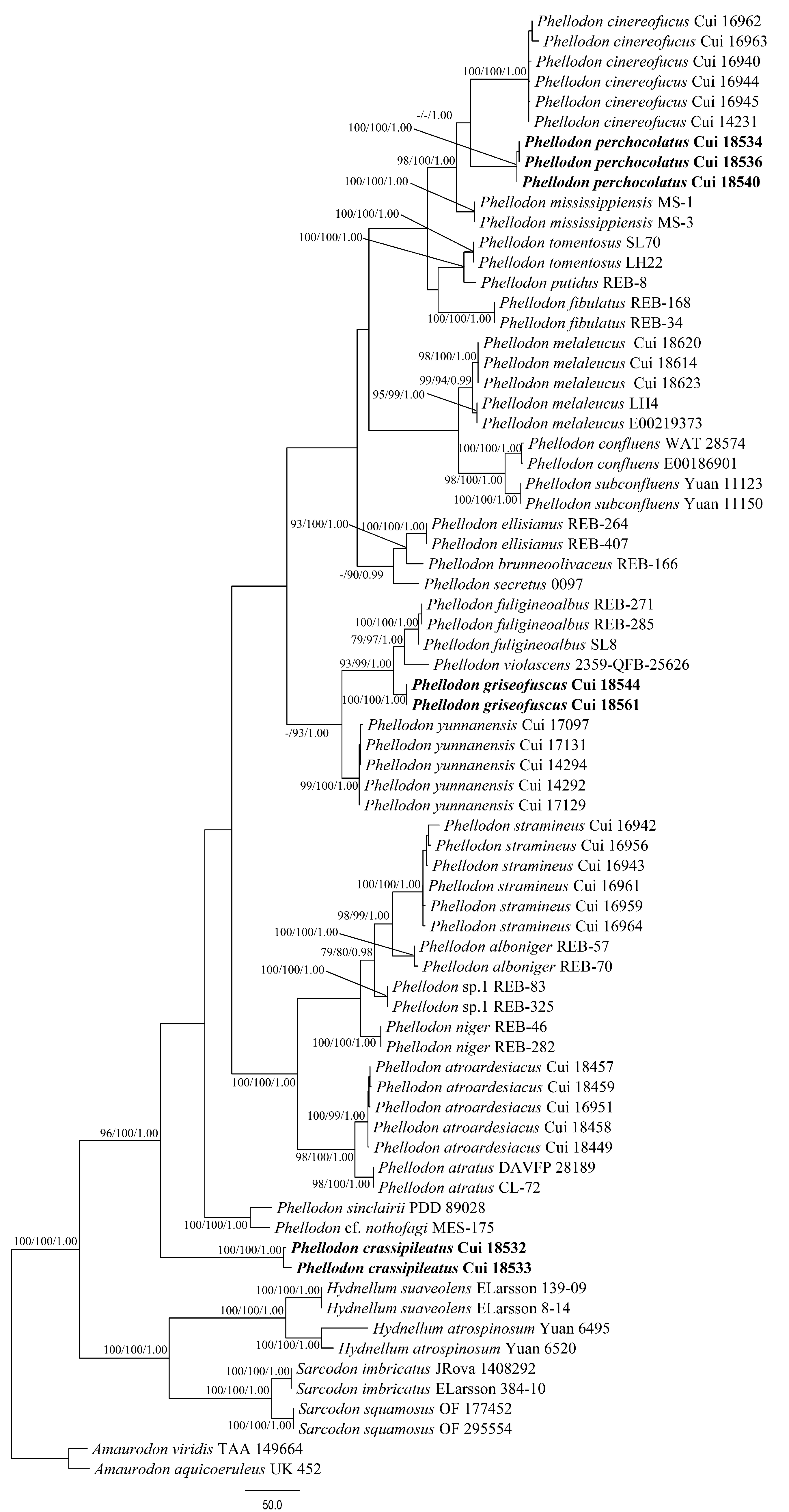
3.1. Phylogenetic Analyses
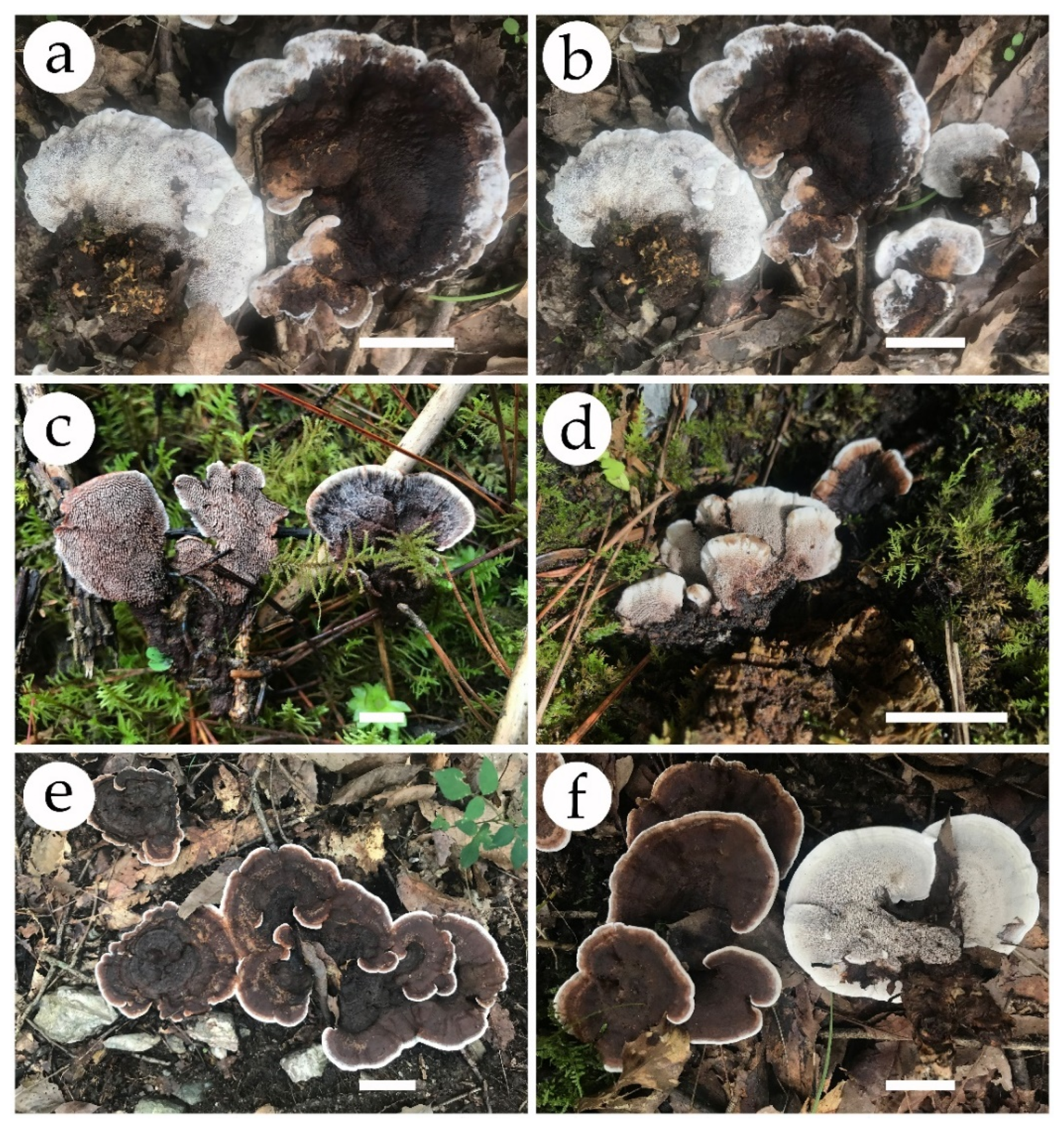
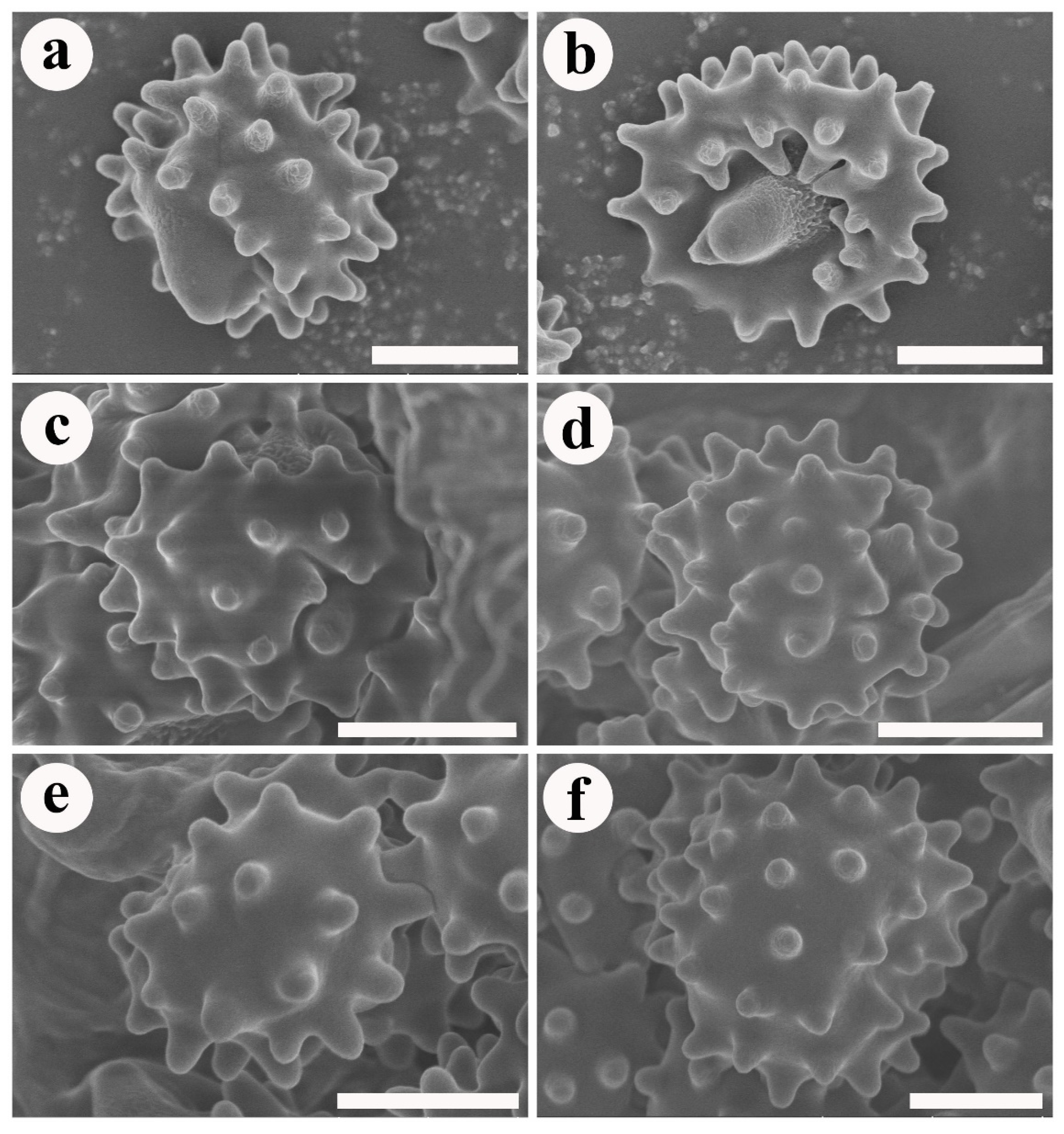
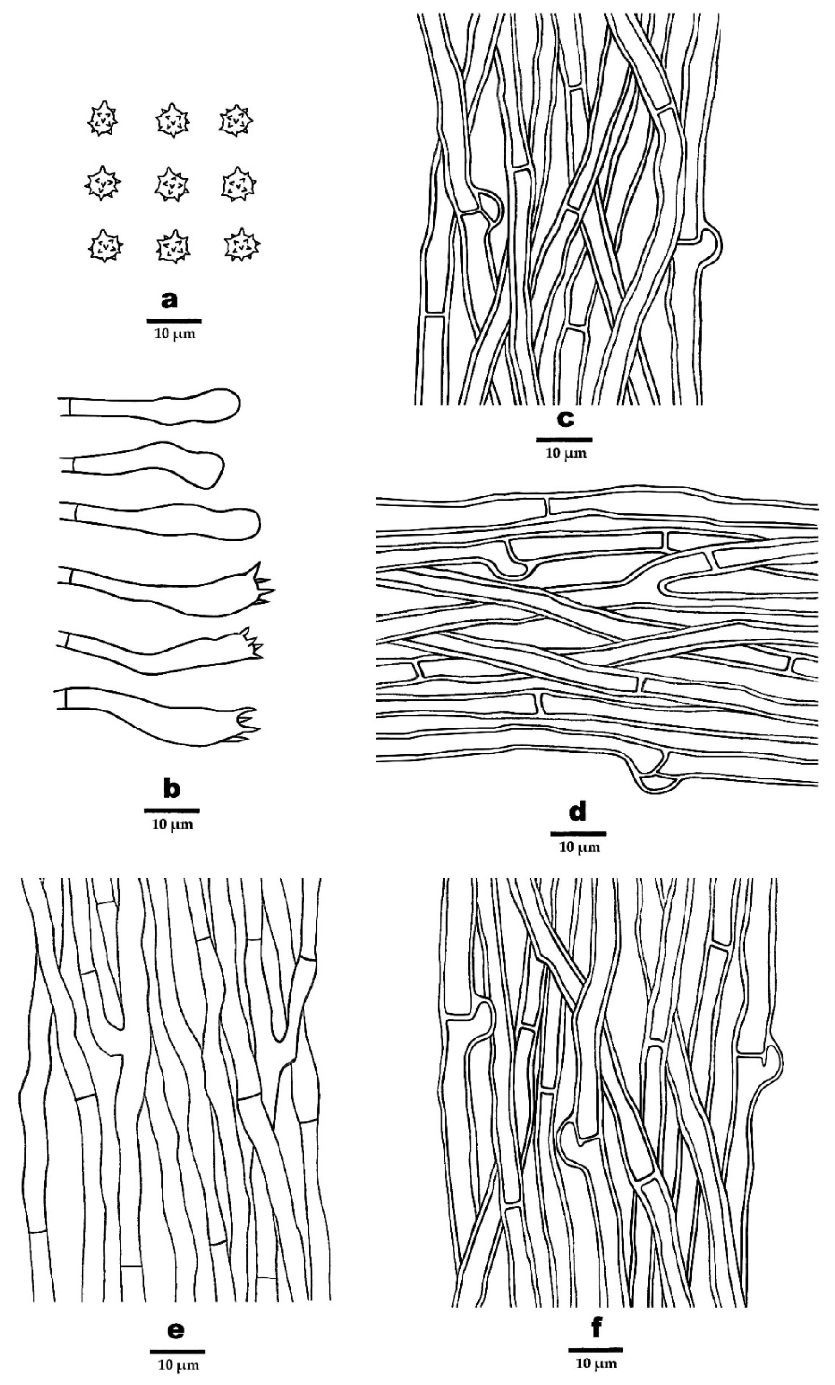
The dataset of ITS included 73 sequences representing 32 taxa. The ITS dataset had an aligned length of 873 characters, of which 376 characters were constant, 46 were variable and parsimony-uninformative, and 451 were parsimony-informative. Maximum parsimony analysis yielded 516 equally parsimonious trees (TL = 1516, CI = 0.547, RI = 0.839, RC = 0.459, HI = 0.453), and 1 of the maximum parsimonious trees is shown in Figure 1. The best fit model selected for these three partitions of ITS sequences was GTR + G for ITS1, JC for 5.8 s, and HKY + G for ITS2. BI resulted in a similar topology with an average standard deviation of split frequencies = 0.007630 to MP analysis. The MP topology is shown with MP (≥75%), ML (≥75%), and BPP (≥0.95) supported values at the nodes (Figure 1).
In the ITS based phylogenetic tree (Figure 1), the three new species P. crassipileatus, P. griseofuscus, and P. perchocolatus formed distinct well-supported lineages distant from other species of Phellodon.
The combined ITS + nLSU + nSSU + RPB1 + RPB2 dataset included sequences from 73 fungal samples representing 32 taxa. The combined dataset had an aligned length of 5639 characters including gaps (873 characters for ITS, 1379 characters for nLSU, 1097 characters for nSSU, 1203 characters for RPB1, 1087 characters for RPB2), of which 4599 characters were constant, 192 were variable and parsimony-uninformative, and 848 were parsimony-informative. Maximum parsimony analysis yielded 12 equally parsimonious trees (TL = 2195, CI = 0.643, RI = 0.866, RC = 0.557, HI = 0.357), and 1 of the maximum parsimonious trees is shown in Figure 2. The best fit model selected for the combined ITS + nLSU + nSSU + RPB1 + RPB2 sequence dataset was GTR + I+G with equal frequency of nucleotides. BI resulted in a similar topology with an average standard deviation of split frequencies = 0.008906 to MP analysis. The MP topology is shown with MP (≥75%), ML (≥75%), and BPP (≥0.95) supported values at the nodes (Figure 2).
The ITS + nLSU + nSSU + RPB1 + RPB2 based phylogenetic tree (Figure 2) produced a topology similar to that generated by the ITS based phylogenetic tree, and confirmed the affinities of the three new species within Phellodon.

3.2. Taxonomy

Phellodon crassipileatus B.K. Cui and C.G. Song, sp. nov., Figure 3a, Figure 4a,b, and Figure 5.
MycoBank: 843670
Diagnosis—This species is characterized by its pale brown to dark brown pileal surface, thick pileus, tomentose pileal margin, white spines, and the presence of clamp connections in generative hyphae of pileal surface, context, and stipe.
Etymologycrassipileatus (Lat.): refers to the thick pileus.
Holotype—CHINA. Sichuan Province, Pingwu County, Bazi, on the ground of forest dominated by trees of Quercus sp., alt. 1190 m, 18 September 2020, Cui 18533 (BJFC 035394).
Fruiting body—Basidiomata annual, centrally or eccentrically stipitate, solitary or gregarious, with a fenugreek odor when dry. Pileus infundibuliform, up to 6.5 cm in diam, 2 cm thick at the center. Pileal surface pale brown to dark brown when fresh, becoming dark brown upon drying, azonate, tomentose at the margin; pileal margin blunt or irregular, white when fresh, becoming cream upon drying, up to 1.2 cm wide. Spines soft, white when fresh, becoming fragile, cream to clay-buff upon drying, up to 3 mm long. Context vinaceous grey, tough, up to 6 mm thick. Stipe brown to dark brown in the outer layer, fuscous in the inner layer, cylindrical, glabrous, up to 1.5 cm long, 1 cm in diameter.
Figure 1. Maximum parsimony (MP) phylogram of the Phellodon species based on ITS sequences data. The supported branches are labeled with parsimony bootstrap values higher than 75%, maximum likelihood bootstrap values higher than 75%, and Bayesian posterior probabilities more than 0.95. Bold names = New species.
Jof 08 00429 g001
Figure 2. Maximum parsimony (MP) phylogram of the Phellodon species based on the combined ITS + nLSU + nSSU + RPB1 + RPB2 sequences data. The supported branches are labeled with parsimony bootstrap values higher than 75%, maximum likelihood bootstrap values higher than 75%, and Bayesian posterior probabilities more than 0.95. Bold names = New species.
Jof 08 00429 g002
Hyphal structure—Hyphal system monomitic; generative hyphae mostly with simple septa, occasionally with clamp connections; all the hyphae IKI–, CB–; tissues turned to olive green in KOH. Generative hyphae in pileal surface pale brown, thick-walled, rarely branched, mostly with simple septa, occasionally with clamp connections, parallel, 2–6 µm in diameter. Generative hyphae in context clay-buff to pale brown, thick-walled, occasionally branched, mostly with simple septa, occasionally with clamp connections, 2–5 µm in diameter. Generative hyphae in spines clay-buff to pale brown, thin-walled, branched, with simple septa, more or less parallel along the spines, 2–4 µm in diameter. Generative hyphae in stipe clay-buff to brown, thick-walled, rarely branched, mostly bearing simple septa, occasionally with clamp connections, parallel along the stipe, 2–6 µm in diameter.
Cystidia—Cystidia and other sterile hyphal elements absent.
Basidia—Basidia clavate, bearing four sterigmata and a basal simple septum, 22–45 × 4–7 µm; sterigmata, 1.5–5 µm; basidioles similar to basidia in shape, but slightly smaller.
Spores—Basidiospores subglobose to globose, hyaline, thin-walled, echinulate, IKI–, CB–, (3.5–) 4–5 × 4–5 µm, L = 4.67 µm, W = 4.19 µm, Q = 1–1.25 (n = 60/2, without the ornamentation).
Additional specimen (paratype) examined—China, Sichuan Province, Pingwu County, Bazi, on the ground of forest dominated by trees of Quercus sp., alt. 1190 m, 18 September 2020, Cui 18532 (BJFC 035393).
Ecological habitsP. crassipileatus was found on the ground of forest dominated by trees of Quercus sp., under a humid monsoon-climate in the northern subtropical region.
Figure 3. Basidiomata of Phellodon species. (a,b) P. crassipileatus, (c,d) P. griseofuscus, and (e,f) P. perchocolatus. Scale bars: 2 cm.
Jof 08 00429 g003
Figure 4. SEM of basidiospores of Phellodon species. (a,b) P. crassipileatus, (c,d) P. griseofuscus, and (e,f). P. perchocolatus. Scale bars: 1.5 µm.
Jof 08 00429 g004
Figure 5. Microscopic structures of P. crassipileatus (drawn from the holotype). (a) Basidiospores, (b) Basidia and basidioles, (c). Hyphae from pileal surface, (d). Hyphae from context, (e). Hyphae from spines, and (f) Hyphae from stipe.
Jof 08 00429 g005
Phellodon griseofuscus B.K. Cui and C.G. Song, sp. nov., Figure 3b, Figure 4c,d, and Figure 6.
MycoBank: 843671.
Diagnosis—This species is characterized by its dark brown to black pileal surface, white to pale brown pileal margin, short spines, generative hyphae with both simple septa and clamp connections in spines, and moderately long basidia.
Etymologygriseofuscus (Lat.): refers to the pale brown to dark brown or blackish basidiomata.
Holotype—China, Sichuan Province, Jiuzhaigou County, Jiuzhaigou Nature Reserve, on the ground of forest dominated by trees of Pinus sp., alt. 2400 m, 20 September 2020, Cui 18561 (BJFC 035422).
Fruiting body—Basidiomata annual, centrally or eccentrically stipitate, solitary or gregarious, with strong odor when dry. Pileus infundibuliform, up to 4 cm in diameter, 5 mm thick at the center. Pileal surface pale brown to dark brown or black when fresh and becoming dark grey to mouse-grey upon drying, azonate, fibrillose; margin blunt or irregular, white to pale brown when fresh, vinaceous grey with age, becoming fuscous upon drying, up to 3 mm wide. Spines soft, white when young, brown with age when fresh, becoming fragile, pale mouse-grey upon drying, up to 1 mm long. Context dark grey, tough, up to 2 mm thick. Stipe fuscous in the outer layer, fuscous to black in the inner layer, cylindrical, glabrous, up to 1.5 cm long, 0.6 cm in diameter.
Hyphal structure—Hyphal system monomitic; generative hyphae mostly with simple septa, occasionally with clamp connections; all the hyphae IKI–, CB–; tissues turned to olive green in KOH. Generative hyphae in pileal surface greyish brown, thick-walled, rarely branched, with simple septa, parallel, 3–6 µm in diameter. Generative hyphae in context pale brown, thick-walled, occasionally branched, with simple septa, parallel, 3–5 µm in diameter. Generative hyphae in spines clay-buff, thin-walled, branched, mostly with simple septa, occasionally with clamp connections, more or less parallel along the spines, 2–4 µm in diameter. Generative hyphae in stipe greyish brown, slightly thick-walled, rarely branched, bearing simple septa, subparallel along the stipe, 2–6 µm in diameter.
Cystidia—Cystidia and other sterile hyphal elements absent.
Basidia—Basidia clavate, bearing four sterigmata and a basal simple septum, 22–55 × 5–6 µm; sterigmata, 1.5–5µm; basidioles similar to basidia in shape, but slightly smaller.
Spores—Basidiospores subglobose to globose, hyaline, thin-walled, echinulate, IKI–, CB–, 4–5 × 3.5–4.5 µm, L = 4.4 µm, W = 4 µm, Q = 1–1.25 (n = 60/2, without the ornamentation).
Additional specimen (paratype) examined—China, Sichuan Province, Jiuzhaigou County, Jiuzhaigou Nature Reserve, on the ground of forest dominated by trees of Pinus sp., alt. 2400 m, 19 September 2020, Cui 18544 (BJFC 035405).
Ecological habitsP. griseofuscus was found on the ground of forest dominated by trees of Pinus sp., under the humid climate of the plateau. This species grows in well-watered bryophytes, which are often interspersed with pine needles.
Figure 6. Microscopic structures of P. griseofuscus (drawn from the holotype). (a) Basidiospores, (b) Basidia and basidioles, (c) Hyphae from pileal surface, (d) Hyphae from context, (e) Hyphae from spines, and (f) Hyphae from stipe.
Jof 08 00429 g006
Phellodon perchocolatus B.K. Cui and C.G. Song, sp. nov., Figure 3c, Figure 4e,f, and Figure 7.
MycoBank: 843672
Diagnosis—This species is characterized by its woody and broad pileus, brown to greyish brown pileal surface when fresh, tomentose pileal margin when young and glabrous after mature, and the presence of both simple septa and clamp connections in generative hyphae of spines.
Etymologyperchocolatus (Lat.): refers to the brown to greyish brown pileal surface.
Holotype—China, Sichuan Province, Pingwu County, Bazi, on the ground of forest dominated by trees of Quercus sp., alt. 1190 m, 18 September 2020, Cui 18536 (BJFC 035397).
Fruiting body—Basidiomata annual, centrally or eccentrically stipitate, solitary or gregarious, with a fenugreek odor when dry. Pileus infundibuliform, woody, up to 9 cm in diam, 6.5 mm thick at the center. Pileal surface brown to greyish brown when fresh, becoming fuscous to black upon drying, zonate, tomentose when young, glabrous after the mature; margin blunt or irregular, white when fresh, becoming buff upon drying, up to 5 mm wide. Spines soft, white when fresh, and becoming fragile, pinkish buff to olivaceous buff upon drying, up to 3 mm long. Context vinaceous grey to greyish brown, tough, up to 3 mm thick. Stipe dark brown to fuscous in the outer layer, fuscous in the inner layer, cylindrical, glabrous, up to 4.8 cm long, 1.9 cm in diameter.
Hyphal structure—Hyphal system monomitic; generative hyphae mostly with simple septa, occasionally with clamp connections; all the hyphae IKI–, CB–; tissues turned to olive-green in KOH. Generative hyphae in pileal surface olivaceous-buff, slightly thick-walled, rarely branched, with simple septa, interwoven, 3–7 µm in diameter. Generative hyphae in context olivaceous-buff, thick-walled, occasionally branched, with simple septa, parallel, 2–6 µm in diameter. Generative hyphae in spines olivaceous-buff, thin-walled, branched, mostly with simple septa, occasionally with clamp connections, more or less parallel along the spines, 2–4 µm in diameter. Generative hyphae in stipe clay-buff, thick-walled rarely branched, bearing simple septa, interwoven in the outer layer, parallel along the stipe in the inner layer, 3–8 µm in diameter.
Cystidia—Cystidia and other sterile hyphal elements absent.
Basidia—Basidia clavate, bearing four sterigmata and a basal simple septum, 16–36 × 5–7 µm; sterigmata, 1–4 µm; basidioles similar to basidia in shape, but slightly smaller.
Spores—Basidiospores subglobose to globose, hyaline, thin-walled, echinulate, IKI–, CB–, 4–5 (–5.5) × (3.5–) 4–4.5 (–5) µm, L = 4.7 µm, W = 4.3 µm, Q = 1–1.25 (n = 90/3, without the ornamentation).
Additional specimens (paratypes) examined—China, Sichuan Province, Pingwu County, Bazi, on the ground of forest dominated by trees of Quercus sp., alt. 1190 m, 18 September 2020, Cui 18534 (BJFC 035394) & Cui 18540 (BJFC 035401).
Ecological habitsP. griseofuscus was found in forest dominated by trees of Quercus sp., under a humid monsoon-climate in the northern subtropical region.
Figure 7. Microscopic structures of P. perchocolatus (drawn from the holotype). (a) Basidiospores, (b) Basidia and basidioles, (c). Hyphae from pileal surface, (d) Hyphae from context, (e) Hyphae from spines, and (f) Hyphae from stipe.
Jof 08 00429 g007
Key to species of Phellodon from China
1. Pileal surface colored straw ------------------------------------------------------- P. stramineus
1. Pileal surface differently -------------------------------------------------------------------------- 2
2. Pileal surface blackish blue to dark grey ----------------------------------- P. atroardesiacus
2. Pileal surface different colored ------------------------------------------------------------------ 3
3. Tissues change color in KOH -------------------------------------------------------------------- 4
3. Tissues unchanged in KOH ----------------------------------------------------- P. subconfluens
4. Clamp connections exist -------------------------------------------------------------------------- 5
4. Clamp connections absent ------------------------------------------------------ P. cinereofuscus
5. Clamp connections exist in spines -------------------------------------------------------------- 6
5. Clamp connections not exist in spines --------------------------------------------------------- 7
6. Spines brown after mature ------------------------------------------------------- P. griseofuscus
6. Spines white after mature --------------------------------------------------------P. perchocolatus
7. Pileal surface tomentose and azonate-----------------------------------------P. crassipilieatus
7. Pileal surface glabrous and zonate----------------------------------------------P. yunnanensis

4. Discussion

In this study, phylogenetic analyses of Phellodon were conducted based on the ITS sequences and the combined ITS + nLSU + nSSU + RPB1 + RPB2 sequences to confirm the affinities of the new species and reveal the relationships of Phellodon species.
Phellodon crassipileatus formed a single lineage different from other species of Phellodon in our phylogenetic analyses (Figure 1 and Figure 2). Morphologically, P. crassipileatus is similar to P. griseofuscus in having infundibuliform and dark brown pileus. However, P. griseofuscus can be distinguished from P. crassipileatus by its fibrillose pileus, brown spines after maturity, presence of clamp connections in generative hyphae of spines, and longer basidia (22–55 × 5–6 µm).
Phellodon griseofuscus is closely related to P. violascens (Alb. and Schwein.) A.M. Ainsw. and P. fuligineoalbus (J.C. Schmidt) R.E. Baird in our phylogenetic analyses (Figure 1 and Figure 2). Phellodon violascens is similar to P. griseofuscus in having solitary or gregarious basidiomata and larger basidiospores measuring 4.5–5.4 × 4.3–4.5 µm [22]. However, P. violascens differs from P. griseofuscus by its white to flesh brown basidiomata and lack of clamp connections. Phellodon fuligineoalbus differs from P. griseofuscus by its yellow-white or light brown basidiomata, lack of clamp connections, and larger basidiospores measuring 4–6 × 4–5 µm [40]. Phellodon yunnanensis B.K. Cui and C.G. may be confused with P. griseofuscus in having white to brown spines. However, P. yunnanensis can be distinct from P. griseofuscus by its smaller basidiospores measuring 3.5–4.5 × 3–4 µm [28] and shorter clavate basidia measuring 24–27 × 6–7 µm [28].
Phellodon perchocolatus and P. cinereofuscus B.K. Cui and C.G. Song clustered together and then grouped with P. mississippiensis R.E. Baird, L.E. Wallace and G. Baker, forming a high supported lineage (98% MP, 100% ML, 1.00 BPP) in our phylogenetic trees (Figure 1 and Figure 2). Morphologically, P. cinereofuscus is similar to P. perchocolatus in having infundibuliform basidiomata and white pileal margin. However, P. cinereofuscus differs from P. perchocolatus by its reddish brown to cinnamon brown pileal surface, glabrous basidiomata and lack of clamp connections [28]. Phellodon mississippiensis is similar to P. perchocolatus in having solitary or gregarious basidiomata, and subglobose to globose basidiospores. However, P. mississippiensis can be distinguished from P. perchocolatus by its white, light orange to light brown pileal surface and shorter basidia measuring 16–22 × 5–6 µm [27]. Moreover, tissues in P. mississippiensis turned light to dark brown in KOH while turned to olive green in P. perchocolatus.

5. Conclusions

This study not only fills in the blank of multiple gene fragments of Phellodon, but also enriches the species diversity of the genus, which will promote the taxonomy and phylogeny of the genus. This is the first step to infer the phylogeny of Phellodon on the basis of multiple genes rather than ITS sequences. Therefore, this study provides a basis for further research on Phellodon. However, only a few species of Phellodon with available multiple genes could be used for the analyses, which limited the systematic study of the genus. For the time being, the best gene marker for the identification of Phellodon is ITS, while more samples with more gene markers including TEF, RPB1, and RPB2 are needed to further investigate the species diversity and phylogenetic relationships of Phellodon species.

Author Contributions

B.-K.C. designed the research; B.-K.C., X.-L.H., D.W., Y.-Y.C., S.L., T.-M.X., and C.-G.S. prepared the samples; C.-G.S., S.L., and T.-M.X. conducted the molecular experiments and analyzed the data; C.-G.S. and B.-K.C. drafted the manuscript. All authors have read and agreed to the published version of the manuscript.

Funding

The research is supported by the National Natural Science Foundation of China (Nos. 31870008, 31900017) and Beijing Forestry University Outstanding Young Talent Cultivation Project (No. 2019JQ03016).

Institutional Review Board Statement

Not applicable.

Informed Consent Statement

Not applicable.

Data Availability Statement

The data and results of this study are available upon reasonable request. Please contact the main author of this publication.

Conflicts of Interest

The authors declare no conflict of interest. The funders had no role in the design of the study; in the collection, analyses, or interpretation of data; in the writing of the manuscript, or in the decision to publish the results.

References

  1. Karsten, P.A. Enumeratio boletinearum et polyporearum fennicarum, systemate novo dispositarum. Rev. Mycol. 1881, 3, 16–19. [Google Scholar]
  2. Stalpers, J.A. The aphyllophoraceous fungi I. keys to the species of the Thelephorales. Stud. Mycol. 1993, 35, 1–168. [Google Scholar]
  3. Pegler, D.N.; Roberst, P.J.; Spooner, B.M. British Chanterelles and Tooth Fungi; Royal Botanic Gardens: Kew, UK, 1997. [Google Scholar]
  4. Parfitt, D.; Ainsworth, A.M.; Simpson, D.; Rogers, H.J.; Boddy, L. Molecular and morphological discrimination of stipitate hydnoids in the genera Hydnellum and Phellodon. Mycol. Res. 2007, 3, 761–777. [Google Scholar] [CrossRef] [PubMed]
  5. Van der Heijden, M.; Klironomos, J.; Ursic, M.; Moutoglis, P.; Streitwolf-Engel, R.; Boller, T.; Wiemken, A.; Sanders, I. Mycorrhizal fungal diversity determines plant biodiversity, ecosystem variability and productivity. Nature 1998, 396, 69–72. [Google Scholar] [CrossRef]
  6. Erland, S.; Taylor, A.F.S. Resupinate ectomycorrhizal fungal genera. In Ectomycorrhizal Fungi Key Genera in Profile; Cairney, J.W.G., Chambers, S.M., Eds.; Springer: Berlin/Heidelberg, Germany, 1999; pp. 347–363. [Google Scholar]
  7. Arnolds, E. Decline of ectomycorrhizal fungi in Europe. Agric. Ecosyst. Environ. 1991, 35, 209–244. [Google Scholar] [CrossRef]
  8. Termorshuizen, A.J.; Schaffers, A.P. The decline of sporocarps of ectomycorrhizal fungi in stands of Pinus sylvestris L. in The Netherlands: Possible causes. Nova Hedwig. 1991, 53, 267–289. [Google Scholar]
  9. Arnolds, E. The fate of hydnoid fungi in The Netherlands and Northwestern Europe. Fungal Ecol. 2010, 3, 81–88. [Google Scholar] [CrossRef]
  10. Holec, J.; Kučera, T. Hydnoid fungi of the family Bankeraceae—Their assemblages and vegetation ecology in Central Europe, Czech Republic. Fungal Ecol. 2018, 32, 40–48. [Google Scholar] [CrossRef]
  11. Larsson, K.H.; Svantesson, S.; Miscevic, D.; Kljalg, U.; Larsson, E. Reassessment of the generic limits for Hydnellum and Sarcodon (Thelephorales, Basidiomycota). MycoKeys 2019, 54, 31–47. [Google Scholar] [CrossRef]
  12. Mu, Y.H.; Wu, F.; Yuan, H.S. Hydnaceous fungi of China 7. Morphological and molecular characterization of Phellodon subconfluens sp. nov. from temperate, deciduous forests. Phytotaxa 2019, 414, 280–288. [Google Scholar] [CrossRef]
  13. Banker, H.J. A contribution to a revision of the North American Hydnaceae. Mem. Torrey Bot. Club 1906, 12, 99–194. [Google Scholar]
  14. Coker, W.C.; Beers, A.H. The Stipitate Hydnums of the Eastern United States; University of North Carolina Press: Chapel Hill, NC, USA, 1951; p. 211. [Google Scholar]
  15. Harrison, K.A. New or little known north American stipitate hydnums. Can. J. Bot. 1964, 42, 1205–1233. [Google Scholar] [CrossRef]
  16. Harrison, K.A. A new species of Phellodon possessing clamp connections. Can. J. Bot. 1972, 50, 1219–1221. [Google Scholar] [CrossRef]
  17. Maas Geesteranus, R.A. The stipitate hydnums of the Netherlands. Fungus 1958, 28, 48–61. [Google Scholar]
  18. Maas Geesteranus, R.A. Notes on the hydnums. Persoonia 1960, 1, 341–384. [Google Scholar]
  19. Maas Geeseranus, R.A. Hyphal structures in the hydnums. Persoonia 1962, 2, 476. [Google Scholar]
  20. Maas Geesteranus, R.A. Hydnaceous fungi of the eastern old world. Verh. Kon. Ned. Akad. Wetensch. Afd. Natuurk 1971, 60, 176. [Google Scholar]
  21. Maas Geesteranus, R.A. The terrestrial hydnums of Europe. Verh. Kon. Ned. Akad. Wetensch. Afd. Natuurk 1975, 65, 127. [Google Scholar]
  22. Hrouda, P. Hydnaceous fungi of the Czech Republic and Slovakia. Czech Mycol. 1999, 51, 99–155. [Google Scholar] [CrossRef]
  23. Hrouda, P. Bankeraceae in Central Europe. 1. Czech Mycol. 2005, 57, 57–78. [Google Scholar] [CrossRef]
  24. Hrouda, P. Bankeraceae in Central Europe. 2. Czech Mycol. 2005, 57, 279–297. [Google Scholar] [CrossRef]
  25. Niemellä, T.; Kinnunen, J.; Renvall, P.; Schigel, D. Phellodon secretus (Basidiomycota), a new hydnaceous fungus from northern pine woodlands. Karstenia 2003, 43, 37–44. [Google Scholar] [CrossRef] [Green Version]
  26. Ainsworth, A.M.; Parfitt, D.; Rogers, H.J.; Boddy, L. Cryptic taxa within European species of Hydnellum and Phellodon revealed by combined molecular and morphological analysis. Fungal Ecol. 2010, 3, 65–80. [Google Scholar] [CrossRef]
  27. Baird, R.E.; Wallace, L.E.; Baker, G. Stipitate hydnums of the southern United States 1: Phellodon mississippiensis sp. nov. Mycotaxon 2013, 123, 183–191. [Google Scholar] [CrossRef]
  28. Song, C.G.; Ji, X.; Liu, S.; He, X.L.; Cui, B.K. Taxonomy and molecular phylogeny of Phellodon (Thelephorales) with descriptions of four new species from Southwest China. Forests 2021, 12, 932. [Google Scholar] [CrossRef]
  29. Wang, M. Taxonomy and Phylogeny of Hydnoid Fungi in Hericiaceae from China. Master’s Thesis, Beijing Forestry University, Beijing, China, 2018. [Google Scholar]
  30. Liu, S.; Han, M.L.; Xu, T.M.; Wang, Y.; Wu, D.M.; Cui, B.K. Taxonomy and phylogeny of the Fomitopsis pinicola complex with descriptions of six new species from east Asia. Front. Microbiol. 2021, 12, 644979. [Google Scholar] [CrossRef]
  31. Sun, Y.F.; Costa-Rezende, D.H.; Xing, J.H.; Zhou, J.L.; Zhang, B.; Gibertoni, T.B.; Gates, G.; Glen, M.; Dai, Y.C.; Cui, B.K. Multi-gene phylogeny and taxonomy of Amauroderma s. lat. (Ganodermataceae). Persoonia 2020, 44, 206–239. [Google Scholar] [CrossRef]
  32. Stöger, A.; Schaffer, J.; Ruppitsch, W. A rapid and sensitive method for direct detection of Erwinia amylovora in symptomatic and asymptomatic plant tissues by polymerase chain reaction. J. Phytopathol. 2006, 154, 469–473. [Google Scholar] [CrossRef]
  33. Thompson, J.D.; Gibson, T.J.; Plewniak, F.; Jeanmougin, F.; Higgins, D.G. The Clustal_X windows interface: Flexible strategies for multiple sequence alignment aided by quality analysis tools. Nucleic Acids Res. 1997, 25, 4876–4882. [Google Scholar] [CrossRef] [Green Version]
  34. Hall, T.A. Bioedit: A user-friendly biological sequence alignment editor and analysis program for Windows 95/98/NT. Nucleic Acids Symp. Ser. 1999, 41, 95–98. [Google Scholar]
  35. Swofford, D.L. PAUP*: Phylogenetic Analysis Using Parsimony (*and Other Methods); Version 4.0b10; Sinauer Associates: Sunderland, MA, USA, 2002. [Google Scholar]
  36. Farris, J.S.; Källersjö, M.; Kluge, A.G.; Kluge, A.G.; Bult, C. Testing significance of incongruence. Cladistics 1995, 10, 315–319. [Google Scholar] [CrossRef]
  37. Felsenstein, J. Confidence intervals on phylogenetics: An approach using bootstrap. Evolution 1985, 39, 783–791. [Google Scholar] [CrossRef] [PubMed]
  38. Posada, D.; Crandall, K.A. Modeltest: Testing the model of DNA substitution. Bioinformatics 1998, 14, 817–818. [Google Scholar] [CrossRef] [PubMed] [Green Version]
  39. Nylander, J.A.A. MrModeltest v2. Program. Distributed by the Author; Evolutionary Biology Center, Uppsala University: Uppsala, Sweden, 2004. [Google Scholar]
  40. Baird, R.E.; Wallace, L.E.; Baker, G.; Scruggs, M. Stipitate hydnoid fungi of the temperate southeastern United States. Fungal Divers. 2013, 62, 41–114. [Google Scholar] [CrossRef]
Table 1. A list of species, specimens and GenBank accession numbers of sequences used in this study.
SpeciesSpecimen No.LocalityGenBank Accession No.
ITSnLSUnSSURPB1RPB2
Amaurodon aquicoeruleusUK 452AustraliaAM490944AM490944---
A. viridisTAA 149664RussiaAM490942AM490942---
Hydnellum atrospinosumYuan 6520ChinaMW579912-MW579912--
H. atrospinosumYuan 6495ChinaMW579938MW579885MW579911--
H. suaveolensELarsson 139-09NorwayMK602734MK602734---
H. suaveolensELarsson 8-14SwedenMK602735MK602735---
Phellodon albonigerREB-70USAKC571749----
P. albonigerREB-57USAJN135206
P. atratusCL-72CanadaMK281471----
P. atratusDAVFP 28189CanadaHQ650766----
P. atroardesiacusCui 18449ChinaMZ221189MZ225598MZ225636--
P. atroardesiacusCui 18457ChinaMZ225577MZ225599MZ225637--
P. atroardesiacusCui 18458ChinaMZ225633MZ225600MZ225638--
P. atroardesiacusCui 18459ChinaMZ225634MZ225601MZ225639--
P. atroardesiacusCui 16951ChinaMZ225632MZ225597MZ225635MZ343209MZ343197
P. brunneoolivaceusREB-166USAKC571752----
P. cinereofuscusCui 14231ChinaMZ225579----
P. cinereofuscusCui 16940AustraliaMZ225580MZ225602MZ225640MZ343210MZ343198
P. cinereofuscusCui 16944ChinaMZ225581MZ225603MZ225641MZ343211MZ343199
P. cinereofuscusCui 16945ChinaMZ225582MZ225604MZ225642--
P. cinereofuscusCui 16962ChinaMZ225583MZ225605MZ225643MZ352084MZ343200
P. cinereofuscusCui 16963ChinaMZ225584MZ225606MZ225644MZ352085MZ343201
P. confluensWAT 28574UKEU622361----
P. confluensE00 186901UKEU622362----
P. crassipileatusCui 18532ChinaOL449267OL439037OL439027--
P. crassipileatusCui 18533ChinaOL449268OL439038OL439028--
P. ellisianusREB-264USAKC571757----
P. ellisianusREB-407USAKC571759----
P. fibulatusREB-168USAJN135205----
P. fibulatusREB-34USAKC571761----
P. fuligineoalbusREB-271USAKC571760----
P. fuligineoalbusREB-285USAJN135196----
P. fuligineoalbusSL8-EU622316----
P. griseofuscusCui 18544ChinaOL449265OL439035OL439025OL456229OL449087
P. griseofuscusCui 18561ChinaOL449266OL439036OL439026--
P. melaleucusLH4UKEU622368----
P. melaleucusE00219373UKEU622369----
P. melaleucusCui 18614ChinaOL449262OL439032OL439022OL456228-
P. melaleucusCui 18620ChinaOL449263OL439033OL439023--
P. melaleucusCui 18623ChinaOL449264OL439034OL439024--
P. mississippiensisMS-1USAJN247563----
P. mississippiensisMS-3USAJN247564----
P. nigerREB-46USAJN135202----
P. nigerREB-282USAKC571766----
P. cf. nothofagiMES-175ChileMH930224----
P. perchocolatusCui 18534ChinaOL449259OL439029OL439020OL456227-
P. perchocolatusCui 18536ChinaOL449260OL439030---
P. perchocolatusCui 18540ChinaOL449261OL439031OL439021--
P. putidusREB-8USAJN135200----
P. secretus0097RussiaMG597404----
P. sinclairiiPDD 89028New ZealandGU222291----
P. stramineusCui 16942ChinaMZ225585MZ225607MZ225645MZ352086-
P. stramineusCui 16943ChinaMZ225586MZ225608MZ225646MZ352087MZ343202
P. stramineusCui 16956ChinaMZ225587MZ225609MZ225647MZ352088MZ343203
P. stramineusCui 16959ChinaMZ225588MZ225610MZ225648MZ352089MZ343204
P. stramineusCui 16961ChinaMZ225589MZ225611MZ225649MZ352090MZ343205
P. stramineusCui 16964ChinaMZ225590MZ225612MZ225650MZ352091-
P. subconfluensYuan 11123ChinaMK677464----
P. subconfluensYuan 11150ChinaMK677465----
Phellodon sp.1REB-83USAKC571747----
Phellodon sp.1REB-325USAKC571748----
P. tomentosusSL70UKEU622381----
P. tomentosusLH22UKEU622382 ---
P. yunnanensisCui 14292ChinaMZ225591----
P. yunnanensisCui 14294ChinaMZ225592----
P. yunnanensisCui 17097ChinaMZ225593MZ225613MZ225651-MZ343206
P. yunnanensisCui 17129ChinaMZ225594MZ225614MZ225652-MZ343207
P. yunnanensisCui 17131ChinaMZ225595MZ225615MZ225653-MZ343208
P. violascens2359-QFB-25626CanadaKM406977----
Sarcodon imbricatusJRova 1408292SwedenMK602746MK602746---
S. imbricatusELarsson 384-10NorwayMK602747MK602747---
S. squamosusOF 177452NorwayMK602768MK602768---
S. squamosusOF 295554NorwayMK602769MK602769---
New sequences are shown in bold.
Publisher’s Note: MDPI stays neutral with regard to jurisdictional claims in published maps and institutional affiliations.

