Article

First Record of Aspergillus fijiensis as an Entomopathogenic Fungus against Asian Citrus Psyllid, Diaphorina citri Kuwayama (Hemiptera: Liviidae)

1
Guangdong Key Laboratory of Animal Conservation and Resource Utilization, Guangdong Public Laboratory of Wild Animal Conservation and Utilization, Institute of Zoology, Guangdong Academy of Science, Guangzhou 510260, China
2
College of Plant Protection, South China Agricultural University, Guangzhou 510642, China
*
Author to whom correspondence should be addressed.
These authors contributed equally to this work.
J. Fungi 2022, 8(11), 1222; https://doi.org/10.3390/jof8111222
Submission received: 5 September 2022 / Revised: 8 November 2022 / Accepted: 8 November 2022 / Published: 19 November 2022
(This article belongs to the Special Issue Plant Fungi: Impact on Agricultural Production)

Abstract

:
The Asian citrus psyllid Diaphorina citri Kuwayama (Hemiptera: Liviidae) is the most widespread and devastating pest species in citrus orchards and is the natural vector of the phloem-limited bacterium that causes Huanglongbing (HLB) disease. Thus, reducing the population of D. citri is an important means to prevent the spread of HLB disease. Due to the long-term use of chemical control, biological control has become the most promising strategy. In this study, a novel highly pathogenic fungal strain was isolated from naturally infected cadavers of adult D. citri. The species was identified as Aspergillus fijiensis using morphological identification and phylogenetic analysis and assigned the strain name GDIZM-1. Tests to detect aflatoxin B1 demonstrated that A. fijiensis GDIZM-1 is a non-aflatoxin B1 producer. The pathogenicity of the strain against D. citri was determined under laboratory and greenhouse conditions. The results of the laboratory study indicated that nymphs from the 1st to 5th instar and adults of D. citri were infected by A. fijiensis GDIZM-1. The mortality of nymphs and adults of D. citri caused by infection with A. fijiensis increased with the concentration of the conidial suspension and exposure time, and the median lethal concentration (LC50) and median lethal time (LT50) values gradually decreased. The mortality of D. citri for all instars was higher than 70%, with high pathogenicity at the 7th day post treatment with 1 × 10⁸ conidia/mL. The results of the greenhouse pathogenicity tests showed that the survival of D. citri adults was 3.33% on the 14th day post-treatment with 1 × 10⁸ conidia/mL, which was significantly lower than that after treatment with the Metarhizium anisopliae GDIZMMa-3 strain and sterile water. The results of the present study revealed that the isolate of A. fijiensis GDIZM-1 was effective against D. citri and it provides a basis for the development of a new microbial pesticide against D. citri after validation of these results in the field.

1. Introduction

The Asian citrus psyllid Diaphorina citri Kuwayama (Hemiptera: Liviidae) is a major pest of Citrus reticulata Blanco, Murraya exotica Jack and other Rutaceae plants [1]. It is also the main transmission vector of citrus Huanglongbing (HLB), which causes huge economic losses to citrus production [2,3]. D. citri has a long lifespan, rapid reproduction and severe generation overlap [4]. At present, chemical pesticides are still considered to be the main means of controlling D. citri [5]. However, the long-term and large-scale use of chemical pesticides will not only increase the resistance of D. citri but also cause environmental pollution, imbalance of the field community structure and other negative effects on the ecosystem [6,7,8]. Therefore, it has become a new research priority to find an environmentally friendly approach for the management of D. citri and to improve the sustainable development of the citrus industry.
Entomopathogenic fungi are pathogenic micro-organisms that can infect and penetrate the host and cause disease, which ultimately leads to the death of insects [9]. In the theory of integrated pest management (IPM), entomopathogenic fungi have become one of the most critical tools in the population control of agricultural pests [10,11,12]. Under natural conditions, entomopathogenic fungi mainly adhere to the surface of the host through spores. Under an appropriate temperature and humidity, conidia absorb water and germinate and differentiate into appressoria and penetration pegs to provide mechanical pressure. At the same time, they also secrete chitinase, protease, lipase and other hydrolases to help hyphae to penetrate the host cuticle and endodermis into the hemolymph [13,14,15]. The mycelial proliferates in the hemocoel, consuming the nutrients in the host, weakening the host insects, reducing their resistance, and producing metabolic toxins, such as destruxin and ciclosporin, inhibiting the cell-mediated immunity defense system of the host, reducing the activity of detoxification enzymes in humoral immunity, destroying the cell morphological structure and physiological function of the host, causing death [16,17,18]. After the death of the insect, entomopathogenic fungi will continue to grow until the hyphae invade all of the tissues and organs and penetrate the host cuticle to produce conidia [12]. Then, the conidia form a new infection cycle by natural transmission, infecting other hosts [19,20].
Entomopathogenic fungi are the largest group of entomopathogenic micro-organisms [21]. They have the advantages of widely existing, a broad-spectrum action, long duration of efficacy, relatively safe for nontarget organisms and less likelihood of the pest developing resistance [22]. Entomopathogenic fungi have become an important resource for controlling agricultural and forestry pests due to their unique insecticidal methods and high efficiency of epidemic potential [9,23]. Currently, more than 1000 species have been reported. For example, Metarhizium anisopliae, Beauveria bassiana and Cordyceps fumosorosea have good control effects on Locusta migratoria Meyen, Aphis gossypii Glover, Spodoptera frugiperda and Trialeurodes vaporariorum Westwood and have been applied to the green control of agricultural pests [9,24,25,26]. In recent years, a large number of entomopathogenic fungi have been reported for the control of D. citri. Paecilomyces variotii, Hirsutella citriformis and Akanthomyces lecanii have the ability to infect D. citri [27,28,29,30]. In addition, B. bassiana and M. anisopliae, which have been commercially produced and applied, can also be used for the control of D. citri. The existing experimental results showed that these two kinds of entomopathogenic fungi could significantly reduce the population density of D. citri and achieve the green control effect of using entomopathogenic fungi to control insects [31,32]. Although some of the entomopathogenic fungi described above have been reported to be effective against D. citri, there is also a need to find new sources of entomopathogenic fungi to develop biological control methods for D. citri. However, there are few reports on the pathogenicity of Aspergillus species against D. citri. Therefore, screening strains with high pathogenicity against D. citri is of great significance for the field control of D. citri and the development of fungal insecticides in the future.
Aspergillus species are diverse fungi that are widely distributed in nature. The most recent research indicates that Aspergillus species are available for the biological control of L. migratoria, Spodoptera litura and Dolichoderus thoracicus, including Aspergillus flavus, Aspergillus nomius and Aspergillus oryzae [33,34,35]. In this study, we collected a fresh, naturally infected adult D. citri cadaver in a lemon orchard. The main aims of our work were (a) to isolate and identify a novel entomopathogenic fungus from D. citri, (b) to detect its production of aflatoxin B1, (c) to study its biological characteristics and determine its pathogenicity against D. citri, and (d) to provide a biological control strategy for D. citri. This study is expected to provide a useful reference for the biological control of D. citri., and to provide a reasonable theoretical basis and technical support for the comprehensive management of pests.

2. Materials and Methods

2.1. D. citri Cadaver Collection and Isolation of Aspergillus Species

A fresh, naturally infected cadaver D. citri adult was collected from a leaf in a lemon orchard, which was located on Maofeng Mountain (113°46′49″ E, 23°29′11″ N), Baiyun District, Guangzhou, China. The D. citri cadaver was infiltrated in 70% ethanol for 30 s, washed with sterile water three times and dried with sterile filter paper. Then, the D. citri cadaver was placed on PDA medium in a biochemical incubator for 1–2 days at 25 ± 1 °C in the dark until mycelia grew around the insect body and then the mycelia were selected and transferred to a new PDA plate for culture. Then, a monoconidial culture was obtained. This isolated and purified strain was designated “GDIZM-1” and deposited in Guangdong Microbial Culture Collection Center (GDMCC) with the conservation number GDMCC 62135.
The GDIZM-1 strain was plated on potato dextrose agar (PDA) medium and cultured in an incubator for 10 days at 25 ± 1 °C. The fungal conidia were suspended in 10 mL sterile water containing 0.1% Tween 80 (v/v), and the conidia were evenly dispersed by magnetic stirrers for 30 min to break up the conidial clumps to ensure a homogenous suspension. The conidial suspension was then filtered. After treatment, an optical microscope (ZEISS, Axio Imager 2, Germany) was used to calculate the total conidial germination rate of the conidia, which should be above 95% [36]. Then, the conidial suspension was adjusted to 1 × 10⁷ conidia/mL suspension.
The adult and nymph of D. citri were immersed in a 1 × 10⁷ conidia/mL suspension for 2 s. The excess conidial suspension was dried with sterile filter paper and the insects were transferred to young leaves. The D. citri were separately reared in an incubator (25 ± 1 °C, 75 ± 5% RH, L:D = 14:10). The infection and spore growth on the 1st- to 5th-instar nymphs and adults of D. citri were observed and recorded with a stereomicroscope (ZEISS, SteREO Discovery. V20, Germany) and identification was based on the phenotypic characteristics and morphology of the mycelia and conidia grown from D. citri [37,38].

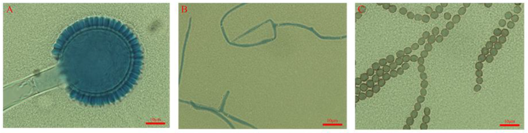
2.2. Morphological Observation

To measure the growth rate and conidial yield, the GDIZM-1 strain was first cultivated on SDAY medium at 27 °C for 10 days. The colony diameter was measured daily. Then, the conidia were collected by filtration from a water suspension containing 0.1% Tween 80 (v/v), and quantified using a hemocytometer. Lactophenol cotton blue staining was used to prepare the slides, and the conidial and sporulation structures of the strain were observed at 100× magnification under an optical microscope (ZEISS, Axio Imager 2, Germany). Both examinations were replicated three times.

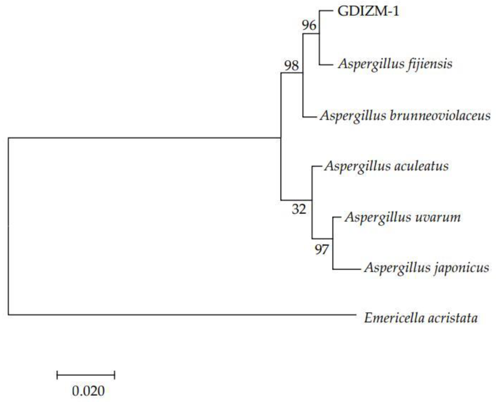
2.3. DNA Extraction and Phylogenetic Analysis of the GDIZM-1 Strain

Total DNA of the GDIZM-1 strain was isolated from samples of the test strains using a fungal DNA kit, following the manufacturer’s instructions (Fungal DNA Kit; Sangon Biotech, Shanghai, China). The DNA-specific sequence of this study consists of three genes: internal transcribed spacer (ITS), translation elongation factor 1-α (TEF1-α) and RNA polymerase II second largest subunit (RPB2). The purified DNA specimens were amplified with primers ITS4-F (5′-TCCTCCGCTTATTGATATGC-3′), ITS5-R (5′-GGAAGTAAAAGTCGTAACAAGG-3′); EF-1983-F (5′-GCYCCYGGHCAYGGTGAYTYAT-3′), EF-12218-R (5′-ATGCACCRACRGCRACRGTYTG-3′); fRPB2–5F (5′-GAYGAYMGWGATCAYTTYGG-3′), fRPB2–7cR (5′-CCCATRGCTTGYTTRCCCAT-3′) [39,40,41,42]. Each PCR mixture (50 μL) contained 25 µL 2 × Ultra Taq PCR MasterMix, 1 µL each primer, 1 µL DNA, and 22 µL ddH2O (TaKaRa, Kusatsu, Shiga, Japan). ITS gene sequence amplification was performed with an initial denaturation of 3 min at 94 °C followed by 35 cycles of 30 s at 94 °C, 30 s at 55 °C, and 45 s at 72 °C and a final extension of 10 min at 72 °C. The TEF1-α gene sequence amplification was performed with an initial denaturation of 10 min at 95 °C followed by 40 cycles of 30 s at 94 °C, 30 s at 55 °C, and 1 min at 72 °C and a final extension of 10 min at 72 °C. The RPB2 gene sequence amplification was performed with an initial denaturation of 10 min at 95 °C followed by 40 cycles of 30 s at 94 °C, 30 s at 50 °C, and 1 min at 72 °C and a final extension of 10 min at 72 °C. The PCR products were separated by 1.0% agarose gel electrophoresis, stained with Gold View in 1 × TAE buffer (Sangon, Shanghai, China), and photographed under UV light. Then, the target PCR products were sent to The Beijing Genomics Institute (BGI; Shenzhen, China) for complete sequencing with PCR primers.
The resulting sequences were checked and aligned using Lasergene v7.1 (DNASTAR, Inc., Madison, Wisconsin USA). The ITS, TEF1-α and RPB2 similarity of the sequences were compared with other fungal homologous sequences (Table 1) using the “BLAST” tool on the National Center for Biotechnology Information website (https://blast.ncbi.nlm.nih.gov/Blast.cgi, accessed on 15 March 2022). Based on the ITS, TEF1 and RPB2 marker genes, the phylogenetic tree was constructed using the maximum likelihood (ML) method of MEGA7: Molecular Evolutionary Genetics Analysis version 7.0 for bigger datasets (Kumar et al. 1994) [43], Institute of Molecular Evolutionary Genetics, Pennsylvania State University, USA. Node support was assessed using a bootstrap procedure of 1000 replicates [44,45]. Emericella acristata strains were used as the outgroup in the phylogenetic analysis.

2.4. Detection of Aflatoxin B1

The presence of aflatoxin B1 was detected by a competitive enzyme-linked immunosorbent assay, according to the manufacturer’s instructions provided with the aflatoxin B1 test kit (EKT-010, Pribolab, Qingdao, China). Extraction and detection were performed according to the manufacturer’s instructions. The GDIZM-1 strain was cultured on PDA medium in an incubator for 10 days at 25 ± 1 °C followed by preparation of 1 × 10⁸ conidia/mL suspension. Sterilized Czapek’s broth, 100 mL, (containing/L: K2HPO4 1 g, FeSO4 0.01 g, Na2SO4 3 g, sucrose 30 g, MgSO4 ∙7H2O 0.5 g, and KCl 0.5 g), added to Erlenmeyer flasks (250 mL), was inoculated with 1 mL of conidial suspension of GDIZM-1 strain followed by incubation at 150 rpm and 27 °C for 1 week following Wu et al. [46]. A culture of the aflatoxin B1 producer A. flavus ATCC 28,539 grown in Czapek’s broth was used as a control. After 1 week of growth, fermentation broth (1 mL) was extracted with 10 mL of methanol for 10 min. Then, mixture was centrifuged in an Eppendorf 5804R centrifuge (Eppendorf, Framingham, MA, USA) at 10,000 rpm for 10 min, and the resultant supernatant was collected. Afterward, 200 μL of clear extract diluted with 800 μL of sample dilution buffer was directly subjected to detection of aflatoxin B1 [47,48]. The optical densities (OD) were measured at 450 nm using an MPP spectrophotometer (PowerWave HT, BioTek, Winooski, USA). The content of aflatoxin B1 in the fermentation broth can be determined by comparing the OD value of the sample with the OD value of the standard product provided by the kit. All standard solutions and sample solutions were analyzed in triplicate wells on a plate, and the whole experiment was conducted three times.

2.5. Plants and Insects for Testing

The Murraya paniculata (L) jack plants used in this study were purchased from Chentian Nursery, Baiyun district, Guangzhou, China and potted in a greenhouse (temperature 25 ± 1 °C, 75 ± 5% RH, L:D =14:10) with nutrient soil as the substrate (perlite:vermiculite:nutrient soil = 1:1:3). These plants were regularly fertilized, watered and pruned.
The colony of D. citri was collected from the campus of Sun Yat-sen University and transferred to M. paniculata seedlings in a greenhouse at the Institute of Zoology, Guangdong Academy of Sciences. Nymphs of D. citri were divided into younger nymphs (1st-2nd instar), middle nymphs (3rd-4th instar) and older nymphs (5th instar) according to their morphological characteristics under a stereomicroscope for uniformity in instar before use in bioassays.

2.6. Pathogenicity Test

2.6.1. Laboratory Bioassays on D. citri

Experiments were performed in an incubator (25 ± 1 °C, 75 ± 5% RH, L:D = 14:10). Six life stages of D. citri were used: younger nymphs (1st-2nd instar), middle nymphs (3rd-4th instar) and older nymphs (5th instar) and mature adults, which were selected for the bioassays. The conidial suspensions of the GDIZM-1 strain were diluted to five concentrations with sterile distilled water (1 × 10⁴, 1 × 10⁵, 1 × 10⁶, 1 × 10⁷, and 1 × 10⁸ conidia/mL). Sterile water containing 0.1% Tween 80 was used as a control (ck). The 20 insects of the different developmental stages of D. citri were immersed in each concentration of conidial suspension or sterile water for 2 s. The excess conidial suspension was dried with sterile filter paper and the insects were transferred to young leaves of M. paniculata and bagged. Each test was replicated three times. The D. citri infected by the GDIZM-1 strain were observed daily. The dead individuals were removed and placed in sterilized Petri dishes to promote fungal growth, and whether it was caused by fungal infection by the GDIZM-1 strain was determined.

2.6.2. Greenhouse Bioassays on D. citri

The pathogenicity determination of the GDIZM-1 strain against D. citri adults was performed in a greenhouse. The Metarhizium anisopliae GDIZMMa-3 strain, which we have already preserved in the laboratory of the Institute of Zoology, Guangdong Academy of Science, was used as a positive control. The conidial suspensions of the GDIZM-1 strain and M. anisopliae GDIZMMa-3 strain were diluted with sterile water to 1 × 10⁸ conidia/mL. A total of 20 D. citri adults were evenly sprayed with the individual conidial suspensions or sterile water containing 0.1% Tween 80 as a control (ck) in each bioassay, with each assay replicated three times. The D. citri infected by the two entomopathogenic fungi were observed daily, and the dead individuals were removed and placed in sterilized Petri dishes filled with moist filter paper to promote fungal growth and confirm the mortality was due to infection by the fungal isolates.

2.7. Data Analysis

The mortality and survival of D. citri after exposure to the tested strains were calculated. The GDIZM-1 strain was used to determine their LC50 and LT50 values on the nymphal stage and adults of D. citri using a biological assay procedure of probit regression analysis with statistical software SPSS 25.0 [49]. They were subjected to one-way analysis of variance (ANOVA) using Duncan’s highly significant difference test at a 95% level of significance. SPSS 25.0 software was used to perform the data analysis and to calculate the homogeneous letters. The results were considered to be statistically significant when p values were <0.05. Student’s t test was used to analyze the differences in the OD values in aflatoxin B1 detection experiments and the mortality difference of D. citri at different developmental stages after treatment with various concentrations of the A. fijiensis GDIZM-1 strain.

3. Results

3.1. Morphological Identification of Infected D. citri

The morphological characteristics of the GDIZM-1 strain are shown in Figure 1. The fungal colonies had radial grooves. The mycelial texture was dense and flat. The frontal side of the colony was white in the early stage and tawny and powdery in the late stage. The color of the mature colony gradually deepened and became earthy yellow (Figure 1). The diameter of the A. fijiensis strain colony was 83.70 mm after 5 days of incubation on SDAY, and the sporulation was 3.86 × 10⁸ conidia/mL after 10 days of incubation (Table 2). The conidiophores had podocytes. The conidiophores were straight with a size of 200–1100 μm × 8–16 μm and inflated to uniseriate globose vesicles with a diameter of 20–60 μm on the top. The conidia were coarsely ellipsoidal to slightly fusiform with a diameter of 3–5 μm and linked into a chain (Figure 2).
The infection test using D. citri nymphs and adults in the laboratory showed that the insects moved slowly and suffered a slight spasm during the early stages of infection. Infected adult D. citri clung tightly to leaves until they were completely covered with hyphae and finally died. Microscopic observation showed hyphae and conidia growing from the intersegmental membranes of the leg and abdomen of infected D. citri after 48–72 h. Then, D. citri was wrapped by mycelia, including the antennae and wings, after 10 days of infection. The morphological identification and infection observation indicated that the GDIZM-1 strain was A. fijiensis (Figure 3).

3.2. Sequencing and Phylogenetic Analysis

DNA fragment sequencing results showed that the ITS gene, TEF1 gene and RPB2 gene of the GDIZM-1 strain were 549 bp, 802 bp and 942 bp, respectively. The DNA sequences were then submitted to GenBank, where they were assigned the accession numbers OM925539, ON000912 and ON000911. The DNA sequences by BLAST comparison in GenBank showed that the GDIZM-1 strain was 99~100% homologous to the A. fijiensis strain. The ITS gene, TEF1 gene and RPB2 gene sequences were concatenated to construct a neighbor-joining tree. Phylogenetic analysis indicated that the GDIZM-1 strain clustered with the A. fijiensis strain clade (Figure 4), which supported our morphological identification that the GDIZM-1 isolate is an A. fijiensis strain.

3.3. Aflatoxin B1 Detection

The standard curve showed a good negative linear relationship between the optical density (OD) values and the concentration of aflatoxin B1 such that the presence of aflatoxin B1 in the sample lowered the OD values, indicating that the detection method used is feasible (Figure 5A). There was no significant difference between the OD values of the metabolite produced by the GDIZM-1 strain grown in Czapek’s broth for 1 week and that of aflatoxin B1 in the standard solution containing 0 ppb aflatoxin B1. In contrast, the OD values of the aflatoxin B1 producer A. flavus ATCC28539 and that of aflatoxin B1 in the standard solution containing 0.1 ppb of aflatoxin B1 were extremely significantly lower when grown under the same conditions as the GDIZM-1 strain (Figure 5B). Based on the above results, we believe that A. fijiensis GDIZM-1 is a non-aflatoxin B1 producer.

3.4. Pathogenicity Analysis of the GDIZM-1 Strain against D. citri

3.4.1. Pathogenicity Determination in the Laboratory

The bioassay results showed that the A. fijiensis GDIZM-1 strain had high pathogenicity to both nymphs and adults of D. citri. The mortality of nymphs and adults of D. citri gradually increased with an increasing conidial concentration (Figure 6). The LC50 values of the nymphs and adults of D. citri were different, from high to low: adult > older nymphs (5th instar) > middle nymphs (3rd-4th instar) > younger nymphs (1st-2nd instar) (Table 3). When treated with a low concentration (1 × 10⁵ conidia/mL) of the A. fijiensis GDIZM-1 strain, after 7 days the mortality of the nymphs and adults of D. citri was more than 45%, which was significantly higher than that of the control (younger nymphs, t = 9.430, p = 0.001; middle nymphs, t = 15.500, p < 0.001; older nymphs, t = 12.500, p < 0.001; adult nymphs, t = 16.971, p < 0.001). With the increase in conidial concentration, the mortality of D. citri increased, and the mortality of the nymphs and adults of D. citri was more than 70% after 7 days when treated with the A. fijiensis GDIZM-1 strain with 1 × 10⁸ conidia/mL, which was significantly higher than that of the control (younger nymphs, t = 22.361, p < 0.001; middle nymphs, t = 24.500, p < 0.001; older nymphs, t = 11.225, p < 0.001; adult nymphs, t = 13.789, p < 0.001).
The results of the lethal time effect of the different conidial concentrations of the A. fijiensis GDIZM-1 strain on the nymphs and adults of D. citri showed that the LT50 of D. citri was significantly shortened with increasing concentrations of the A. fijiensis GDIZM-1 strain (Figure 7A). The LT50 of the nymphs and adults of D. citri were different, from long to short: adult > older nymphs (5th instar) > middle nymphs (3rd-4th instar) > younger nymphs (1st-2nd instar) (Figure 7B). These results show that the A. fijiensis GDIZM-1 strain had high pathogenicity against D. citri and that the mortality of D. citri increased with an increasing conidial concentration and treatment time with the A. fijiensis GDIZM-1 strain. The LT50 and LC50 of D. citri decreased with an increase in the developmental stage of D. citri, during which the A. fijiensis GDIZM-1 strain was applied.

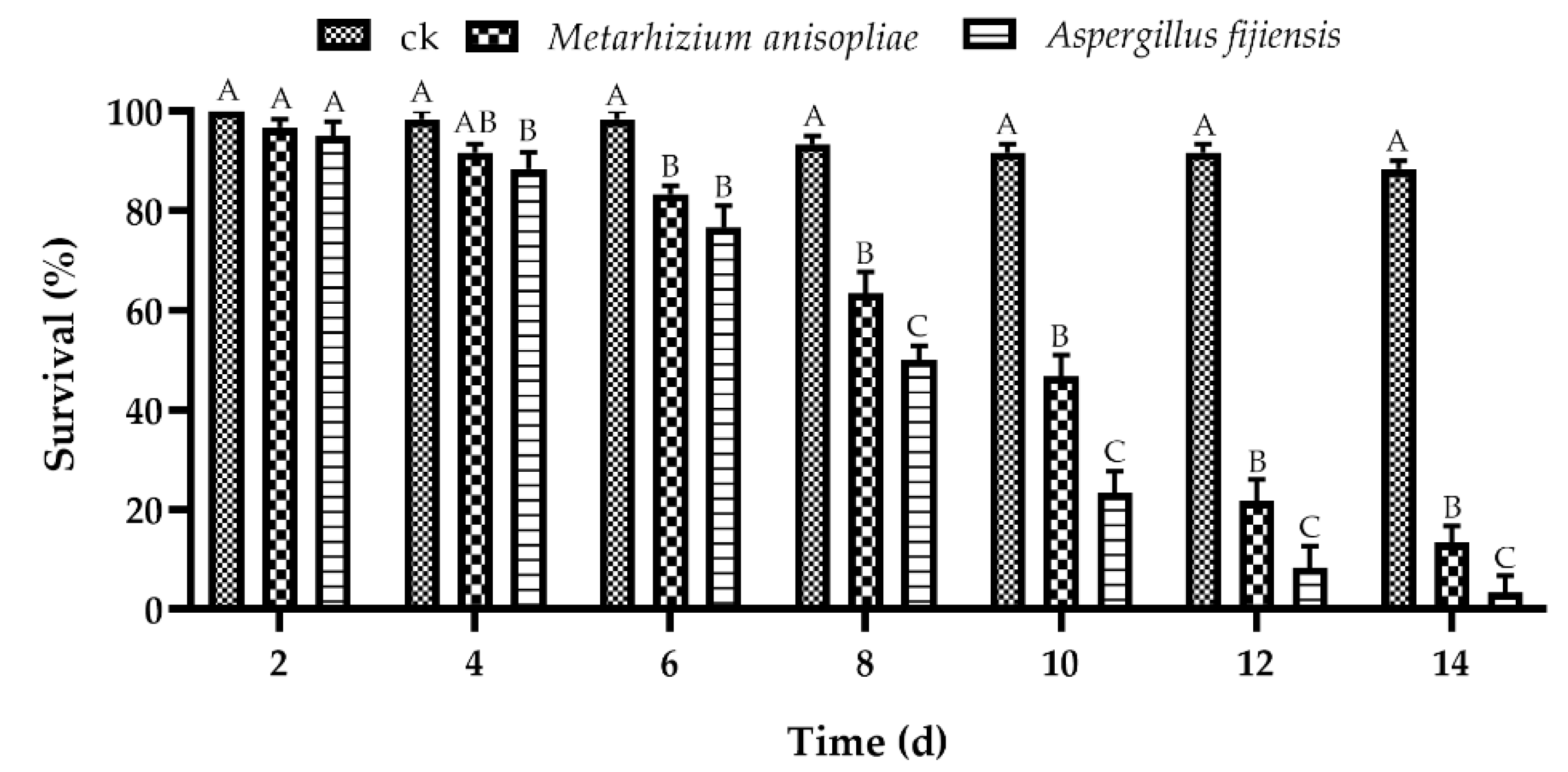
3.4.2. Efficacy of A. fijiensis and M. anisopliae against D. citri in Greenhouse Trials

In this study, we evaluated the pathogenicity of the A. fijiensis GDIZM-1 strain and the M. anisopliae GDIZMMa-3 strain against D. citri at 1 × 10⁸ conidia/mL. The results showed that the survival of D. citri adults decreased with increasing treatment time (Figure 8). The insecticidal effects of the A. fijiensis GDIZM-1 strain and M. anisopliae GDIZMMa-3 strain were relatively slow, but gradually increased with time, and the insecticidal effect of the A. fijiensis GDIZM-1 strain against D. citri was significantly higher than that of the M. anisopliae GDIZMMa-3 strain. When infected with the A. fijiensis GDIZM-1 strain, the survival of D. citri adults was only 3.33% on the 14th day, which was significantly lower than the survival of the M. anisopliae GDIZMMa-3 strain and the control (F = 259.00, df = 2, 6, p < 0.001). These results showed that the A. fijiensis GDIZM-1 strain had better pathogenicity against D. citri adults than the M. anisopliae GDIZMMa-3 strain under greenhouse conditions.

4. Discussion

D. citri is an important vector involved in the natural spread of HLB disease, which is a devastating disease of citrus. Currently, there are no effective control measures for HLB disease, so the prevention and control of D. citri and stopping the infection cycle of HLB disease pathogens are important means for the comprehensive management of HLB disease [50,51]. At present, the majority of farmers mainly rely on chemical pesticides for the management of agricultural pests [52], but the side effects of the continuous use of chemical insecticides have attracted people’s attention to biological control. Entomopathogenic fungi are widely found in nature and are a green and safe natural biological resource. Entomopathogenic fungi have become one of the most critical tools to control D. citri due to their wide host range and environmental friendliness [53,54].
Most fungi reproduce in sexual and asexual ways, and their sexual spores and surrounding tissue structure are the main basis for the classification of fungi. However, the sexual stage of some fungi degenerates and disappears without forming sporulation structures, and the sexual reproduction stage during growth is not always easy to observe [51]. Therefore, asexual conidial and sporulation structures have become an important basis for the identification of fungal strains [55,56]. However, the morphological characteristics cannot always accurately distinguish closely related species of fungi [57,58]. The ITS gene has been found to be a suitable site for identifying Aspergillus genus, but it is not sufficient to distinguish related species so a multi-locus approach is needed [59].
In this study, we identified and isolated an entomopathogenic fungus, GDIZM-1, from a cadaver of D. citri through field investigation in a lemon orchard. Based on ITS, TEF1 and RPB2 sequences were used to construct a multigene phylogenetic tree, which has a higher reliability than a single-gene phylogenetic tree. Finally, through morphological identification and molecular identification, it was determined that the pathogenic fungus was a new entomopathogenic strain of the species A. fijiensis. The colony morphological characteristics and phenotype of the A. fijiensis GDIZM-1 strain were very similar to the reported descriptions of A. fijiensis strains, and its colony colors, colony textures and conidial surfaces were consistent with the results of Varga et al. [60]. This species was first reported in the USA. It has been found in soil on the Fiji Islands and on Lactuca sative in Indonesia and in indoor air, but it has never been reported in China in the past. In addition, it has a substantial economic value, as it includes fermenters of foodstuffs and key cell factories for the production of β-fructofuranosidase [60,61].
The insecticidal mechanisms of entomopathogenic fungi mainly include two types: one is that fungal pathogens proliferate and grow in large numbers after parasitism on healthy insects, absorb nutrients from the host, and finally lead to the death of the host due to lack of nutrition; the other is that the pathogenic fungi produce a variety of toxic metabolites, resulting in the death of the insects by poisoning [9,19]. The toxins and fungal metabolites produced by Aspergillus species can also be used to control some pests. For example, mycotoxins, such as aflatoxin B, are toxic to Periplaneta americana, and ochratoxin, citrinin and patulin are toxic to Drosophila melanogaster [62]. Mensah and Young [63] showed that oil-based extracts of Aspergillus species were toxic to Bemisia tabaci adults. Kaur et al. [64] reported that an ethyl acetate extract of Aspergillus niger adversely affected the survival and development of Spodoptera litura and showed antifeedant and toxic effects of A. niger metabolites. The results of this study showed that the infected D. citri showed slow movement, slight spasms and convulsions and eventually led to death. At present, the cause of death of the infected D. citri remains unclear, and further research is needed.
Numerous studies have reported that Aspergillus species have high pathogenicity against different insects, indicating that the fungus has promising biological control potential in pest management [32,33,34]. The results of our bioassay showed that with the increase in the conidial concentration of the A. fijiensis GDIZM-1 strain, the mortality of D. citri increased, and the median lethal time was shortened. It had high pathogenicity against both nymphs and adults of D. citri. Its pathogenicity against D. citri was comparable to that of Cordyceps javanica, Hirsutella citriformis and C. fumosorosea isolated from D. citri, and the growth rate and spore yield are higher than those of C. javanica [20,21,65]. This is similar to the control effect of azadirachtin and other commonly used pesticides on D. citris [66], so it has the potential to be developed into a new biopesticide resource.
Comparing the virulence results of the A. fijiensis GDIZM-1 strain to the nymphs and adults of D. citri, it was found that its insecticidal ability against younger nymphs of D. citri was higher than that against older nymphs and adults, which may be related to the nutrition and structure of the integument, defense mechanism and microflora composition on the body surface at different developmental stages of D. citri [67,68,69]. The nymphs of D. citri have weak activity on twigs or buds and are easily infected by fungal spores. Moreover, the honeydew secreted by D. citri provides nutrients and favorable conditions for the infection cycle of fungal spores. Therefore, the best opportunity to use entomopathogenic fungi to control D. citri is the peak period of younger nymphs to reduce the population of D. citri rapidly.
The results of greenhouse trials showed that the pathogenicity of the A. fijiensis GDIZM-1 strain against D. citri under semifield conditions was lower than that of the indoor effect, which may be related to environmental factors, such as temperature, humidity and ultraviolet radiation [70,71]. Compared with the control effect of the M. anisopliae GDIZMMa-3 strain against D. citri, the A. fijiensis GDIZM-1 strain had significant advantages, which may be due to strain distinctions, host species, temperature, and soil type in various studies [72,73]. Next, it is necessary to verify its specific prevention effect in the field. At the same time, it can also be combined or alternatively used with oil emulsions and chemical pesticides to reduce pesticide residues and pest resistance and achieve the reduction and synergistic effect of pesticides [74].
The green control of pests is the basic concept of pest control in recent years, and the use of entomopathogenic fungi to control agricultural pests has become an important mean. In the present study, the results showed that A. fijiensis GDIZM-1 had a good lethal effect against D. citri at different developmental stages. Furthermore, researchers found that Aflatoxins belong to a group of toxic and carcinogenic secondary metabolites produced by Aspergillus species [75,76,77,78], which pose major health and economic problems worldwide [79,80,81,82]. Aflatoxin B1 has been classified as a Group 1 carcinogen by the International Agency for Research on Cancer [83]. Since the aflatoxin B1 has strong carcinogenicity and toxicity, these potential risks should be considered before utilizing A. fijiensis GDIZM-1 to control D. citri near human populations. However, the aflatoxin B1 detection results showed that the A. fijiensis GDIZM-1 strain is a non-aflatoxin B1 producer. The present study provided the first systematic report of an A. fijiensis GDIZM-1 strain as an entomopathogenic fungus and a newly discovered pathogen of D. citri. We consider that A. fijiensis GDIZM-1 has the potential to be developed into a biocontrol agent. Currently, the safety of the A. fijiensis GDIZM-1 strain against humans and nontarget organisms is not clear. Therefore, it is necessary not only to evaluate whether it substances harmful to humans and animals and whether it can have a negative impact on the environment and ecology but also to verify its actual control effect in the field. Only after the above issues are clarified can we further develop this new resource and promote the application of biological pesticides.

5. Conclusions

In this study, we isolated and identified the A. fijiensis GDIZM-1 strain with high pathogenicity against D. citri from naturally infected cadavers of D. citri adults, which enriches the existing resource library of entomopathogenic fungi. The results of the laboratory and semifield bioassays show that the A. fijiensis GDIZM-1 strain has promising biological control effects on D. citri and has the potential to be developed into a new biological control agent. Our study is expected to provide a biocontrol option for D. citri.

Author Contributions

Conceptualization, X.M.; Methodology, G.O. and A.I.; Software, H.L. (Hao Liuand); H.L. (Huilin Lu) and F.C.; Validation, J.Y., F.C. and H.L. (Huilin Lu); Formal analysis, J.Y. and A.I.; Investigation, J.Y. and F.C.; Resources, X.M. and G.O.; Data curation, J.Y., H.L. (Hao Liuand) and H.L. (Huilin Lu); Writing—original draft preparation, J.Y. and A.I.; Writing—review and editing, H.L. (Hao Liuand) and A.I.; Supervision, X.M.; Project administration, X.M. and G.O.; Funding acquisition, X.M. and G.O. All authors have read and agreed to the published version of the manuscript.

Funding

This research was funded by grants from the open competition program of top ten critical priorities of Agricultural Science and Technology Innovation for the 14th Five-Year Plan of Guangdong Province (2022SDZG06), The Innovation Team Project of Modern Agricultural Industrial Technology System of Guangdong Province (2022KJ113) and the Natural Science Foundation of Guangdong Province (2019A1515011776).

Institutional Review Board Statement

Not applicable.

Informed Consent Statement

Not applicable.

Data Availability Statement

The raw data supporting the conclusions of this manuscript will be made available by the authors, without undue reservation, to any qualified researcher upon request.

Conflicts of Interest

The authors declare no conflict of interest. The funders had no role in the design of the study; in the collection, analyses, or interpretation of the data; in the writing of the manuscript; or in the decision to publish the results.

References

  1. Nava, D.E.; Torres, M.L.G.; Rodrigues, M.D.L.; Bento, J.M.S.; Parra, J.R.P. Biology of Diaphorina citri (Hem., Psyllidae) on different hosts and at different temperatures. J. Appl. Entomol. 2007, 131, 709–715. [Google Scholar] [CrossRef]
  2. Hall, D.G.; Richardson, M.L.; Ammar, E.D.; Halbert, S.E. Asian citrus psyllid, Diaphorina citri, vector of citrus huanglongbing disease. Entomol. Exp. Appl. 2013, 146, 207–223. [Google Scholar] [CrossRef]
  3. Ammar, E.D.; Hall, D.G.; Hosseinzadeh, S.; Heck, M. The quest for a non-vector psyllid: Natural variation in acquisition and transmission of the huanglongbing pathogen ‘Candidatus Liberibacter asiaticus’ by Asian citrus psyllid isofemale lines. PLoS ONE 2018, 13, e0195804. [Google Scholar] [CrossRef] [PubMed]
  4. Tsai, J.H.; Liu, Y.H. Biology of Diaphorina citri (Homoptera: Psyllidae) on four host plants. J. Econ. Entomol. 2000, 93, 1721–1725. [Google Scholar] [CrossRef] [PubMed]
  5. Boina, D.R.; Bloomquist, J.R. Chemical control of the Asian citrus psyllid and of huanglongbing disease in citrus. Pest Manag. Sci. 2015, 71, 808–823. [Google Scholar] [CrossRef]
  6. Chen, X.D.; Stelinski, L.L. Resistance management for Asian citrus psyllid, Diaphorina citri Kuwayama, in Florida. Insects 2017, 8, 103. [Google Scholar] [CrossRef] [Green Version]
  7. Naeem, A.; Afzal, M.B.S.; Freed, S.; Hafeez, F.; Zaka, S.M.; Ali, Q.; Anwar, H.M.Z.; Iftikhar, A.; Nawaz, M. First report of thiamethoxam resistance selection, cross resistance to various insecticides and realized heritability in Asian citrus psyllid Diaphorina citri from Pakistan. Crop Prot. 2019, 121, 11–17. [Google Scholar] [CrossRef]
  8. Li, Z.; Zhang, Y.; Zhao, Q.; Wang, C.; Cui, Y.; Li, J.; Chen, A.; Liang, G.; Jiao, B. Occurrence, temporal variation, quality and safety assessment of pesticide residues on citrus fruits in China. Chemosphere 2020, 258, 127381. [Google Scholar] [CrossRef]
  9. Idrees, A.; Afzal, A.; Qadir, Z.A.; Li, J. Bioassays of Beauveria bassiana Isolates against the Fall Armyworm, Spodoptera frugiperda. J. Fungi 2022, 8, 717. [Google Scholar] [CrossRef]
  10. Kumar, S.; Kumar, S.; Bhandari, D.; Gautam, M.P. Entomopathogens, pathological symptoms and their role in present scenario of agriculture: A review. Int. J. Curr. Microbiol. App. Sci. 2020, 9, 2110–2124. [Google Scholar] [CrossRef]
  11. Deka, B.; Baruah, C.; Babu, A. Entomopathogenic microorganisms: Their role in insect pest management. Egypt. J. Biol. Pest Control 2021, 31, 121. [Google Scholar] [CrossRef]
  12. Idrees, A.; Qadir, Z.A.; Akutse, K.S.; Afzal, A.; Hussain, M.; Islam, W.; Waqas, M.S.; Bamisile, B.S.; Li, J. Effectiveness of Entomopathogenic Fungi on Immature Stages and Feeding Performance of Fall Armyworm, Spodoptera frugiperda (Lepidoptera: Noctuidae) Larvae. Insects 2021, 12, 1044. [Google Scholar] [CrossRef] [PubMed]
  13. Wosten, H.A.B. Hydrophobins: Multipurpose proteins. Annu. Rev. Microbiol. 2001, 55, 625–646. [Google Scholar] [CrossRef] [Green Version]
  14. Holder, D.J.; Keyhani, N.O. Adhesion of the entomopathogenic fungus Beauveria (Cordyceps) bassiana to substrata. Appl. Environ. Microbiol. 2005, 71, 5260–5266. [Google Scholar] [CrossRef] [PubMed] [Green Version]
  15. Ortiz-Urquiza, A.; Keyhani, N.O. Action on the surface: Entomopathogenic fungi versus the insect cuticle. Insects 2013, 4, 357–374. [Google Scholar] [CrossRef] [PubMed] [Green Version]
  16. Gillespie, J.P.; Bailey, A.M.; Cobb, B.; Vilcinskas, A. Fungi as elicitors of insect immune responses. Arch. Insect Biochem. Physiol. 2000, 44, 49–68. [Google Scholar] [CrossRef]
  17. Kershaw, M.J.; Moorhouse, E.R.; Bateman, R.; Reynolds, S.E.; Charnley, A.K. The role of destruxins in the pathogenicity of Metarhizium anisopliae for three species of insect. J. Invertebr. Pathol. 1999, 74, 213–223. [Google Scholar] [CrossRef]
  18. Qu, S.; Wang, S. Interaction of entomopathogenic fungi with the host immune system. Dev. Comp. Immunol. 2018, 83, 96–103. [Google Scholar] [CrossRef]
  19. Mora, M.A.E.; Castilho, A.M.C.; Fraga, M.E. Classification and infection mechanism of entomopathogenic fungi. Arq. Inst. Biol. 2018, 84, e0552015. [Google Scholar] [CrossRef] [Green Version]
  20. Conceschi, M.R.; D’Alessandro, C.P.; Moral, R.d.A.; Demétrio, C.G.B.; Júnior, I.D. Transmission potential of the entomopathogenic fungi Isaria fumosorosea and Beauveria bassiana from sporulated cadavers of Diaphorina citri and Toxoptera citricida to uninfected D. citri adults. BioControl 2016, 61, 567–577. [Google Scholar] [CrossRef]
  21. Ou, D.; Zhang, L.H.; Guo, C.F.; Chen, X.S.; Ali, S.; Qiu, B.L. Identification of a new Cordyceps javanica fungus isolate and its toxicity evaluation against Asian citrus psyllid. Microbiologyopen 2019, 8, e00760. [Google Scholar] [CrossRef] [PubMed] [Green Version]
  22. Islam, W.; Adnan, M.; Shabbir, A.; Naveed, H.; Abubakar, Y.S.; Qasim, M.; Tayyab, M.; Noman, A.; Nisar, M.S.; Khan, K.A.; et al. Insect-fungal-interactions: A detailed review on entomopathogenic fungi pathogenicity to combat insect pests. Microb. Pathog. 2021, 159, 105122. [Google Scholar] [CrossRef] [PubMed]
  23. Batta, Y.A.; Kavallieratos, N.G. The use of entomopathogenic fungi for the control of stored-grain insects. Int. J. Pest Manag. 2017, 64, 77–87. [Google Scholar] [CrossRef]
  24. Jiang, W.; Peng, Y.; Ye, J.; Wen, Y.; Liu, G.; Xie, J. Effects of the entomopathogenic fungus Metarhizium anisopliae on the mortality and immune response of Locusta migratoria. Insects 2019, 11, 36. [Google Scholar] [CrossRef] [PubMed] [Green Version]
  25. Im, Y.; Park, S.E.; Lee, S.Y.; Kim, J.C.; Kim, J.S. Early-stage defense mechanism of the cotton aphid Aphis gossypii against infection with the insect-killing fungus Beauveria bassiana JEF-544. Front. Immunol. 2022, 13, 907088. [Google Scholar] [CrossRef]
  26. Kim, J.S.; Je, Y.H.; Skinner, M.; Parker, B.L. An oil-based formulation of Isaria fumosorosea blastospores for management of greenhouse whitefly Trialeurodes vaporariorum (Homoptera: Aleyrodidae). Pest Manag. Sci. 2013, 69, 576–581. [Google Scholar] [CrossRef]
  27. Cortez-Madrigal, H.; Sánchez-Saavedra, J.M.; Díaz-Godínez, G.; Mora-Aguilera, G. Enzymatic activity and pathogenicity of entomopathogenic fungi from central and southeastern Mexico to Diaphorina citri (Hemiptera: Psyllidae). Southwest. Entomol. 2014, 39, 491–502. [Google Scholar] [CrossRef]
  28. Gandarilla-Pacheco, F.L.; Galán-Wong, L.J.; López-Arroyo, J.I.; Rodríguez-Guerra, R.; Quintero-Zapata, I. Optimization of pathogenicity tests for selection of native isolates of entomopathogenic fungi isolated from citrusgrowing areas of México on adults of Diaphorina citri Kuwayama (Hemiptera: Liviidae). Fla. Entomol. 2013, 96, 187–195. [Google Scholar] [CrossRef]
  29. Hussain, M.; Akutse, K.S.; Lin, Y.; Chen, S.; Huang, W.; Zhang, J.; Idrees, A.; Qiu, D.; Wang, L. Susceptibilities of Candidatus Liberibacter asiaticus-infected and noninfected Diaphorina citri to entomopathogenic fungi and their detoxification enzyme activities under different temperatures. Microbiologyopen 2018, 7, e00607. [Google Scholar] [CrossRef]
  30. Naeem, A.; Freed, S.; Akmal, M. Biochemical analysis and pathogenicity of entomopathogenic fungi to Diaphorina citri Kuwayama (Hemiptera: Liviidae). Entomol. Res. 2020, 50, 245–254. [Google Scholar] [CrossRef]
  31. Ausique, S.J.J.; D’Alessandro, C.P.; Conceschi, M.R.; Mascarin, G.M.; Delalibera, I. Efficacy of entomopathogenic fungi against adult Diaphorina citri from laboratory to field applications. J. Pest Sci. 2017, 90, 947–960. [Google Scholar] [CrossRef]
  32. Ibarra-Cortes, K.H.; Guzman-Franco, A.W.; Gonzalez-Hernandez, H.; Ortega-Arenas, L.D.; Villanueva-Jimenez, J.A.; Robles-Bermudez, A. Susceptibility of Diaphorina citri (Hemiptera: Liviidae) and its parasitoid Tamarixia radiata (Hymenoptera: Eulophidae) to entomopathogenic fungi under laboratory conditions. Neotrop. Entomol. 2018, 47, 131–138. [Google Scholar] [CrossRef] [PubMed]
  33. Zhang, P.; You, Y.; Song, Y.; Wang, Y.; Zhang, L. First record of Aspergillus oryzae (Eurotiales: Trichocomaceae) as an entomopathogenic fungus of the locust, Locusta migratoria (Orthoptera: Acrididae). Biocontrol Sci. Technol. 2015, 25, 1285–1298. [Google Scholar] [CrossRef]
  34. Lin, W.J.; Chiu, M.C.; Lin, C.C.; Chung, Y.K.; Chou, J.Y. Efficacy of Entomopathogenic fungus Aspergillus nomius against Dolichoderus thoracicus. BioControl 2021, 66, 463–473. [Google Scholar] [CrossRef]
  35. Karthi, S.; Vaideki, K.; Shivakumar, M.S.; Ponsankar, A.; Thanigaivel, A.; Chellappandian, M.; Vasantha-Srinivasan, P.; Muthu-Pandian, C.K.; Hunter, W.B.; Senthil-Nathan, S. Effect of Aspergillus flavus on the mortality and activity of antioxidant enzymes of Spodoptera litura Fab.(Lepidoptera: Noctuidae) larvae. Pestic. Biochem. Physiol. 2018, 149, 54–60. [Google Scholar] [CrossRef]
  36. Ali, S.; Zhang, C.; Wang, Z.Q.; Wang, X.M.; Wu, J.H.; Cuthbertson, A.G.S.; Shao, Z.F.; Qiu, B.L. Toxicological and biochemical basis of synergism between the entomopathogenic fungus Lecanicillium muscarium and the insecticide matrine against Bemisia tabaci (Gennadius). Sci. Rep. 2017, 7, 46558. [Google Scholar] [CrossRef]
  37. Barra, P.; Rosso, L.; Nesci, A.; Etcheverry, M. Isolation and identification of entomopathogenic fungi and their evaluation against Tribolium confusum, Sitophilus zeamais, and Rhyzopertha dominica in stored maize. J. Pest Sci. 2012, 86, 217–226. [Google Scholar] [CrossRef]
  38. Wang, W.; Zhou, L.; Dong, G.; Chen, F. Isolation and identification of entomopathogenic fungi and an evaluation of their actions against the larvae of the fall webworm, Hyphantria cunea (Drury)(Lepidoptera: Arctiidae). BioControl 2019, 65, 101–111. [Google Scholar] [CrossRef]
  39. White, T.J.; Bruns, T.; Lee, S.J.W.T.; Taylor, J. Amplification and direct sequencing of fungal ribosomal RNA genes for phylogenetics. PCR Protoc. Guide Methods Appl. 1990, 18, 315–322. [Google Scholar]
  40. Carbone, I.; Kohn, L.M. A method for designing primer sets for speciation studies in filamentous ascomycetes. Mycologia 1999, 91, 553–556. [Google Scholar] [CrossRef]
  41. Rehner, S.A.; Minnis, A.M.; Sung, G.H.; Luangsa-ard, J.J.; Devotto, L.; Humber, R.A. Phylogeny and systematics of the anamorphic, entomopathogenic genus Beauveria. Mycologia 2011, 103, 1055–1073. [Google Scholar] [CrossRef] [PubMed]
  42. Liu, Y.J.; Whelen, S.; Hall, B.D. Phylogenetic relationships among ascomycetes: Evidence from an RNA polymerse II subunit. Mol. Biol. Evol. 1999, 16, 1799–1808. [Google Scholar] [CrossRef] [PubMed] [Green Version]
  43. Kumar, S.; Tamura, K.; Nei, M. MEGA: Molecular Evolutionary Genetics Analysis software for microcomputers. Bioinformatics 1994, 10, 189–191. [Google Scholar] [CrossRef] [PubMed] [Green Version]
  44. Zhang, W.; Sun, Z. Random local neighbor joining: A new method for reconstructing phylogenetic trees. Mol. Phylogenet. Evol. 2008, 47, 117–128. [Google Scholar] [CrossRef]
  45. Zharkikh, A.; Li, W.H. Estimation of confidence in phylogeny: The complete-and-partial bootstrap technique. Mol. Phylogenet. Evol. 1995, 4, 44–63. [Google Scholar] [CrossRef]
  46. Wu, J.; Yang, B.; Xu, J.; Cuthbertson, A.G.S.; Ali, S. Characterization and toxicity of crude toxins produced by cordyceps fumosorosea against Bemisia tabaci (Gennadius) and Aphis craccivora (Koch). Toxins 2021, 13, 220. [Google Scholar] [CrossRef]
  47. Wang, C.; Huang, Y.; Zhao, J.; Ma, Y.; Xu, X.; Wan, Q.; Li, H.; Yu, H.; Pan, B. First record of Aspergillus oryzae as an entomopathogenic fungus against the poultry red mite Dermanyssus gallinae. Vet. Parasitol. 2019, 271, 57–63. [Google Scholar] [CrossRef]
  48. Liu, B.H.; Hsu, Y.T.; Lu, C.C.; Yu, F.Y. Detecting aflatoxin B1 in foods and feeds by using sensitive rapid enzyme-linked immunosorbent assay and gold nanoparticle immunochromatographic strip. Food Control 2013, 30(1), 184–189. [Google Scholar] [CrossRef]
  49. Liang, G.; Fu, W.; Wang, K. Analysis of t-test misuses and spss operations in medical research papers. Burn. Trauma 2019, 7, 31. [Google Scholar] [CrossRef] [Green Version]
  50. Carmo-Sousa, M.; Cortés, M.T.B.; Lopes, J.R.S. Understanding psyllid transmission of Candidatus Liberibacter as a basis for managing huanglongbing. Trop. Plant Pathol. 2020, 45, 572–585. [Google Scholar] [CrossRef]
  51. Graham, J.; Gottwald, T.; Setamou, M. Status of huanglongbing (HLB) outbreaks in Florida, California and Texas. Trop. Plant Pathol. 2020, 45, 265–278. [Google Scholar] [CrossRef]
  52. Idrees, A.; Qadir, Z.A.; Afzal, A.; Ranran, Q.; Li, J. Laboratory efficacy of selected synthetic insecticides against second instar invasive fall armyworm, Spodoptera frugiperda (Lepidoptera: Noctuidae) larvae. PLoS ONE 2022, 17, e0265265. [Google Scholar] [CrossRef] [PubMed]
  53. Wendel, J.; Cisneros, J.; Jaronski, S.; Vitek, C.; Ciomperlik, M.; Flores, D. Screening commercial entomopathogenic fungi for the management of Diaphorina citri populations in the Lower Rio Grande Valley, Texas, USA. BioControl 2022, 67, 225–235. [Google Scholar] [CrossRef]
  54. Orduño-Cruz, N.; Guzmán-Franco, A.W.; Rodríguez-Leyva, E.; Alatorre-Rosas, R.; González-Hernández, H.; Mora-Aguilera, G. In vivo selection of entomopathogenic fungal isolates for control of Diaphorina citri (Hemiptera: Liviidae). Biol. Control 2015, 90, 1–5. [Google Scholar] [CrossRef]
  55. Ojeda-Lopez, M.; Chen, W.; Eagle, C.E.; Gutierrez, G.; Jia, W.L.; Swilaiman, S.S.; Huang, Z.; Park, H.S.; Yu, J.H.; Canovas, D.; et al. Evolution of asexual and sexual reproduction in the Aspergilli. Stud. Mycol. 2018, 91, 37–59. [Google Scholar] [CrossRef]
  56. Hazen, K.C. Methods for fungal identification in the clinical mycology laboratory. Clin. Microbiol. Newsl. 1996, 18, 137–141. [Google Scholar] [CrossRef]
  57. Aharwar, A.; Parihar, D.K. Talaromyces verruculosus tannase production, characterization and application in fruit juices detannification. Biocatal. Agric. Biotechnol. 2019, 18, 101014. [Google Scholar] [CrossRef]
  58. Klich, M.; Mendoza, C.; Mullaney, E.; Keller, N.; Bennett, J.W. A new sterigmatocystin-producing emericella variant from agricultural desert soils. Syst. Appl. Microbiol. 2001, 24, 131–138. [Google Scholar] [CrossRef]
  59. Balajee, S.A.; Houbraken, J.; Verweij, P.E.; Hong, S.B.; Yaghuchi, T.; Varga, J.; Samson, R.A. Aspergillus species identification in the clinical setting. Stud. Mycol. 2007, 59, 39–46. [Google Scholar] [CrossRef] [Green Version]
  60. Varga, J.; Frisvad, J.C.; Kocsube, S.; Brankovics, B.; Toth, B.; Szigeti, G.; Samson, R.A. New and revisited species in Aspergillus section Nigri. Stud. Mycol. 2011, 69, 1–17. [Google Scholar] [CrossRef] [Green Version]
  61. Coetzee, G.; Smith, J.J.; Görgens, J.F. Influence of codon optimization, promoter, and strain selection on the heterologous production of a β-fructofuranosidase from Aspergillus fijiensis ATCC 20611 in Pichia pastoris. Folia Microbiol. 2022, 67, 339–350. [Google Scholar] [CrossRef] [PubMed]
  62. Srivastava, C.N.; Maurya, P.; Sharma, P.; Mohan, L. Prospective role of insecticides of fungal origin: Review. Entomol. Res. 2009, 39, 341–355. [Google Scholar] [CrossRef]
  63. Mensah, R.K.; Young, A. Microbial control of cotton pests: Use of the naturally occurring entomopathogenic fungus Aspergillus sp. (bc 639) in the management of Bemisia tabaci (Genn.) (Hemiptera: Aleyrodidae) and beneficial insects on transgenic cotton crops. Biocontrol Sci. Technol. 2017, 27, 844–866. [Google Scholar] [CrossRef]
  64. Kaur, T.; Kaur, J.; Kaur, A.; Kaur, S. Larvicidal and growth inhibitory effects of endophytic Aspergillus niger on a polyphagous pest, Spodoptera litura. Phytoparasitica 2016, 44, 465–476. [Google Scholar] [CrossRef]
  65. Cruz-Juárez, G.; Maldonado-Blanco, M.G.; Rodríguez-Guerra, R.; de la Torre-Zavala, S.; Avilés-Arnaut, H.; Flores-González, M.d.S. Mutation to increase sporulation of a strain of Hirsutella citriformis from Mexico and evaluation against Diaphorina citri. Southwest. Entomol. 2018, 43, 891–904. [Google Scholar] [CrossRef]
  66. Santos, M.S.; Zanardi, O.Z.; Pauli, K.S.; Forim, M.R.; Yamamoto, P.T.; Vendramim, J.D. Toxicity of an azadirachtin-based biopesticide on Diaphorina citri Kuwayama (Hemiptera: Liviidae) and its ectoparasitoid Tamarixia radiata (Waterston) (Hymenoptera: Eulophidae). Crop Prot. 2015, 74, 116–123. [Google Scholar] [CrossRef]
  67. Cafarchia, C.; Immediato, D.; Iatta, R.; Ramos, R.A.; Lia, R.P.; Porretta, D.; Figueredo, L.A.; Dantas-Torres, F.; Otranto, D. Native strains of Beauveria bassiana for the control of Rhipicephalus sanguineus sensu lato. Parasites Vectors 2015, 8, 80. [Google Scholar] [CrossRef] [Green Version]
  68. Meyer, J.M.; Hoy, M.A.; Boucias, D.G. Isolation and characterization of an Isaria fumosorosea isolate infecting the Asian citrus psyllid in Florida. J. Invertebr. Pathol. 2008, 99, 96–102. [Google Scholar] [CrossRef]
  69. Freimoser, F.M.; Hu, G.; St. Leger, R.J. Variation in gene expression patterns as the insect pathogen Metarhizium anisopliae adapts to different host cuticles or nutrient deprivation in vitro. Microbiology 2005, 151, 361–371. [Google Scholar] [CrossRef] [Green Version]
  70. Bidochka, M.J.; Small, C.L.N.; Spironello, M. Recombination within sympatric cryptic species of the insect pathogenic fungus Metarhizium anisopliae. Environ. Microbiol. 2005, 7, 1361–1368. [Google Scholar] [CrossRef]
  71. Lord, J.C. Low humidity, moderate temperature, and desiccant dust favor efficacy of Beauveria bassiana (Hyphomycetes: Moniliales) for the lesser grain borer, Rhyzopertha dominica (Coleoptera: Bruchidae). Biol. Control. 2005, 34, 180–186. [Google Scholar] [CrossRef]
  72. Erler, F.; Ates, A.O. Potential of two entomopathogenic fungi, Beauveria bassiana and Metarhizium anisopliae (Coleoptera: Scarabaeidae), as biological control agents against the June beetle. J. Insect Sci. 2015, 15, 44. [Google Scholar] [CrossRef] [PubMed] [Green Version]
  73. Kim, J.; Oh, J.; Yoon, D.H.; Sung, G.H. Suppression of a methionine synthase by calmodulin under environmental stress in the entomopathogenic fungus Beauveria bassiana. Environ. Microbiol. Rep. 2017, 9, 612–617. [Google Scholar] [CrossRef] [PubMed]
  74. Wu, J.; Yang, B.; Zhang, X.; Cuthbertson, A.G.S.; Ali, S. Synergistic interaction between the entomopathogenic fungus Akanthomyces attenuatus (Zare & Gams) and the botanical insecticide Matrine against Megalurothrips usitatus (Bagrall). J. Fungi 2021, 7, 536. [Google Scholar] [CrossRef]
  75. Ghallab, A.; Hassan, R.; Myllys, M.; Albrecht, W.; Friebel, A.; Hoehme, S.; Hofmann, U.; Seddek, A.l.; Braeuning, A.; Kuepfer, L.; et al. Subcellular spatio-temporal intravital kinetics of aflatoxin B1 and ochratoxin A in liver and kidney. Arch. Toxicol. 2021, 95, 2163–2177. [Google Scholar] [CrossRef]
  76. Fouad, A.M.; Ruan, D.; El-Senousey, H.A.K.; Chen, W.; Jiang, S.Q.; Zheng, C.T. Harmful effects and control strategies of aflatoxin b1 produced by Aspergillus flavus and Aspergillus parasiticus strains on poultry: Review. Toxins 2019, 11, 176. [Google Scholar] [CrossRef] [Green Version]
  77. Rustom, I.Y.S. Aflatoxin in food and feed: Occurrence, legislation and inactivation by physical methods. Food Chem. 1997, 59, 57–67. [Google Scholar] [CrossRef]
  78. Wood, G.E. Mycotoxins in foods and feeds in the United States. J. Anim. Sci. 1992, 70, 3941–3949. [Google Scholar] [CrossRef] [Green Version]
  79. Li, F.; Yoshizawa, T.; Kawamura, O.; Luo, X.; Li, Y. Aflatoxins and fumonisins in corn from the high-incidence area for human hepatocellular carcinoma in Guangxi, China. J. Agric. Food Chem. 2001, 49, 4122–4126. [Google Scholar] [CrossRef]
  80. Wild, C.P.; Gong, Y.Y. Mycotoxins and human disease: A largely ignored global health issue. Carcinogenesis 2010, 31, 71–82. [Google Scholar] [CrossRef]
  81. Wu, H.C.; Wang, Q.; Yang, H.I.; Ahsan, H.; Tsai, W.Y.; Wang, L.Y.; Chen, S.Y.; Chen, C.J.; Santella, R.M. Aflatoxin B1 exposure, hepatitis B virus infection, and hepatocellular carcinoma in Taiwan. Cancer Epidemiol. Biomarkers Prev. 2009, 18, 846–853. [Google Scholar] [CrossRef] [PubMed]
  82. Creppy, E.E. Update of survey, regulation and toxic effects of mycotoxins in Europe. Toxicol. Lett. 2002, 127, 19–28. [Google Scholar] [CrossRef]
  83. Anttila, A.; Bhat, R.V.; Bond, J.A.; Borghoff, S.J.; Bosch, F.X.; Carlson, G.P.; Castegnaro, M.; Cruzan, G.; Gelderblom, W.C.A.; Hass, U.; et al. Some traditional herbal medicines, some mycotoxins, naphthalene and styrene. IARC Monogr. Eval. Carcinog. Risks Hum. 2002, 82, 1–556. [Google Scholar]
Figure 1. The morphology of the A. fijiensis GDIZM-1 strain on SDAY. (A,B) Colony of the A. fijiensis GDIZM-1 strain on SDAY for 3 days; (C,D) Colony of the A. fijiensis GDIZM-1 strain on SDAY for 10 days. Scale bar of A, B, C and D = 10 mm.
Jof 08 01222 g001
Figure 2. The microscopic morphology of the A. fijiensis GDIZM-1 strain on SDAY. (A) Conidiophores and uniseriate globose vesicles; (B) Podocytes; (C) Conidial chains. Scale bar of (AC) = 10 μm.
Jof 08 01222 g002
Figure 3. The infection phenotype of D. citri nymphs and adults with the A. fijiensis GDIZM-1 strain (1 × 10⁷ conidia/mL). Panels (AF) are the 1st, 2nd, 3rd, 4th and 5th nymphs and mature adults of D. citri on the 10th day after infection. Scale bar of (A) = 100 μm; scale bars of (BE) = 200 μm; scale bar of (F) = 500 μm.
Jof 08 01222 g003
Figure 4. Maximum likelihood (ML) phylogram of the concatenated alignment of ITS, TEF1, and RPB2 sequences for the GDIZM-1 strain. E. acristata was used as the outgroup. The percentage of replicate trees in which the associated taxa clustered together in the bootstrap test (1000 replicates) are shown next to the branches.
Jof 08 01222 g004
Figure 5. Standard curve and aflatoxin B1 in the metabolites of the GDIZM-1 strain. (A). Ai represents the OD value of aflatoxin B1 contained in each aflatoxin standard (0.1, 0.3, 0.9, 2.7, 8.1 ppb); A0 represents the OD value of aflatoxin B1 in the standard containing 0 ppb of aflatoxin B1; lgc represents the logarithm of the concentration. (B). Comparisons of the optical density (OD) values of aflatoxin B1 by aflatoxin B1-producing A. flavus ATCC28539 grown in CA broth for 1 week (A. flavus) and the GDIZM-1 strain grown under the same conditions. Standard solutions M1 and M2 contained 0 ppb and 0.1 ppb aflatoxin B1. The OD values were obtained using ELISA, according to the aflatoxin B1 test kit manufacturer’s instructions. Data represent the means ± SEs from three replicates, each of which used three wells on the plate. Asterisks and NS indicate significant and nonsignificant differences, respectively, as determined by t tests (** = p < 0.01; ns = p > 0.05).
Jof 08 01222 g005
Figure 6. The mortality (means ± SEs) of D. citri at different developmental stages treated with various concentrations of the A. fijiensis GDIZM-1 strain. The data in the figure are the mortality of D. citri on the 7th day after infection. One-way ANOVA and Duncan’s new multiple range method were used to analyze the differences among the different treatments. Different capital letters indicate that the difference was extremely significant (p < 0.01).
Jof 08 01222 g006
Figure 7. The median lethal time (Means ± SEs) of D. citri different developmental stages treated with various concentrations of the A. fijiensis GDIZM-1 strain. (A). The median lethal time of a conidial suspension with different concentrations applied to D. citri at the same developmental stages. (B). The median lethal time of conidial suspensions with the same concentration applied to D. citri at different developmental stages. One-way ANOVA and Duncan’s new multiple range method were used to analyze the differences among the different treatments. Different lowercase letters indicate that the difference was significant (p < 0.05). Different capital letters indicate that the difference was extremely significant (p < 0.01).
Jof 08 01222 g007
Figure 8. The survival (Means ± SEs) of Diaphorina citri adults infected with Aspergillus fijiensis GDIZM-1 strain and Metarhizium anisopliae GDIZMMa-3 strain (all are 1 × 10⁸ conidia/mL) under greenhouse conditions. One-way ANOVA and Duncan’s new multiple range method were used to analyze the differences among the different treatments. Different capital letters indicate that the difference was extremely significant (p < 0.01).
Jof 08 01222 g008
Table 1. The reference entomopathogenic fungi used in phylogenetic analysis and their GenBank accession numbers for ITS, TEF1-α and RPB2.
SpeciesGeneBank Accession Number
ITSTEF1-αRPB2
Aspergillus brunneoviolaceusMT102843HE984384KX650010
Aspergillus aculeatusKY320594HE984398MK340898
Aspergillus japonicusKX621981HE984394MN969079
Aspergillus fijiensisMH856458HE984402HE984375
Aspergillus fijiensisOM925539 *ON000912 *ON000911 *
Aspergillus uvarumMZ541955HE984397HE984364
Emericella acristataEF652446KM882998KU867032
* This is the GenBank accession number of the GDIZM-1 strain.
Table 2. Colony diameter and sporulation at Day 10 (means ± SEs) of the A. fijiensis GDIZM-1 strain.
StrainColony Diameter (mm)Growth Rate (mm/d)Sporulation
(×108 Conidia/mL)
GDIZM-183.70 ± 0.4716.74 ± 0.103.75 ± 0.30
Table 3. Regression equations of the pathogenicities of the Aspergillus fijiensis GDIZM-1 strain against the different developmental stages of Diaphorina citri after 7 days of infection.
Insect StagesToxicity Regression EquationLC50
(Conidia/mL)
95% Confidence Interval (Conidia/mL)χ2p
1st~2nd instar nymph y = 0.30 x 1.15   6.40 × 1035.08 × 10−1~7.40 × 1040.150.99
3rd~4th instar nymph y = 0.27 x 1.08 1.15 × 1041.81 × 10−1~1.44 × 1050.220.98
5th instar nymph y = 0.28 x 1.43 1.20 × 1050.86 × 103~9.76 × 1050.080.99
Adult y = 0.28 x 1.52 2.77 × 1055.45 × 103~2.48 × 1060.480.92
Publisher’s Note: MDPI stays neutral with regard to jurisdictional claims in published maps and institutional affiliations.

