
Thiolation of Myco-Synthesized Fe3O4-NPs: A Novel Promising Tool for Penicillium expansium Laccase Immobilization to Decolorize Textile Dyes and as an Application for Anticancer Agent


Abstract
:1. Introduction
2. Materials and Methods
2.1. Materials and Chemicals
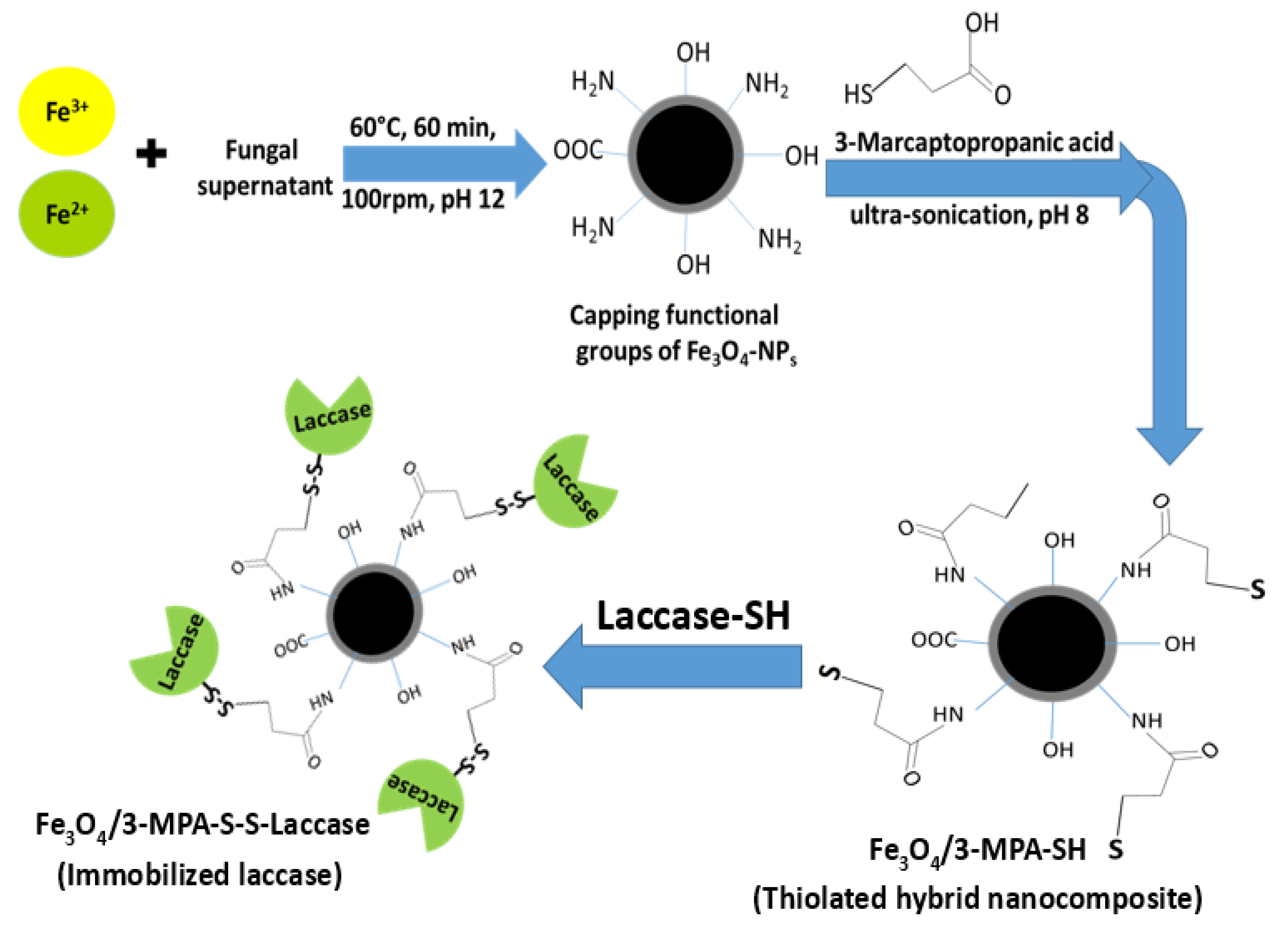
2.2. Bio-Synthesis of Thiol-Functionalized Magnetic Nanoparticles
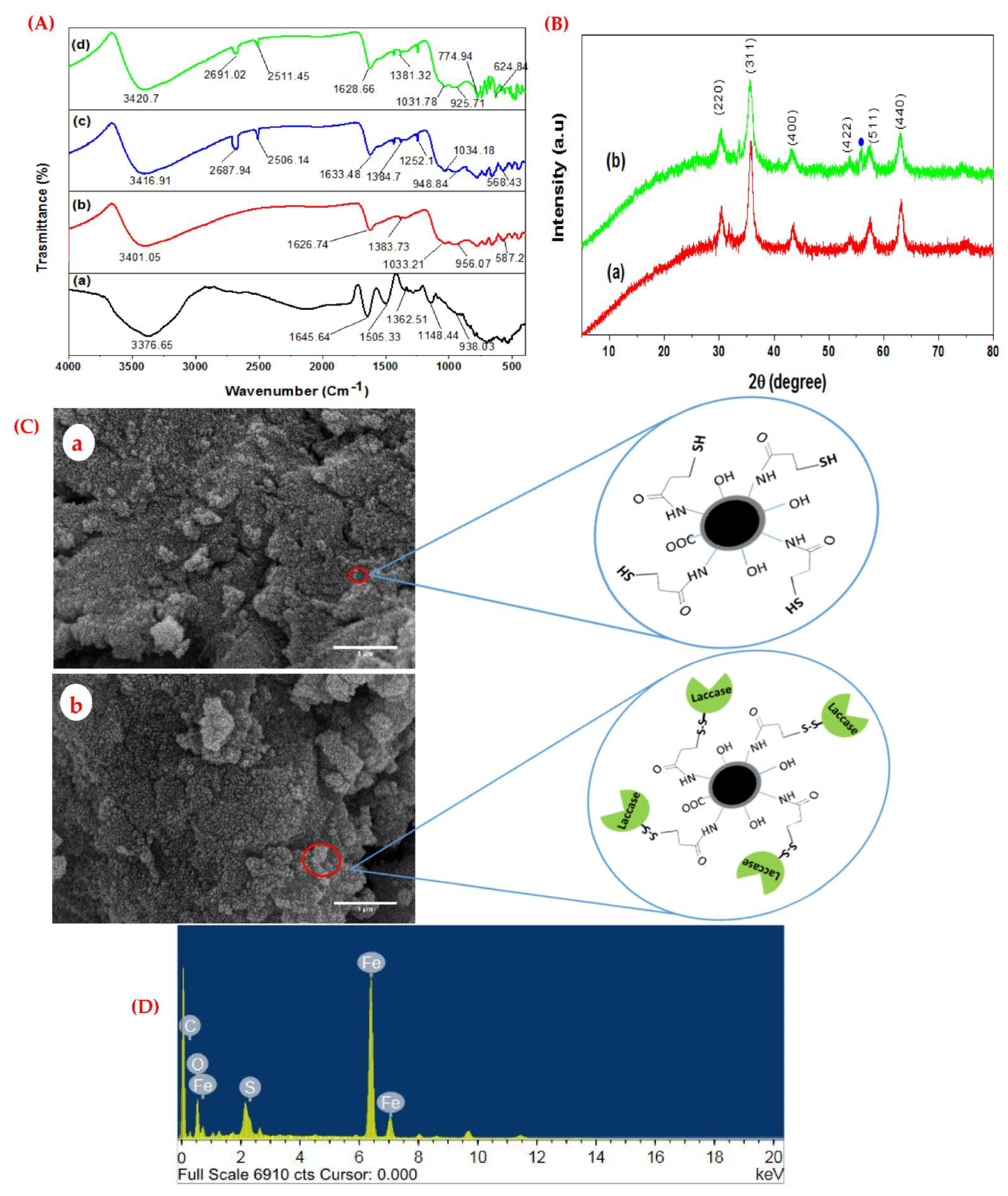
2.3. Characterization of Functionalized Magnetic Nanocomposite
2.4. Biotechnological Applications of Fe3O4/3-MPA-SH in Laccase Immobilization
2.4.1. Screening for the Most Potential Laccase Producer Fungal Isolate
2.4.2. Laccase Activity Assay and Protein Determination
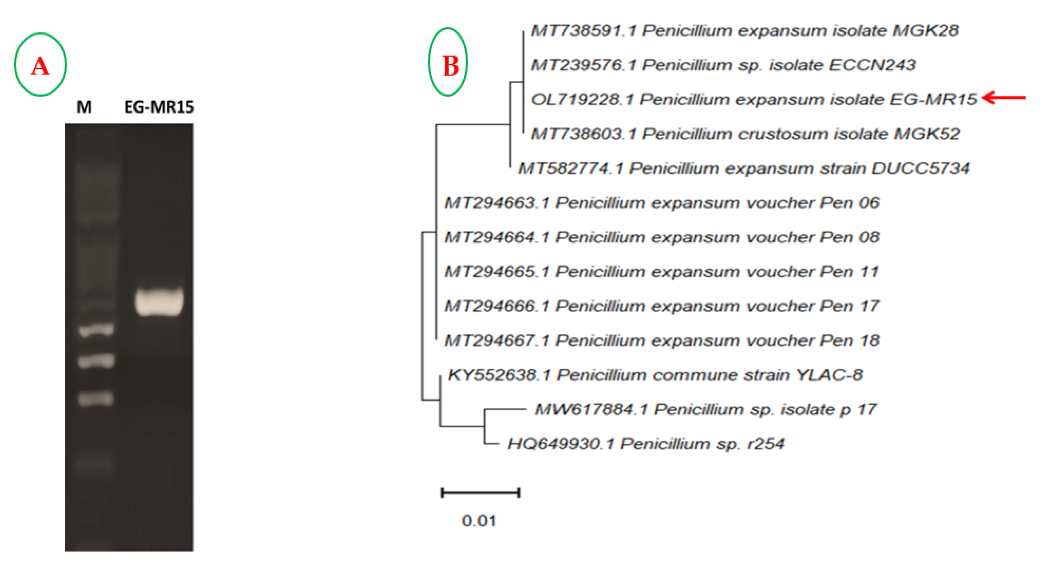
2.4.3. Identification and Deposition of the Most Potential Laccase-Producing Isolate
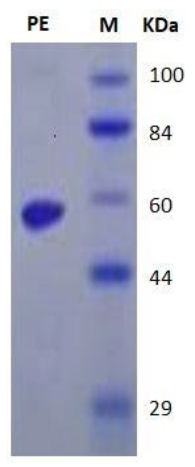
2.4.4. Extraction, Purification, and Molecular Homogeneity of Laccase
2.4.5. Immobilization of Laccase on a Thiolated Functionalized Magnetic Nanosupport
2.4.6. Characterization of Free Laccase and Fe3O4/3-MPA-S-S-Lac
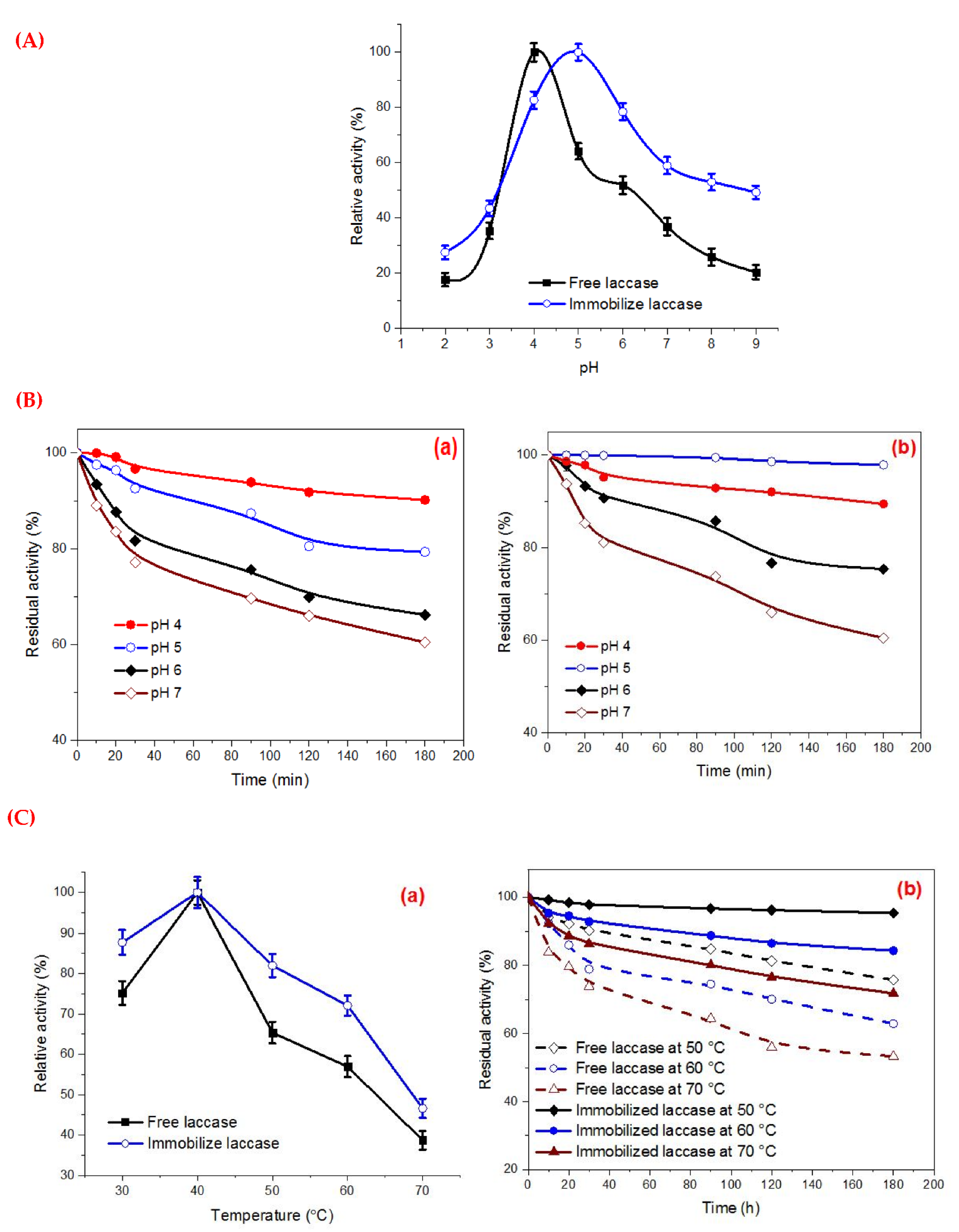
pH Optima and pH Stability
Optimum Temperature and Thermal Stability
Determination of Km and Vmax
Effect of Different Organic Solvents on Enzyme Stability
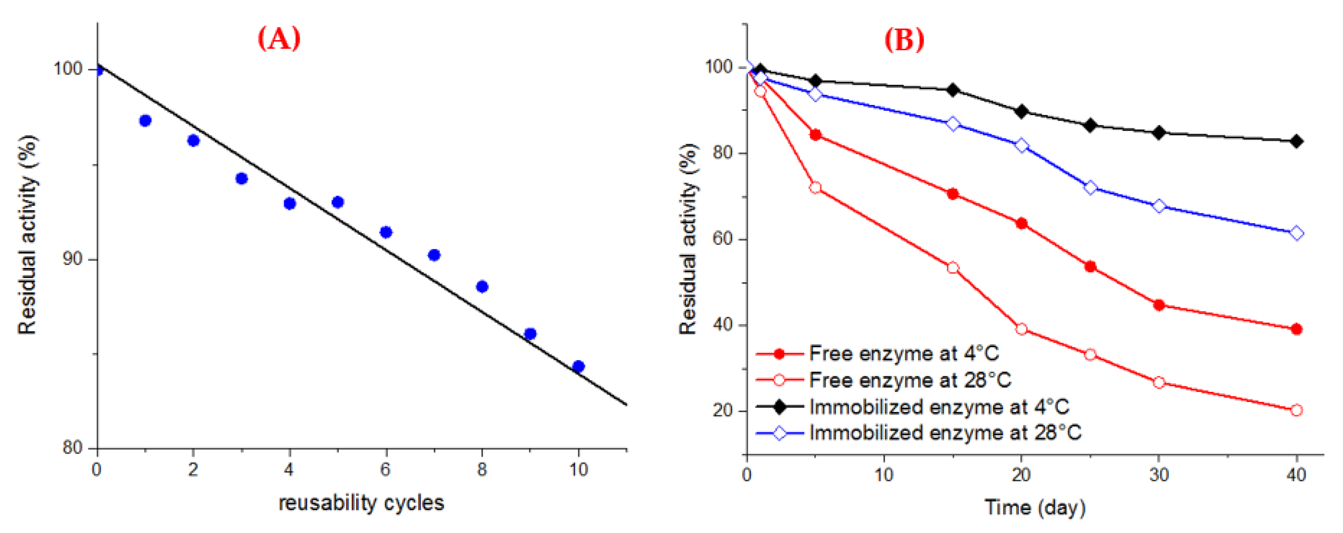
Operational Stability (Reusability)
Storage Stability
Decolorization of Synthetic Dyes
Reusability Assessment of Fe3O4/3-MPA-S-S-Laccase
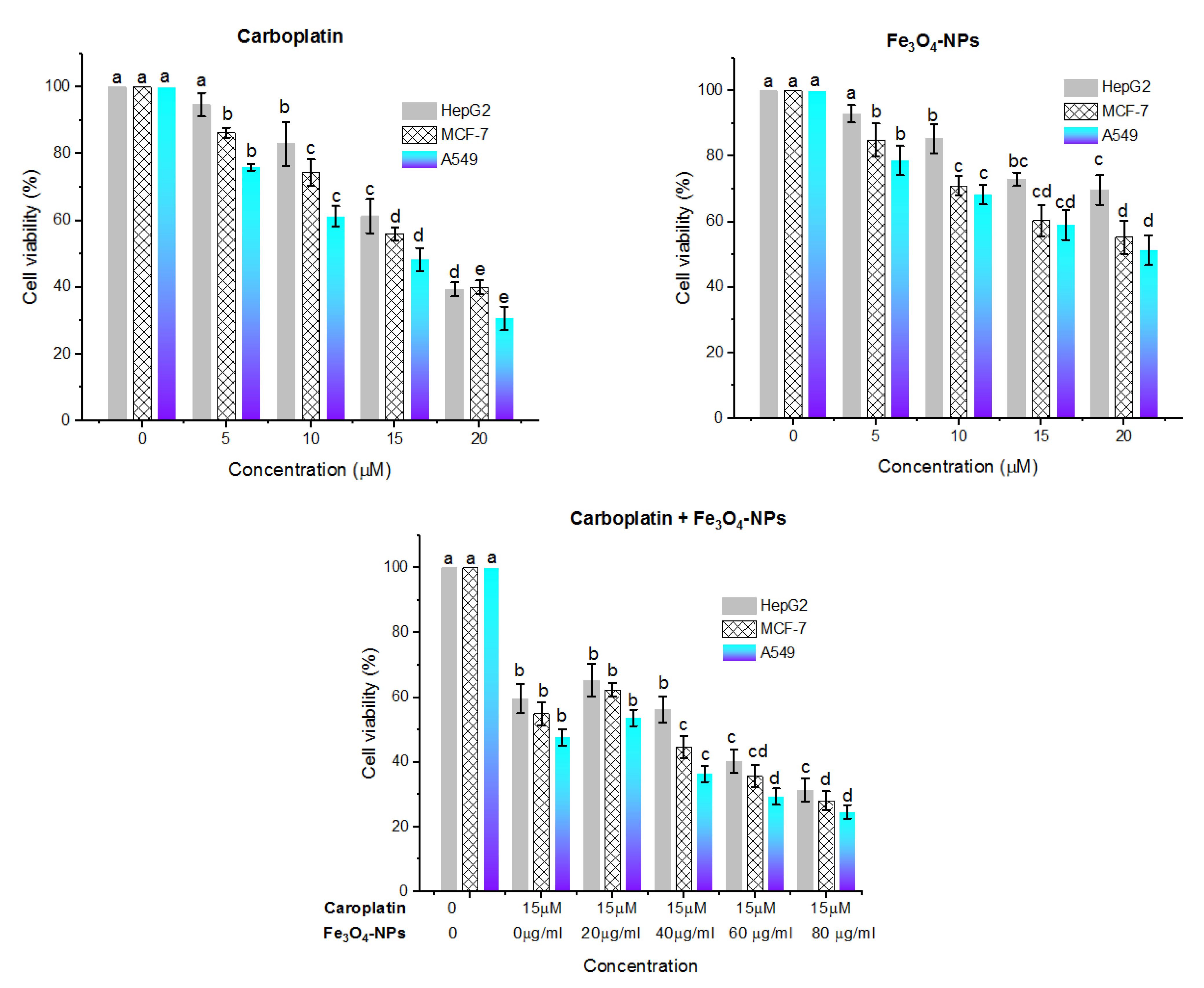
2.5. Cytotoxic Effect
2.6. Statistical Analysis
3. Results and Discussion
3.1. Synthesis
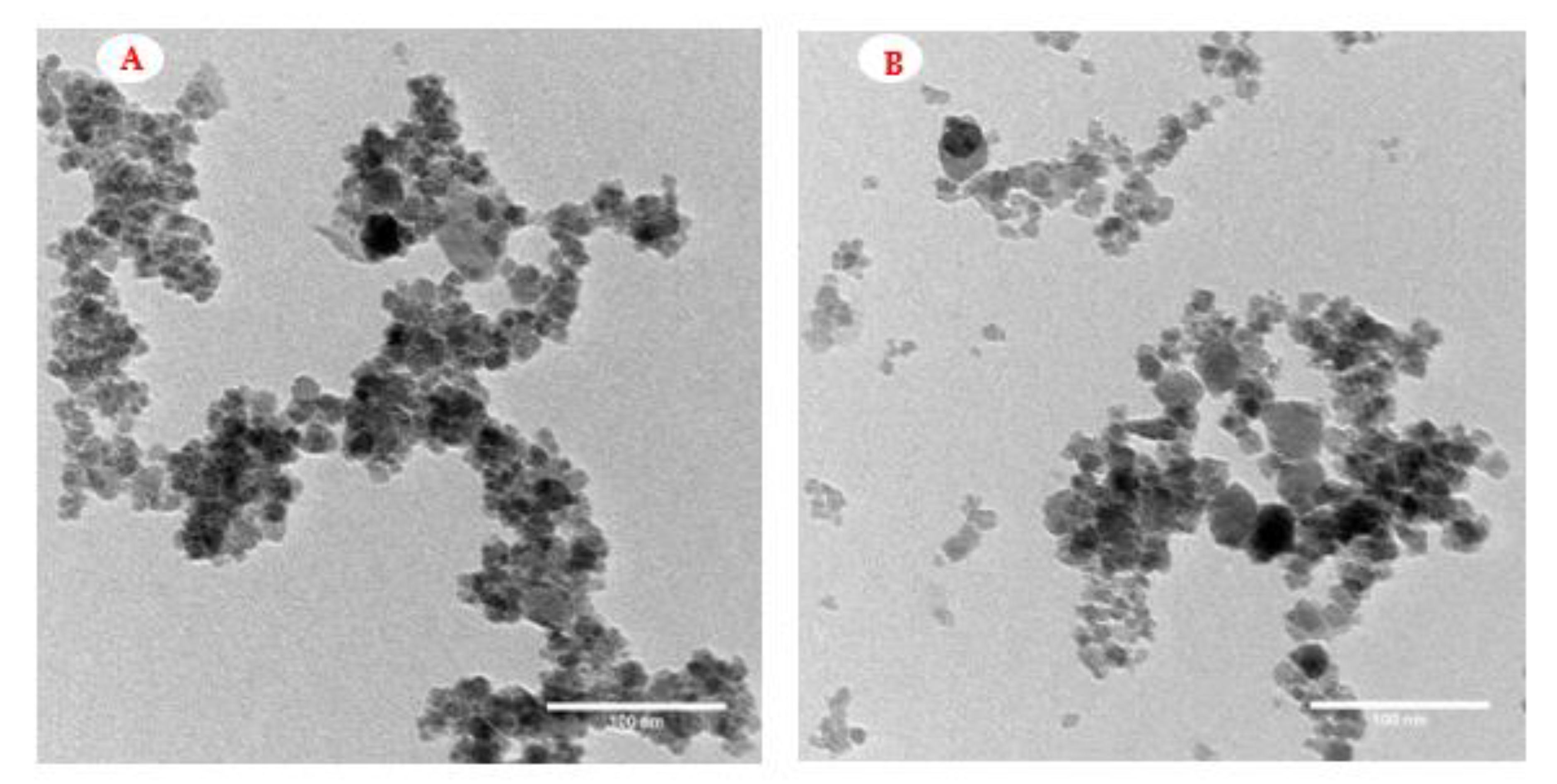
3.2. Characterization of the Nanosupport for Laccase Immobilization
3.3. Screening for the Most Potent Laccase Producing Isolate
3.4. Production of Purified Laccase from P. expansium
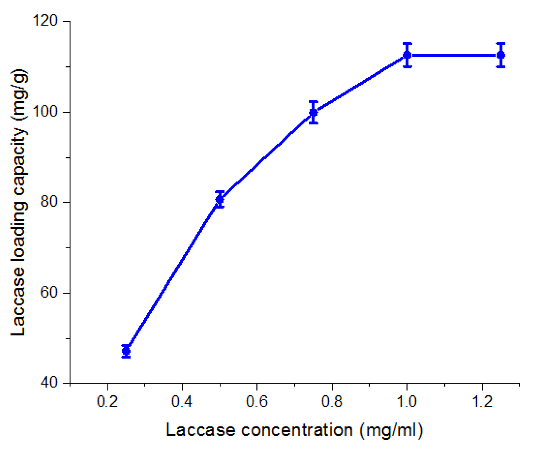
3.5. Immobilization of Laccase on Fe3O4/3-MPA-SH
3.6. Biochemical Characterization of the Free and Immobilized Laccase
3.6.1. pH Optima and pH Stability
3.6.2. Temperature Optima and Thermostability
3.6.3. Kinetic Parameters
3.6.4. Influence of Different Organic Solvents on Laccase Stability
3.6.5. Operational Stability of Immobilized Laccase
3.6.6. Storage Stability
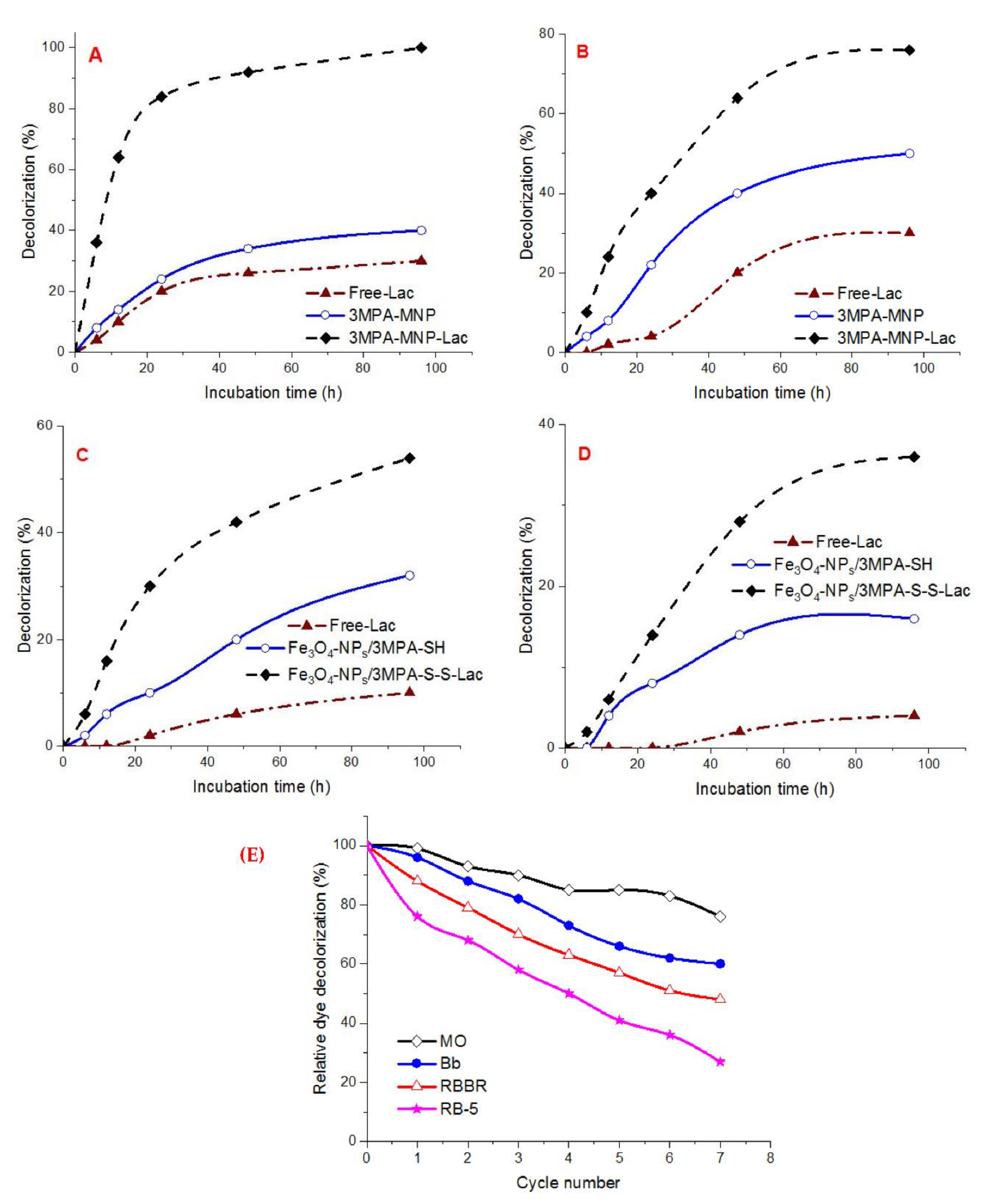
3.7. Biotechnological Application of Free Laccase, Fe3O4/3MPA-SH, and Fe3O4/3-MPA-S-S-Laccase for Catalytic Decolorization of Dyes
Reusability Assessment of the Covalent-Immobilized Laccase for Catalytic Decolorization of Dyes
3.8. Cytotoxic Effect
4. Conclusions
Author Contributions
Funding
Institutional Review Board Statement
Informed Consent Statement
Data Availability Statement
Conflicts of Interest
References
- Kadam, A.A.; Sharma, B.; Shinde, S.K.; Ghodake, G.S.; Saratale, G.D.; Saratale, R.G.; Kim, D.-Y.; Sung, J.-S. Thiolation of Chitosan Loaded over Super-Magnetic Halloysite Nanotubes for Enhanced Laccase Immobilization. Nanomaterials 2020, 10, 2560. [Google Scholar] [CrossRef]
- Sheng, S.; Farinas, E.T. Laccase and Its Mutant Displayed on the Bacillus subtilis Spore Coat for Oxidation of Phenolic Compounds in Organic Solvents. Catalysts 2021, 11, 606. [Google Scholar] [CrossRef]
- Hassan, S.E.-D.; Fouda, A.; Saied, E.; Farag, M.; Eid, A.M.; Barghoth, M.G.; Awad, M.A.; Hamza, M.F.; Awad, M.F. Rhizopus Oryzae-mediated green synthesis of magnesium oxide nanoparticles (MgO-NPs): A promising tool for antimicrobial, mosquitocidal action, and tanning effluent treatment. J. Fungi 2021, 7, 372. [Google Scholar] [CrossRef] [PubMed]
- Datta, S.; Veena, R.; Samuel, M.S.; Selvarajan, E. Immobilization of laccases and applications for the detection and remediation of pollutants: A review. Environ. Chem. Lett. 2021, 19, 521–538. [Google Scholar] [CrossRef]
- Qiu, X.; Wang, S.; Miao, S.; Suo, H.; Xu, H.; Hu, Y. Co-immobilization of laccase and ABTS onto amino-functionalized ionic liquid-modified magnetic chitosan nanoparticles for pollutants removal. J. Hazard. Mater. 2021, 401, 123353. [Google Scholar] [CrossRef] [PubMed]
- El-Shora, H.M.; El-Sharkawy, R.M. Tyrosinase from Penicillium chrysogenum: Characterization and application in phenol removal from aqueous solution. J. Gen. Appl. Microbiol. 2020, 66, 323–329. [Google Scholar] [CrossRef] [PubMed]
- Liu, Y.-Q.; Maulidiany, N.; Zeng, P.; Heo, S. Decolourization of azo, anthraquinone and triphenylmethane dyes using aerobic granules: Acclimatization and long-term stability. Chemosphere 2021, 263, 128312. [Google Scholar] [CrossRef]
- Tabla-Hernández, J.; Rodríguez-Espinosa, P.; Hernandez-Ramirez, A.; Mendoza-Pérez, J.A.; Cano-Aznar, E.; Martínez-Tavera, E. Treatment of Eutrophic water and wastewater from Valsequillo Reservoir, Puebla, Mexico by Means of Ozonation: A multiparameter approach. Water 2018, 10, 1790. [Google Scholar] [CrossRef] [Green Version]
- Ulu, A.; Birhanli, E.; Boran, F.; Köytepe, S.; Yesilada, O.; Ateş, B. Laccase-conjugated thiolated chitosan-Fe3O4 hybrid composite for biocatalytic degradation of organic dyes. Int. J. Biol. Macromol. 2020, 150, 871–884. [Google Scholar] [CrossRef]
- Vantamuri, A.; Kaliwal, B. Purification and characterization of laccase from Marasmius species BBKAV79 and effective decolorization of selected textile dyes. 3 Biotech 2016, 6, 1–10. [Google Scholar] [CrossRef] [PubMed] [Green Version]
- Ali, I.; Peng, C.; Khan, Z.M.; Sultan, M.; Naz, I. Green synthesis of phytogenic magnetic nanoparticles and their applications in the adsorptive removal of crystal violet from aqueous solution. Arab. J. Sci. Eng. 2018, 43, 6245–6259. [Google Scholar] [CrossRef]
- Siddeeg, S.M.; Tahoon, M.A.; Mnif, W.; Ben Rebah, F. Iron oxide/chitosan magnetic nanocomposite immobilized manganese peroxidase for decolorization of textile wastewater. Processes 2020, 8, 5. [Google Scholar] [CrossRef] [Green Version]
- Su, J.; Noro, J.; Fu, J.; Wang, Q.; Silva, C.; Cavaco-Paulo, A. Exploring PEGylated and immobilized laccases for catechol polymerization. AMB Express 2018, 8, 1–9. [Google Scholar] [CrossRef] [PubMed]
- El-Shora, H.M.; El-Sharkawy, R.M. Evaluation of Putative Inducers and Inhibitors toward Tyrosinase from two Trichoderma species. Jordan J. Biol. Sci. 2020, 13, 7–12. [Google Scholar]
- Stefanov, I.; Pérez-Rafael, S.; Hoyo, J.; Cailloux, J.; Santana Pérez, O.O.; Hinojosa-Caballero, D.; Tzanov, T. Multifunctional enzymatically generated hydrogels for chronic wound application. Biomacromolecules 2017, 18, 1544–1555. [Google Scholar] [CrossRef] [Green Version]
- Mikkelsen, M.S.; Blaakaer, J.; Petersen, L.K.; Schleiss, L.G.; Iversen, L.H. Pharmacokinetics and toxicity of carboplatin used for hyperthermic intraperitoneal chemotherapy (HIPEC) in treatment of epithelial ovarian cancer. Pleura Peritoneum 2020, 5, 20200137. [Google Scholar] [CrossRef] [PubMed]
- Tharmalingam, M.D.; Matilionyte, G.; Wallace, W.H.; Stukenborg, J.-B.; Jahnukainen, K.; Oliver, E.; Goriely, A.; Lane, S.; Guo, J.; Cairns, B. Cisplatin and carboplatin result in similar gonadotoxicity in immature human testis with implications for fertility preservation in childhood cancer. BMC Med. 2020, 18, 374. [Google Scholar] [CrossRef] [PubMed]
- Kojima, K.; Takahashi, S.; Saito, S.; Endo, Y.; Nittami, T.; Nozaki, T.; Sobti, R.C.; Watanabe, M. Combined effects of Fe3O4 nanoparticles and chemotherapeutic agents on prostate cancer cells in vitro. Appl. Sci. 2018, 8, 134. [Google Scholar] [CrossRef] [Green Version]
- El-Shora, H.M.; El-Sharkawy, R.M.; Khateb, A.M.; Darwish, D.B. Production and immobilization of β-glucanase from Aspergillus niger with its applications in bioethanol production and biocontrol of phytopathogenic fungi. Sci. Rep. 2021, 11, 21000. [Google Scholar]
- Ayub, A.; Raza, Z.A.; Majeed, M.I.; Tariq, M.R.; Irfan, A. Development of sustainable magnetic chitosan biosorbent beads for kinetic remediation of arsenic contaminated water. Int. J. Biol. Macromol. 2020, 163, 603–617. [Google Scholar] [CrossRef]
- Bradford, M.M. A rapid and sensitive method for the quantitation of microgram quantities of protein utilizing the principle of protein-dye binding. Anal. Biochem. 1976, 72, 248–254. [Google Scholar] [CrossRef]
- Pitt, J. The Genus Penicillium and Its Teleomorphic States Eupenicillium and Talaromyces; Academic Press: London, UK; New York, NY, USA, 1979. [Google Scholar]
- Laemmli, U.K. Cleavage of structural proteins during the assembly of the head of bacteriophage T4. Nature 1970, 227, 680–685. [Google Scholar] [CrossRef]
- Chen, L.; Zou, M.; Hong, F.F. Evaluation of fungal laccase immobilized on natural nanostructured bacterial cellulose. Front. Microbiol. 2015, 6, 1245. [Google Scholar] [CrossRef] [PubMed] [Green Version]
- Muthuvelu, K.S.; Rajarathinam, R.; Selvaraj, R.N.; Rajendren, V.B. A novel method for improving laccase activity by immobilization onto copper ferrite nanoparticles for lignin degradation. Int. J. Biol. Macromol. 2020, 152, 1098–1107. [Google Scholar] [CrossRef]
- Lineweaver, H.; Burk, D. The determination of enzyme dissociation constants. J. Am. Chem. Soc. 1934, 56, 658–666. [Google Scholar] [CrossRef]
- Xie, Y.; Liu, D.; Cai, C.; Chen, X.; Zhou, Y.; Wu, L.; Sun, Y.; Dai, H.; Kong, X.; Liu, P. Size-dependent cytotoxicity of Fe3O4 nanoparticles induced by biphasic regulation of oxidative stress in different human hepatoma cells. Int. J. Nanomed. 2016, 11, 3557. [Google Scholar]
- Ebadi, S.; Henschke, N.; Forogh, B.; Ansari, N.N.; Tulder, M.W.; Babaei-Ghazani, A.; Fallah, E. Therapeutic ultrasound for chronic low back pain. Cochrane Database Syst. Rev. 2020, 7. [Google Scholar] [CrossRef]
- Fouda, A.; Hassan, S.E.-D.; Abdel-Rahman, M.A.; Farag, M.M.; Shehal-Deen, A.; Mohamed, A.A.; Alsharif, S.M.; Saied, E.; Moghanim, S.A.; Azab, M.S. Catalytic degradation of wastewater from the textile and tannery industries by green synthesized hematite (α-Fe2O3) and magnesium oxide (MgO) nanoparticles. CRBIOT 2021, 3, 29–41. [Google Scholar] [CrossRef]
- Federer, C.; Kurpiers, M.; Bernkop-Schnürch, A. Thiolated chitosans: A multi-talented class of polymers for various applications. Biomacromolecules 2020, 22, 24–56. [Google Scholar] [CrossRef]
- Alnadari, F.; Xue, Y.; Zhou, L.; Hamed, Y.S.; Taha, M.; Foda, M.F. Immobilization of β-glucosidase from Thermatoga maritima on chitin-functionalized magnetic nanoparticle via a novel thermostable chitin-binding domain. Sci. Rep. 2020, 10, 1663. [Google Scholar] [CrossRef]
- Zhang, K.; Yang, W.; Liu, Y.; Zhang, K.; Chen, Y.; Yin, X. Laccase immobilized on chitosan-coated Fe3O4 nanoparticles as reusable biocatalyst for degradation of chlorophenol. J. Mol. Struct. 2020, 1220, 128769. [Google Scholar] [CrossRef]
- Daâssi, D.; Rodríguez-Couto, S.; Nasri, M.; Mechichi, T. Biodegradation, Biodegradation of textile dyes by immobilized laccase from Coriolopsis gallica into Ca-alginate beads. Int. Biodeterior 2014, 90, 71–78. [Google Scholar] [CrossRef]
- Chairin, T.; Nitheranont, T.; Watanabe, A.; Asada, Y.; Khanongnuch, C.; Lumyong, S. Purification and characterization of the extracellular laccase produced by Trametes polyzona WR710–1 under solid-state fermentation. J. Basic Microbiol. 2014, 54, 35–43. [Google Scholar] [CrossRef] [PubMed]
- Mainak, M.; Rintu, B. Purification and biochemical characterization of a newly produced yellow laccase from Lentinus squarrosulus MR13. 3 Biotech 2015, 5, 227–236. [Google Scholar]
- Abdelgalil, S.A.; Attia, A.R.; Reyed, R.M.; Soliman, N.A. Biotechnology, Partial purification and biochemical characterization of a new highly acidic NYSO laccase from Alcaligenes faecalis. J. Genet. Eng. Biotechnol. 2020, 18, 1–11. [Google Scholar] [CrossRef]
- Cao, P.; Liu, H.; Wu, D.; Wang, X. Immobilization of laccase on phase-change microcapsules as self-thermoregulatory enzyme carrier for biocatalytic enhancement. Chem. Eng. J. 2021, 405, 126695. [Google Scholar] [CrossRef]
- Zofair, S.F.F.; Arsalan, A.; Khan, M.A.; Alhumaydhi, F.A.; Younus, H. Immobilization of laccase on Sepharose-linked antibody support for decolourization of phenol red. Int. J. Biol. Macromol. 2020, 161, 78–87. [Google Scholar] [CrossRef]
- Ghodake, G.S.; Yang, J.; Shinde, S.S.; Mistry, B.M.; Kim, D.-Y.; Sung, J.-S.; Kadam, A.A. Paper waste extracted α-cellulose fibers super-magnetized and chitosan-functionalized for covalent laccase immobilization. Bioresour. Technol. 2018, 261, 420–427. [Google Scholar] [CrossRef] [PubMed]
- Kunamneni, A.; Ghazi, I.; Camarero, S.; Ballesteros, A.; Plou, F.J.; Alcalde, M.J.P.B. Decolorization of synthetic dyes by laccase immobilized on epoxy-activated carriers. Process Biochem. 2008, 43, 169–178. [Google Scholar] [CrossRef] [Green Version]
- Alnadari, F.; Xue, Y.; Almakas, A.; Mohedein, A.; Samie, A.; Abdel-Shafi, M.; Abdin, M. Large batch production of Galactooligosaccharides using β-glucosidase immobilized on chitosan-functionalized magnetic nanoparticle. J. Food Biochem. 2021, 45, e13589. [Google Scholar] [CrossRef]
- Guzik, U.; Hupert-Kocurek, K.; Wojcieszyńska, D. Immobilization as a strategy for improving enzyme properties-application to oxidoreductases. Molecules 2014, 19, 8995–9018. [Google Scholar] [CrossRef] [Green Version]
- Chamoli, S.; Yadav, E.; Saini, J.K.; Verma, A.K.; Navani, N.K.; Kumar, P. Magnetically recyclable catalytic nanoparticles grafted with Bacillus subtilis β-glucosidase for efficient cellobiose hydrolysis. Int. J. Biol. Macromol. 2020, 164, 1729–1736. [Google Scholar] [CrossRef] [PubMed]
- Jaiswal, N.; Pandey, V.P.; Dwivedi, U.N. Immobilization of papaya laccase in chitosan led to improved multipronged stability and dye discoloration. Int. J. Biol. Macromol. 2016, 86, 288–295. [Google Scholar] [CrossRef] [PubMed]
- Valencia, P.; Ibañez, F. Estimation of the Effectiveness Factor for Immobilized Enzyme Catalysts through a Simple Conversion Assay. Catalysts 2019, 9, 930. [Google Scholar] [CrossRef] [Green Version]
- Rodakiewicz-Nowak, J.; Kasture, S.; Dudek, B.; Haber, J. Effect of various water-miscible solvents on enzymatic activity of fungal laccases. J. Mol. Catal. B Enzym. 2000, 11, 1–11. [Google Scholar] [CrossRef]
- Wan, Y.-Y.; Lu, R.; Xiao, L.; Du, Y.-M.; Miyakoshi, T.; Chen, C.-L.; Knill, C.J.; Kennedy, J.F. Effects of organic solvents on the activity of free and immobilised laccase from Rhus vernicifera. Int. J. Biol. Macromol. 2010, 47, 488–495. [Google Scholar] [CrossRef] [PubMed]
- Wu, E.; Li, Y.; Huang, Q.; Yang, Z.; Wei, A.; Hu, Q. Laccase immobilization on amino-functionalized magnetic metal organic framework for phenolic compound removal. Chemosphere 2019, 233, 327–335. [Google Scholar] [CrossRef] [PubMed]
- Zhou, J.; Wu, Y.; Zhang, Q.; Xu, G.; Ni, Y. Co-immobilized alcohol dehydrogenase and glucose dehydrogenase with resin extraction for continuous production of chiral diaryl alcohol. Appl. Biochem. Biotechnol. 2021, 193, 2742–2758. [Google Scholar] [CrossRef] [PubMed]
- Sato, A.; Itcho, N.; Ishiguro, H.; Okamoto, D.; Kobayashi, N.; Kawai, K.; Kasai, H.; Kurioka, D.; Uemura, H.; Kubota, Y. Magnetic nanoparticles of Fe3O4 enhance docetaxel-induced prostate cancer cell death. INT J. Nanomed. 2013, 8, 3151. [Google Scholar]
- Abbas, H.S.; Krishnan, A. Magnetic nanosystems as a therapeutic tool to combat pathogenic fungi. Adv. Pharm. Bull. 2020, 10, 512. [Google Scholar] [CrossRef]
- Arakha, M.; Pal, S.; Samantarrai, D.; Panigrahi, T.K.; Mallick, B.C.; Pramanik, K.; Mallick, B.; Jha, S. Antimicrobial activity of iron oxide nanoparticle upon modulation of nanoparticle-bacteria interface. Sci. Rep. 2015, 5, 1–12. [Google Scholar] [CrossRef] [PubMed] [Green Version]
- Caldeirão, A.C.M.; Araujo, H.C.; Arias, L.S.; Ramírez Carmona, W.; Miranda, G.P.; Oliveira, S.H.P.; Pessan, J.P.; Monteiro, D.R. Nanocarriers of Miconazole or Fluconazole: Effects on three-species Candida biofilms and cytotoxic effects in vitro. J. Fungi 2021, 7, 500. [Google Scholar] [CrossRef] [PubMed]
- Shiota, M.; Yokomizo, A.; Naito, S. Oxidative stress and and rogen receptor signaling in the development and progression of castration-resistant prostate cancer. Free Radic. Biol. Med. 2011, 51, 1320–1328. [Google Scholar] [CrossRef] [PubMed]


 ), Fe3O4-NPs/3MPA-SH (
  ) and Fe3O4-NPs/3MPA-S-S-Lac (
  ), (E) Repeated decolorization cycles of environmental pollutants (MO, Bb, RBBR, and RB-5) in the presence of a redox mediator by Fe3O4/3-MPA-S-S-Lac.
), Fe3O4-NPs/3MPA-SH ( ) and Fe3O4-NPs/3MPA-S-S-Lac ( ), (E) Repeated decolorization cycles of environmental pollutants (MO, Bb, RBBR, and RB-5) in the presence of a redox mediator by Fe3O4/3-MPA-S-S-Lac.
), (E) Repeated decolorization cycles of environmental pollutants (MO, Bb, RBBR, and RB-5) in the presence of a redox mediator by Fe3O4/3-MPA-S-S-Lac.
), Fe3O4-NPs/3MPA-SH ( ) and Fe3O4-NPs/3MPA-S-S-Lac ( ), (E) Repeated decolorization cycles of environmental pollutants (MO, Bb, RBBR, and RB-5) in the presence of a redox mediator by Fe3O4/3-MPA-S-S-Lac.


| Purification Steps | Total Activity (U) | Total Protein (mg) | Specific Activity (U/mg) | Purification Fold | Yield (%) |
|---|---|---|---|---|---|
| Crude enzyme | 2254.5 | 224.64 | 10.03 | 1 | 100 |
| Ammonium sulfate (75%) | 1566 | 49.59 | 31.94 | 3.18 | 69 |
| DEAE-cellulose | 951.6 | 6.422 | 148.17 | 14.76 | 42 |
| Sephadex G-200 | 625.3 | 1.885 | 331.72 | 33.05 | 27 |
| Support | Laccase Loading (mg/g) | References |
|---|---|---|
| Fe3O4/3MPA-SH | 112 | PS |
| LA-Au/PDA@SiO2-MEPCM | 50 | [37] |
| Magnetic biochar | 27 | [32] |
| Magnetized-chitosan-grafted hallohalloysite nanotube | 100 | [1] |
| Magnetic-chitosan | 32 | [32] |
| MACS-NIL-Cu-Laccase | 47 | [38] |
| Chitosan-functionalized supermagneti cellulose | 73 | [39] |
| Sepabeads EC-EP3 | 32.6 | [40] |
| Dilbeads NK | 17.8 | [40] |
| Substrate | Free-Laccase | Fe3O4-NPs/3-MPA-S-S-Laccase | ||||||
|---|---|---|---|---|---|---|---|---|
| Kcat (S−1) | Km (mM) | Vmax (U/mg Protein/min) | Kcat/Km (mM−1 S−1) | Kcat (S−1) | Km (mM) | Vmax (U/mg Protein/min) | Kcat/Km (mM−1 S−1) | |
| ABTS | 45.05 | 4.15 | 29.06 | 10.87 | 48.45 | 2.60 | 27.03 | 18.65 |
| Catechol | 23.71 | 1.31 | 14.22 | 18.14 | 24.84 | 0.93 | 14.90 | 26.75 |
| Residual Activity (%) | Concentrations of Organic Solvents (%) | ||||||||||||||
|---|---|---|---|---|---|---|---|---|---|---|---|---|---|---|---|
| Acetone | Methanol | Ethanol | |||||||||||||
| 10 | 20 | 30 | 40 | 50 | 10 | 20 | 30 | 40 | 50 | 10 | 20 | 30 | 40 | 50 | |
| Free laccase | 98.6 | 93.3 | 89.6 | 85.7 | 79.8 | 87.6 | 74.3 | 61.1 | 46.9 | 32.2 | 94.4 | 88.3 | 80.5 | 75.2 | 63.8 |
| Fe3O4/3-MPA-S-S-laccase | 99.6 | 96.1 | 93.6 | 90.7 | 84.6 | 91.1 | 78.2 | 65.1 | 49.8 | 35.3 | 96.1 | 93.9 | 89.4 | 84.8 | 79.3 |
Publisher’s Note: MDPI stays neutral with regard to jurisdictional claims in published maps and institutional affiliations. |
© 2022 by the authors. Licensee MDPI, Basel, Switzerland. This article is an open access article distributed under the terms and conditions of the Creative Commons Attribution (CC BY) license (https://creativecommons.org/licenses/by/4.0/).
Share and Cite
El-Shora, H.M.; Khateb, A.M.; Darwish, D.B.; El-Sharkawy, R.M. Thiolation of Myco-Synthesized Fe3O4-NPs: A Novel Promising Tool for Penicillium expansium Laccase Immobilization to Decolorize Textile Dyes and as an Application for Anticancer Agent. J. Fungi 2022, 8, 71. https://doi.org/10.3390/jof8010071
El-Shora HM, Khateb AM, Darwish DB, El-Sharkawy RM. Thiolation of Myco-Synthesized Fe3O4-NPs: A Novel Promising Tool for Penicillium expansium Laccase Immobilization to Decolorize Textile Dyes and as an Application for Anticancer Agent. Journal of Fungi. 2022; 8(1):71. https://doi.org/10.3390/jof8010071
Chicago/Turabian StyleEl-Shora, Hamed M., Aiah M. Khateb, Doaa B. Darwish, and Reyad M. El-Sharkawy. 2022. "Thiolation of Myco-Synthesized Fe3O4-NPs: A Novel Promising Tool for Penicillium expansium Laccase Immobilization to Decolorize Textile Dyes and as an Application for Anticancer Agent" Journal of Fungi 8, no. 1: 71. https://doi.org/10.3390/jof8010071