Article

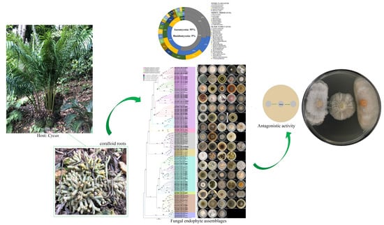
Diversity, Phylogeny and Antagonistic Activity of Fungal Endophytes Associated with Endemic Species of Cycas (Cycadales) in China

1
South China Botanical Garden, Chinese Academy of Sciences, Guangzhou 510650, China
2
Fairy Lake Botanical Garden, Chinese Academy of Sciences, Shenzhen 518004, China
3
University of Chinese Academy of Sciences, Beijing 100049, China
4
Department of Biological Sciences, College of Science, University of Santo Tomas, Manila 1008, Philippines
5
Fungal Biodiversity, Ecogenomics and Systematics (FBeS) Group, Research Center for the Natural and Applied Sciences, University of Santo Tomas, Manila 1008, Philippines
*
Author to whom correspondence should be addressed.
J. Fungi 2021, 7(7), 572; https://doi.org/10.3390/jof7070572
Submission received: 21 June 2021 / Revised: 13 July 2021 / Accepted: 14 July 2021 / Published: 18 July 2021
(This article belongs to the Special Issue Diversity and Classification of Environmental Fungi)

Abstract

:
The culture-based approach was used to characterize the fungal endophytes associated with the coralloid roots of the endemic Cycas debaoensis and Cycas fairylakea from various population sites in China. We aim to determine if the assemblages of fungal endophytes inside these endemic plant hosts are distinct and could be explored for bioprospecting. The isolation method yielded a total of 284 culturable fungal strains. Identification based on the analysis of the internal transcribed spacer (ITS) rDNA showed that they belonged to two phyla, five classes, eight orders and 22 families. At least 33 known genera and 62 different species were confirmed based on >97% ITS sequence similarity. The most frequent and observed core taxa in the two host species regardless of their population origin were Talaromyces, Penicillium, Fusarium, Pochonia and Gliocladiopsis. Seventy percent was a rare component of the fungal communities with only one or two recorded isolates. Contrary to common notions, diversity and fungal richness were significantly higher in C. debaoensis and C. fairylakea collected from a botanical garden, while the lowest was observed in C. debaoensis from a natural habitat; this provides evidence that garden management, and to a minor extent, ex-situ conservation practice, could influence fungal endophyte communities. We further selected nineteen fungal isolates and screened for their antagonistic activities via a co-cultivation approach against the phytopathogens, Diaporthe sp. and Colletotrichum sp. Among these, five isolates with high ITS similarity matches with Hypoxylon vinosupulvinatum (GD019, 99.61%), Penicillium sp. (BD022, 100%), Penicillifer diparietisporus (GD008, 99.46%), Clonostachys rogersoniana (BF024, 99.46%) and C. rosea (BF011, 99.1%), which showed exceptional antagonistic activities against the phytopathogenic fungi with a significant inhibition rate of 70–80%. Taken together, our data presented the first and most comprehensive molecular work on culturable fungal endophytes associated with the coralloid roots of cycads. Our study also demonstrated that about 5% of fungal endophytes were not detected by the high-throughput sequencing approach, implying the equal importance of a culture-dependent approach to study fungal communities of cycads. We further highlighted the potential role of endemic and rare plants to discover and isolate unique plant-associated fungal taxa with excellent biocontrol properties.

Graphical Abstract

1. Introduction

Complex and heterogeneous assemblages of fungal communities that do not elicit symptoms of disease are hidden intracellularly in the internal tissues of plants [1,2]. They are termed as fungal endophytes as they spend part or the entirety of their life cycle inside healthy tissues of plants [1]. Fungal endophytes also represent an important component of the root microbiota and provide benefits to their hosts by acting as biocontrol agents, plant growth promoters, environmental stress regulators, and host protectors [3,4]. Root endophytic fungi were reported in different plants across a wide range of environments. For instance, mounting evidence showed a highly diverse species of root endophytes in plants thriving in hostile environments, such as deserts [5,6], salt marshes [7,8], arid and geothermal environments [9,10] and karst rocks [11]. The functional symbiosis or the establishment of mutualistic relationships between root fungal endophytes with their hosts are crucial for their survival and adaptation to these extremophilic environments [11].
The diversity and community assemblages of fungal endophytes are said to be filtered by a multitude of factors. The interactions that exist between them were reported to be influenced by both environmental and physiological factors resulting in a balance and beneficial relations between parties [12]. For instance, previous studies demonstrated that soil properties, nutrient availability, root exudates and traits and local climatic conditions, e.g., temperature and precipitation, are vital drivers that structure endophytic microfungal communities in plant roots [13,14,15,16,17,18]. Geographical distance also has a significant effect on fungal communities where plant hosts occupying the same geographic region with nearly similar climatic conditions result in similar endophytic fungal communities [17]. In some studies, these abiotic factors are coupled with host-related factors such as genetic differences among inter/intraspecific hosts and influence the entire plant-fungal association [18,19]. This is particularly interesting to investigate especially since some plants belonging to a single species revealed both genetic distinctness and high dissimilar fungal communities [19].
Fungal endophytes recovered from a wide lineage of plants have been investigated for bioprospecting [2,20,21,22,23]. Particularly on gymnosperms, early studies have reported the production of secondary metabolites with strong antimicrobial activity [24,25]. Interestingly, multiple studies in the past showed that endophytic fungal communities associated with native and endemic plants may have the greatest promise for exploitation as they have the potential to host rare and unique assemblage of fungal communities. Some of the fungal endophytes that were isolated from native or endemic plants exemplarily produced antioxidant, anticancer and antimicrobial compounds [21,25,26,27,28,29]. This reflects the additional need to particularly investigate fungal communities associated with endemic host plants.
Cycas is the sole genus of the cycad family Cycadaceae [30], with several members endemic in one geographical region. Of the 120 accepted species, Cycas debaoensis Y.C. Zhong and C.J. Chen and Cycas fairylakea D.Y. Wang are rare, critically endangered and endemic to China [30,31]. Their natural populations were also found in different localities with varying climatic conditions. C. debaoensis was distributed at the open karst areas near the border of Guangxi and Yunnan provinces, while C. fairylakea grew naturally in moist closed forests in the East Guangdong province [30,31]. Environmental conditions with extreme drought and poor nutrients are the characteristics of the extant cycads’ habitats millions of years ago [32]. Cycads are then believed to evolve a differentiated organ called coralloid roots, which are morphologically distinguished by dichotomous branching, coral-like forms and apogeotropic growth [32]. These special roots were found to shelter a diverse group of endosymbiotic microorganisms. In fact, it is most recognized with its relationship to the symbiotic cyanobacteria manifesting a macroscopically visible green zone around its cortex [32]. An increase in attention relative to genomic research and conservation studies is particularly given for Cycas due to its rarity, taxonomic and evolutionary uniqueness, high degree of endangerment and extinction risk [33,34,35].
Microbial studies carried out on coralloid roots were relatively focused on either the root-cyanobacterial association [32] or the overall estimation of microbial diversity based on high-throughput sequencing [36,37,38,39]. Recently, a functional-based metagenomic study revealed that the coralloid roots of some Cycas spp. particularly contained microbes responsible for metal homeostasis, carbon metabolism and stress responses [40]. In this context, studies on culturable microbes, such as fungal endophytes from endemic cycads, merit equal importance as they could be explored for biodiscovery and the bioprospecting of bioactive metabolites. Therefore, the present study investigated the assemblages of culturable fungal endophytes in the coralloid roots of C. debaoensis and C. fairylakea collected from different sites in China. The plants are distributed in exclusively small geographical locations and are therefore expected to contain host-specific endophytes. Specifically, this study aims to (1) identify the diversity, phylogeny and taxonomic composition of fungal endophytes associated with the coralloid roots of C. debaoensis and C. fairylakea; (2) to examine whether or not the hosts’ identity, population or sampling sites, i.e., at local and regional scales, and transplantation influence the pattern of diversity and community composition of fungal endophytes; and (3) screen for the antagonistic potential of the selected fungal endophytes against phytopathogens for biocontrol and bioprospecting.

2. Materials and Methods

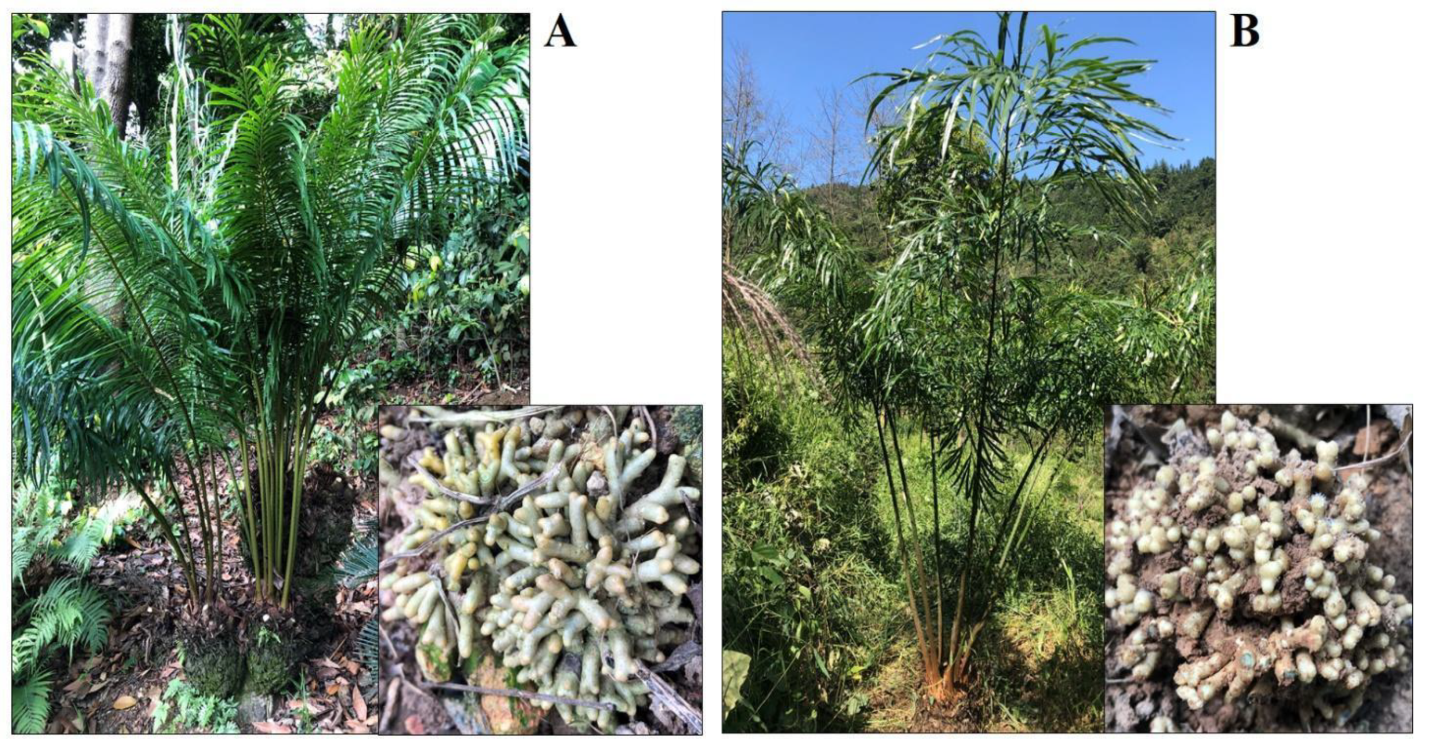
2.1. Sampling Site Descriptions and Sample Collection

Samples of coralloid roots were taken from the two host species, C. debaoensis and C. fairylakea (Figure 1) from different population or sampling sites, i.e., an ex-situ conservation botanic garden, a natural habitat and a reintroduction site where propagated populations from a plant nursery in a botanical garden were reintroduced to the natural habitat. These propagated populations were derived from seeds collected in their natural habitat and grown in plant nurseries in a botanical garden before re-planting in the reintroduction site. The sampling sites are located in the Guangxi and Guangdong provinces in China. Details of the host species and sampling localities are provided in Table 1. The sampling sites in the Guangxi province possess a distance of 1100 km from the sampling sites in the Guangdong province. The former region in South China has a mountainous terrain and is home to spectacular karst landforms and natural reserves. The latter region facing the South China Sea is diverse and intersected by several basins with scattered alluvial valleys. Both regions have subtropical to tropical climate. In Guangxi, the summers are generally long, hot and humid lasting from April to October. The average annual temperature ranges from 17 °C to 23 °C and the annual precipitation ranges from 1080 mm to 1730 mm. In the Guangdong province, a more humid climatic condition marked by high temperature and abundant rainfall, especially between the months of April to June, is experienced. It also has a prolonged hot but wet summer with an annual rainfall of 1400–2000 mm. The average annual temperature is 22 °C. Six plant individuals with thick and healthy coralloid roots were selected per population site. From each plant host, two to three clusters of roots with a small amount of soil were collected and placed inside clean Ziplock bags, kept in an ice chest, brought to the laboratory and immediately refrigerated at 4 °C until further processing.

2.2. Coralloid Root Processing and Fungal Endophyte Isolation

The coralloid roots were cut into small clusters, washed under running tap water and gently scrubbed with a soft bristle brush to remove adhering soil and other debris. Under sterile conditions, the roots were first rinsed in cold milliQ water and the root surface was chemically treated according to the method described [15], with some modifications: sodium hypochlorite (NaOCl, ~2% active chlorine) for five minutes, 70% ethyl alcohol for five minutes and sterile milliQ water four times for 1–2 min each time. The roots were then blot-dried using sterile paper towels and aseptically dissected into thin longitudinal segments with approximate uniform sizes (~5 cm long, ~2 mm thick) using sterile blades. The root segments were then plated on a half-strength Malt Extract Agar (MEA, BD Difco, BD Shanghai Co., Ltd., Shanghai, China) amended with 400 µg ml−1 antibiotic streptomycin to prevent bacterial contamination. A total of 36 root segments were cut and placed on six petri dishes per host individual. Overall, a total of 1080 root segments, 648 from C. debaoensis (6 plant individuals × 36 root segments × host species × 3 sites) and 432 from C. fairylakea (6 plant individuals × 36 root segments × host species × 2 sites), were utilized in this study. Six root segments were equidistantly placed onto the surface of each plated medium using sterile forceps. Sterility control plates were also prepared by plating water from the final wash and imprinting the roots before dissection into segments onto separate and clean MEA with antibiotic. To rule out the possible effect of air contamination, two clean MEA plates were left open while performing the experiment. The isolation and control plates were then incubated in a growth chamber at 28 ± 2 °C. Incubation and checking of plates were performed up to 4 weeks to allow the isolation of slow growing fungi. The absence of fungal colony growth in the sterility control plates indicates a successful and efficient surface sterilization [41]. Observation of the culture plates was conducted every day to observe emerging hypha from the edge of the coralloid roots. Isolation was immediately performed by cutting a small portion on the edge of fungal hypha and transferring it to a freshly prepared medium but without antibiotics. Purification of fungal endophytes was carried out using hyphal/spore tip method. All isolates were cultured and maintained in agar slants at the Cycad Symbiotic Microorganism Lab (CSML) in the Research Building of the Fairy Lake Botanical Garden, Shenzhen.

2.3. Morphocultural Characterization and Morphotype Groupings of Fungal Endophytes

After successful isolation and purification of fungal endophytes, all isolates were grown on Potato Dextrose Agar (PDA, BD Difco, BD Shanghai Co., Ltd.). The axenic isolates were then observed and grouped according to their distinct colonial features and assigned to a specific group code. The morphological characteristics that were considered for morphotype groupings were the colony appearance, color, form, elevation and margin. The color and appearance of the colony on top and below (reverse) were also noted [41]. Representatives from each morphotype groups were subsequently grown for DNA extraction and molecular identification.

2.4. DNA Extraction, PCR Amplification and ITS Gene Sequencing

Genomic DNA was extracted from a 7–10 day old fungal culture representing 80 morphospecies following the modified procedure [42]. Approximately 100 mg of fresh fungal mycelium was used and ground in sterile mortar and pestle with liquid nitrogen. The crushed fungal mycelium was then put in a 2 mL sterilized microcentrifuge tube with 60 mg sterile glass bead and 700 µL of extraction buffer with the following composition: 100 mM Tris HCl (pH 8.0); 50 mM EDTA, 3% SDS. Homogenization was performed once on a tissue homogenizer (Tissuelyser 24, Jingxin Tech., Yiyang, China) at 6 m/s for 30 s. Centrifugation of the fungal tissue homogenate was then carried out for 10 min at 12,000 rpm. Approximately 600 uL of supernatant was then transferred to a new microcentrifuge tube and treated with 2 uL of RNase A (10 mg/mL). DNA extraction was performed twice by putting equal volume of PCI (Phenol Chloroform Isoamylol: 25:24:1). Precipitation was performed with chilled absolute ethyl alcohol at −20 °C for 1 h. The DNA pellet was washed twice with 70% ethyl alcohol, air dried and dissolved in 1× TE (pH 8.0) buffer. The ribosomal DNA internal transcribed spacer (ITS) region (ITS1–5.8S–ITS2) was amplified using the forward primer ITS1 (5′-TCCGTAGGTGAACCTGCGG-3′) and reverse primer ITS4 (5′-TCCTCCGCTTATTGATATGC-3′) [43]. Following a previous study [44], the final reaction volume was 25 µL containing 2.5 µL 10× PCR (Mg2+free) buffer, 2 µL of 25 mM MgCl2 (Takara), 1.5 µL dNTP mixture (2.5 mM), 1 µL of each primer (10 pmoles), 0.2 µL (5 U/mL) of Taq polymerase, 1.5 µL template DNA and 15.3 µL sterile dH2O. PCR amplification was carried out on a BIO-RAD T100 Thermal Cycler with initial denaturation for 5 min at 94 °C, followed by 35 cycles (35 s at 94 °C, 50 s at 52 °C, 1 min at 72 °C) and a final extension of 5 min at 72 °C. The quality of PCR products was then checked on 0.8% agarose gel in 1× TAE buffer. The amplicons were sent to TSINGKE Biotechnology Co., Ltd. (Guangdong, China) and sequenced in both directions using the abovementioned primers on ABI 3730 Automated Sequencer.

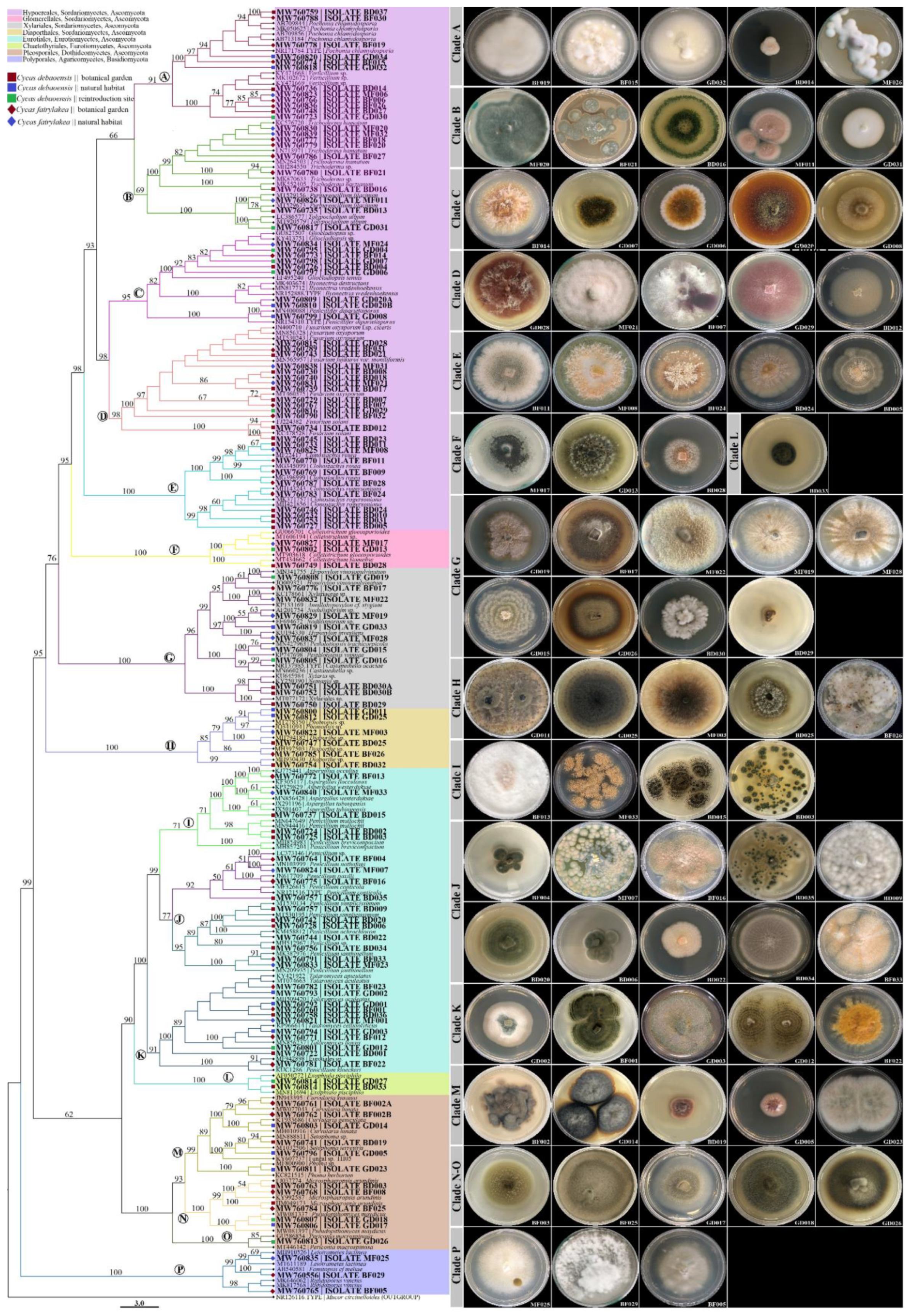
2.5. Molecular Identification and Phylogenetic Analysis of Fungal Endophytes

A total of 120 fungal sequences were generated from this study in which the 119 sequences were deposited to the National Center for Biotechnology Information (NCBI) Taxonomy Database (http://www.ncbi.nlm.nih.gov/taxonomy) GenBank with accession numbers MW750556, MW760722–MW760840. We then compared and identified our sequences with the data deposited in NCBI databases via the BLAST algorithm and downloaded the closest match sequences (≥97% identity). Alignment of the sequences was performed using the Auto algorithm of MAFFT v7.450 [45] and manually edited in Geneious Prime 2020.2 [46]. Phylogenetic reconstruction was then performed with the Maximum Likelihood (ML) topological method. The best fit model used for the ITS sequence alignment were the general time reversible and gamma distributed with invariant sites (GTR+G+I) as determined by the Bayesian information criterion (BIC) in the model selection tab of IQ-TREE v6.10 (iqtree.cibiv.univie.ac.at) [47]. Bootstrap analyses and calculations were based on 1000 replicates. The ML was run on IQ-TREE v6.10 [47] and the resulting phylogenetic tree consensus file was viewed in FigTree v1.4.3, rooted with Mucor circinelloides (phylum Mucoromycota). Accession numbers of sequences retrieved from GenBank are also included in our phylogenetic tree.

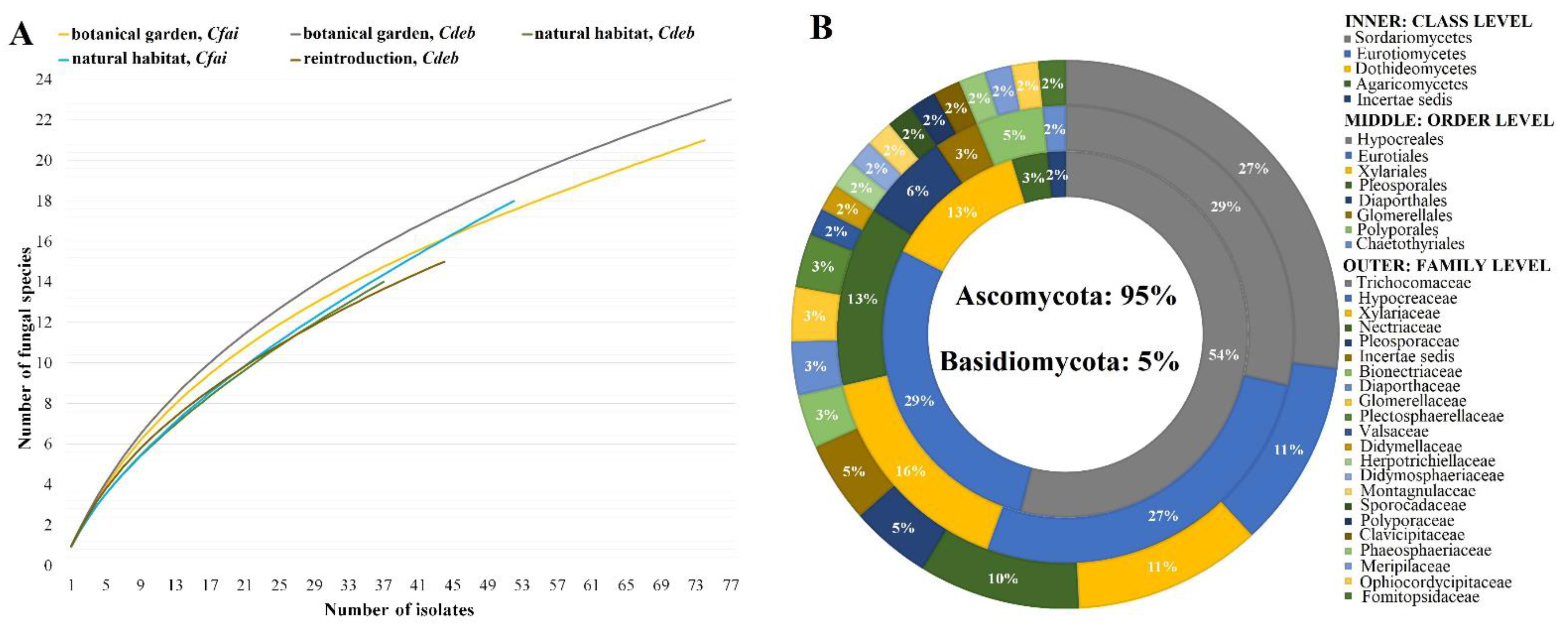
2.6. Statistical and Diversity Analysis

The colonization rate (CR) of fungal endophytes was determined as the total number of isolates recovered from a given site divided by the total number of root segments prepared in that particular site multiplied by 100. The isolation frequency (IF) was also computed by the number of isolates recovered from a given site divided by the overall number of isolates multiplied by 100. In addition, comparisons of the fungal endophyte composition and diversity between the host species (C. debaoensis vs. C. fairylakea) and populations sites (botanical garden vs. reintroduction site vs. natural habitat) were calculated using the species richness (S), Shannon–Wiener (H’) and Fisher’s-Alpha (FA) diversity statistics. The number of species recovered from a particular population site or host species represented the species richness. The Shannon and Fisher’s alpha indices were computed using the relative proportion of fungal species in a specific population site and host species. The taxonomic diversity index through the computation of Species (S) over Genus (G) (S/G) ratio was also determined per host species and population sites. Moreover, the β-diversity using the non-metric multidimensional scaling (NMDS) based on Bray–Curtis dissimilarity matrices was performed using the datasets from population sites and host species. The difference in the diversity was statistically analyzed by performing the analysis of variance (p < 0.05) using Paleontological statistics software PAST (version 4.03) [48].
The datasets from our previous study [39], in which we reported fungal composition using high throughput sequencing, were also extracted and compared with the datasets obtained using the culture-dependent technique in the present study to provide insights on the overall community composition and structure of fungal endophytes associated with the cycads. We simply expressed the data as a Venn diagram to show whether we recovered those taxa that were recorded by culture-independent molecular methods.

2.7. In Vitro Co-Cultivation Assay between Fungal Endophytes and the Test Pathogens

Endophytic fungal cultures were screened to test their ability to suppress mycelial growth of fungal pathogens on a direct confrontation using the in vitro co-cultivation assay. Fungal co-cultivating technique is a quick and efficient method to mimic microbial interactions in their natural environment and allow the identification of microbes with antimicrobial activities [49,50,51,52]. In the present study, only fungal endophytes with distinct colonial characteristic features, e.g., pigmented colony and production of coloring/exudates on media, were subjected to antagonistic screening [52]. In addition, we selected the fungal endophytes based on recent published literatures and focused on isolates that were less-studied (very few existing studies on their activities were conducted), rare taxa (fungal endophytes with single isolate or were not commonly reported) and those that were reported to exhibit strong bioactivities against other microorganisms. Therefore, nineteen isolated fungal endophytes were examined. Meanwhile, the two fungal phytopathogens were recovered from the symptomatic-diseased leaf of C. debaoensis and were identified as Diaporthe sp. strain CdP01 and Colletotrichum sp. strain CdP02. Fungal discs (6 mm) were produced from the cultures of phytopathogens and the challenged root endophytic isolates. The fungal endophyte disc was placed on the center of the plate with half-strength PDA. The discs of the two phytopathogens were placed in the same manner in the same plate at a distance of approximately 3 cm from the endophyte. Control plates were also produced with only inoculated test phytopathogens in the absence of endophytic fungus. The assay was conducted with three biological replicates. Observations were carried out for ten days. The radial growths of the mycelia of the test pathogen on a control (r1) and in the direction of the endophytic fungus (r2) were measured and the percent inhibition in mycelial growth was calculated based on the formula I% = [(r1 − r2)/r1] × 100 [52]. Furthermore, the interactions of the fungal isolates during direct confrontations on co-cultivation assay were characterized based on the six types classification system [53]. These include the following: (1) Interaction type A: characterized by mutual intermingling growth of fungi with no visible sign of antagonism or inhibition of both fungi; (2) Type B: a mutual inhibition upon contact or at a distance between fungi with no to very minimal clear zone (usually <2 mm); (3) Type C: observed with inhibition of one fungus on contact with the inhibited species continued to grow at a significantly reduced rate, while the inhibitor species grew at a slightly, reduced rate or unchanged; (4) Type D: a mutual inhibition between fungi at a distance producing significantly clear zones of >2 mm; (5) Type E: inhibition of one fungus upon contact with another, the inhibitor species continuing to grow at a reduced rate through the inhibited colony; (6) Type F: inhibition of one species upon contact or at a distance and the inhibitor species continuing to grow at an unchanged rate through or over the inhibited fungus.

3. Results

3.1. Fungal Isolates Characterization and Taxonomic Assignment

A total of 284 fungal endophytes were recovered from 1018 coralloid root segments examined from the two host species of Cycas (n = 30) collected in different population sites (Figure 2A). Overall, root samples from the host plants in the botanical garden yielded more isolates (IF) and significant higher colonization rates (CR) (IF = 27%, CR = 36% for C. debaoensis; IF = 26%, CR = 34% for C. fairylakea) than the samples from individuals in natural habitat (IF = 13%, CR = 17% for C. debaoensis; IF = 18%, CR = 24% for C. fairylakea) and from the reintroduced populations (IF = 15%, CR = 20% for C. debaoensis) (Table 2). C. debaoensis (IF = 56%, CR = 24%) had a higher isolation frequency and a lower colonization rate than C. fairylakea (IF = 44%, CR = 29%). Our isolates are composed of spore-forming species and mycelia sterilia on potato dextrose agar. Mycelia sterilia are fungal endophytes that failed to sporulate even after prolonged grown in culture media. They were observed based on their morphocultural characteristics, such as growth pattern, colony appearance, form, margin and color (front and reverse). The fungal isolates that share the same colonial features and obtained from the same host species and site were grouped together and considered to belong to the same morphotaxa. Overall, 120 isolates (42% of the total isolates) were maintained and subsequently sequenced for molecular identification and phylogenetic analysis. The high-quality ITS sequence data in combination with its cultural appearance were used to assign each isolate to a corresponding taxonomic unit. The closest match species and other information were identified on the BLASTn search (Supplementary Table S1). A similarity value of ≥97% was observed among sequenced fungal isolates except for GD016 with 95% percent identity against its nearest BLAST hit (Supplementary Table S1).
The majority of the fungal isolates belonged to Ascomycota (95% of the total isolates) and a few to Basidiomycota (5%) (Figure 2B). Furthermore, a total of five classes, eight orders and 22 families were identified. The fungal classification with the highest proportion was Sordariomycetes (54%), followed by Eurotiomycetes (29%) and Dothideomycetes (13%). A small proportion of isolates, with 3% and 2%, were classified as Agaricomycetes and as unclassified incertae sedis, respectively. At the family level, Trichocomaceae (27%) was the most diverse, followed by Hypocreaceae (11%), Xylariaceae (11%) and Nectriaceae (10%) (Figure 2B). The same number of isolates (5%) was classified into Pleosporaceae and as incertae sedis. The other families in the phylum Ascomycota were identified with ≤3% abundance (Figure 2B). Isolates in the phylum Basidiomycota were distributed into families of Fomitopsidaceae (3%), Polyporaceae (2%) and Meripilaceae (2%).

3.2. Phylogenetic Analysis of Fungal Endophytes Associated to Cycas debaoensis and Cycas fairylakea

We combined our fungal sequences with the sequences of close relatives available in NCBI GenBank to construct the ITS phylogenetic tree. The fungal endophytes from the coralloid roots of C. debaoensis and C. fairylakea from different population sites were represented by eight different fungal orders (as shown by different colors in the phylogenetic tree; Figure 3). Four orders, i.e., Hypocreales, Glomerellales, Xylariales and Diaporthales, represented the class Sordariomycetes; two orders, i.e., Eurotiales and Chaetothyriales, represented the Eurotiomycetes; a single fungal order Pleosporales represented the Dothideomycetes; and Polyporales represented the Agaricomycetes. Furthermore, in order to describe the phylogenetic placement of the fungal endophytes, we partitioned and marked the clades in the phylogenetic tree with letters that represent a total of sixteen clades. Eight clades were generated in Sordariomycetes (clade A–H), four clades represent the class Eurotiomycetes (clade I–L), three for Dothideomycetes (clade M–O) and one for Agaricomycetes (clade P) and the last clade represented the outgroup Mucoromycota (Figure 3). The cultural appearances of the fungal isolates were also provided in Figure 3 and labeled with the same letter as in the clade in the phylotree.
Further analysis of the tree showed that fungal endophytes in the coralloid roots belonged to at least 33 genera. Hypocreales is the most represented fungal order with 55 isolates (46% of the total isolates) and 10 genera. In clade A, six endophyte sequences (=6 isolates) were noted and were grouped to Pochonia chlamydosporia and Verticillium sp. Clade B has seven morphologically distinct isolates and clusters with Trichoderma, five of which showed a close relationship to Trichoderma hamatum, one with unidentified species of Trichoderma and one with T. harzianum (clade B). In the same clade (B), doubletons and singletons (fungal species with two or one isolate/s) clustered to Purpureocillium lilacinum and Tolypocladium album with 100% of bootstrapping. Nine endophyte sequences clustered in clade C, six of which are closely related to Gliocladiopsis sp., two sequences to Ilyonectria sp. and a single isolate recovered from natural habitat of C. debaoensis clustered with Penicillifer diparietisporus with 100% of bootstrapping. One of the clades with the highest number of sequences grouped with the genus Fusarium specifically to F. oxysporum (=12 isolates) and F. solani (=2 isolates) with 98% and 100% bootstrap support, respectively (clade D). Interestingly, various morphologically distinct isolates matched to F. oxysporum were observed in this study (see fungal morphotypes in Figure 3, clade D). The last clade (E) in the order Hypocreales is composed of ten sequences strongly clustered to Clonostachys, where half of the sequences joined the C. rogersoniana with 99% of bootstrapping and the other half to C. rosea with 100% of bootstrapping. Isolates from C. rosea were observed with both white-to-gray and orange colonies (see fungal cultural morphology in Figure 3, clade E).
In a different clade (F), six sequences formed the order Glomerellales in which three sequences from the present study showed close relationship to Colletotrichum gloeosporioides. The second highest number of isolates in Sordariomycetes was observed with fungal order Xylariales and represented by a single clade (clade G). At least eight species supported with morphologically distinct colonies (see fungal morphotypes in Figure 3, clade G) were composed of one to three isolates within the genera Annulohypoxylon, Castanediella, Hypoxylon, Nemania, Nodulisporium, Pestalotiopsis and Xylaria and were identified in this clade. In another clade (H) representing the order Diaporthales, three sequences clustered to Diaporthe and three sequences to Phomopsis with high bootstrap support. Interestingly, among the genera identified, Penicillium and its teleomorph Talaromyces in the order Eurotiales were the most represented with a total number of 24 isolates belonging to nine and four distinct species, respectively (see clade I–L and the corresponding morphotypes; Figure 3). In the same order, three isolates have been identified as closely related to three different Aspergillus spp. with 100% bootstrap values. A separate clade (L) consisted of two sequences displaying high relatedness (100% bootstrapping) to Exophiala pisciphila, which is a member of dark septate endophyte in the order Chaetothyriales. Furthermore, eleven isolates belonged to order Pleosporales in the class Dothideomycetes and represented three clades (M–O). The first clade in this order consists of five sequences in which two of these grouped to Curvularia spp. and a singleton grouped to Phoma. Two isolates showed high relatedness to multiple but closely related genera such as Setophoma and Alternaria and to an unidentified fungal strain (Th05; KY607737) from a root sample. Clade N is composed of five sequences, three of which showed similarities with 99% bootstrapping to Microsphaeropsis arundinis and two sequences to Pseudopithomyces maydicus with 100% bootstrap values. Clade O is composed of a singleton isolate grouped with the fungal species Periconia macrospinosa. Four sequences of Basidiomycetous fungi in order Polyporales (clade P) were exclusively recovered from the roots of Cycas fairylakea. Two of these sequences grouped with Rigidoporus vinctus and Leiotrametes lactinea, while two isolates strongly clustered with Fomitopsis cf meliae.

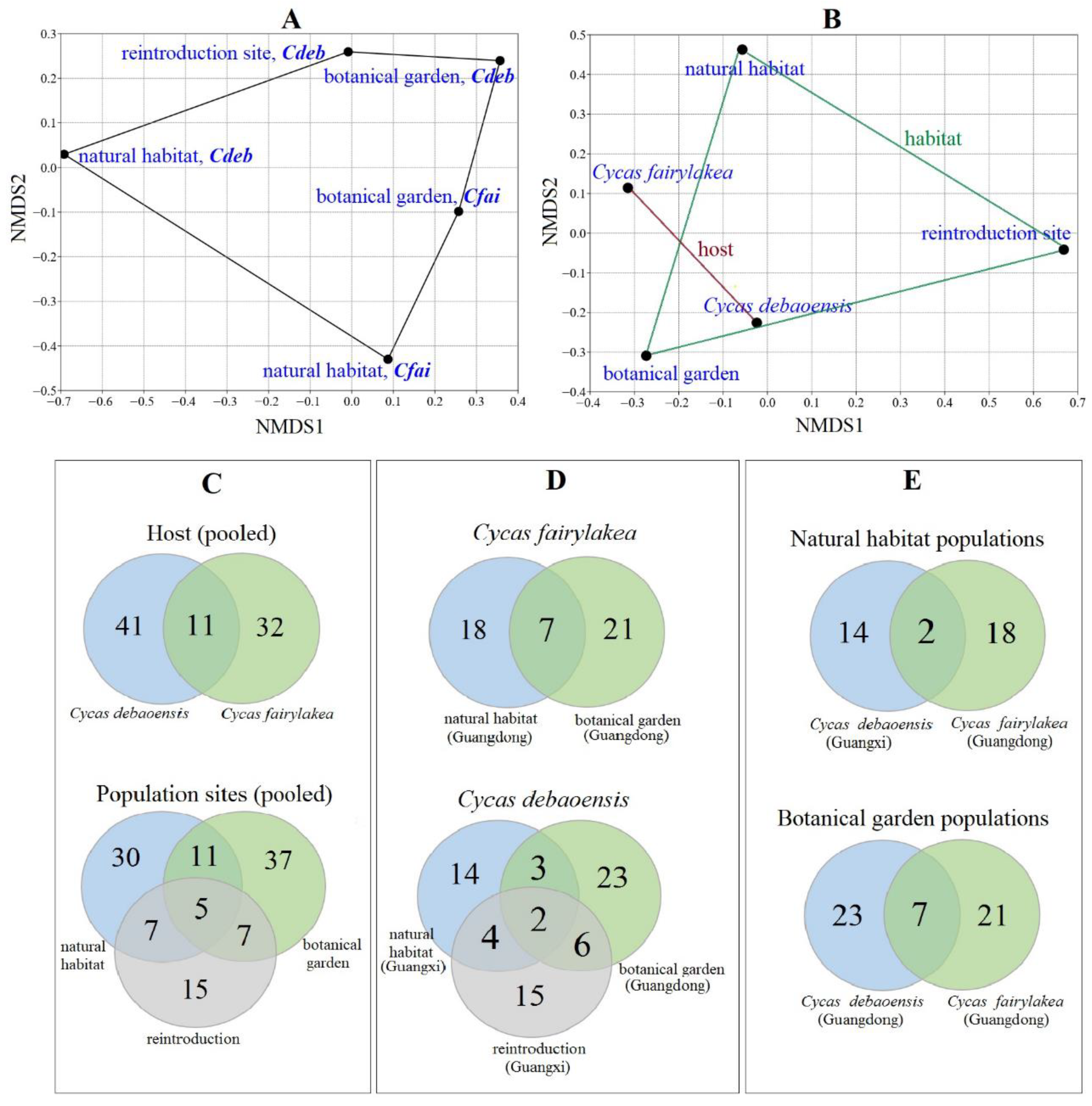
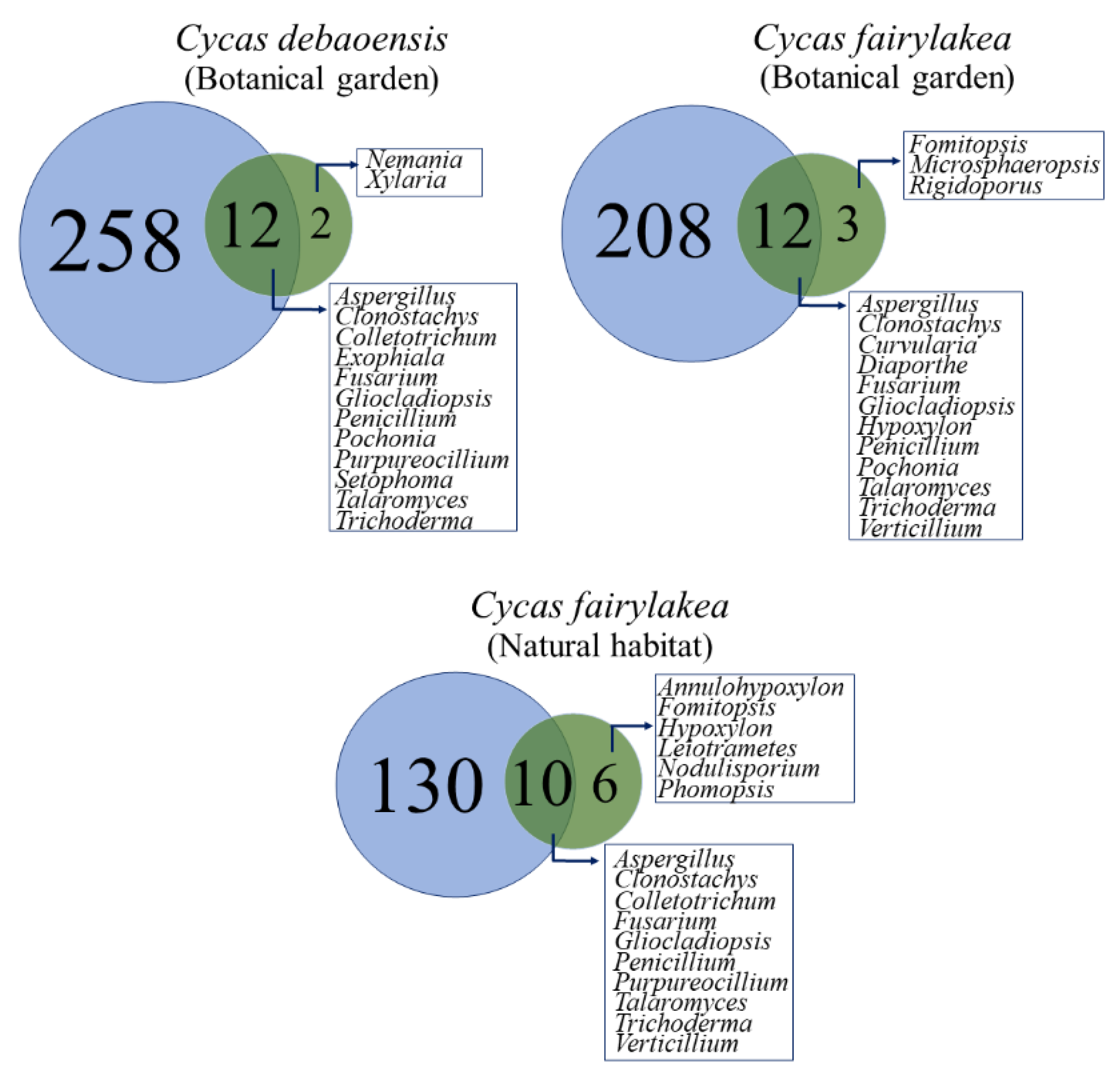
3.3. Diversity and Community Analysis of Fungal Endophytes

The findings of our study showed that both C. debaoensis and C. fairylakea shelters various fungal species. A comparative taxonomic and species diversity analyses between hosts and population sites was performed using different estimators, i.e., S/G ratio or Taxonomic Diversity Index (TDI), Shannon–Wiener (H’) and Fisher’s Alpha (FA) diversity indices. The highest endophytic community diversities and fungal richness were noted for C. debaoensis (H’ = 2.7, FA = 11.1) followed by C. fairylakea (H’ = 2.5, FA = 9.8), with their samples both collected from the botanical garden (Table 2). In fact, a comparable number of species were recorded in these samples, both with 15 genera and with 23 and 21 species, respectively. This was followed by C. fairylakea from natural habitats with diversity values of H’ = 2.2, FA = 9.8 and fungal composition of 18 species and 16 genera (Table 2). The least diverse fungal assemblage was hosted by C. debaoensis from their population sites in the Guangxi province with comparable results (natural habitat: H’ = 2.2, FA = 8.2; reintroduced: H’ = 2.2, FA = 8.0). Fifteen taxa and 13 genera were identified in the reintroduction site, while 14 species and 12 genera were identified in natural habitat (wild population). Furthermore, the differences on the S/G ratio or TDI values between population sites, except for the C. debaoensis from the botanical garden, were not statistically significant, revealing that the taxonomic diversities among population sites were the same.
Ordination from NMDS revealed that the fungal endophytes in each host species from respective population sites showed unique and overlapping endophytic fungal communities (Figure 4A,B). A higher similarity was noticed in pooled datasets between the host species than population sites. This was statistically not supported by performing a non-parametric analysis of similarities (ANOSIM) which reflected no significant differences observed on fungal communities in between population sites (R = 0.5, p = 0.31) and host species (R = 0.08, p = 0.39). In addition, the analysis of similarities showed that the fungal community assemblage was also not significantly different (R = 0.42, p = 0.20) over the studied regional population sites, i.e., between the Guangdong and Guangxi provinces. Interestingly, a series of Venn diagrams highlighted the number of shared and unique fungal taxa between the host species and the regional and local population sites (Figure 4C–E). Only 11 species of fungal endophytes were present in both Cycas species, while only five taxa were shared between the three population sites (Figure 4C). This quantifies to a percentage of 17% and 8% shared species among host and population sites, respectively. Evaluation of fungal composition dissimilarities was also carried out for each host species per population site. Interestingly, seven species were shared between C. fairylakea collected from botanical garden and natural habitat from Guangdong province (Figure 4D). Only two species were common in C. debaoensis collected from the three population sites, four taxa were shared between reintroduction and natural habitat and both geographical sites were located in the Guangxi province (Figure 4D). Forty-five (73%) of the 62 fungal species were exclusively isolated from either host species in a single population site.
Taxonomic compositional similarities were also evaluated. Talaromyces aculeatus was the predominant and the only isolated fungal species which appeared in both host species across all population sites. The other shared fungal species were recorded in both hosts but not in all sites. For instance, Gliocladiopsis sp., Verticillium sp. and Fusarium oxysporum were recovered from four collecting sites except in C. debaoensis from the natural habitat. Pochonia chlamydosporia was absent in C. fairylakea from its natural population. Among the population sites, Clonostachys rogersoniana and Diaporthe spp. were exclusively recorded from both hosts in the botanical garden. Different species representing the genera Nodulisporium and Phomopsis were recovered only from the natural habitat of the two hosts. We also found a very limited number of taxa shared per population sites per geographical region. Clonostachys rosea was the only species common in Guangdong province and was not recorded in coralloid root samples from the Guangxi province. On the other hand, Ilyonectria sp. and Pseudopithomyces maydicus were exclusively reported in population sites in Guangxi. Others have been found in multiple sites but are exclusive to a specific host. For Cycas debaoensis, the exclusive species include Exophiala pisciphila and Alternaria/Setophoma spp. Fungal species exclusive to C. fairylakea were Trichoderma spp., Fomitopsis cf meliae, Microsphaeropsis arundinis and Penicillium janthinellum. In addition, all basidiomycetous fungi in this study were exclusively recovered from C. fairylakea. Other fungal taxa were recorded exclusively in one collection site. For example, the genera Castanediella, Periconia and Tolypocladium were unique in the reintroduction site of C. debaoensis. Similarly, the species representing Penicillifer, Pestalotiopsis and Phoma were found exclusively in the natural habitat of C. debaoensis, while Annulohypoxylon and Leiotrametes were unique in the natural habitat of C. fairylakea.

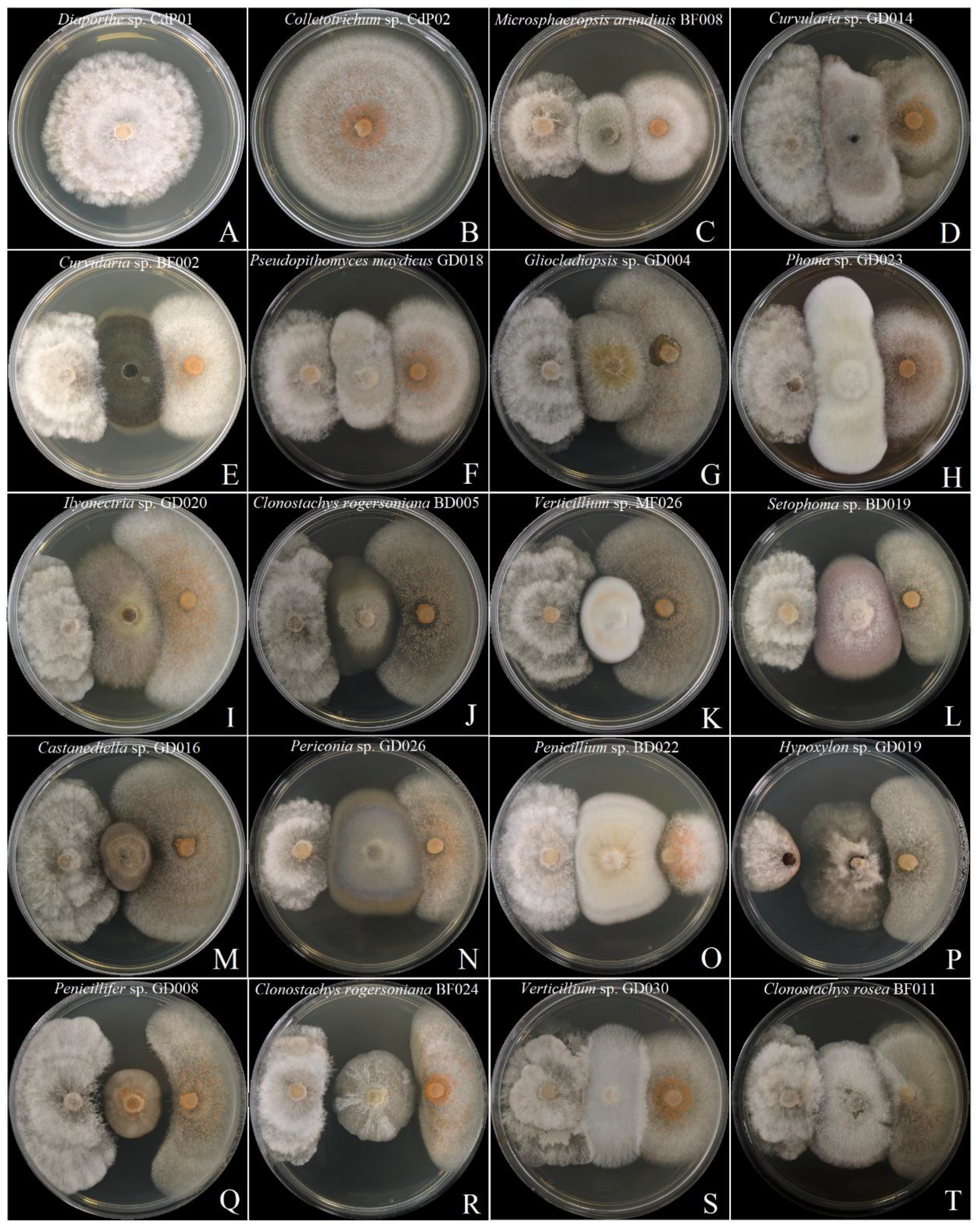
3.4. Screening of Antagonistic Potential Using Co-Cultivation Assay

One of the goals of our study was to discover fungal endophytes from endemic plant species with promising bioactivities. For this reason, the antagonistic activities of nineteen fungal endophyte isolates against the phytopathogens Diaporthe sp. CdP01 and Colletotrichum sp. CdP02 previously isolated from the diseased leaves of C. debaoensis were tested using the co-cultivation assay. The interactions between the fungal endophytes and the phytopathogens were characterized according to the criterion as described in our Materials and Methods Section. Results showed that six fungal species representing the genera Curvularia, Microsphaeropsis, Ilyonectria, Phoma and Pseudopithomyces showed type B interaction and mutually inhibited the two fungal phytopathogens (Figure 5, Table 3). In other cases, Clonostachys rogersoniana strain BD005 and Gliocladiopsis sp. showed Type B interaction against Diaporthe sp. but a different activity against Colletotrichum sp. In the separate co-cultivation plates of the two phytopathogens and the fungal strains, namely Hypoxylon sp., Alternaria/Setophoma spp., Verticillium sp. strain MF026, Penicillium sp., Castanediella sp. and Periconia sp., the fungal interaction type C has been observed (Figure 5). This interaction was characterized by inhibition of Diaporthe sp. and Colletotrichum sp. upon contact with the fungal endophytes where both phytopathogens continued to grow at a significantly reduced rate and the fungal endophytes (inhibitor species) grew also at a slightly reduced rate. Interestingly, several isolates showed promising activities and displayed Type E and F interactions. For instance, Verticillium sp. strain GD030 showed type E interaction and mutually inhibited the colony of the two phytopathogens upon contact but continued to grow at a reduced rate and advances over the colony of Diaporthe sp. The hypha of Gliocladiopsis sp. showed the same activity against Colletotrichum sp. Meanwhile, Penicillifer sp. and a different strain of C. rogersoniana BF024 inhibited the two phytopathogens at a distance while these isolates continued to grow at normal pace. In fact, a significant zone of inhibition (>2mm) was observed between these fungal endophytes and the phytopathogens. The fungal isolate C. rosea also manifested the type F interaction and further colonized the hyphal surface of the pathogens.
It is also interesting to note that pigment production was observed at the margin of some fungal isolates during physical confrontations with other isolates in the co-cultivation assay. For example, a dark pink pigmentation was observed at the margin between the colonies of Alternaria/Setophoma spp. and the phytopathogen, Colletotrichum sp. (Supplementary Figure S1). The hyphal portion of M. arundinis in contact with the phytopathogen Diaporthe sp. was also observed with black pigmentation after extending the assay incubation to 14 days (Supplementary Figure S1). On the contrary, the hypha of the phytopathogen Diaporthe sp. produced black pigmentation after its contact with fungal endophytes Verticillium sp. and C. rosea (Supplementary Figure S1). Meanwhile, the fungal endophyte species Ilyonectria sp. and Phoma sp. secreted an orange to red pigmentation onto the medium in its antagonism assay with the two phytopathogens.
The in vitro co-cultivation assay revealed that all 19 endophytic isolates inhibited the mycelial growth of the two pathogens, but the degree of inhibition varied per endophyte. The inhibition percentages (I%) of the fungal endophytes against Diaporthe sp. and Colletotrichum sp. were calculated with the results shown in Table 3. Fourteen (74%) and ten (53%) isolates showed 50–65% inhibition percentage against Diaporthe sp. and Colletotrichum sp., respectively. Hypoxylon sp. was recorded with the highest I% against Diaporthe sp. at 87.7%. This was followed by Penicillifer sp. and C. rogersoniana BF024 with I% of 69% and 66%, respectively. Meanwhile, the colonial growth of the Colletotrichum sp. was significantly inhibited at 80% inhibition percentage by C. rogersoniana (strain BF024), followed by Penicillium sp., Penicillifer sp. and C. rosea. In terms of fungal interactions, eight (42%) and six (32%) fungal endophytes exhibited type B interactions against Diaporthe sp. and Colletotrichum sp., respectively. Seven (37%) and eight (42%) exhibited type C. One fungal endophyte showed type E interaction against the two phytopathogens while three endophytes were recorded with type F interaction.

4. Discussion

The specialized coralloid roots of cycads appear to be hotspots for fungal communities as demonstrated in the recent studies employing next generation sequencing [37,38,39]. These studies also suggest that endemic Cycas spp. may harbor rare, specific or potentially novel fungal taxa which could be explored for bioprospecting and biocontrol of plant pathogens. The present study therefore employs a culture-dependent technique to study the diversity and molecular phylogeny of endophytic fungi in the specialized roots of two endemic Cycas host species in China. This technique has also been used to generate good collection of viable and culturable endophytic fungal communities, which is a requirement for any bioprospecting projects, and to test their ecological functions in the latter part of the experiments. Furthermore, the fungal communities associated with C. debaoensis from the wild and reintroduced populations in the province of Guangxi in China were also reported for the first time in this study.
The fungal endophytes associated with C. debaoensis and C. fairylakea were represented mostly by the phylum Ascomycota and comparatively with few taxa from Basidiomycota, corroborating the same results of previous studies on root-associated fungal endophyte communities [7,11,38,39,54]. In addition, Sordariomycetes was the most prevalent class of endophytic species in our study, followed by Eurotiomycetes, Dothideomycetes and Agaricomycetes as shown previously. Overall, a total of 62 species and 33 genera were molecularly identified from 30 plant individuals of the two Cycas hosts collected from different population sites. Previous study also reported 346 strains and 37 genera of fungal endophytes isolated from the endemic C. panzhihuaensis from Sichuan, China [54], and thus our study reflected the expected number of fungal genera in cycads. However, out of these only 58 strains and 16 genera were reported to have been isolated from the coralloid roots. In particular, the genera Diaporthe, Aspergillus, Trichoderma, Alternaria, Curvularia and Fusarium were previously reported in coralloid roots and were also identified in this study. Four other genera such as Paecilomyces (Purpureocillium), Periconia, Phoma and Pestalotiopsis were also identified in the present study but have been reported in other tissues, i.e., normal root, bulb, leaves and megasporophyll of C. panzhihuaensis [54]. Interestingly, about 70% of our reported fungal genera were not previously isolated, suggesting a taxonomically diverse fungal endophyte community recovered from the coralloid roots of the endemic Cycas species in Guangdong and Guangxi.
The genera Talaromyces and Fusarium that dominates the two Cycas spp. were common and ubiquitous in a wide range of plant host species [7,12,15,27,41,54]. As we might expect, these taxa were likely to be transmitted by the coralloid roots from the surrounding soil and house to its inner compartments as they have been frequently isolated in soil samples [14,51]. Interestingly, the high recurrence of fungal species Talaromyces aculeatus, Fusarium oxysporum, Gliocladiopsis sp. and Pochonia sp. across population sites may be attributed to the generalist status of these taxa or, as we have hypothesized, may also suggest their potential permanent association to cycads. In fact, Fusarium sp. was also among the single taxon of fungal endophytes that dominates the coralloid roots of C. panzhihuaensis from individuals in wild and botanical garden in the Sichuan Province [54]. The genera Verticillium, Penicillium and Trichoderma were also reported in the coralloid roots of Cycas. Fungal species under these genera were common saprophytes and pathogens or often colonized plant hosts as endophytes [12]. Interestingly, other genera, namely Colletotrichum, Diaporthe and Phomopsis, mostly inhabit above-ground organs [1,15,21,55], but were recorded from the coralloid roots. A possible migration of microbial associates within host tissues was reported in a few studies [15,56]. One such example is the detection of a high number of shared fungi in between above-ground and below-ground tissues in the exotic plant Ageratina adenophora, suggesting the potential transmission of fungal endophytes from air to leaves to stem to roots or vice versa [15]. The same pattern was observed among six forbs species but showed evident systematic growth of fungal endophytes through vertical transmission in seeds and recovered the same fungal taxa from the leaves [57]. Systemic growth of fungal endophytes in many plants remains poorly understood and, therefore, it would be particularly interesting to study whether the fungal endophytes associated to cycads were seed-transmitted, especially since the reproduction of these plants was primarily dependent on seed propagation. Alternatively, since the coralloid roots are sometimes found above the surface of the soil, it is also likely that some of the fungal endophytes commonly infecting the above-ground tissues via airborne fungal spores have non-systemically infected the coralloid roots similar to many foliar and stem endophytes [19,41,55], thus explaining the presence of some of the most common fungal communities thriving within the coralloid roots.
Rarely occurring taxa with singleton or doubletons isolates were also found to be members of fungal communities of the coralloid roots and exclusively found in one or two population sites. Particularly interesting are the rare taxa that were isolated in the limestone area in Guangxi. Phoma spp., which is a common root endophyte [13], has only a single isolate from C. debaoensis in the karst habitat. Furthermore, lesser known fungal genera, namely Castanediella, Penicillifer, Pseudopithomyces, Ilyonectria and Periconia, were only recovered from the population sites in Guangxi. Some of these genera have been previously reported in relatively few plants found in the same or nearby region, such as Panax notoginseng from Yunnan [58] and the wild Dysosma versipellis from Guangxi [28], indicating a potential biogeographical pattern of these fungal taxa. On the other hand, other taxa we recorded, such as Pseudopithomyces, were previously recovered from the aerial stilt root of mangrove (Rhizophora apiculata) [59] and the high abundances of Periconia were found in roots of grasses [60,61], suggesting its strong affinity to roots. Interestingly, Penicillifer has few new records recovered from soil samples in Bangladesh [62] and Korea [63]. To the best of our knowledge, reports on its association with plants, especially in roots, are very few in number and consists of mostly unpublished data. Its recovery from the roots of cycads along with other fungal endophytes could suggest their role to the hosts in adapting to the karst environment since many fungi provide service to plants, especially in unfavorable environments [6,9,37]. For example, some fungal endophytes may facilitate the adaptation of native Colobanthus quitensis to an environment with low temperature and extreme aridity [9]. Similarly, the presence of fungal endophytes in the special roots of cycads equally play a pivotal role that allow them to withstand the harsh environment and to further improve their growth, particularly in the condition of their habitat 300 million years ago [37,38].
The differences in the fungal communities between host and sites were more distinctive when the taxonomic composition is considered compared to the diversity values, as the numbers are influenced by the biases brought by the traditional culture-based methods in investigating fungal diversity [15,44]. In fact, the present study only recovered a very small portion of the total fungal communities from the coralloid roots when compared to the high-throughput sequencing datasets in the earlier study carried out by the same authors, although the similarity of the fungal taxa that were isolated was high since some of the plants used in the present study came from the samples in the previous metagenome-based study [39]. Direct comparison reveals that 60–80% of the fungal genera reported here (Figure 6) were also detected with high relative abundance in the study with culture-independent approach (see Supplementary Table S2). However, the genera, namely Cladosporium, Mycena, Scytalidium and Simplicillium, were previously detected with high relative abundance but were not recovered using the culture-based approach. Similarly, several fungal endophytes, about 10–20% (Figure 6), were successfully isolated but were not detected in the previous study. The high-throughput sequencing may have possibly recovered these fungi but comparisons to databases have resulted in poor taxonomic information as was confirmed by the many OTUs recorded but were only assignable to a higher taxonomic ranking, e.g., class, order and family level [39]. Therefore, we highly suggest incorporating the two approaches (culture-dependent and culture-independent) when conducting biodiversity studies to obtain a complete and broader picture of the fungal endophyte community since these two different approaches complement the limitations of each other [64].
As clearly illustrated, the botanical garden samples have the highest number of isolates, community richness and diversity, while no significant difference was found between reintroduction site and natural habitat. However, the species estimator curves also did not reach the point of saturation (Figure 2A), indicating that more species are still expected for isolation. Several relevant factors including controlled growing conditions and differing habitat management may have initiated the increase in fungal assemblages in the botanical garden resulting in higher isolates as was observed with previous studies [41,44] and in our study. Neighboring plant richness in the botanical garden may equally contribute to this pattern as they can be donors of fungal propagules relative to one another [44]. It is also worth noting that samples from Guangdong province have more fungal endophytes yield than samples from the Guangxi province. The functional requirements and the local climatic conditions may have driven the assembly of endophytic fungi in plant tissues [15,16], which might also explain this result. It is worth recalling that Guangdong possesses humid and moist conditions (see Table 1 for reference) when compared to Guangxi, which has a relatively dry environment. The former conditions support fungal growth [15,16] and may have even promoted the germination of dormant spores that are typically inactive under natural dry habitat conditions [65], thereby resulting in the high fungal richness associated with the Guangdong samples. In addition, the Guangxi region with karst topology has been reported with extremely poor nitrogen and phosphorus elements and poor water conservation resulting in a dried-up soil surface [66]. Since fungal community is strongly driven by available phosphorus and water [67], these conditions may perhaps cause a decline in fungal biomass in the soil, which directly impacts the available inocula that may infect plant endosphere, such as roots, and therefore have supported our previous explanation as to why a lower richness was detected for Guangxi samples. However, this idea needs further studies as fungal endophytes, particularly the root-associated fungi, are often known to promote plant growth through the acquisition of minerals and would expectedly be advantageous in nutrient-poor habitats.
In addition, the Venn diagrams directly reflect the very small number of shared taxa between population sites suggesting that fungal endophyte communities in the coralloid roots of cycads may also be dependent on host-related factors as shown previously in other studies [41,44]. Host species identity and phylogenetic signals were reported to play a significant role on foliar and root associated endophytes [41,68,69]. An evidence showing the dissimilarity in phyllosphere fungal communities in Mussaenda pubescens was found to be positively correlated with the detected genetic differentiation among its regional populations [19]. Experiments on Arabidopsis revealed that host genotypes have little effects on the composition of its root endophytic microbes [68]. Similarly, a recent study showed that the host genotype was found to significantly contribute to the shaping of the root communities of fungal endophytes in wheat cultivars [69]. In Cycadaceae, the use of ISSR molecular markers in previous studies showed that the degree of genetic variation is high in between populations of the same species [33,34,35], indicating a clear plant genetic distance. Determining the genetic differences among the populations of Cycas fairylakea and C. debaoensis used in the present study can specifically determine the influence of hosts genotype on the assemblage of fungal endophytes in their coralloid roots and certainly merits future studies.
What is of interest is that our findings also highlight the possible influence of transplantation as we observed moderate varying microfungal communities between plants in the natural habitat and the grown seedlings that were planted in the botanical garden or reintroduction site. In fact, contrary to our predictions, the populations of C. debaoensis grown in the natural habitat and the populations for which the seeds were propagated, raised in nursery in a botanical garden for some time and then planted to the reintroduction site slightly differed in fungal composition, with only four shared taxa (28%). The similarity is comparatively low in spite of the fact that the reintroduction site was just 8 km away from the natural habitat and therefore was recorded to share the same environmental and climate conditions. Similarly, the C. debaoensis and C. fairylakea in the botanic garden, which we would expect otherwise, showed no significant difference in the values of their diversity and had only seven shared taxa (30%). We speculate that transplantation, including the time it was carried out, may have contributed to the changes of fungal communities associated to the Cycas host species across their population sites. The same pattern was observed in a previous study reporting different fungal communities between the roots of the grasses Bouteloua species raised in greenhouses and then transplanted to a natural field [70]. In support of this, a prior experimental study demonstrated that the transplanted plants required a certain period of time to be well-adapted in the new habitat where it takes at least six months to establish a distinct rhizosphere communities conformed from the resident soil communities [71,72]. Since soil, particularly the rhizosphere, is the imminent source of microfungal inocula for root infection, perhaps more time is needed to horizontally acquire it for their plant compartments, such as the roots. Therefore, this possibly explains our result that due to the time difference in introduction, the fungal communities in the C. fairylakea transplanted to the garden for 2.5 years from the time of sampling are distinct from those plants that were planted at longer period of time in the botanical garden, such as C. debaoensis which was planted for 16 years. This may also support the case of the individuals of C. debaoensis in Guangxi, which are at 12 years of post-transplantation to the reintroduction site and are displaying different fungal communities than compared to the natural habitat. The fungal endophytes present in their roots prior to transplantation were perhaps replaced by a different community acquired in the new habitat after long-time establishment. Our assumptions however require more experimental support due to the sample size, which limits our ability to draw further assessments. Certainly, experimental validation in which various experimental factors, e.g., the use of even-aged individuals, determination of the fungal communities on the nursery-grown cycads before and after transplantations must be considered for future studies.
We also conducted a co-cultivation assay to test the ability of fungal endophytes associated with Cycas spp. to be potential biocontrol agents of two phytopathogens Colletotrichum sp. and Diaporthe sp. The genera Colletotrichum and Diaporthe are important causative agents of anthracnose to many crops and a grapevine dieback disease, respectively [73,74]. Increasing evidence showed that fungal endophytes recovered from native or endemic plants may hold significant interest for their bioactive compound production and were shown to be useful in attacking pathogens [21,26,28,75]. For instance, three fungal endophytes from endemic plants in Central Andean Precordillera were found to secrete volatile compounds and suppressed the sporulation of filamentous necrotrophic fungus Botrytis cinerea [75]. Likewise, fungal isolates recovered from native Chilean gymnosperms also showed positive inhibition activity against several pathogens [25]. Specifically, Verticillium sp. isolated from roots of wild Rehmannia glutinosa showed strong antifungal activity against the pathogen Pyricularia oryzae [76]. In our study, the two Cycas spp. endemic to China harbored taxonomically distinct fungal communities in their coralloid roots that also can potentially produce a broad spectrum of secondary metabolites. In fact, the fungal interactions between the endophytes and the phytopathogens in the co-cultivation assay showed that some of the fungal endophytes are excellent candidates for bioprospecting. For example, fungal endophytes representing the genera Periconia, Alternaria/Setophoma, Penicillium, Verticillium and Hypoxylon out-grew the phytopathogens and restricted the growth of their mycelium. The mechanism possessed by these isolates may be similar to fungal endophytes with known antagonistic activity and inhibits the pathogens through competition, antibiosis and/or mycoparasitism [49,50,51,52]. In fact, some of the isolated genera, e.g., Penicillium and Alternaria, have been reported as potential biocontrol agents of several plant diseases [52,77]. In addition, previous studies revealed diverse compounds naturally recovered from Periconia macrospinosa and Hypoxylon spp., which potentially exhibited antimicrobial activities [78,79]. Interestingly, Clonostachys rogersoniana BF024, C. rosea and Penicillifer sp. were found to exhibit striking activities against the two phytopathogens (Figure 5). As was elucidated in the review, Clonostachys rogersoniana and C. rosea are known to produce a variety of compounds with known antibacterial, antinematodal, anti-dinoflagellate and cytotoxic activities [80,81,82]. In fact, C. rosea has been found effective against many kinds of fungal pathogens and the analysis of transcriptome data showed the presence of plenty of biocontrol genes [80,81]. No study has been reported specifically on the antifungal activity of C. rogersoniana. It is interesting because our study further demonstrated that the antagonistic activities of fungal endophytes from the coralloid roots are strain-specific, as exemplified by the different activities shown by C. rogersoniana strains BD005 and BF024 against the two pathogens. This species is known to produce gliocladiosin and rogersonin, which showed moderate antibacterial activity against Klebsiella pneumoniae and Bacillus subtilis [82], and could also be the same compounds responsible for the significant inhibition of strain BF024 against the two phytopathogens in our study. In addition, another interesting result was observed with a rare and slow-growing fungal species Penicillifer diparietisporus, which has the great potential to produce promising secondary metabolites as shown by the significant high inhibition percentage in the present study. What is of special interest is that this species also produces a distinct woody odor, indicating that it could be a lucrative source of bioactive compounds.
Some of the fungal isolates in a co-cultivation assay were observed with pigment productions on their hyphae suggesting that one fungus is releasing a compound to further impede the growth of the other. A similar observation was shown in the study of Hamzah et al. [52], where an endophytic fungus Alternaria macrospora secreted a yellow pigmentation on the medium during its contact with the pathogenic Fusarium solani since day three of the dual culture assay. Fungal pigmentations were reported to act as fungal armor and assisted fungi in sustaining adverse conditions or stressful environments [83]. In some experimental studies, fungal endophytes provided thermal protections to plants and demonstrated that ability to produce melanin pigmentation that possibly dissipates heat along the hyphae and/or complex with oxygen radicals generated during heat stress [84,85]. The production of pigmentations of some fungal endophytes in the co-cultivation assay was potentially a chemical defense for protecting their colonies against the other microbes, especially as a means of survival under the natural conditions of their host plant.

5. Conclusions

We revealed a phylogenetically and unique group of cultivable endophytic fungal communities colonizing the apogeotropic coralloid roots of the endemic Cycas debaoensis and C. fairylakea. The endophytic fungal assemblage was dissimilar between hosts species among population sites and were possibly influenced by a couple of factors, including local climatic conditions and age of transplantation. Our study further demonstrated that fungal endophytes from endemic hosts as in the case of our two Cycas spp. can be promising sources of bioactive compounds. In fact, our study tested, for the first time, the antagonistic activities of fungal endophytes recovered from the coralloid roots of Chinese Cycas spp. and our results showed that they may be considered as effective antagonists or biocontrol agents. Therefore, further investigation of their secondary metabolites, particularly for identifying and elucidating the compounds from those fungal isolates that show pronounced activities in co-cultivation assay, is needed. Our study fills the research gap by providing comprehensive data on the diversity and communities of cultivable fungal endophytes, including their possible ecological relationship to their host cycads.

Supplementary Materials

The following are available online at https://www.mdpi.com/article/10.3390/jof7070572/s1, Figure S1: Reverse plates illustrating the results of the in vitro co-cultivation assay between the fungal endophytes and phytopathogens; Table S1: BLAST analysis showing the maximum nucleotide identity of representative fungal isolates based on ITS rDNA sequences; Table S2: List of fungal taxa recovered by culture-independent method, the datasets were extracted from the study of Pecundo et al. [39].

Author Contributions

Conceptualization, M.H.P., T.E.E.d.C., T.C. and N.L.; methodology and software, M.H.P.; validation, T.E.E.d.C., T.C., K.I.N. and N.L.; data analysis, M.H.P., T.E.E.d.C. and N.L.; investigation and data curation, M.H.P.; writing—original draft, M.H.P., writing—review and editing, M.H.P., T.E.E.d.C., K.I.N., T.C., H.R. and N.L.; project administration and funding acquisition, H.R., T.C. and N.L. All authors have read and agreed to the published version of the manuscript.

Funding

This research was supported by the National Forestry and Grassland Administration for Rescues and Breeding of Wild Rare and Endangered Species (Li N. 2017, 2018, 2019), the Shenzhen Fairy Lake Botanical Garden for Metagenomics Studies on Microbial Diversity in the Coralloid Roots in Cycadales (Li N. FLSF-2020-02), the Shenzhen Urban Management Bureau (Li N. 201411) and the Shenzhen Key Laboratory of Subtropical Plant Diversity, Fairy Lake Botanical Garden, Chinese Academy of Sciences (Chen T. 2017–2020). There was no additional external funding received for this study.

Institutional Review Board Statement

Not applicable.

Informed Consent Statement

Not applicable.

Data Availability Statement

All sequences generated in this study were submitted to GenBank. Other data presented in this study are available in Supplementary Materials here.

Acknowledgments

M.H.P. is thankful to the CAS-TWAS President’s Fellowship for the doctoral scholarship. All the authors sincerely thank Jianwei Rong for her assistance to M.H.P. and T.C. during sample collection at the Meilin Reservoir, Shenzhen, and Zaoyan Lu, Lanqian Yu and Zuyu Qin for accompanying M.H.P. and N.L. during the collection in Guangxi.

Conflicts of Interest

The authors declare no conflict of interest.

References

  1. Petrini, O. Fungal Endophytes of Tree Leaves. In Microbial Ecology of Leaves; Springer: New York, NY, USA, 1991; pp. 179–197. [Google Scholar]
  2. dela Cruz, T.E.E.; Notarte, K.I.; Apurillo, C.C.S.; Tarman, K.; Bungihan, M.E. Biomining fungal endophytes from tropical plants and seaweeds for drug discovery. In Biodiversity and Biomedicine; Academic Press: Cambridge, MA, USA, 2020; pp. 51–62. [Google Scholar]
  3. Mayerhofer, M.S.; Kernaghan, G.; Harper, K. The effects of fungal root endophytes on plant growth: A meta-analysis. Mycorrhiza 2012, 23, 119–128. [Google Scholar] [CrossRef]
  4. Hardoim, P.R.; Van Overbeek, L.S.; Berg, G.; Pirttilä, A.M.; Compant, S.; Campisano, A.; Döring, M.; Sessitsch, A. The Hidden World within Plants: Ecological and Evolutionary Considerations for Defining Functioning of icrobial Endophytes. Microbiol. Mol. Biol. Rev. 2015, 79, 293–320. [Google Scholar] [CrossRef] [Green Version]
  5. González-Teuber, M.; Vilo, C.; Bascuñán-Godoy, L. Molecular characterization of endophytic fungi associated with the roots of Chenopodium quinoa inhabiting the Atacama Desert, Chile. Genom. Data 2017, 11, 109–112. [Google Scholar] [CrossRef] [PubMed]
  6. Ali, A.H.; Radwan, U.; El-Zayat, S.; El-Sayed, M.A. Desert Plant-Fungal Endophytic Association: The Beneficial Aspects to their Hosts. Biol. Forum 2018, 10, 138–145. [Google Scholar]
  7. Macia-Vicente, J.G.; Jansson, H.-B.; Abdullah, S.K.; Descals, E.; Salinas, J.; Lopez-Llorca, L.V. Fungal root endophytes from natural vegetation in Mediterranean environments with special reference to Fusarium spp. FEMS Microbiol. Ecol. 2008, 64, 90–105. [Google Scholar] [CrossRef] [PubMed] [Green Version]
  8. Maciá-Vicente, J.G.; Ferraro, V.; Burruano, S.; Lopez-Llorca, L.V. Fungal Assemblages Associated with Roots of Halophytic and Non-halophytic Plant Species Vary Differentially Along a Salinity Gradient. Microb. Ecol. 2012, 64, 668–679. [Google Scholar] [CrossRef] [PubMed] [Green Version]
  9. Hereme, R.; Morales-Navarro, S.; Ballesteros, G.; Barrera, A.; Ramos, P.; Gundel, P.; Molina-Montenegro, M.A. Fungal Endophytes Exert Positive Effects on Colobanthus quitensis Under Water Stress but Neutral Under a Projected Climate Change Scenario in Antarctica. Front. Microbiol. 2020, 11, 264. [Google Scholar] [CrossRef]
  10. Zhou, W.-N.; White, J.; Soares, M.; Torres, M.S.; Zhou, Z.-P.; Li, H.-Y. Diversity of fungi associated with plants growing in geothermal ecosystems and evaluation of their capacities to enhance thermotolerance of host plants. J. Plant Interac. 2015, 10, 305–314. [Google Scholar] [CrossRef] [Green Version]
  11. Yao, Y.Q.; Lan, F.; Qiao, Y.M.; Wei, J.G.; Huang, R.S.; Li, L.B. Endophytic fungi harbored in the root of Sophora tonkinensis Gapnep: Diversity and biocontrol potential against phytopathogens. MicrobiologyOpen 2017, 6, e00437. [Google Scholar] [CrossRef]
  12. Rodriguez, R.J.; White, J.F., Jr.; Arnold, A.E.; Redman, A.R.A. Fungal endophytes: Diversity and functional roles. New Phytol. 2009, 182, 314–330. [Google Scholar] [CrossRef]
  13. Macia-Vicente, J.G.; Jansson, H.-B.; Mendgen, K.; Lopez-Llorca, L.V. Colonization of barley roots by endophytic fungi and their reduction of take-all caused by Gaeumannomyces graminis var. tritici. Can. J. Microbiol. 2008, 54, 600–609. [Google Scholar] [CrossRef] [Green Version]
  14. Van Der Heijden, M.G.A.; Bardgett, R.D.; Van Straalen, N.M. The unseen majority: Soil microbes as drivers of plant diversity and productivity in terrestrial ecosystems. Ecol. Lett. 2008, 11, 296–310. [Google Scholar] [CrossRef]
  15. Fang, K.; Miao, Y.-F.; Chen, L.; Zhou, J.; Yang, Z.-P.; Dong, X.-F.; Zhang, H.-B. Tissue-Specific and Geographical Variation in Endophytic Fungi of Ageratina adenophora and Fungal Associations with the Environment. Front. Microbiol. 2019, 10, 2919. [Google Scholar] [CrossRef] [Green Version]
  16. David, A.S.; Seabloom, E.; May, G. Plant Host Species and Geographic Distance Affect the Structure of Aboveground Fungal Symbiont Communities, and Environmental Filtering Affects Belowground Communities in a Coastal Dune Ecosystem. Microb. Ecol. 2016, 71, 912–926. [Google Scholar] [CrossRef] [PubMed]
  17. Herrera, J.; Poudel, R.; Nebel, K.A.; Collins, S.L. Precipitation increases the abundance of some groups of root-associated fungal endophytes in a semiarid grassland. Ecosphere 2011, 2, 50. [Google Scholar] [CrossRef]
  18. Gallart, M.; Adair, K.L.; Love, J.; Meason, D.; Clinton, P.; Xue, J.; Turnbull, M.H. Host Genotype and Nitrogen form Shape the Root Microbiome of Pinus radiata. Microb. Ecol. 2018, 75, 419–433. [Google Scholar] [CrossRef]
  19. Qian, X.; Duan, T.; Sun, X.; Zheng, Y.; Wang, Y.; Hu, M.; Yao, H.; Ji, N.; Lv, P.; Chen, L.; et al. Host genotype strongly influences phyllosphere fungal communities associated with Mussaenda pubescens var. alba (Rubiaceae). Fungal Ecol. 2018, 36, 141–151. [Google Scholar] [CrossRef]
  20. Notarte, K.; Nakao, Y.; Yaguchi, T.; Bungihan, M.; Suganuma, K.; dela Cruz, T.E.E. Trypanocidal activity, cytotoxicity and histone modifications induced by malformin A1 isolated from the marine-derived fungus Aspergillus tubingensis IFM 63452. Mycosphere 2017, 8, 111–120. [Google Scholar] [CrossRef]
  21. Notarte, K.I.; Devanadera, M.K.; Mayor, A.B.; Cada, M.C.; Pecundo, M.H.; Macabeo, A.P. Toxicity, antibacterial, and antioxidant activities of fungal endophytes Colletotrichum and Nigrospora spp. isolated from Uvaria grandiflora. Philipp. J. Sci. 2019, 148, 503–510. [Google Scholar]
  22. Notarte, K.I.; Yaguchi, T.; Suganuma, K.; dela Cruz, T.E.E. Antibacterial, cytotoxic and trypanocidal activities of marine-derived fungi isolated from Philippine macroalgae and seagrasses. Acta Bot. Croat. 2018, 77, 141–151. [Google Scholar] [CrossRef] [Green Version]
  23. Ramirez, C.S.P.; Notarte, K.I.R.; dela Cruz, T.E.E. Antibacterial activities of mangrove leaf endophytic fungi from Luzon Island, Philippines. Studies in Fungi 2020, 3, 320–331. [Google Scholar] [CrossRef]
  24. Schmeda-Hirschmann, G.; Hormazábal, E.; Astudillo, L.; Rodriguez, J.; Theoduloz, C. Secondary metabolites from endophytic fungi isolated from the Chilean gymnosperm Prumnopitys andina (Lleuque). World J. Microbiol. Biotechnol. 2005, 21, 27–32. [Google Scholar] [CrossRef]
  25. Hormazábal, E.; Piontelli, E. Endophytic fungi from Chilean native gymnosperms: Antimicrobial activity against human and phytopathogenic fungi. World J. Microbiol. Biotechnol. 2009, 25, 813–819. [Google Scholar] [CrossRef]
  26. Duan, X.; Xu, F.; Qin, D.; Gao, T.; Shen, W.; Zuo, S.; Yu, B.; Xu, J.; Peng, Y.; Dong, J. Diversity and bioactivities of fungal endophytes from Distylium chinense, a rare waterlogging tolerant plant endemic to the Three Gorges Reservoir. BMC Microbiol. 2019, 19, 1–14. [Google Scholar] [CrossRef] [Green Version]
  27. Cui, J.L.; Guo, T.T.; Ren, Z.X.; Zhang, N.S.; Wang, M.L. Diversity and antioxidant activity of culturable endophytic fungi from alpine plants of Rhodiola crenulata, R. angusta, and R. sachalinensis. PLoS ONE 2015, 10, e0118204. [Google Scholar] [CrossRef] [PubMed] [Green Version]
  28. Tan, X.-M.; Zhou, Y.-Q.; Zhou, X.-L.; Xia, X.-H.; Wei, Y.; He, L.-L.; Tang, H.-Z.; Yu, L.-Y. Diversity and bioactive potential of culturable fungal endophytes of Dysosma versipellis; a rare medicinal plant endemic to China. Sci. Rep. 2018, 8, 1–9. [Google Scholar] [CrossRef] [PubMed] [Green Version]
  29. Venkateswarulu, N.; Shameer, S.; Bramhachari, P.; Basha, S.T.; Nagaraju, C.; Vijaya, T. Isolation and characterization of plumbagin (5- hydroxyl- 2- methylnaptalene-1,4-dione) producing endophytic fungi Cladosporium delicatulum from endemic medicinal plants. Biotechnol. Rep. 2018, 20, e00282. [Google Scholar] [CrossRef] [PubMed]
  30. Zhong, Y.C.; Chen, C.J. Cycas debaoensis Y. C. Zhong & C. J. Chen—A New Cycad from China. Acta Phytotax. 1997, 35, 571. [Google Scholar]
  31. Wang, D.Y. Taxonomy of Cycas in China. In Cycads in China; Wang, F.X., Liang, H.B., Chen, T.Q., Wang, D.Y., Eds.; Guangdong Science and Technology Press: Guangdong, China, 1996; pp. 13–19. [Google Scholar]
  32. Chang, A.C.G.; Chen, T.; Li, N.; Duan, J. Perspectives on Endosymbiosis in Coralloid Roots: Association of Cycads and Cyanobacteria. Front. Microbiol. 2019, 10, 1888. [Google Scholar] [CrossRef] [Green Version]
  33. Jian, S.; Liu, N.; Gao, Z.; Wei, Q.; Xie, Z.; Wu, M.; Ren, H. Biological characteristics of wild Cycas fairylakea population in Guangdong Province, China. Front. Biol. China 2006, 1, 430–433. [Google Scholar] [CrossRef]
  34. Jianguang, X.; Shuguang, J.; Nian, L. Genetic variation in the endemic plant Cycas debaoens is on the basis of ISSR analysis. Aust. J. Bot. 2005, 53, 141–145. [Google Scholar] [CrossRef]
  35. Xiao, L.; Ge, X.; Gong, X.; Hao, G.; Zheng, S. ISSR Variation in the Endemic and Endangered Plant Cycas guizhouensis (Cycadaceae). Ann. Bot. 2004, 94, 133–138. [Google Scholar] [CrossRef] [Green Version]
  36. Gutiérrez-García, K.; Bustos-Díaz, E.D.; Corona-Gomez, J.A.; Ramos-Aboites, H.; Selem, N.; Cruz-Morales, P.; Pérez-Farrera, M.; Barona-Gómez, F.; Cibrián-Jaramillo, A. Cycad Coralloid Roots Contain Bacterial Communities Including Cyanobacteria and Caulobacter spp. That Encode Niche-Specific Biosynthetic Gene Clusters. Genome Biol. Evol. 2019, 11, 319–334. [Google Scholar] [CrossRef] [Green Version]
  37. Zheng, Y.; Chiang, T.-Y.; Huang, C.-L.; Gong, X. Highly diverse endophytes in roots of Cycas bifida (Cycadaceae), an ancient but endangered gymnosperm. J. Microbiol. 2018, 56, 337–345. [Google Scholar] [CrossRef]
  38. Zheng, Y.; Gong, X. Niche differentiation rather than biogeography shapes the diversity and composition of microbiome of Cycas panzhihuaensis. Microbiome 2019, 7, 1–19. [Google Scholar] [CrossRef] [Green Version]
  39. Pecundo, M.H.; Chang, A.C.G.; Chen, T.; dela Cruz, T.E.E.; Ren, H.; Li, N. Full-Length 16S rRNA and ITS Gene Sequencing Revealed Rich Microbial Flora in Roots of Cycas spp. in China. Evol. Bioinform. 2021, 17, 1–16. [Google Scholar] [CrossRef]
  40. Chang, A.C.G.; Pecundo, M.H.; Duan, J.; Ren, H.; Chen, T.; Li, N. GeoChip 5.0 microarray: A descriptive overview of genes present in coralloid root microbiomes of Cycas debaoensis and Cycas fairylakea. ELBA Bioflux 2020, 12, 5–20. [Google Scholar]
  41. Solis, M.J.L.; dela Cruz, T.E.E.; Schnittler, M.; Unterseher, M. The diverse community of leaf-inhabiting fungal endophytes from Philippine natural forests reflects phylogenetic patterns of their host plant species Ficus benjamina, F. elastica and F. religiosa. Mycoscience 2016, 57, 96–106. [Google Scholar] [CrossRef]
  42. Aamir, S. A rapid and efficient method of fungal genomic DNA extraction, suitable for PCR based molecular methods. Plant Pathol. Quar. 2015, 5, 74–81. [Google Scholar] [CrossRef]
  43. White, T.J.; Bruns, T.; Lee, S.; Taylor, J. Amplification and direct sequencing of fungal ribosomal RNA genes for phylogenetics. In PCR Protocols, a Guide to Methods and Applications; Innis, M.A., Gelfand, D.H., Sninsky, J.J., White, T.J., Eds.; Academic Press: New York, NY, USA, 1990; pp. 315–322. [Google Scholar]
  44. Solis, M.J.L.; Yurkov, A.; dela Cruz, T.E.E.; Unterseher, M. Leaf-inhabiting endophytic yeasts are abundant but unevenly distributed in three Ficus species from botanical garden greenhouses in Germany. Mycol. Prog. 2014, 14, 1019. [Google Scholar] [CrossRef]
  45. Katoh, K.; Standley, D.M. MAFFT multiple sequence alignment software version 7: Improvements in performance and usability. Mol. Biol. Evol. 2013, 30, 772–780. [Google Scholar] [CrossRef] [PubMed] [Green Version]
  46. Kearse, M.; Moir, R.; Wilson, A.; Stones-Havas, S.; Cheung, M.; Sturrock, S.; Buxton, S.; Cooper, A.; Markowitz, S.; Duran, C.; et al. Geneious Basic: An integrated and extendable desktop software platform for the organization and analysis of sequence data. Bioinformatics 2012, 28, 1647–1649. [Google Scholar] [CrossRef] [PubMed]
  47. Trifinopoulos, J.; Nguyen, L.T.; von Haeseler, A.; Minh, B.Q. W-IQ-TREE: A fast online phylogenetic tool for maximum likelihood analysis. Nucleic Acids Res. 2016, 44, W232–W235. [Google Scholar] [CrossRef] [Green Version]
  48. Hammer, Ø.; Harper, D.A.T.; Ryan, P.D. PAST: Paleontological Statistics Software Package for Education and Data Analysis. Palaeontol. Electron. 2001, 4, 9. [Google Scholar]
  49. Dagamac, N.H.A.; Sogono, P.G.; Cabalfin, R.C.B.; Adducul, A.C.Y.; dela Cruz, T.E.E. Fungal Root Endophytes from Musa spp. as Biological Control Agents against the Plant Pathogen Fusarium oxysporum. Acta Manil. 2010, 56, 27–35. [Google Scholar] [CrossRef]
  50. De Mesa, R.B.C.; Espinosa, I.R.; Agcaoili, M.C.R.R.; Calderon, M.A.T.; Pangilinan, M.V.B.; De Padua, J.C.; dela Cruz, T.E.E. Antagonistic activities of needle-leaf fungal endophytes against Fusarium spp. MycoAsia 2020, 6, 1–11. [Google Scholar]
  51. dela Cruz, T.E.E.; Din, H.J.F.; Aril-dela Cruz, J.V. Microbes for Sustainable Agriculture: Isolation and Identification of Beneficial Soil- and Plant-Associated Microorganisms. In SEARCA Professorial Chair Lecture Monograph No. 6; SEARCA: Laguna, Philippines, 2021. [Google Scholar]
  52. Hamzah, T.N.T.; Lee, S.Y.; Hidayat, A.; Terhem, R.; Faridah-Hanum, I.; Mohamed, R. Diversity and Characterization of Endophytic Fungi Isolated from the Tropical Mangrove Species, Rhizophora mucronata, and Identification of Potential Antagonists Against the Soil-Borne Fungus, Fusarium solani. Front. Microbiol. 2018, 9, 1707. [Google Scholar] [CrossRef]
  53. Wheeler, K.A.; Hocking, A.D. Interactions among xerophilic fungi associated with dried salted fish. J. Appl. Bacteriol. 1993, 74, 164–169. [Google Scholar] [CrossRef]
  54. Zheng, Y.; Wu, B.; Yu, Z.; Jiang, S.; Yang, Y. Study on the Diversity of Endophytic Fungi in Cycas panzhihuaensis L. J. Life Sci. Res. 2013, 17, 406–411. [Google Scholar]
  55. Apurillo, C.C.S.; Cai, L.; dela Cruz, T.E.E. Diversity and bioactivities of mangrove fungal endophytes from Leyte and Samar, Philippines. Philipp. Sci. Lett. 2019, 12, 33–48. [Google Scholar]
  56. Chi, F.; Shen, S.-H.; Cheng, H.-P.; Jing, Y.-X.; Yanni, Y.G.; Dazzo, F.B. Ascending Migration of Endophytic Rhizobia, from Roots to Leaves, inside Rice Plants and Assessment of Benefits to Rice Growth Physiology. Appl. Environ. Microbiol. 2005, 71, 7271–7278. [Google Scholar] [CrossRef] [Green Version]
  57. Hodgson, S.; De Cates, C.; Hodgson, J.; Morley, N.J.; Sutton, B.C.; Gange, A.C. Vertical transmission of fungal endophytes is widespread in forbs. Ecol. Evol. 2014, 4, 1199–1208. [Google Scholar] [CrossRef] [Green Version]
  58. Zheng, Y.-K.; Miao, C.-P.; Chen, H.-H.; Huang, F.-F.; Xia, Y.-M.; Chen, Y.-W.; Zhao, L.-X. Endophytic fungi harbored in Panax notoginseng: Diversity and potential as biological control agents against host plant pathogens of root-rot disease. J. Ginseng Res. 2017, 41, 353–360. [Google Scholar] [CrossRef] [PubMed]
  59. Doilom, M. Can ITS sequence data identify fungal endophytes from cultures? A case study from Rhizophora apiculata. Mycosphere 2017, 8, 1869–1892. [Google Scholar] [CrossRef]
  60. Jumpponen, A.; Herrera, J.; Porras-Alfaro, A.; Rudgers, J. Biogeography of root-associated fungal endophytes. In Biogeography of Mycorrhizal Symbiosis; Tedersoo, L., Ed.; Springer: Cham, FL, USA, 2017; pp. 195–222. [Google Scholar]
  61. Knapp, D.G.; Imrefi, I.; Boldpurev, E.; Csíkos, S.; Akhmetova, G.; Berek-Nagy, P.J.; Otgonsuren, B.; Kovács, G.M. Root-Colonizing Endophytic Fungi of the Dominant Grass Stipa krylovii From a Mongolian Steppe Grassland. Front. Microbiol. 2019, 10, 2565. [Google Scholar] [CrossRef] [PubMed]
  62. Islam, S.M.N.; Haque, M.A.; Chowdhury, M.Z.H.; Mishu, N.J.; Hossain, M.M. A Hypocrealean Fungus Penicillifer diparietisporus, First Reported in Bangladesh. Bangladesh J. Sci. Res. 2020, 31–33, 63–65. [Google Scholar]
  63. Das, K.; Back, C.G.; Lee, S.Y.; Jung, H.Y. Penicillifer diparietisporus: A New Record from Field Soil in Korea. Korean J. Mycol. 2019, 46. [Google Scholar] [CrossRef]
  64. Dissanayake, A.J.; Purahong, W.; Wubet, T.; Hyde, K.D.; Zhang, W.; Xu, H.; Zhang, G.; Fu, C.; Liu, M.; Xing, Q.; et al. Direct comparison of culture-dependent and culture-independent molecular approaches reveal the diversity of fungal endophytic communities in stems of grapevine (Vitis vinifera). Fungal Divers. 2018, 90, 85–107. [Google Scholar] [CrossRef]
  65. Pither, J.; Pickles, B.J. The paleosymbiosis hypothesis: Host plants can be colonized by root symbionts that have been inactive for centuries to millenia. FEMS Microbiol. Ecol. 2017, 93, 1–9. [Google Scholar] [CrossRef]
  66. Li, Y.-B.; Shao, J.-A.; Yang, H.; Bai, X. The relations between land use and karst rocky desertification in a typical karst area, China. Environ. Earth Sci. 2008, 57, 621–627. [Google Scholar] [CrossRef]
  67. Tian, Q.; Taniguchi, T.; Shi, W.-Y.; Li, G.; Yamanaka, N.; Du, S. Land-use types and soil chemical properties influence soil microbial communities in the semiarid Loess Plateau region in China. Sci. Rep. 2017, 7, srep45289. [Google Scholar] [CrossRef] [PubMed] [Green Version]
  68. Bulgarelli, D.; Rott, M.; Schlaeppi, K.; Van Themaat, E.V.L.; Ahmadinejad, N.; Assenza, F.; Rauf, P.; Huettel, B.; Reinhardt, R.; Schmelzer, E.; et al. Revealing structure and assembly cues for Arabidopsis root-inhabiting bacterial microbiota. Nat. Cell Biol. 2012, 488, 91–95. [Google Scholar] [CrossRef] [PubMed]
  69. Latz, M.A.; Kerrn, M.H.; Sørensen, H.; Collinge, D.B.; Jensen, B.; Brown, J.K.; Madsen, A.M.; Jørgensen, H.J.L. Succession of the fungal endophytic microbiome of wheat is dependent on tissue-specific interactions between host genotype and environment. Sci. Total Environ. 2021, 759, 143804. [Google Scholar] [CrossRef] [PubMed]
  70. Chung, Y.A.; Jumpponen, A.; Rudgers, J.A. Divergence in Diversity and Composition of Root-Associated Fungi Between Greenhouse and Field Studies in a Semiarid Grassland. Microb. Ecol. 2018, 78, 122–135. [Google Scholar] [CrossRef]
  71. Schöps, R.; Goldmann, K.; Herz, K.; Lentendu, G.; Schöning, I.; Bruelheide, H.; Wubet, T.; Buscot, F. Land-Use Intensity Rather Than Plant Functional Identity Shapes Bacterial and Fungal Rhizosphere Communities. Front. Microbiol. 2018, 9, 2711. [Google Scholar] [CrossRef]
  72. Schöps, R.; Goldmann, K.; Korell, L.; Bruelheide, H.; Wubet, T.; Buscot, F. Resident and phytometer plants host comparable rhizosphere fungal communities in managed grassland ecosystems. Sci. Rep. 2020, 10, 919. [Google Scholar] [CrossRef] [PubMed]
  73. Zakaria, L. Diversity of Colletotrichum Species Associated with Anthracnose Disease in Tropical Fruit Crops—A Review. Agriculture 2021, 11, 297. [Google Scholar] [CrossRef]
  74. Manawasinghe, I.S.; Dissanayake, A.J.; Li, X.; Liu, M.; Wanasinghe, D.; Xu, J.; Zhao, W.; Zhang, W.; Zhou, Y.; Hyde, K.D.; et al. High Genetic Diversity and Species Complexity of Diaporthe Associated with Grapevine Dieback in China. Front. Microbiol. 2019, 10, 1936. [Google Scholar] [CrossRef]
  75. Vidal, A.; Parada, R.; Mendoza, L.; Cotoras, M. Endophytic Fungi Isolated from Plants Growing in Central Andean Precordillera of Chile with Antifungal Activity against Botrytis cinerea. J. Fungi 2020, 6, 149. [Google Scholar] [CrossRef]
  76. You, F.; Han, T.; Wu, J.-Z.; Huang, B.-K.; Qin, L.-P. Antifungal secondary metabolites from endophytic Verticillium sp. Biochem. Syst. Ecol. 2009, 37, 162–165. [Google Scholar] [CrossRef]
  77. Gharsallah, H.; Ksentini, I.; Naayma, S.; Taieb, K.H.; Abdelhedi, N.; Schuster, C.; Triki, M.A.; Ksantini, M.; Leclerque, A. Identification of fungi in Tunisian olive orchards: Characterization and biological control potential. BMC Microbiol. 2020, 20, 1–13. [Google Scholar] [CrossRef] [PubMed]
  78. Anderson, J.R.; Edwards, R.L.; Whalley, A.J.S. Metabolites of the higher fungi. Part 21. 3-Methyl-3,4-dihydroisocoumarins and related compounds from the ascomycete family xylariaceae. J. Chem. Soc. Perkin Trans. 1983, 1, 2185–2192. [Google Scholar] [CrossRef]
  79. Inose, K.; Tanaka, K.; Koshino, H.; Hashimoto, M. Cyclopericodiol and new chlorinated melleins isolated from Periconia macrospinosa KT3863. Tetrahedron 2019, 75, 130470. [Google Scholar] [CrossRef]
  80. Sun, Z.-B.; Li, S.-D.; Ren, Q.; Xu, J.-L.; Lu, X.; Sun, M.-H. Biology and applications of Clonostachys rosea. J. Appl. Microbiol. 2020, 129, 486–495. [Google Scholar] [CrossRef] [PubMed] [Green Version]
  81. Sun, Z.-B.; Sun, M.-H.; Li, S.-D. Draft Genome Sequence of Mycoparasite Clonostachys rosea Strain 67-1. Genome Announc. 2015, 3, e00546-15. [Google Scholar] [CrossRef] [PubMed] [Green Version]
  82. Wang, Y.; Ren, J.; Li, H.; Pan, Y.; Liu, X.; Che, Y.; Liu, G. The disruption of verM activates the production of gliocladiosin A and B in Clonostachys rogersoniana. Org. Biomol. Chem. 2019, 17, 6782–6785. [Google Scholar] [CrossRef]
  83. Pombeiro-Sponchiado, S.R.; Sousa, G.S.; Andrade, J.C.R.; Lisboa, H.F.; Gonçalves, R.C.R. Production of Melanin Pigment by Fungi and Its Biotechnological Applications. In Melanin; IntechOpen: London, UK, 2017. [Google Scholar]
  84. Davidson, J.F.; Whyte, B.; Bissinger, P.H.; Schiestl, R.H. Oxidative stress is involved in heat-induced cell death in Saccharomyces cerevisiae. Proc. Natl. Acad. Sci. USA 1996, 93, 5116–5121. [Google Scholar] [CrossRef] [Green Version]
  85. Redman, R.S.; Vervoort, V.S.; Beachem, M.A.; Edwards, P.S.; Ladd, S.; Miller, K.E.; De Mollerat, X.; Clarkson, K.; Dupont, B.; Schwartz, C.E.; et al. Thermotolerance Generated by Plant/Fungal Symbiosis. Science 2002, 298, 1581. [Google Scholar] [CrossRef]
Figure 1. Host species of Cycas and the appearance of their coralloid roots growing above the surface of the soil. (A) Cycas fairylakea from the ex-situ conservation site in National Cycad Germplasm Conservation Center, Fairy Lake Botanical Garden, Guangdong, and (B) Cycas debaoensis from the reintroduction site in Debao County, Guangxi.
Jof 07 00572 g001
Figure 2. (A) Rarefaction curve showing the species richness of fungal endophytes plotted against the number of isolates; datasets are based on the host species (Cycas debaoensis and Cycas fairylakea) per their collection sites (botanical garden, natural habitat and reintroduction site). (B) Taxonomic distribution of the fungal taxa (n = 120) from the coralloid roots of the two endemic Cycas spp.
Jof 07 00572 g002
Figure 3. A maximum likelihood (ML) analysis showing the phylogenetic relationships of root fungal endophytes. The tree has a total of 233 fungal sequences, in which 119 fungal sequences came from the present study, 113 reference sequences of close relatives came from the NCBI database and the sequence from the type specimen of Mucor circinelloides (NR126116; phylum Mucoromycota; order Mucorales) as the outgroup. The bootstrap support values are indicated at the nodes. In bold font are the isolate code and sequence accession number of the fungal endophytes obtained from the coralloid roots of Cycas debaoensis and Cycas fairylakea. Fungal isolates are further highlighted in different colors according to the order classification. The tip shape of each isolate in the tree is a key in determining its source of host species and collection origin. Images on the right of the phylotree are the morphotypes of the representative fungal endophytes per assigned clade.
Jof 07 00572 g003
Figure 4. NMDS visualization based on Bray–Curtis analysis. The graphs showed the (A) ordination of the fungal communities between host species per population sites. (B) Plots are based on the pooled datasets of host Cycas debaoensis and Cycas fairylakea, regardless of the population sites and population sites, i.e., natural habitat, reintroduction site and botanical garden regardless of the host species. Venn diagrams illustrated the number of shared and unique taxa. Comparison was carried out between (C) pooled datasets from host species and population sites, (D) host species and (E) population sites over geographical region.
Jof 07 00572 g004
Figure 5. Results of the co-cultivation assay between nineteen fungal endophytes and the two fungal phytopathogens. (A,B) Control plates of the phytopathogens isolated from the diseased-symptomatic leaves of Cycas debaoensis. The two leaf fungal pathogens were co-cultivated with the fungal endophytes on PDA plates and incubated for 10 days at 26 ± 2 °C (CT). (CI) Displayed Type B interaction against the two pathogens; note that Gliocladiopsis GD004 displayed Type E interaction against Colletotrichum sp. (G). Type C interaction was observed in (JP), while Type E and F interactions were observed in (QT) against the two phytopathogens.
Jof 07 00572 g005
Figure 6. Venn diagrams expressing the number of shared and unique fungal endophytes between culture-independent (in blue) and culture-dependent (in green) methods. We listed the fungal genera recovered by both methods and those that were recovered solely by the culture-dependent method. The list of fungal taxa unique to culture-independent method is provided in Supplementary Table S2. The datasets based on the culture-independent method was obtained from the previous study in overlaps with some plants used in the present study collected from the same sites [39].
Jof 07 00572 g006
Table 1. Data on the host species and sample collection sites.
HostPopulation SitesLocalityGPS/AltitudeHabitat DescriptionCollection Date
Cycas
debaoensis
botanical garden1 NCGCC, FLBG, Shenzhen, Guangdong22°34′36.1″ N 114°10′51.1″ E
60 ± 30 m.a.s.l.
Living collections of cycads in a controlled and managed environment; individuals used for the study were planted in 2004.26 January 2020
natural habitatDebao County, Baise, Guangxi23°29′29.2″ N 106°12′50.8″ E
905 ± 24 m.a.s.l.
Dispersed in open landscapes and on limestone hills with shrubs and trees; some cycad plants growing on cracks or in thin soil near the edges of limestone.22 October 2020
reintroduction site1 Huanglian mountain nature reserve, Debao County, Baise, Guangxi23°33′47.1″ N 106°14′09.8″ E
885 ± 8 m.a.s.l.
Scattered on open slopes with grasses and sparsely covered by shrubs and small trees; local population was reintroduced in 2008.22 October 2020
Cycas fairylakeaBotanical garden1 NCGCC, FLBG, Shenzhen, Guangdong22°34′36.4″ N 114°10′50.6″ E
55 ± 20 m.a.s.l.
Living collections of cycads in controlled and managed environment; individuals used for the study were planted in 2017.26 January 2020
natural habitatMeilin Reservoir Park, Shenzhen, Guangdong22°34′38.5″ N 114°00′34.2″ E
114 ± 23 m.a.s.l.
Growing on slopes of ridges and cliffs along river valleys and understory of moist closed subtropical forests at low elevations.21 January 2020
1 National Cycad Germplasm Conservation Center (NCGCC); Fairy Lake Botanical Garden (FLBG).
Table 2. Records and diversity of fungal endophytes associated with the hosts Cycas spp. from different population sites. Values followed by the same letter are not significantly different, p < 0.05.
1 Sample Source2 Records of Fungal Endophytes
RGSS/G RatioH’FACR (%)IF (%)
Cdeb, Nh3712141.2 a2.2 a8.2 a1713
Cfai, Nh5216181.1 a2.2 a9.8 b2418
Cdeb, Bg7715231.5 b2.7 b,c11.1 c3627
Cfai, Bg7415211.4 a,b2.5 c9.8 b3426
Cdeb, Re4413151.2 a2.2 a8.0 a2015
Cdeb (Pooled)15829411.4 a3.0 a17.8 a2456
Cfai (Pooled)12621321.5 a2.7 b13.8 b2944
1 Host: Cdeb: Cycas debaoensis; Cfai: Cycas fairylakea; Population site: Nh: natural habitat; Bg: botanical garden; Re: reintroduction site. 2 Records of Fungal Endophytes: R = individuals/records, G = number of genera, S = number of species, H’ = Shannon diversity index, FA = Fisher’s Alpha diversity index, CR = colonization rate, IF = isolation frequency.
Table 3. Results of the in vitro co-cultivation assay of nineteen selected endophytes against the two phytopathogens. The type of fungal interaction for each isolate against each pathogen and the percentage of growth inhibition (I%) ± standard deviation (SD) was indicated. See Figure 3 for the description of the clades.
Species Name and Isolate CodeCladeDiaporthe sp. Strain CdP01Colletotrichum sp. Strain CdP02
Percent Inhibition (I%) ± SDType of InteractionPercent Inhibition (I%) ± SDType of Interaction
Castanediella sp. GD016G58.7 ± 1.89C63.3 ± 1.57C
Clonostachys rogersoniana BD005E52 ± 1.63B62.2 ± 1.50C
Clonostachys rogersoniana BF024E66.7 ± 1.89F82.1 ± 1.50F
Clonostachys rosea BF011E64.3 ± 2.05F80 ± 1.63F
Curvularia sp. BF002M50 ± 1.63B56.7 ± 2.72B
Curvularia sp. GD014M51.6 ± 0.47B53.9 ± 2.1B
Gliocladiopsis sp. GD004C56 ± 3.27B68.3 ± 1.36E
Hypoxylon sp. GD019G87.33 ± 0.94C70.3 ± 1.89C
Ilyonectria sp. GD020C60 ± 3.77B61.7 ± 1.70B
Microsphaeropsis arundinis BF008N42.7 ± 3.77B51.1 ± 2.83B
Penicillifer sp. GD008C69.3 ± 3.30F81.1 ± 3.14F
Penicillium sp. BD022J64 ± 2.83C81.5 ± 2.51C
Periconia sp. GD026O62 ± 2.83C69.3 ± 1.89C
Phoma sp. GD023M61. 3 ± 4.99B65.6 ± 4.16B
Pseudopithomyces maydicus GD018N58.7 ± 1.89B65.6 ± 1.57B
Alternaria/Setophoma sp. GD005M61.3 ± 1.89C75.6 ± 1.57C
Alternaria/Setophoma sp. BD019M62. 3 ± 0.94C71.1 ± 2.83C
Verticillium sp. MF026A53.3 ± 1.89C65.56 ± 1.57C
Verticillium sp. GD030A49.3 ± 1.89E58 ± 2.79E
Publisher’s Note: MDPI stays neutral with regard to jurisdictional claims in published maps and institutional affiliations.

