
Waste Rose Flower and Lavender Straw Biomass—An Innovative Lignocellulose Feedstock for Mycelium Bio-Materials Development Using Newly Isolated Ganoderma resinaceum GA1M


 , ,
, , 
 , , , ,
Abstract
:1. Introduction
2. Materials and Methods
2.1. Materials
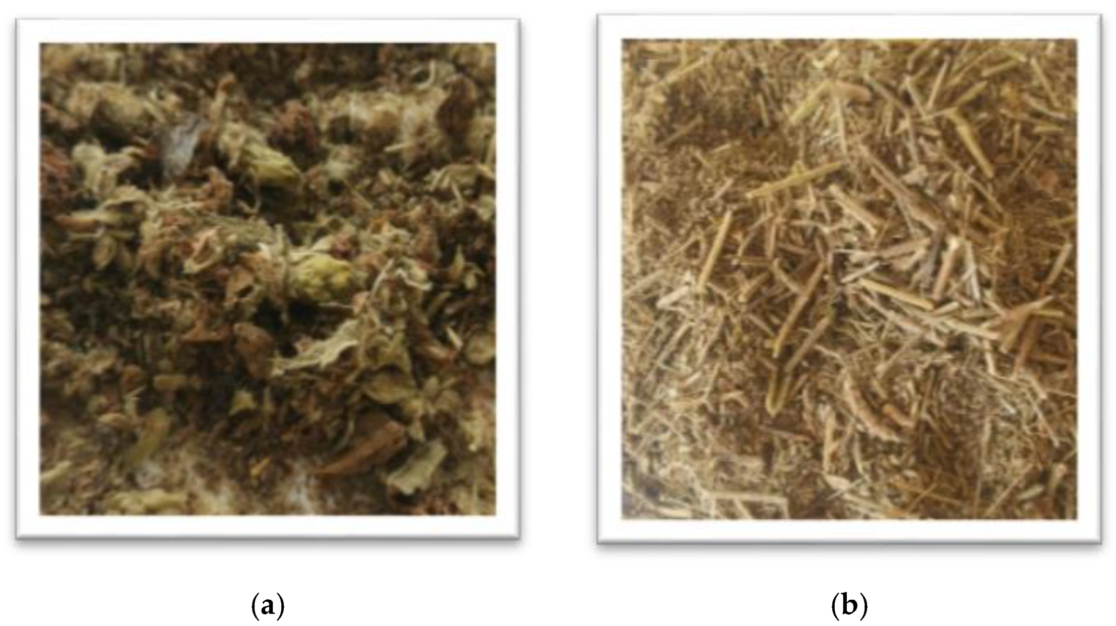
2.1.1. Substrates
2.1.2. Fungal Isolate
2.2. Substrate Preparation Procedure
2.2.1. Preparation for Cultivation
2.2.2. Preparation for Chemical Analysis
2.3. Chemical Characterization of the Substrate
2.3.1. Proximate Composition Determination
2.3.2. Determination of Total and Individual Polyphenols
- (1)
- Non-volatile substances—0.2 mL ethanol extract was lyophilized and 50 μL pyridine and 50 μL N,O-Bis-(trimethylsilyl)-trifluoroacetamide (BSTFA) were added. The sample was incubated at 70 °C for 40 min. For analysis 1.0 μL from the solution was injected on a gas chromatograph Agilent GC 7890 with mas-selective detector Agilent MD 5975 and column HP-5ms (30 m with diameter 0.32 mm and 0.25 μm thicknesses). The following temperature regimen was used—initial temperature 100 °C (hold for 2 min) then increase to 180 °C with 15 °C/min (hold for 1 min) and increase of the temperature to 300 °C with 5 °C/min (hold for 10 min); injector and detector temperatures—250 °C, helium was used s carrier gas at 1.0 mL/min. The scanning range of the mass-selective detector was m/z = 50–550 in split-split mode (10:1).
- (2)
- Volatile substances—1.0 mL ethanol extract was extracted with 1.0 mL dichloromethane (triple). The combined organic layers were dried under vacuum at 30 °C. To the dry residue 100 μL dichloromethane was added. For analysis 1.0 μL from the solution was injected on a gas chromatograph Agilent GC 7890 with mas-selective detector Agilent MD 5975 and column HP-5ms. The following temperature regimen was used—the initial temperature was 40 °C and then increase to 300 °C with 5 °C/min (hold for 10 min); injector and detector temperatures—250 °C, helium was used as carrier gas at 1.0 mL/min. The scanning range of the mass-selective detector was m/z = 40–400 in splitless mode. The individual compounds were identified by comparing the retention times and the relative index (RI) with those of standard substances and mas-spectral data from libraries of The Golm Metabolome Database (http://csbdb.mpimp-golm.mpg.de/csbdb/gmd/gmd.html, accessed on 5 November 2020) and NIST’08 (National Institute of Standards and Technology, Gaithersburg, MD, USA).
2.4. Molecular Identification of the Basidiomycete Isolate by ITS1-5.8S-ITS2 rRNA Gene Sequence Analysis
2.5. Solid-State Cultivation for Mycelium Bio-Composite Obtaining
2.5.1. Inoculum Preparation
2.5.2. Inoculation of the Substrates and Mycelium Growth
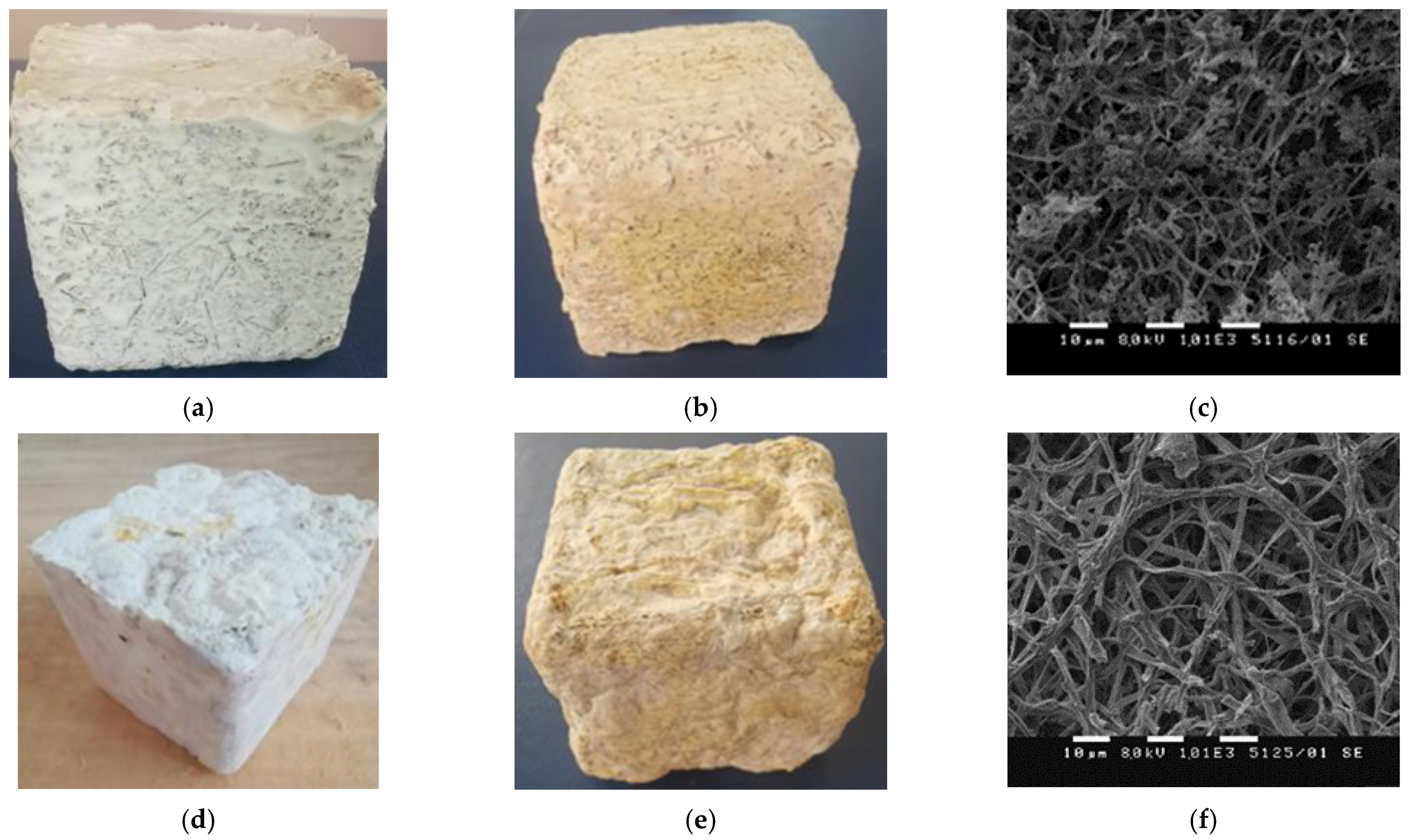
2.6. Scanning Electron Microscopy (SEM)
2.7. Fourier Transform Infrared Spectroscopy (FTIR)
2.8. Basic Physical and Mechanical Characterization
2.9. Statistical Analysis
3. Results and Discussion
3.1. Chemical Characterization of the HERF and SDLS and Assessment of Their Potential as Feedstock for Mycelium Growth and New Bio-Materials Development
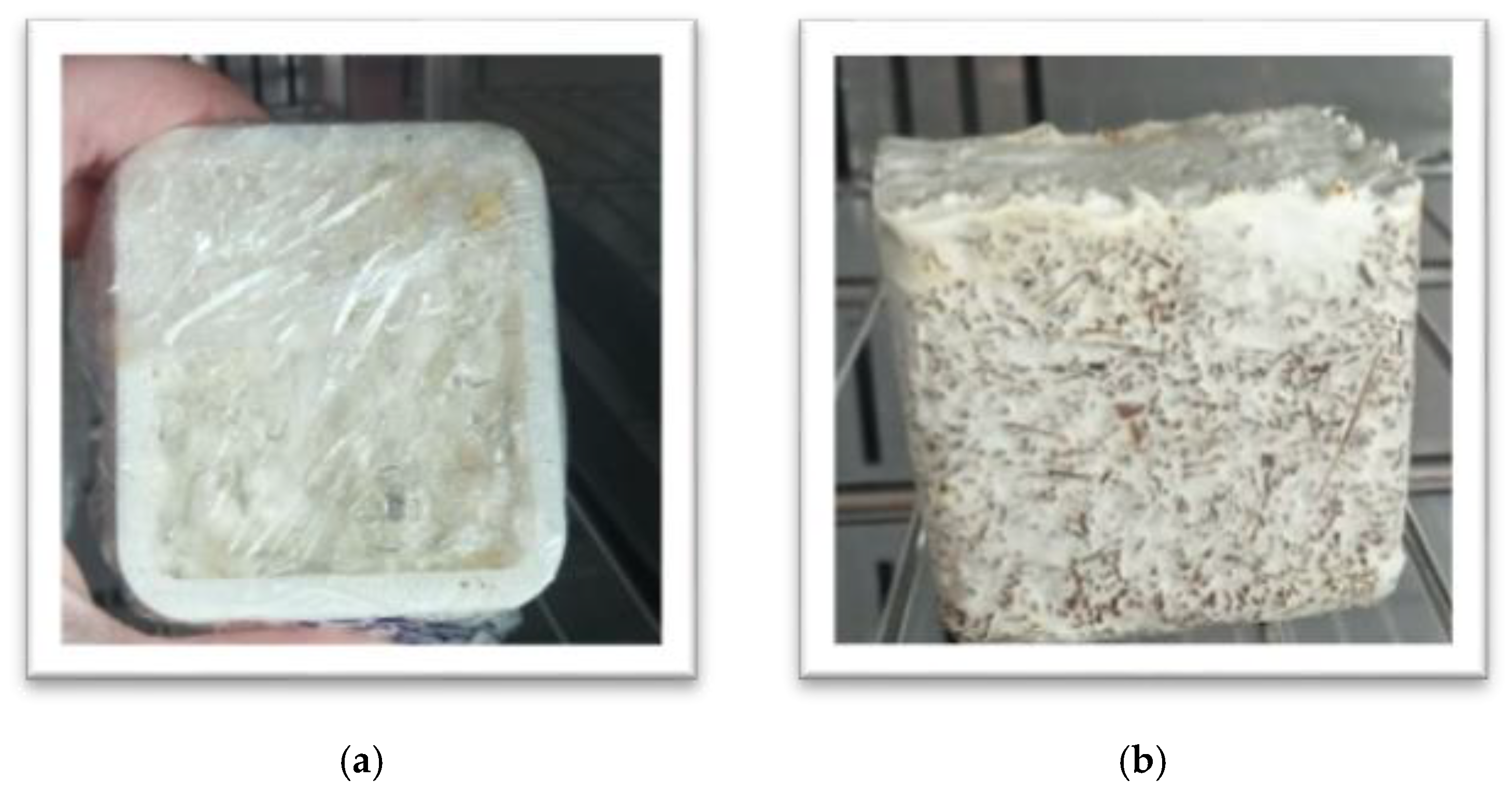
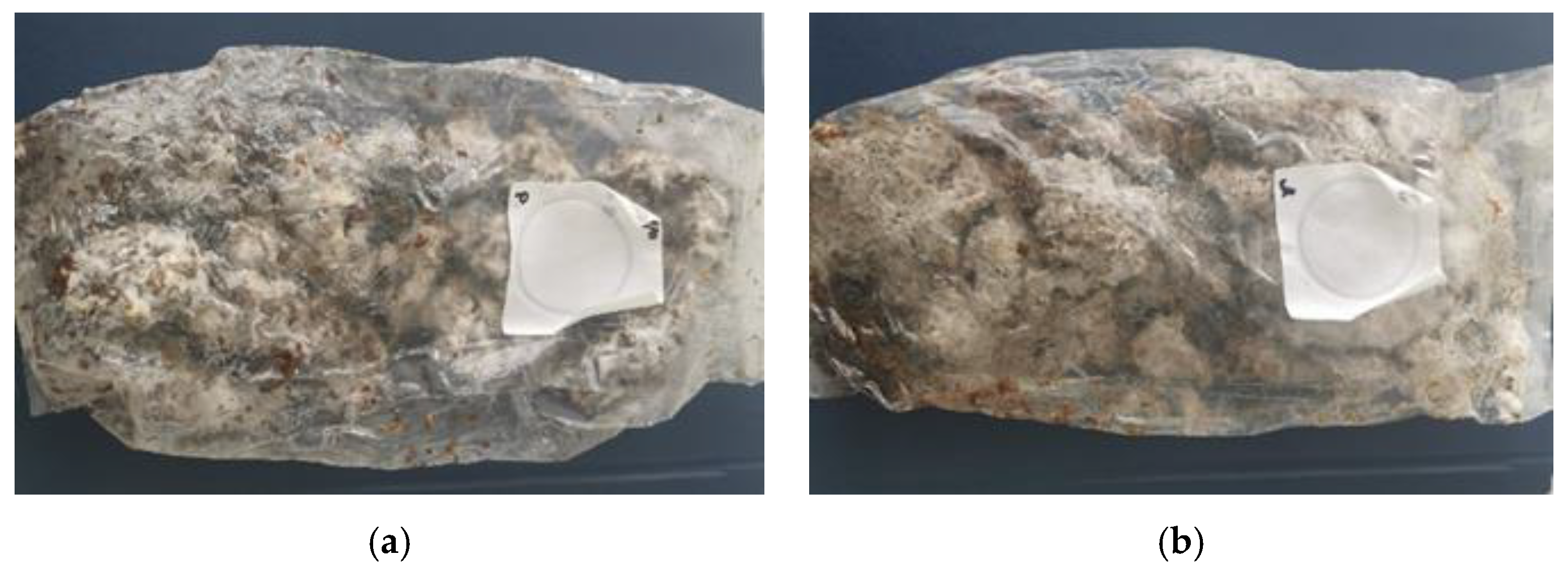
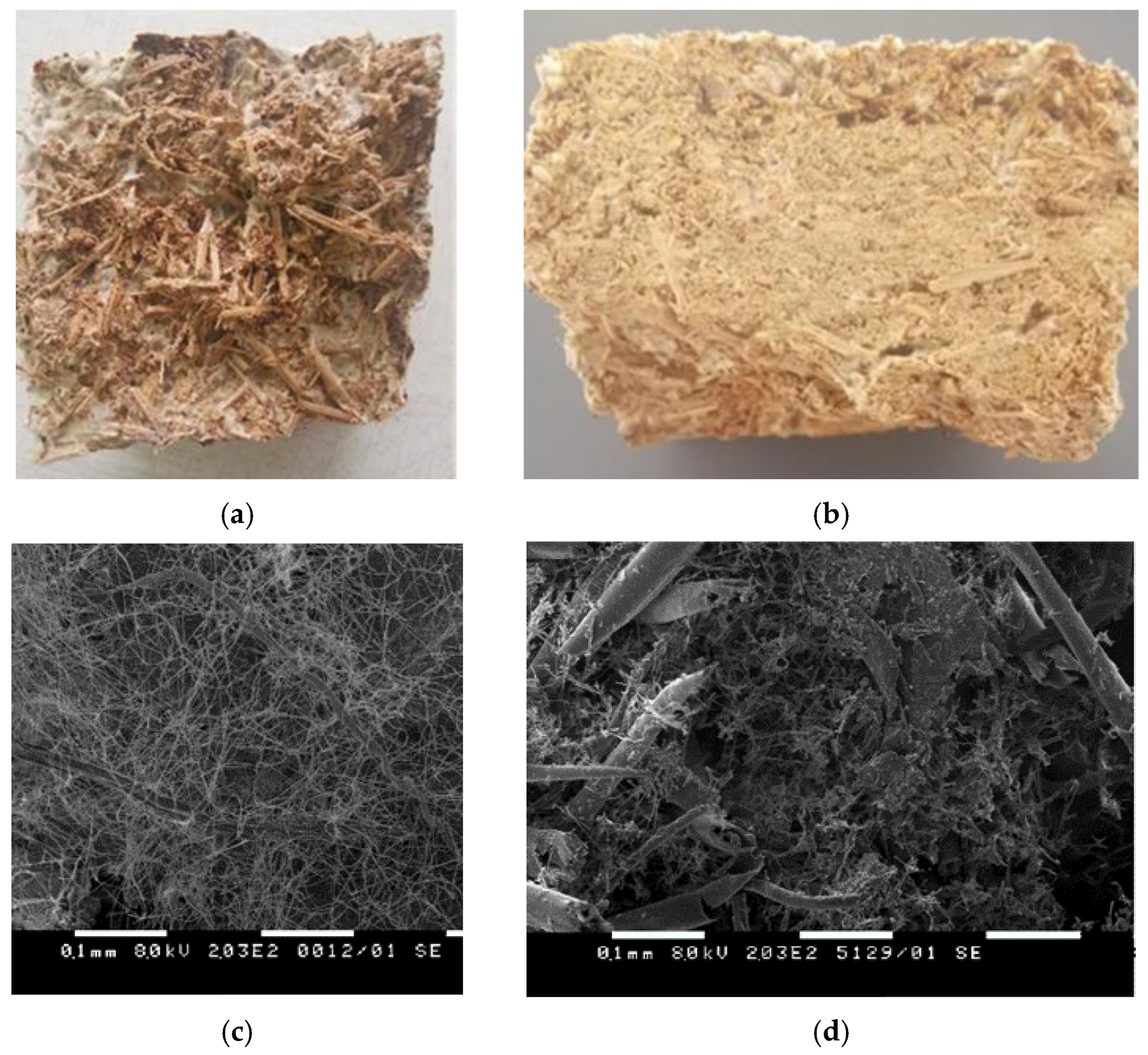
3.2. Solid State Cultivation of Ganoderma Resinaceum GA1M on HERF and SDLS for Mycelium-Based Bio-Composite Formation
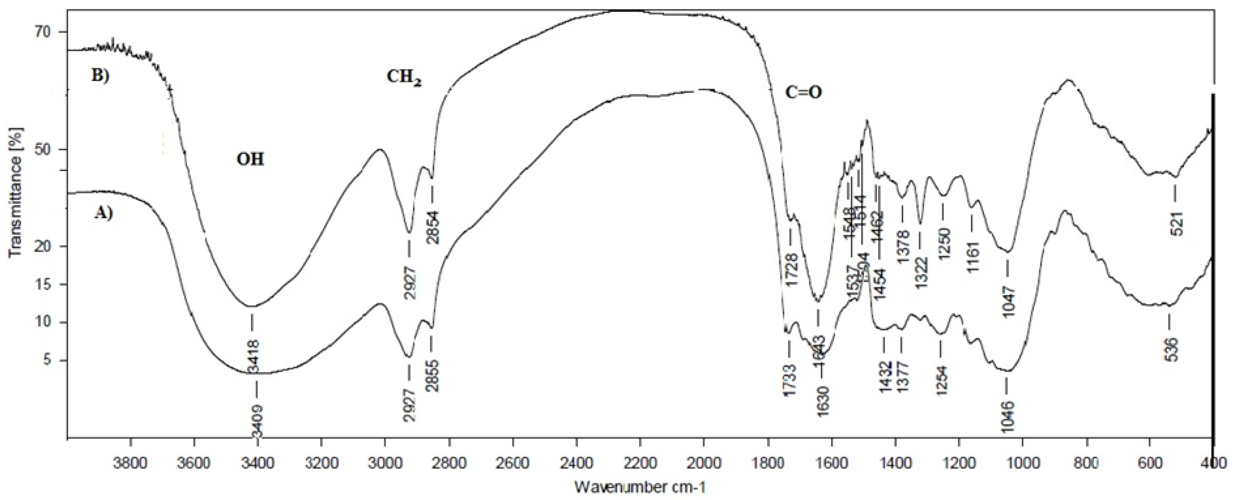
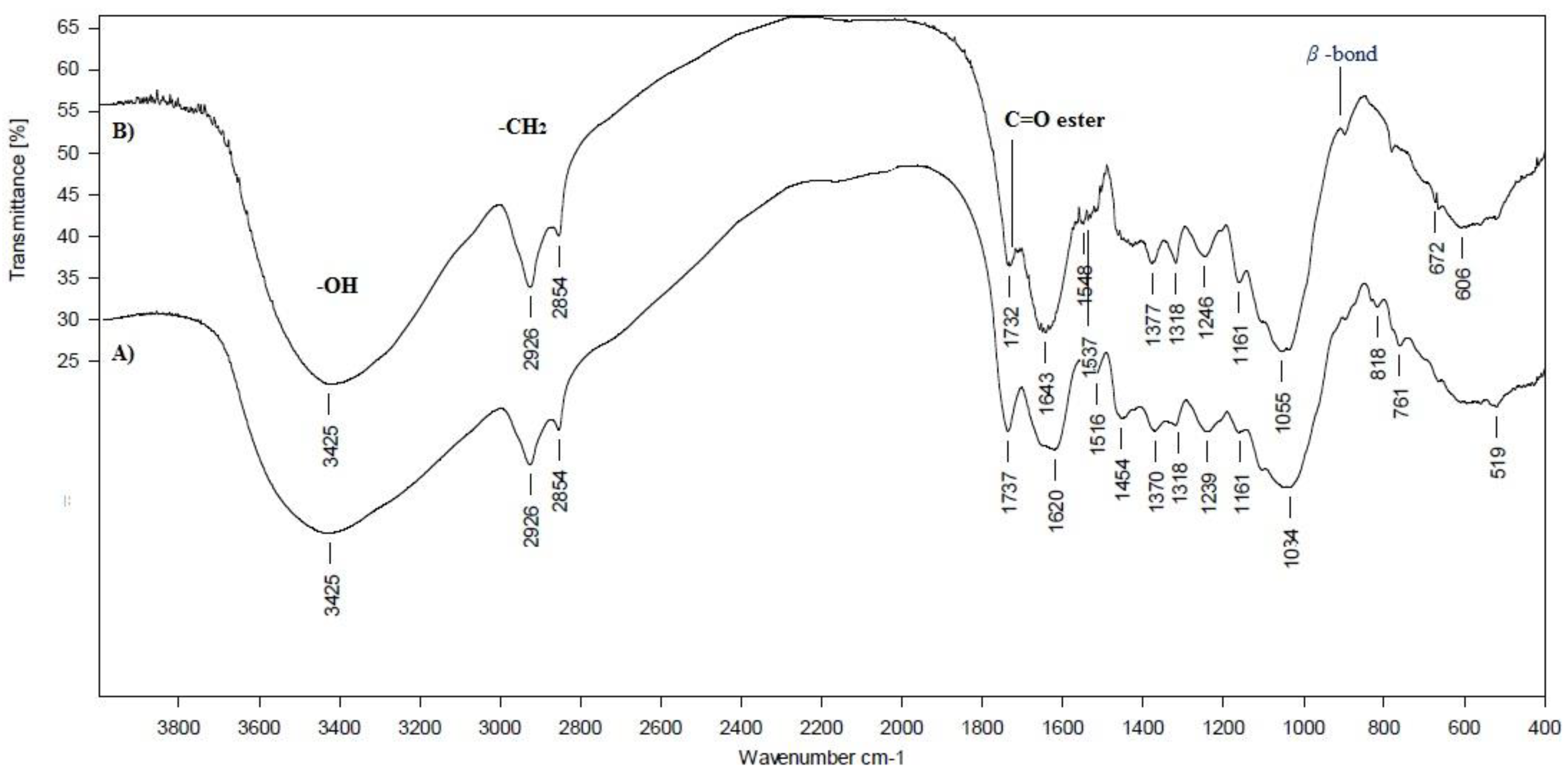
3.3. FTIR Spectroscopy
3.4. Basic Physical and Mechanical Characterization of the Mycelium HERF/SDLS-Based Bio-Composites
4. Conclusions
Author Contributions
Funding
Institutional Review Board Statement
Informed Consent Statement
Data Availability Statement
Acknowledgments
Conflicts of Interest
References
- Slavov, A.; Vasileva, I.; Stefanov, L.; Stoyanova, A. Valorization of wastes from the rose oil industry. Rev. Environ. Sci. Bio/Technology 2017, 16, 309–325. [Google Scholar] [CrossRef]
- Lesage-Meessen, L.; Bou, M.; Sigoillot, J.-C.; Faulds, C.B.; Lomascolo, A. Essential oils and distilled straws of lavender and lavandin: A review of current use and potential application in white biotechnology. Appl. Microbiol. Biotechnol. 2015, 99, 3375–3385. [Google Scholar] [CrossRef]
- Slavov, A.; Denev, P.; Panchev, I.; Shikov, V.; Nenov, N.; Yantcheva, N.; Vasileva, I. Combined recovery of polysaccharides and polyphenols from Rosa damascena wastes. Ind. Crop. Prod. 2017, 100, 85–94. [Google Scholar] [CrossRef]
- Sun, W.; Tajvidi, M.; Hunt, C.; McIntyre, G.; Gardner, D.J. Fully Bio-Based Hybrid Composites Made of Wood, Fungal Mycelium and Cellulose Nanofibrils. Sci. Rep. 2019, 9, 3766. [Google Scholar] [CrossRef]
- Zolotovsky, K. Guided Growth: Design and Computation of Biologically Active Materials. Ph.D. Thesis, Massachusetts Institute of Technology, Cambridge, MA, USA, 2017. [Google Scholar]
- Haneef, M.; Ceseracciu, L.; Canale, C.; Bayer, I.S.; Heredia-Guerrero, J.A.; Athanassiou, A. Advanced Materials from Fungal Mycelium: Fabrication and Tuning of Physical Properties. Sci. Rep. 2017, 7, srep41292. [Google Scholar] [CrossRef]
- Jones, M.; Huynh, T.; Dekiwadia, C.; Daver, F.; John, S. Mycelium Composites: A Review of Engineering Characteristics and Growth Kinetics. J. Bionanoscience 2017, 11, 241–257. [Google Scholar] [CrossRef]
- Jones, M.; Bhat, T.; Huynh, T.; Kandare, E.; Yuen, K.K.R.; Wang, C.-H.; John, S. Waste-derived low-cost mycelium composite construction materials with improved fire safety. Fire Mater. 2018, 42, 816–825. [Google Scholar] [CrossRef]
- Jones, M.; Huynh, T.; John, S. Inherent species characteristic influence and growth performance assessment for mycelium composite applications. Adv. Mater. Lett. 2018, 9, 71–80. [Google Scholar] [CrossRef]
- Jones, M.P.; Lawrie, A.C.; Huynh, T.T.; Morrison, P.D.; Mautner, A.; Bismarck, A.; John, S. Agricultural by-product suitability for the production of chitinous composites and nanofibers utilising Trametes versicolor and Polyporus brumalis mycelial growth. Process. Biochem. 2019, 80, 95–102. [Google Scholar] [CrossRef]
- Jones, M.; Mautner, A.; Luenco, S.; Bismarck, A.; John, S. Engineered mycelium composite construction materials from fungal biorefineries: A critical review. Mater. Des. 2020, 187, 108397. [Google Scholar] [CrossRef]
- Manan, S.; Ullah, M.W.; Ul-Islam, M.; Atta, O.M.; Yang, G. Synthesis and applications of fungal mycelium-based advanced functional materials. J. Bioresour. Bioprod. 2021, 6, 1–10. [Google Scholar] [CrossRef]
- Islam, M.R.; Tudryn, G.; Bucinell, R.; Schadler, L.; Picu, R.C. Morphology and mechanics of fungal mycelium. Sci. Rep. 2017, 7, 13070. [Google Scholar] [CrossRef] [Green Version]
- Grimm, D.; Wösten, H.A.B. Mushroom cultivation in the circular economy. Appl. Microbiol. Biotechnol. 2018, 102, 7795–7803. [Google Scholar] [CrossRef] [Green Version]
- Perez, R.; Luccioni, M.; Kamakaka, R.; Clamons, S.; Gaut, N.; Stirling, F.; Adamala, K.P.; Silver, P.A.; Endy, D. Enabling community-based metrology for wood-degrading fungi. Fungal Biol. Biotechnol. 2020, 7, 1–15. [Google Scholar] [CrossRef] [Green Version]
- Higgins, C.; Margot, H.; Warnquist, S.; Obeysekare, E.; Mehta, K. Mushroom cultivation in the developing world: A comparison of cultivation technologies. In Proceedings of the 2017 IEEE Global Humanitarian Technology Conference (GHTC), IEEE, San Jose, CA, USA, 19–22 October 2017; pp. 1–7. [Google Scholar]
- Bruscato, C.; Malvessi, E.; Brandalise, R.N.; Camassola, M. High performance of macrofungi in the production of mycelium-based biofoams using sawdust—Sustainable technology for waste reduction. J. Clean. Prod. 2019, 234, 225–232. [Google Scholar] [CrossRef]
- Liu, R.; Long, L.; Sheng, Y.; Xu, J.; Qiu, H.; Li, X.; Wang, Y.; Wu, H. Preparation of a kind of novel sustainable mycelium/cotton stalk composites and effects of pressing temperature on the properties. Ind. Crop. Prod. 2019, 141, 111732. [Google Scholar] [CrossRef]
- Alves, R.M.E.; Alves, M.L.; Campos, M.J. Morphology and Thermal Behaviour of New Mycelium-Based Composites with Different Types of Substrates. In Proceedings of the 2nd International Conference on Experimental and Computational Mechanics in Engineering, Banda Aceh, India, 13–14 October 2020; Gabler: Wiesbaden, Germany; pp. 189–197. [Google Scholar]
- Tacer-Caba, Z.; Varis, J.J.; Lankinen, P.; Mikkonen, K.S. Comparison of novel fungal mycelia strains and sustainable growth substrates to produce humidity-resistant biocomposites. Mater. Des. 2020, 192, 108728. [Google Scholar] [CrossRef]
- Santos, I.S.; Nascimento, B.L.; Marino, R.H.; Sussuchi, E.M.; Matos, M.P.; Griza, S. Influence of drying heat treatments on the mechanical behavior and physico-chemical properties of mycelial biocomposite. Compos. Part B Eng. 2021, 217, 108870. [Google Scholar] [CrossRef]
- Appels, F.V.; Camere, S.; Montalti, M.; Karana, E.; Jansen, K.; Dijksterhuis, J.; Krijgsheld, P.; Wösten, H.A. Fabrication factors influencing mechanical, moisture- and water-related properties of mycelium-based composites. Mater. Des. 2019, 161, 64–71. [Google Scholar] [CrossRef]
- Girometta, C.; Picco, A.M.; Baiguera, R.M.; Dondi, D.; Babbini, S.; Cartabia, M.; Pellegrini, M.; Savino, E. Physico-Mechanical and Thermodynamic Properties of Mycelium-Based Biocomposites: A Review. Sustainability 2019, 11, 281. [Google Scholar] [CrossRef] [Green Version]
- Nava, J.A.L.; González, J.M.; Chacón, X.R.; Luna, J.A.N. Assessment of Edible Fungi and Films Bio-Based Material Simulating Expanded Polystyrene. Mater. Manuf. Process. 2015, 31, 1085–1090. [Google Scholar] [CrossRef]
- Jiang, L.; Walczyk, D.; McIntyre, G.; Bucinell, R.; Tudryn, G. Manufacturing of biocomposite sandwich structures using mycelium-bound cores and preforms. J. Manuf. Process. 2017, 28, 50–59. [Google Scholar] [CrossRef]
- Läkk, H.; Krijgsheld, P.; Montalti, M.; Wösten, H. Fungal based biocomposite for habitat structures on the Moon and Mars. In Proceedings of the 69th International Astronautical Congress (IAC), Bremen, Germany, 1–5 October 2018. [Google Scholar]
- Ridzqo, I.F.; Susanto, D.; Panjaitan, T.H.; Putra, N. Sustainable Material: Development Experiment of Bamboo Composite through Biologically Binding Mechanism. IOP Conf. Series: Mater. Sci. Eng. 2020, 713, 012010. [Google Scholar] [CrossRef]
- Attias, N.; Danai, O.; Abitbol, T.; Tarazi, E.; Ezov, N.; Pereman, I.; Grobman, Y.J. Mycelium bio-composites in industrial design and architecture: Comparative review and experimental analysis. J. Clean. Prod. 2020, 246, 119037. [Google Scholar] [CrossRef]
- Lesage-Meessen, L.; Bou, M.; Ginies, C.; Chevret, D.; Navarro, D.; Drula, E.; Bonnin, E.; Del Río, J.C.; Odinot, E.; Bisotto, A.; et al. Lavender- and lavandin-distilled straws: An untapped feedstock with great potential for the production of high-added value compounds and fungal enzymes. Biotechnol. Biofuels 2018, 11, 217. [Google Scholar] [CrossRef] [PubMed] [Green Version]
- Ratiarisoa, R.V.; Magniont, C.; Ginestet, S.; Oms, C.; Escadeillas, G. Assessment of distilled lavender stalks as bioaggregate for building materials: Hygrothermal properties, mechanical performance and chemical interactions with mineral pozzolanic binder. Constr. Build. Mater. 2016, 124, 801–815. [Google Scholar] [CrossRef]
- Pla, M.F.D.E.; González, P.; Sette, P.; Portillo, F.; Rojas, A.M.; Gerschenson, L.N. Effect of processing on physico-chemical characteristics of dietary fibre concentrates obtained from peach (Prunus persica L.) peel and pulp. Food Res. Int. 2012, 49, 184–192. [Google Scholar] [CrossRef]
- Singleton, V.L.; Rossi, J.A. Colorimetry of total phenolics with phosphomolybdic-phosphotungstic acid reagents. Am. J. Enol. Vitic. 1965, 16, 144–158. [Google Scholar]
- Stefanova, P.; Taseva, M.; Georgieva, T.; Gotcheva, V.; Angelov, A. A Modified CTAB Method for DNA Extraction from Soybean and Meat Products. Biotechnol. Biotechnol. Equip. 2013, 27, 3803–3810. [Google Scholar] [CrossRef] [Green Version]
- Toju, H.; Tanabe, A.; Yamamoto, S.; Sato, H. High-Coverage ITS Primers for the DNA-Based Identification of Ascomycetes and Basidiomycetes in Environmental Samples. PLoS ONE 2012, 7, e40863. [Google Scholar] [CrossRef] [Green Version]
- Altschul, S.F.; Gish, W.; Miller, W.; Myers, E.W.; Lipman, D.J. Basic local alignment search tool. J. Mol. Biol. 1990, 215, 403–410. [Google Scholar] [CrossRef]
- Slavov, A.; Panchev, I.; Kovacheva, D.; Vasileva, I. Physico-chemical characterization of water-soluble pectic extracts from Rosa damascena, Calendula officinalis and Matricaria chamomilla wastes. Food Hydrocoll. 2016, 61, 469–476. [Google Scholar] [CrossRef]
- Calvo-Flores, F.G.; Dobado, J.A.; Isac-García, J.; Martín-Martínez, F.J. Lignin and Lignans as Renewable Raw Materials; Wiley: Hoboken, NJ, USA, 2015. [Google Scholar]
- Singh, T.; Singh, A.P. White and Brown Rot Fungi as Decomposers of Lignocellulosic Materials and Their Role in Waste and Pollution Control. Advances in Endophytic Fungal Research 2016, 233–247. [Google Scholar] [CrossRef]
- Yang, Z.; Zhang, F.; Still, B.; White, M.; Amstislavski, P. Physical and Mechanical Properties of Fungal Mycelium-Based Biofoam. J. Mater. Civ. Eng. 2017, 29, 04017030. [Google Scholar] [CrossRef]
- Elsacker, E.; Vandelook, S.; Brancart, J.; Peeters, E.; De Laet, L. Mechanical, physical and chemical characterisation of myceli-um-based composites with different types of lignocellulosic substrates. PLoS ONE 2019, 14, e0213954. [Google Scholar] [CrossRef] [Green Version]
- Slavov, A.; Vasileva, I.; Denev, P.; Dinkova, R.; Teneva, D.; Ognyanov, M.; Georgiev, Y. Polyphenol-rich extracts from essential oil industry wastes. Bulg. Chem. Commun. 2021, 52, 78–83. [Google Scholar]
- Lelivelt, R.J.J.; Lindner, G.; Teuffel, P.; Lamers, H. The production process and compressive strength of Mycelium-based materials. In Proceedings of the First International Conference on Bio-based Building Materials, Clermont-Ferrand, France, 22–25 June 2015; pp. 1–6. [Google Scholar]
- Feng, N.-J.; Zhai, H.; Lai, Y.-Z. On the chemical aspects of the biodelignification of wheat straw with Pycnoporus sanguineus and its combined effects with the presence of Candida tropicalis. Ind. Crop. Prod. 2016, 91, 315–322. [Google Scholar] [CrossRef] [Green Version]
- Pelletier, M.; Holt, G.; Wanjura, J.; Lara, A.; Tapia-Carillo, A.; McIntyre, G.; Bayer, E. An evaluation study of pressure-compressed acoustic absorbers grown on agricultural by-products. Ind. Crop. Prod. 2017, 95, 342–347. [Google Scholar] [CrossRef]
- Demir, I.; Doğan, C. Physical and mechanical properties of hempcrete. Open Waste Manag. J. 2020, 13, 26–34. [Google Scholar] [CrossRef]
- Ziegler, A.R.; Bajwa, S.G.; Holt, G.A.; McIntyre, G. Evaluation of physico-mechanical properties of mycelium reinforced green biocomposites made from cellulosic fibers. Appl. Eng. Agric. 2016, 32, 931–938. [Google Scholar] [CrossRef]
- Stevulova, N.; Cigasova, J.; Purcz, P.; Schwarzova, I.; Kacik, F.; Geffert, A. Water absorption behavior of hemp hurds composites. Mater. 2015, 8, 2243–2257. [Google Scholar] [CrossRef] [Green Version]
- Pakkala, T.A.; Lahdensivu, J. Long-term water absorption tests for frost insulation materials taking into account frost attack. Case Stud. Constr. Mater. 2014, 1, 40–45. [Google Scholar] [CrossRef] [Green Version]
- Amstislavski, P.; Yang, Z.; White, M.D. United States Patent Application Pubblication. WO 2017/132523 Al, 2017. [Google Scholar]
- Available online: https://www.specifiedby.com/styrene-packaging-insulation-ltd/stylite-eps-geofill-void-formers/geofill-compressive-strength-factsheet_4eeaa0ea.pdf (accessed on 11 May 2021).


| HERF, g/100 g DW | SUM | SDLS, g/100 g DW | SUM | |
|---|---|---|---|---|
| Fibers (total) | 75.14 ± 0.19 b | - | 81.54 ± 0.18 a | - |
| Uronic acids | 8.95 ± 0.17 a | - | 3.54 ± 0.12 b | - |
| Ash | 3.10 ± 0.16 b | - | 6.59 ± 0.11 a | - |
| Non-cellulosic polysaccharides | 14.57 ± 0.2 a | 62.27 ± 0.21 b | 13.79 ± 0.15 a | 76.43 ± 0.21 a |
| Cellulose | 29.13 ± 0.17 b | 38.16 ± 0.21 a | ||
| Lignin | 18.57 ± 0.16 b | 24.48 ± 0.14 a |
| HERF, g/100 g DW | SDLS, g/100 g DW | |
|---|---|---|
| Total polyphenols | 5.70 ± 0.20 b | 1.14 ± 0.07 a |
| Quercetin | 0.30 ± 0.002 b | 0.03 ± 0.001 a |
| Quercetin-3-β-glucoside | 0.48 ± 0.002 b | 0.04 ± 0.001 a |
| Rutin | 0.98 ± 0.002 | nd |
| Myricetin | 0.09 ± 0.001 b | 0.028 ± 0.001 a |
| Kaempferol | 0.04 ± 0.001 b | 0.01 ± 0.001 a |
| Catechin | 0.33 ± 0.002 b | 0.33 ± 0.001 a |
| Epicatechin | 0.23 ± 0.001 b | 0.25 ± 0.001 a |
| Neochlorogenic acid | 0.13 ± 0.002 a | 0.16 ± 0.001 b |
| 3,4-dihydroxy benzoic acid | 0.20 ± 0.001 a | 0.51 ± 0.001 b |
| p-Coumaric acid | 0.05 ± 0.001 | nd |
| Ferulic acid | 0.07 ± 0.001 a | 0.16 ± 0.001 b |
| Gallic acid | 0.12 ± 0.002 a | 0.29 ± 0.002 b |
| Rosmarinic acid | 0.05 ± 0.001 a | 0.13 ± 0.001 b |
| Cinnamic acid | 0.02 ± 0.001 | nd |
| Compound | RI | HERF | SDLS |
|---|---|---|---|
| α-Pinene | 940 | 0.87 ± 0.08 a | 0.25 ± 0.06 b |
| β-Pinene | 980 | 0.61 ± 0.05 a | 1.54 ± 0.08 b |
| β-Myrcene | 991 | 0.34 ± 0.07 a | 1.19 ± 0.09 b |
| p-Cymene | 1019 | - | 0.54 ± 0.07 |
| Limonene | 1025 | - | 3.55 ± 0.15 |
| Eucalyptol | 1031 | - | 3.18 ± 0.16 |
| cis-beta-Ocimene | 1040 | - | 5.41 ± 0.21 |
| trans-beta-Ocimene | 1050 | - | 3.37 ± 0.19 |
| γ-Terpinene | 1062 | 0.91 ± 0.08 a | 0.38 ± 0.06 b |
| cis-Linalool oxide | 1073 | - | 0.19 ± 0.05 |
| trans-Linalool oxide | 1078 | - | 0.29 ± 0.05 |
| Terpinene | 1087 | 2.00 ± 0.10 | - |
| β-Linalool | 1097 | 3.84 ± 0.10 a | 18.91 ± 0.15 b |
| Phenethyl alcohol | 1110 | 21.07 ± 0.17 | - |
| cis-Rose oxide | 1112 | 0.45 ± 0.05 | - |
| trans-Rose oxide | 1127 | 0.25 ± 0.04 | - |
| Camphor | 1146 | - | 0.48 ± 0.07 |
| Borneol | 1169 | - | 0.58 ± 0.10 |
| Lavandulol | 1171 | - | 6.12 ± 0.21 |
| Terpin-4-ol | 1178 | 0.81 ± 0.07 a | 3.10 ± 0.11 b |
| α-Terpineol | 1189 | 0.51 ± 0.05 a | 3.13 ± 0.09 b |
| β-Citronellol | 1228 | 11.26 ± 0.18 | - |
| Nerol | 1230 | 3.78 ± 0.15 | - |
| Geraniol | 1255 | 8.24 ± 0.21 a | 0.28 ± 0.10 b |
| Linalyl acetate, dihydro- | 1275 | - | 18.14 ± 0.16 |
| (±)-Lavandulyl acetate | 1290 | - | 4.93 ± 0.13 |
| Citronellyl acetate | 1354 | 0.19 ± 0.05 | - |
| Eugenol | 1356 | 2.84 ± 0.09 | - |
| Neryl acetate | 1364 | 2.09 ± 0.08 a | 0.95 ± 0.08 b |
| Geranyl acetate | 1383 | 0.63 ± 0.07 a | 2.94 ± 0.11 b |
| β-Bourbonene | 1384 | 3.16 ± 0.18 a | 0.20 ± 0.09 b |
| β-Cubebene | 1389 | 5.90 ± 0.15 | - |
| β-Elemene | 1390 | 0.51 ± 0.04 | - |
| Methyl eugenol | 1401 | 0.44 ± 0.05 | - |
| β-Caryophyllene | 1419 | 1.58 ± 0.12 a | 7.20 ± 0.18 b |
| α-Humulene (α-Caryophyllene) | 1454 | 0.34 ± 0.05 a | 5.06 ± 0.12 b |
| Germacrene D | 1479 | 0.39 ± 0.06 a | 2.76 ± 0.09 b |
| α-Farnesene | 1508 | 0.56 ± 0.05 a | 0.27 ± 0.04 b |
| β-Bisabolene | 1510 | 0.18 ± 0.04 a | 0.20 ± 0.03 a |
| trans-Nerolidol | 1564 | 2.60 ± 0.10 a | 0.27 ± 0.08 b |
| Spathulenol | 1575 | 1.63 ± 0.12 a | 0.19 ± 0.07 b |
| Caryophyllene oxide | 1580 | 0.32 ± 0.05 a | 0.30 ± 0.04 a |
| γ-Eudesmol | 1631 | 0.30 ± 0.04 a | 0.42 ± 0.05 a |
| β-Eudesmol | 1649 | 0.25 ± 0.05 a | 0.22 ± 0.03 a |
| α-Eudesmol | 1651 | 0.89 ± 0.03 a | 0.34 ± 0.05 b |
| Farnesol | 1714 | 0.34 ± 0.04 a | 0.55 ± 0.07 a |
| n-Nonadecane | 1901 | 4.29 ± 0.18 a | 0.17 ± 0.04 b |
| n-Eicosane | 2000 | 3.83 ± 0.15 a | 0.32 ± 0.08 b |
| n-Heneicosane | 2100 | 0.27 ± 0.06 a | 0.34 ± 0.08 a |
| n-Docosane | 2200 | 0.75 ± 0.08 | - |
| n-Tricosane | 2300 | 4.46 ± 0.21 | - |
| n-Tetracosane | 2400 | 1.60 ± 0.12 | - |
| n-Pentacosane | 2500 | 1.41 ± 0.12 | - |
| n-Hexacosane | 2600 | 1.41 ± 0.11 | - |
| Assignment | Mycelium Component (Main Contribution) | HERF | SDLS | Mycelium HERF-Based Bio-Composite | Mycelium SDLS-Based Bio-Composite |
|---|---|---|---|---|---|
| Vibration Frequency, cm−1 | |||||
| O-H stretching vibration of intra and inter hydrogen bond | Polysaccharides | 3429 | 3409 | 3425 | 3418 |
| C-H stretches in methyl and methylene groups CH2, asymmetric stretching | Lipids, Polysaccharides | 2966 | 2927 | 2926 | 2927 |
| CH2 symmetric stretching | Lipids, Polysaccharides | 2854 | 2855 | 2854 | 2854 |
| C=O stretching in xylans (hemicellulose), pectins; ester bonds | Lipids, Polysaccharides | 1737 | 1733 | 1732 | 1728 |
| (amide I in β -sheets secondary structures) Absorbed O-H associated with lignin or cellulose; a characteristic band for cis- HRC = CR’H bonds, | Proteins; Lipids, Polysaccharides | 1652; 1620 | 1630 | 1665 1643 | 1643 |
| Amide II | Proteins | - | - | 1548 | 1548 |
| a characteristic band of the type C=C; C=C stretching of aromatic ring (syringyl) in lignin C=C stretcing of aromatic ring (guaiacyl) in lignin C=C stretching of aromatic ring (syringyl) in lignin | Polysaccharides | 1516 | 1516 | 1537;1548 | 1537;1548 |
| νC-Hs(CH2) in pyranise ring, | Polysaccharides | 1454 | 1432 | - | 1454;1462 |
| C-H bending in chitin, cellulose and hemicellulose | Polysaccharides | 1370 | 1377 | 1377 | 1378 |
| CH2 wagging in cellulose | Polysaccharides | 1318 | 1318 | - | - |
| PO2- asymmetric stretching, C-O stretching in lignin and xylan, Nucleic acids | Nucleic acids Polysaccharides | 1239 | 1246 | 1254 | 1250 |
| 1C-O-C vibration in cellulose and hemicellulose | Polysaccharides | 1150 | 1161 | 1152 | 1161 |
| - | 1114 | - | 1110 | ||
| C-O valence vibration from C3-O3H | Polysaccharides | 1057 | 1055 | 1046 | 1047 |
| C-O stretching in cellulose | Polysaccharides | 1053 | 1055 | 1055 | 1047 |
| C-C stretching | Polysaccharides | 1034 | 1055 | 1046 | 1047 |
| β(COH), β(CH) of C-1, νs(COC) in glycosidic linkage, ring modes | Polysaccharides | 928 | 916 | 916 | 919 |
| Anomer C-group, Glucan β-anomer C-H bending, C-H deformation in cellulose, ν(CC), β(CCH) | Polysaccharides | 899 | 878 | 855 | 842 |
| CH2-rocking | Polysaccharides | 761 | - | 769 | - |
| Mycelium Bio-Composite | Apparent Densityρa | Capillary AbsorptionWc | Water Absorption | Compressive Resistance at 10% Deformation σ10 | |||
|---|---|---|---|---|---|---|---|
| 1 Day | 28 Days | ||||||
| Wva,1d | Wma,1d | Wva,28d | Wma,28d | ||||
| kg/m3 | kg/m2 | %vol. | %wt. | %vol. | %wt. | kPa | |
| HERF | 462 ± 10.2 | 3.4 ± 0.38 | 20.3 ± 3.45 | 43.9 ± 7.5 | 58.2 ± 7.2 | 126.0 ± 15.7 | 1029 ± 51 |
| SDLS | 347 ± 3.7 | 6.5 ± 0.20 | 39.8 ± 5.72 | 114.6 ± 14.5 | 85.0 ± 3.5 | 245.0 ± 10.1 | 718 ± 22 |
Publisher’s Note: MDPI stays neutral with regard to jurisdictional claims in published maps and institutional affiliations. |
© 2021 by the authors. Licensee MDPI, Basel, Switzerland. This article is an open access article distributed under the terms and conditions of the Creative Commons Attribution (CC BY) license (https://creativecommons.org/licenses/by/4.0/).
Share and Cite
Angelova, G.; Brazkova, M.; Stefanova, P.; Blazheva, D.; Vladev, V.; Petkova, N.; Slavov, A.; Denev, P.; Karashanova, D.; Zaharieva, R.; et al. Waste Rose Flower and Lavender Straw Biomass—An Innovative Lignocellulose Feedstock for Mycelium Bio-Materials Development Using Newly Isolated Ganoderma resinaceum GA1M. J. Fungi 2021, 7, 866. https://doi.org/10.3390/jof7100866
Angelova G, Brazkova M, Stefanova P, Blazheva D, Vladev V, Petkova N, Slavov A, Denev P, Karashanova D, Zaharieva R, et al. Waste Rose Flower and Lavender Straw Biomass—An Innovative Lignocellulose Feedstock for Mycelium Bio-Materials Development Using Newly Isolated Ganoderma resinaceum GA1M. Journal of Fungi. 2021; 7(10):866. https://doi.org/10.3390/jof7100866
Chicago/Turabian StyleAngelova, Galena, Mariya Brazkova, Petya Stefanova, Denica Blazheva, Veselin Vladev, Nadejda Petkova, Anton Slavov, Petko Denev, Daniela Karashanova, Roumiana Zaharieva, and et al. 2021. "Waste Rose Flower and Lavender Straw Biomass—An Innovative Lignocellulose Feedstock for Mycelium Bio-Materials Development Using Newly Isolated Ganoderma resinaceum GA1M" Journal of Fungi 7, no. 10: 866. https://doi.org/10.3390/jof7100866