
Prognostic Value of Cardiac Magnetic Resonance Feature Tracking Strain in Aortic Stenosis
 ,
,
Abstract
:1. Introduction
2. Materials and Methods
2.1. Cardiovascular Magnetic Resonance
2.2. Statistical Analysis
3. Results
4. Discussion
5. Limitations
6. Conclusions
Author Contributions
Funding
Institutional Review Board Statement
Informed Consent Statement
Data Availability Statement
Conflicts of Interest
References
- Coffey, S.; Cairns, B.J.; Iung, B. The Modern Epidemiology of Heart Valve Disease. Heart 2016, 102, 75–85. [Google Scholar] [CrossRef] [PubMed]
- Sverdlov, A.L.; Ngo, D.T.; Chapman, M.J.; Ali, O.A.; Chirkov, Y.Y.; Horowitz, J.D. Pathogenesis of Aortic Stenosis: Not Just a Matter of Wear and Tear. Am. J. Cardiovasc. Dis. 2011, 1, 185–199. [Google Scholar] [PubMed]
- Dweck, M.R.; Boon, N.A.; Newby, D.E. Calcific Aortic Stenosis: A Disease of the Valve and the Myocardium. J. Am. Coll. Cardiol. 2012, 60, 1854–1863. [Google Scholar] [CrossRef] [PubMed]
- Rassi, A.N.; Pibarot, P.; Elmariah, S. Left Ventricular Remodelling in Aortic Stenosis. Can. J. Cardiol. 2014, 30, 1004–1011. [Google Scholar] [CrossRef] [PubMed]
- Calin, A.; Mateescu, A.D.; Popescu, A.C.; Bing, R.; Dweck, M.R.; Popescu, B.A. Role of Advanced Left Ventricular Imaging in Adults with Aortic Stenosis. Heart 2020, 106, 962–969. [Google Scholar] [CrossRef]
- Musa, T.A.; Treibel, T.A.; Vassiliou, V.S.; Captur, G.; Singh, A.; Chin, C.; Dobson, L.E.; Pica, S.; Loudon, M.; Malley, T.; et al. Myocardial Scar and Mortality in Severe Aortic Stenosis: Data from the BSCMR Valve Consortium. Circulation 2018, 138, 1935–1947. [Google Scholar] [CrossRef]
- Barone-Rochette, G.; Piérard, S.; De Meester de Ravenstein, C.; Seldrum, S.; Melchior, J.; Maes, F.; Pouleur, A.-C.; Vancraeynest, D.; Pasquet, A.; Vanoverschelde, J.-L.; et al. Prognostic Significance of LGE by CMR in Aortic Stenosis Patients Undergoing Valve Replacement. J. Am. Coll. Cardiol. 2014, 64, 144–154. [Google Scholar] [CrossRef]
- Runge, V.M. Safety of the Gadolinium-Based Contrast Agents for Magnetic Resonance Imaging, Focusing in Part on Their Accumulation in the Brain and Especially the Dentate Nucleus. Investig. Radiol. 2016, 51, 273–279. [Google Scholar] [CrossRef]
- Herrmann, S.; Störk, S.; Niemann, M.; Lange, V.; Strotmann, J.M.; Frantz, S.; Beer, M.; Gattenlöhner, S.; Voelker, W.; Ertl, G.; et al. Low-Gradient Aortic Valve Stenosis Myocardial Fibrosis and Its Influence on Function and Outcome. J. Am. Coll. Cardiol. 2011, 58, 402–412. [Google Scholar] [CrossRef]
- Cramariuc, D.; Gerdts, E.; Davidsen, E.S.; Segadal, L.; Matre, K. Myocardial Deformation in Aortic Valve Stenosis: Relation to Left Ventricular Geometry. Heart 2010, 96, 106–112. [Google Scholar] [CrossRef]
- Magne, J.; Cosyns, B.; Popescu, B.A.; Carstensen, H.G.; Dahl, J.; Desai, M.Y.; Kearney, L.; Lancellotti, P.; Marwick, T.H.; Sato, K.; et al. Distribution and Prognostic Significance of Left Ventricular Global Longitudinal Strain in Asymptomatic Significant Aortic Stenosis: An Individual Participant Data Meta-Analysis. JACC Cardiovasc. Imaging 2019, 12, 84–92. [Google Scholar] [CrossRef] [PubMed]
- Ring, L.; Abu-Omar, Y.; Kaye, N.; Rana, B.S.; Watson, W.; Dutka, D.P.; Vassiliou, V.S. Left Atrial Function Is Associated with Earlier Need for Cardiac Surgery in Moderate to Severe Mitral Regurgitation: Usefulness in Targeting for Early Surgery. J. Am. Soc. Echocardiogr. 2018, 31, 983–991. [Google Scholar] [CrossRef] [PubMed]
- Vassiliou, V.S.; Flynn, P.D.; Raphael, C.E.; Newsome, S.; Khan, T.; Ali, A.; Halliday, B.; Bruengger, A.S.; Malley, T.; Sharma, P.; et al. Lipoprotein(a) in Patients with Aortic Stenosis: Insights from Cardiovascular Magnetic Resonance. PLoS ONE 2017, 12, e0181077. [Google Scholar] [CrossRef] [PubMed]
- Otto, C.M.; Nishimura, R.A.; Bonow, R.O.; Carabello, B.A.; Erwin, J.P.; Gentile, F.; Jneid, H.; Krieger, E.V.; Mack, M.; McLeod, C.; et al. 2020 ACC/AHA Guideline for the Management of Patients With Valvular Heart Disease. J. Am. Coll. Cardiol. 2021, 77, e25–e197. [Google Scholar] [CrossRef] [PubMed]
- Dweck, M.R.; Joshi, S.; Murigu, T.; Alpendurada, F.; Jabbour, A.; Melina, G.; Banya, W.; Gulati, A.; Roussin, I.; Raza, S.; et al. Midwall Fibrosis Is an Independent Predictor of Mortality in Patients with Aortic Stenosis. J. Am. Coll. Cardiol. 2011, 58, 1271–1279. [Google Scholar] [CrossRef] [PubMed]
- Nazir, S.A.; Shetye, A.M.; Khan, J.N.; Singh, A.; Arnold, J.R.; Squire, I.; McCann, G.P. Inter-Study Repeatability of Circumferential Strain and Diastolic Strain Rate by CMR Tagging, Feature Tracking and Tissue Tracking in ST-Segment Elevation Myocardial Infarction. Int. J. Cardiovasc. Imaging 2020, 36, 1133–1146. [Google Scholar] [CrossRef]
- Backhaus, S.J.; Metschies, G.; Billing, M.; Kowallick, J.T.; Gertz, R.J.; Lapinskas, T.; Pieske, B.; Lotz, J.; Bigalke, B.; Kutty, S.; et al. Cardiovascular Magnetic Resonance Imaging Feature Tracking: Impact of Training on Observer Performance and Reproducibility. PLoS ONE 2019, 14, e0210127. [Google Scholar] [CrossRef]
- Thaden, J.J.; Nkomo, V.T.; Enriquez-Sarano, M. The Global Burden of Aortic Stenosis. Prog. Cardiovasc. Dis. 2014, 56, 565–571. [Google Scholar] [CrossRef]
- Tsampasian, V.; Grafton-Clarke, C.; Gracia Ramos, A.E.; Asimakopoulos, G.; Garg, P.; Prasad, S.; Ring, L.; McCann, G.P.; Rudd, J.; Dweck, M.R.; et al. Management of Asymptomatic Severe Aortic Stenosis: A Systematic Review and Meta-Analysis. Open Heart 2022, 9, e001982. [Google Scholar] [CrossRef]
- Vassiliou, V.S.; Pavlou, M.; Malley, T.; Halliday, B.P.; Tsampasian, V.; Raphael, C.E.; Tse, G.; Vieira, M.S.; Auger, D.; Everett, R.; et al. A Novel Cardiovascular Magnetic Resonance Risk Score for Predicting Mortality Following Surgical Aortic Valve Replacement. Sci. Rep. 2021, 11, 20183. [Google Scholar] [CrossRef]
- Stokke, T.M.; Hasselberg, N.E.; Smedsrud, M.K.; Sarvari, S.I.; Haugaa, K.H.; Smiseth, O.A.; Edvardsen, T.; Remme, E.W. Geometry as a Confounder When Assessing Ventricular Systolic Function: Comparison Between Ejection Fraction and Strain. J. Am. Coll. Cardiol. 2017, 70, 942–954. [Google Scholar] [CrossRef] [PubMed]
- Onishi, T.; Saha, S.K.; Delgado-Montero, A.; Ludwig, D.R.; Onishi, T.; Schelbert, E.B.; Schwartzman, D.; Gorcsan, J. Global Longitudinal Strain and Global Circumferential Strain by Speckle-Tracking Echocardiography and Feature-Tracking Cardiac Magnetic Resonance Imaging: Comparison with Left Ventricular Ejection Fraction. J. Am. Soc. Echocardiogr. 2015, 28, 587–596. [Google Scholar] [CrossRef] [PubMed]
- Giannini, C.; Petronio, A.S.; Talini, E.; De Carlo, M.; Guarracino, F.; Grazia, M.; Donne, D.; Nardi, C.; Conte, L.; Barletta, V.; et al. Early and Late Improvement of Global and Regional Left Ventricular Function after Transcatheter Aortic Valve Implantation in Patients with Severe Aortic Stenosis: An Echocardiographic Study. Am. J. Cardiovasc. Dis. 2011, 1, 264–273. [Google Scholar] [PubMed]
- Schattke, S.; Baldenhofer, G.; Prauka, I.; Zhang, K.; Laule, M.; Stangl, V.; Sanad, W.; Spethmann, S.; Borges, A.C.; Baumann, G.; et al. Acute Regional Improvement of Myocardial Function after Interventional Transfemoral Aortic Valve Replacement in Aortic Stenosis: A Speckle Tracking Echocardiography Study. Cardiovasc. Ultrasound 2012, 10, 15. [Google Scholar] [CrossRef]
- Kempny, A.; Diller, G.-P.; Kaleschke, G.; Orwat, S.; Funke, A.; Radke, R.; Schmidt, R.; Kerckhoff, G.; Ghezelbash, F.; Rukosujew, A.; et al. Longitudinal Left Ventricular 2D Strain Is Superior to Ejection Fraction in Predicting Myocardial Recovery and Symptomatic Improvement after Aortic Valve Implantation. Int. J. Cardiol. 2013, 167, 2239–2243. [Google Scholar] [CrossRef] [PubMed]
- Ng, A.C.T.; Prihadi, E.A.; Antoni, M.L.; Bertini, M.; Ewe, S.H.; Marsan, N.A.; Leung, D.Y.; Delgado, V.; Bax, J.J. Left Ventricular Global Longitudinal Strain Is Predictive of All-Cause Mortality Independent of Aortic Stenosis Severity and Ejection Fraction. Eur. Heart J. Cardiovasc. Imaging 2018, 19, 859–867. [Google Scholar] [CrossRef] [PubMed]
- Løgstrup, B.B.; Andersen, H.R.; Thuesen, L.; Christiansen, E.H.; Terp, K.; Klaaborg, K.-E.; Poulsen, S.H. Left Ventricular Global Systolic Longitudinal Deformation and Prognosis 1 Year after Femoral and Apical Transcatheter Aortic Valve Implantation. J. Am. Soc. Echocardiogr. 2013, 26, 246–254. [Google Scholar] [CrossRef]
- Poulin, F.; Carasso, S.; Horlick, E.M.; Rakowski, H.; Lim, K.-D.; Finn, H.; Feindel, C.M.; Greutmann, M.; Osten, M.D.; Cusimano, R.J.; et al. Recovery of Left Ventricular Mechanics after Transcatheter Aortic Valve Implantation: Effects of Baseline Ventricular Function and Postprocedural Aortic Regurgitation. J. Am. Soc. Echocardiogr. Off. Publ. Am. Soc. Echocardiogr. 2014, 27, 1133–1142. [Google Scholar] [CrossRef]
- Romano, S.; Judd, R.M.; Kim, R.J.; Kim, H.W.; Klem, I.; Heitner, J.F.; Shah, D.J.; Jue, J.; White, B.E.; Indorkar, R.; et al. Feature-Tracking Global Longitudinal Strain Predicts Death in a Multicenter Population of Patients With Ischemic and Nonischemic Dilated Cardiomyopathy Incremental to Ejection Fraction and Late Gadolinium Enhancement. JACC Cardiovasc. Imaging 2018, 11, 1419–1429. [Google Scholar] [CrossRef]
- Reindl, M.; Tiller, C.; Holzknecht, M.; Lechner, I.; Beck, A.; Plappert, D.; Gorzala, M.; Pamminger, M.; Mayr, A.; Klug, G.; et al. Prognostic Implications of Global Longitudinal Strain by Feature-Tracking Cardiac Magnetic Resonance in ST-Elevation Myocardial Infarction. Circ. Cardiovasc. Imaging 2019, 12, e009404. [Google Scholar] [CrossRef]
- Le, T.-T.; Huang, W.; Singh, G.K.; Toh, D.-F.; Ewe, S.H.; Tang, H.C.; Loo, G.; Bryant, J.A.; Ang, B.; Tay, E.L.-W.; et al. Echocardiographic Global Longitudinal Strain Is Associated With Myocardial Fibrosis and Predicts Outcomes in Aortic Stenosis. Front. Cardiovasc. Med. 2021, 8, 750016. [Google Scholar] [CrossRef] [PubMed]
- Bing, R.; Cavalcante, J.L.; Everett, R.J.; Clavel, M.A.; Newby, D.E.; Dweck, M.R. Imaging and Impact of Myocardial Fibrosis in Aortic Stenosis. JACC Cardiovasc. Imaging 2019, 12, 283–296. [Google Scholar] [CrossRef] [PubMed]
- Wang, Y.; Zhang, M.; Chen, H.; Li, H. Prognostic Value of Global Longitudinal Strain in Asymptomatic Aortic Stenosis: A Systematic Review and Meta-Analysis. Front. Cardiovasc. Med. 2022, 9, 778027. [Google Scholar] [CrossRef] [PubMed]
- Vollema, E.M.; Sugimoto, T.; Shen, M.; Tastet, L.; Ng, A.C.T.; Abou, R.; Marsan, N.A.; Mertens, B.; Dulgheru, R.; Lancellotti, P.; et al. Association of Left Ventricular Global Longitudinal Strain With Asymptomatic Severe Aortic Stenosis. JAMA Cardiol. 2018, 3, 839–847. [Google Scholar] [CrossRef]
- Cole, G.D.; Dhutia, N.M.; Shun-Shin, M.J.; Willson, K.; Harrison, J.; Raphael, C.E.; Zolgharni, M.; Mayet, J.; Francis, D.P. Defining the Real-World Reproducibility of Visual Grading of Left Ventricular Function and Visual Estimation of Left Ventricular Ejection Fraction: Impact of Image Quality, Experience and Accreditation. Int. J. Cardiovasc. Imaging 2015, 31, 1303–1314. [Google Scholar] [CrossRef] [PubMed]
- Thorstensen, A.; Dalen, H.; Amundsen, B.H.; Aase, S.A.; Stoylen, A. Reproducibility in Echocardiographic Assessment of the Left Ventricular Global and Regional Function, the HUNT Study. Eur. J. Echocardiogr. 2010, 11, 149–156. [Google Scholar] [CrossRef] [PubMed]
- Potter, E.; Marwick, T.H. Assessment of Left Ventricular Function by Echocardiography: The Case for Routinely Adding Global Longitudinal Strain to Ejection Fraction. JACC Cardiovasc. Imaging 2018, 11, 260–274. [Google Scholar] [CrossRef]
- Gavara, J.; Rodriguez-Palomares, J.F.; Valente, F.; Monmeneu, J.V.; Lopez-Lereu, M.P.; Bonanad, C.; Ferreira-Gonzalez, I.; del Garcia, B.B.; Rodriguez-Garcia, J.; Mutuberria, M.; et al. Prognostic Value of Strain by Tissue Tracking Cardiac Magnetic Resonance After ST-Segment Elevation Myocardial Infarction. JACC Cardiovasc. Imaging 2018, 11, 1448–1457. [Google Scholar] [CrossRef]
- Schuster, A.; Hor, K.N.; Kowallick, J.T.; Beerbaum, P.; Kutty, S. Cardiovascular Magnetic Resonance Myocardial Feature Tracking: Concepts and Clinical Applications. Circ. Cardiovasc. Imaging 2016, 9, 1–9. [Google Scholar] [CrossRef]
- Buss, S.J.; Breuninger, K.; Lehrke, S.; Voss, A.; Galuschky, C.; Lossnitzer, D.; Andre, F.; Ehlermann, P.; Franke, J.; Taeger, T.; et al. Assessment of Myocardial Deformation with Cardiac Magnetic Resonance Strain Imaging Improves Risk Stratification in Patients with Dilated Cardiomyopathy. Eur. Heart J. Cardiovasc. Imaging 2015, 16, 307–315. [Google Scholar] [CrossRef]
- Vassiliou, V.S.; Perperoglou, A.; Raphael, C.E.; Joshi, S.; Malley, T.; Everett, R.; Halliday, B.; Pennell, D.J.; Dweck, M.R.; Prasad, S.K. Midwall Fibrosis and 5-Year Outcome in Moderate and Severe Aortic Stenosis. J. Am. Coll. Cardiol. 2017, 69, 1755–1756. [Google Scholar] [CrossRef] [PubMed]
- Howard, T.; Majmundar, M.; Sarin, S.; Kumar, A.; Ajay, A.; Krishnaswamy, A.; Reed, G.W.; Harb, S.C.; Harmon, E.; Dykun, I.; et al. Predictors of Major Adverse Cardiovascular Events in Patients With Moderate Aortic Stenosis: Implications for Aortic Valve Replacement. Circ. Cardiovasc. Imaging 2023, 16, 557–565. [Google Scholar] [CrossRef] [PubMed]
- Strange, G.; Stewart, S.; Celermajer, D.; Prior, D.; Scalia, G.M.; Marwick, T.; Ilton, M.; Joseph, M.; Codde, J.; Playford, D. Poor Long-Term Survival in Patients With Moderate Aortic Stenosis. J. Am. Coll. Cardiol. 2019, 74, 1851–1863. [Google Scholar] [CrossRef] [PubMed]


| Demographics | Whole Cohort (N = 194) | Moderate AS (N = 53) | Severe AS (N = 141) |
|---|---|---|---|
| Age, years | 72.9 ± 12.9 | 68.1 ± 14.7 | 74.7 ± 11.8 |
| Male, n (%) | 118 (60.8) | 38 (71.7) | 80 (56.7%) |
| Hypertension, n (%) | 87 (44.8) | 30 (56.6) | 57 (40.4) |
| Diabetes, n (%) | 43 (22.2) | 9 (17) | 34 (24.1) |
| Hypercholesterolaemia, n (%) | 96 (49.5) | 24 (45.3) | 72 (51.1%) |
| Chronic kidney disease, n (%) | 33 (17) | 8 (15.1) | 25 (17.7) |
| Atrial fibrillation, n (%) | 41 (21.1) | 8 (15.1) | 33 (23.4) |
| Previous coronary artery bypass, n (%) | 22 (11.3) | 5 (9.4) | 17 (12.1) |
| Previous percutaneous coronary intervention, n (%) | 27 (13.9) | 4 (7.5) | 23 (16.3) |
| Pharmacotherapy | |||
| ACE-I/ARB, n (%) | 91 (46.9) | 29 (54.7) | 62 (44) |
| Betablocker, n (%) | 67 (34.5) | 23 (43.4) | 44 (31.2) |
| Aldosterone antagonist, n (%) | 30 (15.5) | 10 (18.9) | 20 (14.2) |
| Statins, n (%) | 121 (62.4) | 34 (64.2) | 87 (61.7) |
| Diuretic, n (%) | 71 (36.6) | 12 (22.6) | 59 (41.8) |
| Warfarin, n (%) | 27 (13.9) | 11 (20.8) | 16 (11.3) |
| CMR data | |||
| CMR aortic valve area, cm2 | 0.89 ± 0.28 | 1.25 ± 0.24 | 0.76 ± 0.16 |
| LVEF, % | 57 ± 19 | 58 ± 20 | 56±18 |
| LV mass, g | 143 ± 73 | 180 ± 66 | 128 ± 69 |
| Indexed LV mass | 75.9 ± 34.7 | 92.7 ± 28.5 | 69.7 ± 34.8 |
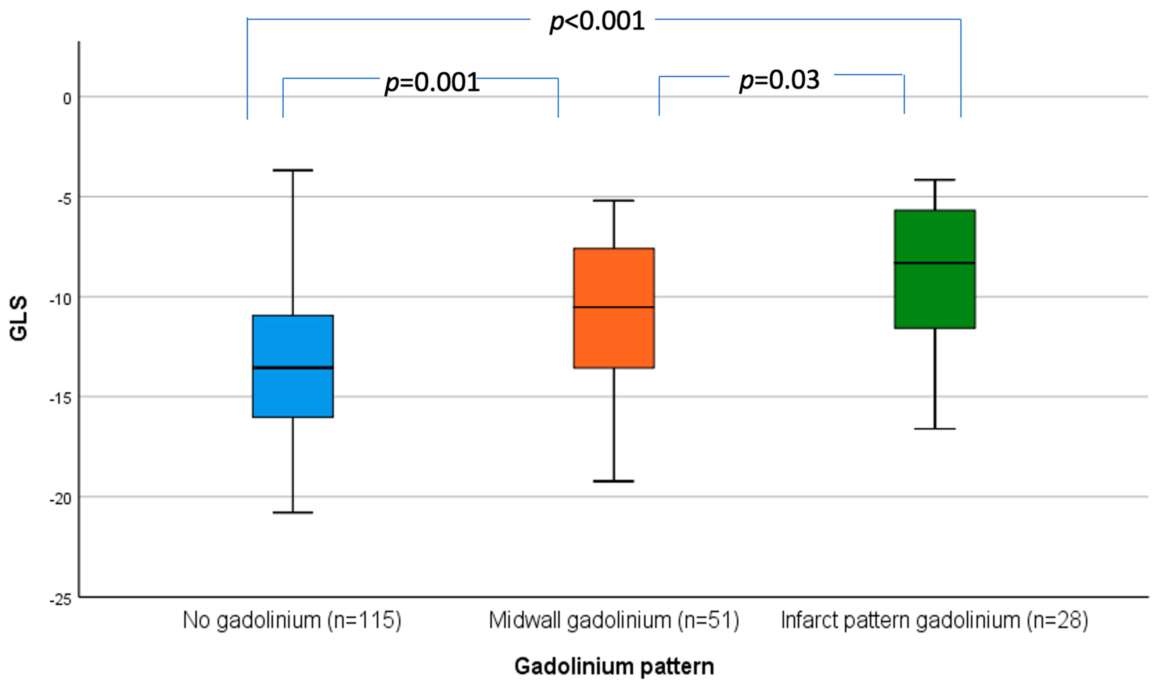
| No myocardial fibrosis, n (%) | 115 (59.3) | 21 (39.6) | 94 (66.7) |
| Midwall fibrosis, n (%) | 51 (26.3) | 22 (41.5) | 29 (20.6) |
| Infarction pattern fibrosis, n (%) | 28 (14.4) | 10 (18.9) | 18 (12.8) |
| LGE mass | 4.7 ± 8.5 | 6.4 ± 8.7 | 4 ± 8.4 |
| LGE, % | 2.9 ± 6.6 | 3.4 ± 5.1 | 2.7 ± 7.1 |
| GLS | −12.2 ± 4.1 | −12.3 ± 4.3 | −12.1 ± 4.1 |
| RVEF, % | 59 ± 11.8 | 56 ± 11.2 | 60 ± 11.7 |
| Indexed LA volume | 60.6 ± 22.9 | 65.3 ± 25.4 | 58.8 ± 21.7 |
| Indexed LVEDV | 127.9 ± 61.2 | 106.3 ± 49.8 | 136.0 ± 63.3 |
| Indexed LVESV | 48.0 ± 35.3 | 48.0 ± 38.7 | 48.4 ± 34.1 |
| Intervention | |||
| None, n (%) | 38 (19.6) | 22 (41.5) | 16 (11.3) |
| TAVR, n (%) | 92 (47.4) | 4 (7.5) | 88 (62.4) |
| SAVR, n (%) | 64 (33) | 27 (50.9) | 37 (26.2) |
| Variable | All-Cause Mortality | Cardiac Mortality | ||
|---|---|---|---|---|
| p Value | HR [95% CI] | p Value | HR [95% CI] | |
| Male | 0.241 | 0.773 [0.502, 1.189] | 0.425 | 0.744 [0.413, 1.452] |
| Age | <0.001 * | 1.044 [1.022, 1.066] | 0.009 * | 1.042 [1.010, 1.074] |
| Hypertension | 0.011 * | 1.747 [1.134, 2.692] | 0.150 | 1.600 [0.844, 3.034] |
| Diabetes | 0.473 | 1.199 [0.731, 1.965] | 0.483 | 1.295 [0.629, 2.667] |
| Hypercholesterolaemia | 0.953 | 1.013 [0.660, 1.554] | 0.825 | 0.931 [0.492, 1.760] |
| Chronic kidney disease | 0.003 * | 2.097 [1.278, 3.440] | 0.003 * | 2.867 [1.445, 5.689] |
| Atrial fibrillation | 0.526 | 1.177 [0.711, 1.947] | 0.914 | 0.958 [0.440, 2.086] |
| Previous CABG | 0.477 | 1.249 [0.677, 2.301] | 0.433 | 1.416 [0.593, 3.378] |
| Previous PCI | 0.018 * | 1.933 [1.122, 3.330] | 0.029 * | 2.290 [1.088, 4.820] |
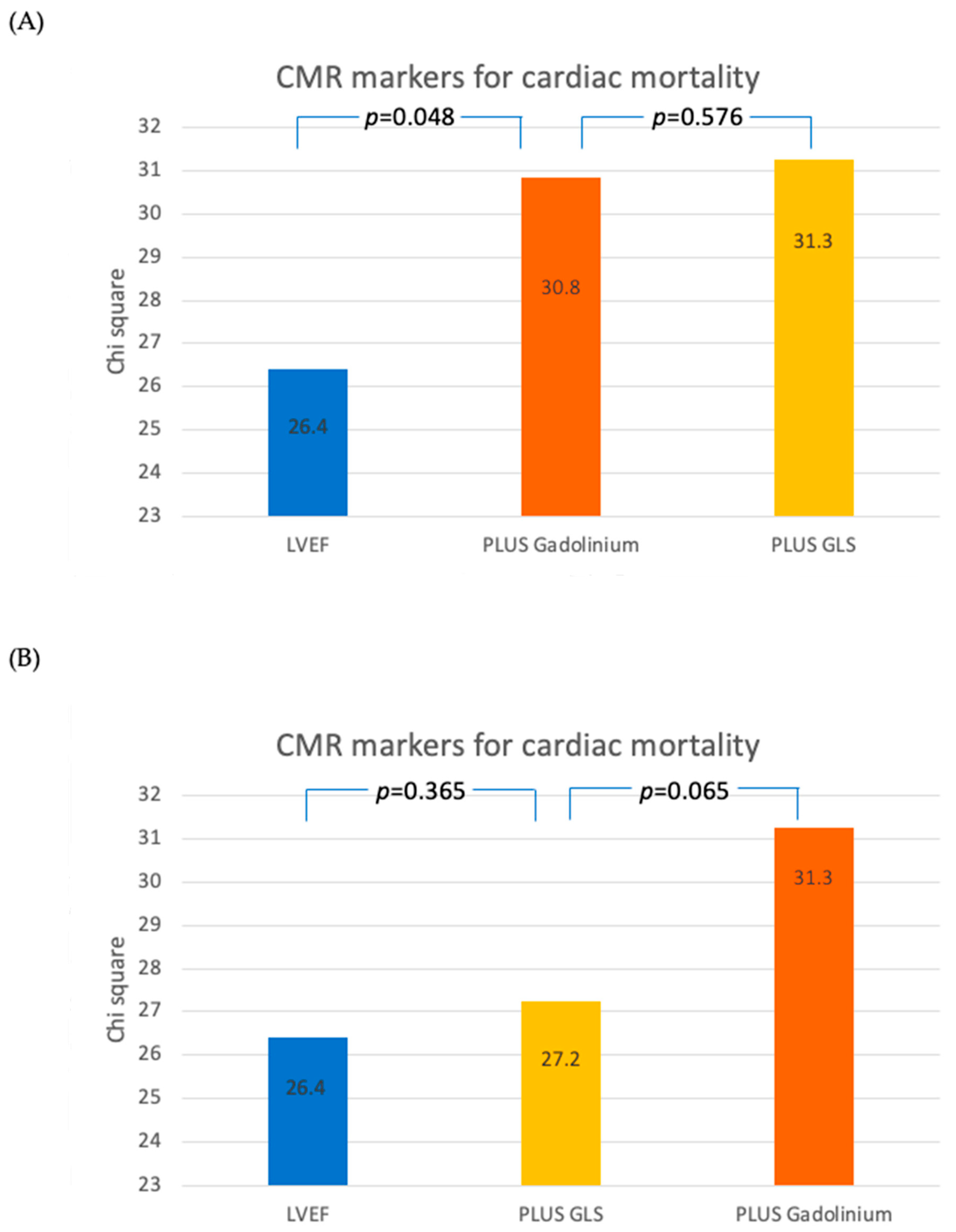
| Gadolinium (midwall or infarct pattern) | 0.001 * | 2.138 [1.391, 3.286] | 0.001 * | 2.890 [1.507, 5.541] |
| Gadolinium mass | 0.024 * | 1.024 [1.003, 1.045] | 0.042 * | 1.030 [1.001, 1.060] |
| Gadolinium % | 0.134 | 6.710 [0.557, 80.858] | 0.168 | 9.740 [0.384, 247.061] |
| Indexed LV mass | 0.429 | 1.002 [0.996, 1.009] | 0.252 | 1.005 [0.996, 1.014] |
| Indexed LA volume | 0.015 * | 1.010 [1.002, 1.018] | 0.047 * | 1.011 [1.000, 1.023] |
| LVEF | <0.001 * | 0.974 [0.963, 0.985] | <0.001 * | 0.960 [0.944, 0.976] |
| Indexed LVEDV | 0.822 | 1.000 [0.997, 1.004] | 0.785 | 1.001 [0.996, 1.006] |
| Indexed LVESV | 0.008 * | 1.007 [1.002, 1.013] | 0.001 * | 1.013 [1.006, 1.020] |
| RVEF | <0.001 * | 0.968 [0.951, 0.985] | 0.002 * | 0.962 [0.938, 0.986] |
| GLS | <0.001 * | 1.126 [1.067, 1.188] | <0.001 * | 1.201 [1.104, 1.306] |
| GRS | <0.001 * | 0.970 [0.955, 0.985] | <0.001 * | 0.951 [0.926, 0.976] |
| GCS | <0.001* | 1.120 [1.070, 1.173] | <0.001 * | 1.170 [1.090, 1.256] |
| CMR AS severity | 0.351 | 0.809 [0.518, 1.263] | 0.261 | 0.692 [0.364, 1.316] |
| ACE-I/ARB | 0.546 | 1.143 [0.741, 1.763] | 0.524 | 0.811 [0.426, 1.545] |
| Betablocker | 0.824 | 1.052 [0.672, 1.647] | 0.723 | 1.124 [0.589, 2.143] |
| Statin | 0.194 | 1.365 [0.854, 2.182] | 0.775 | 0.910 [0.477, 1.736] |
| Aldosterone antagonist | 0.018 * | 1.879 [1.114, 3.169] | 0.001 * | 3.252 [1.669, 6.336] |
| Diuretic | 0.002 * | 2.114 [1.329, 3.363] | 0.084 | 1.798 [0.923, 3.499] |
| Aortic valve intervention (SAVR or TAVR) | <0.001 * | 0.329 [0.210, 0.516] | <0.001 * | 0.174 [0.093, 0.325] |
| Variable | All-Cause Mortality | Cardiac Mortality | ||
|---|---|---|---|---|
| p Value | HR [95% CI] | p Value | HR [95% CI] | |
| Age | <0.001 * | 1.044 [1.021, 1.067] | 0.020 * | 1.041 [1.006, 1.078] |
| Gadolinium (midwall or infarct pattern) | 0.018 * | 1.752 [1.100, 2.790] | 0.080 | 1.899 [0.927, 3.892] |
| Chronic kidney disease | 0.130 | 1.486 [0.889, 2.484] | 0.113 | 1.778 [0.872, 3.624] |
| Indexed LA volume | 0.760 | 1.002 [0.992, 1.012] | 0.891 | 0.999 [0.984, 1.014] |
| LVEF | 0.353 | 0.991 [0.972, 1.010] | 0.119 | 0.977 [0.949, 1.006] |
| GLS | 0.376 | 1.040 [0.953, 1.135] | 0.587 | 1.039 [0.905, 1.192] |
Disclaimer/Publisher’s Note: The statements, opinions and data contained in all publications are solely those of the individual author(s) and contributor(s) and not of MDPI and/or the editor(s). MDPI and/or the editor(s) disclaim responsibility for any injury to people or property resulting from any ideas, methods, instructions or products referred to in the content. |
© 2024 by the authors. Licensee MDPI, Basel, Switzerland. This article is an open access article distributed under the terms and conditions of the Creative Commons Attribution (CC BY) license (https://creativecommons.org/licenses/by/4.0/).
Share and Cite
Tsampasian, V.; Merinopoulos, I.; Ravindrarajah, T.; Ring, L.; Heng, E.L.; Prasad, S.; Vassiliou, V.S. Prognostic Value of Cardiac Magnetic Resonance Feature Tracking Strain in Aortic Stenosis. J. Cardiovasc. Dev. Dis. 2024, 11, 30. https://doi.org/10.3390/jcdd11010030
Tsampasian V, Merinopoulos I, Ravindrarajah T, Ring L, Heng EL, Prasad S, Vassiliou VS. Prognostic Value of Cardiac Magnetic Resonance Feature Tracking Strain in Aortic Stenosis. Journal of Cardiovascular Development and Disease. 2024; 11(1):30. https://doi.org/10.3390/jcdd11010030
Chicago/Turabian StyleTsampasian, Vasiliki, Ioannis Merinopoulos, Thuwarahan Ravindrarajah, Liam Ring, Ee Ling Heng, Sanjay Prasad, and Vassilios S. Vassiliou. 2024. "Prognostic Value of Cardiac Magnetic Resonance Feature Tracking Strain in Aortic Stenosis" Journal of Cardiovascular Development and Disease 11, no. 1: 30. https://doi.org/10.3390/jcdd11010030