
Left Ventricle Segmental Longitudinal Strain and Regional Myocardial Work Index Could Help Determine Mitral Valve Prolapse Patients with Increased Risk of Ventricular Arrhythmias
 , , ,
, , ,
Abstract
:1. Introduction
2. Materials and Methods
2.1. Study Design and Patient Population
2.2. HOLTER-ECG Monitoring
2.3. Echocardiography
2.4. Statistics
3. Results
3.1. Baseline Characteristics
3.2. Echocardiography Parameters
3.3. Predictors of Arrhythmic Episodes
4. Discussion
5. Study Limitations
6. Conclusions
Supplementary Materials
Author Contributions
Funding
Institutional Review Board Statement
Informed Consent Statement
Data Availability Statement
Conflicts of Interest
References
- Freed, L.A.; Levy, D.; Levine, R.A.; Larson, M.G.; Evans, J.C.; Fuller, D.L.; Lehman, B.; Benjamin, E.J. Prevalence and clinical outcome of mitral valve prolapse. N. Engl. J. Med. 1999, 341, 1–7. [Google Scholar] [CrossRef]
- Levine, R.A.; Triulzi, M.O.; Harrigan, P.; Weyman, A.E. The relationship of mitral annular shape to the diagnosis of mitral valve prolapse. Circulation 1987, 75, 756–767. [Google Scholar] [CrossRef] [PubMed]
- Nishimura, R.A.; McGoon, M.D.; Shub, C.; Miller, F.A., Jr.; Ilstrup, D.M.; Tajik, A.J. Echocardiographically documented mitral valve prolapse. Long-term follow-up of 237 patients. N. Engl. J. Med. 1985, 313, 1305–1309. [Google Scholar] [CrossRef] [PubMed]
- Anders, S.; Said, S.; Schulz, F.; Püschel, K. Mitral valve prolapse syndrome as cause of sudden death in young adults. Forensic Sci. Int. 2007, 171, 127–130. [Google Scholar] [CrossRef]
- Sriram, C.S.; Syed, F.F.; Ferguson, M.E.; Johnson, J.N.; Enriquez-Sarano, M.; Cetta, F.; Cannon, B.C.; Asirvatham, S.J.; Ackerman, M.J. Malignant bileaflet mitral valve prolapse syndrome in patients with otherwise idiopathic out-of-hospital cardiac arrest. J. Am. Coll. Cardiol. 2013, 62, 222–230. [Google Scholar] [CrossRef]
- Basso, C.; Iliceto, S.; Thiene, G.; Perazzolo Marra, M. Mitral Valve Prolapse, Ventricular Arrhythmias, and Sudden Death. Circulation 2019, 140, 952–964. [Google Scholar] [CrossRef]
- Düren, D.R.; Becker, A.E.; Dunning, A.J. Long-term follow-up of idiopathic mitral valve prolapse in 300 patients: A prospective study. J. Am. Coll. Cardiol. 1988, 11, 42–47. [Google Scholar] [CrossRef]
- Basso, C.; Perazzolo Marra, M.; Rizzo, S.; De Lazzari, M.; Giorgi, B.; Cipriani, A.; Frigo, A.C.; Rigato, I.; Migliore, F.; Pilichou, K.; et al. Arrhythmic Mitral Valve Prolapse and Sudden Cardiac Death. Circulation 2015, 132, 556–566. [Google Scholar] [CrossRef] [PubMed]
- Baumgartner, H.; Falk, V.; Bax, J.J.; De Bonis, M.; Hamm, C.; Holm, P.J.; Iung, B.; Lancellotti, P.; Lansac, E.; Muñoz, D.R.; et al. 2017 ESC/EACTS Guidelines for the management of valvular heart disease. Eur. Heart J. 2017, 38, 2739–2791. [Google Scholar] [CrossRef]
- Otto, C.M.; Nishimura, R.A.; Bonow, R.O.; Carabello, B.A.; Erwin, J.P., 3rd; Gentile, F.; Jneid, H.; Krieger, E.V.; Mack, M.; McLeod, C.; et al. 2020 ACC/AHA Guideline for the Management of Patients with Valvular Heart Disease: A Report of the American College of Cardiology/American Heart Association Joint Committee on Clinical Practice Guidelines. Circulation 2021, 143, e72–e227. [Google Scholar] [CrossRef]
- Grigioni, F.; Enriquez-Sarano, M.; Ling, L.H.; Bailey, K.R.; Seward, J.B.; Tajik, A.J.; Frye, R.L. Sudden death in mitral regurgitation due to flail leaflet. J. Am. Coll. Cardiol. 1999, 34, 2078–2085. [Google Scholar] [CrossRef]
- Antoine, C.; Benfari, G.; Michelena, H.I.; Maalouf, J.F.; Nkomo, V.T.; Thapa, P.; Enriquez-Sarano, M. Clinical Outcome of Degenerative Mitral Regurgitation: Critical Importance of Echocardiographic Quantitative Assessment in Routine Practice. Circulation 2018, 138, 1317–1326. [Google Scholar] [CrossRef] [PubMed]
- Enriquez-Sarano, M.; Avierinos, J.F.; Messika-Zeitoun, D.; Detaint, D.; Capps, M.; Nkomo, V.; Scott, C.; Schaff, H.V.; Tajik, A.J. Quantitative determinants of the outcome of asymptomatic mitral regurgitation. N. Engl. J. Med. 2005, 352, 875–883. [Google Scholar] [CrossRef]
- Han, H.; Ha, F.J.; Teh, A.W.; Calafiore, P.; Jones, E.F.; Johns, J.; Koshy, A.N.; O'Donnell, D.; Hare, D.L.; Farouque, O.; et al. Mitral Valve Prolapse and Sudden Cardiac Death: A Systematic Review. J. Am. Heart Assoc. 2018, 7, e010584. [Google Scholar] [CrossRef] [PubMed]
- Essayagh, B.; Sabbag, A.; Antoine, C.; Benfari, G.; Yang, L.-T.; Maalouf, J.; Asirvatham, S.; Michelena, H.; Enriquez-Sarano, M. Presentation and Outcome of Arrhythmic Mitral Valve Prolapse. J. Am. Coll. Cardiol. 2020, 76, 637–649. [Google Scholar] [CrossRef]
- Sabbag, A.; Essayagh, B.; Barrera, J.D.R.; Basso, C.; Berni, A.; Cosyns, B.; Deharo, J.-C.; Deneke, T.; Di Biase, L.; Enriquez-Sarano, M.; et al. EHRA expert consensus statement on arrhythmic mitral valve prolapse and mitral annular disjunction complex in collaboration with the ESC Council on valvular heart disease and the European Association of Cardiovascular Imaging endorsed by the Heart Rhythm Society, by the Asia Pacific Heart Rhythm Society, and by the Latin American Heart Rhythm Society. Europace 2022, 24, euac125. [Google Scholar] [CrossRef]
- Perazzolo Marra, M.; Basso, C.; De Lazzari, M.; Rizzo, S.; Cipriani, A.; Giorgi, B.; Lacognata, C.; Rigato, I.; Migliore, F.; Pilichou, K.; et al. Morphofunctional Abnormalities of Mitral Annulus and Arrhythmic Mitral Valve Prolapse. Circ. Cardiovasc. Imaging 2016, 9, e005030. [Google Scholar] [CrossRef]
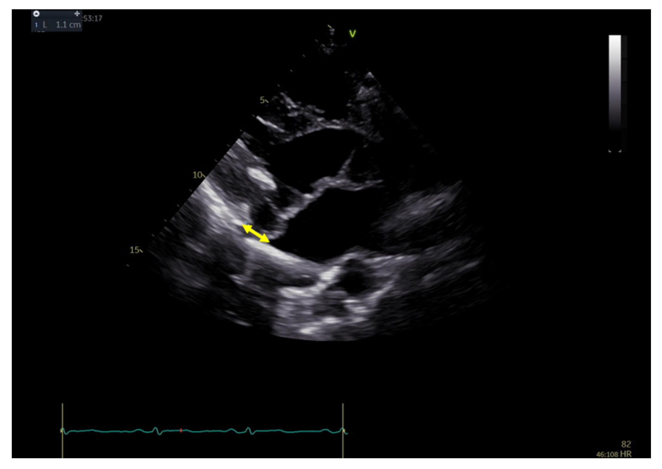
- Carmo, P.; Andrade, M.J.; Aguiar, C.; Rodrigues, R.; Gouveia, R.; Silva, J.A. Mitral annular disjunction in myxomatous mitral valve disease: A relevant abnormality recognizable by transthoracic echocardiography. Cardiovasc. Ultrasound 2010, 8, 53. [Google Scholar] [CrossRef] [PubMed]
- Essayagh, B.; Sabbag, A.; Antoine, C.; Benfari, G.; Batista, R.; Yang, L.-T.; Maalouf, J.; Thapa, P.; Asirvatham, S.; Michelena, H.I.; et al. The Mitral Annular Disjunction of Mitral Valve Prolapse: Presentation and Outcome. JACC Cardiovasc. Imaging 2021, 14, 2073–2087. [Google Scholar] [CrossRef]
- Bennett, S.; Thamman, R.; Griffiths, T.; Oxley, C.; Khan, J.N.; Phan, T.; Patwala, A.; Heatlie, G.; Kwok, C.S. Mitral annular disjunction: A systematic review of the literature. Echocardiography 2019, 36, 1549–1558. [Google Scholar] [CrossRef]
- Bharati, S.; Granston, A.S.; Liebson, P.R.; Loeb, H.S.; Rosen, K.M.; Lev, M. The conduction system in mitral valve prolapse syndrome with sudden death. Am. Heart J. 1981, 101, 667–670. [Google Scholar] [CrossRef] [PubMed]
- Sheppard, M.N.; Steriotis, A.K.; Sharma, S. Letter by Sheppard et al. Regarding Article, “Arrhythmic Mitral Valve Prolapse and Sudden Cardiac Death”. Circulation 2016, 133, e458. [Google Scholar] [CrossRef]
- Alenazy, A.; Eltayeb, A.; Alotaibi, M.K.; Anwar, M.K.; Mulafikh, N.; Aladmawi, M.; Vriz, O. Diagnosis of Mitral Valve Prolapse: Much More than Simple Prolapse. Multimodality Approach to Risk Stratification and Therapeutic Management. J. Clin. Med. 2022, 11, 455. [Google Scholar] [CrossRef] [PubMed]
- Hutchins, G.M.; Moore, G.W.; Skoog, D.K. The association of floppy mitral valve with disjunction of the mitral annulus fibrosus. N. Engl. J. Med. 1986, 314, 535–540. [Google Scholar] [CrossRef]
- Angelini, A.; Ho, S.Y.; Anderson, R.H.; Becker, A.E.; Davies, M.J. Disjunction of the mitral annulus in floppy mitral valve. N. Engl. J. Med. 1988, 318, 188–189. [Google Scholar] [CrossRef] [PubMed]
- Han, H.; Parsons, S.A.; Teh, A.W.; Sanders, P.; Neil, C.; Leong, T.; Koshy, A.N.; Vohra, J.K.; Kalman, J.M.; Smith, K.; et al. Characteristic Histopathological Findings and Cardiac Arrest Rhythm in Isolated Mitral Valve Prolapse and Sudden Cardiac Death. J. Am. Heart Assoc. 2020, 9, e015587. [Google Scholar] [CrossRef]
- Bui, A.H.; Roujol, S.; Foppa, M.; Kissinger, K.V.; Goddu, B.; Hauser, T.H.; Zimetbaum, P.J.; Ngo, L.H.; Manning, W.J.; Nezafat, R.; et al. Diffuse myocardial fibrosis in patients with mitral valve prolapse and ventricular arrhythmia. Heart 2017, 103, 204–209. [Google Scholar] [CrossRef]
- Figliozzi, S.; Georgiopoulos, G.; Lopes, P.M.; Bauer, K.B.; Moura-Ferreira, S.; Tondi, L.; Mushtaq, S.; Censi, S.; Pavon, A.G.; Bassi, I.; et al. Myocardial Fibrosis at Cardiac MRI Helps Predict Adverse Clinical Outcome in Patients with Mitral Valve Prolapse. Radiology 2022, 306, 220454. [Google Scholar] [CrossRef]
- Garg, P.; Swift, A.J.; Zhong, L.; Carlhäll, C.-J.; Ebbers, T.; Westenberg, J.; Hope, M.D.; Bucciarelli-Ducci, C.; Bax, J.J.; Myerson, S.G. assessment of mitral valve regurgitation by cardiovascular magnetic resonance imaging. Nat. Rev. Cardiol. 2020, 17, 298–312. [Google Scholar] [CrossRef]
- Scatteia, A.; Pascale, C.E.; Gallo, P.; Pezzullo, S.; America, R.; Cappelletti, A.M.; Vecchia, L.A.D.; Guarini, P.; Dellegrottaglie, S. Abnormal Papillary Muscle Signal on Cine MRI As a Typical Feature of Mitral Valve Prolapse. Sci. Rep. 2020, 10, 9166. [Google Scholar] [CrossRef]
- Vriz, O.; Eltayeb, A.; Landi, I.; Anwar, K.; Alenazy, A.; Hiristova, K.; Kasprzak, J.; D'Andrea, A.; Amro, B.; Limongelli, G.; et al. Transthoracic echocardiography for arrhythmic mitral valve prolapse: Phenotypic characterization as first step. Echocardiography 2022, 39, 1158–1170. [Google Scholar] [CrossRef]
- Zienciuk-Krajka, A.; Daniłowicz-Szymanowicz, L.; Dorniak, K.; Owczuk, R.; Kaufmann, D.; Zacharek, D.; Dąbrowska-Kugacka, A.; Figura-Chmielewska, M.; Nowak, R.; Kempa, M.; et al. Clinical characteristics of patients with arrhythmic mitral valve prolapse in a single tertiary center: Prevalence of electrocardiographic and myocardial abnormalities. Kardiol. Pol. 2021, 79, 693–696. [Google Scholar] [CrossRef] [PubMed]
- Dejgaard, L.A.; Skjølsvik, E.T.; Lie, Ø.H.; Ribe, M.; Stokke, M.K.; Hegbom, F.; Scheirlynck, E.S.; Gjertsen, E.; Andresen, K.; Helle-Valle, T.M.; et al. The Mitral Annulus Disjunction Arrhythmic Syndrome. J. Am. Coll. Cardiol. 2018, 72, 1600–1609. [Google Scholar] [CrossRef] [PubMed]
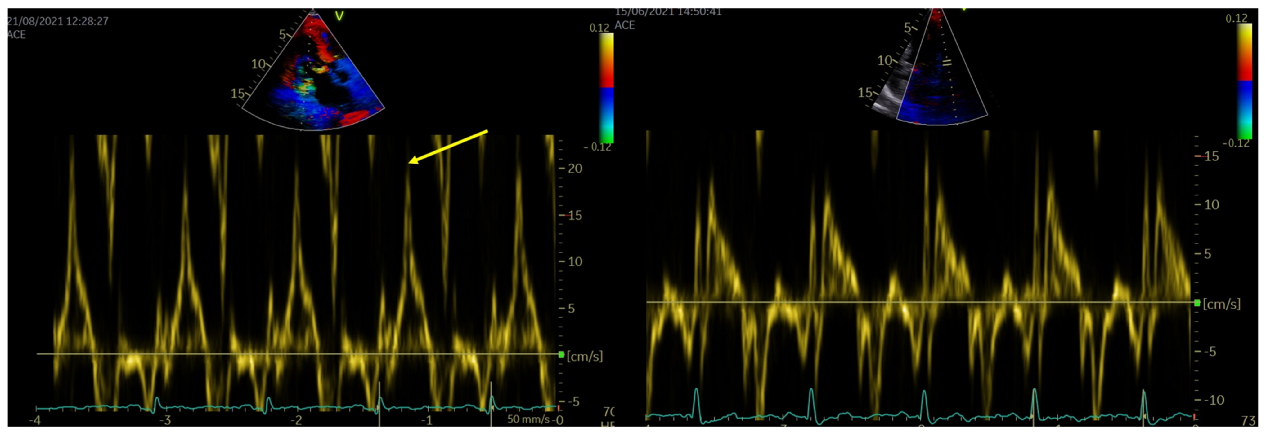
- Muthukumar, L.; Rahman, F.; Jan, M.F.; Shaikh, A.; Kalvin, L.; Dhala, A.; Jahangir, A.; Tajik, A.J. The Pickelhaube sign. Novel Echocardiographic Risk Marker for Malignant Mitral Valve Prolapse. JACC Cardiovasc. Imaging 2017, 10, 1078–1079. [Google Scholar] [CrossRef] [PubMed]
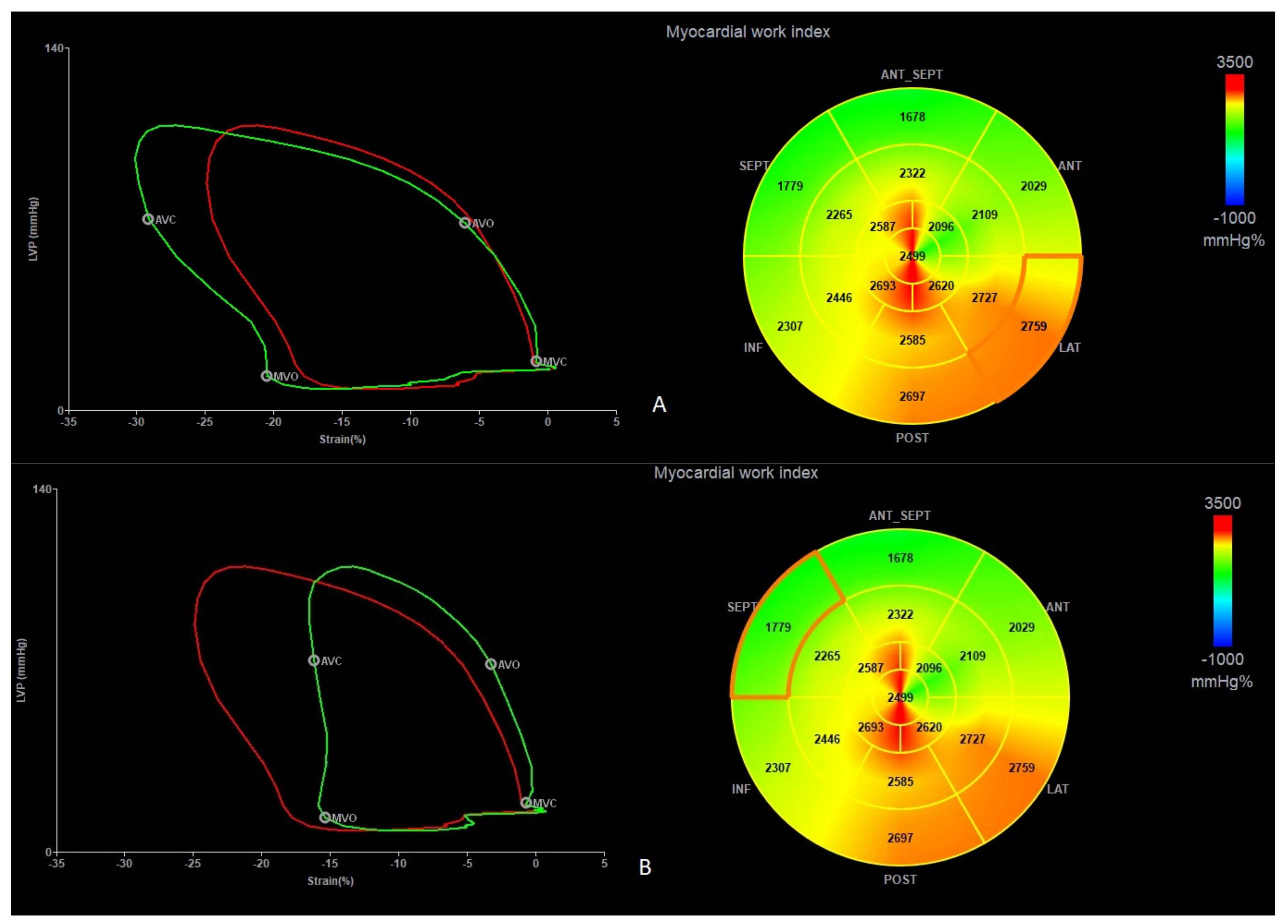
- Russell, K.; Eriksen, M.; Aaberge, L.; Wilhelmsen, N.; Skulstad, H.; Remme, E.W.; Haugaa, K.H.; Opdahl, A.; Fjeld, J.G.; Gjesdal, O.; et al. A novel clinical method for quantification of regional left ventricular pressure-strain loop area: A noninvasive index of myocardial work. Eur. Heart J. 2012, 33, 724–733. [Google Scholar] [CrossRef]
- Loncaric, F.; Marciniak, M.; Nunno, L.; Mimbrero, M.; Fernandes, J.F.; Fabijanovic, D.; Sanchis, L.; Doltra, A.; Montserrat, S.; Cikes, M.; et al. Distribution of myocardial work in arterial hypertension: Insights from non-invasive left ventricular pressure-strain relations. Int. J. Cardiovasc. Imaging 2021, 37, 145–154. [Google Scholar] [CrossRef]
- Zoghbi, W.A.; Adams, D.; Bonow, R.O.; Enriquez-Sarano, M.; Foster, E.; Grayburn, P.A.; Hahn, R.T.; Han, Y.; Hung, J.; Lang, R.M.; et al. Recommendations for Noninvasive Evaluation of Native Valvular Regurgitation: A Report from the American Society of Echocardiography Developed in Collaboration with the Society for Cardiovascular Magnetic Resonance. J. Am. Soc. Echocardiogr. 2017, 30, 303–371. [Google Scholar] [CrossRef]
- Lang, R.M.; Badano, L.P.; Mor-Avi, V.; Afilalo, J.; Armstrong, A.; Ernande, L.; Flachskampf, F.A.; Foster, E.; Goldstein, S.A.; Kuznetsova, T.; et al. Recommendations for Cardiac Chamber Quantification by Echocardiography in Adults: An Update from the American Society of Echocardiography and the European Association of Cardiovascular Imaging. Eur. Heart J. Cardiovasc. Imaging 2015, 16, 233–271. [Google Scholar] [CrossRef]
- Voigt, J.-U.; Pedrizzetti, G.; Lysyansky, P.; Marwick, T.H.; Houle, H.; Baumann, R.; Pedri, S.; Ito, Y.; Abe, Y.; Metz, S.; et al. Definitions for a Common Standard for 2D Speckle Tracking Echocardiography: Consensus Document of the EACVI/ASE/Industry Task Force to Standardize Deformation Imaging. J. Am. Soc. Echocardiogr. 2015, 28, 183–193. [Google Scholar] [CrossRef]
- Leitman, M.; Lysiansky, M.; Lysyansky, P.; Friedman, Z.; Tyomkin, V.; Fuchs, T.; Adam, D.; Krakover, R.; Vered, Z. Circumferential and longitudinal strain in 3 myocardial layers in normal subjects and in patients with regional left ventricular dysfunction. J. Am. Soc. Echocardiogr. 2010, 23, 64–70. [Google Scholar] [CrossRef]
- Belghiti, H.; Brette, S.; Lafitte, S.; Reant, P.; Picard, F.; Serri, K.; Courregelongue, M.; Dos Santos, P.; Douard, H.; Roudaut, R.; et al. Automated function imaging: A new operator-independent strain method for assessing left ventricular function. Arch. Cardiovasc. Dis. 2008, 101, 163–169. [Google Scholar] [CrossRef]
- Suga, H. Total mechanical energy of a ventricle model and cardiac oxygen consumption. Am. J. Physiol. 1979, 236, H498–H505. [Google Scholar] [CrossRef]
- Manganaro, R.; Marchetta, S.; Dulgheru, R.; Ilardi, F.; Sugimoto, T.; Robinet, S.; Cimino, S.; Go, Y.Y.; Bernard, A.; Kacharava, G.; et al. Echocardiographic reference ranges for normal noninvasive myocardial work indices: Results from the EACVI NORRE study. Eur. Heart J. Cardiovasc. Imaging 2019, 20, 582–590. [Google Scholar] [CrossRef] [PubMed]
- Morbach, C.; Sahiti, F.; Tiffe, T.; Cejka, V.; Eichner, F.A.; Gelbrich, G.; Heuschmann, P.U.; Störk, S.; On behalf of the STAAB consortium. Myocardial work-correlation patterns and reference values from the population-based STAAB cohort study. PLoS ONE 2020, 15, e0239684. [Google Scholar] [CrossRef] [PubMed]
- Yuda, S.; Sato, Y.; Abe, K.; Kawamukai, M.; Kouzu, H.; Muranaka, A.; Kokubu, N.; Hashimoto, A.; Tsuchihashi, K.; Watanabe, N.; et al. Inter-vendor variability of left ventricular volumes and strains determined by three-dimensional speckle tracking echocardiography. Echocardiography 2014, 31, 597–604. [Google Scholar] [CrossRef]
- Eriksson, M.J.; Bitkover, C.Y.; Omran, A.S.; David, T.E.; Ivanov, J.; Ali, M.J.; Woo, A.; Siu, S.; Rakowski, H. Mitral annular disjunction in advanced myxomatous mitral valve disease: Echocardiographic detection and surgical correction. J. Am. Soc. Echocardiogr. 2005, 18, 1014–1022. [Google Scholar] [CrossRef] [PubMed]
- Lancellotti, P.; Garbi, M. Malignant Mitral Valve Prolapse: Substrates to Ventricular Remodeling and Arrhythmias. Circ. Cardiovasc. Imaging 2016, 9, e005248. [Google Scholar] [CrossRef]
- Lauretta, L.; Casalino, G.; Amzulescu, M.; David-Cojocariu, A.; Unger, P. How to improve tissue Doppler imaging sensitivity to detect the Pickelhaube sign. Eur. Heart J. Cardiovasc. Imaging 2020, 21, 746. [Google Scholar] [CrossRef]
- Nutter, D.O.; Wickliffe, C.; Gilbert, C.A.; Moody, C.; King, S.B., 3rd. The pathophysiology of idiopathic mitral valve prolapse. Circulation 1975, 52, 297–305. [Google Scholar] [CrossRef]
- Ermakov, S.; Gulhar, R.; Lim, L.; Bibby, D.; Fang, Q.; Nah, G.; Abraham, T.P.; Schiller, N.B.; Delling, F.N. Left ventricular mechanical dispersion predicts arrhythmic risk in mitral valve prolapse. Heart 2019, 105, 1063–1069. [Google Scholar] [CrossRef]
- Huttin, O.; Pierre, S.; Venner, C.; Voilliot, D.; Sellal, J.-M.; Aliot, E.; Sadoul, N.; Juillière, Y.; Selton-Suty, C. Interactions between mitral valve and left ventricle analysed by 2D speckle tracking in patients with mitral valve prolapse: One more piece to the puzzle. Eur. Heart J. Cardiovasc. Imaging 2017, 18, 323–331. [Google Scholar] [CrossRef]
- van Wijngaarden, S.E.; Abou, R.; Hiemstra, Y.L.; Ajmone Marsan, N.; Bax, J.J.; Delgado, V. Regional Left Ventricular Myocardial Mechanics in Degenerative Myxomatous Mitral Valve Disease: A Comparison Between Fibroelastic Deficiency and Barlow's Disease. JACC Cardiovasc. Imaging 2018, 11, 1362–1364. [Google Scholar] [CrossRef] [PubMed]
- Vaidya, V.R.; DeSimone, C.V.; Damle, N.; Naksuk, N.; Syed, F.F.; Ackerman, M.J.; Ponamgi, S.P.; Nkomo, V.T.; Suri, R.M.; Noseworthy, P.A.; et al. Reduction in malignant ventricular arrhythmia and appropriate shocks following surgical correction of bileaflet mitral valve prolapse. J. Interv. Card Electrophysiol. 2016, 46, 137–143. [Google Scholar] [CrossRef]
- Franz, M.R.; Cima, R.; Wang, D.; Profitt, D.; Kurz, R. Electrophysiological effects of myocardial stretch and mechanical determinants of stretch-activated arrhythmias. Circulation 1992, 86, 968–978, Erratum in Circulation 1992, 86, 1663. [Google Scholar] [CrossRef] [PubMed]
- Morningstar, J.E.; Gensemer, C.; Moore, R.; Fulmer, D.; Beck, T.C.; Wang, C.; Moore, K.; Guo, L.; Sieg, F.; Nagata, Y.; et al. Mitral Valve Prolapse Induces Regionalized Myocardial Fibrosis. J. Am. Heart Assoc. 2021, 10, e022332. [Google Scholar] [CrossRef] [PubMed]
- Papadopoulos, K.; Özden Tok, Ö.; Mitrousi, K.; Ikonomidis, I. Myocardial Work: Methodology and Clinical Applications. Diagnostics 2021, 11, 573. [Google Scholar] [CrossRef]
- Muthukumar, L.; Jahangir, A.; Jan, M.F.; Perez Moreno, A.C.; Khandheria, B.K.; Tajik, A.J. Association Between Malignant Mitral Valve Prolapse and Sudden Cardiac Death: A Review. JAMA Cardiol. 2020, 5, 1053–1061. [Google Scholar] [CrossRef]
- Borzì, D.D.; Saladino, S.; Losi, V.; Faro, D.C.; Monte, I.P. Strain and Myocardial Work Index during Echo Exercise to Evaluate Myocardial Function in Athletes. J. Cardiovasc. Echogr. 2022, 32, 82–88. [Google Scholar] [CrossRef]


| MVP All n = 72 | |
|---|---|
| Age (years old) | 40 (33–49) |
| Female sex, n (%) | 51 (71) |
| Previous medical history and clinical data | |
| Palpitations, n (%) | 48 (67) |
| Presyncope, n (%) | 13 (18) |
| Syncope n (%) | 10 (14) |
| History of sudden cardiac arrest, n (%) | 17 (24) |
| Sudden cardiac death in a family history, n (%) | 4 (6) |
| Implantable cardioverter-defibrillator, n (%) | 17 (24) |
| Hypertension, n (%) | 6 (8) |
| Coronary artery disease, n (%) | 1 (1) |
| Hiperlipidemia, n (%) | 4 (6) |
| Atrial fibrillation/flutter, n (%) | 3 (4) |
| Medications | |
| Beta-blockers, n (%) | 52 (72) |
| Angiotensin converting enzyme inhibitors/sartans, n (%) | 1 (1) |
| Spironolactone, n (%) | 6 (8) |
| Cordarone/Sotalol/Propafenon, n (%) | 6 (8) |
| Diuretics, n (%) | 1 (1) |
| Statins, n (%) | 5 (7) |
| MAD (+) n = 47 | MAD (−) n = 25 | p | |
|---|---|---|---|
| Age (years old) | 39 (32–46) | 41 (37–52) | 0.074 |
| Female sex, n (%) | 34 (72) | 17 (68) | 0.787 |
| Previous medical history and clinical data | |||
| Palpitations, n (%) | 37 (79) | 11 (44) | 0.004 |
| Presyncope, n (%) | 11 (23) | 2 (8) | 0.196 |
| Syncope n (%) | 9 (19) | 1 (4) | 0.149 |
| History of sudden cardiac arrest, n (%) | 12 (26) | 5 (20) | 0.773 |
| Sudden cardiac death in a family history, n (%) | 3 (6) | 1 (4) | 1.000 |
| Implantable cardioverter-defibrillator, n (%) | 13 (28) | 4 (16) | 0.384 |
| Hypertension, n (%) | 5 (11) | 1 (4) | 0.658 |
| Coronary artery disease, n (%) | 1 (2) | 0 (0) | 1.000 |
| Hiperlipidemia, n (%) | 1 (2) | 3 (12) | 0.117 |
| Atrial fibrillation/flutter, n (%) | 2 (4) | 1 (4) | 1.000 |
| Beta-blockers, n (%) | 35 (75) | 17 (68) | 0.589 |
| Angiotensin converting enzyme inhibitors/sartans, n (%) | 1 (2) | 0 (0) | 1.000 |
| Spironolactone, n (%) | 5 (11) | 1 (4) | 0.658 |
| Cordarone/Sotalol/Propafenon, n (%) | 5 (11) | 1 (4) | 0.658 |
| Diuretics, n (%) | 0 (0) | 1 (4) | 0.347 |
| Statins, n (%) | 2 (4) | 3 (12) | 0.334 |
| MAD+ n = 47 | MAD− n = 25 | p | |
|---|---|---|---|
| Heart rate mean (minimal–maximal) (bpm) | 74 (63–83) | 70 (67–76) | 0.4904 |
| Presence of any ventricular arrhythmias, n (%) | 43 (91) | 13 (52) | <0.001 |
| Premature ventricular contractions (average number) | 2600 (937–5250) | 48 (7–752) | 0.002 |
| NSVT presence, n (%) | 25 (53) | 4 (16) | 0.002 |
| NSVT—cycle length (ms) | 200 (177–254) | 371 (297–411) | 0.035 |
| Polymorphic ventricular arrhythmia | 22 (47) | 0 (0) | <0.001 |
| MVP n = 72 | Healthy n = 20 | p | MAD+ n = 47 | MAD− n = 25 | p * | p ** | p *** | |
|---|---|---|---|---|---|---|---|---|
| Bileaflet | 48 (67%) | − | − | 37 (79%) | 11 (44%) | 0.026 | − | − |
| Barlow disease | 11 (26%) | − | − | 10 (21%) | 1 (4%) | 0.068 | − | − |
| Trivial regurgitation | 36 (50%) | − | − | 19 (40%) | 17 (68%) | 0.043 | − | − |
| Mild regurgitation | 33 (46%) | − | − | 24 (51%) | 9 (36%) | 0.232 | − | − |
| LAV index (ml/m2) | 29 (23–40) | 20 (18–23) | <0.000 | 34 (23–40) | 25 (22–30) | 0.051 | 0.001 | 0.022 |
| LVEDd (mm) | 49 (46–54) | 46 (41–46) | <0.000 | 50 (47–54) | 49 (46–51) | 0.074 | <0.000 | 0.006 |
| LVESd (mm) | 34 (29–38) | 28 (26–30) | <0.000 | 35 (29–39) | 34 (29–37) | 0.363 | 0.001 | 0.001 |
| e′ (cm/s) | 12 (10–14) | 14 (10–16) | 0.084 | 12 (10–14) | 13 (10–14) | 0.482 | 0.065 | 0.080 |
| E/e′ | 5.9 (4.9–6.9) | 6.3 (5–7.6) | 0.233 | 5.9 (5.0–6.8) | 5.9 (4.9–8.0) | 0.418 | 0.267 | 0.409 |
| S′ lat (cm/s) | 12 (8–18) | 8 (7–9) | <0.000 | 16 (12–22) | 8 (7–10) | <0.000 | <0.000 | 0.064 |
| S′ sept (cm/s) | 9 (8–10) | 8 (8–9) | 0.088 | 9 (8–11) | 8 (7–8) | <0.002 | <0.018 | 0.202 |
| Pickelhaube sign, n (%) | 30 (42%) | - | - | 30 (64%) | - | - | - | - |
| RVID (mm) | 36 (33–40) | 26 (23–29) | <0.000 | 36 (33–39) | 35 (33–41) | 0.492 | <0.000 | <0.000 |
| LVEF (%) | 57 (54–61) | 62 (60–65) | <0.000 | 56 (54–61) | 58 (54–62) | 0.275 | <0.001 | <0.004 |
| GLS (%) | −22 (−23–−10) | −22 (−22–−20) | 0.445 | −22 (−23–−21) | −20 (−22–−19) | <0.027 | 0.128 | <0.036 |
| PSD (ms) | 44 (34–54) | 34 (27–38) | 0.005 | 46 (36–56) | 38 (31–46) | 0.051 | <0.002 | 0.064 |
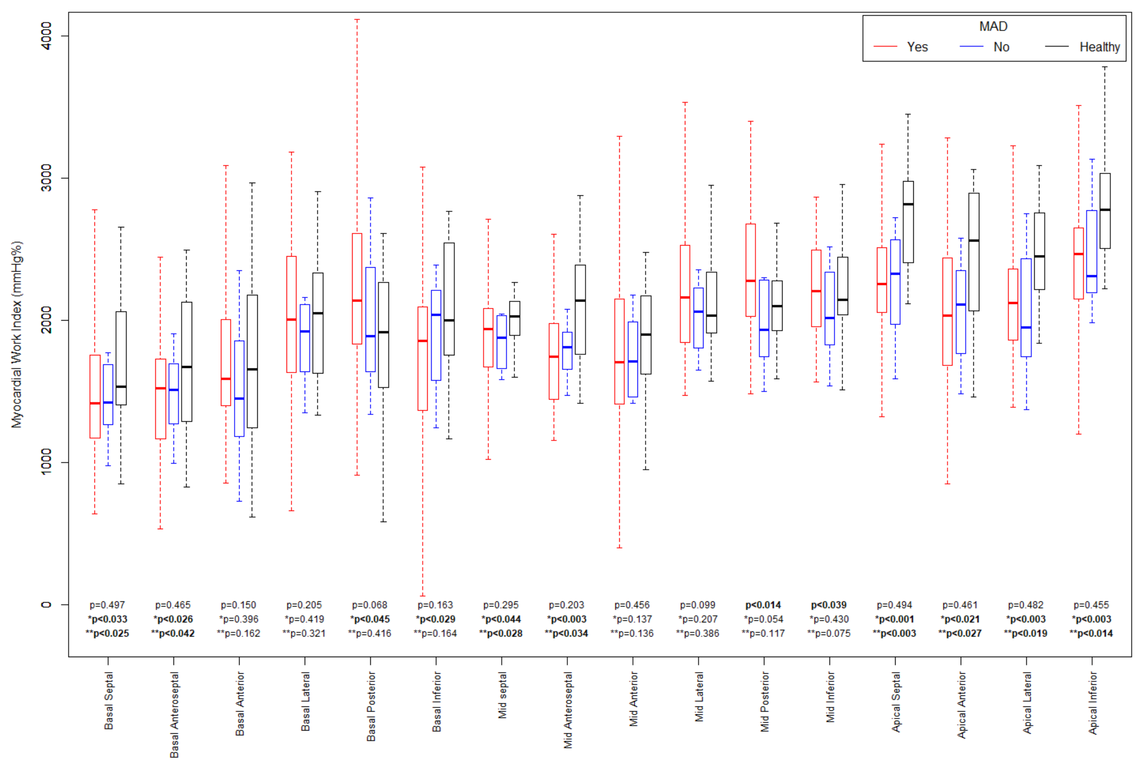
| GWI (mmHg%) | 2001 (1775–2206) | 2127 (2073–2354) | 0.018 | 2052 (1811–2202) | 1910 (1754–2200) | 0.214 | <0.028 | <0.009 |
| GCW (mmHg%) | 2112 (1823–2324) | 2145 (2006–2392) | 0.099 | 2241 (1874−2363) | 1874 (1797–2204) | 0.064 | 0.235 | <0.037 |
| GWW (mmHg%) | 124 (83–192) | 143 (95 −188) | 0.268 | 129 (87–197) | 114 (79–163) | 0.199 | 0.349 | 0.179 |
| GWE (%) | 94 (91–96) | 94 (90–95) | 0.403 | 93 (90–96) | 94 (92–96) | 0.278 | 0.482 | 0.278 |
| Cut-off | AUC% | Sensitivity | Specificity | PPV | NPV | OR | p | |
|---|---|---|---|---|---|---|---|---|
| GLS (%) | −20 | 71.7 (58.7–84.40) | 0.51 | 0.92 | 0.91 | 0.54 | 11.6 (2.39–56.1) | <0.001 |
| MAD distance (mm) | 10 | 70.0 (55.2–84.6) | 0.62 | 0.68 | 0.65 | 0.65 | 3.54 (1.09–11.51) | 0.042 |
| Peak segmental strain (%) | ||||||||
| Basal lateral | −25 | 75.5 (62.3–88.8) | 0.97 | 0.46 | 0.75 | 0.92 | 12.15 (3.78–273.8) | <0.001 |
| Mid lateral | −25 | 71.0 (57.6–84.4) | 0.79 | 0.56 | 0.74 | 0.64 | 4.93 (1.63–15.0) | 0.006 |
| Mid posterior | −25 | 70.9 (58.3–83.6) | 0.76 | 0.56 | 0.72 | 0.61 | 4.10 (1.38–12.17) | 0.015 |
| Mid inferior | −23 | 75.9 (64.1–87.7) | 0.62 | 0.88 | 0.89 | 0.58 | 11.2 (2.84–44.1) | <0.001 |
| MW index (mmHg%) | ||||||||
| Basal lateral | 2200 | 72.6 (58.2–87.1) | 0.85 | 0.62 | 0.79 | 0.71 | 9.17 (2.76–30.43) | <0.001 |
| Mid lateral | 2500 | 70.0 (52.5–83.1) | 0.92 | 0.46 | 0.73 | 0.79 | 10.15 (2.44–42.24) | <0.001 |
| Mid posterior | 2400 | 66.8 (52.8–80.8) | 0.84 | 0.5 | 0.73 | 0.67 | 5.33 (1.63–17.43) | 0.009 |
| Mid inferior | 2400 | 66.1 (52.1–80.2) | 0.82 | 0.5 | 0.73 | 0.63 | 4.57 (1.46–14.35) | 0.011 |
| OR | p | |
|---|---|---|
| Basal lateral Peak segmental strain −25% + MW index 2200 mmHg% | 32.15 (3.78–273.8) | <0.001 |
| Mid lateral Peak segmental strain −25% + MW index 2500 mmHg% | 10.43 (2.28–38.93) | <0.001 |
| Mid posterior Peak segmental strain −25% + MW index 2400 mmHg% | 6.09 (1.79–20.74) | 0.004 |
| Mid inferior Peak segmental strain −22% + MW index 2400 mmHg% | 5.50 (1.69–17.93) | 0.005 |
Disclaimer/Publisher’s Note: The statements, opinions and data contained in all publications are solely those of the individual author(s) and contributor(s) and not of MDPI and/or the editor(s). MDPI and/or the editor(s) disclaim responsibility for any injury to people or property resulting from any ideas, methods, instructions or products referred to in the content. |
© 2023 by the authors. Licensee MDPI, Basel, Switzerland. This article is an open access article distributed under the terms and conditions of the Creative Commons Attribution (CC BY) license (https://creativecommons.org/licenses/by/4.0/).
Share and Cite
Daniłowicz-Szymanowicz, L.; Zienciuk-Krajka, A.; Wabich, E.; Fijałkowski, M.; Fijałkowska, J.; Młodziński, K.; Raczak, G. Left Ventricle Segmental Longitudinal Strain and Regional Myocardial Work Index Could Help Determine Mitral Valve Prolapse Patients with Increased Risk of Ventricular Arrhythmias. J. Cardiovasc. Dev. Dis. 2023, 10, 181. https://doi.org/10.3390/jcdd10040181
Daniłowicz-Szymanowicz L, Zienciuk-Krajka A, Wabich E, Fijałkowski M, Fijałkowska J, Młodziński K, Raczak G. Left Ventricle Segmental Longitudinal Strain and Regional Myocardial Work Index Could Help Determine Mitral Valve Prolapse Patients with Increased Risk of Ventricular Arrhythmias. Journal of Cardiovascular Development and Disease. 2023; 10(4):181. https://doi.org/10.3390/jcdd10040181
Chicago/Turabian StyleDaniłowicz-Szymanowicz, Ludmiła, Agnieszka Zienciuk-Krajka, Elżbieta Wabich, Marcin Fijałkowski, Jadwiga Fijałkowska, Krzysztof Młodziński, and Grzegorz Raczak. 2023. "Left Ventricle Segmental Longitudinal Strain and Regional Myocardial Work Index Could Help Determine Mitral Valve Prolapse Patients with Increased Risk of Ventricular Arrhythmias" Journal of Cardiovascular Development and Disease 10, no. 4: 181. https://doi.org/10.3390/jcdd10040181