

Electrocardiographic and other Noninvasive Hemodynamic Markers in Decompensated CHF Patients
 , ,
, ,
Abstract
:1. Introduction
2. Materials and Methods
3. Statistical Analysis
4. Results
5. Discussion
6. Conclusions
7. Limitations
Author Contributions
Funding
Institutional Review Board Statement
Informed Consent Statement
Data Availability Statement
Conflicts of Interest
References
- Ambrosy, A.P.; Fonarow, G.C.; Butler, J.; Chioncel, O.; Greene, S.J.; Vaduganathan, M.; Nodari, S.; Lam, C.S.P.; Sato, N.; Shah, A.N.; et al. The global health and economic burden of hospitalizations for heart failure: Lessons learned from hospitalized heart failure registries. J. Am. Coll. Cardiol. 2014, 63, 1123–1133. [Google Scholar] [CrossRef] [PubMed]
- Virani, S.S.; Alonso, A.; Benjamin, E.J.; Bittencourt, M.S.; Callaway, C.W.; Carson, A.P.; Chamberlain, A.M.; Chang, A.R.; Cheng, S.; Delling, F.N.; et al. American Heart Association Council on Epidemiology and Prevention Statistics Committee and Stroke Statistics Subcommittee. Heart Disease and Stroke Statistics-2020 Update: A Report from the American Heart Association. Circulation 2020, 141, e139–e596. [Google Scholar] [CrossRef] [PubMed]
- Setoguchi, S.; Stevenson, L.W.; Schneeweiss, S. Repeated hospitalizations predict mortality in the community population with heart failure. Am. Heart J. 2007, 154, 260–266. [Google Scholar] [CrossRef] [PubMed]
- Gheorghiade, M.; De Luca, L.; Fonarow, G.C.; Filippatos, G.; Metra, M.; Francis, G.S. Pathophysiologic targets in the early phase of acute heart failure syndromes. Am. J. Cardiol. 2005, 96, 11G–17G. [Google Scholar] [CrossRef]
- Roy, R.; McDonaugh, B.; O’Gallagher, K. COVID-19 and the heart. Br. Med. Bull. 2022, 144, 4–11. [Google Scholar] [CrossRef]
- Hughes, Z.; Simkowski, J.; Mendapara, P.; Fink, N.; Gupta, S.; Youmans, Q.; Khan, S.; Wilcox, J.; Mutharasan, R.K. Racial and Socioeconomic Differences in Heart Failure Hospitalizations and Telemedicine Follow-up During the COVID-19 Pandemic: Retrospective Cohort Study. JMIR Cardio 2022, 6, e39566. [Google Scholar] [CrossRef]
- Piccirillo, G.; Moscucci, F.; Bertani, G.; Lospinuso, I.; Mastropietri, F.; Fabietti, M.; Sabatino, T.; Zaccagnini, G.; Crapanzano, D.; Di Diego, I.; et al. Short-Period Temporal Dispersion Repolarization Markers Predict 30-Days Mortality in Decompensated Heart Failure. J. Clin. Med. 2020, 9, 1879. [Google Scholar] [CrossRef]
- Piccirillo, G.; Moscucci, F.; Mariani, M.V.; Di Iorio, C.; Fabietti, M.; Mastropietri, F.; Crapanzano, D.; Bertani, G.; Sabatino, T.; Zaccagnini, G.; et al. Hospital mortality in decompensated heart failure. A pilot study. J. Electrocardiol. 2020, 61, 147–152. [Google Scholar] [CrossRef]
- McDonagh, T.A.; Metra, M.; Adamo, M.; Gardner, R.S.; Baumbach, A.; Böhm, M.; Burri, H.; Butler, J.; Čelutkienė, J.; Chioncel, O.; et al. 2021 ESC Guidelines for the diagnosis and treatment of acute and chronic heart failure. Eur. Heart J. 2021, 42, 3599–3726. [Google Scholar] [CrossRef]
- Piccirillo, G.; Moscucci, F.; Bertani, G.; Lospinuso, I.; Sabatino, T.; Zaccagnini, G.; Crapanzano, D.; Diego, I.D.; Corrao, A.; Rossi, P.; et al. Short-period temporal repolarization dispersion in subjects with atrial fibrillation and decompensated heart failure. Pacing Clin. Electrophysiol. 2021, 44, 327–333. [Google Scholar] [CrossRef]
- Piccirillo, G.; Moscucci, F.; Carnovale, M.; Corrao, A.; Di Diego, I.; Lospinuso, I.; Sciomer, S.; Rossi, P.; Magrì, D. Glucose dysregulation and repolarization variability markers are short-term mortality predictors in decompensated heart failure. Cardiovasc. Endocrinol. Metab. 2022, 11, e0264. [Google Scholar] [CrossRef] [PubMed]
- Piccirillo, G.; Moscucci, F.; Carnovale, M.; Corrao, A.; Di Diego, I.; Lospinuso, I.; Caltabiano, C.; Mezzadri, M.; Rossi, P.; Magrì, D. Short-Period Temporal Dispersion Repolarization Markers in Elderly Patients with Decompensated Heart Failure. La Clin. Ter. 2022, 173, 356–361. [Google Scholar] [CrossRef]
- Prineas, R.J. The Minnesota Code Manual of Electrocardiographic Findings: Standards and Procedures for Measurement and Classification; Wright, J., Ed.; Springer: Boston, MA, USA, 1982; pp. 172–176. [Google Scholar]
- Darouian, N.; Narayanan, K.; Aro, A.L.; Reinier, K.; Uy-Evanado, A.; Teodorescu, C.; Gunson, K.; Jui, J.; Chugh, S.S. Delayed intrinsicoid deflection of the QRS complex is associated with sudden cardiac arrest. Heart Rhythm 2016, 13, 927–932. [Google Scholar] [CrossRef] [Green Version]
- Aiken, A.V.; Goldhaber, J.I.; Chugh, S.S. Delayed intrinsicoid deflection: Electrocardiographic harbinger of heart disease. Ann. Noninvasive Electrocardiol. 2022, 27, e12940. [Google Scholar] [CrossRef]
- O’Neal, W.T.; Qureshi, W.T.; Nazarian, S.; Kawel-Boehm, N.; Bluemke, D.A.; Lima, J.A.; Soliman, E.Z. Electrocardiographic Time to Intrinsicoid Deflection and Heart Failure: The Multi-Ethnic Study of Atherosclerosis. Clin. Cardiol. 2016, 39, 531–536. [Google Scholar] [CrossRef] [Green Version]
- Januzzi, J.L., Jr.; Chen-Tournoux, A.A.; Christenson, R.H.; Doros, G.; Hollander, J.E.; Levy, P.D.; Nagurney, J.T.; Nowak, R.M.; Pang, P.S.; Patel, D.; et al. N-Terminal Pro-B-Type Natriuretic Peptide in the Emergency Department: The ICON-RELOADED Study. J. Am. Coll. Cardiol. 2018, 71, 1191–1200. [Google Scholar] [CrossRef] [PubMed]
- Baumert, M.; Porta, A.; Vos, M.A.; Malik, M.; Couderc, J.P.; Laguna, P.; Piccirillo, G.; Smith, G.L.; Tereshchenko, L.G.; Volders, P.G. QT interval variability in body surface ECG: Measurement, physiological basis, and clinical value: Position statement and consensus guidance endorsed by the European Heart Rhythm Association jointly with the ESC Working Group on Cardiac Cellular Electrophysiology. Europace 2016, 18, 925–944. [Google Scholar] [CrossRef] [PubMed]
- Piccirillo, G.; Moscucci, F.; Fiorucci, C.; Di Iorio, C.; Mastropietri, F.; Magrì, D. Time- and frequency-domain analysis of beat to beat P-wave duration, PR interval and RR interval can predict asystole as form of syncope during head-up tilt. Physiol. Meas. 2016, 37, 1910–1924. [Google Scholar] [CrossRef] [PubMed] [Green Version]
- Piccirillo, G.; Moscucci, F.; Fiorucci, C.; D’Alessandro, G.; Pascucci, M.; Magrì, D. P wave analysis and left ventricular systolic function in chronic heart failure. Possible insights form the P wave–PP interval spectral coherence. Minerva Cardioangiol. 2016, 64, 525–533. [Google Scholar]
- Piccirillo, G.; Magrì, D.; D’Alessandro, G.; Fiorucci, C.; Moscucci, F.; Di Iorio, C.; Mastropietri, F.; Parrotta, I.; Ogawa, M.; Lin, S.F.; et al. Oscillatory behavior of P wave duration and PR interval in experimental congestive heart failure: A preliminary study. Physiol. Meas. 2018, 39, 035010. [Google Scholar] [CrossRef]
- Piccirillo, G.; Moscucci, F.; Magrì, D. Air Pollution Role as Risk Factor of Cardioinhibitory Carotid Hypersensitivity. Atmosphere 2022, 13, 123. [Google Scholar] [CrossRef]
- Malik, M.; Bigger, J.T.; Camm, A.J.; Kleiger, R.E.; Malliani, A.; Moss, A.J.; Schwartz, P.J. Heart rate variability. Standards of measurement, physiological interpretation, and clinical use. Task Force of the European Society of Cardiology and the North American Society of Pacing and Electrophysiology. Eur. Heart J. 1996, 17, 354–381. [Google Scholar] [CrossRef] [Green Version]
- Piccirillo, G.; Luparini, R.L.; Celli, V.; Moisè, A.; Lionetti, M.; Marigliano, V.; Cacciafesta, M. Effects of carvedilol on heart rate and blood pressure variability in subjects with chronic heart failure. Am. J. Cardiol. 2000, 86, 1392–1395. [Google Scholar] [CrossRef] [PubMed]
- Piccirillo, G.; Di Giuseppe, V.; Nocco, M.; Lionetti, M.; Moisè, A.; Naso, C.; Tallarico, D.; Marigliano, V.; Cacciafesta, M. Influence of aging and other cardiovascular risk factors on baroreflex sensitivity. J. Am. Geriatr. Soc. 2001, 49, 1059–1065. [Google Scholar] [CrossRef]
- Piccirillo, G.; Ogawa, M.; Song, J.; Chong, V.J.; Joung, B.; Han, S.; Magrì, D.; Chen, L.S.; Lin, S.F.; Chen, P.S. Power spectral analysis of heart rate variability and autonomic nervous system activity measured directly in healthy dogs and dogs with tachycardia-induced heart failure. Heart Rhythm 2009, 6, 546–552. [Google Scholar] [CrossRef] [Green Version]
- Piccirillo, G.; Rossi, P.; Mitra, M.; Quaglione, R.; Dell’Armi, A.; Di Barba, D.; Maisto, D.; Lizio, A.; Barillà, F.; Magrì, D. Indexes of temporal myocardial repolarization dispersion and sudden cardiac death in heart failure: Any difference? Ann. Noninvasive Electrocardiol. 2013, 18, 130–139. [Google Scholar] [CrossRef] [PubMed]
- Piccirillo, G.; Moscucci, F.; D’Alessandro, G.; Pascucci, M.; Rossi, P.; Han, S.; Chen, L.S.; Lin, S.F.; Chen, P.S.; Magrì, D. Myocardial repolarization dispersion and autonomic nerve activity in a canine experimental acute myocardial infarction model. Heart Rhythm 2014, 11, 110–118. [Google Scholar] [CrossRef] [Green Version]
- Charloux, A.; Lonsdorfer-Wolf, E.; Richard, R.; Lampert, E.; Oswald-Mammosser, M.; Mettauer, B.; Geny, B.; Lonsdorfer, J. A new impedance cardiograph device for the non-invasive evaluation of cardiac output at rest and during exercise: Comparison with the “direct” Fick method. Eur. J. Appl. Physiol. 2000, 82, 313–320. [Google Scholar] [CrossRef]
- Hsu, A.R.; Barnholt, K.E.; Grundmann, N.K.; Lin, J.H.; McCallum, S.W.; Friedlander, A.L. Sildenafil improves cardiac output and exercise performance during acute hypoxia, but not normoxia. J. Appl. Physiol. 2006, 100, 2031–2040. [Google Scholar] [CrossRef]
- Lepretre, P.M.; Koralsztein, J.P.; Billat, V.L. Effect of exercise intensity on relationship between VO2max and cardiac output. Med. Sci. Sports Exerc. 2004, 36, 1357–1363. [Google Scholar] [CrossRef] [Green Version]
- Tonelli, A.R.; Alnuaimat, H.; Li, N.; Carrie, R.; Mubarak, K.K. Value of impedance cardiography in patients studied for pulmonary hypertension. Lung 2011, 189, 369–375. [Google Scholar] [CrossRef] [PubMed]
- Gordon, N.; Abbiss, C.R.; Maiorana, A.J.; Marston, K.J.; Peiffer, J.J. Intrarater Reliability and Agreement of the Physioflow Bioimpedance Cardiography Device during Rest, Moderate and High-Intensity Exercise. Kinesiology 2018, 50, 140–149. [Google Scholar]
- Leão, R.N.; Silva, P.M.D. Impedance Cardiography in the Evaluation of Patients with Arterial Hypertension. Int. J. Cardiovasc. Sci. 2019, 32, 61–69. [Google Scholar] [CrossRef]
- Anand, G.; Yu, Y.; Lowe, A.; Kalra, A. Bioimpedance analysis as a tool for hemodynamic monitoring: Overview, methods and challenges. Physiol. Meas. 2021, 42, 03TR01. [Google Scholar] [CrossRef] [PubMed]
- Pérez-Riera, A.R.; De Abreu, L.C.; Barbosa-Barros, R.; Nikus, K.C.; Baranchuk, A. R-Peak Time: An Electrocardiographic Parameter with Multiple Clinical Applications. Ann. Noninvasive Electrocardiol. 2016, 21, 10–19. [Google Scholar] [CrossRef] [PubMed]
- Mortara, A.; La Rovere, M.T.; Signorini, M.G.; Pantaleo, P.; Pinna, G.; Martinelli, L.; Ceconi, C.; Cerutti, S.; Tavazzi, L. Can power spectral analysis of heart rate variability identify a high risk subgroup of congestive heart failure patients with excessive sympathetic activation? A pilot study before and after heart transplantation. Br. Heart J. 1994, 71, 422–430. [Google Scholar] [CrossRef] [Green Version]
- La Rovere, M.T.; Pinna, G.D.; Maestri, R.; Mortara, A.; Capomolla, S.; Febo, O.; Ferrari, R.; Franchini, M.; Gnemmi, M.; Opasich, C.; et al. Short-term heart rate variability strongly predicts sudden cardiac death in chronic heart failure patients. Circulation 2003, 107, 565–570. [Google Scholar] [CrossRef] [Green Version]
- Piccirillo, G.; Nocco, M.; Moisè, A.; Lionetti, M.; Naso, C.; Di Carlo, S.; Marigliano, V. Influence of vitamin C on baroreflex sensitivity in chronic heart failure. Hypertension 2003, 41, 1240–1245. [Google Scholar] [CrossRef] [Green Version]
- Piccirillo, G.; Magrì, D.; Naso, C.; Di Carlo, S.; Moisè, A.; De Laurentis, T.; Torrini, A.; Matera, S.; Nocco, M. Factors influencing heart rate variability power spectral analysis during controlled breathing in patients with chronic heart failure or hypertension and in healthy normotensive subjects. Clin. Sci. 2004, 107, 183–190. [Google Scholar] [CrossRef] [Green Version]
- Piccirillo, G.; Magrì, D.; Di Carlo, S.; De Laurentis, T.; Torrini, A.; Matera, S.; Magnanti, M.; Bernardi, L.; Barillà, F.; Quaglione, R.; et al. Influence of cardiac-resynchronization therapy on heart rate and blood pressure variability: 1-year follow-up. Eur. J. Heart Fail. 2006, 8, 716–722. [Google Scholar] [CrossRef] [Green Version]
- Van der Meer, N.J.; Oomen, M.W.; Vonk Noordegraaf, A.; Pijpers, R.J.; Plaizier, M.A.; De Vries, P.M. Does impedance cardiography reliably estimate left ventricular ejection fraction? J. Clin. Monit. 1996, 12, 5–9. [Google Scholar] [CrossRef] [PubMed]
- Pickett, B.R.; Buell, J.C. Usefulness of the impedance cardiogram to reflect left ventricular diastolic function. Am. J. Cardiol. 1993, 71, 1099–1103. [Google Scholar] [CrossRef] [PubMed]


| Subjects with | Subjects without | ||
|---|---|---|---|
| Acute Decompensated Heart Failure | |||
| N: 87 | N: 53 | p value | |
| Age, years | 83 ± 10 | 83 ± 9 | 0.884 |
| M/F n | 38/49 | 23/30 | 0.974 |
| BMI, kg/m2 | 26 ± 5 | 25 ± 5 | 0.814 |
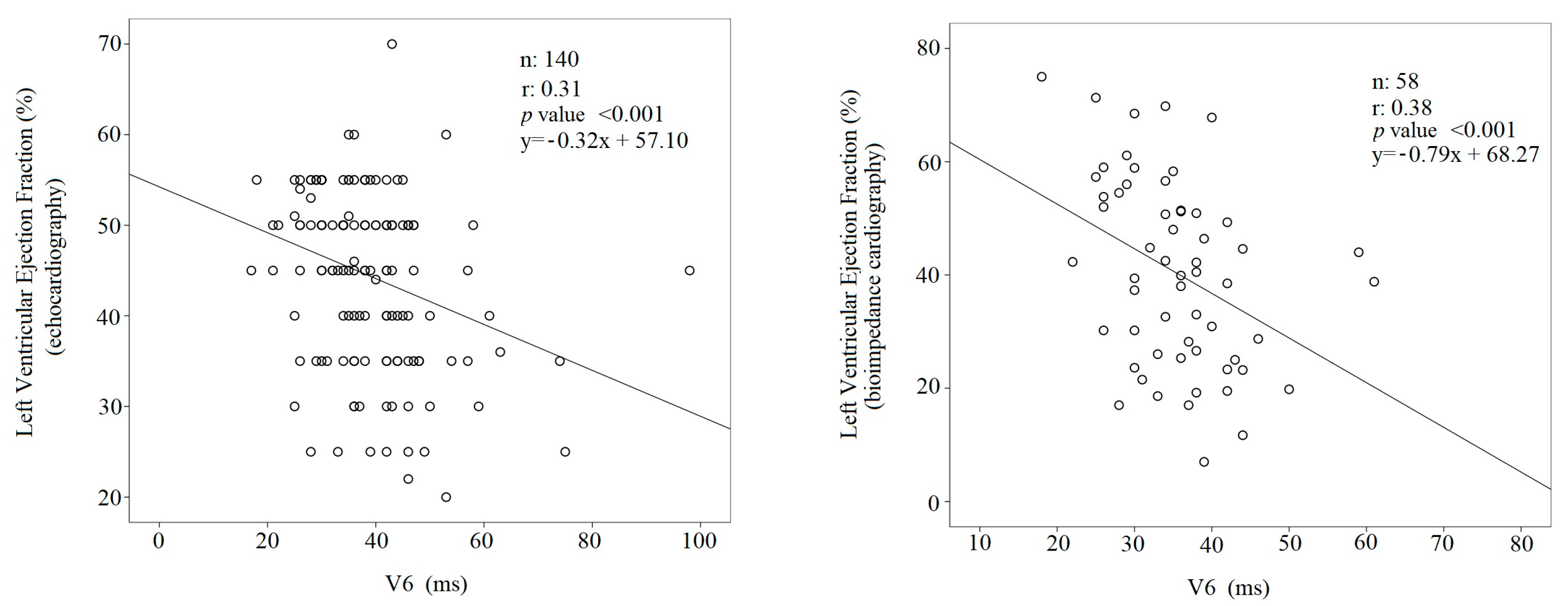
| Left Ventricular Ejection Fraction % | 42 ± 10 | 48 ± 8 | <0.001 |
| Left Ventricular Mass Index, (g/m2) | 129 ± 33 | 134 ± 28 | 0.440 |
| Left Ventricular End-Diastolic Diameter, (mm) | 53 ± 8 | 52 ± 6 | 0.462 |
| Posterior Wall Thickness, (mm) | 11 ± 2 | 1 ± 1 | 0.253 |
| Interventricular Septum Thickness, (mm) | 12 ± 1 | 12 ± 1 | 0.511 |
| Left Atrial Transversal Diameter, (mm) | 47 ± 6 | 45 ± 6 | 0.133 |
| Tricuspid Annular Plane Systolic Excursion, (mm) | 20 ± 5 | 21 ± 3 | 0.194 |
| Tricuspid Regurgitation Peak Gradient, (mmHg) | 48 ± 15 | 37 ± 9 | 0.001 |
| NT-pro BNP, pg/mL | 4930 {9170} | 512 {790} | <0.001 |
| C-reactive Protein, (mg/dL) | 4 {9} | 5 {9} | 0.085 |
| High sensitivity cardiac troponin, (pg/L) | 55 {94} | 29 {25} | <0.001 |
| Serum potassium, (mmol/L) | 4.10 ± 0.64 | 4.14 ± 0.56 | 0.716 |
| Serum Calcium, (mmol/L) | 2.20 ± 0.77 | 2.10 ± 0.15 | 0.716 |
| Creatinine Clearance, (mL/m) | 42 ± 25 | 54 ± 22 | 0.005 |
| Fasting Glucose, (mmol/L) | 7.2 ± 2.5 | 6.1 ± 1.9 | 0.011 |
| HbA1c (%) | 6.3 ± 1.6 | 5.8 ± 1.2 | 0.070 |
| Total Cholesterol, (mmol/L) | 3.35 ± 0.95 | 4.00 ± 0.90 | 0.006 |
| HDL-cholesterol, (mmol/L) | 0.99 ± 0.39 | 1.19 ± 0.41 | 0.030 |
| LDL-cholesterol, (mmol/L) | 1.67 ± 0.65 | 2.19 ± 0.74 | 0.002 |
| Triglycerides, (mmol/L) | 1.97 ± 1.60 | 1.50 ± 0.84 | 0.126 |
| PaO2/FiO2 ratio, | 329 ± 123 | 327 ± 76 | 0.921 |
| A-ADO2, mmHg | 45 (62) | 34 (31) | 0.102 |
| Reduced LVEF, n (%) | 41 (47) | 11 (21) | 0.002 |
| Mildly reduced LVEF, n (%) | 14 (16) | 8 (15) | 0.875 |
| Preserved LVEF, n (%) | 32 (37) | 34 (64) | 0.002 |
| Hypertension, n (%) | 65 (75) | 42 (79) | 0.540 |
| Hypercholesterolemia. n (%) | 39 (45) | 18 (34) | 0.204 |
| Diabetes, n (%) | 40 (46) | 16 (30) | 0.064 |
| Renal Insufficiency, n (%) | 48 (55) | 12 (23) | <0.001 |
| Known Myocardial Ischemia History, n (%) | 33 (38) | 13 (25) | 0.102 |
| Valve Diseases, | 25 (29) | 13 (25) | 0.587 |
| Premature Supraventricular Complexes, n (%) | 8 (9) | 2 (4) | 0.227 |
| Premature Ventricular Complexes, n (%) | 20 (23) | 7 (13) | 0.155 |
| Permanent Atrial fibrillation, n (%) | 35 (40) | 10 (20) | 0.009 |
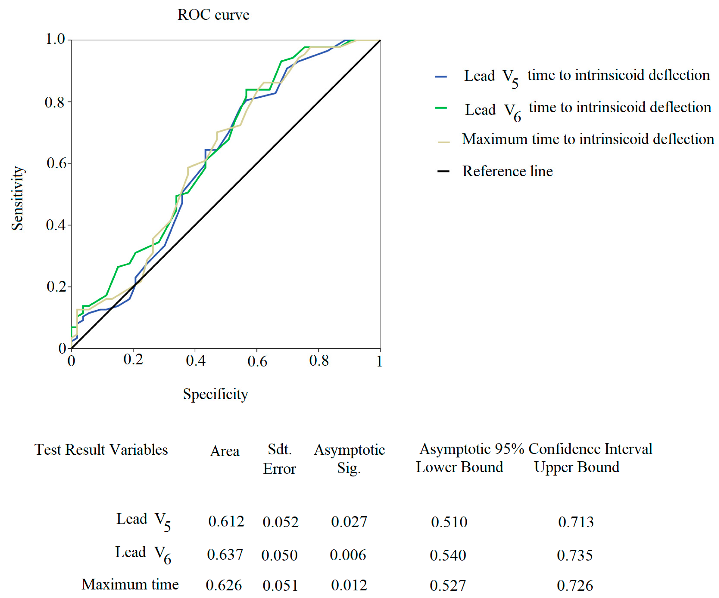
| Time to Intrisicoid Deflection >50 ms, n(%) | 14 (16) | 5 (9) | 0.265 |
| Deceased Subjects, n (%) | 29 (33) | 3 (6) | <0.001 |
| β-blockers, n (%) | 63 (72) | 28 (53) | 0.018 |
| Furosemide, n (%) | 72 (83) | 33 (62) | 0.007 |
| ACE/Sartans, n(%) | 24 (28) | 27 (51) | 0.005 |
| Aldosterone antagonists, n (%) | 15 (17) | 3 (6) | 0.047 |
| Potassium, n (%) | 8 (9) | 4 (8) | 0.735 |
| Nitrates, n (%) | 12 (14) | 9 (17) | 0.608 |
| Digoxin, n (%) | 5 (6) | 2 (4) | 0.603 |
| Statins, n (%) | 15 (28) | 23 (26) | 0.810 |
| Antiplatelet drugs, n (%) | 28 (32) | 22 (42) | 0.264 |
| Oral Anticoagulants, n (%) | 24 (28) | 12 (23) | 0.516 |
| Diltiazem or Verapamil, n (%) | 2 (2) | 3 (6) | 0.299 |
| Dihydropyridine Calcium channel blockers, n (%) | 11 (13) | 12 (23) | 0.122 |
| Propafenone, n (%) | 1 (1) | 1 (2) | 0.721 |
| Amiodarone, n (%) | 6 (7) | 3 (6) | 0.772 |
| Valsartan/Sacubitril, n (%) | 2 (2) | 1 (2) | 0.870 |
| Gliflozin, n (%) | 1 (1) | 0 (0) | 0.999 |
| Subjects with | Subjects without | ||
|---|---|---|---|
| Acute Decompensated Heart Failure | |||
| N: 87 | N: 53 | p value | |
| Lead I, ms | 41 ± 9 | 41 ± 14 | 0.951 |
| Lead II, ms | 42 ± 12 | 39 ± 9 | 0.138 |
| Lead III, ms | 42 ± 12 | 38 ± 11 | 0.062 |
| Lead aVR, ms | 40 ± 9 | 37 ± 10 | 0.160 |
| Lead aVL, ms | 39 ± 10 | 39 ± 13 | 0.921 |
| Lead aVF, ms | 40 ± 9 | 39 ± 13 | 0.617 |
| Lead V1, ms | 42 ± 14 | 38 ± 16 | 0.124 |
| Lead V2, ms | 41 ± 15 | 39 ± 14 | 0.391 |
| Lead V3, ms | 40 ± 13 | 39 ± 14 | 0.196 |
| Lead V4, ms | 39 ± 11 | 36 ± 12 | 0.112 |
| Lead V5, ms | 40 ± 9 | 35 ± 11 | 0.011 |
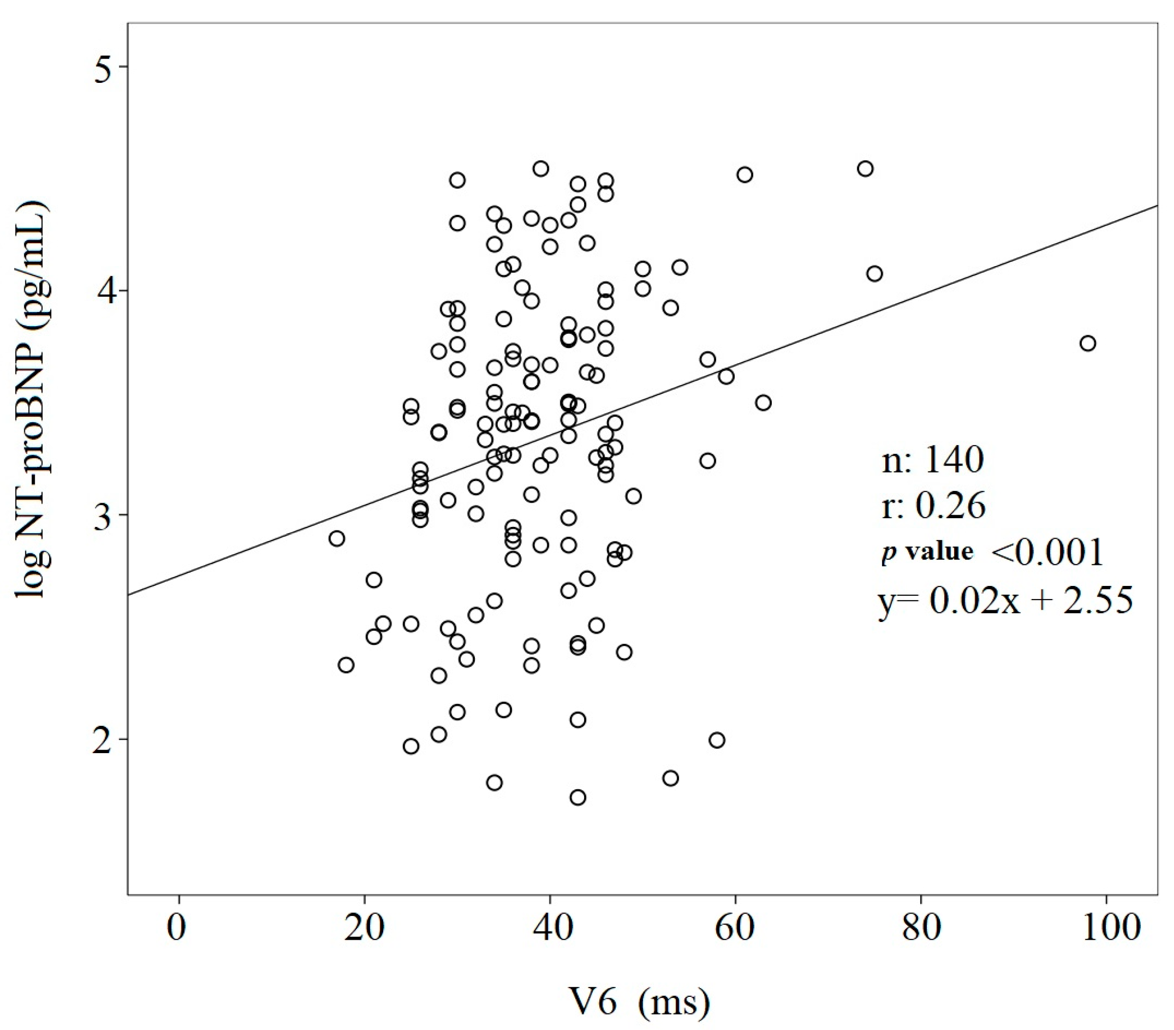
| Lead V6, ms | 41 ± 11 | 35 ± 9 | 0.002 |
| Maximum R-Peak Time, ms | 42 ± 11 | 37 ± 10 | 0.004 |
| Subjects with | Subjects without | ||
|---|---|---|---|
| Acute Decompensated Heart Failure | |||
| N:87 | N:53 | p value | |
| RR mean, ms | 826 ± 183 | 887 ± 146 | 0.039 |
| QR mean, ms | 40 ± 11 | 38 ± 11 | 0.172 |
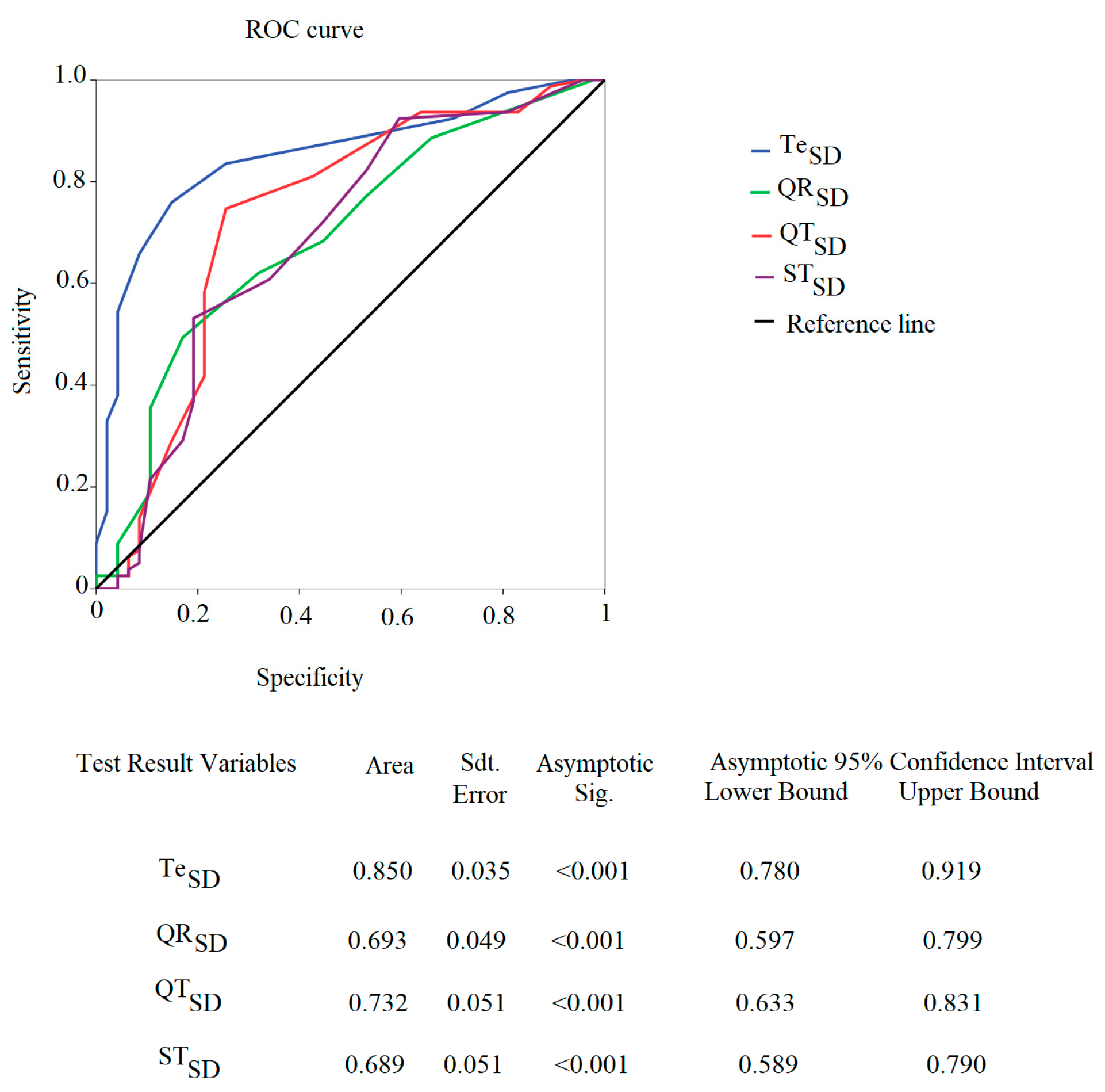
| QRSD, ms2 | 5 (4) | 3 (4) | <0.001 |
| QRS mean, ms | 90 ± 18 | 86 ± 18 | 0.197 |
| QRSSD, ms2 | 7 (5) | 5 (4) | <0.001 |
| QT mean, ms | 443 ± 75 | 425 ± 56 | 0.171 |
| QTSD, ms2 | 10 (5) | 6 (4) | <0.001 |
| ST mean, ms | 349 ± 71 | 337 ± 48 | 0.301 |
| STSD, ms2 | 9 (5) | 6 (4) | <0.001 |
| Te mean, ms | 106 ± 31 | 96 ± 15 | 0.021 |
| TeSD, ms2 | 8 (4) | 5 (2) | <0.001 |
| Subjects with | Subjects without | ||
|---|---|---|---|
| Acute Decompensated Heart Failure | |||
| N: 37 | N: 36 | p value | |
| RRSD, ms2 | 16 (9) | 23 (19) | 0.006 |
| TP, ms2 | 242 (292) | 506 (1051) | 0.007 |
| VLF, ms2 | 165 (257) | 291 (702) | 0.031 |
| LF, ms2 | 26 (44) | 90 (210) | 0.006 |
| HF, ms2 | 21 (42) | 44 (85) | 0.030 |
| LF CF, Hz | 0.11 ± 0.02 | 0.09 ± 0.03 | 0.020 |
| HF CF, Hz | 0.29 ± 0.10 | 0.28 ± 0.07 | 0.465 |
| LF/HF, | 1.33 (1.53) | 1.79 (2.87) | 0.214 |
| LF, nu | 45 (20) | 54 (38) | 0.151 |
| HF, nu | 33 (24) | 30 (23) | 0.282 |
| Variables | B | Wald | Multivariable Analysis Odd Ratio (95% CI) | p Values |
|---|---|---|---|---|
| QT | 0.02 | 5.97 | 1.02 (1.00–1.03) | 0.015 |
| Te | −0.07 | 11.09 | 0.93 (0.89–0.97) | 0.001 |
Disclaimer/Publisher’s Note: The statements, opinions and data contained in all publications are solely those of the individual author(s) and contributor(s) and not of MDPI and/or the editor(s). MDPI and/or the editor(s) disclaim responsibility for any injury to people or property resulting from any ideas, methods, instructions or products referred to in the content. |
© 2023 by the authors. Licensee MDPI, Basel, Switzerland. This article is an open access article distributed under the terms and conditions of the Creative Commons Attribution (CC BY) license (https://creativecommons.org/licenses/by/4.0/).
Share and Cite
Piccirillo, G.; Moscucci, F.; Mezzadri, M.; Caltabiano, C.; Di Diego, I.; Carnovale, M.; Corrao, A.; Stefano, S.; Scinicariello, C.; Giuffrè, M.; et al. Electrocardiographic and other Noninvasive Hemodynamic Markers in Decompensated CHF Patients. J. Cardiovasc. Dev. Dis. 2023, 10, 125. https://doi.org/10.3390/jcdd10030125
Piccirillo G, Moscucci F, Mezzadri M, Caltabiano C, Di Diego I, Carnovale M, Corrao A, Stefano S, Scinicariello C, Giuffrè M, et al. Electrocardiographic and other Noninvasive Hemodynamic Markers in Decompensated CHF Patients. Journal of Cardiovascular Development and Disease. 2023; 10(3):125. https://doi.org/10.3390/jcdd10030125
Chicago/Turabian StylePiccirillo, Gianfranco, Federica Moscucci, Martina Mezzadri, Cristina Caltabiano, Ilaria Di Diego, Myriam Carnovale, Andrea Corrao, Sara Stefano, Claudia Scinicariello, Marco Giuffrè, and et al. 2023. "Electrocardiographic and other Noninvasive Hemodynamic Markers in Decompensated CHF Patients" Journal of Cardiovascular Development and Disease 10, no. 3: 125. https://doi.org/10.3390/jcdd10030125