
Development of a Multiplex Quantitative PCR for Detecting Porcine Epidemic Diarrhea Virus, Transmissible Gastroenteritis Virus, and Porcine Deltacoronavirus Simultaneously in China

Abstract
:Simple Summary
Abstract
1. Introduction
2. Materials and Methods
2.1. Primers and Probes
2.2. Standard Strains and Clinical Samples
2.3. Extraction of RNA and Obtained cDNA
2.4. Recombinant Plasmid Construction
2.5. Optimization of Conditions for Multiplex qPCR
2.6. Standard Curves
2.7. Specificity
2.8. Sensitivity
2.9. Repeatability
2.10. Clinical Sample Detection
3. Results
3.1. Optimization of Conditions for Multiplex qPCR
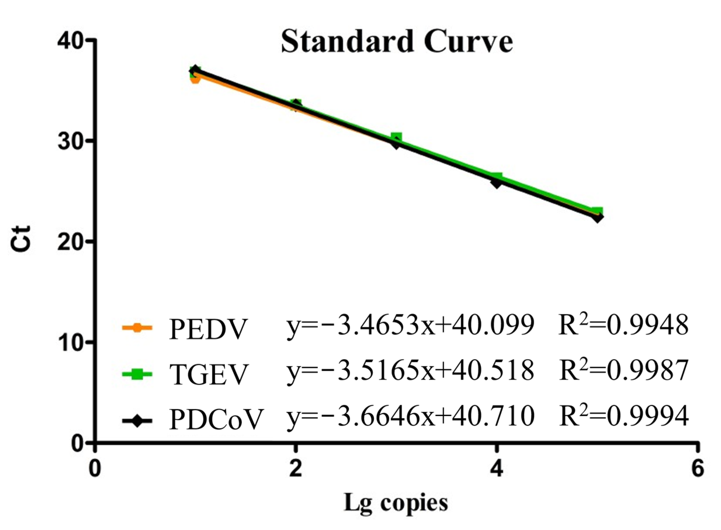
3.2. Standard Curves
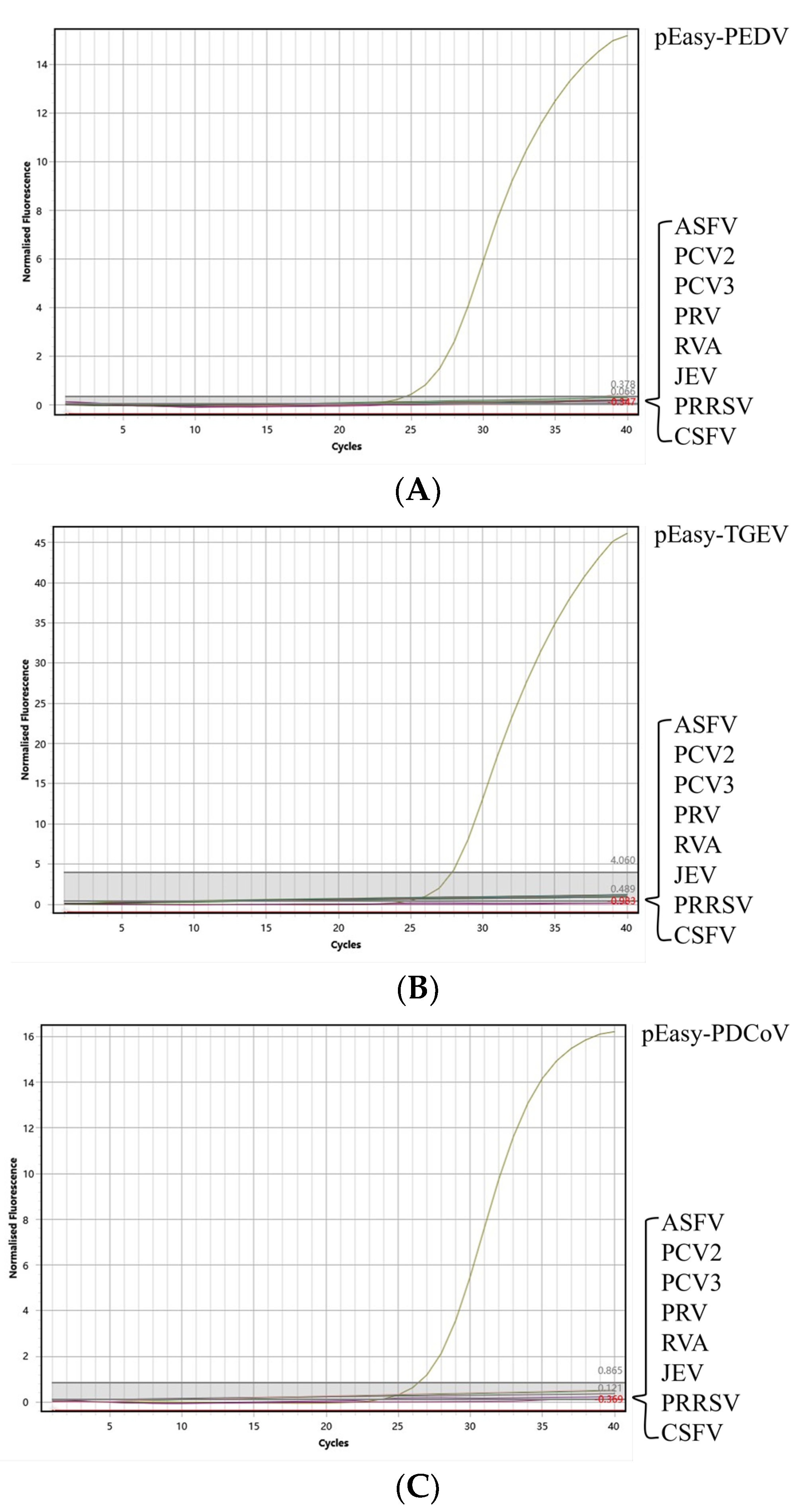
3.3. Specificity
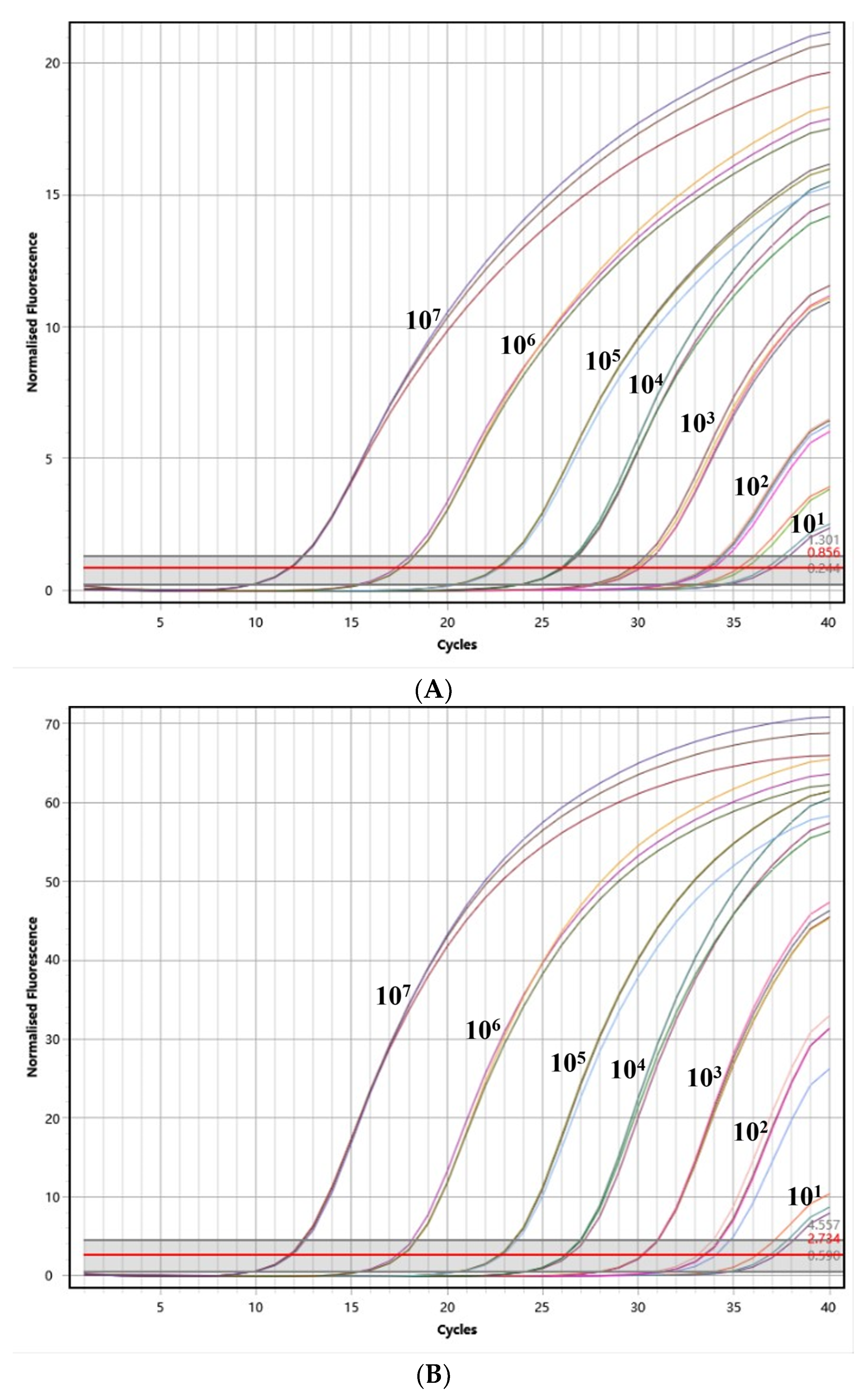
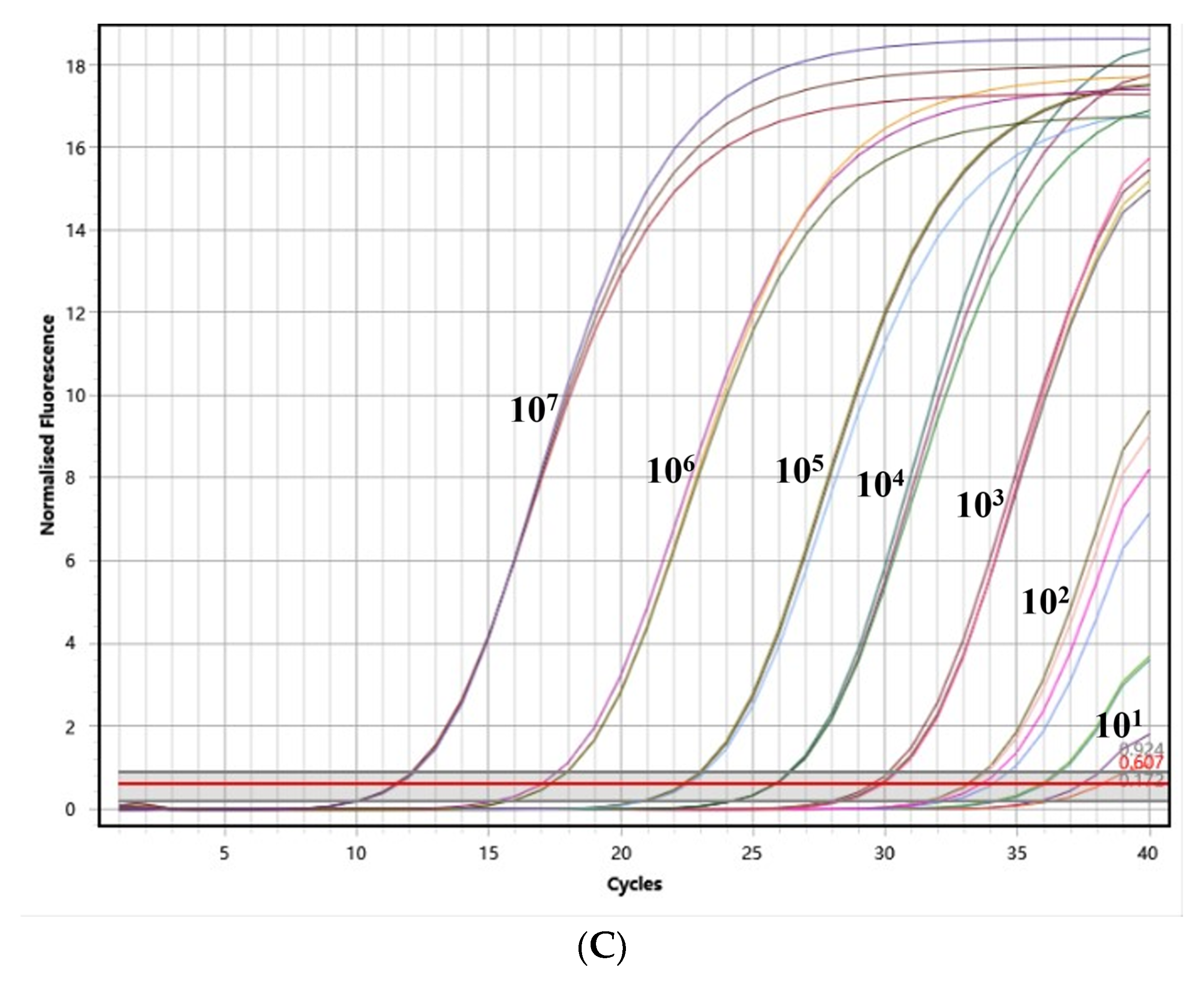
3.4. Sensitivity
3.5. Repeatability
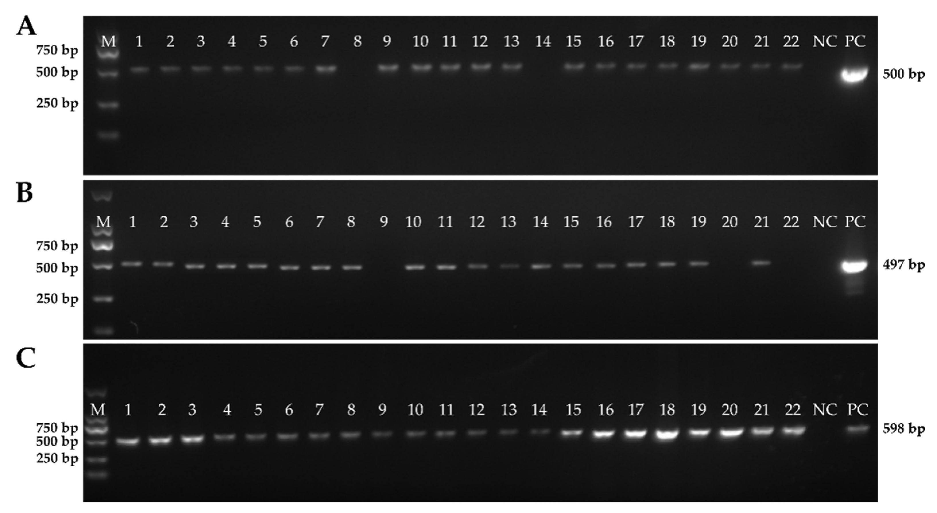
3.6. Detection of the Clinical Samples
4. Discussion
5. Conclusions
Supplementary Materials
Author Contributions
Funding
Institutional Review Board Statement
Informed Consent Statement
Data Availability Statement
Conflicts of Interest
References
- Zhang, J.; Tsai, Y.L.; Lee, P.Y.A.; Chen, Q.; Zhang, Y.; Chiang, C.J.; Shen, Y.H.; Li, F.C.; Chang, H.F.G.; Gauger, P.C.; et al. Evaluation of two singleplex reverse transcription-Insulated isothermal PCR tests and a duplex real-time RT-PCR test for the detection of porcine epidemic diarrhea virus and porcine deltacoronavirus. J. Virol. Methods 2016, 234, 34–42. [Google Scholar] [CrossRef] [PubMed]
- Liu, Q.; Wang, H.Y. Porcine enteric coronaviruses: An updated overview of the pathogenesis, prevalence, and diagnosis. Vet. Res. Commun. 2021, 45, 75–86. [Google Scholar] [CrossRef] [PubMed]
- Wang, D.; Fang, L.R.; Xiao, S.B. Porcine epidemic diarrhea in China. Virus Res. 2016, 226, 7–13. [Google Scholar] [CrossRef]
- Yang, Y.L.; Yu, J.Q.; Huang, Y.W. Swine enteric alphacoronavirus (swine acute diarrhea syndrome coronavirus): An update three years after its discovery. Virus Res. 2020, 285, 198024. [Google Scholar] [CrossRef] [PubMed]
- Kocherhans, R.; Bridgen, A.; Ackermann, M.; Tobler, K. Completion of the porcine epidemic diarrhoea coronavirus (PEDV) genome sequence. Virus Genes 2001, 23, 137–144. [Google Scholar] [CrossRef] [Green Version]
- Alonso, S.; Izeta, A.; Sola, I.; Enjuanes, L. Transcription regulatory sequences and mRNA expression levels in the coronavirus transmissible gastroenteritis virus. J. Virol. 2002, 76, 1293–1308. [Google Scholar] [CrossRef] [Green Version]
- Garwes, D.J. Transmissible gastroenteritis. Vet. Rec. 1988, 122, 462–463. [Google Scholar] [CrossRef]
- Zhang, J. Porcine deltacoronavirus: Overview of infection dynamics, diagnostic methods, prevalence and genetic evolution. Virus Res. 2016, 226, 71–84. [Google Scholar] [CrossRef]
- Lin, C.; Saif, L.J.; Marthaler, D.; Wang, Q. Evolution, antigenicity and pathogenicity of global porcine epidemic diarrhea virus strains. Virus Res. 2016, 226, 20–39. [Google Scholar] [CrossRef] [Green Version]
- Niederwerder, M.C.; Hesse, R.A. Swine enteric coronavirus disease: A review of 4years with porcine epidemic diarrhoea virus and porcine deltacoronavirus in the United States and Canada. Transbound. Emerg. Dis. 2018, 65, 660–675. [Google Scholar] [CrossRef] [Green Version]
- Li, C.; Lu, H.; Geng, C.; Yang, K.; Liu, W.; Liu, Z.; Yuan, F.; Gao, T.; Wang, S.; Wen, P.; et al. Epidemic and Evolutionary Characteristics of Swine Enteric Viruses in South-Central China from 2018 to 2021. Viruses 2022, 14, 1420. [Google Scholar] [CrossRef] [PubMed]
- Zhang, F.; Luo, S.; Gu, J.; Li, Z.; Li, K.; Yuan, W.; Ye, Y.; Li, H.; Ding, Z.; Song, D.; et al. Prevalence and phylogenetic analysis of porcine diarrhea associated viruses in southern China from 2012 to 2018. BMC VET RES 2019, 15, 470. [Google Scholar] [CrossRef] [PubMed] [Green Version]
- Shi, Y.; Li, B.; Tao, J.; Cheng, J.; Liu, H. The Complex Co-infections of Multiple Porcine Diarrhea Viruses in Local Area Based on the Luminex xTAG Multiplex Detection Method. Front. Vet. Sci. 2021, 8, 602866. [Google Scholar] [CrossRef] [PubMed]
- Kim, S.H.; Kim, I.J.; Pyo, H.M.; Tark, D.S.; Song, J.Y.; Hyun, B.H. Multiplex real-time RT-PCR for the simultaneous detection and quantification of transmissible gastroenteritis virus and porcine epidemic diarrhea virus. J. Virol. Methods 2007, 146, 172–177. [Google Scholar] [CrossRef]
- Masuda, T.; Tsuchiaka, S.; Ashiba, T.; Yamasato, H.; Fukunari, K.; Omatsu, T.; Furuya, T.; Shirai, J.; Mizutani, T.; Nagai, M.; et al. Development of one-step real-time reverse transcriptase-PCR-based assays for the rapid and simultaneous detection of four viruses causing porcine diarrhea. Jpn. J. Vet. Res. 2016, 64, 5–14. [Google Scholar]
- Pan, Z.; Lu, J.; Wang, N.; He, W.T.; Zhang, L.; Zhao, W.; Su, S. Development of a TaqMan-probe-based multiplex real-time PCR for the simultaneous detection of emerging and reemerging swine coronaviruses. Virulence 2020, 11, 707–718. [Google Scholar] [CrossRef]
- Wang, Y.; Xu, L.; Noll, L.; Stoy, C.; Porter, E.; Fu, J.; Feng, Y.; Peddireddi, L.; Liu, X.; Dodd, K.A.; et al. Development of a real-time PCR assay for detection of African swine fever virus with an endogenous internal control. Transbound. Emerg. Dis. 2020, 67, 2446–2454. [Google Scholar] [CrossRef]
- Huang, Y.L.; Pang, V.F.; Pan, C.H.; Chen, T.H.; Jong, M.H.; Huang, T.S.; Jeng, C.R. Development of a reverse transcription multiplex real-time PCR for the detection and genotyping of classical swine fever virus. J. Virol. Methods 2009, 160, 111–118. [Google Scholar] [CrossRef]
- Turlewicz-Podbielska, H.; Pomorska-Mól, M. Porcine Coronaviruses: Overview of the State of the Art. Virol. Sin. 2021, 36, 833–851. [Google Scholar] [CrossRef]
- Kim, O.; Choi, C.; Kim, B.; Chae, C. Detection and differentiation of porcine epidemic diarrhoea virus and transmissible gastroenteritis virus in clinical samples by multiplex RT-PCR. Vet. Rec. 2000, 146, 637–640. [Google Scholar] [CrossRef]
- Zhao, J.; Shi, B.J.; Huang, X.G.; Peng, M.Y.; Zhang, X.M.; He, D.N.; Pang, R.; Zhou, B.; Chen, P.Y. A multiplex RT-PCR assay for rapid and differential diagnosis of four porcine diarrhea associated viruses in field samples from pig farms in East China from 2010 to 2012. J. Virol. Methods 2013, 194, 107–112. [Google Scholar] [CrossRef]
- Zhao, Y.; Liu, F.; Li, Q.; Wu, M.; Lei, L.; Pan, Z. A multiplex RT-PCR assay for rapid and simultaneous detection of four RNA viruses in swine. J. Virol. Methods 2019, 269, 38–42. [Google Scholar] [CrossRef] [PubMed]
- Liu, G.; Jiang, Y.; Opriessnig, T.; Gu, K.; Zhang, H.; Yang, Z. Detection and differentiation of five diarrhea related pig viruses utilizing a multiplex PCR assay. J. Virol. Methods 2019, 263, 32–37. [Google Scholar] [CrossRef] [PubMed]
- Nan, P.; Wen, D.; Opriessnig, T.; Zhang, Q.; Yu, X.; Jiang, Y. Novel universal primer-pentaplex PCR assay based on chimeric primers for simultaneous detection of five common pig viruses associated with diarrhea. Mol. Cell. Probes 2021, 58, 101747. [Google Scholar] [CrossRef] [PubMed]
- Si, G.; Niu, J.; Zhou, X.; Xie, Y.; Chen, Z.; Li, G.; Chen, R.; He, D. Use of dual priming oligonucleotide system-based multiplex RT-PCR assay to detect five diarrhea viruses in pig herds in South China. AMB Express 2021, 11, 99. [Google Scholar] [CrossRef] [PubMed]
- Ding, G.; Fu, Y.; Li, B.; Chen, J.; Wang, J.; Yin, B.; Sha, W.; Liu, G. Development of a multiplex RT-PCR for the detection of major diarrhoeal viruses in pig herds in China. Transbound. Emerg. Dis. 2020, 67, 678–685. [Google Scholar] [CrossRef] [Green Version]
- Jia, S.; Feng, B.; Wang, Z.; Ma, Y.; Gao, X.; Jiang, Y.; Cui, W.; Qiao, X.; Tang, L.; Li, Y.; et al. Dual priming oligonucleotide (DPO)-based real-time RT-PCR assay for accurate differentiation of four major viruses causing porcine viral diarrhea. Mol. Cell. Probes 2019, 47, 101435. [Google Scholar] [CrossRef]
- Huang, X.; Chen, J.; Yao, G.; Guo, Q.; Wang, J.; Liu, G. A TaqMan-probe-based multiplex real-time RT-qPCR for simultaneous detection of porcine enteric coronaviruses. Appl. Microbiol. Biot. 2019, 103, 4943–4952. [Google Scholar] [CrossRef] [Green Version]
- Zhu, J.H.; Rawal, G.; Aljets, E.; Yim-Im, W.; Yang, Y.L.; Huang, Y.W.; Krueger, K.; Gauger, P.; Main, R.; Zhang, J. Development and Clinical Applications of a 5-Plex Real-Time RT-PCR for Swine Enteric Coronaviruses. Viruses 2022, 14, 1536. [Google Scholar] [CrossRef]
- Su, M.; Li, C.; Qi, S.; Yang, D.; Jiang, N.; Yin, B.; Guo, D.; Kong, F.; Yuan, D.; Feng, L.; et al. A molecular epidemiological investigation of PEDV in China: Characterization of co-infection and genetic diversity of S1-based genes. Transbound. Emerg. Dis. 2020, 67, 1129–1140. [Google Scholar] [CrossRef] [Green Version]


| Primers and probes | Sequences (5′ end to 3′ end) | Length (bp) | Use |
|---|---|---|---|
| PEDV-F | CCCGTTGATGAGGTGATTG | 500 | Amplification of M |
| PEDV-R | TTGGCGACTGTGACGAAAT | ||
| PEDV-qF | GACGCGCTTCTCACTACTTC | 134 | qPCR for the detection of M |
| PEDV-qR | TGTACGCCAGTAGCAACCTT | ||
| PEDV-probe | FAM-TGCAGACCTGTCGGCCCATCA-BHQ1 | ||
| TGEV-F | GTCAACCCATAGCCTCAA | 497 | Amplification of S |
| TGEV-R | GCCACTAAGTAGCGTCCT | ||
| TGEV-qF | ACATAGTGGGTGTACCGTCTG | 140 | qPCR for the detection of S |
| TGEV-qR | GCCACTAAGTAGCGTCCTGT | ||
| TGEV-probe | CY5-AGCACTGACAAATCGTGCACACCA-BHQ2 | ||
| PDCoV-F | TACTCATCCTCAGTTTCGTG | 598 | Amplification of N |
| PDCoV-R | ACCCGTCTTCTCAGTGTCT | ||
| PDCoV-qF | CAGTTTCGTGGCAATGGAGT | 79 | qPCR for the detection of N |
| PDCoV-qR | TGGTGTAACGCAGCCAGTAG | ||
| PDCoV-probe | HEX-CCGCTTAACTCCGCCATCAAACCCG-BHQ1 | ||
| ACTB-qF | CCCTGGAGAAGAGCTACGAG | 175 | qPCR for the detection of β-Actin |
| ACTB-qR | AGGTCCTTCCTGATGTCCAC | ||
| ACTB-probe | Texas Red-CGGCAACGAGCGCTTCCGGT-BHQ2 |
| Component | Volume (μL) |
|---|---|
| 2 × AceQ qPCR probe master mix | 10 |
| TGEV-qF/qR (10 μM) | 0.6 (0.3 μM) |
| TGEV-Probe (10 μM) | 0.3 (0.15 μM) |
| PEDV-qF/qR (10 μM) | 0.6 (0.3 μM) |
| PEDV-Probe (10 μM) | 0.3 (0.15 μM) |
| PDCoV-qF/qR (10 μM) | 0.6 (0.3 μM) |
| PDCoV-Probe (10 μM) | 0.3 (0.15 μM) |
| Template DNA | 2 |
| ddH2O | Up to 20 |
| Plasmids | Concentration (Virus copies/μL) | Intra-group | Inter-group | ||||
|---|---|---|---|---|---|---|---|
| Mean Ct | S.D. | CV (%) | Mean Ct | S.D. | CV (%) | ||
| PEDV | 102 | 32.355 | 0.189 | 0.6 | 32.759 | 0.286 | 0.8 |
| 104 | 25.208 | 0.042 | 0.2 | 25.386 | 0.242 | 0.9 | |
| 106 | 16.418 | 0.193 | 1.2 | 16.969 | 0.431 | 2.5 | |
| TGEV | 102 | 32.063 | 0.183 | 0.6 | 32.576 | 0.381 | 1.2 |
| 104 | 24.719 | 0.058 | 0.2 | 25.309 | 0.429 | 1.7 | |
| 106 | 16.421 | 0.167 | 1.0 | 17.047 | 0.452 | 2.7 | |
| PDCoV | 102 | 32.361 | 0.03 | 0.0009 | 32.526 | 0.133 | 0.004 |
| 104 | 25.176 | 0.005 | 0.0002 | 25.428 | 0.250 | 0.009 | |
| 106 | 17.279 | 0.093 | 0.005 | 17.16 | 0.085 | 0.004 | |
| Detection Method | Number of Positive Samples | ||
|---|---|---|---|
| PEDV | TGEV | PDCoV | |
| Multiplex qPCR | 268 | 75 | 210 |
| Reference methods | 258 | 71 | 201 |
| Agreements | 96.27% | 94.67% | 95.71% |
| Pathogens | Number of Positive Samples | Infection Rate (%) |
|---|---|---|
| PEDV | 91 | 19.70 |
| TGEV | 4 | 0.87 |
| PDCoV | 47 | 10.17 |
| PEDV/TGEV | 15 | 3.25 |
| PEDV/PDCoV | 107 | 23.16 |
| TGEV/PDCoV | 1 | 0.22 |
| PEDV/TGEV/PDCoV | 55 | 11.90 |
| β-Actin | 462 | 100 |
| In total | 462 | / |
| Province | Amount | Positive samples | Negative samples | ||
|---|---|---|---|---|---|
| PEDV | TGEV | PDCoV | |||
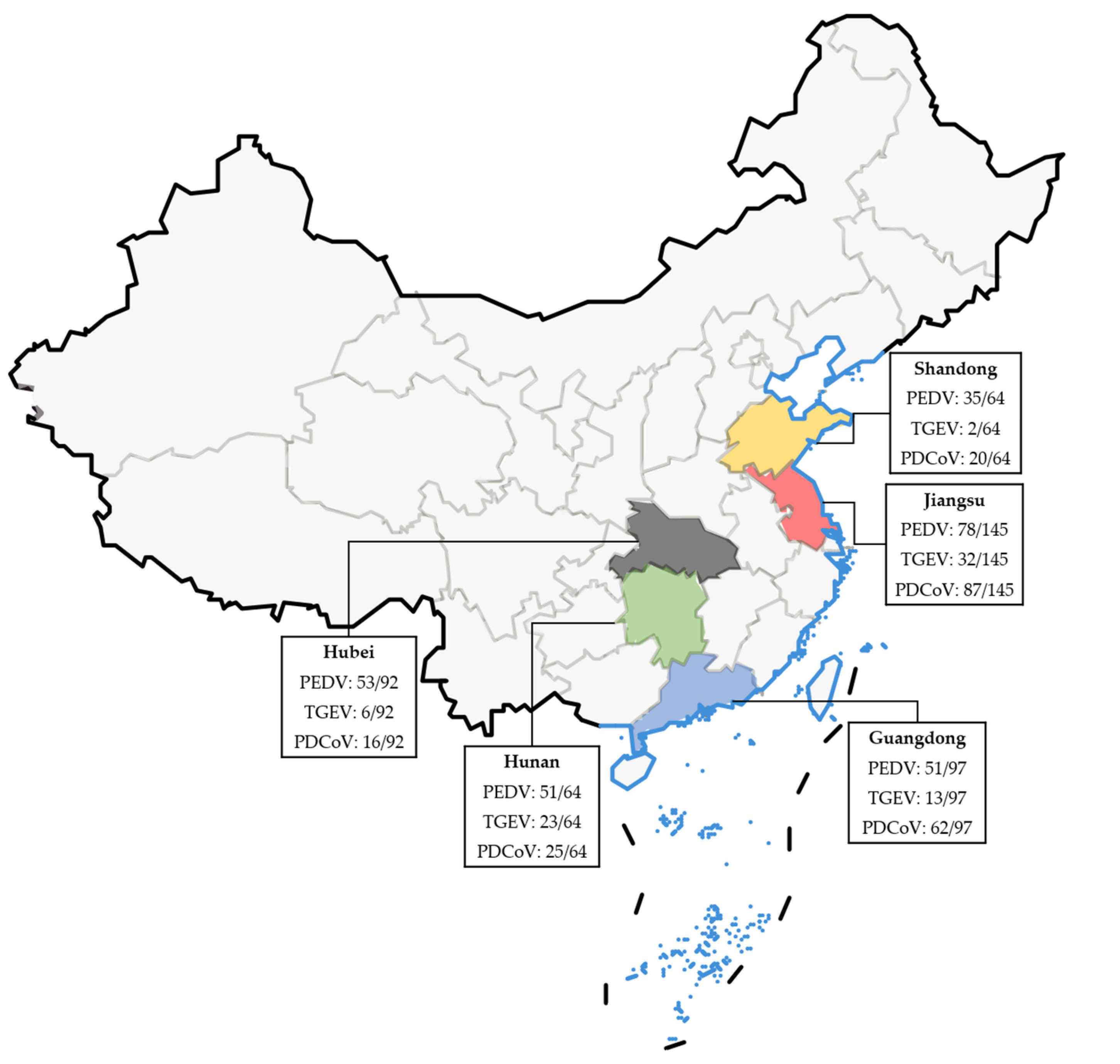
| Jiangsu | 145 | 78 | 32 | 87 | 52 |
| Guangdong | 97 | 51 | 13 | 62 | 25 |
| Hubei | 92 | 53 | 6 | 16 | 34 |
| Shandong | 64 | 35 | 2 | 20 | 28 |
| Hunan | 64 | 51 | 23 | 25 | 3 |
| Sum | 462 | 268 | 76 | 210 | 142 |
Disclaimer/Publisher’s Note: The statements, opinions and data contained in all publications are solely those of the individual author(s) and contributor(s) and not of MDPI and/or the editor(s). MDPI and/or the editor(s) disclaim responsibility for any injury to people or property resulting from any ideas, methods, instructions or products referred to in the content. |
© 2023 by the authors. Licensee MDPI, Basel, Switzerland. This article is an open access article distributed under the terms and conditions of the Creative Commons Attribution (CC BY) license (https://creativecommons.org/licenses/by/4.0/).
Share and Cite
Chen, J.; Liu, R.; Liu, H.; Chen, J.; Li, X.; Zhang, J.; Zhou, B. Development of a Multiplex Quantitative PCR for Detecting Porcine Epidemic Diarrhea Virus, Transmissible Gastroenteritis Virus, and Porcine Deltacoronavirus Simultaneously in China. Vet. Sci. 2023, 10, 402. https://doi.org/10.3390/vetsci10060402
Chen J, Liu R, Liu H, Chen J, Li X, Zhang J, Zhou B. Development of a Multiplex Quantitative PCR for Detecting Porcine Epidemic Diarrhea Virus, Transmissible Gastroenteritis Virus, and Porcine Deltacoronavirus Simultaneously in China. Veterinary Sciences. 2023; 10(6):402. https://doi.org/10.3390/vetsci10060402
Chicago/Turabian StyleChen, Jianpeng, Rongchao Liu, Huaicheng Liu, Jing Chen, Xiaohan Li, Jianfeng Zhang, and Bin Zhou. 2023. "Development of a Multiplex Quantitative PCR for Detecting Porcine Epidemic Diarrhea Virus, Transmissible Gastroenteritis Virus, and Porcine Deltacoronavirus Simultaneously in China" Veterinary Sciences 10, no. 6: 402. https://doi.org/10.3390/vetsci10060402