Article

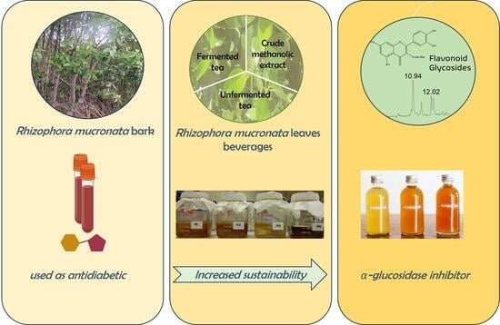
α-Glucosidase Inhibitory Activity of Tea and Kombucha from Rhizophora mucronata Leaves

by
Yunita Eka Puspitasari
1,2,3,*,
Emmy Tuenter
1,*,
Annelies Breynaert
1,
Kenn Foubert
1,
Herawati Herawati
2,
Anik Martinah Hariati
4,
Aulanni’am Aulanni’am
2,5,
Tess De Bruyne
1 and
Nina Hermans
1
1
Natural Products and Food Research & Analysis (NatuRA), University of Antwerp, 2610 Antwerp, Belgium
2
Doctoral Program of Environmental Studies, Universitas Brawijaya, Malang 65145, Indonesia
3
Department of Fisheries Processing Technology, Faculty of Fisheries and Marine Sciences, Universitas Brawijaya, Malang 65145, Indonesia
4
Department of Aquaculture, Faculty of Fisheries and Marine Sciences, Universitas Brawijaya, Malang 65145, Indonesia
5
Biochemistry Laboratory, Faculty of Sciences, Universitas Brawijaya, Malang 65145, Indonesia
*
Authors to whom correspondence should be addressed.
Beverages 2024, 10(1), 22; https://doi.org/10.3390/beverages10010022
Submission received: 22 February 2024 / Accepted: 13 March 2024 / Published: 15 March 2024
(This article belongs to the Topic Ways to Achieve Healthy and Sustainable Diets)

Abstract

:
A decoction of Rhizophora mucronata Lam. mangrove bark is used as an antidiabetic treatment in Asia. Kombucha tea is a fermented beverage, which is also claimed to be antidiabetic. In this work, the potency of R. mucronata leaves as α-glucosidase inhibitor was studied to assess whether it could be a suitable alternative to the use of R. mucronata bark. α-glucosidase inhibitory activities were determined for three extracts prepared from R. mucronata leaves, being the unfermented tea of R. mucronata leaves, the fermented kombucha tea and an 80% methanolic extract of the residual R. mucronata leaves. Flavonoid glycosides were identified in tea powder, kombucha tea and in the crude methanolic extract. Both the unfermented tea and the kombucha tea after 7 days of fermentation inhibited α-glucosidase with IC50 values of 0.12 ± 0.02 mg/mL and 0.09 ± 0.04 mg/mL, respectively. The methanolic extract showed a stronger α-glucosidase inhibitory activity compared to the kombucha tea and tea powder with an IC50 value of 0.0435 ± 0.0007 mg/mL. Acarbose, used as a positive control, inhibited α-glucosidase with an IC50 value of 2.4 ± 0.2 mg/mL. It was found that the three types of preparations of R. mucronata all were potent α-glucosidase inhibitors.

Graphical Abstract

1. Introduction

The use of mangrove plants, growing in tropical and subtropical coastal brackish water, as foods and folk medicines has risen due to increased scientific evidence regarding bioactive compounds, pharmacological studies, and food processing technology. The theaflavin content of herbal teas prepared from Rhizophora lamarckii, R. apiculata, R. mucronata, Bruguiera cylindrica and Ceriops decandra leaves, as an alternative raw material for tea instead of the traditionally used Camellia sinensis leaves, was investigated [1]. High levels of theaflavin were reported for R. mucronata (1.0%) and C. decandra (1.9%). Mangrove tea has been prepared from Rhizophora stylosa fruit by a local mangrove society in Indonesia since 2012 [2], as well as from R. mucronata fruit [3]. Herbal teas prepared from R. mucronata, R. apiculata, and R. annamalayana mangrove leaves, instead of C. sinensis, were studied for their antimicrobial activity against Escherichia coli and Salmonella typhi, as well as for their antioxidant properties (total antioxidant activity, total reducing power, and radical scavenging activity). The biological activity was attributed to the presence of phenols and flavonoids in the leaves [4].
Mangrove indeed is rich in polyphenols and known as a source of tannins [5]. Phytochemical investigations of R. mucronata leaves also led to the identification of several constituents such as α-amyrin, β-amyrin, oleanolic acid and betulin [6], ajmalicine, vindoline, catharanthine and serpentine [7], coumarins, xanthones, benzoic acid, and flavanones [8] and quinizarin [9].
Kombucha tea originates from China and is a fermented beverage, which has been gaining popularity over the past decade, not only in Asia but also in occidental countries, due to its potential health benefits. Nowadays, kombucha is available in Europe and the United States, and is marketed as a functional beverage, providing an alternative for carbonated soft drinks [10,11]. As the fermentation process proceeds, the kombucha taste changes from sweet into sour [12]. Kombucha tea is prepared from sweetened tea and a cellulosic pellicle, containing symbiotic acetic acid bacteria and yeasts (called SCOBY or kombucha), placed on top of the tea [13]. The sweetened tea can be made from black or green C. sinensis tea [14], or oolong C. sinensis tea [15]. Interestingly, kombucha tea from C. sinensis presents a wide range of biological activities, including antidiabetic properties [16,17,18]. Apart from black or green tea, other herbal sources have been fermented, such as coconut water [19], oak leaves (Quercus convallata and Q. arizonica) [20], and snake fruit (Salacca zalacca) juice [21].
Some commercial α-glucosidase inhibitors, including acarbose, miglitol and voglibose, are used as antidiabetic drugs, but also show adverse effects, such as flatulence and abdominal discomfort [22,23]. Fermented beverages, like kombucha, could be interesting alternatives in the management of diabetes. Indeed, administration of snake fruit (Salacca zalacca) kombucha to diabetic rats for 28 days decreased their fasting plasma glucose levels by 31–59% [24], and starch hydrolase activity decreased after 7 days administration of coconut water kombucha, indicating antidiabetic properties for both preparations [19]. In addition, kombucha tea prepared from black tea showed a higher hypoglycaemic and hypolipidemic activity (by suppression of pancreatic α-amylase activity and delaying absorption of LDL-cholesterol and triglycerides and increasing HDL-cholesterol significantly), compared to black tea itself [16]. Moreover, blood glucose levels of diabetic rats treated with R. mucronata leaves were decreased [25,26,27]. Furthermore, R. mucronata bark, applied in traditional medicine in the Philippines and Vietnam to treat diabetes, was assessed for its α-glucosidase inhibiting property. Its aqueous and ethanolic extracts significantly inhibited α-glucosidase with IC50 values of 3.3 ± 0.6 µg/mL and 0.08 ± 1.82 µg/mL, respectively [28,29]. Antidiabetic properties by α-glucosidase inhibition of herbal tea and of kombucha tea made from R. mucronata fruit were reported, with the fermented kombucha tea showing a stronger inhibition than the herbal tea [3].
The mangrove ecosystem provides herbal medicine and food for human consumption. However, unfortunately, mangrove plants, including R. mucronata are able to absorb and accumulate heavy metals, depending on the plant species, age and exposure time [30]. Heavy metals, including As, Cd, and Pb, were detected in tea leaves and tea infusions collected from different countries, but the levels were still below the WHO (World Health Organization) limit for herbal medicines (2007). Consuming tea prepared from R. mucronata leaves may thus provide health benefits, but the presence of toxic compounds or heavy metals at the same time poses a health risk. In order to take this aspect into account, the heavy metal content (Pb, Hg and Cd) was analyzed in the course of this study, as recommended by WHO [31].
R. mucronata, among other Asian mangrove plants, has been utilized as a folk medicine and its decoction is used in a variety of ailments, including diabetes [26,28,29,32,33,34,35]. In addition, the fermented tea prepared from R. mucronata fruit showed stronger antidiabetes activity than the herbal tea made from the same fruit [3].
Several studies already proved the potency of R. mucronata bark to inhibit α-glucosidase activity [28,29], which is one of the human gastrointestinal enzymes involved in the breakdown of dietary starch into glucose, which increases the blood glucose level. However, extensive consumption of R. mucronata bark leads to deforestation, mangrove degradation and disruption of the marine environment. Twenty percent of the world’s mangrove areas are found in Indonesia, possibly making it the most important mangrove habitat in the world. However, these areas have been decreasing over the last six centuries leading to the loss of more than 70% of mangrove area [36]. Thus, the search for more sustainable alternatives which inhibit α-glucosidase activity and which can serve as an antidiabetic health product is desirable [37,38]. Therefore, the purpose of this study is to investigate the phytochemical composition and the antidiabetic potential of unfermented and fermented tea, prepared from leaves of R. mucronata.

2. Materials and Methods

2.1. Chemicals

Solvents including methanol (≥99.8%) and acetonitrile (≥99.8%) were purchased from Acros Organics (Geel, Belgium). Formic acid (98%) and dimethylsulfoxide (DMSO) were obtained from Acros Organics (Geel, Belgium). Water was dispensed by a Milli-Q system (Rephile) and was passed through a 0.2 μm membrane filter before use. Reference standards including benzoic acid, p-hydroxybenzoic acid, salicylic acid, coumarin, cinnamic acid, protocatechuic acid, p-coumaric acid, vanillic acid, gallic acid, caffeic acid, ferulic acid, syringic acid, sinapic acid, apigenin, emodin, naringenin, luteolin, catechin, epicatechin, quercetin, taxifolin, isorhamnetin, chlorogenic acid, stigmasterol, quercitrin, procyanidin B2, rutin, and tannic acid were purchased from Extrasynthese (Lyon, France), Sigma-Aldrich (Bornem, Belgium), Santa Cruz Biotechnology (Heidelberg, Germany), or Carl Roth (Karlsruhe, Germany). Acarbose (≥95%), α-glucosidase, and p-nitrophenyl-α-D-glucopyranoside were purchased from Sigma-Aldrich (Steinheim, Germany); transparent 96 well plates (Nunc) were from Thermo Scientific (Reskide, Denmark) and Na2HPO4·2H2O from Merck (Darmstadt, Germany). The SCOBY (art.nr 905, batch number 58706) was purchased from Wellness drinks (Frankfurt am Main, Germany).

2.2. Plant Material

Fresh R. mucronata leaves of the first, second and third levels from shoots were collected in the village of Penunggul, Pasuruan, Indonesia in September 2016. They were identified and authenticated taxonomically at the Herbarium Bogoriensis, Research Center for Biology-Indonesian Institute of Sciences, where the sample (Np. 248/IPH.1.02/If.07/XII/2016) was deposited. The leaves were dried under the sun (±24 °C for 5 days), resulting in a moisture content of 9.9%. Then, they were grinded with a disk mill and sieved with a sieve with mesh size 0.3–0.5 mm.

2.3. Preparation of Unfermented Tea Powder

Dried R. mucronata leaves (1.2 kg) were extracted by decoction (steeping in 12 L hot water (90 °C for 20 min)), analogous to the traditional preparation of R. mucronata bark. After centrifugation at 3000 rpm for 5 min and filtration, the filtrate was lyophilized to obtain tea powder, and it was stored at 4 °C. The residue was used to prepare the methanolic extract.

2.4. Preparation of Kombucha Tea

Fermentation of R. mucronata tea powder was conducted based on Watawana et al. [19] with slight modifications. The unfermented tea powder (9.36 g) was suspended in 3.6 L boiling hot water for 15 min in a water bath. Sucrose 10% (w/v) was added; the tea was stirred till complete dissolution of the sugar, and was then chilled to a temperature of 25 ± 3 °C. Once the sweetened tea had been chilled, a kombucha pellicle (SCOBY) was placed on the surface of the tea for fermentation. Four samples were prepared in parallel in this fermentation experiment: sample 1 (ST1) and sample 2 (ST2) (experiment in duplicate) containing tea and pellicle; a blank sample (BS), containing water (instead of tea) and pellicle, and a negative control (NC) containing tea, but no pellicle. In total, 12 jars were prepared; 3 of each of the four different samples, to allow assessment of the samples after three different fermentation durations (0, 7 or 14 days). The jars were covered with cheese cloth, which was tightly fastened with an elastic band. Fermentation was carried out in an incubator with the temperature maintained at 25 ± 2 °C. Samples were taken on days 0, 7 and 14. After fermentation, the kombucha pellicle and tea were separated for further analysis by taking the pellicle from the jar. All samples were stored at −80 °C.

2.5. Preparation of the Methanolic Extract

The residue of R. mucronata leaves (502 g) was macerated with 13 L 80% methanol. Maceration was conducted in ambient conditions in the lab, without agitation, and at room temperature. The ratio of maceration was 1:100 (m/v). The macerate was filtered and the filtrate was evaporated under reduced pressure to obtain the crude extract which was subsequently freeze dried, yielding 107 g of extract. Concentration of the macerate was performed using an IKA Vacuubrand rotary evaporator (VWR-Avantor, Leuven, Belgium) and lyphilisation was carried out by a Lyovapor L-200 (Buchi, Hendrik-Ido-Ambacht, The Netherlands). The 80% methanolic extract was stored at 4 °C.

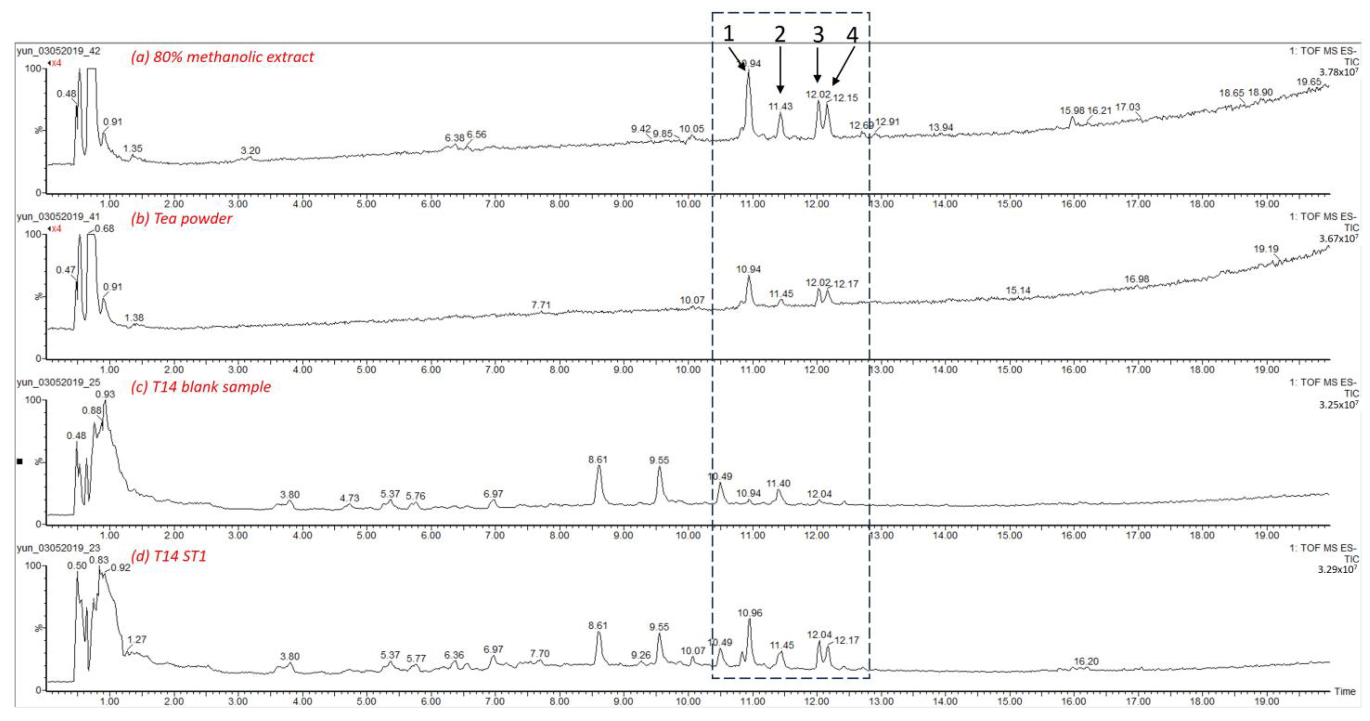
2.6. UPLC-HRMS Analysis

Ultra Performance Liquid Chromatography—High Resolution Mass Spectrometry analysis of unfermented tea, kombucha tea and the methanolic extract of R. mucronata leaves was performed on an Acquity UPLC system coupled to a XEVO G2-XS QTOF mass spectrometer (Waters, Milford, MA, USA). Samples for LC-MS analysis were prepared by dissolution of dried tea powder and methanolic extract in 80% MeOH at a concentration of 1 mg/mL. As for the fermented tea samples, 0.5 mL of the kombucha tea was diluted with MeOH (1:2), then centrifuged at 14,000 rpm for 10 min and at 4 °C, and subsequently the supernatant was diluted twice.
Separation was achieved on a BEH Shield RP18 1.7µm column (2.1 × 100 mm, Waters, Milford, MA, USA). The mobile phase was composed of H2O + 0.1% formic acid (FA) (A) and ACN + 0.1% FA (B) and the gradient was set as follows: 2% B (0–1 min), 26% B (14 min), 65% B (24 min), 100% B (26–29 min), and 2% B (31–36 min). The flow rate was 0.4 mL/min. Mass spectra were recorded in MSE mode in both ESI+ and ESI- with a scan range from m/z 50 to 1500 with the capillary voltage set at 1.0 kV for the positive and 0.8 kV for the negative ionisation mode. Other parameters were set as follows: cone voltage 40 V, cone gas (N2) flow 50 L/h, desolvation gas (N2) flow 1000 L/h, source temperature 120 °C, and desolvation temperature 550 °C. MassLynx 4.1 (Waters, Milford, MA, USA) was used for controlling the instrument and for subsequent data analysis.
The samples were injected, along with a mix of 28 reference compounds. Identification of compounds was carried out by comparison with experimental data obtained for the reference compounds (retention time, m/z value, fragmentation pattern) and by comparison with databases such as MassBank, ReSpect, MetFrag, and ChEBI and reports in the literature.

2.7. α-Glucosidase Inhibition Assay

The in vitro α-glucosidase inhibition assay was performed in 96-well plates. Phosphate buffer 0.1 M was prepared with Na2HPO4.2H2O, and the pH was adjusted to 6.8 with NaOH (1 M). The tea powder, methanolic extract and Kombucha tea samples were prepared in phosphate buffer which contained 6% DMSO, at concentrations ranging between 0.03 and 0.20 mg/mL. For tea powder and methanolic extract, the dried material was reconstituted in the phosphate buffer with 6% DMSO, while Kombucha teas were diluted to reach the mentioned concentration test range, as deduced from the initial Kombucha tea concentration (dilution factors ranging from 10× to 100×). The test sample (50 µL) was incubated with 100 µL α-glucosidase (0.2 U/mL) in phosphate buffer (Sigma Aldrich, Steinheim, Germany) at 37 °C for 15 min. Then, 50 µL substrate (p-nitrophenyl α-D-glucopyranoside) (PNPG, 5 mM stock) was added into the solution. After 30 min incubation at 37 °C, the hydrolysis of PNPG to release p-nitrophenol (PNP) was monitored at 405 nm, with a Bio Tek Eon microplate reader (BIO-TEK, Synergy HT, Vermont, USA) with Gen 5 version 2.06 software. The blank sample was prepared with phosphate buffer instead of α-glucosidase. Acarbose was used as positive control, and all experiments were performed in triplicate. IC50 values were calculated using GraphPad Prism version 6 software. They were expressed as mean ± SD of triplicate measurements.

2.8. Heavy Metal Analysis

Heavy metal content of dried leaves and unfermented tea powder was analyzed by the Standard National Indonesia (SNI) method SNI 19-2896-1998: Testing for metal contamination in food. The analysis was carried out using a Thermo Scientific Atomic Absorption Spectrophotometer (AAS) iCE 3000 (Kyoto, Japan). The elements Pb, Cd and Hg were analyzed.

3. Results

3.1. Identification of Phytochemicals by UPLC-HRMS

UPLC-HRMS analysis was conducted to (tentatively) identify and compare the major compounds present in the unfermented tea powder, kombucha tea, and methanolic extract. An overview of the identified compounds and their chromatographic and spectroscopic data is provided in Table 1 and Figure 1. The results show that four compounds were identified in all samples. Compound 1 was identified in ESI- mode as rutin (quercetin-3-O-rutinoside) by comparison with the reference compound. It was detected at a retention time of 10.95 min and showed a molecular ion with m/z 609.1438 ([M-H]). Rutin has previously been found in R. mucronata leaves, as reported by Gurudeeban et al. [39].
Compounds 2, 3 and 4 were identified as flavonoid O-glycosides, as indicated by neutral losses of 146, 162, and 308 Da, which represent a deoxyhexoside, hexoside and deoxyhexose-hexoside moiety, respectively [40]. Compound 2 showed an [M-H] ion at retention time 11.45 min with m/z 447.0930 and was identified as a tetrahydroxyflavone-O-hexoside. Its fragment ion [M-H-162] at m/z 285 corresponded to the tetrahydroxyflavone aglycone, after loss of a hexosyl moiety (162 Da) [41]. According to the literature, both luteolin and kaempferol aglycons were reported in R. mucronata [39], and the exact identification based on LC-MS analysis alone was not possible.
Compounds 3 and 4 showed monoisotopic ions at m/z 593.1494 and 623.1584 ([M-H]), respectively, while in the fragmentation spectrum, signals were found at m/z 285 and 315, corresponding to the respective aglycone moieties of these compounds. These fragment ions were formed after the neutral loss of a moiety with a size of 308 Da, which could correspond to a deoxyhexose-hexose disaccharide moiety [40], possibly a rutinoside moiety [42]. Another MS/MS fragment was observed at m/z 447 [M-H-146]- corresponding to the loss of a deoxyhexose moiety. However, the exact identification of these sugar moieties could not be performed by LC-MS analysis alone. Compound 3 was thus proposed to be tetrahydroxyflavone-O-deoxyhexose-O-hexoside. Compound 4 was tentatively identified as tetrahydroxy-methoxyflavone-O-deoxyhexose-O-hexoside. The fragment ion at m/z 315 indicated a tetrahydroxy-methoxyflavone aglycone moiety [43]. The four tentatively identified compounds were present in unfermented and fermented tea, as well as in the methanolic extract.

3.2. Effect of Kombucha Tea on α-Glucosidase Activity

The inhibitory activity of tea powder, kombucha tea and 80% methanolic extract on α-glucosidase is shown in Table 2. The IC50 values for the tea powder and the methanolic extract were 0.12 ± 0.02 mg/mL and 0.0435 ± 0.0007 mg/mL, respectively, thus showing a much stronger potency against the activity of α-glucosidase than acarbose (IC50 2.4 ± 0.2 mg/mL). Methanolic extract is more effective in inhibiting α-glucosidase compared to the tea powder and kombucha tea prepared from R. mucronata leaves. For kombucha tea, α-glucosidase inhibitory activity did not change with longer fermentation time. In contrast to an earlier finding by [19], this study did not demonstrate that in kombucha tea, prepared from R. mucronata leaves by fermentation, the α-glucosidase inhibitory activity increases [19].

3.3. Heavy Metal Analysis

In this study, non-essential heavy metal concentrations were determined in mangrove leaves and tea powder by means of AAS. The heavy metal content of both leaves and unfermented tea are given in Table 3. WHO guidelines on assessing quality of herbal medicines recommended limits of heavy metals for lead ≤10 mg/kg and for cadmium ≤0.3 mg/kg [31]. The monograph for herbal drugs of the European Pharmacopoeia [44] mentioned a limit of 0.1 mg/kg mercury in herbal drugs. The measured concentrations of heavy metals in leaves and unfermented tea did not exceed the limits given in both sources (Table 3). The leaves exhibited a higher Pb, Cd and Hg content than the tea powder. These results are consistent with a previous study, which revealed the reduction of heavy metals in food products during the cooking process, which depended on the cooking conditions (time, temperature and cooking medium) [45].

4. Discussion

Medicinal plants used in the treatment of diabetes mellitus can act via different mechanisms. In this work, the α-glucosidase inhibitory potency of three different preparations of R. mucronata leaves, including unfermented tea powder, a fermented kombucha tea and a methanolic extract, were evaluated. A previous study reported that king coconut water (Cocos nucifera) kombucha, after 7 days of fermentation, inhibited α-glucosidase activity more prominently than prior to fermentation [19]. The fermentation process is also known to modify the secondary metabolite profile. Previous studies conducted by [17] reported that the total phenolic and flavonoid content of kombucha tea prepared from black tea increased after fermentation. It was hypothesized that the complex polyphenols are metabolized into smaller molecules, thus increasing total phenolic and flavonoid content [17]. These results are in line with a study demonstrating that enzymes secreted by microorganisms are involved in the degradation of (-)-epigallocatechin gallate (EGCG) to (-)-epigallocatechin (EGC) and of (-)-epicatechin gallate (ECG) to (-)-epicatechin (EC) during kombucha fermentation [46]. However, the results of the current study were not in agreement with this, since fermentation did not affect the α-glucosidase inhibitory activity of the tea prepared from R. mucronata leaves. On the other hand, this result is in line with those reported by [20] who stated that both an infusion of oak leaves and the fermented leaves inhibited α-glucosidase activity by 98–99% and the activity was related to the presence of natural antidiabetic compounds. According to the authors, the levels of phenolic compounds, such as gallic acid, quercetin and catechin, were not altered by fermentation and no significant difference in total phenolic content between the infusion and the fermented oak leaves was found.
Both bark and root of R. mucronata are boiled and consumed as a traditional natural remedy against diabetes in various countries. Mangrove is exploited extensively for human use, which thereby poses a threat to mangrove ecosystems. An important drawback of the utilization of bark and root is the risk of deforestation, while the restoration of mangrove can only be accomplished by planting [38]. As previously reported, the ethanolic extract of R. mucronata bark suppressed α-glucosidase activity with an IC50 value of 126.6 ± 18.2 µg/mL [23]. The results obtained here for leaf extracts of R. mucronata are in line with this previous study, as it was found that both unfermented tea powder (IC50 0.12 ± 0.02 mg/mL) and the 80% MeOH extract (IC50 0.0435 ± 0.0007 mg/mL) have potency against α-glucosidase. Therefore, the in vitro biological activity of mangrove leaves as α-glucosidase inhibitor was demonstrated, which could be a therapeutic approach for decreasing postprandial hyperglycemia by retarding the absorption of glucose. The substitution of R. mucronata bark by leaves could thus provide a more sustainable alternative for the use of mangrove in the treatment of diabetes. In this investigation, the preparation of kombucha from R. mucronate leaves and its α-glucosidase inhibiting activity have been described for the first time.
With regard to the phytochemical composition of R. mucronata leaves, four flavonoids have been tentatively identified in kombucha tea, tea powder and the 80% MeOH crude extract, namely rutin, tetrahydroxyflavone-O-hexoside, tetrahydroxyflavone-O-deoxyhexose-O-hexoside, and tetrahydroxy-methoxyflavone-O-deoxyhexose-O-hexoside. The aglycones of these compounds all belong to the group of flavones. Several flavonoids, including rutin, isorhamnetin, quercetin, myricetin, kaempferol, and luteolin were previously identified in R. apiculata leaves [47], and isorhamnetin-3-O-glucoside was identified in R. racemosa [48]. A previous study determined that the bark and root of R. mangle primarily contained tannins, while flavonoids are the most common secondary metabolites of leaves [49]. Furthermore, rutin, kaempferol and luteolin were previously identified in R. mucronata leaves [39], while Vittaya et al. carried out a qualitative comparison of the phytochemical composition of R. mucronata leaf, bark, pods, and twigs [50].
Flavonoid glycosides, isolated from several plants such as Scabiosa prolifera, Melalleuca leucadendra, Polygonatum verticillatum, Sabia parviflora and Psychotria luzoniensis, showed various biological activities, including antioxidant activity, immunostimulant activity and cytotoxic activity [51,52,53,54,55]. Rutin, kaempferol, and isorhamnetin were previously shown to display a more prominent α-glucosidase inhibition compared to acarbose [56,57]. The α-glucosidase inhibitory IC50 value for kaempferol was 12 µM [57]. The flavonoid structure influences the α-glucosidase inhibitory activity: flavonoids with a 3-OH group proved to be more effective α-glucosidase inhibitors [56,57]. In addition, the α-glucosidase activity is Increased when an unsaturated C ring and a hydroxyl moiety in the B ring are present [56,57,58,59]. Furthermore, the presence of a hydroxyl group in position 5 (of the flavonoid A ring) enhanced the inhibitory activity [56]. Flavonol glycosides that were screened as α-glucosidase inhibitors include rutin, isolated from Mallotus furetianus tea, which inhibited α-glucosidase activity with an IC50 value of 188.07 µg/mL [60]. Also, Acer buergerianum and Acer truncatum leaves, containing kaempferol, inhibited α-glucosidase activity with IC50 values of 2.12 µg/mL and 1.35 µg/mL, respectively, which were lower than acarbose (IC50 584.29 µg/mL) [61]. Thus, the presence of flavonoid glycosides in the leaves of R. mucronata may in part be responsible for the observed α-glucosidase inhibitory activity. The phytochemical compounds of R. mucronata leaves have potential as natural nutraceuticals to prevent diabetes mellitus because of their high α-glucosidase inhibitory activity.
Food safety is one of the essential matters to be considered in the development of (functional) foods and herbal medicines. Heavy metals, including Cd, Hg and Pb, could be introduced into herbal drugs through a contaminated environment (water, soil). Since heavy metals (including Pb, Hg, Cd) are known to accumulate in R. mucronata, because the plant grows in the mangrove ecosystem [62], it is of particular importance to monitor its heavy metal content. The WHO proposed heavy metal limits of 10 mg/kg for Pb and 0.3 mg/kg for Cd in herbal medicines [31]. In several countries, such as China and Singapore, the maximum allowed concentration of Hg is 0.5 ppm in herbal medicines [31,63]. Several pharmacopoeias established heavy metal limits for herbal drugs, including the European Pharmacopoeia. According to the European Pharmacopoeia monograph for herbal drugs, limits for lead, cadmium and mercury are 5.0 mg/kg, 0.5 mg/kg, and 0.1 mg/kg, respectively. In addition, the European Commission established limits for heavy metals in food supplements as defined by Commission Regulation (EC) No.1881/2006 and the following limits have been set: Pb 3.0 mg/kg, Cd 1.0 mg/kg and Hg 0.10 mg/kg [64]. Based on the heavy metal analysis carried out in the current work, Pb, Hg and Cd concentrations in leaves and unfermented tea were found to be below the limits proposed by the WHO, EC 1881/2006 and Ph. Eur monograph for herbal drugs. Therefore, R. mucronata leaves, harvested from Penunggul, East Java, Indonesia and its unfermented tea are found to be safe for consumption.

5. Conclusions

The phytochemical profiling of R. mucronata leaves led to the tentative identification of four flavonoid glycosides. The α-glucosidase inhibitory activity of the unfermented tea powder and kombucha tea, as well as the 80% methanolic extract of R. mucronata leaves, is reported here for the first time. The current study showed that R. mucronata leaves are a promising marine natural product, which can serve as an alternative source of α-glucosidase inhibitors to manage blood glucose levels. Given the similar activity compared to R. mucronata root and bark, leaves may constitute an interesting alternative in the treatment of diabetes. In this way, deforestation can be limited, and a sustainable use of mangrove ecosystems becomes feasible. Thus, these findings can contribute to the development of healthy beverages and functional foods based on mangrove resources.

Author Contributions

Conceptualization, Y.E.P., E.T., K.F., A.B., T.D.B. and N.H.; methodology, Y.E.P. and E.T.; formal analysis, Y.E.P.; investigation, Y.E.P.; resources, Y.E.P.; data curation, Y.E.P., K.F., A.B. and E.T.; writing—original draft preparation, Y.E.P.; writing—review and editing, Y.E.P., E.T., K.F., A.B., T.D.B., A.A., A.M.H. and N.H.; visualization, Y.E.P.; supervision, N.H., T.D.B., A.A., A.M.H. and H.H.; project administration, N.H., T.D.B. and A.A.; funding acquisition, Y.E.P. All authors have read and agreed to the published version of the manuscript.

Funding

This research was funded by the Ministry of Finance, the Indonesia Endowment Fund for Education (LPDP) and the Indonesian Ministry of Research, Technology and Higher Education (RISTEKDIKTI) through a doctoral scholarship (PRJ-4270/LPDP.3/2016) for Y.E.P.

Data Availability Statement

Data are contained within the article.

Conflicts of Interest

The authors declare no conflicts of interest.

References

  1. Kathiresan, K. Studies on tea from mangrove leaves. Environ. Ecol. 1995, 13, 321–323. [Google Scholar]
  2. Miranti, D.I.; Ichiura, H.; Ohtani, Y. The Bioactive Compounds and Antioxidant Activity of Food Products of Rhizophora stylosa Fruit (Coffee and Tea Mangrove). Int. J. For. Res. 2018, 2018, 2315329. [Google Scholar] [CrossRef]
  3. Harisman, E.K.; Puspitasari, Y.E. The kombucha from Rhizophora mucronata Lam herbal tea: Characteristics and the potential as an antidiabetic beverage. J. Pharm. Pharm. Res. 2020, 8, 410–421. [Google Scholar]
  4. Arulkumar, A.; Kumar, K.S.; Paramasivam, S. Antibacterial and invitro antioxidant potential of Indian mangroves. Biocatal. Agric. Biotechnol. 2020, 23, 101491. [Google Scholar] [CrossRef]
  5. Bibi, S.N.; Fawzi, M.M.; Gokhan, Z.; Rajesh, J.; Nadeem, N.; Rengasamy Kannan, R.R.; Albuquerque, R.D.D.G.; Pandian, S.K. Ethnopharmacology, Phytochemistry, and Global Distribution of Mangroves-A Comprehensive Review. Mar. Drugs 2019, 17, 231. [Google Scholar] [CrossRef] [PubMed]
  6. Ghosh, A.; Misra, S.; Dutta, A.K.; Choudhury, A. Pentacyclic triterpenoids and sterols from seven species of mangrove. Phytochemistry 1985, 24, 1725–1727. [Google Scholar] [CrossRef]
  7. Gurudeeban, S.; Ramanathan, T.; Satyavani, K. Antimicrobial and Radical Scavenging Effects of Alkaloid Extracts from Rhizophora mucronata. Pharm. Chem. J. 2015, 49, 34–37. [Google Scholar] [CrossRef]
  8. Taniguchi, K.; Funasaki, M.; Kishida, A.; Sadhu, S.K.; Ahmed, F.; Ishibashi, M.; Ohsaki, A. Two new coumarins and a new xanthone from the leaves of Rhizophora mucronata. Bioorg. Med. Chem. Lett. 2018, 28, 1063–1066. [Google Scholar] [CrossRef] [PubMed]
  9. Sachithanandam, V.; Lalitha, P.; Parthiban, A.; Muthukumaran, J.; Jain, M.; Misra, R.; Mageswaran, T.; Sridhar, R.; Purvaja, R.; Ramesh, R. A comprehensive in silico and in vitro studies on quinizarin: A promising phytochemical derived from Rhizophora mucronata Lam. J. Biomol. Struct. Dyn. 2022, 40, 7218–7229. [Google Scholar] [CrossRef] [PubMed]
  10. Kapp, J.M.; Sumner, W. Kombucha: A systematic review of the empirical evidence of human health benefit. Ann. Epidemiol. 2019, 30, 66–70. [Google Scholar] [CrossRef]
  11. Kim, J.; Adhikari, K. Current trends in kombucha: Marketing perspectives and the need for improved sensory research. Beverages 2020, 6, 15. [Google Scholar] [CrossRef]
  12. Dufresne, C.; Farnworth, E. Tea, Kombucha, and health: A review. Food Res. Int. 2000, 33, 409–421. [Google Scholar] [CrossRef]
  13. Reiss, J. Influence of different sugars on the metabolism of the tea fungus. Z. Lebensm. Unters. Forsch. 1994, 198, 258–261. [Google Scholar] [CrossRef]
  14. Kallel, L.; Desseaux, V.; Hamdi, M.; Stocker, P.; Ajandouz, E.H. Insights into the fermentation biochemistry of Kombucha teas and potential impacts of Kombucha drinking on starch digestion. Food Res. Int. 2012, 49, 226–232. [Google Scholar] [CrossRef]
  15. Koh, L.W.; Wong, L.L.; Loo, Y.Y.; Kasapis, S.; Huang, D. Evaluation of different teas against starch digestibility by mammalian glycosidases. J. Agric. Food Chem. 2010, 58, 148–154. [Google Scholar] [CrossRef]
  16. Aloulou, A.; Hamden, K.; Elloumi, D.; Ali, M.B.; Hargafi, K.; Jaouadi, B.; Ayadi, F.; Elfeki, A.; Ammar, E. Hypoglycemic and antilipidemic properties of kombucha tea in alloxan-induced diabetic rats. BMC Complement. Altern. Med. 2012, 12, 63. [Google Scholar] [CrossRef]
  17. Bhattacharya, S.; Gachhui, R.; Sil, P.C. Effect of Kombucha, a fermented black tea in attenuating oxidative stress mediated tissue damage in alloxan induced diabetic rats. Food Chem. Toxicol. 2013, 60, 328–340. [Google Scholar] [CrossRef]
  18. Hosseini, S.A.; Gorjian, M.; Rasouli, L.; Shirali, S. A comparison between the effect of green tea and Kombucha prepared from green tea on the weight of diabetic rats. Biosci. Biotechnol. Res. Asia 2015, 12, 141–146. [Google Scholar] [CrossRef]
  19. Watawana, M.I.; Jayawardena, N.; Gunawardhana, C.B.; Waisundara, V.Y. Enhancement of the antioxidant and starch hydrolase inhibitory activities of king coconut water (Cocos nucifera var. aurantiaca) by fermentation with kombucha “tea fungus”. Int. J. Food Sci. Technol. 2016, 51, 490–498. [Google Scholar] [CrossRef]
  20. Gamboa-Gómez, C.I.; Simental-Mendía, L.E.; González-Laredo, R.F.; Alcantar-Orozco, E.J.; Monserrat-Juarez, V.H.; Ramírez-España, J.C.; Gallegos-Infante, J.A.; Moreno-Jiménez, M.R.; Rocha-Guzmán, N.E. In vitro and in vivo assessment of anti-hyperglycemic and antioxidant effects of Oak leaves (Quercus convallata and Quercus arizonica) infusions and fermented beverages. Food Res. Int. 2017, 102, 690–699. [Google Scholar] [CrossRef]
  21. Zubaidah, E.; Afgani, C.A.; Kalsum, U.; Srianta, I.; Blanc, P.J. Comparison of in vivo antidiabetes activity of snake fruit Kombucha, black tea Kombucha and metformin. Biocatal. Agric. Biotechnol. 2019, 17, 465–469. [Google Scholar] [CrossRef]
  22. DiNicolantonio, J.J.; Bhutani, J.; O’Keefe, J.H. Acarbose: Safe and effective for lowering postprandial hyperglycaemia and improving cardiovascular outcomes. Open Heart 2015, 2, e000327. [Google Scholar] [CrossRef]
  23. Priebe, M.G.; Eelderink, C.; Wachters-Hagedoorn, R.E.; Vonk, R.J. Starch Digestion and Applications of Slowly Available Starch. In Starch in Food: Structure, Function and Applications, 2nd ed.; Sjoo, M., Nilsson, L., Eds.; Woodhead Publishing: Cambridge, UK, 2018; pp. 805–826. Available online: https://www.elsevier.com/books/starch-in-food/sjoo/978-0-08-100868-3 (accessed on 24 March 2020).
  24. Zubaidah, E.; Apriyadi, T.E.; Kalsum, U.; Widyastuti, E.; Estiasih, T.; Srianta, I.; Blanc, P.J. In vivo evaluation of snake fruit Kombucha as hyperglycemia therapeutic agent. Int. Food Res. J. 2018, 25, 453–457. [Google Scholar]
  25. Adhikari, A.; Ray, M.; Kumar, T.; Sharmistha, S. Anti-diabetic activity of Rhizophora mucronata leaves in streptozotocin-nicotinamide induced animal model. J. Middle East N. Afr. Sci. 2018, 4, 4–10. [Google Scholar]
  26. Pandey, A.K.; Gupta, P.P.; Lal, V.K. Hypoglycemic effect of Rhizophora mucronata in streptozotocin induced diabetic rats. J. Complement. Integr. Med. 2014, 11, 179–183. [Google Scholar] [CrossRef] [PubMed]
  27. Ray, M.; Adhikari, A.; Sur, T.K.; Mondal, C.; Pathak, A.; Das, A.K. Pharmacognostic and anti-hyperglycemic evaluation of the leaves of Sunderban mangrove, Rhizophora mucronata L. Rev. Mex. Fis. 2014, 60, 2289–2294. [Google Scholar]
  28. Lawag, I.L.; Aguinaldo, A.M.; Naheed, S.; Mosihuzzaman, M. α-Glucosidase inhibitory activity of selected Philippine plants. J. Ethnopharmacol. 2012, 144, 217–219. [Google Scholar] [CrossRef] [PubMed]
  29. Trinh, B.T.D.; Staerk, D.; Jager, A.K. Screening for potential α-glucosidase and α-amylase inhibitory constituents from selected Vietnames plants used to treat type 2 diabetes. J. Ethnopharmacol. 2016, 186, 189–195. [Google Scholar] [CrossRef]
  30. Paz-Alberto, A.M.; Celestino, A.B.; Sigua, G.C. Phytoremediation of Pb in the sediment of a mangrove ecosystem. J. Soils Sediments 2014, 14, 251–258. [Google Scholar] [CrossRef]
  31. World Health Organization. WHO Guidelines for Assessing Quality of Herbal Medicines with Reference to Contaminants and Residues; World Health Organization: Geneva, Switzerland, 2007. [Google Scholar]
  32. Pattanaik, C.; Reddy, C.S.; Dhal, N.K.; Das, R. Utilisation of mangrove forests in Bhitarkanika wildlife sanctuary, Orissa. Indian J. Tradit. Knowl. 2008, 7, 598–603. [Google Scholar]
  33. Adhikari, A.; Ray, M.; Das, A.; Sur, T. Antidiabetic and antioxidant activity of Rhizophora mucronata leaves (Indian sundarban mangrove): An in vitro and in vivo study. AYU (Int. Q. J. Res. Ayurveda) 2016, 37, 76. [Google Scholar] [CrossRef]
  34. Anjaneyulu, A.S.R.; Anjaneyulu, V.; Rao, V.L. New beyerane and isopimarane diterpenoids from Rhizophora mucronata. J. Asian Nat. Prod. Res. 2002, 4, 53–60. [Google Scholar] [CrossRef] [PubMed]
  35. Gurib-Fakim, A.; Subratty, H.; Narod, F.; Govinden-Soulange, J.; Mahomoodally, F. Biological activity from indigenous medicinal plants of Mauritius. Pure Appl. Chem. 2005, 77, 41–51. [Google Scholar] [CrossRef]
  36. Ilman, M.; Dargusch, P.; Dart, P. Onrizal A historical analysis of the drivers of loss and degradation of Indonesia’s mangroves. Land Use Policy 2016, 54, 448–459. [Google Scholar] [CrossRef]
  37. Gill, A.M.; Tomlinson, P.B. Studies on the Growth of Red Mangrove (Rhizophora mangle L.) 3. Phenology of the Shoot. Biotropica 1971, 3, 109. [Google Scholar] [CrossRef]
  38. Biswas, S.R.; Mallik, A.U.; Choudhury, J.K.; Nishat, A. A unified framework for the restoration of Southeast Asian mangroves-bridging ecology, society and economics. Wetl. Ecol. Manag. 2009, 17, 365–383. [Google Scholar] [CrossRef]
  39. Gurudeeban, S.; Kaliamurthi, S.; Sheik, H.S.; Thiruganasambandam, R. Molecular docking, isolation and biological evaluation of Rhizophora mucronata flavonoids as anti-nociceptive agents. Biomed. Prev. Nutr. 2014, 4, 555–560. [Google Scholar] [CrossRef]
  40. Vukics, V.; Guttman, A. Structural Characterization of Flavonoid Glycosides by Multi-Stages Mass Spectrometry. Mass Spectrom. Rev. 2010, 29, 1–16. [Google Scholar] [CrossRef]
  41. Tourino, S.; Fuguet, E.; Jauregui, O.; Saura-Calixto, F.; Cascante, M.; Torres, J.L. High-resolution liquid chromatography/electrospray ionization time-of-flight mass spectrometry combined with liquid chromatography/electrospray ionization tandem mass spectrometry to identify polyphenols from grape antioxidant dietary fiber. Rapid Commun. Mass Spectrom. 2008, 22, 3489–3500. [Google Scholar] [CrossRef]
  42. Hanhineva, K.; Rogachev, I.; Kokko, H.; Mintz-Oron, S.; Venger, I.; Kärenlampi, S.; Aharoni, A. Non-targeted analysis of spatial metabolite composition in strawberry (Fragaria × ananassa) flowers. Phytochemistry 2008, 69, 2463–2481. [Google Scholar] [CrossRef]
  43. Lin, L.-Z.; Chen, P.; Ozcan, M.; Harnly, J.M. Chromatographic Profiles and Identification of New Phenolic Components of Ginkgo biloba Leaves and Selected Products. J. Agric. Food Chem. 2008, 56, 6671–6679. [Google Scholar] [CrossRef]
  44. European Pharmacopeia Herbal Drugs. Monograph 1433. Pharmaeuropa 2008, 20, 302–303. [Google Scholar]
  45. Hajeb, P.; Sloth, J.J.; Shakibazadeh, S.; Mahyudin, N.A.; Afsah-Hejri, L. Toxic elements in food: Occurrence, binding, and reduction approaches. Compr. Rev. Food Sci. Food Saf. 2014, 13, 457–472. [Google Scholar] [CrossRef] [PubMed]
  46. Jayabalan, R.; Marimuthu, S.; Swaminathan, K. Changes in content of organic acids and tea polyphenols during kombucha tea fermentation. Food Chem. 2007, 102, 392–398. [Google Scholar] [CrossRef]
  47. Satyavani, K.; Gurudeeban, S.; Ramanathan, T.; Muthusankar, A. Influence of Rhizophora apiculata Flavonoids on Chemical and Thermal Induced Nociceptive Models. Br. J. Pharm. Res. 2015, 7, 102–109. [Google Scholar] [CrossRef]
  48. Chiavaroli, A.; Sinan, K.I.; Zengin, G.; Mahomoodally, M.F.; Sadeer, N.B.; Etienne, O.K.; Cziáky, Z.; Jekő, J.; Glamocilja, J.; Sokovic, M.; et al. Identification of Chemical Profiles and Biological Properties of Rhizophora racemosa g. Mey. Extracts Obtained by Different Methods and Solvents. Antioxidants 2020, 9, 533. [Google Scholar] [CrossRef] [PubMed]
  49. Cruz, S.M.; Marroquín, N.; Alvarez, L.E.; Chang, D.E.; Cáceres, A. Evaluation of Mangrove (Rhizophora mangle L.) products as coloring, antimicrobial and antioxidant agents. Int. J. Phytocosmetics Nat. Ingred. 2015, 2, 12. [Google Scholar] [CrossRef]
  50. Vittaya, L.; Charoendat, U.; Janyong, S.; Ui-eng, J.; Leesakul, N. Comparative analyses of saponin, phenolic, and flavonoid contents in various parts of Rhizophora mucronata and Rhizophora apiculata and their growth inhibition of aquatic pathogenic bacteria. J. Appl. Pharm. Sci. 2022, 12, 111–121. [Google Scholar] [CrossRef]
  51. Al-Qudah, M.A.; Otoom, N.K.; Al-Jaber, H.I.; Saleh, A.M.; Abu Zarga, M.H.; Afifi, F.U.; Abu Orabi, S.T. New flavonol glycoside from Scabiosa prolifera L. aerial parts with in vitro antioxidant and cytotoxic activities. Nat. Prod. Res. 2017, 31, 2865–2874. [Google Scholar] [CrossRef]
  52. Hashim, A.N.; Swilam, N.F.; Moustafa, E.S.; Bakry, S.M.; Labib, R.M.; Barakat, H.H.; Singab, A.B.; Linscheid, M.W.; Nawwar, M.A. A cytotoxic flavonol glycoside from Melaleuca leucadendra leaves extract with immunostimulant activity. Pharmazie 2018, 73, 61–64. [Google Scholar] [CrossRef]
  53. Sharma, S.; Joshi, R.; Kumar, D. Quantitative analysis of flavonols, flavonol glycoside and homoisoflavonoids in Polygonatum verticillatum using UHPLC-DAD-QTOF-IMS and evaluation of their antioxidant potential. Phytochem. Anal. 2020, 31, 333–339. [Google Scholar] [CrossRef]
  54. Sun, Q.; Pan, G.; Xu, W.; Lu, X.; Bai, C.; Liu, M.; Chen, Y. Isolation and structure elucidation of a new flavonol glycoside from Sabia Parviflora. Nat. Prod. Res. 2021, 35, 2408–2413. [Google Scholar] [CrossRef]
  55. Ramil, R.J.D.; Ramil, M.D.I.; Konno, T.; Murata, T.; Kobayashi, K.; Buyankhishig, B.; Agrupis, S.C.; Sasaki, K. A new hexenoic acid glycoside with cytotoxic activity from the leaves of Psychotria luzoniensis. Nat. Prod. Res. 2021, 35, 5036–5041. [Google Scholar] [CrossRef] [PubMed]
  56. Proenca, C.; Freitas, M.; Ribeiro, D.; Oliveira, E.F.T.; Sousa, J.L.C.; Tom, S.M.; Ramos, M.J.; Silva, A.M.S.; Fernandes, P.A.; Fernandes, E. α-Glucosidase inhibition by flavonoids: An in vitro and in silico structure—Activity relationship study. J. Enzym. Inhib. Med. Chem. 2017, 32, 1216–1228. [Google Scholar] [CrossRef] [PubMed]
  57. Tadera, K.; Minami, Y.; Takamatsu, K.; Matsuoka, T. Inhibition of α-glucosidase and α-amylase by flavonoids. J. Nutr. Sci. Vitaminol. 2006, 52, 149–153. [Google Scholar] [CrossRef] [PubMed]
  58. Li, Y.Q.; Zhou, F.C.; Gao, F.; Bian, J.S.; Shan, F. Comparative evaluation of quercetin, isoquercetin and rutin as inhibitors of α-glucosidase. J. Agric. Food Chem. 2009, 57, 11463–11468. [Google Scholar] [CrossRef] [PubMed]
  59. Şöhretoğlu, D.; Sari, S. Flavonoids as alpha-glucosidase inhibitors: Mechanistic approaches merged with enzyme kinetics and molecular modelling. Phytochem. Rev. 2020, 19, 1081–1092. [Google Scholar] [CrossRef]
  60. Li, S.; Zhang, W.; Wang, R.; Li, C.; Lin, X.; Wang, L. Screening and identification of natural α -glucosidase and α -amylase inhibitors from partridge tea (Mallotus furetianus Muell-Arg) and in silico analysis. Food Chem. 2022, 388, 133004. [Google Scholar] [CrossRef] [PubMed]
  61. Zhang, L.; Xu, L.; Ye, Y.H.; Zhu, M.F.; Li, J.; Tu, Z.C.; Yang, S.H.; Liao, H. Phytochemical profiles and screening of α-glucosidase inhibitors of four Acer species leaves with ultra-filtration combined with UPLC-QTOF-MS/MS. Ind. Crops Prod. 2019, 129, 156–168. [Google Scholar] [CrossRef]
  62. Maldonado-Román, M.; Jiménez-Collazo, J.; Malavé-Llamas, K.; Musa-Wasil, J.C. Mangroves and Their Response to a Heavy Metal Polluted Wetland in The North Coast of Puerto Rico. J. Trop. Life Sci. 2012, 6, 210–218. [Google Scholar] [CrossRef]
  63. Gasser, U.; Klier, B.; Kühn, A.V.; Steinhoff, B. Current findings on the heavy metal content in herbal drugs. Pharmeur. Sci. Notes 2009, 49, 37–50. [Google Scholar]
  64. Commission Regulation Commission Regulation (EC) No 1881/2006 of 19 December 2006 setting maximum levels for certain contaminants in foodstuffs. Off. J. Eur. Union 2006, 362, 5–24.
Figure 1. Total ion chromatograms of 80% methanolic extract (a), tea powder (b), Kombucha blank sample after 14 days of incubation (c) and Kombucha tea sample after 14 days of fermentation (d). Rutin (quercetin-3-O-rutinoside) (1), tetrahydroxyflavone-O-hexoside (2), tetrahydroxyflavone-O-deoxyhexose-O-hexoside (3) and tetrahydroxy-methoxyflavone-O-deoxyhexose-O-hexoside (4).
Beverages 10 00022 g001
Table 1. Phytochemicals of tea powder, kombucha tea and the methanolic extract prepared from R. mucronata leaves identified by UPLC-HRMS in ESI- mode ([M-H] ions).
RT (min) NameMeasured
m/z
Calculated
m/z
Error
(ppm)
Molecular
Formula
10.94Rutin (Quercetin-3-O-rutinoside)609.1433609.14773.5C27H29O16
11.45Tetrahydroxyflavone-O-hexoside447.0918447.0927−2.5C21H19O11
12.04Tetrahydroxyflavone-O-deoxyhexose-O-hexoside593.1494593.1506−2.0C27H29O15
12.11Tetrahydroxy-methoxyflavone-O-deoxyhexose-O-hexoside623.1584623.1612−4.5C28H31O16
Table 2. α-glucosidase inhibitory activity (IC50-value in mg/mL) of R. mucronata kombucha tea before and after 7 and 14 days of fermentation.
Kombucha TeaTea Powder80% MeOH ExtractAcarbose
0 Days7 Days14 Days
ST10.13 ± 0.010.09 ± 0.040.116 ± 0.006
ST20.10 ± 0.010.09 ± 0.020.112 ± 0.006
NC0.124 ± 0.0030.081 ± 0.0090.127± 0.007
0.12 ± 0.020.0435 ± 0.00072.4 ± 0.2
Table 3. Heavy metal concentration in R. mucronata leaves and tea powder (mean ± SD).
ElementLeaves Level (ppm)Tea Powder Level (ppm)Limit Level (ppm)References
Cd0.0117 ± 0.00060.0026 ± 0.00010.3[31]
Pb0.1112 ± 0.00360.0063 ± 0.003110.0[31]
Hg0.0006 ± 0.0001ND *0.1[44]
* ND (not detected) with LOD < 0.00003.
Disclaimer/Publisher’s Note: The statements, opinions and data contained in all publications are solely those of the individual author(s) and contributor(s) and not of MDPI and/or the editor(s). MDPI and/or the editor(s) disclaim responsibility for any injury to people or property resulting from any ideas, methods, instructions or products referred to in the content.

